effects of pasteurella haemolytica on pulmonary vascular ... · recent information suggests that...
TRANSCRIPT
i
Effects of Pasteurella haemolytica on Pulmonary Vascular Adrenergic Mechanisms
By
Ernest Reginald Rogers
Dissertation submitted to the Graduate Faculty of the Virginia Polytechnic Institute and StateUniversity in partial fulfillment of the requirements for the degree of:
Doctor of Philosophy
in
Veterinary Medical Sciences: Pharmacology
APPROVED BY:
--------------------------------- ---------------------------------Peter Eyre, Chairman Hugo P. Veit, Chairman
---------------------------- -----------------------------Jeff R. Bloomquist Lydia L. Donaldson
------------------------------ ----------------------------- Nammalwar Sriranganathan W. Dee Whittier
August 2003
Blacksburg, Virginia
Keywords: adrenergic receptors, bovine respiratory disease complex, immunopharmacology,immunotoxicology, inflammation, Mannheimia haemolytica, Pasteurella haemolytica, pneumonicpasteurellosis, vascular adrenergic pharmacology

ii
Effects of Pasteurella haemolytica on Pulmonary Vascular Adrenergic Mechanisms
By
Ernest Reginald Rogers
Peter Eyre, Chairman Hugo P. Veit, Chairman
ABSTRACT
Pneumonic pasteurellosis is a significant disease in beef production medicine. The mostrecent information suggests that this disease is a $700 million dollar per year economic burden inbovine food animal production The medical and pathological characteristics of this disease are welldocumented. Many pathological findings associated with pneumonic pasteurellosis may beexplained by disruption of the pulmonary vascular adrenergic system. However, only a limitedamount of research has addressed the adrenergic system and its relationship to the etiology andpathophysiology of this disease. In an attempt to further investigate the contributions of thevascular adrenergic receptor mechanism to the development of pneumonic pasteurellosis a series ofsix experiments have been completed.
It is to be noted, that in 1999 the organism Pasteurella haemolytica was renamedMannheimia haemolytica. The name change was based on the taxonomic features of the organismfrom other closely related organisms, in particular Pasteurella multocida.. The differences notedwere identified and described by Dr. Mannheim in 1974. The familiarity of the past nomenclatureand the lack of familiarity for the new nomenclature suggests that the more commonly recognizedname of Pasteurella haemolytica should be used throughout this document.
Scientific evidence suggests that the disruption of the normal homeostatic mechanisms ofthe pulmonary vasculature to beta adrenergic agents may be part of the etiology of pneumonicpasteurellosis. The dynamics and kinetics of the involvement of the beta receptors, followingprophylactic vaccination and in the disease state, has yet to be fully investigated with respect to theevents associated with pneumonic pasteurellosis.
Evaluation of the time frame of the onset and duration of the events associated with thedisruption of pulmonary vascular beta adrenergic receptor mechanisms revealed that an escalating
iii
level of dysfunction occurs over the first 24-48 hour period after exposure to parenteral Pasteurellahaemolytica and lasts for at least 21 days.
A component ofP.haemolytica organism or contained in the vaccine using the organism islikely associated with the disruption of vascular beta adrenergic mechanism. This factor is, as yet,not specifically identified, however the likely culprit is the lipid A moiety of the endotoxin. Usingthe well defined and purified Escherichia coli endotoxin, trials were run to examine the effect ofendotoxin on the pharmacological response of vascular associated beta adrenergic receptormechanisms. The effects of Escherichia coli endotoxin, administered parenterally, on betaadrenergic receptor mechanisms were pharmacologically indistinguishable from those effectsfollowing parenterally administered Pasteurella haemolytica.
The nature of the disruption in the beta adrenergic receptor remains a mystery. The receptormechanism involves at least two second messengers to initiate vascular relaxation. Initial activationof the beta adrenergic receptor with a beta selective drug starts a cascade of events involvingadenylylate cyclase and cyclic adenylylate monophosphate (cAMP) and nitric oxide. A disruptionin the receptor mechanism, as a result of the parenteral administration of Pasteurella haemolytica,which is "upstream" of adenylyl cyclase, would result in a diminished amount of cAMP whencompared to the unvaccinated negative controls. An investigation of cAMP accumulation, at thereceptor level was inconclusive.
The assessment of some previously used vaccines has demonstrated that there is an, as yetunidentified virulence factor, associated with these vaccines that results in the pharmacologicaldisruption of beta adrenergic receptor mechanisms. Two newer vaccines, Once PMH® and OneShot® have been evaluated and there is evidence to suggest that these currently used vaccines alsohave the ability to disrupt beta adrenergic receptor mechanisms in rats.
The effects of parenteral P. haemolytica on the alpha-2 adrenergic receptor mechanism, isdescribed. The alpha-2 receptor mechanism, unlike the beta receptor mechanism appears toincrease the amount of vasoconstriction. The possibility that the alpha-2 adrenergic receptor couldalso mediate vasorelaxation under certain conditions was investigated. The evidence suggests thatin the presence of high alpha-1 mediated vascular tone, the alpha -2 receptor can causevasorelaxation. Evidence, from other scientists active in this area of investigation, suggests that avasorelaxation response may be mediated by nitric oxide. Elimination of the nitric oxide mediatedrelaxation may offer an explanation for the increased vasoconstriction noted with alpha-2 selectivedrugs after exposure to parenteral P. haemolytica.
iv
Finally, the importance of the beta adrenergic receptor to the disease process is addressedby elucidation of one of the mechanisms by which Micotil 300® (tilmicosin phosphate) acts toimprove cattle with symptomatic pneumonic pasteurellosis. The rapid improvement of animals onMicotil 300®, with-in 24 hours suggests that there is a mechanism beyond the antimicrobial effectof the drug that mediates the clinical improvement. Evaluation of the effect of Micotil 300®
demonstrates a pharmacologically measurable amount of beta adrenergic activity with respect to thebovine pulmonary artery and vein.
Based on the conclusions drawn as a result of these experiments, the adrenergic system ingeneral, and the beta adrenergic system in particular are important to the development of pneumonicpasteurellosis in cattle. The beta adrenergic system is affected by endotoxin. Further, thesereceptors maybe responsible for the mediation of the pathological and clinical signs associated withpneumonic pasteurellosis.
In conclusion, these investigations have suggested, that it is likely that a disruption in thehomeostatic mechanisms mediated by the beta and alpha-2 adrenergic receptors are intimatelyinvolved in the development of post vaccination receptor failure as well as the pathophysiologyassociated with pneumonic pasteurellosis in cattle.
v
DEDICATIONS AND ACKNOWLEDGMENTS
I am dedicating this work to four individuals who have been of great support and encouragement inmy pursuit of education.
Mr. Roy Rooklin Rogers (deceased) and Mrs. Dorothy Lorraine Rogers (deceased), my parents,who are always with me.
Dr. Daniel Urtnowski (deceased) and Mrs Judy Urtnowski, friends, who always are there for me.
First, to my graduate committee Chairmen and Members. Each of you have supported meacademically and personally through this program. The counsel and assistance is muchappreciated. The two most influential persons were the two chairmen Drs. Peter Eyre and HugoVeit, both of whom gave me the freedom to make mistakes and the guidance and support to fulfill agoal. Though one’s graduate committee are the obvious individuals offering the needed energy,guidance and counsel, fortunately many others have also played a major role in my program.
The individuals of the Graduate Office (VMRCVM) and Department of BiomedicalSciences and Pathobiology have been some of my best supporters. Many thanks to Drs. John Lee,Lud Eng, Thomas Caceci, John Robertson, Steve Smith, Bonnie Smith, Jeff Wilcke, Linda Price andmany others. The staff of the Dean's Office Joyce Morgan, Regina Wimmer, Donna Pitt andKaren Mabry. There are many technical areas in the College of Veterinary Medicine that haveindividuals who not only support their individual areas of expertise but have been wonderfulsources of information and inspiration. In particular Jerry Baber, Truman Capone, Don Massieand Terry Lawrence of the Audio-Visual Department, Ginny Viers and Kathy Lowe of the ElectronMicroscopy Laboratory, Diane Flaherty and Kam Ko of the Clinical Toxicology Laboratory.
I further realize the importance of camaraderie and must thank the many students withwhom I've commiserated long into the night over failed experiments and administrative mix-ups.Drs. Willie McCain, Subhendra Singh, Arati Kumath and Michael Kirby and the many otherfriends I've made during my journey. It would be difficult for me to include all the names in thisshort acknowledgment, please realize I appreciate all of the people who have enterd my life. Finally,but by no means least, the sacrifice of all the animals who, without knowledge or consent, gave theirlives to further science. I respect their sacrifice.
Thank-you for your support.

vi
TABLE OF CONTENTSABSTRACT .....................................................................................................................iiDEDICATION.................................................................................................................vTABLE OF CONTENTS.................................................................................................viLIST OF ABBREVIATIONS ..........................................................................................x
I. INTRODUCTION........................................................................................................1
II. REVIEW OF RELATED LITERATURE....................................................................3A) Overview of the Problem..............................................................................................3B) Pasteurella haemolytica...............................................................................................5C) Lungs and the Pulmonary Vasculature.........................................................................12D) Lung Defense Mechanisms.........................................................................................16E) Pulmonary Vascular Control Mechanisms...................................................................23F) Beta Receptors Mechanisms ........................................................................................27G) The Possible Significance of Beta Adrenergic Dysfunction in Bovine RespiratoryDisease Complex and Pneumonic Pasteurellosis..............................................................34
III. EXPERIMENTS ........................................................................................................53
A). Evaluation of the beta adrenergic response of vascular smooth muscle followingexposure to Pasteurella haemolytica A-1 in the rat...........................................................53
B).Evaluation of Escherichia coli (O 55/B5) endotoxin in inducing beta adrenergic receptormechanism mediated dysfunction of vascular smooth muscle of ex-vivo rat aorta. ...........94
C). Changes in beta adrenergic stimulated cyclic adenylylate monophophate levels (cAMP),in ex-vivo aortic rings, following parenteral exposure to Pasteurella haemolytica A-1 in rats...........................................................................................................................................106
D). Examination of two currently used Pasteurella haemolytica vaccines in the assessmentof their potential for beta adrenergic uncoupling activity in the rat aorta............................122
E). Possible physiological interaction of the alpha-2 adrenergic system and Nonadrenergic-Noncholinergic (NANC) mechanisms of the aortic vascular control systems...................137
F). Beta adrenoceptor activity of Micotil 300® in bovine pulmonary vascular tissue, apossible adjunct to therapy................................................................................................151
IV. GENERAL DISCUSSION........................................................................................179
V. REFERENCES............................................................................................................184
VI. VITAE ........................................................................................................................203
vii
Figure Caption Page#1 Schematic representation of endotoxin 11
#2 G-protein receptor with seven anti-parallelalpha-helices 30
#3 Structures of a classical agonist, isoproterenoland non-depolarizing antagonist, propranolol 32
#1.1 Comparison of data over all days 64
#1.2 Comparison of mean data at 10-5 molar isoproterenol 65
#1.3 Comparison of data over all concentrations ofPasteurella haemolytica 66
#1.4 Comparison of mean data at 10-5 molar concentrationof isoproterenol 67
#1.5 EM-Negative control 74
#1.6 EM-Positive control 75
#1.7 EM-Experimental day #1 76
#1.8 EM-Experimental day #2 77
#1.9 EM-Experimental day #4 78
#1.10 EM-Experimental day #5 79
#1.11 EM-Experimental day #6 80
#1.12 EM-Experimental day #7 81
#1.13 EM-Experimental day #14 82
viii
#1.14 EM-Experimental day #21 83
#1.15 EM-Negative control 105 CFU 84
#1.16 EM-Positive control 0 CFU 85
#1.17 EM-101 CFU 86
#1.18 EM-102 CFU 87
#1.19 EM-103 CFU 88
#1.20 EM-104 CFU 89
#2.1 Comparison of 200 EUE. coli endotoxin withPH positive and negative controls 102
#3.1 Comparison of data between vaccinated andnon-vaccinated controls groups exposed to thecontrol treatment-Krebs-Henseleit 113
#3.2 Comparison of data between vaccinated andnon-vaccinated groups exposed to isoproterenol 114
#3.3 Comparison of data between vaccinated andnon-vaccinated groups exposed to forskolin 115
#4.1 Comparison of induction of beta adrenergicdysfunction by One Shot® and PH 128
#4.2 Comparison of the induction of beta adrenergicdysfunction by Once PMH® and PH 129
#5.1 Comparison of clonidine induced relaxation withand without the nitric oxide antagonist L-NAME 145
ix
#T.1 Table 1: Subject Profiles 156
#6.1 Comparison of Micotil 300® induced relaxation in thebovine pulmonary artery with and without propranolol 163
#6.2 Comparison of Micotil 300® induced relaxation in thebovine pulmonary vein with and without propranolol 164
#6.3 Comparison of dobutamine induced relaxation onbovine pulmonary artery with and without atenolol 165
#6.4 Comparison of dobutamine induced relaxation onthe bovine pulmonary vein with and withoutpropranolol 166
#6.5 Comparison of dobutamine induced relaxationin the bovine pulmonary artery and vein 167
#6.6 Comparison of terbutaline induced relaxation inthe bovine pulmonary artery with and withoutICI-118,551 168
#6.7 Comparison of terbutaline induced relaxationin the bovine pulmonary vein with and withoutICI-118,551 169
#6.8 Comparison of terbutaline induced relaxation in thebovine pulmonary artery and vein 170
#6.9 Comparison of Micotil 300® induced relaxation inthe bovine pulmonary vein with and withoutICI-118,551 171
#6.10 Comparison of Micotil 300® induced relaxation in thebovine pulmonary vein with and withoutICI-118,551 172
x
LIST OF ABBREVIATIONS
a-1 alpha-1 adrenergic receptor
a-2 alpha-2 adrenergic receptor
ANOVA analysis of variance
APC antigen presenting cell
ARDS acute respiratory distress syndrome
ATP adenosine triphosphate
BAR beta adrenergic receptor
BARC beta adrenergic receptor complex
BAL broncho-alveolar lavage
BALT broncho-alveolar associated lymphatic tissue
BHI brain heart infusion broth
BRDC bovine respiratory disease complex
BRSV bovine respiratory syncitial virus
BSA bovine serum albumin
BVD bovine viral diarrhea
Ca++ calcium
C5A complement component
C3A complement component
cAMP cyclic adenosine monophosphate
cAPK cAMP dependent protein kinase
cGMP cyclic guanosine monophosphate
CFU colony forming units
DAG diacylglycerol
ECAM endothelial cell adhesion molecule
EC50 / ED50 effective concentration or effective dose for 50% of subjects
EC Escherichia coli
xi
EDRF endothelial derived relaxation factor
EU endotoxin unit
G-a alpha protein of the G-protein receptor mechanism
G-b beta protein of the G-protein receptor mechanism
G-g gamma protein of the G-protein receptor mechanism
GDP guanosine diphosphate
GTP guanosine triphosphate
ICAM intracellular adhesion molecule
Ig-A immunoglobulin -A
Ig-E immunoglobulin-E
Ig-G immunoglobulin -G
IL-1 interleukin-1
IL-6 interleukin-6
IP3 inositol triphosphate
KDO 2-keto-3-deoxycytulosonic acid
L-NAME Nw-nitro-L-arginine methyl ester
LPS lipopolysaccharide, endotoxin
MAC membrane attack complex
MHC major histocompatability complex
MLCK myosin light chain kinase
MODS multiple organ dysfunction syndrome
NANC nonadrenergic noncholinergic
NFkB nuclear factor kappa B
NO nitric oxide
NOS nitric oxide synthetase
PAF platelet activating factor

xii
PAM pulmonary alveolar macrophages
PH Pasteurella haemolytica, (Mannheimia haemolytica)
PI-3 parainfluenza 3 virus
PLC phospholipase C
PMN polymorphonuclear, neutrophil
ScEM scanning electron micrographs
SEM standard error of the mean
TCR T cell receptor
TF tissue factor
TNF-alpha Tumor Necrosis Factor alpha
VCAM vascular cell adhesion molecule
VIP vasoactive intestinal peptide

1
INTRODUCTION
Bovine Respiratory Disease Complex and more specifically pneumonic pasteurellosis (also
known as shipping fever or transit fever) has been recognized in cattle occurring as a unique series
of events that are described by a complex, multifactorial equation that as yet, is not completely
understood. The disease pneumonic pasteurellosis is defined by the presence of Pasteurella
haemolytica (PH) in the lower respiratory tract simultaneously with the diagnosis of
fibrinopurulent broncho-pneumonia. Exposure of the host to Pasteurella haemolytica is not
sufficient to induce the disease. Further, the bacterium Pasteurella haemolytica A-1(PH), is a
commensal found on the mucosal surface of the nasal and pharyngeal areas of most healthy and
susceptible ruminant species. Many investigators have demonstrated that the action of respiratory
viruses (Infectious Bovine Rhinotracheitis (IBR), Parainfluenza-3 (PI-3), Bovine Viral Diarrhea
Virus (BVD), and Bovine Syncytial Respiratory Virus (BSRV)) act synergistically with PH to
initiate pneumonic pasteurellosis. The viral component appears to disrupt the physical, cytological
and immunological pulmonary defense mechanisms rendering the host more susceptible to bacterial
invasion. Other factors such as physical stressors including; vaccination, castration, crowding,
starvation, dehydration, cold exposure, and transportation also enhance the likelihood of pneumonia
in cattle. Stressors that act to increase circulating cortisol can result in a decrease of pulmonary
defenses.
Previous studies, completed in this laboratory, demonstrated that live Pasteurella
haemolytica A-1 (approximately 105 colony forming units) injected intraperitoneally causes both
beta and alpha-2 adrenergic receptor dysfunction in rats. Similar results have been described for
the pulmonary vasculature in sheep. Disruption of vascular associated adrenergic mechanisms
appears to be a component in the molecular pathogenesis of pneumonic pasteurellosis.
2
Lipopolysaccharide (LPS) or endotoxin, of Pasteurella haemolytica A-1 may be the active
bacterial cellular component that causes adrenergic dysfunction. Beta adrenergic receptors are
integral to many metabolic and physiological functions including: vascular smooth muscle
relaxation, inhibition of the leukocyte chemotaxis, modification of fluid extravasation in the lung,
and modulation of coagulation events. One of the mechanisms by which these events are likely to
be induced is through a change in the relative concentrations of the two intracellular second
messengers, cyclic adenosine monophosphate (cAMP) and cyclic guanosine monophosphate
(cGMP). PH endotoxin has been demonstrated to disrupt the cellular concentrations of cAMP and
cGMP.
3
II. REVIEW OF RELATED LITERATURE
A) Overview of the Problem
The Bovine Respiratory Disease Complex (BRDC) is a group of undifferentiated diseases
of young cattle. This group of diseases is characterized by signs and symptoms such as nasal
discharge, coughing, fever, lethargy and non-specific lung sounds (crackles and wheezes) in the
cranial and ventral lung fields. Pneumonic pasteurellosis is characterized by the isolation of PH as
the predominant bacterial agent involved with a fibrinous bronchopneumonia in cattle (Blood 1988).
Bovine Respiratory Disease Complex has been a significant problem for years in feedlot
yearlings (Jensen 1976A; Jensen 1976B) and in cow-calf operations (Salman 1991B) in the United
States and Canada. Recent estimates of calf mortality due to respiratory infections have been given
to be 7.6%, and represents 9.8% of costs of all diseases monitored (Wittum 1993).
The high cost is, in part, due to the greater age-increased dollars invested per calf at the time
of illness or death (Salman 1991A; Salman 1991C). Data indicate that the incidence of BRDC is
greater in calves (cattle under 500 lbs) than in adults and surprisingly greater among dairy calves
than beef calves (USDA 1992; Dargatz 1996) . Total lost revenue, due to death from bovine
respiratory disease, to American producers was approximately $100 million dollars in 1986
(Wohlgemuth 1987). More recently the United States Department of Agriculture, Center for
Epidemiology and Animal Health (USDA-CEAH) estimates the cost of BRDC to the United States
to be between $400-500 million dollars per year (Dargatz 1996).
Prevention of pneumonic pasteurellosis has been based on improving preconditioning
methods, herd management practices (Wilson 1985; Loan 1992; Smith 1992; Radostits 1994) and
the use of prophylactic vaccination (Wilson 1985; Smith 1988; Shewen 1988A; AVMA Council
Report 1993). Preconditioning is directed at decreasing stress, increasing the plane of nutrition and
4
improving weight gain, feed efficiency and disease resistance (Wilson 1985; Loan 1992; Smith
1992; Radostits 1994).
The efficiency of vaccines in reducing morbidity and mortality as a consequence of
pneumonic pasteurellosis among food animals is controversial. Vaccines are thought to act by
enhancing immunologically based disease resistance in the medium to long term period (months to
years) without having to expose susceptible individuals to the disease itself.
Recent studies from this laboratory suggested that parenteral administration of Pasteurella
haemolytica A-1 may initiate subclinical changes in the pulmonary vasculature of calves (Weekley
1993A; 1993D), sheep (Weekley 1991A; 1991B) and the aorta of rats (Weekley 1993B). These
subtle pathophysiological changes appear to increase disease susceptibility and incidence in the
immediate post-vaccination period.
A positive economic and disease reducing effect of vaccines for pneumonic pasteurellosis,
have been reported (Smith 1988; Shewen 1988B; Cravens 1993; Cravens 1993A). Other scientists,
using epidemiological data, presented evidence that vaccines for pneumonic pasteurellosis may not
be as effective as previously thought (Martin 1983; Thorlarkson 1990). These findings may be
due to a period of potential post-vaccination impairment that may occur, simultaneously in time,
with a high stress environment. Any vaccine related physiological /vascular impairment may be
deleterious to normal pulmonary physiological and anatomical defenses so when combined with the
immunodepressant effects of stress could result in increased morbidity and mortality (Weekley
1993B) . In the scientific literature, which considers prophylactic vaccination an effective tool, the
immediate post-vaccination intervals were stress free (Smith 1988; Shewen 1988B; Cravens 1993;
Cravens 1993A). This stress-free environment may be very different than that found in actual
practice where the producer may gather the animals, vaccinate, castrate, implant and then
immediately transport to the feedlots (Dyer 1982; Shoo 1989).
5
Vaccine efficacy is dependent on many factors. The PH bacteria are present as normal
nasal pharyngeal flora. Given their close anatomical proximity to the lung, they are potentially able
to infect any animal that is immunocompromised before the establishment of the protective
immunity of the prophylactic vaccination (10-21 days) (Evermann 1988; Tizard 2000). Therefore,
this results in an increase in disease susceptibility due to a vaccine induced, stress mediated,
compromised homeostasis, that has been reported as a vaccine failure (Martin 1983). It is likely
that the interaction of stress and disruption of the beta adrenergic receptors, during the PH vaccine
post-exposure period, is the cause of the increases in morbidity and decreased weight gain noted by
Thorlarkson (Thorlarkson 1990) and increases in mortality noted by Martin (Martin 1980; Martin
1983).
Previous work completed in this laboratory reported that exposure to PH modifies the beta
adrenergic receptor (BAR) response of the pulmonary vasculature to vasoactive drugs. Altered
adrenergic vasoreactivity could possibly contribute to the development of pulmonary congestion
and inflammation associated with increased susceptibility to pneumonic pasteurellosis. Parenteral
PH also appears to cause a disruption in the integrity of the pulmonary endothelium in sheep and
calves (Weekley 1991; 1991A; 1991B; 1993A; 1993D) and the aortic endothelium in rats
(Weekley 1993B). These results suggest that exposure to PH may induce conditions within the
pulmonary and systemic vasculature that increase the risk of pulmonary disease in the short term.
B) Pasteurella haemolytica
Pasteurellacae are gram negative, non-motile coccobacilli or rods. These bacteria are
facultative anaerobes and are fermentative. Pasteurellacae may be divided into several species
including: P. haemolytica, P. multocida, P. pneumotropica, and many others that are relatively host
species specific (Mutters 1989). PH are animal pathogens primarily of the ruminant respiratory
system however, infections of man, rodents, dogs and lagomorphs have been reported. Pasteurella
6
haemolytica have been found to be part of the normal nasopharyngeal flora, however these
organisms are rarely found in significant numbers in the lungs of healthy unstressed calves
(Corbeil 1985; Shewen 1993) .
A proposal in the recent scientific literature suggested that Pasteurella haemolytica be
renamed as Mannheimia haemolytica (Angen 1999). This proposal is put forward based on a re-
evaluation of the taxonomic dissimilarities between Pasteurella haemolyica and Pasteurella
multocida. Thismay be valid, however, most veterinary clinicians and researchers are most familiar
and currently using the original nomenclature. Though the Angen (1999) paper is accepted at this
time I will complete this dissertation using the more recognized clinical terminology of Pasteurella
haemolytica.
Pasteurella haemolytica A1 is the primary bacterial agent isolated in pneumonic
pasteurellosis however, infections leading to fibrinous bronchopneumonia can also result from
Pasteurella multocida (Dungworth 1983; Wiksie 1985). Pasteurella haemolytica is easily
identified as a smooth round, grayish to off-white colony that initiates beta-hemolysis on a
Colombia blood agar plate made containing 5% sheep blood. This same organism fails to grow on
McConkey's medium (Joklik 1993; Shewen 1993). PH falls into one of two distinct biotypes
based on its ability to ferment different sugars. Biotype A has the ability to ferment the sugar
arabinose while biotype T has the ability to ferment trehalose. Further classification is based on
serotype. Serotype is defined by soluble or extractable capsular surface antigens using a passive
haemagglutination procedure or rapid plate agglutination test. Sixteen serotypes are recognized
currently and are designated by the numbers 1 through 16. Those serotypes numbered 3, 4, 10, 15
are all trehalose fermenters, biotype T. The remaining serotypes are arabinose fermenters, biotype
A. Some PH cannot be adequately serotyped to be identified within the current classification
scheme (Joklik 1993; Shewen 1993). The most common isolate found associated with pneumonic

7
pasteurellosis is biotype A serotype 1 in cattle and biotype A serotype 2 in sheep (Dungworth
1983; Wiksie 1985).
Four virulence factors have been described for PH. These include: fimbriae, polysaccharide
capsule, leukotoxin, and endotoxin ( lipopolysaccharide ). Fimbriae are capsular protein
appendages serving as adhesins that interact with specific receptors on selective eukaryotic cells. It
is thought that fimbriae enhance adherence of the organism to the respiratory mucosa (Confer
1990). Two types of fimbriae are recognized to be associated with PH biotype A, serotype 1. One
type are large rigid (12nm in length) and narrower (5nm) are more flexible fimbria (Morck 1987) .
Though present in all growing colonies this factor is labile in its' presence. It has been
demonstrated that shaking a viable culture during the log growth phase can inhibit the formation of
fibriae (Confer 1990).
A second major virulence factor associated with PH is capsular polysaccharide or the
glycocalyx. Glycocalyx associated with PH grown in-vivo is greater in thickness than that grown
in-vitro (Morck 1989). Capsular polysaccharide appears to also vary with the different serotypes
and may be a major antigenic factor in the immunological protection induced by prophylactic
vaccination. This was not explained in a series of experiments. The glycocalyx is a complex
polysaccharide that enhances the attachment of PH to the alveolar luminal membrane and bronchial
epithelial surfaces (Morck 1987; Morck 1989; Confer 1990). Capsular polysaccharide appears to
increase neutrophil migration though it decreased the PMN's ability to kill PH A1 (Chae 1990).
Further, encapsulated PH were less susceptible to serum agglutination, complement-mediated
killing (MAC attack), and phagocytosis by neutrophils (Confer 1990). The capsular portion of PH
may also be involved in the pathogenesis of pneumonic pasteurellosis and the resulting mortality by
inducing anaphylaxis in cattle (Rice Conlon 1993). Again, this factor was not investigated further
within the research performed for this dissertation.
8
The third major virulence factor of PH-A1 is the leukotoxin. Leukotoxin is a high
molecular weight multiprotein complex, secreted from the bacterial cell and is toxic to both ruminant
leukocytes and platelets (Shewen 1982; Clinkenbeard 1989A; Confer 1990; Clinkenbeard 1991;
Shewen 1993). Leukotoxin appears to be elaborated by all strains of PH (Clinkenbeard 1989A).
This is a heat labile molecule that, in low doses, appears to inhibit neutrophil mediated phagocytosis,
oxygen free radical production and leukocyte proliferation. At higher concentrations there is cell
death (ruminant leukocytes and platelets) by lysis (Shewen 1982; Clinkenbeard 1989A; Confer
1990; Clinkenbeard 1991; Shewen 1993). Lysis occurs secondarily to the formation of
transmembrane pores that result in the disruption of the potassium, sodium, calcium and water
homeostatic equilibration across the leukocyte cell membrane. This can occur within several
minutes of the exposure of leukocytes to the toxin and ceases, due to decreased elaboration, within
60 minutes. It has been shown that Ca++ is required for lysis of neutrophils (Confer 1990). The
subsequent release of neutrophil lytic enzymes and oxygen free radicals are both tissue damaging
and chemotactic for other leukocytes increasing the likelyhood of autogenous tissue damage
(Confer 1990). Platelet destruction due to the leukotoxin can lead to the activation of the
coagulation system through the elaboration of fibrinogen resulting in the generation of thrombin,
perivascular and extra-vascular fibrin leakage (Confer 1990; Clinkenbeard 1991). However, unlike
endotoxin, leukotoxin appears to have no deleterious effect on the vascular endothelial cells
(Clinkenbeard 1992). Within the pulmonary alveolar spaces and vasculature, leukotoxin reduces
cellular defenses, neutrophils and alveolar macrophages. Also the generation of thrombi is
secondary to the leukotoxin induction of the intravascular coagulation system and the destruction of
platelets (Confer 1990).
LPS or endotoxin, the fourth major virulence factor, is a complex amphiphilic moiety,
derived from the wall of gram negative bacteria. Endotoxin is a heat stable, potentially lethal toxin
released on the destruction of a gram negative bacteria (Confer 1990). There are many reasons to
consider LPS as the spearhead in the pathogenesis of pneumonic pasteurellosis. LPS specific
9
antibodies appear to enhance opsonization of bacteria in a mouse model of pasteurellosis (Wilson
1992) . Unfortunately, these antibodies are insufficient in-vivo to protect the animal from the
disease (Confer 1986). Secondly, LPS has been demonstrated to damage bovine endothelial cells
in-vitro (Paulsen 1989; Breider 1990; Breider 1991). There is some evidence to suggest that the
LPS from PH may modify its virulence properties in-vitro when comparing nasopharyngeal
acquired bacteria versus the pathogenic bacteria found in the lower respiratory tract (Lacroix 1993).
Studies with purified endotoxin have demonstrated that intravenous administration in calves can
result in the release of thromboxane A2 , prostaglandins, serotonin, cAMP and cGMP (Emau 1987).
The pathogenic effects of LPS are to some degree, dose dependent . At low concentrations of
endotoxins, phagocytosis is inhibited while at higher concentrations phagocytosis is increased
(Confer 1986). Some of the local pulmonary affects of LPS in sheep administered parenterally
(IV) include; pulmonary inflammation, hyperemia, hemorrhage, edema and congestion (Brogen
1989). LPS can also lead to increased neutrophil adherence to the vascular endothelium as well as
increased arachidonic acid release indicating endothelial cell activation. Activation of the neutrophil
has been associated with an increased severity of lesions in pneumonic pasteurellosis (Slocombe
1982; Breider 1988). The modified neutrophil response, in the presence of LPS, can increase the
release of endogenous vasoactive mediators, the generation and release of oxygen free radicals and
proteolytic enzymes. Response to endotoxin is dependent on several factors including: animal
species, dose, route and rate of administration, and ease of absorption into the circulatory system
(Confer 1990). Some of the changes in the hematological and pulmonary system, in response to
endotoxin, are mediated by immune protective cells and their elaboration of the inflammatory
cytokines: interleukin 1(IL-1), interleukin 6 (IL-6) and Tumor Necrosis Factor-alpha (TNF-alpha).
Cytokines are associated with an acute endogenous phase response leading to inflammation (Emau
1987; Confer 1990; Joklik 1993). The o-polysaccharide chain of LPS has been suspected of
reducing the number of airway beta adrenergic receptors (BAR) in the guinea pig lung (Schreurs
1982; Schreurs 1983).

10
Lipopolysacccharides have three basic structural components, lipid A moiety, the core
region (containing the inner core and outer core) and an o-specific polysaccharide chain that is
unique to each organism (see figure #1).
The lipid A moiety is the major, pharmacologically active, part of the lipopolysaccharide
(Raetz 1990). The lipid A portion activates host defenses, inducing the clinically recognizable
septic signs of sepsis including: hypotension, decompensating cardiac function. Further the lipid A
moiety acts as a pyrogen resulting in fever and a mediator of lung and vascular damage (Lacroix
1993). The lipid A portion of LPS is highly conserved among eubacteria, suggesting its importance
to the survival of these organisms. The hydroxyl group (C6) serves as the attachment site of 2-
keto-3-deoxyocytulosonic acid (KDO). There are two fatty acid chains attached to the glucosamine
disaccharide which result in the hydrophobic properties of the lipid A molecule. The number of
fatty acid substituents vary for the numerous bacterial species and even for bacteria grown in single
culture. These variations in structure may alter toxicity without altering other types of biological
activity (Lacroix 1993). Potency has been demonstrated to increase with up to six fatty acid
substitutions. However, removal of all fatty acids results in a loss of toxicity. Further, the removal
of either the C1 or C4 phosphate groups can also result in loss of toxic activity (Joklik 1993;
Lacroix 1993).
The core region consists of an inner and outer region. The inner core region is bound to the
disaccharide of the pharmacologically active lipid A molecule by the molecule KDO. KDO and the
core structure may be conserved within the organism, although this molecule differs among
bacterial families. Those bacteria lacking the KDO segment of the LPS molecule are non-viable
(Joklik 1993).
The O-specific polysaccharide, a side chain sugar, is responsible for the serotype antigenic
determinants in PH (Chester 1973; Lacroix 1993). Serotypes 2 and 8 lack the o-specific
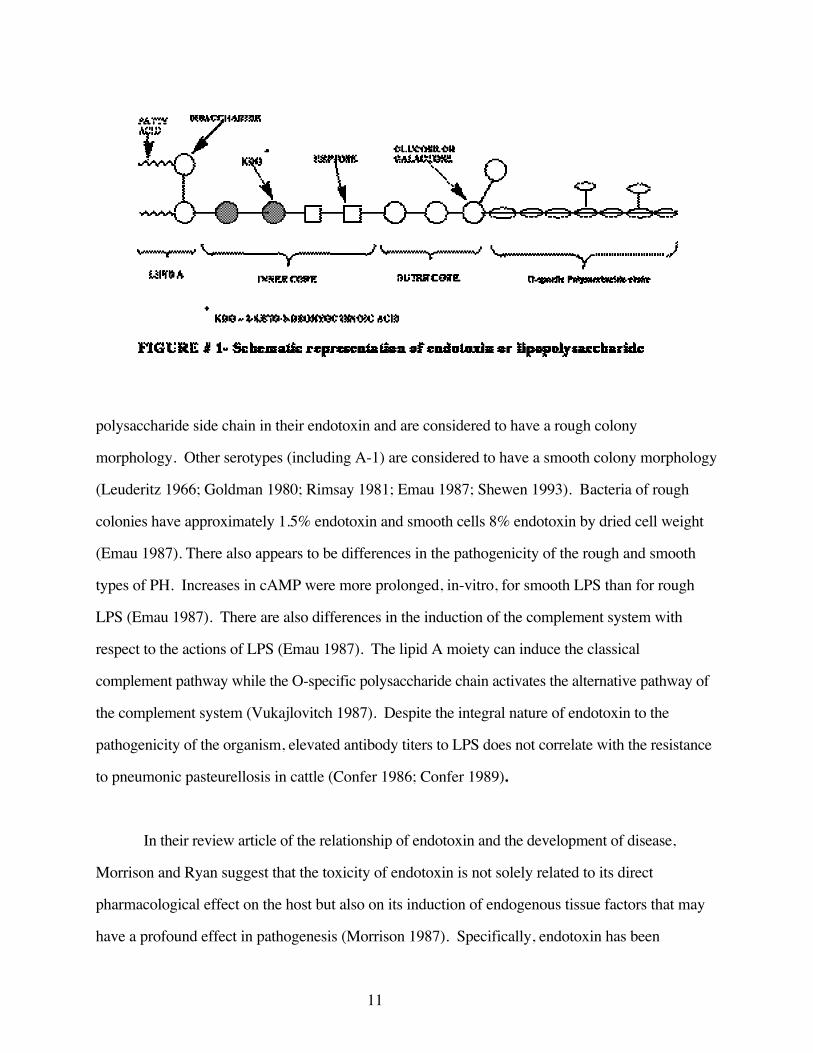
11
polysaccharide side chain in their endotoxin and are considered to have a rough colony
morphology. Other serotypes (including A-1) are considered to have a smooth colony morphology
(Leuderitz 1966; Goldman 1980; Rimsay 1981; Emau 1987; Shewen 1993). Bacteria of rough
colonies have approximately 1.5% endotoxin and smooth cells 8% endotoxin by dried cell weight
(Emau 1987). There also appears to be differences in the pathogenicity of the rough and smooth
types of PH. Increases in cAMP were more prolonged, in-vitro, for smooth LPS than for rough
LPS (Emau 1987). There are also differences in the induction of the complement system with
respect to the actions of LPS (Emau 1987). The lipid A moiety can induce the classical
complement pathway while the O-specific polysaccharide chain activates the alternative pathway of
the complement system (Vukajlovitch 1987). Despite the integral nature of endotoxin to the
pathogenicity of the organism, elevated antibody titers to LPS does not correlate with the resistance
to pneumonic pasteurellosis in cattle (Confer 1986; Confer 1989).
In their review article of the relationship of endotoxin and the development of disease,
Morrison and Ryan suggest that the toxicity of endotoxin is not solely related to its direct
pharmacological effect on the host but also on its induction of endogenous tissue factors that may
have a profound effect in pathogenesis (Morrison 1987). Specifically, endotoxin has been
12
demonstrated to induce the complement anaphylatoxins (C3a, C5a) (Vukajlovitch 1987),
Arachidonic Acid metabolism (Hagmann 1985; Aderem 1986), Tissue Factor activity (Morrison
1978), Platelet Activating Factor (Doebber 1985) the cytokines Interleukin 1 (Bayne 1986) and
Tumor Necrosis Factor-alpha (Beutler 1985; Beutler 1985) .
C) Lungs and the Pulmonary Vasculature
Some unique and species specific features associated with both the bovine pulmonary
physiology and anatomy may significant in of the etiology of shipping fever. There is a common
clinical perception that cattle are at an increased risk for pulmonary infection and disease due to PH.
Bovine pulmonary anatomy and physiology are significantly different than those of man
and other mammals (Veit 1978; Green 1982; Robinson 1984; Lekeux 1993; Weekley 1995; Wren
1995). In the bovine, the right thoracic cavity encompasses four lung lobes (cranial, middle,
accessory and caudal) while the left thoracic cavity has two lung lobes (cranial, caudal) (Robinson
1984; Weekley 1995; Wren 1995). The volume of the bovine lung is small relative to the size and
weight of the animal (Robinson 1984; Weekley 1995; Wren 1995) and there is a lower functional
capacity as compared to other mammals (Tenney 1963; Veit 1978; Robinson 1984; Weekley 1995;
Wren 1995) . The functional capacity is greater in the fully grown adult than the calf. In the calf
up to 42% of the minute-ventilation is dead space (Slocombe 1982) whereas in the young adult
(10.5 months) the dead space ventilation has increased to 68% of the minute ventilation (Hales
1968; Slocombe 1982).
Pulmonary airway anatomy consists of two clinically different divisions, the upper
respiratory tract and the lower respiratory tract (Liggitt 1985). The upper respiratory tract consists
of the oro-pharynx, larynx, trachea and mainstem bronchi. The lower respiratory tract starts below
the mainstem bronchi. The trachea and main stem bronchi divide into successive, sequentially

13
smaller generations. The first generation starts just distal to the cricoid cartilage of the larynx and
each subsequent bifurcation represents another generation. Each bronchus is paralleled by an
artery, vein and nerve numbered for their generation. In general, in mammals, there are
approximately ten to twelve generations of bronchial divisions (this is variable between individuals)
that exist from the trachea to the respiratory bronchioles (Breeze 1985). Supporting cartilage exists
in the conducting airways down to the level of the terminal bronchioles (approximately 1-2 mm in
diameter or 8-10 generations of bronchial bifurcations). At this level, the cartilage diminishes and
tends to form discrete plates that may move dynamically in response to the airway vascular smooth
muscle. Airway smooth muscle is a prominent anatomical and functional feature of the terminal
and respiratory bronchioles in both man and ruminants (Green 1982; Breeze 1985). This airway
smooth muscle diminishes to the level of the alveoli where it is completely absent. Bronchial
smooth muscle tone is under the control of both the autonomic nervous system and systemic
endogenous hormones (Breeze 1985).
In most species, lobar bronchioles arise as part of the complex of bronchioles that is formed
by the termination of the trachea, called the carina. The bovine lung has an additional bronchial
bifurcation (a lobar bronchus) to the right cranial lobe which arises directly from the trachea,
crainial to the carina (Getty 1975; Veit 1978; Schummer 1979; Robinson 1984; Wren 1995) .
Although this may seem to be an innocuous anatomical deviation, direct gravitationally dependent
drainage of tracheo-bronchial secretions into the right cranial lobe may potentially "seed" acini with
bacteria and / or occlude bronchioles with protective mucoid polysaccharides associated with the
mucociliary escalator (described below under lung physical defenses).
The bovine lung has complete interlobular septa and a dearth of inter-alveolar pores, also
called pores of Kohn (Veit 1978; Robinson 1984; Weekley 1995; Wren 1995). This results in
poor collateral communication among the secondary lobules and acini with an increased likelihood
that a single mucoid thrombus could occlude an entire primary lobule (Robinson 1984; Wren

14
1995). Occlusion of an alveolus or terminal bronchiole can result in a ventilation perfusion
mismatch which would cause a local (at the level of the acinus) anaerobic environment. Pulmonary
vascular constriction occurs as a response to alveolar hypoxemia. This pre-capillary
vasoconstriction can cause a functional and physiological shift of blood away from the occluded
alveolus and potentially increases right heart work over time (Veit 1978; Green 1982; Robinson
1984; Breeze 1985; Barnes 1995). The resultant shift in blood away from the alveolus also
diminishes the likelihood of the diapedesis of neutrophils and increases the anaerobic nature of the
acinus.
Dynamic lung compliance is lowered in the normal healthy calf more than in other young
mammals (Lekeux 1993). Dynamic lung compliance is the ability of the lung to affect a volume
change to a given intrathoracic pressure change. A decrease in compliance may occur as a result of
terminal smooth muscle mediated bronchial closure or bronchial obstruction (Lekeux 1993).
Decreased dynamic compliance is an indication of early obstructive airway disease (Geunter 1982;
Green 1982). Thus, the calf must exert more effort (with respect to intrathoracic negative pressure)
for normal inspiration and is therefore more susceptible to vascular blockage secondary to airway
emboli (Veit 1978; Green 1982; Robinson 1984).
The vascular, lymphatic and neural elements parallel each other along the bronchial tree
structure. The lung has two arterial systems functioning within the pulmonary tissue; the first
system is the tissue nutritional blood supply and the second is a functional supply. The nutrient
supply, the bronchial artery, arises from the thoracic aorta to supply oxygen to the pulmonary
tissues. The second or functional blood supply arises from the right ventricle to enter the lungs as
the pulmonary arteries. The arteriol vascular system then branch in concert with the bronchial
airways to eventually form the pre-capillary arterioles, a capillary net and then post-capillary venules
at the level of the alveolus. The functional supply enters the alveolar blood-gaseous exchange area
through the fine capillary bed vessels that are in intimate contact with the respiratory alveolar
15
membrane of the terminal respiratory tract. The barrier between the vascular elements and the
airway elements consists of a double basement membrane with a single layer of airway epithelial
cells and a vascular layer of luminal endothelial cells (Green 1982; Lekeux 1993).
Bovine pulmonary arterial vessels have a significantly greater tunica muscularis than other
mammals (Tucker 1975; Robinson 1984; Weekley 1995; Wren 1995). Pulmonary vascular
reactivity to hypoxia is therefore proportionately greater (Robinson 1984; Wadsworth 1994;
Weekley 1995) as is the ability of the bovine lung to correct a ventilation perfusion mismatch (Veit
1978; Robinson 1984; Weekley 1995). There is a significant vascular collateral circulation at the
level of the pre-capillary and capillary bed due to the anastomosis of the bronchial arterioles with
the pre-capillary arterioles in the bovine (Schummer 1979). Overall these unique anatomical
features of the bovine pulmonary vasculature results in dramatic fluctuations in perfusion /
ventilation ratio and contribute in part to the pathogenesis of pneumonic pasteurellosis.
The pulmonary capillary vasculature also serves as a "vascular filter" for emboli, thrombi
and the cellular elements of the vascular system. Those thrombi filtered by the lungs are removed
by phagocytosis. This is primarily by circulating monocytes and vascular endothelial cells.
Beyond the pulmonary actions, systemic and splenic vascular sequestration of lymphocytes,
granulocytes, platelets and macrophages comprise the physiological and hemostatic attempts to
regulate circulating vascular elements (Irwin 1975; Breeze 1985).
The pulmonary endothelium is a complete, non-fenestrated, single layer, squamous cell
sheet lining the vasculature. Adjacent cells have tight cell junctions with physiologically active
intracellular pores approximately 4 nm in diameter. Ordinarily these cells are impervious to
macromolecules though there is bi-directional transport of ions and macro-molecules (Ryan 1982;
Breeze 1985). The endothelium is a metabolically active site (Ryan 1982). The endothelial cells
have surface caveolae and form pinocytic and phagocytic vessels. Many vasoactive mediators and
16
hormones are removed from the circulation by the endothelium including, serotonin, nor-
epinephrine, prostaglandins (PGE, PGF) and thromboxane (Breeze 1985). As well, pro-
coagulatory molecules such as ADP are inactivated by endothelial cells (Ryan 1982). As a
metabolic site, the endothelium elaborates PGI-2, PGE-2, thromboxane and activates angiotensin-1,
angiotensin-2 (Ryan 1982; Breeze 1985). Under normal conditions the endothelium is actively
non-thrombogenic in nature (Ryan 1982). However, when damaged the endothelium can activate
the both the coagulation and complement systems (Ryan 1982).
In summary the combination of the small alveolar surface area (Veit 1978), the lack of
alveolar communication (pores of Kohn), and the amplification of the vasoconstriction response due
to an increased pulmonary vascular muscular tunica may also contribute to the development of both
an anaerobic environment and a local metabolic acidosis. The possible effect of this combination of
circumstances may be a suppression of bacterial clearance (Thompson 1974) and suggests the
possibility of an alveolar environmental change from an aerobic to an anaerobic environment. This
may result in a changes in the relative pathogenicity of P. haemolytica. Further, it has been
demonstrated that anaerobically challenged macrophages loose their ability to phagocytose bacteria
but retain their ability to generate and release free radicals after cell degeneration (Matthews 1987;
Cazin 1990; Thompson 1992). The simultaneous association of the above described multiple
factors may result in both pulmonary tissue damage as well as a compromise of the lung's physical
antimicrobial defenses in the presence of an increased PH endotoxin challenge (Wessman 1964).
D) Lung Defense Mechanisms
Lung defenses may be divided into three general categories: physical defenses,
immunological defenses and cellular defenses. It is thought that beyond the second generation of
bronchioles the environment is sterile and maintained so by the cellular and immune defenses
(Liggitt 1985).

17
The upper respiratory tract is protected by physical and immunological defenses (Veit 1978;
Robinson 1984; Breeze 1985; Liggitt 1985). The nares are the external entrance to a series of blind
and connecting passages called conchae or turbinates (Schummer 1979). These passages are lined
by a mucus secreting epithelium that traps particles in the air turbulence caused by inhalation. This
is termed turbulent filtration (Liggitt 1985) and is effective in removing particles greater than 10 µm
(Dungworth 1983; Reece 1991). Trapped particles are then carried out of the turbinates on a
mucocilliary blanket and either swallowed or eliminated in a mucoid secretion from the nares
(Liggitt 1985; Reece 1991). A second physical defense of the upper respiratory tract is the
mucociliary escalator. This is a thin layer (approximately 5 mm thick) of mucus carried on cilia that
extend from the terminal bronchioles to the level of the larynx. This system of particle removal
depends on the settling of debris on the mucus carpet in the respiratory airways. The cilia that
move the mucus crainiad arise from the epithelium which secretes mucus. This protective layer of
mucus can vary in composition and consistency with respect to the ratio of mucopolysaccharides
from highly viscous to serous, thereby affecting the mobility of the mucus (Breeze 1985) .
Hydration of the mucociliary escalator is of great importance in maintaining a functional and
efficient barrier to bacteria and debris. Dehydration, hypoxia, hyperoxia or exposure to caustic
irritants (e.g. ammonia or sulfur dioxide) may cause sufficient disruption of cilia and epithelial
physiology to inhibit the effective protection offered by the mucocilliary escalator. This will
therefore predispose an individual animal to an increased risk of pulmonary infections and diseases
(Breeze 1985; Liggitt 1985). Moving through successive bronchiole generations of airways, the
number of cilia decline in density (Breeze 1985). This results in a decreased ability to clear mucus
from the terminal bronchioles (Wiksie 1985). Other elements of the lung defense system including
neutrophils, alveolar macrophages, IgA and lactoferrin are active in the mucoid layer of the upper
airway (Dyer 1982; Liggitt 1985). Though important in pulmonary defense, the mucociliary
escalator is but one of a myriad of defenses active in the protection of the lungs (Thompson 1974).
18
The importance of the mucociliary escalator may lie in its ability to maintain a clear passage in the
upper bronchioles so as to ensure airway patency and remove pathogenic organisms.
Another physical defense of the more cranial portions (major bronchi and trachea) of the
upper respiratory tract is the cough. The cough is a sudden violent contraction of the diaphragm
and the abdominal and respiratory musculature that produces a very quick compression of the
thoracic cavity and diaphragm against a closed or semi-closed glottis. This results in a forceful
expulsion of air through the tracheo-bronchial tree which carries with it debris and mucus which are
then eliminated by swallowing the salivary secretions (Dyer 1982; Liggitt 1985).
Immunological defenses of the upper respiratory tract may be divided into immunological
tissue-based defenses and immunological secretory defenses. The immunological tissue-based
defenses are commonly known as bronchus associated lymphoid tissue (BALT). BALT consists
of two types of tissue, lymphoid nodules and lymphoid aggregates. Lymphoid nodules are
primarily found in the submucosa of the tracheal tree (Schummer 1979; Breeze 1985; Sminia
1989). The nodules are well organized and contain T lymphocytes (approximately 20% ) and B
lymphocytes (approximately 50%) while the remainder of the lymphocytes (30%) are unidentifiable
(Breeze 1985; Liggitt 1985). Lymphoid aggregates and infiltrates are less well defined and
organized, but are often associated with lymphocytes (Breeze 1985; Liggitt 1985; Sminia 1989).
BALT appears to be absent in the neonatal calf but increases with age to a peak at approximately 18
months of age and then again decreases (Anderson 1986). BALT is able to initiate local
immunological defenses, may elaborate primarily IgG and IgA in cattle and may have other, as yet,
unrecognized functions (McDermott 1982; Tizard 2000). BALT communicates through the deep
lymphatics that drain the pulmonary tissues and follow the bronchioles as described above (Liggitt
1985).
19
Secretory defenses of the upper respiratory tract include: IgA, IgG and IgE among other
components (Liggitt 1985; Tizard 2000). In a calf, younger than six weeks, there is a predominance
of IgG throughout the upper respiratory tract however, after six weeks there is a predominance of
IgA in the nasopharyngeal area (Liggitt 1985). IgA, produced by B-lymphocytes, predominates in
the nasopharyngeal areas of the respiratory tract and protects the animal by 1) preventing the
adherence of particles to the mucosa, and 2) enhancing pinocytosis (Breeze 1985). The
predominant secretory antibody of the bronchioles is IgG which protects the respiratory tract by
opsonization of the invading organism (Liggitt 1985; Tizard 2000). IgE mediates local allergic
reactions and in the event of its activation goes on to enhance the formation of IgG (Tizard 2000).
Interferon, another of the secretory defenses, is elaborated in the nasal mucosa (Liggitt 1985). Its
primary activity is to inhibit the replication of foreign organisms (including DNA and RNA viruses)
by interference with nucleic acid synthesis (Tizard 2000).
Cellular defenses of the upper respiratory tract include: neutrophils, macrophages and
lymphocytes which are interspersed among the mucopolysacharides of the mucociliary escalator
(Dyer 1982; Liggitt 1985; Tizard 2000). Neutrophils act primarily by phagocytosing and killing
bacteria by proteases and free radical generation (Liggitt 1985). Their numbers measure less than
2% of fluid collected by bronchial alveolar lavage, however, vascular neutrophils (up to 50% of
circulating leukocytes) may respond to the chemotactic signals released by activated vascular and
pulmonary alveolar macrophages and IgG thereby augmenting the numbers of PMN's in the
bronchioles (Liggitt 1985; Tizard 2000). Macrophages are also found in the mucociliary escalator
fluid though in much lower numbers than is found in the lower respiratory tract (Liggitt 1985;
Tizard 2000). They also attract other cellular defenses by the elaboration of cytokines (particularly
IL-8), immunoglobulins (primarily IgG), chemo-attractant factors and vasoactive amines to initiate
inflammation (Liggitt 1985; Tizard 2000). Lymphocytes may also be active in the upper respiratory
tract. These are B lymphocytes, which exist primarily in the BALT and act to generate IgA, and
20
attract T lymphocytes. Though few in number, lymphocytes serve as an anamnestic surveyor of the
antigens present (Liggitt 1985; Tizard 2000).
The lower respiratory tract defenses (terminal bronchials and alveolae) are predominated by
the activity of the cellular defenses. The primary cell types include: pulmonary alveolar
macrophages (PAM) and lymphocytes (T cells and B cells). The pulmonary alveolar macrophages
act to phagocytose and kill microorganisms (Breeze 1985; Liggitt 1985; Tizard 2000), lymphocytes
are primarily active in immunosurveillance and defense as described above. The immunoglobulins
most commonly noted are IgG and IgA (Liggitt 1985; Tizard 2000).
Pulmonary alveolar macrophages (PAM) lie in a thin layer of surfactant in the alveolus of
the lower respiratory tract and play a primary role in chemotaxis, phagocytosis, antigen processing
and elaboration of cytokines (Breeze 1985). These cells are derived from monocytes and interstitial
macrophages (Bowden 1984; Liggitt 1985). PAMs interact with neutrophils and eosinophils of the
lower respiratory tract. The PAMs are approximately 80% of the cells found at this level and is the
primary defense mechanism of the alveolus (Trigo 1984; Liggitt 1985). Macrophage defense
represents a double edged sword. Elaboration of antibacterial oxygen free radicals and cytokines
can have a damaging effect on the pulmonary microvasculature and airway functioning (Engels
1985; Barnard 1992; Seccombe 1994) . Chemotactic factors, chemical radicals and cytokines act
both to protect the alveolus and incite inflammatory processes and coagulation within the lumen of
the alveolus, the inter-lobular septa and at the immediately adjacent vasculature (Breeze 1985; Yoo
1995). An inflammatory response, at this level, can decrease dynamic compliance of the lung so as
to initiate or facilitate in the obstruction of the terminal bronchioles. There is evidence that
pulmonary alveolar macrophages can increase in number as a result of increased stress. This could
lead to an enhanced defensive and inflammatory response (Boorman 1979; Bowden 1980). Further
the macrophage can divide in-vivo to form a self sustaining pool of cells (Boorman 1979; Bowden
1980; McGuire 1982). The optimal PAM : bacteria killing ratio has been reported to be on the
21
order of 10:1 and effective killing was reduced when the ratio was reversed to 1:10 (Trigo 1984).
This suggests that optimal numbers of PAM must be closely regulated and if diminished could
result in a significant lapse of microbial defense at the level of the alveolus.
In an anaerobic environment it has been demonstrated that superoxide anion release is
decreased while lysozyme release and phagocytosis was unaffected in the killing of Candida
albicans (Thompson 1992). Under anaerobic conditions macrophages, stimulated in-vitro with
phorbol myristic acetate, have been shown to be able to survive for up to 48 hours, if reintroduced to
a normal oxygen level within that time. Macrophages in an anaerobic environment show increases
in production of lactic acid and glucose uptake, at 24 hours, there is cell degeneration as
demonstrated by decreases in the level of cellular ATP (Cazin 1990; Thompson 1992). However,
macrophages are unable to recover either homeostasis or protective functioning after 72 hours in an
anaerobic environment (Cazin 1990). One of the macrophage antimicrobial functions is to
elaborate cytokines (Engels 1985; Kelly 1990; Benburnou 1992; Ward 1993; Yoo 1995). The
elaboration of cytokines, primarily Tumor Necrosis Factor-alpha [TNF-alpha] and Interleukin-1 B
[IL-1B], is arrested in anaerobic conditions (Matthews 1987). This information again suggests that
in an anaerobic environment primary alveolar defenses are reduced or abrogated dependent on the
length of time the anaerobic conditions are present.
The functions of TNF alpha are numerous and complex in an in-vivo system. TNF-alpha is
specifically formed in response to endotoxin challenge (Sharma 1992) and is also elaborated by
vascular endothelium, smooth muscle and other cellular elements (Warner 1989). TNF-alpha acts
to stimulate the production of IL-1 and class 1 MHC antigens (Hamblin 1993). By its presence,
TNF alpha can generate oxygen free radicals though this can be inhibited by anaerobic conditions
(Matthews 1987). It has been demonstrated that TNF-alpha, acting through oxygen free radicals
(Matthews 1987; Larrick 1990; Hamblin 1993) can cause significant vascular, endothelial and
interstitial tissue injury (Barnard 1992; Seccombe 1994). TNF-alpha may also modify
22
vasoreactivity through the inhibition of nitric oxide, a potent member of the endothelial relaxing
factor family (Johnson 1992). Il-1 stimulates B and T lymphocyte activity, activates neutrophils,
vascular endothelial cells and its release is also stimulated by endotoxin and inhibited by drugs with
antioxidant properties (Ku 1990).
The neutrophil, although initially in low numbers in the alveolus, actively responds to the
chemotactic signals of: IL-8, complement and some arachidonic acid products (Liggitt 1985;
Hamblin 1993). The neutrophil has a beneficial effect in the reduction of direct tissue damage by
PH through its antibacterial activity (Breider 1991). The presence of the neutrophil also has the
potential to contribute to both alveolar and vascular tissue damage through the generation and
release of free radicals (Vandenbroucke-Grauls 1987; Warren 1987; Jaeschke 1990; Maheswaran
1993; Granger 1994). The significance of neutrophil mediated damage has been assessed and can
be shown to be reduced in neutrophil deficient animals (Breider 1988; Paller 1989).
Other cellular elements of the lower respiratory tract include T lymphocytes and B
lymphocytes that consist approximately of 10% of the total free cells found in the normal bovine
lung (Liggitt 1985). The function of T cells includes: the activation of PAMs to act as T helper and
T suppresser cells as well as T cytotoxic cells (Breeze 1985; Liggitt 1985). PAMs with T cells, act
as antigen presenting cells, to ensure an appropriate anamnestic response (Liggitt 1985; Tizard
2000). The B cell generates IgA which is of minor importance in the lower respiratory tract as
compared to the nasal pharyngeal (Liggitt 1985). Some plasma cells elaborate IgG. This is the
predominant antibody of the lower respiratory tract of cattle (Liggitt 1985). IgG serves to opsonize
antigens and enhance bacterial phagocytosis (Liggitt 1985; Tizard 2000). The secretory products
of the lower tract are intimately tied to the cellular defenses already described.
Complement is a protective element that can, in early inflammation, be found throughout the
lung including the alveoli (Liggitt 1985). The presence of the complement components C5A and
23
C3A (the anaphylactoids) have been shown be an effective antibacterial mechanism even at low
concentrations. These anaphylactoids may initiate tissue damage through the mediation of
inflammation and coagulation (Liggitt 1985; Parke 1995; Tizard 2000). Other secretory products
of the lower respiratory tract include alpha-1-antitrypsin, alpha-2-macroglobulin, fibronectin,
lactoferrin and surfactant (Liggitt 1985).
E) Pulmonary Vascular Control Mechanisms
The autonomic nervous system is primarily involved in the control of the pulmonary
vasculature. Three systems are interactive in vascular control and include the cholinergic, adrenergic
and non-adrenergic-non-cholinergic (NANC) systems (Barnes 1995). These systems are not only
involved in the regulation of homeostasis, but also have significant input into airway and vascular
dynamic control. Since the primary focus of this dissertation is to investigate the vascular dynamics
of the beta adrenergic receptor, only cursory descriptions will be given of the non-adrenergic and
extra-vascular functions of these autonomic systems.
Cholinergic innervation of the vasculature has been reviewed in the bovine and other
mammals (Fisher 1965). The physiological significance of this system on vascular physiology is
questionable (Bergofsky 1979; Lefkowitz 1991). Paradoxical, simultaneous dilation and
contraction of pulmonary vessels to acetylcholine suggests the complexity of this mechanism of
control. These findings suggests a "vernier type" vascular control mediated by the action of the
cholinergic system on the pulmonary vasculature (Bergofsky 1979).
Nonadrenergic noncholinergic (NANC) mechanisms are also active in the neural-humoral
control of the pulmonary vascular control (Barnes 1986). This control is mediated by the simple
molecules and neurotransmitters, nitric oxide (NO) in cattle and rabbits (Gustafsson 1991) and
vasoactive intestinal peptide (VIP) in cattle (Barnes 1986). These two agents act to cause
24
vasodilatation in response to their release. Substance P, also a neurotransmitter of the NANC
system, has been shown to cause vasodilatation in pigs (Adnot 1989).
Adrenergic control is the third neurological based pulmonary vascular control mechanism
and the focus of this dissertation. The adrenergic system is divided into three types of activity
based on the receptors mediated and its pharmacological specificity. First, the alpha-1 adrenergic
receptor system, defined pharmacologically as responding to alpha-1 adrenergic agonists (e.g.
phenylephrine, methoxamine) and blocked by alpha-1 adrenergic antagonists (e.g. prazosin). The
typical vascular alpha-1 adrenergic receptor acts to cause constriction of the pulmonary vasculature
(Ruffolo 1991). The alpha-2 adrenergic receptor system responds to alpha-2 adrenergic agonists
(e.g. clonidine) and is blocked by alpha-2 adrenergic antagonists (e.g. yohimbine, rauwolscine)
(Ruffolo 1991). The vascular function of the alpha-2 receptor is not completely clear and appears to
be more complex than that of the alpha-1 receptor. Alpha-2 receptors are found directly on the
endothelium, on vascular smooth muscle and pre-synaptically as well (Lefkowitz 1988). The alpha-
2 receptor appears to have a dualistic nature in its activity. It may act to cause vascular relaxation in
the presence of high alpha-1 pulmonary vascular tone or can result in vascular contraction in the
presence of low alpha-1 vascular tone. The beta receptor responds to beta adrenergic agonists (e.g.
isoproterenol or terbutaline) and may be blocked by beta adrenergic antagonists (e.g. propranolol).
The action of the beta receptor is to induce vasorelaxation in the pulmonary vascular bed (Lefkowitz
1974). The beta adrenergic receptor, its functioning or dysfunction with respect to pathogenesis of
pneumonic pasteurellosis will be the primary focus of this dissertation.
All adrenergic receptors are G-protein based receptors. The alpha-2 and beta receptors act
through a common mechanism of adenylyl cyclase, cAMP / cGMP modulation. The alpha-1
receptor acts through the inositol triphosphate / diacylglycerol (IP3 / DAG) mechanism (Ruffolo
1991). The specific effect of each adrenergic receptor type is variable with respect to its anatomical
location.
25
Alpha-1 adrenergic system may be activated by selective agonists (phenylephrine,
methoxamine) (Ross 1996) resulting in pulmonary vasoconstriction (Limbird 1985; Pepperl 1994).
The molecular mechanisms of this stimulation are mediated by G-protein coupled phospholipase C
(PLC). This generates two biochemical products, diacylglycerol (DAG) and inositolphosphate
(IP3). DAG phosphorylates a protein kinase and IP3 acts directly to increase intracellular calcium
to initiate smooth muscle contraction . It has been demonstrated that alpha-1 receptors are
dependent on both intracellular calcium (Ca+ +) stores and extracellular Ca+ + concentration to
induce maximal vasoconstriction (Ruffolo 1991; Hoffman 1995; Hoffman 1996; Ross 1996).
Alpha-2 adrenergic receptors may be activated by an alpha-2 specific agonist (e.g..
clonidine). Clonidine is considered selective for the activation of the alpha-2 receptors at
physiological or low dose pharmacological levels, though it does have some weak alpha-1 agonistic
activity (Ross 1996). Alpha-2 receptors are found on both the pulmonary endothelium and the
vascular smooth muscle (VanHoutte 1989).
The presence and actions of the alpha-2 receptors in the aortic tissue of rats has been
brought into question (Randriantsoa 1981; Dashwood 1985; Martinotti 1991). Dashwood and
Jacobson stated that there were no alpha-2 receptors in the aortic tissue of Wistar rats by
autoradiographic ligand binding studies (Dashwood 1985). Some questions have been raised about
the use of autoradiographic ligand binding studies for alpha-2 adrenoreceptors in view of their
rarity on the aorta of the rat (Randriantsoa 1981). The results of Dashwood and Jacobson may be
affected by factors that have been known to effect the expression of other receptors including age,
species, breed, strain and sex. Martinotti suggests that there are no alpha-2 receptors in the aortic
tissue of 200-220 gram, male, Sprague-Dawley rats though their method of receptor evaluation is
less rigorous than that of the previous study by Dashwood and Jacobson. Martinotti concluded,
based on the lack of response to detomidine (an alpha-2 receptor agonist), that there are few or no
26
alpha-2 receptors in aortic tissue of the rat (Martinotti 1991). However Martinotti's experiment
could represent “zero sum” pharmacological response to an alpha-2 agonist and partial alpha-1
agonist described above. The results of Randriantsoa suggests that the alpha-2 receptor exists in the
rat aorta but may represent a variation in response from a true alpha-2 receptor (Randriantsoa
1981).
In light of these findings, it is likely that the alpha-2 receptor (or possibly a variation of the
alpha-2 receptor) are rare in the rat aorta, though their importance cannot be measured by their
numbers but is apparent by their physiological functioning in-vivo. It is possible that this receptor
acts to buffer the extreme constrictions mediated by the alpha-1 receptor. Further, the alpha-2
receptor may, in fact, be the vascular equivalent of the "fine tuning" knob balancing the effects of
beta-mediated vasodilation and the alpha-1 mediated vasoconstriction. These possibilities have yet
to be investigated experimentally. Another possible explanation for the dual nature of the alpha-2
receptor is that the class of receptors responding to an alpha-2 agonist are variable or heterogeneous
in their presence, numbers and activity on the endothelium of the rat aorta and vary only in
homeostatic importance only in response to some as yet unrecognized signal. It remains to be
discovered what, if any, factor affects the presence and functional expression of the alpha-2 receptor
in the rat aorta.
The beta receptor agonist, isoproterenol, induces significant vasorelaxation in the presence
of high vascular tone in both rat aorta and bovine pulmonary vasculature. High vascular tone may
be induced by preconditioning vessels with potassium (e.g. KCL) a cell and membrane depolarizing
agent. The mechanism by which beta receptor mediated relaxation occurs, in high vascular tone, is
through the activation of adenylylate cyclase via a subunit of the G-protein of the receptor. There is
a subsequent generation of the second messenger cyclic adenosine monophosphate (cAMP). The
cAMP is then thought to activate a protein kinase (myosin light chain kinase) resulting in
27
dephosphorylation, Ca++ sequestration and subsequent vasorelaxation (Hoffman 1995; Hoffman
1996). The beta receptor is discussed in greater detail in the next section.
F) Beta Receptor Mechanisms
G-protein receptors represent a superfamily of receptor proteins that are associated with a
signal transducing guanosine molecule. Each receptor (alpha-1, alpha-2 and beta) is an integral
membrane protein characterized by seven transmembrane alpha-helical segments (Watson 1994).
All adrenergic receptors (alpha and beta) are of the same basic structure with some differences in
their amino acid composition and secondary messenger system (Lefkowitz 1991; Hoffman 1995).
There is similarity among the groups of adrenergic receptors. For instance, alpha-1 and alpha-2
receptors have approximately 45% homology in their amino acid sequence (Langer 1989). It is
likely that there is less homology between beta receptors and their alpha counterparts.
The beta adrenergic receptor polypeptide string consists of seven transmembrane alpha-
helices that are joined by short chains of amino acids (Cuatrecasas 1974; Lefkowitz 1974). The
amino terminus starts outside the plasma membrane and the first a helix spans the cell membrane
while the amino acid loop between the first and second a helices enters the cytosol. The second
alpha helix is in an anti-parallel orientation to the first a helix. This architecture continues with all
alpha helices in an anti-parallel orientation to its immediate adjacent neighbors. This leads to an
accordion type structure of the molecular biology of the G-protein receptor (see figure #2)
(Lefkowitz 1988; Levitzki 1988; Ostrowski 1992; Strader 1994).
The loop between the alpha helix #5 and #6 is elongated and plays an essential role in the
activation of the adrenergic intracellular mechanisms. Specific amino acids on the #5-6 loop are
available to be phosphorylated (the catalytic site) by way of the interaction of an adrenergic agonist
with the receptor protein (Lefkowitz 1988; Levitzki 1988; Ostrowski 1992; Strader 1994).
28
Activation of the beta receptor occurs when an agonist interacts at three separate locations
simultaneously, with receptor amino acids of the transmembrane alpha helices. The amino group of
the agonist forms an ionic bond with the carboxylate side chain of aspartate (A113) in the third a
helical segment. This has been verified by demonstrating that a mutation in A113 inhibits receptor-
agonist interactions (Levitzki 1988). The two hydroxyl groups of the catechol ring of the agonist,
forms hydrogen bonds with two serine residues (S204 and S 207) on the fifth transmembrane alpha
helical segment. Mutation of either of these two serines can also result in the inhibition of the
receptor-agonist binding. Finally, the third interaction is between the agonist and the sixth
transmembrane helix. The interaction at the catechol ring is the formation of a hydrophobic bond
with phenylalanine (F290) of the sixth alpha helix (see diagram of beta adrenergic agonist and
antagonist, figure #3), (Lefkowitz 1988; Levitzki 1988; Ostrowski 1992; Strader 1994).
The binding of the agonist to the beta receptor moiety causes movement among the
transmembrane helices (particularly #5 and #6). This movement results in the activation of the
transducing G-proteins ( Ga stimulatory) and subsequent induction of adenylyl cyclase by the
catalytic site on the #5 / #6 cytoplasmic loop and finally, in the elevation of cAMP.
The protein receptor moiety is adjacent to but separate from the actual G-protein
transducing unit. The G-protein transducing unit consists of the Gg, Gb, and Ga subunits (trimeric
unit). These proteins together are referred to as the Gs protein transduction complex. In an inactive
form, the Gg, Gb remains bound to Ga and guanosine diphosphate (GDP) is attached to the Ga
unit. Once activated, the catalytic site on the cytosolic loop, binds to the trimeric G-protein complex
to initiate the intracellular phase of activation. Upon activation, guanosine diphosphate (GDP)
bound by the Ga subunit is released and replaced by guanosine triphosphate (GTP) which activates
Ga. Ga is an intracellular switch protein that is part of a GTPase superfamily. The activated Ga
unit dissociates from the Gg, Gb subunits and binds to adenylyl cyclase at its catalytic domains.
Hydrolysis of GTP to GDP allows the dissociation of the Ga from adenylyl cyclase and initiates

29
the hydrolysis of ATP to cAMP. The now inactive Ga molecule then returns to associate with the
Gg, Gb units of the transduction protein.
Adenylyl cyclase is an enzyme bound to the intracellular membrane adjacent to the receptor.
Activation of adenylyl cyclase by an agonist results in the intracellular increase of cAMP. An
antagonist, on the other hand, either fails to cause a conformational receptor change or initiates a
conformational change in the receptor or G-protein moiety that results in a non-functional , non-
activated G-protein complex. The most current thinking, with respect to antagonists, is that GDP
fails to be replaced by GTP in the Ga molecule, this results in a failure to activate adenylyl cyclase.
The gram negative bacterial product, pertussus toxin, acts to lock Ga-GDP complex in a bound state
which directly causes receptor functional uncoupling (Levitzki 1988; Simon 1991; Birnbaumer
1992; Hepler 1992; Lodish 1995).
The activation of adenylyl cyclase is one step in the cascade from the receptor-ligand
interaction to the final cellular effect (Lefkowitz 1988; Levitzki 1988). It is important since it is a
step where there may be amplification of the receptor signal (Levitzki 1988; Tang 1992). In the

30
COOH
NH
1 2 3 4 5 6 7
FIGURE #2 - G-Protein receptor with seven anti-parallel alpha-helices. This is thebasic format for adrenergic receptors of the pulmonary vasculature.
EXTRACELLULAR
INTRACELLULAR
CELLULARMEMBRANE
CATALYTIC SITE
LOOP 5-6
case of the beta receptor, cAMP has a direct effect on the cAMP-dependent protein kinases in the
cytosol. These protein kinases, once activated, act to modify cellular activity. Each protein kinase
has two regulatory units and two catalytic subunits. The binding of cAMP to each of the regulatory
subunits results in the activation of the catalytic subunits which then result in the phosphorylation
of specific substrate proteins.
In vascular smooth muscle the phosphorylated proteins modulate intracellular calcium,
resulting in relaxation. Calcium is an essential ingredient in both the contraction and relaxation of
vascular smooth muscle (Morgan 1991). Calcium enters the cytoplasm from either the extracellular
space or from sequestered intracellular stores. In a smooth muscle contraction, the presence of
calcium allows the activation of the myosin light chain kinase (MLCK). The MLCK then acts to
phosphorylate the myosin light chain which then promotes the myosin-actin cross bridging in the
presence of the activated myosin-ATPase. Relaxation occurs by the deactivation of the myosin
31
through dephosphorylation of MLCK and by the sequestering of Ca++ secondary to the action of
cAMP on the cGMP-dependent protein kinase (Lincoln 1991). Dephosphorylation is governed by
factors that are not tied solely to the intracellular calcium level. Further, it has been found that beta
adrenergic agents not only modulate relaxation but also may increase the level of Ca2+ necessary
for MLCK activation (Morgan 1984).
Among the many types of receptors located on a neutrophil, the beta-adrenergic receptor is
functionally most important. It is reported that there are up to 106 stimulatory receptors, including
those for complement degradation products, formyl peptide and immune complexes, on the PMN
cell surface. Further, there are approximately 2x10⁴ inhibitory receptors, including receptors for
prostaglandin E1, and beta adrenergic agonists. Beta receptors are by far the minority:
approximately 103 (5%), of the inhibitory cell surface receptors and .04% of the entire receptor
population on the neutrophil (Galant 1981; Galant 1984; Sklar 1986; Mueller 1988). The beta-2
receptor is the primary adrenergic receptor found on the leukocyte (Motulsky 1982). Beta
receptors have been proposed to be one mechanism for neutrophil and macrophage regulation of
the release of oxygen free radicals (Engels 1985; Ogunbiyi 1986; Borregaard 1988; Barnard 1992;
Opdahl 1993; Seccombe 1994), phagocytosis, chemotaxis, and granular enzyme release (protease,
and beta-glucosidase) (Bourne 1971; Ignarro 1974; Zurier 1974; Hills 1975; Busse 1984; Tecoma
1986; Al-Essa 1995). More recently it has been demonstrated that, by activation of beta adrenergic
receptors on both neutrophils and the eosinophils, it is possible to inhibit leukocyte adhesion to
venules in the rat trachea and thus reduces the amount of plasma leakage to perivascular tissues
(Bowden 1994). The beta adrenergic mediated inhibition of exuberant inflammation by circulating
cells is thought to be important in limiting surrounding tissue destruction (Mueller 1988).
Confirmation that beta receptor mediated leukocyte inhibition occurs was demonstrated by the
restraining effects beta adrenergic agonists had in-vitro and that this activity could be blocked by
propranolol, a beta adrenergic antagonist (Tecoma 1986; Mueller 1988; Opdahl 1993). Further, it
was established that when leukocyte receptor affinity for classical adrenergic agonists was
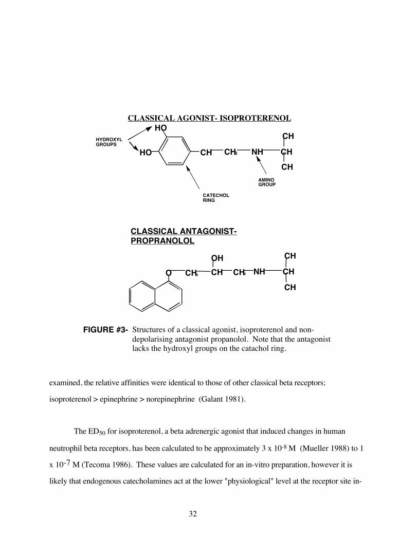
32
CLASSICAL AGONIST- ISOPROTERENOLHO
HO CH CH2 NH CH
CH
CH
HYDROXYLGROUPS
CATECHOLRING
AMINOGROUP
CLASSICAL ANTAGONIST-PROPRANOLOL
O CH2 CH CH2
CH
CH
CHNHOH
FIGURE #3- Structures of a classical agonist, isoproterenol and non-depolarising antagonist propanolol. Note that the antagonistlacks the hydroxyl groups on the catachol ring.
examined, the relative affinities were identical to those of other classical beta receptors;
isoproterenol > epinephrine > norepinephrine (Galant 1981).
The ED50 for isoproterenol, a beta adrenergic agonist that induced changes in human
neutrophil beta receptors, has been calculated to be approximately 3 x 10⁻⁸ M (Mueller 1988) to 1
x 10-7 M (Tecoma 1986). These values are calculated for an in-vitro preparation, however it is
likely that endogenous catecholamines act at the lower "physiological" level at the receptor site in-
33
vivo. There is a difference in the ability of beta agonists to inhibit neutrophil free radical generation
in circulating blood-borne neutrophils when compared to their tissue based counterparts (Al-Essa
1995). In circulating neutrophils, at least 30-40% of the beta receptors on this leukocyte must be
occupied to have an inhibitory effect on the generation of oxygen free radicals (Mueller 1988;
Opdahl 1993; Al-Essa 1995). This is amazing since 40% occupancy of neutrophil beta adrenergic
receptors represents approximately .04 % of the total neutrophil receptor population (calclated
estimate). This small number of specialized receptors hold the ability to mediate the release of
oxygen free radicals over-ruling over 99% of the remaining neutrophil receptors! Further, this
suggests two significant points: first, a minimal disruption of beta adrenergic receptor complex
could have significant physiological and pathological effects and two, naive neutrophils need
minimal endogenous beta adrenergic agonist activity to control the release of oxidative free radicals.
Finally, pharmacological levels of activators are likely to overstate the more subtle endogenous
activity of normal physiological levels of beta adrenergic mediators in the vasculature.
Minimal dysfunction of the neutrophil receptor population may lead to unchecked
generation of oxygen free radicals. The definition of minimal dysfunction in this case is, as yet, to
be experimentally assessed and defined. The disruption of beta receptor mechanisms following
prophylactic vaccination, using live Pasteurella haemolytica, of the vascular smooth muscle has
been described by Weekley (1993E). A damaged vascular beta receptor likely results in a
diminished functional population of receptors in the presence of an increased vascular tunica
muscularis tone (as is the case in the bovine pulmonary vasculature). This can result in excess
venous constriction due to the unchecked action of intact alpha-1 receptors and alpha-2 induced
contraction (Weekley 1993E). Similarly, beta receptor uncoupling in the leukocyte population,
particularly the neutrophils, has the potential to result in an increased release of PMN generated
oxygen free radicals and consequently severe pulmonary tissue derangement, damage and
destruction.

34
Beta receptors are also found on the vascular endothelium and have been demonstrated to
modify extra vascular fluid movement (Mizus 1985; Minnear 1986) and inhibit the coagulation
system through the modification of tissue factor (Busso 1991). Disruption of these receptors, in
the pulmonary vasculature, could result in pulmonary vascular leakage resulting in tissue
congestion, and activation of the coagulation cascade resulting in deleterious thromboembolic
events.
Given the significant and intimate involvement of the beta adrenergic receptor in the
pulmonary vascular homeostatic mechanism, it would appear that the uncoupling of these receptors
could be an essential element in the development of pulmonary pathophysiology in bovine
pneumonic pasteurellosis.
G) Significance of Beta Adrenergic Receptor Dysfunction in the Pathogenesis of Bovine
Respiratory Disease Complex and Pneumonic Pasteurellosis
Pneumonic pasteurellosis is characterized by two distinct sequential pathological events, the
first related to the "invasion of the respiratory system" either singly by the organism Pasteurella
haemolytica as an opportunistic infection, or in concert with a secondary bacterial or viral vector as
has been described previously. The second or perhaps the primary insult is that of tissue
inflammation in the respiratory tract. The hypothesis of this dissertation implies the importance of
the beta adrenergic receptor in the mediation and regulation of the inflammatory aspect of the
disease process of BRDC or pneumonic pasteurellosis. By extension the suggestion is that the
loss of regulation of the pulmonary inflammatory process, as a consequence of the disruption of the
beta adrenergic receptor can result in the excessive damage to bovine lungs as is commonly noted in
this disease process. Inflammation is a complex event that is a consequence of tissue derived
mediators that, when uncontrolled, can account for all of the signs and indications of the morbidity
and mortality of pneumonic pasteurellosis. Scientific acceptance of this thesis may give more

35
specific direction to the development of a new prevention and treatment strategies that ultimately
could result in great improvements in the cost, both pathologically (to the individual animal) and
financially (to the producer), of this devastating disease. I would like to first review the specifics of
the inflammatory process, then suggest the role of the beta adrenergic receptor to this process. This
understanding may then suggest possible future targets of treatment that could prove invaluable in
the prophylaxis, metaphylaxis and treatment of pneumonic pasteurellosis in cattle.
The inflammatory process involves multiple physiological systems including: leukocytes
(primarily neutrophils), pulmonary alveolar macrophages, platelets, T-cells, mast cells, cytokines
among other factors. Also intimately involved in the pulmonary inflammatory response are several
cascade, systems including but not limited to, the arachidonic acid cascade, the coagulation cascade,
the complement cascade and finally the kinin cascade. These two lists of endogenous systems are
not meant to be a comprehensive compilation of all endogenous systems involved in pulmonary
inflammation, but merely a brief overview of what are likely to be the more important systems
involved with pneumonic pasteurellosis. The majority of these systems are integrated into a
complex of inter-related physiological processes at the center of which is the overall modulating
transcription factor, Nuclear Factor kappa B (NF kappa B). The basic elements of pneumonic
pasteurellosis are infection, endogenous inflammatory mediators, Nuclear Factor kappa B and the
beta adrenergic receptor.
Many of the lesions described both on gross and microscopic pathological examination can
be explained in terms of pulmonary vascular homeostatic dysfunction in general, and beta
adrenergic uncoupling in particular. Pneumonic pasteurellosis can lead to either fibrinous
bronchopneumonia or fibronecrotic pneumonia with a lobar pattern. Typical initial gross
pathological lesions include: reddish-black to dark brown-gray consolidated cranial ventral lung
lobes, thickening of the interlobular septa, fibrinous pleuritis, the thorax may contain a straw colored
effusion and areas of necrosis can be seen in the lungs (Rehmtulla 1981; Dungworth 1983; Wiksie

36
1985; Lopez 1986). In most cases resulting in mortality, the gross lesions involve 60-80% of the
total lung (Wiksie 1985). Histopathological lesions include: necrosis with large numbers of
bacteria, thrombosis and leukocyte accumulation have also been noted (Rehmtulla 1981;
Dungworth 1983; Wiksie 1985; Lopez 1986). The pathology described above maybe associated
with events initiated by the disruption of the beta adrenergic receptors of the pulmonary vascular
smooth muscle, endothelium, and circulating leukocytes. This may result resulting in uncontrolled
exuberant inflammatory activity.
Inhibition of the functioning of the mucociliary escalator is likely one contributing factor to
the pathology seen in BRDC (Breeze 1985). Factors such as metabolic or respiratory acidosis can
lead to decrease in the efficacy of the mucociliary escalator (Breeze 1985; Liggitt 1985) . The
mucocilliary protective system may also be interrupted by viral infections in both the bovine (Breeze
1985; Hjerpe 1986; Radostits 1994) and in the equine (Dixon 1992). The interruption of this
physical defense could result in a gravitational dependent drainage to the anterior ventral aspects of
the lung. This is likely to form multiple mucus plugs in the terminal bronchioles which would then
isolate the associated acinus. Without collateral communication (via pores of Kohn) in the bovine
lung, an anaerobic environment is established. This would likely change the nature of any PH
trapped in this environment (Leeper et al 1992, Christman 2001).
In an in-vitro anaerobic environment, PH demonstrates an enhanced toxicity (Waurzyniak
1994, Chang 1987). The proposed increase in PH toxicity is thought to be due to an increase in the
local expression of leukotoxin seen in the early log growth phase (in-vitro) (Waurzyniak 1994,
Chang 1987). This activity declines in the late log to stationary phase of in-vitro bacterial growth
(Chang 1987). These findings support the labile pathogenic nature of PH. To date, only two
studies have been completed on the characterization of PH endotoxin in an anaerobic environment.
The first study (Davies 1992) examined the quantitative changes under anaerobic conditions in-vitro
and found a decrease in the quantity of LPS. This study is lacking on two essential points, first the
37
dynamic and complex interactions of multiple factors including: cytokines, immunological
chemokines, vasoactive autocoids and tissue factors, that in the anaerobic, acidic acinus are unlikely
to be duplicated in-vitro. Secondly, an in-vivo environment is likely to be a facultatively anaerobic
environment rather than a true anaerobic situation. In another investigation, it was shown that
bovine serum albumin (BSA) is a factor in the elaboration of LPS (Davies 1992). Bovine serum
albumin may be elaborated as a consequence of BRDC when PM infects the alveolus (Waurzyniak
1994). As noted previously, the qualitative nature of rough and smooth endotoxin in PH and the
different effects these two types of endotoxin have on cyclic nucleotides is great (Emau 1987),
however no evaluation has been made as to the relative amounts of rough and smooth endotoxin
under anaerobic or aerobic conditions. In a second study, Wessman showed that there is variation
in rough and smooth cell variants in varying oxygen tensions. The more virulent smooth cell
colonies were dominated by the less virulent rough colonies in low oxygen tension (Wessman
1964). In combination, the two studies cited above, suggest that the elaboration of a more virulent
LPS is possibly under anaerobic conditions. This is an indication that changes in the virulence of
PH with respect to the alveolar micro-environment, remains to be investigated in-vivo. It is likely
that the severity of the beta adrenergic receptor dysfunction is related to qualitative changes in PH.
Leukocytes are important in the pathology associated with pulmonary inflammation in
general and pneumonic pasteurellosis specifically and are acted upon by, PAF, IL-8, LPS and many
other endogenous and exogenous mediators. It has been suggested that microvascular damage to
endothelial cells by neutrophil generated elastase is enhanced by endotoxin (Smedly 1986). It has
also been shown that endotoxin can enhance pulmonary vascular sequestration of leukocytes in
sheep (Sigurdsson 1989).
The LPS mediated adrenergic receptor disruption has an effect on circulating and tissue
leukocytes (Slocombe 1985; Breider 1988). The oxygen free radical release by neutrophils is
diminished in the presence of a beta receptor agonist (e.g. isoproterenol) (Engels 1985; Ogunbiyi

38
1986; Borregaard 1988; Barnard 1992; Opdahl 1993; Seccombe 1994). This results in an
unmodulated increase in the generation of oxygen free radicals. Events of this nature have been
shown to enhance tissue damage in the micro-environment in other organs, species and in-vitro
(Bourne 1971; Ignarro 1974; Zurier 1974; Hills 1975; Harlan 1981; Busse 1984; Engels 1985;
Tecoma 1986; Warren 1987; Barnard 1992; Kubes 1993; Granger 1994; Seccombe 1994; Al-Essa
1995). Evidence for the prominent role of neutrophils in the histopathology of respiratory disease
has been noted. Neutrophil deficient animals suffer less vascular and pulmonary damage than
neutrophil sufficient animals with respect to the lungs in calves (Slocombe 1985; Breider 1991;
Maheswaran 1993), in the kidneys (Paller 1989) and liver (Jaeschke 1990) of rats. However, the
authors investigating neutrophil mediated pathology failed to examine the effects of beta adrenergic
agonists and antagonists in modulating the neutrophil.
Other cellular elements such as thymic derived lymphocytes or T-cells have been recognized
as being involved in pulmonary airway allergic inflammation (Stirling and Chung 2000). Though
pneumonic pasteurellosis is unlikely to be mediated by airway allergic mechanisms this may be
one of the many pathways induced that leads to pulmonary and vascular inflammation.
There are three types of T-lymphocytes: the Th-0 (naive cells), as well as the two activated
cells Th-1 and Th-2 (Tizard 2000). The naive T-cells are induced to activity by the presentation of
an antigen by an antigen presenting cell (APC). This then results in the activation of two equally
important co-stimulatory receptors on the Th-0 cell, the CD 4+ and the CD 28 receptor (Green
1982). The antigen presenting cell that is most likely involved in this process is the pulmonary
alveolar macrophage. The activated Th-0 cell is then transformed into either a Th-1 cell, in an IL-12
and interferon-gamma predominated environment or a Th-2 cell in an IL-4 environment. The Th-1
cells are a natural counterbalance to the Th-2 pro-inflammatory cells (Stirling 2000). Successful
generation of the Th-2 cell requires that the APC activates both the CD 4+ and the CD 28 receptor
of the naive T-cell (Harding 1992). The distinction between the Th-1 cells and the Th-2 cells is
39
related to the cytokine they differentially secret (Mosmann 1986; Mosmann 1991). Failure to
activate both T-cell receptors (TCR) would result in a less than efficient Th-2 immunological
response (Humbert 1999). The Th-2 cells generate multiple cytokines (including IL-4, IL-5, IL-9,
IL-10, and IL-13) that promote inflammation and the further generation of the Th-2 cell type of
lymphocyte. The IL-4 cytokine promotes the generation of the Th-2 lymphocyte preferentially over
the Th-1 lymphocyte. Airway inflammation is mediated through mast cell activation, immigration of
eosinophils and IgE (Chung 1999) as well as the activation of surface markers and the increase in
goblet cells resulting in increased intra-luminal mucus (Kips 1999; Bosquet 2000) all induced by
the activities of the cytokines. It is therefore clear that a shift in the balance from the Th-1 to the
Th-2 lymphocytes could result in an escalation of pulmonary inflammation. As well, the increased
generation of mucus in the bovine lung, could result in isolation of individual alveoli thereby
creating an anaerobic environment which would promote changes in pulmonary alveolar
macrophages leading to further alveolar and vascular inflammation. Though a factor in the
development of pneumonic pasteurellosis, the significance of the various T-cell populations and
their activities has yet to be investigated with respect to BRDC.
Control of the Th-1/Th-2 by beta adrenergic receptors has been investigated in murine
Thymic helper cells and reveals that there is a differential expression of the beta receptor on the Th-
1 / Th-2 cells (Ramer-Quinn 1997). Murine Th-1 resting and activated cells appear to have beta
adrenergic receptors associated with the down regulation of the IL-2 cytokine, whereas this does not
appear to be the case with respect to the to the Th-2 resting cell (Sanders 1997). IL-2 has been
demonstrated to be the initiator of the "vascular leak" syndrome from the lung vasculature (Parke
1995). The results presented for beta adrenergic receptors in murine Th-1 cells are related to the
cells found in the lymph nodes and have yet to be confirmed for bovine pulmonary resident Th-1
and Th-2 cells.
40
Platelet Activating Factor (PAF) has been recognized for its role in pulmonary inflammation
(Pinkard 1982) and experimental pneumonic pasteurellosis in calves (McClenahan 2000). PAF has
multiple effects including induction of platelet activation, neutrophil aggregation,
bronchoconstriction, increasing pulmonary vascular resistance and the initiation of pulmonary
edema (Vargaftig 1981; Pinkard 1982; Suquet 1983; Foster 1992; Maxwell 1993). In addition,
PAF also is a co-stimulator of TNF-alpha induced PMN release of oxidative factors (von Asmuth
1995) and has a direct effect on pulmonary artery endothelial cells (Grigorian 1987). The
implications of the pathophysiology of PAF with respect to pneumonic pasteurellosis in calves has
recently been discussed in McClenahan (McClenahan 2000).
The interaction of PAF, IL-8 and activated complement factors are synergistic affect with
respect to the intravascular attraction of PMNs. As previously noted, several authors have
demonstrated that in neutrophil deficient animals, there are a significantly decreased number of
pneumonic lesions in the bovine lungs (Slocombe 1985; Breider 1988). The decrease in lesions
parallels the decreases in fibrin, erythrocyte and leukocyte adhesion, septal necrosis and finally
decreased albumin concentration in BAL fluid (Slocombe 1985; McAdams 1988; Weiss 1991).
PAF also has direct effects on the ex-vivo cells including desensitization of the beta adrenergic
receptors on the pulmonary endothelial cells (Grigorian 1987). LPS in concert with PAF are two of
the essential elements in pneumonic pasteurellosis. Pasteurella haemolytica LPS acts through IL-8
and enhances a PMN margination response in the bovine lung (Yamamoto T.; O. Kayikawa 1998).
PAF not only initiates the pulmonary vascular emigration of PMNs but also acts with IL-8 and
TNF-alpha to enhance leukocyte emigration and increase the release of PMN oxidative enzymes
which leads to pulmonary inflammation (von Asmuth 1995). The emigration of platelets and
activated complement fragments initiates the coagulation cascade resulting in the disposition of
thrombin and fibrin (von Asmuth 1995; McIntyre 1997). In addition, PAF results in the release of
thromboxane, prostacyclin and other cyclo-oxygenase products of the aracidonic acid cycle
41
(Grigorian 1987; van de Weerdt 1999). In total these endogenous products result in enhanced
pulmonary inflammation.
The effects of PAF in the pathophysiology of pneumonic pasteurellosis has been evaluated
by the use of the PAF inhibitor WEB 2086 (McClenahan 2000). It has been established that PAF
is a major factor in the pathophysiology of pneumonic pasteurellosis. By inhibiting PAF, it has
been established that there was a reduction of the lung lesions, attenuation of the increase in micro-
vascular permeability and a reduction of PMN infiltration. Some of the effects of WEB 2086 may
have an affect on leukocyte deformability, PMN size and CD 11b expression with respect to PMNs
(McClenahan 2000).
The complement cascade is activated by both the coagulation cascade and endotoxins
prevalent in pneumonic pasteurellosis. The activity of the complement cascade as a mediator of
pulmonary inflammation has been extensively investigated (Marks 1989; Foreman 1994; Czermak
1998; Hamacher 1998). Complement is another factor in the overall inflammatory activity of the
lung. This cascade is often initiated by LPS and involves the complement components, C5a, C3b,
C4b and their respective receptors (Czermak 1998; Hamacher 1998).
It has been demonstrated that complement can mediate LPS induced endothelial and
leukocyte adhesion factors (Ward 1989; Varki 1992) and endothelial cell activation (Marks 1989;
Foreman 1994). Brauer et al, have elucidated the pro-inflammatory properties of complement
induced by endotoxin and mediated by the Membrane Attack Complex (MAC) (Brauer 2000). The
components of the complement cascade involved in this pro-inflammatory action is most likely to
be the anaphylaxatoxins C3a and C5a (Marks 1989; Foreman 1994) and represent the pro-
inflammatory mediators of the complement cascade affecting the lungs.

42
A third factor C6 is integral to the inflammatory and immuno-pathological events process.
Brauer et al identified two groups of rats, one group is the natural condition, with the complement
factor C6, and the second group represents animals with a deficiency in this same factor (Brigham
1986; Brauer 1994; Brauer 1995). C6 is the complement factor thought to activate the MAC and
this subsequently enhances the production of TNF-alpha and endothelial leakage (Kilgore 1995).
These findings have important consequences with respect to inflammation in lungs. First,
the interaction of the complement system in the inflammation of the lung is supported. Second, the
technique of an intra-peritoneal injection of PH endotoxin initiates systemic, multifactorial events
likely including: induction of cytokines, selectins and induction of neutrophil activity. Finally the
conclusions of this study indicate that the pro-inflammatory systems, induced by endotoxin, are
very likely redundant. The redundant nature of inflammation is supported by the identification
Nuclear Factor kappa-B (NFkB).
Pathological studies have confirmed the presence of both thrombin and fibrin that are
results of the coagulation cascade and BRDC. These findings suggest the importance of the
regulation / inhibition of the coagulation cascade, but also the importance of the interrelationship
and inhibition of the cascade by other elements in sequence of pulmonary inflammation.
The coagulation cascade can be initiated by several elements of the pneumonic
pasteurellosis disease syndrome. The most significant element of pasteurellosis that initiates
coagulation is LPS, via an increase in the Hageman factor (De la Cardena 1993). The Hageman
factor also increases on exposure to denuded endothelium, perivascular cells, smooth muscle, tissue
factor via an increase in Factor VIII (Drake 1989), activation by factors of the complement cascade,
PMN release of elastase, IL-6. Tissue factor (TF) expression increases in monocytes and
endothelial cells due to increases in mRNA, cytokines, IL-1 beta, TNF-alpha (Nawroth 1986;
Scarpeti 1989; Fei 1993). The TF gene contains sites of activation by NFkB. These sites also exist
43
in monocytes and endothelial cells (Mackman 1991; Orthner 1995). The el;aboration of NFkB by
reactive oxygen intermediates released by actvated macrophages and neutrophils (Mackman 1991;
Orthner 1995), then feeds back to an increase in tissue factor (Muller 1993) enhancing the
inflammation in the pulmonary tissue.
The consequence of the coagulation cascade is the generation of thrombin. Thrombin is
generated at sites of vascular injury. Thrombin has endogenous effects including: enhanced
fibrinolytic activity, anticoagulant activity as well as it stimulates endothelial cell synthesis resulting
in an inhibition of platelet activation and an increase in vasodilatation. Further, thrombin is active in
promoting inflammation to facilitate the process of wound healing (Carney 1992), increase vascular
permeability and increase inflammatory cells at the site of injury (Malik 1986) . The effects of
thrombin on vascular associated cells is mediated through the thrombin receptor (Bahou 1993).
The thrombin receptor is a member of the G-protein receptor family and is found on
platelets, endothelial cells, smooth muscle cells and fibroblasts (Bahou 1993). The consequences of
the activation of the thrombin receptor leads to a dual signaling path resulting in an increased
turnover of phospholipase-C mediated phosphoinositol (Huang 1987) and an inhibition of
adenylylate cyclase (Huang 1987; Brass 1988; Brass 1991). Thrombin also causes an increase in
trans-endothelial permeability which is antagonized by the activation of a beta adrenergic
stimulating agent, isoproterenol (Minnear 1993).
Finally nitric oxide is elaborated by endothelial in response to increases coagulation and
LPS activity (Minnear 1993, Huang 1987). An increase in nitric oxide mediated vascular relaxation
(Tesfamarium 1993) increases selectin expression, and therefore, leukocyte adherence (Shenker
1994; Zimmerman.and Paulson 1994), increases prostacyclin production (Ngaiza 1991) and the
availability of Von Willebrand's factor as a result of the increase of the P-selectin (Storck 1995).
44
Activated beta receptors on the vascular endothelial cells, have also been demonstrated to
inhibit the elaboration of tissue factor the first step of the extrinsic pathway of coagulation (Majno
1961; Busso 1991). Investigators have demonstrated that injured endothelial cells release
thromboplastin and prothrombin which is converted to thrombin then fibrinogen intravascularly
(Majno 1961). The simultaneous occurrence of the disruption of the beta adrenergic receptor
function on the endothelial cells and endotoxin induced endothelial damage is very likely to enhance
the activation of the extrinsic coagulation system leading to a higher potential for coagulopathy. It
has also been demonstrated that in high stress states such as induced by: exercise, crowding,
decreased pulmonary compliance or increased ventilation perfusion mismatch, a pulmonary
metabolic acidosis may develop which can cause further disruption of neutrophil and vascular beta
adrenergic receptors (Davies 1984; Davies 1986; Hendricks 1986; Deitch 1987; Davies 1988).
Local metabolic acidosis is a logical consequence of either unbalanced adrenergic mediated vascular
constriction, or of intravascular coagulation following exposure to Pasteurella haemolytica
organisms (Weekley 1991B; Weekley 1993B; Weekley 1999). The possibility of enhanced
cytokine expression, particularly IL-8, a neutrophil chemo-attractant (Xing, 1994) may add to the
local vascular and interstitial pulmonary damage by recruiting more neutrophils to the local area
initiating a degenerating oxidative mediated spiral resulting in severe tissue pathology.
Overall the significance of the coagulation cascade to pneumonic pasteurellosis is that these
events can be synergistic with the other pro-inflammatory endogenous events. The coagulation
cascade is central to both the infective and inflammatory aspects of pneumonic pasteurellosis.
Finally, the significance of the inhibition of adenylylate cyclase by thrombin is equivalent to the
inhibition of the beta adrenergic receptor.
Cytokine release and production are initiated by the immediate infection phase of
pneumonic pasteurellosis. The initiation of cytokine activity is very likely due to the endotoxin
moiety ofPasteurella haemolytica. Initially, three cytokines appear to be active in initiating
45
inflammation. The inflammatory cytokines include IL-1B, TNF-alpha, IL-6. These are released
due to the influence of LPS on the pulmonary macrophage (Goldblum 1991). Though the
inflammatory cytokines are of importance initially, these cytokines act in concert with a neutrophil
attractant cytokine, IL-8. The activity of IL-8 mediates the infiltration of neutrophils to the area of
inflammation for phagocytosis. The cytokine cascade doesn't end with these initial events. The
inflammatory cytokines can facilitate the release of other cytokines through the activity of LPS, T
helper cells, neutrophils and multiple other endogenous elements involved in infection and
inflammation. This demonstrates the interactive, interdependent nature of infection and
inflammation in pneumonic pasteurellosis in cattle.
It is clear that all of the elements above are intimately related. One event is either initiated by
an element of inflammation and infection, or is an immediate or downstream step of a cascade event
initiated by inflammation. This thesis is presented to elucidate the importance of the beta adrenergic
receptor to the control and or manipulation of the inflammatory consequences of the respiratory
disease, pneumonic pasteurellosis. With respect to the overall pathophysiological process of this
disease, the inflammatory cascade and consequences is likely of greater importance than infection .
The clear inter-dependence of endogenous factors, once initiated by exogenous inflammatory
factors, results in the activation of the full consequence of inflammation. In the host, inflammation
is likely the most important mechanism in the pathophysiology of pneumonic pasteurellosis in
cattle. Further, the normal feed-forward and feed-back controls, some mediated by the beta
adrenergic receptors, maybe disrupted leading to unembraced inflammation. The consequences of
inflammation due to beta adrenergic receptor dysfunction may be a cause of morbidity and
mortality in BRDC.
Recently the many elements of the inflammatory cascade has been tied to a central
controlling factor, NFkB. NFkB is a transcription factor found in the cytosol of many cells. Once
activated, NFkB, initiates multiple factors that contribute, enhance and prolong the inflammatory

46
process. NFkB was first described by (Sen 1986) and functions as a rapid response against
environmental challenge and regulating inflammation (Baeuerle 1997).
NFkB is found in all cells. The cells that are most likely significant to the pathophysiology
of BRDC are the lymphocytes (B and T cells), pulmonary alveolar macrophages (Lee 1998;
Lentsch 1999; Abraham 2000) and endothelial cells (Weber 2000).
In effect, NFkB mediates a rapid mobilization of the acute inflammatory response (Abraham
2000). Endothelial cell activation of NFkB leads to activation of extrinsic tissue factor (Abraham
2000). NFkB is activated by many triggers, including LPS (Lee 1998; Abraham 2000), systemic
hemorrhage (Moine 1997), hypoxia via PAMs (Lentsch 1999), leukotriene B4, thrombin, TNF-
alpha, and IL-1 (Moine 1997; Abraham 2000). Each of these endogenous entities is also enhanced
by the induction of NFkB and therefore constitutes a positive feedback loop (Lee 1998). This
feedback can, if unchecked, create an escalating series of events leading to exuberant inflammation
The activity and activation of NFkB is well understood. A trigger, as described above,
would attach to an extracellular receptor. This receptor then activates an intracellular heterodimer
complex (I Kappa B, NF Kappa B). This complex then degrades, secondarily to phosphorylation,
to two separate molecules I Kappa B and the activated NF Kappa B. The activated NF Kappa B
then moves from the cytoplasm to enter the nucleus via nuclear pores to bind on the Kappa B
binding sites of the genetic material (Matthews 1995; Baldwin 1996; Akira 1997). The binding of
the NFkB dimer to the mRNA leads to an up-regulation of protein leading to increased
inflammation (Lee 1998; Abraham 2000; Christman 2000; Sadikot 2000; Christman 2000A). I
Kappa B acts as a concentration-dependent negative feedback system by inhibiting transcription by
binding the NFkB (Arenzana-Seisdedos 1997; Tran 1997).
47
Proteins induced include the pro-inflammatory cytokines, chemokines (IL-8), cell adhesion
molecules (ELAM-1, VCAM-1, ICAM-1), acute phase proteins (complement) oxidative stress
related enzymes (COX2, NOS, lipoxygenase) and an up-regulation of tissue factor (in endothelial
cells) (Grimm 1993; Lee 1998; Abraham 2000; Christman 2000; Christman 2000A). It should be
noted that TNF-alpha and IL-1 act as a positive feed back for NFkB (Blackwell 1997). This
suggests one method that could lead to exuberant inflammation.
Work recently completed in human patients with Acute Respiratory Distress Syndrome
(ARDS), has demonstrated an increase in activated NF Kappa B in pulmonary alveolar
macrophages (Schwartz 1996). As previously noted, the PAM may also be the source of the
initiation of exuberant inflammation secondary to hypoxia. Recent experiments, in humans, have
supported this concept (Schwartz 1996). Hypoxia has lead to an increase in the number of
macrophages and monocytes in an anaerobic micro-environment (Lewis 1999). Others have
suggested that PAMs, in other respiratory diseases (e.g. asthma) in humans, may be the initiating
event in ARDS (Christman 2000A). In many pulmonary diseases of man, there have been
increases in the numbers of macrophages, as well as a decrease in their systemic cellular half-life,
circulation time and an increase in TNF-alpha production (Van Furth 1989), TNF-alpha receptors
(Ghezzi 1991; Scannell 1995; Scannell 1999), and an increase in the lymphocytes CD11b and CDe
receptors (Scannell 1995). Hypoxia increases the generation of TNF-alpha within the PAM, which
in a hypoxic environment there is exuberant inflammation leading to avascular necrosis (Van Furth
1989). These changes may not be seen on initial broncho-alveolar lavage in-vitro since minimal
changes in PAM are expected to occur in the first 72 hours in an anaerobic environment (Cazin
1990). However changes in PAM have been shown to occur much earlier. In rabbits, there is a
decrease in phagocytic activity in PAMs in hypoxia after only an hour (Leeper-Woodford 1992) .
It should be noted that a minimal basal release of NFkB occurs in the PAM even in an aerobic
environment (Christman 2000). This information along with an increase in PAM-NFkB and
cytokine production in the face of decreasing antimicrobial activity suggests the possibility that PH-
48
LPS destruction is occuring while the pro-inflammatory agents increasing in the vascular micro-
environment. These events are likely synergistic and may explain the ravages of the pathogenesis
of pneumonic pasteurellosis without the need for significant infection. Hypoxia leads to an
increase in NFkB due to an induced degradation of negative feedback molecule I Kappa B (Grimm
1993). It is well known that LPS in gram negative sepsis will activate the coagulation system
(Taylor 1990). Lung injury decreases with a decrease in the number of PAMs, as well there are
proportional decreases in NFkB and its products TNF-alpha, adhesion molecules, and PMN
accumulation (probably secondarily to the decrease elaboration of IL-8) (Lentsch 1999). There is
some discrepancy between studies with respect to the relationship between NFkB and IL-8,
chemokine. Though all studies agree that NFkB can induce IL-8, the relationship may not be a
directly linear ratio or may be multifactorial (Christman 2000). In fact, the best prognostic indicator
of a fatal outcome in ARDS is based on NFkB binding activity in peripheral monocytes (Bohrer
1997). PGE2 an anti-inflammatory protaglandin increases with an increase in NFkB represents a
negative feedback system with respect to inflammation (Grimm 1993).
The activity of the beta adrenergic receptor on immunologically active cells including:
PAMs, PMNs and lymphocytes is to depress or control inflammatory activity (Barnett 1997). The
role of the beta adrenergic receptor directly with respect to NFkB has been investigated, but remains
obscure with relation to pulmonary vascular disease (Baeuerle 1997). In one set of experiments, it
was suggested that an increase in cAMP could result in a decrease in NFkB through a measured
decrease in I kappa B the deactivating portion of the initial heterodimer. This is considered weak
evidence of beta adrenergic receptor activity in the modulation of NFkB activity. There is some
other circumstantial evidence for the beta adrenergic modulation of NFkB pro-inflammatory
activity. A second study showed that hemorrhage and LPS result in an increase in both NFkB and
cAMP in peripheral PMNs (Shenkar 1999). The source of the cAMP was presumed to be the beta
adrenergic mechanism. It was shown that cAMP leads to a CREB complex that then directly
connected with NFkB activation is accomplished by two different mechanisms. These results
49
suggest that hemorrhage activation of NF kappa B is mediated by Beta adrenergic receptor
(Abraham 2000). This paper offers a suggestion that an increase in cAMP will have an inhibitory
effect on the pro-inflammatory activity of NFkB. The paper by LeTulzo suggests another
mechanism for the modulation of NFkB by cAMP. The action is proposed to be a decrease in
NFkB activity secondary to phosphorylation of the I Kappa-b alpha to I Kappa B which, in an
increased concentration, serves to reform the heterodimer with NFkB to become inactive (Ollivier
1996; Le Tuzlo 1997; Parry 1997).
The significance of the beta adrenoreceptor with respect to NFkB has yet to be evaluated
with respect to the pathogenesis of BRDC. However, it has been demonstrated that thrombin can
inhibit a BAR activity in PMNs leading to exuberant degranulation and this event is mediated by
NF Kappa B and Il-8 (Ollivier 1996). Finally, an increase in the BARC intermediate, cAMP, has
been shown to increase many adhesion molecules (ELAM-1, VCAM-1 and ICAM-1) (Ollivier
1996). This evidence is highly suggestive that the BAR is integral to the modulation of NFkB.
Further, it is clear that NFkB is integral to the multi-factorial events resulting in vascular and
pulmonary inflammation. It remains to be demonstrated whether disruption of the BARC by any
mechanism may result in exuberant inflammation via the NFkB.
Beta receptors are clearly important in the modulation of both vascular and leukocytic
homeostatic activity. There is evidence that PH endotoxin may uncouple the beta adrenergic
receptor from its ability to generate cAMP via adenylyl cyclase. This uncoupling would result in a
decreased receptor expression (van Heuven-Nolsen 1986; Forse 1989, Sales 1989). It has been
suggested that the uncoupling, by E.coli endotoxin, of the cardiac beta adrenergic receptor occurs
between the beta receptor and adenylylate cyclase, with no significant change in binding site affinity
(Harlan 1981). The diminished cardiac response of the beta-1 cardiac adrenergic receptor is distal
to the agonist-receptor interaction. Direct adenyl cyclase stimulation, by sodium floride, is
unaffected by E.coli LPS with respect to the BAR complex (Campbell 1993). This additional
50
evidence supports the concept that beta adrenergic dysfunction contributes to the lack of cardiac
response to beta-adrenergic agonists in endotoxemia leading to impaired myocardial function
(Romano 1985; Campbell 1993; Bensard 1994). It is likely that the pulmonary vascular system
may be similarly affected by endotoxin. The deficits described by Weekley (1993E) may be due
to G-protein receptor deficits associated with the uncoupling of the beta adrenergic signal
transduction system.
The importance of the pulmonary homeostatic beta receptors in the pathogenesis of BRDC
should not be underestimated. It has been shown that in endotoxemia the beta-2 adrenergic
selective agonist terbutaline can reduce microvascular serum leakage (Sigurdsson 1988). This beta-
2 agonist is likely to act at all beta receptors although for the purposes of BRDC, the majority of
ameliorating effects would likely occur on the endothelial based beta receptors (Steinberg 1984;
Mizus 1985; Minnear 1986). Also, following the administration of terbutaline, there is a reduction
in the negative hemodynamic and respiratory effects of endotoxin administered to sheep
(Sigurdsson 1989). There is a similar beta adrenergic mediated anti-inflammatory effect on the
macrophage cytokine product, TNF-alpha (Hu 1991; Severn 1992). This suggests that treatment of
an endotoxemic animal with a beta-2 selective agonist may be beneficial in alleviating the acute
symptoms and tissue pathology seen in BRDC and also associated with endotoxemia.
The importance of the beta receptor in the regulation of the homeostatic elements of the
endothelium, vascular smooth muscle, platelets, neutrophils, and macrophages is well established in
multiple species. The beta adrenergic receptor, though important in various pulmonary vascular
homeostatic mechanisms, has largely been ignored with respect to the pathophysiology of bovine
pneumonic pasteurellosis. Weekley, in the articles cited earlier, has shown that the activity of PH
in-vivo can disrupt beta adrenoreceptors on vascular smooth muscle in several species. A relatively
new macrolide antibiotic, tilmicosin phosphate (Micotil® 300), has been shown to have exceptional
efficacy in the treatment of bovine pneumonic pasteurellosis. In preliminary, unpublished
51
experiments, this antibiotic also appeared to have significant beta adrenoceptor activity which may
account for its clinical efficacy in cattle and toxicity in species other than the bovine.
The sum of the findings presented above is that the investigation of the beta-adrenoceptor is
not only relevant to the pathogenesis of pneumonic pasteurellosis but may be a new line of
investigation for the prophylactic, metaphylactic and treatment modalities for bovine respiratory
disease. This research is to evaluate beta adrenoceptor dysfunction as a major mechanism in the
pathogenesis of BRDC. BAR control inflammation through many redundant, multifactorial
mechanisms the most important of which is NFkB. Beta receptors may be disrupted by many
factors though, with respect to pneumonic pasteurellosis, thrombin and LPS appear to be the most
crucial elements. The disruption of beta receptors may be the one significant factor resulting in
vascular and pulmonary homeostatic disruption. It is likely that exuberant inflammation, that results
in the morbidity and mortality of pneumonic pasteurellosis, culminates in the devastating economic
consequences noted for this disease. The ability to understand, modulate or control inflammation
through either the manipulation of the beta adrenergic receptors or NFkB is the target for the
development of effective treatments, metaphylaxis and prophylaxis for BRDC.
I would propose that NFkB should be the target of treatment modalities for pneumonic
pasteurellosis. Further, disruption of the BAR is one of the key element in the development of
runaway inflammation and the interrelationship between NFkB and the BAR the subject of intense
investigation with respect to the pathogenesis of BRDC. Many experiments demonstrate and
support the prospect that the disruption of the BAR is the cornerstone of excessive inflammation
involving the action of the NFkB molecule.
The advantages of an approach investigating the relationship of NFkB and the beta
adrenergic receptors are clear. First, the use of antibiotics, in food animals, is currently under public
scrutiny and criticism. This is particularly in the European Common Market countries, where it is
52
banned from use. Second, as pointed out earlier, the inflammatory process can account for more
lesions related to this disease than the consequences of bacterial invasion (infective) process. To
adequately mute the inflammatory process, one must recognize that much of the control in the
normal animal is by way of the beta adrenergic receptor and NFkB and that this mechanism is
disabled in the infected animal by the activity of endotoxin and thrombin. This dissertation
investigates the hypothesis that the beta adrenergic receptor could be the primary target for the
treatment for pneumonic pasteurellosis. Further, the modification or up-regulation of the NFkB
system is likely to be of significant consequence in the pathogensesis of pneumonic pasterellosis.
I therefore propose, that unregulated tissue destruction, as a result of uncoupled beta
adrenergic receptors, is a major factor in the pathogenesis of pneumonic pasteurellosis. Further, the
activation of cellular, pulmonary and vascular beta adrenergic receptors is likely to be beneficial in
the treatment and prophylactic prevention of BRDC and pneumonic pasteurellosis.

53
III. EXPERIMENTS COMPLETED
A). EVALUATION OF THE BETA ADRENERGIC RESPONSE OF VASCULAR
SMOOTH MUSCLE FOLLOWING EXPOSURE TO PASTEURELLA HAEMOLYTICA
A-1 IN THE RAT
Earlier studies by Weekley et al demonstrated that a disturbance in in-vitro vascular smooth
muscle relaxation occurs after prophylactic vaccination with Pasteurella haemolytica (serotype A,
biotype 1) in several species (Weekley 1991A; 1993A; 1993B). The dysfunction was described as
a reduction in vascular smooth muscle relaxation as a response to the beta adrenergic agonist
isoproterenol. The clinical significance and molecular mechanisms of this deficit have yet to be
investigated.
The anatomy of the ruminant lung is different from that of other mammals. The bovine
lung is anatomically much smaller with respect to the body mass than other animals. This suggests
that the lung could be more susceptible to disease since its metabolic workload is proportionally
greater than other mammalian species. In the ruminant acinus there are no pores of Kohn which, in
other species, facilitate inter-alveolar communication and enhance air circulation to the vasculature
(Robinson 1984; Weekley 1995; Wren 1995). Since the majority of vascular relaxation and
constriction is mediated "down stream" of the post-arteriolar capillaries at the level of the micro-
capillaries and post-capillary venules (Green 1982) a decrease in blood flow in combination with
the isolation of the alveoli from the bronchiolar tree can result in an interstitial tissue metabolic
acidosis (Green 1982). Further, a decreased flow of blood, or disruption of laminar flow results in
the rolling and eventual diapedesis of leukocytes, particularly neutrophils (Hogg 1993). Beta
adrenergic receptors have been demonstrated to blunt the chemiluminescent response to stimulating
agents and thus may act as a "safety switch" preventing exuberant free radical and protease

54
liberation. If uncontrolled this may lead to extensive tissue damage (Engels 1985; Ogunbiyi 1986;
Borregaard 1988; Barnard 1992; Opdahl 1993; Seccombe 1994).
The beta receptor exists in close association with the endothelium (Steinberg 1984) and is
involved in the regulation and inhibition of thrombin mediated vascular permeability (Mizus 1985;
Minnear 1986). Other beta adrenergic receptor mechanisms modulate the destructive action of
neutrophils in immunologically mediated events (Busse 1984; Mueller 1988; Bowden 1994).
Reductions in vascular perfusion, as a result of an increased vascular tone may have deleterious
metabolic consequences. Disruption of the beta adrenergic mechanism, at the endothelium level,
can result in increased vascular tone in an indirect manner.
Given the importance of the beta adrenergic system, in regulation of microvascular events, it
would appear that disruption of this receptor system has the potential to result in severe tissue
damage, enhance the disease processes and precipitate the debilitation of the animal. The
dysfunction of beta adrenergic mechanisms may be an important step in this pathogenic mechanism
leading to pneumonic pasteurellosis in ruminants and may also be a consequence of post
vaccination exposure. The implications of the original work by Weekley (1993B), when considered
with the epidemiological evidence that monovalent Pasteurella haemolytica A-1 (PH) based
vaccines lack immunoprotective efficacy and results in decreased weight gain, (Martin 1983;
Thorlarkson 1990) suggests that there may be a positive relationship between vaccination and
decreased animal thriftiness. One factor in this complex equation may be the disruptive effect that
some monovalent PH vaccines appear to have on beta adrenergic system on the rat aorta. Similar
dysfunctional changes in response to prophylactic PH vaccination in cattle and sheep have been
demonstrated following intramuscular injection (Weekley, 1993A; 1993B; 1993E).
The nature of the disruption of beta adrenergic receptor mechanisms has yet to be
investigated. One may suspect that a functional disruption can occur as a single event in time
55
resulting in the disruption of the beta adrenergic mechanism. It is reasonable to expect that there is
a minimum level of dose of Pasteurella haemolytica A1 necessary to induce beta adrenergic
receptor system disruption.
This dissertation will describe the time course of the initiation and duration of the disruption
of the beta receptor. Further investigations will assess the vaccine dose necessary to cause a
pharmacologically defined disruption of the beta adrenergic receptor mechanism following the
intraperitoneal injection of PH in the rat. Tissue relaxation and electron microscopic morphological
changes in the vascular endothelium, in ex vivo aortic segments, will be examined with respect to
vaccination kinetics and dose.
METHODS AND MATERIALS
A) Preparation of Vaccine
Pasteurella haemolytica organisms were subcultured from a commercial vaccine Precon®
(A.H. Robbins Co., Richmond Virginia, lyophilized vaccine). This was accomplished by growth of
the initial vaccine in broth overnight in a sterile Erlenmeyer flask (1liter calf brain, heart infusion
broth [BHI] in an incubator at 370 C with gentle swirling in an incubated orbital shaker for 12
hours). An aliquot of fifty milliliters of the resulting broth culture was then removed and
transferred to a sterile Erlenmeyer flask containing 500 milliliter volume of BHI, and incubated at
370C with gentle swirling. This second broth culture was then evaluated for turbidity representing
bacterial growth, using a Klett meter every sixty minutes until there was no change between two
consecutive readings. At each Klett meter reading, an aliquot of the sample (500 microliters) was
obtained using sterile technique and the number of colony forming units (CFU / milliliter) was
determined by a serial dilution and plating of each dilution. The final product was then centrifuged
at 3000 x g for 15 minutes and the supernatant decanted and discarded. The remaining organisms
56
were then resuspended in a BHI aliquot of 1 milliliter in a polyethylene tube and frozen at -700C
until needed. Twenty-four hours before vaccination, one aliquot of the prepared vaccine was thawed
to room temperature over several hours first on ice then in cold tap water. The vial was then
disinfected by the external spraying of 70% ethanol and placed in a laminar airflow biohazard hood.
Once in the biohazard hood aseptic conditions were maintained throughout all subsequent
procedures. Serial logarithmic dilutions were made of the aliquot and plated on bovine blood agar
(blood was obtained from PH naive animals) and evaluated for the number of colony forming units
of PH. This procedure allowed the assessment of the number of colony forming units per milliliter
(CFU/ml). This allowed the appropriate dilution of the vaccination dose by sterile normal saline.
Animals were injected with the appropriate dose of bacteria administered intraperitoneally. The
injected volume never exceeded 1.0 ml. Randomly, re-evaluation of the number of colony forming
units (CFU) per milliliter was calculated from the aliquot remaining after intraperitoneal injection.
In all cases the CFU's assessed before vaccination were with-in 10% of those calculated post-
vaccination.
B) Preparation of Drugs and Solutions
Krebs-Henseleit buffer was used in the biophysical evaluation and dissection of the ex-vivo
aortic rings. The composition of the Krebs-Henseleit solution (in one liter of sterile double distilled
water in a sterile 5 liter Erlenmeyer flask as follows: 6.9 grams NaCl ; 0.35 grams KCl; 0.28 CaCl;
0.14 grams MgSO4; 2.09 grams NaHCO3; 0.16 grams KH2PO4; 2.0 grams dextrose as described
by Weekley (1994). This solution was aseptically mixed immediately before use.
Potassium chloride was used for initial smooth muscle contraction. This stock solution was
prepared in sterile, double distilled water to ensure a 30 millimolar concentration when 200
microliters of stock KCl solution was added to 30 milliliters of the Krebs Henseleit bathing
solution.
57
Isoproterenol, a beta adrenergic receptor agonist, was used to evaluate the beta adrenergic
mechanism of isolated, ex-vivo, aortic vascular rings. This solution was prepared in sterile, double
distilled water. Serial decreasing logarithmic concentrations of isoproterenol were made from the
initial 10-2 molar solution. The serial dilutions were made by taking 100 microliters of the initial
stock solution and diluting this in 900 microliters of sterile double distilled water sequentially to 10-
9 molar solution.
C) Treatments and Subjects
Adult male Sprague-Dawley rats (Harlan Sprague-Dawley Inc., Indianapolis, Indiana)
approximately 150-175 g, on arrival, were used in this series of experiments. The animals initially
were housed in a quarantine area for a one week for acclimatization to the new facility. All animals
were examined physically for external signs of disease. Any animals showing any external signs
of disease were eliminated from this study. The acclimatization period involved a 12:12 light dark
photo period, room temperature was 220 +/- 30 C.
I) Examination of the time course of beta adrenergic receptor disruption
Pasteurella haemolytica biotype A serotype 1, obtained from Precon® live lyophilized
vaccine, at a concentration of 105 CFU's, was injected intraperitoneally into subjects. Concentration
of bacteria in each injection was evaluated by serial logarithmic dilution of previously grown and
frozen (-70 C0 ) aliquots of PH. After injection random aliquots were re-evaluated to assure that
there were approximately 105 CFU in the injection solution.
Forty rats were vaccinated on a single day over a period of one hour. Each animal was
assigned to one of ten experimental groups (n=4) identified as "Day" 0, 1, 2, 3, 4, 5, 6, 7, 14, or 21.
58
Day 1 represents the first day post vaccination (twenty-four hours), day 2 represents the second day
post vaccination and so on through Day 21. Both negative (day 0, no exposure to PH) and positive
controls (day 3, post-exposure to PH at concentration of 105 CFU) were obtained according to the
procedure described by Weekley and Eyre (1993B)
II) Examination of the dose ofPasteurella haemolytica necessary to initiate beta
adrenergic receptor dysfunction
Six experimental groups were formed by assigning animals (n=4) to a group. Each group
was exposed to one experimental treatment . Serial logarithmic concentrations of PH (from 0 CFU
(negative control) through 105 CFU (positive control) yielded the six experimental treatments (0
CFU, 101 CFU, 102 CFU, 103 CFU, 104 CFU, 105 CFU) used in this experiment. Pasteurella
haemolytica biotype A serotype 1 at a concentration of 105 CFU's was serially diluted to give the
appropriate concentration of colony forming units to be injected intraperitoneally into rats. All
groups were evaluated at 72 hours post parenteral exposure to PH.
D) Tissue Collection and Measurement Techniques
Rats were anesthetized with sodium pentobarbital (80 mg/kg) immediately followed by a
separate administration of 25 U of sodium heparin intraperitoneally. Once a deep surgical plane of
anesthesia was reached the abdomen was opened to reveal the diaphragm. The approach to the
thorax and thoracic aorta was through the diaphragm. The ventrum of the thorax was removed to
completely expose the thoracic contents. Once exposed, the aorta from the base of the heart to the
last rib was carefully removed ensuring that no damage or stretching occurred. A segment of the
aorta, from the base of the heart to the distal portion of the thoracic limb, was then placed in warm
aerated Krebs-Henseleit (370+/- 2oC, 95% oxygen: 5% carbon dioxide) for further dissection and
removal of extraneous fat and other superficial tissue. All aortic rings were obtained from the base
59
of the heart. The rings were carefully measured to be 3 mm in length. Exceptional care was taken
to ensure that there was no stretching or damage to the endothelium. The segment was then used
for biophysical testing. A second segment immediately adjacent to the 3 mm segment used for
pharmacological testing was then removed from each aorta for examination by electron microscopy.
The 3 mm aortic ring removed for the biophysical testing was then mounted between two
glass hooks. Biophysical testing involved the assessment of the change in tension (isometric
contraction) of the aortic ring due to the addition of known concentrations of various drugs. The
ring was placed between the two hooks in a thirty-five milliliter tissue bath containing Krebs-
Henseleit bicarbonate buffer. One hook was attached to a fixed point at the bottom of the tissue
chamber while the other hook was attached to an electronic transducer that was calibrated to give
output in grams tension. The tissue was placed in an insulated heated tissue bath aerated with 95%
oxygen and 5% carbon dioxide through each of four individually isolated tissue baths. A constant
tissue bath temperature of 37.0oC (+/-2oC) was maintained by circulating water through a
temperature controlled water pump and sequentially through the outer heating jacket of each tissue
bath. Each tissue ring was subjected to 2 grams resting tension for two hours and allowed to
equilibrate. The Krebs-Henseleit buffer was replaced with fresh buffer every 15-20 minutes
throughout equilibration and between testing sessions. Testing started immediately after the
equilibration period. The aortic rings, in the tissue bath, were contracted with 200 µl of potassium
chloride (KCl, 30 millimolar) and allowed to equilibrate to a stable level over 30 minutes. After
equilibration to the contraction effects of the KCl, sequential increasing log molar doses of
isoproterenol were added.
Electronically calibrated analog signals were transmitted from the transducer connected to
the glass hook through an analog digital converter (MacLab A/D signal converter, World Precision
Instruments). Digitized information was captured by MacLab data acquisition software (MacLab
2.51) on a Model M5011 Macintosh SE computer (1MB RAM, 800 K drive, 20 sc hard disk) in
60
real time. Records of vascular smooth muscle activity, as a cumulative response to log molar
increases in isoproterenol were obtained and the results stored on a 3.5 inch computer disk.
E) Scanning Electron Microscopy
The sections of aorta harvested for electron microscopic examination were immediately
placed in a solution of 5% gluteraldehyde and Krebs-Henseleit solution. Once fixed, (minimum
time 7 days) the tissue was washed with 0.1 mol/L sodium cacodylate buffer (pH 7.3) then
dehydrated through a series of increasing concentrations of ethanol. Tissue was then freeze dried
using liquid carbon dioxide and a vacuum, mounted on aluminum stubs, sputter coated with gold in
such a manner that the aortic luminal surface was exposed for examination. Electron microscopic
examination was completed using a JOEL 100 scanning electron microscope.
These sections were examined for any changes indicating endothelial structural abnormality
for both the kinetic and dosage experiments. Each tissue was examined for any signs of damage as
a result of dissection, handling or the experimental glass hooks. In no case were any signs of
damage due to the handling techniques observed. Photographs were made of samples that were
representative of the experimental group (n=4) from which they came. All photographs are
identified by numbers, referenced to the original experimental group and have 10 micrometer
reference bars for ease of comparison.
F) Statistical Analysis of Data
Data were analyzed in several ways. Initially the data were compared for each experimental
treatment at the isoproterenol concentration of 10-5 molar. In pilot studies this concentration of
isoproterenol resulted in a data point in the middle of the generated dose response curve and so was
selected to be an appropriate point for comparison of different groups. The comparison was done
61
by use of an analysis of variance (ANOVA) using group (n=4) means and standard error of the
mean. This comparison was done to evaluate if there was a significant change in aortic ring tone at
10-5 molar isoproterenol among treatment groups. Comparison of the values at 0 molar
isoproterenol and 10-5 molar isoproterenol were made to assess the amount of relaxation from the
initial baseline for each treatment. These comparisons were accomplished by use of Student "t"
test. This comparison was to evaluate if there was a significant change with increasing logarithmic
doses of isoproterenol.
RESULTS
A) Examination of the time course of beta adrenergic receptor disfunction
The time course of the response of vascular smooth muscle to beta adrenergic stimulation is
complex over increasing doses of isoproterenol (figure #1). Within group variation for overall
group responses, was assessed by comparing the change in tension (relaxation or constriction) at
the two extremes of the dose range for isoproterenol (0 molar, baseline and 10-5 molar). This
comparison was done by use of a Student "t" test. The negative control (day-0) demonstrates the
classical sigmoid curve for beta adrenergic mediated vascular smooth muscle (figure #1) response.
In this case, relaxation was observed and was found to be significantly different (P< 0.0001) from
the baseline. The positive control (day-3) indicated no relaxation in response to isoproterenol
mediated beta adrenergic stimulation. This confirms the findings of Weekley et al. (1993A, 1993B
and 1993E ) in previous experiments. The lack of response to isoproterenol also occurred at day
21 between the two extremes of isoproterenol concentration. For days 1, 2, 4, 5, 6, 7, and 14, there
was no apparent isoproterenol mediated relaxation. Further, each of these days demonstrated a
significant increase in tension at 10-3 molar isoproterenol when compared to baseline (p< 0.05). On
day 1, there was a trend toward relaxation between the molar isoproterenol concentrations of 10-7 M
and 10-5 M. Day one demonstrated relaxation to the isoproterenol stimulation, as the negative

62
control, however there was a significant increase in tension, reversal of potency (p< 0.0001) noted
above 10-5 molar isoproterenol.
Using an ANOVA it was determined that the variance among the tissue tension means (at
10-5 M isoproterenol) was significantly greater than expected by chance (p< 0.0001). Pairwise
comparisons by the Student "t" test. for each group mean (n=4) indicated a significant difference
between day 0 (negative control) and all other groups (p< 0.001) except day 1 where the difference
was insignificant (p> 0.05, figure #2). Day 1 was significantly different from all other groups,
except day 0.
B) Examination of the concentration of Pasteurella haemolytica AI on beta adrenergic
system disruption
A two way ANOVA was performed on the isoproterenol dose on the experimental data
from each PH dose group. Statistical, within groups, comparison of the extent of relaxation over
the range of concentrations of isoproterenol was completed (figure #3). The negative control (0
CFU PH) showed significant relaxation (p< 0.0001) as compared to all other experimental
conditions at 10-5 molar isoproterenol. There was also a significant increase (p< 0.001) in
contraction of the group of experimental tissues (101 CFU to 104 CFU) beyond the potassium
chloride pre-contraction (baseline) condition. Finally, there were no significant differences in
baseline tension (p> 0.05) for the 105 CFU (positive control) experimental tissues versus the other
exprimental treatment conditions (101 CFU through 104 CFU). A graphical description of
experimental data across the increasing logarithmic concentrations of isoproterenol is presented in
figure #3.
A one way analysis of variance (ANOVA) of groups at the 10-5 molar isoproterenol
concentration determined that the variance among the means was significantly greater than expected
63
by chance (p< 0.0001). The means were then compared in a pair-wise fashion using the Student-
Newman-Keuls multiple comparisons test. The negative control (0 CFU) was significantly
different from all other groups (p< 0.001) and demonstrated a relatively complete relaxation. The
positive control (105 CFU) was not significantly different from groups 102 CFU to 104 CFU. The
group exposed to the concentration 101 CFU was also found to be intermediate to the negative
control and all other groups. Though not statistically significant there was a steady progressive
increase in the inhibition of relaxation to a maximum at 104 CFU. The positive control (105 CFU)
showed a relative decrease in the inhibition of the beta adrenergic mediated smooth muscle response
to isoproterenol with respect to groups 102 CFU to 104 CFU (figure #4).
C) Examination of the morphological changes associated with the time from
exposure and the dose of the exposure
Tissues collected for each subject were examined. The luminal endothelium of each tissue
was observed under a scanning electron microscope. After microscopic examination, each tissue in a
group (n=4) a representative section of the endothelium was identified and photographed. After all
tissues had been examined the photographs were then reviewed for trends or patterns that might
parallel treatment levels.
Examination of the photographs (figures #5-14) with respect to increasing time from
exposure to Pasteurella haemolytica A1 appears to indicate increasing fibrous elements on the
endothelial surface that is consistent with fibrin. Further, on day 7 it appears as though there are
vesicles associated with the endothelial surface as described by Weekley (1993A). These
ulcerations / vesicles have been associated in time with the disruption of the beta adrenergic mediated
smooth muscle relaxation. The photo-micrographs are presented below (figures #5-20).
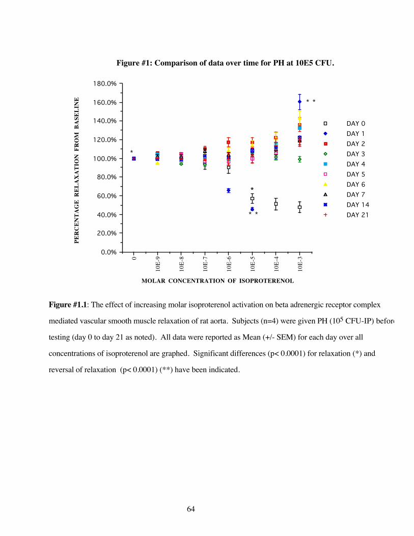
64
0
10E-
9
10E-
8
10E-
7
10E-
6
10E-
5
10E-
4
10E-
3
0.0%
20.0%
40.0%
60.0%
80.0%
100.0%
120.0%
140.0%
160.0%
180.0%
DAY 0DAY 1DAY 2DAY 3DAY 4DAY 5DAY 6DAY 7 DAY 14DAY 21
Figure #1: Comparison of data over time for PH at 10E5 CFU.
MOLAR CONCENTRATION OF ISOPROTERENOL
PER
CEN
TAG
E R
ELA
XA
TIO
N F
RO
M B
ASE
LIN
E
* *
* *
*
*
Figure #1.1: The effect of increasing molar isoproterenol activation on beta adrenergic receptor complex
mediated vascular smooth muscle relaxation of rat aorta. Subjects (n=4) were given PH (105 CFU-IP) before
testing (day 0 to day 21 as noted). All data were reported as Mean (+/- SEM) for each day over all
concentrations of isoproterenol are graphed. Significant differences (p< 0.0001) for relaxation (*) and
reversal of relaxation (p< 0.0001) (**) have been indicated.
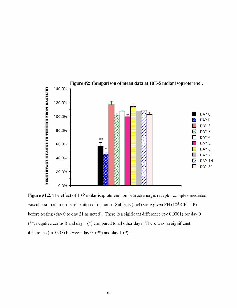
65
0%0.0%
20.0%
40.0%
60.0%
80.0%
100.0%
120.0%
140.0%
DAY 0DAY1DAY 2DAY 3DAY 4DAY 5DAY 6DAY 7DAY 14DAY 21
Figure #2: Comparison of mean data at 10E-5 molar isoproterenol.
PER
CEN
TAG
E C
HA
NG
E IN
TEN
SIO
N F
RO
M B
ASE
LIN
E
**
*
Figure #1.2: The effect of 10-5 molar isoproterenol on beta adrenergic receptor complex mediated
vascular smooth muscle relaxation of rat aorta. Subjects (n=4) were given PH (105 CFU-IP)
before testing (day 0 to day 21 as noted). There is a sigificant difference (p< 0.0001) for day 0
(**, negative control) and day 1 (*) compared to all other days. There was no significant
difference (p> 0.05) between day 0 (**) and day 1 (*).
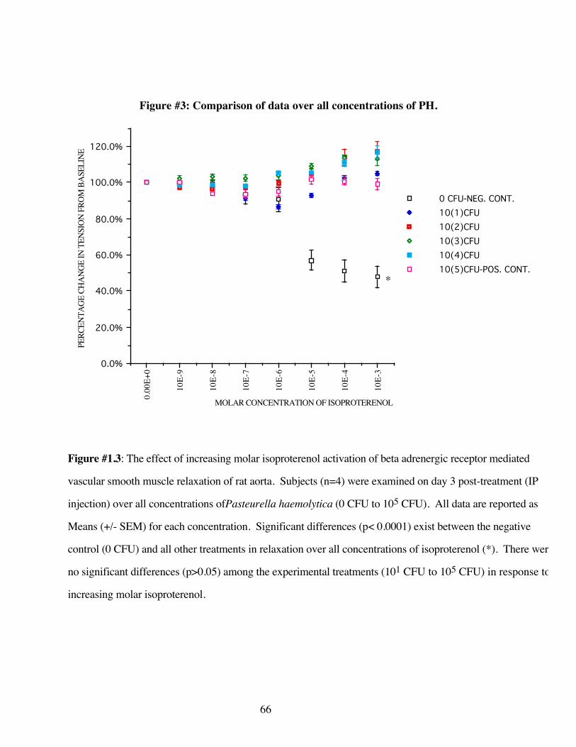
66
0.0
0E+0
10E-
9
10E-
8
10E-
7
10E-
6
10E-
5
10E-
4
10E-
30.0%
20.0%
40.0%
60.0%
80.0%
100.0%
120.0%
0 CFU-NEG. CONT.10(1)CFU10(2)CFU10(3)CFU10(4)CFU10(5)CFU-POS. CONT.
Figure #3: Comparison of data over all concentrations of PH.
MOLAR CONCENTRATION OF ISOPROTERENOL
PERC
ENTA
GE C
HANG
E IN
TEN
SION
FRO
M B
ASEL
INE
*
Figure #1.3: The effect of increasing molar isoproterenol activation of beta adrenergic receptor mediated
vascular smooth muscle relaxation of rat aorta. Subjects (n=4) were examined on day 3 post-treatment (IP
injection) over all concentrations ofPasteurella haemolytica (0 CFU to 105 CFU). All data are reported as
Means (+/- SEM) for each concentration. Significant differences (p< 0.0001) exist between the negative
control (0 CFU) and all other treatments in relaxation over all concentrations of isoproterenol (*). There were
no significant differences (p>0.05) among the experimental treatments (101 CFU to 105 CFU) in response to
increasing molar isoproterenol.
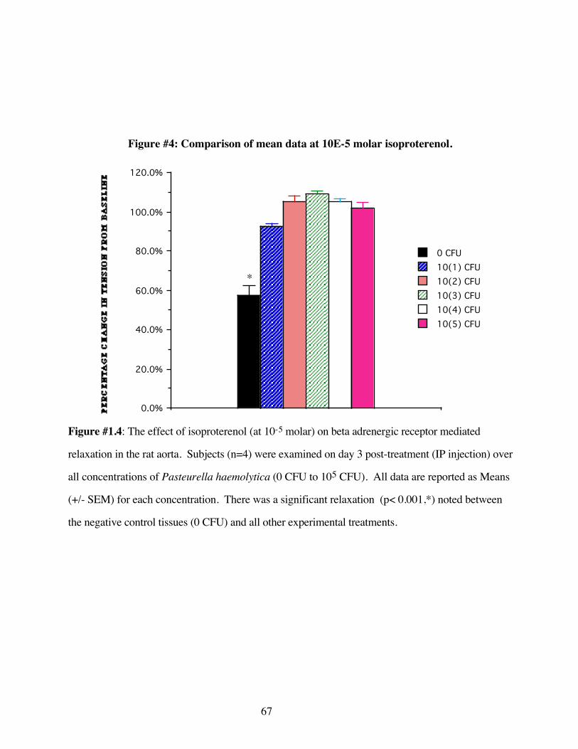
67
0%0.0%
20.0%
40.0%
60.0%
80.0%
100.0%
120.0%
0 CFU10(1) CFU10(2) CFU10(3) CFU10(4) CFU10(5) CFU
Figure #4: Comparison of mean data at 10E-5 molar isoproterenol.
PER
CEN
TAG
E C
HA
NG
E IN
TEN
SIO
N F
RO
M B
ASE
LIN
E
*
Figure #1.4: The effect of isoproterenol (at 10-5 molar) on beta adrenergic receptor mediated
relaxation in the rat aorta. Subjects (n=4) were examined on day 3 post-treatment (IP injection) over
all concentrations of Pasteurella haemolytica (0 CFU to 105 CFU). All data are reported as Means
(+/- SEM) for each concentration. There was a significant relaxation (p< 0.001,*) noted between
the negative control tissues (0 CFU) and all other experimental treatments.

68
Similar examination of scanning electron micrographs for the experiment involving
increasing doses of Pasteurella haemolytica A1 were reviewed These photographs showed
increasing amount of the cellular elements and fibrin-like material on the vascular luminal
endothelium with increasing concentrations of Pasteurella haemolytica.(see figure #16-21).
DISCUSSION
A) Time Course of Beta Adrenergic Receptor System Dysfunction
Vascular adrenergic smooth muscle tone is not likely the product of a single receptor type or
single drug class. All adrenergic receptors interact to result in the final vascular smooth muscle
response that is measured. It is likely that the predominant initiators of relaxation of vascular tone
are the beta adrenergic receptors which are activated by isoproterenol (a synthetic mixed beta
adrenergic agonist). The predominant receptor initiating vascular constriction is the alpha-1
receptor. Finally, alpha-2 receptors appear to cause constriction in low vascular tone but initiate
relaxation during high vascular tone (results not presented here). Further to confound this issue, the
pharmacological agonists available are selective, not specific for each adrenergic receptor. This
suggests that these agonistic adrenergic drugs may have interactions with other than their specific
adrenergic receptors at higher pharmacological concentrations.
Comparison of these data to those of previous researchers, reveals the same pattern as that
described by Weekley for the negative and positive control groups (Weekley 1993E). After day 2,
the loss of the smooth muscle relaxation response to isoproterenol cannot be denied. The negative
control aortic rings demonstrated a typical relaxation throughout increasing concentrations of
isoproterenol. The data on day 1 (figures #1 and #2) demonstrate a partial relaxation and then a
reversal of the relaxation (reversal of potency) to a contraction at the higher concentrations of
isoproterenol. This evidence suggests that the nature of the disruption of the beta adrenergic

69
mechanism the disruption of the pharmacologically mediated smooth muscle relaxation response
occurs over 24-48 hours. This data of reversal of potency suggests that disruption of the adrenergic
mechanism is not complete but dose dependent or related to receptor recruitment and may be
overcome by increasing pharmacological doses.
The relaxation seen on day one offers several possible explanations. Endotoxin, one of the
virulence factors of PH has been known to affect levels of cyclic adenosine monophosphate
(cAMP). On day one, the effect of the treatment caused an enhanced, (possibly supersensitivity)
response in the beta receptors which subsequently fail to respond by day 2. This is a previously
reported phenomenon and is seen in denervation super-reactivity in whole animal preparations.
However, a review of the literature fails to describe this event in beta receptors. Another explanation
may be that the early actions of the vaccine primarily affect the adrenergic receptors responsible for
vascular contraction (alpha-1). Early temporary reduction of the tonic activity of alpha one receptors
would result in the data obtained for day 1 at these lower isoproterenol concentrations.
The first time point of maximum contraction of vascular tissue occurs two days after
parenteral exposure to PH . This indicates that previous experiments, evaluated at three days, have
obtained the expected results but have yet to characterize the maximal effect of inhibition of
relaxation of vascular tissue. There are relative, though not statistically significant, increases in the
amount of inhibition of the smooth muscle relaxation response at day 2 and day 7 (see figure #2).
The relaxation response may be related to fluctuations in the disruption of beta adrenergic system to
exposure to PH.
At the higher doses of isoproterenol there is a tendency for the tissues to demonstrate an
increased constriction. At higher doses of isoproterenol (10-4 and 10 -3 molar concentration) the
selectivity for beta receptors may be generalized to other receptors including the alpha-1 receptor.
This generalization of isoproterenol may result in an increase in alpha-1 activity and an immediate

70
contraction response. Similar results can be noted for all days except the negative and positive
controls.
The question of the duration of the disruption of the beta receptor has been investigated.
Cursory evaluation of the data may suggest a tendency to recovery from days 2 to 5 (figure #2).
This trend reverses on day 6 with increasing levels of beta adrenergic receptor dysfunction to day
21. Decreased vascular relaxation after the parenteral administration of PH may occur for several
reasons. If the beta adrenergic receptor protein is rendered non-functional, return to function may
require the regeneration of entirely new BARCs. If the disruption is beyond the receptor protein, in
the receptor mechanism cascade, the disruption may require regeneration of the appropriate
molecules for full functioning. Several steps in the biochemical cascade may be particularly
susceptible to Pasteurella haemolytica disruption. The disruption of the post-beta adrenergic
receptor mechanism, the G protein, could result in failure of vasorelaxation. The disruption of the
secondary messenger system, downstream of the G-protein, involving adenylylate cyclase and
cAMP may be interrupted and therefore hinder vascular relaxation. There is some evidence that
Pasteurella haemolytica can have deleterious effects on cAMP. The post cAMP mechanisms are
only beginning to be investigated. The possibility exists that there is a disruption of the steps
involving the myosin light chain kinase, the calcium (another second messenger) or the mechanism
of the vascular smooth muscle relaxation proper. At this juncture none of these possibilities have
been investigated with respect to the parenteral exposure to PH.
B) Dose of bacteria to initiate beta adrenergic receptor disruption
This series of experiments suggest that disruption is not an all or none phenomenon. The
first dose increment (101 CFUs) reveals a statistically significant initial relaxation followed by a
sudden reversal of the relaxation resulting in a final overall contraction at and above 10-5 molar
concentration of isoproterenol. This data suggests a partial disruption of the beta adrenergic

71
receptor mechanism. All other results (102 to 105 CFU) indicate no significant relaxation at 10-5
molar isoproterenol. In fact there is a significant contraction (P< 0.01) for all groups, except the
negative and positive controls at and above 10-5 molar isoproterenol concentration.
C) Examination of the morphological changes with respect to time
The morphological characteristics of the experimental treatment are more clear cut. The
negative control shows no evidence of fibrin formation. The first observation of an increase in
cellular elements is evident on Day 1. The thesis of this paper is that the beta adrenergic system is
rendered dysfunctional after parenteral exposure to PH. The beta receptor has also been
demonstrated to exist in close association with the cells of the endothelium (Steinberg 1984) and is
involved in the regulation of vascular permeability, the inhibition of thrombin mediated vascular and
the permeability to albumin (Mizus 1985; Minnear 1986). The inhibition of the beta adrenergic
mechanisms would therefore likely result in the disruption of the control of vascular permeability
and increases in thrombogenic events on the endothelial surface. Examination of the endothelial
surface reveals an increase in linear "fibrin like" strands. These events appear to increase with
increasing time. The events can be explained by the disruption of endothelial based beta adrenergic
receptors that inhibit thrombogenic events intravascularly. The fibrin-like material on day 6 has a
multi-stranded appearance and represents an increase in fiber diameter. The fibrin strands decrease
significantly at days 14 and are not visible by day 21. These data appear to support the thesis that
the beta adrenergic receptors are disrupted by parenteral exposure to Pasteurella haemolytica.
On day 7 there appeared to be a "delamination" or "vesicular formation" of the luminal
surface and an apparent exposure of subendothelial tissues of the aorta. These "vesicles" were also
noted by Weekley et al (1993A) The vesicles are of unknown origin however similar lesions have
been noted in the pumonary vasculature after infection with Haemophilus somnus in cattle (Griffin,
1996) and have also been noted on endothelial surfaces of animals that have not been exposed to
72
PH. It is therefore possible that these "vesicles or blebs" are non-specific responses to either
immunological, endotoxin or stressor challenges. A possible scenario for their origin is that
unregulated beta receptors on the surface of the endothelium fail to prevent cellular elements,
particularly neutrophils from interacting with the endothelial surface. As has been noted the beta
receptor of the neutrophil acts to reduce extraneous and exuberant release of oxidative enzymes.
The disruption of the beta receptors on the neutrophil would therefore be unable to prevent the
exuberant release of the oxidative enzymes by the neutrophil. Oxidative enzymes released even in
limited quantities should result in endothelial cell damage which may appear as vesicles. There is
some support in the literature for this position as it has been demonstrated that neutrophil deficient
animals appear to demonstrate less gross and microscopic pathological damage than animals with
normal levels of neutrophils (Breider, 1991).
The evaluation of morphological events over time appears to involve the development of
fibrin like strands increasing in density from day 1 through day 7. Day 7 appears to be the first day
where blebbing, or disruption of the endothelial surface is noted. By day 14 the fibrin strands
appear to be decreased in number and by day 21 are non-existent. Overall there is an increase in
cellularity from day 1 through day 6. The endothelium appears to swell decreasing the
topographical features as time progresses. A thrombogenic event is likely to follow a sequential
pattern of increasing fibrin and cellularity intravascularly. These events may be followed by
elaboration and release of intracellular substances that can result in delamination of the endothelial
surface resulting in the blebbing noted. These results support the hypothesis that fibrin is a
consequence of the initiation of throbo-embolic/endothelial activity. The blebbing is likely a
consequence of increased cellularity, increased expression of ICAM, ELAM receptors resulting in
the vesicle formation, delamination or blebbing.
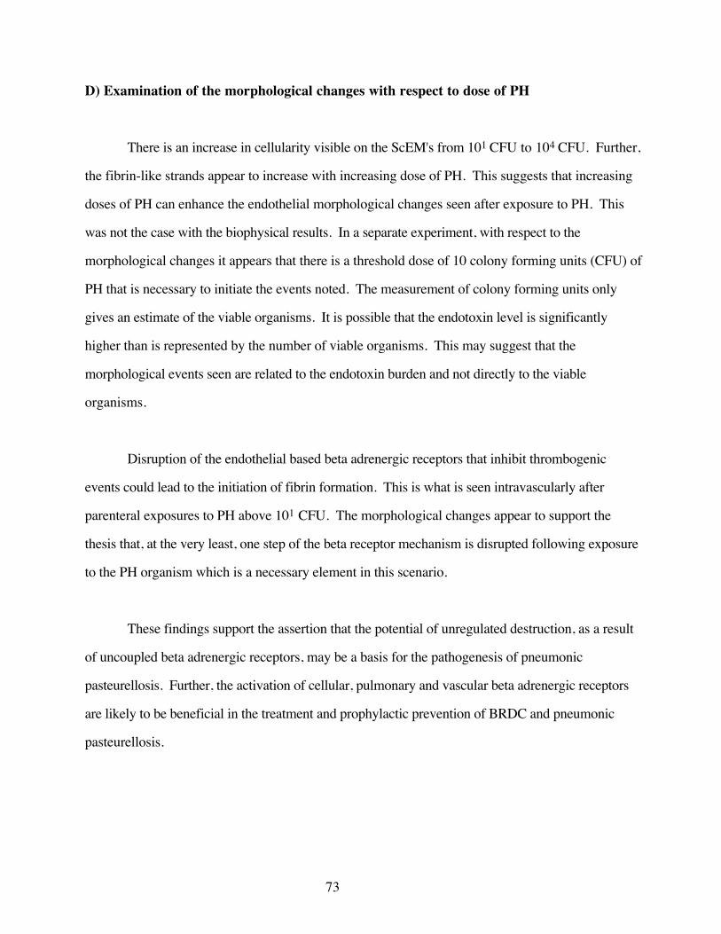
73
D) Examination of the morphological changes with respect to dose of PH
There is an increase in cellularity visible on the ScEM's from 101 CFU to 104 CFU. Further,
the fibrin-like strands appear to increase with increasing dose of PH. This suggests that increasing
doses of PH can enhance the endothelial morphological changes seen after exposure to PH. This
was not the case with the biophysical results. In a separate experiment, with respect to the
morphological changes it appears that there is a threshold dose of 10 colony forming units (CFU) of
PH that is necessary to initiate the events noted. The measurement of colony forming units only
gives an estimate of the viable organisms. It is possible that the endotoxin level is significantly
higher than is represented by the number of viable organisms. This may suggest that the
morphological events seen are related to the endotoxin burden and not directly to the viable
organisms.
Disruption of the endothelial based beta adrenergic receptors that inhibit thrombogenic
events could lead to the initiation of fibrin formation. This is what is seen intravascularly after
parenteral exposures to PH above 101 CFU. The morphological changes appear to support the
thesis that, at the very least, one step of the beta receptor mechanism is disrupted following exposure
to the PH organism which is a necessary element in this scenario.
These findings support the assertion that the potential of unregulated destruction, as a result
of uncoupled beta adrenergic receptors, may be a basis for the pathogenesis of pneumonic
pasteurellosis. Further, the activation of cellular, pulmonary and vascular beta adrenergic receptors
are likely to be beneficial in the treatment and prophylactic prevention of BRDC and pneumonic
pasteurellosis.
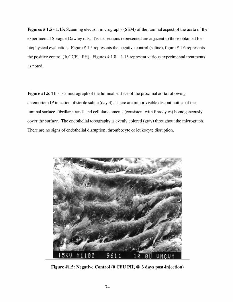
74
Figures # 1.5 - 1.13: Scanning electron micrographs (SEM) of the luminal aspect of the aorta of the
experimental Sprague-Dawley rats. Tissue sections represented are adjacent to those obtained for
biophysical evaluation. Figure # 1.5 represents the negative control (saline), figure # 1.6 represents
the positive control (105 CFU-PH). Figures # 1.8 – 1.13 represent various experimental treatments
as noted.
Figure #1.5: This is a micrograph of the luminal surface of the proximal aorta following
antemortem IP injection of sterile saline (day 3). There are minor visible discontinuities of the
luminal surface, fibrillar strands and cellular elements (consistent with fibrocytes) homogeneously
cover the surface. The endothelial topography is evenly colored (gray) throughout the micrograph.
There are no signs of endothelial disruption, thrombocyte or leukocyte disruption.
Figure #1.5: Negative Control (0 CFU PH, @ 3 days post-injection)

75
Figure # 1.6: This is a micrograph of the luminal surface of the proximal aorta following
antemortem IP (3 day) injection of 105 CFU PH. There are significant disruptions of the endothelial
surface at 11 and 12 o'clock positions. There are circular / spherical cell-like adhesions consistent in
size and shape to thrombocytes at 1-2 and 7-8 o'clock positions. Other cell like adhesion are seen at
6-9 and 3 o'clock positions, these are consistent with leukocytes. Flattened cell like adhesions with
fibrillar-like attachments are consistent with fibrocytes. The endothelial topography is not as smooth
as the negative control. There are increased depth and size of rugae (endothelial folds).
..
Figure # 1.6: Positive control (105 CFU PH, @ 3 days post-injection)
76
Figure # 1.7: This micrograph represents a 24 hour post injection (105 CFU PH) section of the
proximal luminal aorta Initial changes noted in this micrograph are increased fibrillar attachment to
the luminal surface, attachment of several biconcave cell-like discs (consistent with RBC's) and
multiple "sphere-like" adhesions (consistent with thrombocytes). There appears to be an increase in
fibrillar cellular activity (consistent with increased fibrocyte activity) at the endothelial / luminal
surface.
Figure # 1.7: Experimental day #1.

77
Figure # 1.8: This micrograph at 48 hours post injection (105 CFU PH, IP) shows a continuation
of the increased fibrillar activity over the luminal surface noted previously (figure # 1.7). The cell-
like elements (consistent with leukoctyes) appear creanated secondary to preparation artifacts.
Fibrillar elements are increased over previous days. There are endothelial discontinuities creating a
moth-eaten appearance to the topography. The lesions at 1, 3 and 7 o'clock positions suggest
endothelial ulceration or vesicular formation.
Figure # 1.8: Experimental day #2
78
Figure # 1.9: Day 4 following antemortem injection (105-CFU PH, IP) demonstrates significant
disruption of the endothelial surface at the 12 and 1 o'clock positions. The ulcerated areas of the
endothelial surface are consistant with disrupted bullae or ulceration's. Fibrillar elements are
consistant with those noted in the 2 and 3 day micrographs.
Figure # 1.9: Experimental day #4

79
Figure # 1.10: This micrograph, antemortem day 7 post injection (105 CFU PH, IP), shows an
increase in the fibrillar components on the endothelial surface as compared to previous days
micrographs.. There appear to be ulcerations of the endothelium at 11 and 12 o'clock. Spherical
cell-like elements can be seen throughout the micrograph, consistant with erythrocytes fibrocytes
and leukocytes. Overall there appears to be less organization to the direction and thickness of the
fibrils noted previously (figures # 1.8 and # 1.9). The topography of the endothelial surface has
been obscured by the fibrillar elements.
Figure # 1.10: Experimental day #5

80
Figure # 1.11: This micrograph has thicker (3-4 micron) fibrils in a network that is parallel to the
rugae (endothelial folds) of the endothelial surface. The thicker fibrils appear to be organized
multiple strands of the smaller (1 micron) fibrillar elements. There are two areas of bullae / vesicular
rupture of the endothelial surface that can be seen at approximately 1 and 3 o'clock. Overall the
topography of the surface is smoother than that of the negative control suggesting endothelial cell
swelling.
Figure # 1.11: Experimental day #6

81
Figure # 1.12: This micrograph day 7 post injection (IP 105 CFU-PH) demonstrates the maximum
level of endothelial damage noted over all days examined (day 1 through day 21). There are multiple
ruptured bullae, ulcerative areas exposing tissue consistant with a basement membrane. The luminal
surface of the vascular endothelium appears to be devoid of rugae (endothelial folds) suggesting
endothelial swelling. There is a minimal amount of fibrillar material scattered over the surface of the
endothelium.
Figure # 1.12: Experimental day #7
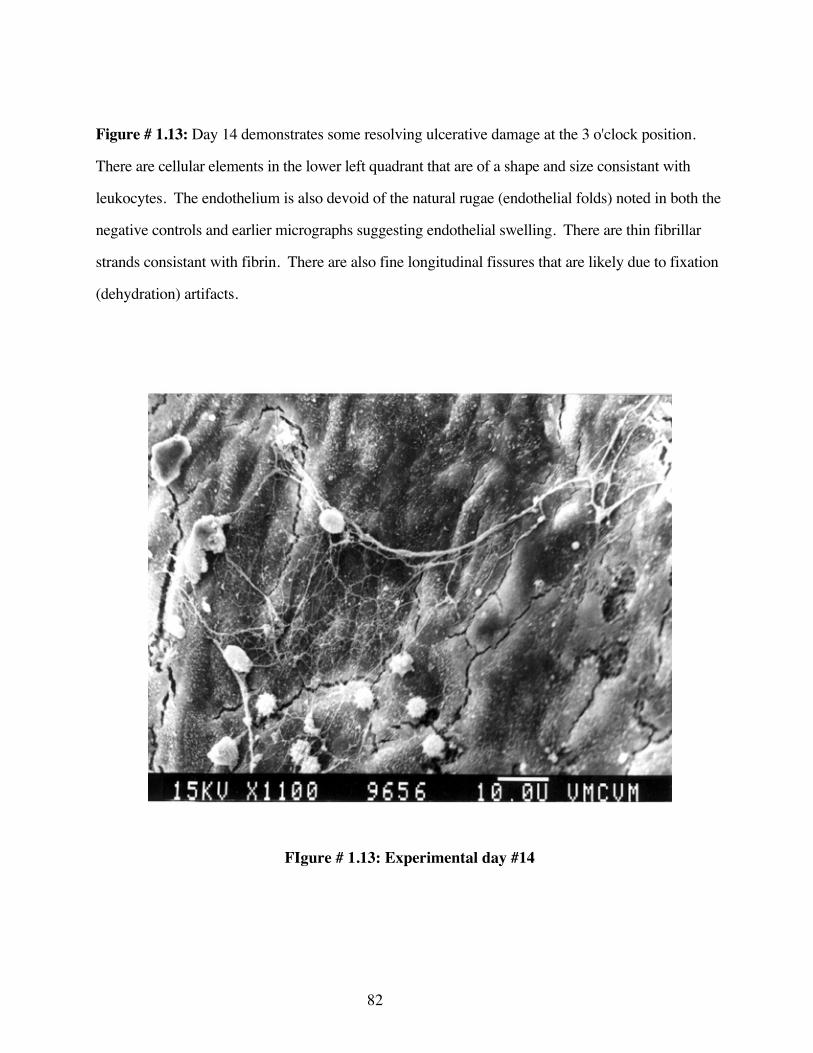
82
Figure # 1.13: Day 14 demonstrates some resolving ulcerative damage at the 3 o'clock position.
There are cellular elements in the lower left quadrant that are of a shape and size consistant with
leukocytes. The endothelium is also devoid of the natural rugae (endothelial folds) noted in both the
negative controls and earlier micrographs suggesting endothelial swelling. There are thin fibrillar
strands consistant with fibrin. There are also fine longitudinal fissures that are likely due to fixation
(dehydration) artifacts.
FIgure # 1.13: Experimental day #14
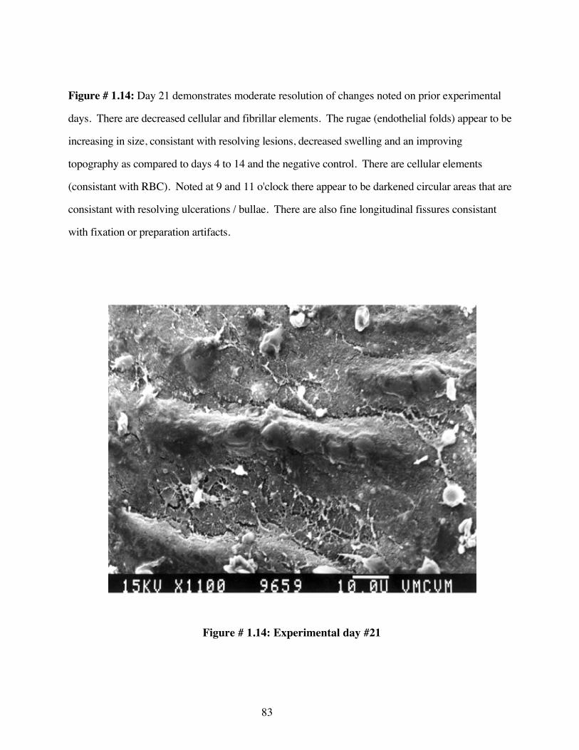
83
Figure # 1.14: Day 21 demonstrates moderate resolution of changes noted on prior experimental
days. There are decreased cellular and fibrillar elements. The rugae (endothelial folds) appear to be
increasing in size, consistant with resolving lesions, decreased swelling and an improving
topography as compared to days 4 to 14 and the negative control. There are cellular elements
(consistant with RBC). Noted at 9 and 11 o'clock there appear to be darkened circular areas that are
consistant with resolving ulcerations / bullae. There are also fine longitudinal fissures consistant
with fixation or preparation artifacts.
Figure # 1.14: Experimental day #21
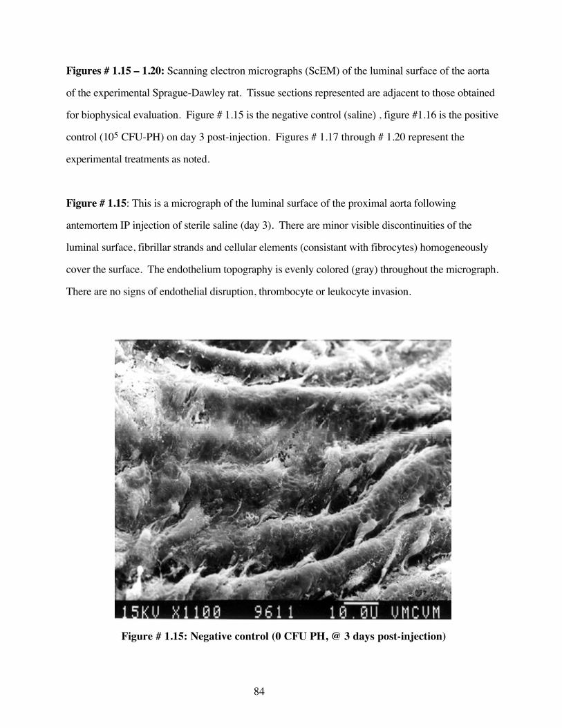
84
Figures # 1.15 – 1.20: Scanning electron micrographs (ScEM) of the luminal surface of the aorta
of the experimental Sprague-Dawley rat. Tissue sections represented are adjacent to those obtained
for biophysical evaluation. Figure # 1.15 is the negative control (saline) , figure #1.16 is the positive
control (105 CFU-PH) on day 3 post-injection. Figures # 1.17 through # 1.20 represent the
experimental treatments as noted.
Figure # 1.15: This is a micrograph of the luminal surface of the proximal aorta following
antemortem IP injection of sterile saline (day 3). There are minor visible discontinuities of the
luminal surface, fibrillar strands and cellular elements (consistant with fibrocytes) homogeneously
cover the surface. The endothelium topography is evenly colored (gray) throughout the micrograph.
There are no signs of endothelial disruption, thrombocyte or leukocyte invasion.
Figure # 1.15: Negative control (0 CFU PH, @ 3 days post-injection)
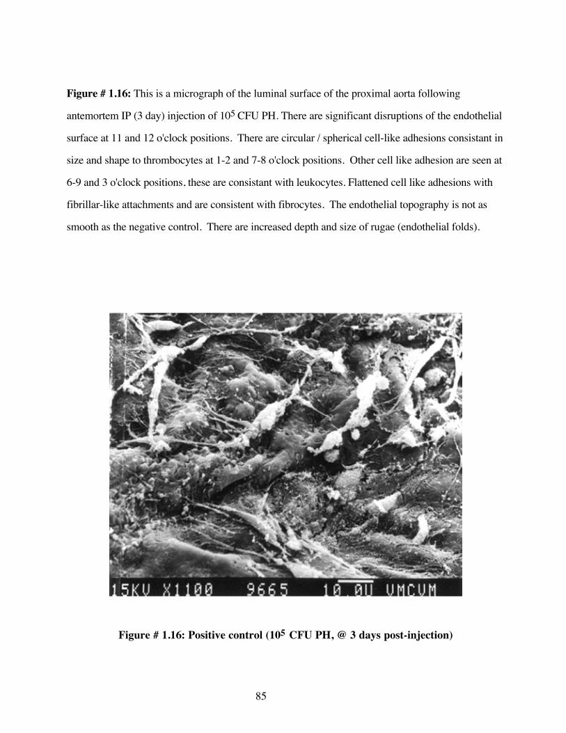
85
Figure # 1.16: This is a micrograph of the luminal surface of the proximal aorta following
antemortem IP (3 day) injection of 105 CFU PH. There are significant disruptions of the endothelial
surface at 11 and 12 o'clock positions. There are circular / spherical cell-like adhesions consistant in
size and shape to thrombocytes at 1-2 and 7-8 o'clock positions. Other cell like adhesion are seen at
6-9 and 3 o'clock positions, these are consistant with leukocytes. Flattened cell like adhesions with
fibrillar-like attachments and are consistent with fibrocytes. The endothelial topography is not as
smooth as the negative control. There are increased depth and size of rugae (endothelial folds).
Figure # 1.16: Positive control (105 CFU PH, @ 3 days post-injection)
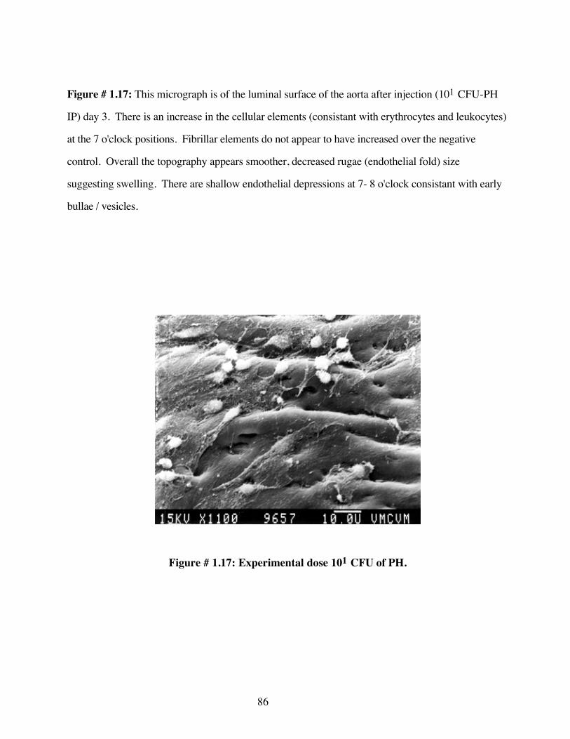
86
Figure # 1.17: This micrograph is of the luminal surface of the aorta after injection (101 CFU-PH
IP) day 3. There is an increase in the cellular elements (consistant with erythrocytes and leukocytes)
at the 7 o'clock positions. Fibrillar elements do not appear to have increased over the negative
control. Overall the topography appears smoother, decreased rugae (endothelial fold) size
suggesting swelling. There are shallow endothelial depressions at 7- 8 o'clock consistant with early
bullae / vesicles.
Figure # 1.17: Experimental dose 101 CFU of PH.
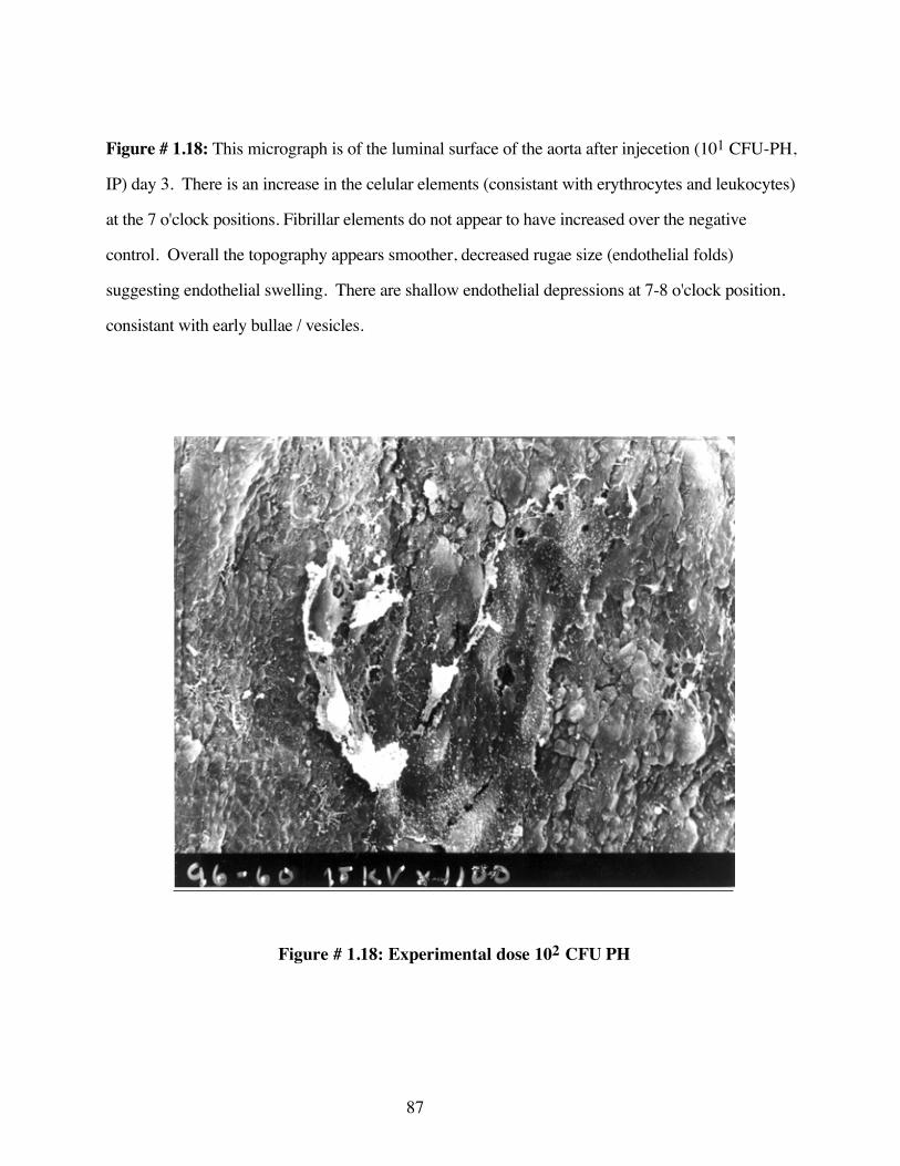
87
Figure # 1.18: This micrograph is of the luminal surface of the aorta after injecetion (101 CFU-PH,
IP) day 3. There is an increase in the celular elements (consistant with erythrocytes and leukocytes)
at the 7 o'clock positions. Fibrillar elements do not appear to have increased over the negative
control. Overall the topography appears smoother, decreased rugae size (endothelial folds)
suggesting endothelial swelling. There are shallow endothelial depressions at 7-8 o'clock position,
consistant with early bullae / vesicles.
Figure # 1.18: Experimental dose 102 CFU PH

88
Figure # 1.19: This micrograph (103 CFU-PH IP) demonstrates evidence of increasing luminal /
cellular activity. There are multiple disc-like cells with an apparent concave surface (consistant with
erythrocytes) and spherical cells with small "projection" (consistant with leukocytes) The
endothelium also appears to have a flattened topography. There are some irregular circular
endothelial depressions or ulcerations at the 4, 7 and 12 o'clock positions. Overall there appears to
be an increased level of cellular activity and endothelial swelling.
Figure # 1.19: Experimental dose 103 CFU PH
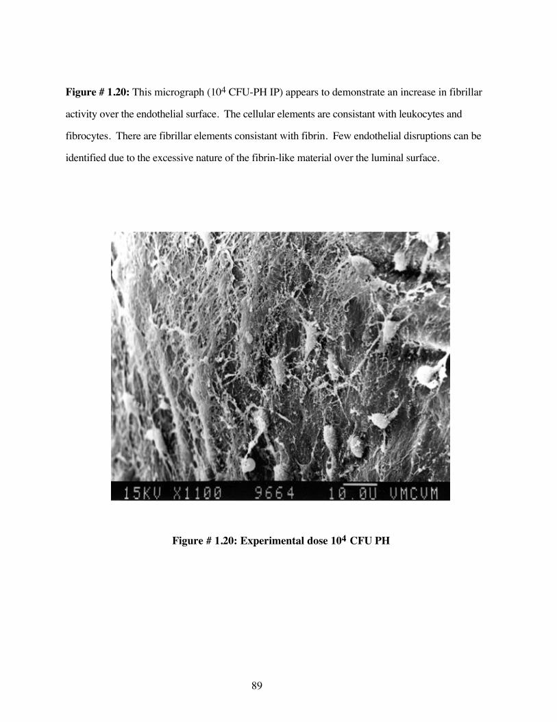
89
Figure # 1.20: This micrograph (104 CFU-PH IP) appears to demonstrate an increase in fibrillar
activity over the endothelial surface. The cellular elements are consistant with leukocytes and
fibrocytes. There are fibrillar elements consistant with fibrin. Few endothelial disruptions can be
identified due to the excessive nature of the fibrin-like material over the luminal surface.
Figure # 1.20: Experimental dose 104 CFU PH
90
CONCLUSIONS
It is unclear as to whether the biophysical effects associated with isoproterenol occur solely
as a result of the disruption of the beta adrenergic system alone or are a combination of the
disruption of the beta adrenergic receptor and alpha-2 and possibly even alpha-1 adrenergic receptor
dysfunction. The data indicating abrupt change from relaxation to an increase in tension (reversal of
the trend), may be due to some interaction of isoproterenol at the alpha adrenergic receptor at higher
concentrations. Isoproterenol is a beta adrenergic selective, not specific, drug. However, at higher
concentrations the drug becomes less selective and has alpha receptor activity possibly confounding
the drug selectivity (e.g. above 10-4 molar). This suggests that either the alpha-1 or alpha-2
receptors are responding to the beta adrenergic agonist or that the beta receptor activity for the
remaining population of active receptors has been maximized for relaxation and are demonstrating a
pharmacological failure. It is very difficult to isolate a single receptor type of the vascular system
pharmacologically. In most cases, neuropharmacological activities are dynamic and interaction
among receptor types is a common confound in these experiments.
It is apparent from the data that the maximal pharmacological effect of antemortem
intraperitoneal injection of PH occurs at two days (48 hours) after exposure to 102 CFUs in the rat.
The maximal endothelial, cellular and fibrillar (micro-anatomical) events, at the luminal surface,
consistent with endothelial disruption, increase from day 2 or 3 through day 21 and appear to be at a
maximum on day 7 (figure #12) or after 103 CFU PH-IP. The exact duration of the
pharmacological / micro-anatomical disruption was not able to be determined within the limits of the
present experiment. It is most probable that the duration of the beta adrenergic receptor system is in
proportion to the quantity and duration of exposure to PH and LPS. It is likely that some
component of an individual's immunological status (e.g. neutrophil population, cortisol
concentration, level of cytokine release) are factors in the duration and extent of the disruption of the
adrenergic mechanisms. It must first be ascertained whether the physiological effects noted, are a

91
result of pure beta adrenergic and endothelial disruption or if there is a component of alpha-1 or
alpha-2 adrenergic system failure. It is less likely that the disruption of the vascular adrenergic
dynamic control mechanisms are a consequence of only one receptor mechanism. The vascular
smooth muscle homeostatic control system is modulated by the adrenergic, cholinergic and the non-
adrenergic non-cholinergic (NANC) system. It is therefore very likely that multiple system changes
are involved in the disruption of the beta adrenergic receptor mediated smooth muscle relaxation
response.
The morphological results are consistent with earlier finding (Weekley 1993E). The
changes noted are consistent temporally, with the disruption of beta adrenergic receptor mechanisms
and the initiation of micropathology. This observation is consistent with inflammation and is likely
to involve the endothelium, smooth muscle and immunologically active cellular elements.
These results support the hypothesis that disruption of beta adrenergic receptors is mediated
by either PH or an element intimately associated with PH (e.g. LPS). The observation that the beta
adrenergic receptor mechanism is disrupted for up to 21 days at levels of PH parenteral exposure of
101 CFU suggest that some whole unit vaccines have the potential to induce changes that my be
deleterious to an animal's health. The consequences of this study are that the disruption of the beta
receptor mechanism associated with the vascular smooth muscle is likely to have occurred but is
unlikely to be the sole mechanism involved in the pathogenesis of either post vaccination illness or
the pathogenesis of pneumonic pasteurellosis. There is indirect evidence to suggest that beta
receptor systems, beyond those of the vascular smooth muscle receptor mechanisms are affected.
Potentially the most devastating consequence of BARC disruption is the failure of the limiting of
leukocyte, and more specifically neutrophil, oxidative enzyme and elastase release. Further,
morphological evidence suggests that there are deleterious thrombogenic events that occur at the
surface of the endothelium and increase in severity up to day 7 and then continually improves until
day 21. Similar findings are noted for increasing PH antemortem injections.
92
References
Barnard, M. L, S. Matalon (1992). “Mechanisms of extracellular reactive oxygen species injury tothe pulmonary microvasculature.” Journal of Applied Physiology 72(5): 1724-9.
Borregaard, N. (1988). The respiratory burst. The Respiratory Burst and its PhysiologicalSignificance. , A.J. Sbarra. (Editor) New York, Plenum Press: p.1-8.
Bowden, J. J., I. Sulakvelidze, D.M. McDonald (1994). “Inhibition of neutrophil and eosinophiladhesion to venules of rat trachea by beta-2 adrenergic agonist formoterol.” Journal of AppliedPhysiology 77(1): 397-405.
Busse, W. W., J.M. Sosman (1984). “Isoproterenol inhibition of isolated human neutrophilfunction.” Journal of Allergy and Clinical Immunology 73: 404-12.
Engels, F., R.S. Oosting, F.P. Nijkamp (1985). “Pulmonary macrophages induce deterioration ofguinea pig tracheal beta adrenergic function through release of oxygen free radicals.” EuropeanJournal of Pharmacology 111: 143-4.
Green, J. F. (1982). Fundamental Cardiovascular and Pulmonary Physiology. Philadelphia, Lea andFebiger.
Hogg, N. (1993). “A model of leukocyte adhesion to vascular endothelium.” Current Topics inMicrobiology and Immunology 184: 79-86.
Martin, S. W. (1983). “Vaccination; is it effective in preventing respiratory disease or influencingweight gains in feedlot calves.” Canadian Veterinary Journal 24: 10-19.
Minnear, F. L., A. Johnson, A.B. Malik (1986). “Beta adrenergic modulation of pulmonarytransvascular fluid and protein exchange.” Journal of Applied Physiology 60: 266-74.
Mizus, I., W.Summer, I. Farrukh, J.R. Michael, G.H. Gurtner (1985). “Isoproterenol oraminophylline attenuate pulmonary edema after acid lung injury.” American Review of RespiratoryDisease 131: 256-9.
Mueller, H., H.J. Motulsky, L.A. Sklar (1988). “The potency and kinetics of the beta adrenergicreceptors on human neutrophils.” Molecular Pharmacology 34: 347-53.
Ogunbiyi, P. O. (1986). Effects of microbial infections on guinea pig and calf homeostasis:implications for therapy., Doctoral Dissertation, Faculty of Graduate Studies. Guelph, University ofGuelph: 280.
Opdahl, H., H.B. Benestad and G. Nicolayson (1993). “Effect of beta-adrenergic agents on humanneutrophil granulocyte activation with N-formyl-methionyl-leucyl-phenylalanine and phorbolmyristate acetate.” Pharmacology and Toxicology 72: 221-8.
Robinson, N. E., R.F. Slocomb, F.J. Derksen (1984). Physiology of the bovine lung. BovineRespiratory Disease : A Symposium. R. W. Loan. College Station, Texas, Texas A&M UniversityPress: 163-222.
Schummer, A., R.Nickel, W.O. Sack (1979). The Viscera of the Domestic Mammals. New York,Springer-Verlag.
93
Seccombe, J. F., P.J. Pearson, H. V. Schaff (1994). “Oxygen radical mediated vascular injuryselectively inhibits receptor dependent release of nitric oxide from canine coronary arteries.” Journalof Thoracic and Cardiovascular Surgery 107: 505-9.
Steinberg, S. F., E.A. Jaffe, J.P. Bilezikian (1984). “Endothelial cells contain beta adrenergicadrenoreceptors.” Naunyn-Schmiedebergs Archives of Pharmacology 325: 310-3.
Thorlarkson, B., W. Martin, D. Peters (1990). “A field trial to evaluate the efficacy of a commercialPasteurella haemolytica bacterial extract in preventing bovine respiratory disease.” CanadianJournal of Veterinary Research 31: 573-9.
Weekley, L. B., P. Eyre, H.P. Veit, N. Sriranganathan (1991A). “Exposure of sheep to Pasteurellahaemolytica alters ex vivo pulmonary vascular smooth muscle alpha adrenergic responses: role ofvascular endothelium.” Research Communications in Chemical Pathology and Pharmacology 74(1):121-4.
Weekley, L. B., P.Eyre, H.P. Veit, N. Sriranganathan (1993A). “Morphological and functionaldisturbances in pulmonary vascular endothelium following exposure of sheep to an avirulent strainof Pasteurella haemolytica.” Journal of Comparative Pathology 108(1): 65-72.
Weekley, L. B., P.Eyre (1993B). “Vaccination with live Pasteurella haemolytica alters vascularadrenergic reactivity.” Research Communications in Chemical Pathology and Pharmacology 79(3):389-92.
Weekley, L. B., P. Eyre (1993E). “Disturbances in ex vivo vascular smooth muscle responsesfollowing exposure to Pasteurella haemolytica vaccines.” Journal of Veterinary Pharmacology andTherapeutics 16: 446-53.
Weekley, L. B. (1994). Personal Communication.
Weekley, L. B., H. P.Veit (1995). “Potential morphologic and physiologic factors that maypredispose the bovine lung to respiratory diease.” The Compendium 17(7): 974-983.
Wren, G. (1995). “Understanding the bovine respiratory system.” Bovine Veterinarian November:6-9.
94
B). EVALUATION OF ESCHERICHIA COLI (O 55/B5) ENDOTOXIN ININDUCING BETA ADRENERGIC RECEPTOR MECHANISM MEDIATEDDYSFUNCTION OF VASCULAR SMOOTH MUSCLE OF EX-VIVO RAT AORTA
Live Pasteurella haemolytica (PH) injected intraperitoneally in the rat, has been
demonstrated to cause a disruption of the vascular beta adrenergic system associated with smooth
muscle (Weekley 1993A; 1993B; 1993E). In the intact pulmonary vascular system the beta
adrenergic receptor acts to mediate vascular dilation. On the arterial side this vasodilation increases
the functional blood flow to the lung. The post-arterial beta adrenergic receptor mechanism also
causes vasodilation where the consequence is increased drainage of blood from the lung which
prevents pulmonary hypertension and subsequent pulmonary congestion. Therefore any disruption
of venous dilation has the potential to cause either pulmonary vascular congestion or pulmonary
interstial edema, two of the many classical signs of pneumonic pasteurellosis.
Many authors have examined the epidemiology of the efficacy of several monovalent PH
vaccines. The results of these studies suggest that vaccination is less than efficacious in preventing
disease in the short term and may even predispose the animal to an increased risk of disease
(Martin 1983; Thorlarkson 1990). It has been demonstrated that parenteral exposure to a live
vaccine, Precon® (live vaccine), and an endotoxin free vaccine, Presponse® (leukotoxoid rich),
causes disruption of beta adrenergic receptors associated with vascular smooth muscle in several
species (Weekley 1991A; 1993B). Beta receptor mechanisms are associated not only with the
vascular smooth muscle, but also have been demonstrated to ameliorate extravasation of fluid,
inhibit thrombin interactions at the endothelial surface and mediate excessive release of destructive
enzymes and oxygen free radicals from leukocytes, particularly neutrophils. The importance of the
beta receptor system to pulmonary homeostasis is well established. The beta adrenergic receptor
mechanism disruption may be significant factor in the post-vaccination increase in disease
morbidity and mortality, the pathogenesis of pneumonic pasteurellosis and the severity of
pulmonary vascular lesion development.

95
The question of which component of the monovalent pneumonic pasteurellosis vaccines
being examined is causing the beta adrenergic dysfunction has yet to be investigated. Each
manufactured vaccine is unique both in its immunomodulatory elements and in the chemical
adjuvants used to enhance the host immunoprotective reaction. Four virulence factors have been
described for PH A-1 (PH). These include: fimbriae, polysaccharide capsule, leukotoxin, and
lipopolysaccharide (endotoxin, LPS). The most likely portion of the vaccine to cause beta
adrenergic complex failure is endotoxin. However scientific information concerning Presponse®
indicated that this vaccine was endotoxin free as assessed by the Limulus Ameobocyte Lysate assay
(LAL) (Cyanamid 1992). Researchers have suggested that Presponse® is not endotoxin free but
may, coincidentally, contain a factor that causes a false negative test in the LAL assay (Maheswaran
et al 1995). Endotoxin has been demonstrated to disrupt the beta adrenergic receptor system of
cardiac beta adrenergic mechanisms (van Heuven-Nolsen 1986; Campbell 1993). The evaluation of
endotoxin as a factor in the disruption of the vascular smooth muscle associated beta adrenergic
receptor mechanisms now appears to be an important question to investigate.
Endotoxins have four common elements among the gram negative bacteria that share this
feature. All endotoxins contain four basic components, lipid A moiety, the inner core, the outer core
and the o-specific polysaccharide chain. The species differences in endotoxin are found in the
sequences associated with the o-specific-polysaccharide side chain. The lipid A moiety of
endotoxin is likely the essential factor causing the defect noted by Weekley. Due to difficulties in
isolating and purifying PH endotoxin, Eschericia coli (EC) endotoxin has been selected for use.
EC endotoxin has been shown to uncouple beta adrenergic receptors of the myocardium (Romano
1985; Shepard 1987; Forse 1989; Campbell 1993; Bensard 1994). The extraction and purification
of EC endotoxin has been standardized and well characterized and is available commercially. Two
studies have directly compared the effects of EC endotoxin to that of PH and found that the EC
endotoxin was less virulent than the PH endotoxin (Rimsay 1981; Lacroix 1993). Unfortunately,
96
neither of these studies investigated the functioning of the vascular beta receptors with respect to the
activity. It is for these reasons that EC endotoxin was chosen for this study. This paper proposes
to examine the effect of purified EC endotoxin alone as one factor affecting the functioning of
vascular beta adrenergic receptors associated with smooth muscle in the rat aorta previously
described by Weekley (1991A; 1993A; 1993B).
MATERIALS AND METHODS
A) Treatments and Subjects
Adult male Sprague-Dawley rats (Harlan Sprague-Dawley Inc., Indianapolis, Indiana) 150-
175g were used in this series of experiments. The animals initially were housed in a quarantine
area for a 1 week for acclimatization to the new facility and observe for physical signs of disease.
The acclimatization period involved a 12:12 light dark photo period, room temperature was 220 C
+/- 30 C. Each experimental animal was injected, intraperitoneally, with 200 micro endotoxin units
( µEU) of EC endotoxin in sterile, endotoxin free saline (O55:B5, Sigma L2880). Negative control
animals were injected with endotoxin-free normal sterile saline, in the same manner as the
experimental animals. The positive controls were injected with 105 colony forming units (CFU) of
PH intraperitoneally as described by Weekley (1993E). There were four animals in each of the
three groups (n=4)
B) Preparation of Drugs and Solutions
The PH suspension was evaluated for its endotoxin content by use of the Limulus
Ameobocyte Lysate assay (QCL-1000, Bio-Whittaker Co.). A standard preparation of 105 colony
forming units was evaluated at several concentrations by log-serial dilutions. Standardized
equivalent EU Escherichia coli endotoxin (Sigma L2880, O55:B5) was added to sterile endotoxin
97
free saline and vortexed in a sterile injection vial to ensure complete mixing. The appropriate
quantity (200 µEU) of EC endotoxin was then immediately injected, intraperitoneally in the
experimental animals.
Tissues were carefully dissected and suspended in Krebs-Henseleit saline buffer. This
buffer was composed of one liter of sterile double distilled water in a sterile 5 liter Erlenmeyer flask
plus: 6.9 grams NaCl ; 0.35 grams KCl; 0.28 CaCl; 0.14 grams MgSO4; 2.09 grams NaHCO3;
0.16 grams KH2PO4; 2.0 grams dextrose, as described by Weekley (1994). This solution was
aseptically mixed immediately before use.
Potassium chloride was used for initial smooth muscle contraction. This solution was
prepared in sterile, double distilled water (30 millimolar concentration when 200 microliters of stock
KCl solution is added to 30 milliliters of the bathing solution).
Isoproterenol, a mixed beta adrenergic receptor agonist, was used to evaluate the beta
receptor functioning of isolated, ex-vivo, aortic vascular tissue. This solution was prepared in
sterile, double distilled water. Serial decreasing logarithmic concentrations of isoproterenol were
made from the initial 10-2 molar solution. The serial dilutions were made by taking 100 microliters
of the initial stock solution and diluting this in 900 microliters of sterile double distilled water
sequentially to 10-9 molar solution.
C) Tissue Collection and Measurement Techniques
Subjects were anesthetized to a surgical plane of anesthesia with sodium pentobarbital (80
mg/kg, IP) which was then followed immediately by a second injection of 25 IU of sodium heparin
(IP). When at a deep surgical plane of anesthesia, the abdomen was opened to reveal the
diaphragm. The approach to the thorax and thoracic aorta was through the diaphragm. The

98
ventrum of the thorax was removed to completely expose the thoracic contents. The aorta was
removed from the base of the heart to the last rib ensuring that minimal damage or stretching
occurred. The entire vessel was the placed in warm aerated Krebs-Henseleit (370 +/-2oC, mixed
with 95% oxygen: 5% carbon dioxide) for further dissection and complete removal of extraneous
fat and superficial tissue. All aortic rings were obtained from the base of the heart. The rings were
carefully measured to be 3 mm in width. The dissection process was performed with exceptional
care to ensure that stretching or damage to the endothelium was avoided.
The 3 mm aortic ring, removed for biophysical testing, was placed between two glass hooks
in a thirty-five milliliter tissue bath. Changes in tension (isometric contraction) of the aortic ring
were measured following the addition of known concentrations of various drugs. One hook was
attached to a fixed point at the bottom of the tissue chamber while the other hook was attached to an
electronic transducer that was calibrated to give output in grams tension. The tissue was placed in
an insulated heated tissue bath containing Krebs-Henseleit bicarbonate buffer The buffer was
aerated with 95% oxygen and 5% carbon dioxide through each of four individually isolated tissue
baths. A constant tissue bath temperature of 37 O+/- 2o C was maintained by circulating water
through a temperature controlled water pump and sequentially through the outer heating jacket of
each tissue bath. Each tissue ring initially was placed in the tissue bath under 2 grams resting
tension for two hours and allowed to equilibrate. The Krebs-Henseleit buffer was replaced every
15-20 minutes throughout equilibration and testing. Testing started immediately after the
equilibration period. The aortic rings, in the tissue bath, were exposed to 200 µl of potassium
chloride (KCl, 30 millimolar) and allowed to equilibrate to this treatment over 30 minutes. After the
tissue had equilibrated to the KCl, the recording was started and sequential increasing molar doses
of isoproterenol were added. Isoproterenol hydrochloride was used to evaluate the response of beta
adrenergic receptors in causing smooth muscle relaxation.
99
Electronically calibrated analog signals were transmitted from the transducer connected to
the glass hook through an analog digital converter (MacLab A/D signal converter, World Precision
Instruments). Digitized information was captured by MacLab data acquisition software (MacLab
2.51) on a Model M5011 Macintosh SE computer (1MB RAM, 800 K drive, 20 megabyte hard
disk) in real time. Records of vascular smooth muscle activity, as a cumulative dose response to
log molar increases in isoproterenol, were obtained and the results stored on a 3.5 inch computer
disk.
D) Statistical Analysis of Data
Data were analyzed in several ways. Initially the data were compared for each experimental
treatment at the isoproterenol concentration of 10-5 molar. In pilot studies this concentration of
isoproterenol appeared to approximate the effective concentration 50% (EC 50) and so was selected
to be an appropriate point for comparison of different treatments for the experimental protocols.
The comparison was completed by use of an analysis of variance (ANOVA) using group (n=4)
means and standard error of the mean. This comparison was to evaluate significant differences
among treatment groups.
Further analysis consisted of a pair wise comparison of the values of 0 molar isoproterenol
and 10-5 molar isoproterenol. Pair wise comparisons were accomplished by use of Student-
Newman-Keuls test. This comparison was to evaluate change, with respect to relaxation or
contraction, with increasing logarithmic doses of isoproterenol
RESULTS
Comparison of the effects of Escherichia coli endotoxin (EC, 055:B5) with the whole
organism preparation of PH (105 CFU) suggests that the EC endotoxin mediates similar beta
100
receptor deficits as was noted to be caused by IP injection of a suspension of the whole PH
organism (figure #1). The mechanisms and pathogenesis of this deficit remains to be investigated.
The negative control resulted in the expected smooth muscle relaxation in response to
increasing logarithmic molar isoproterenol. The positive control failed to respond to the
isoproterenol cumulative dose response curve, as expected. Statistical comparison of the three
groups at 10-5 molar concentration of isoproterenol revealed that there is a highly significant
difference between the negative control and the positive control (p< 0.001). The aortic rings from
the experimental animals treated with the EC endotoxin were identical to the positive control animals
treated with PH endotoxin at 10-5 molar isoproterenol (no significant difference, p> 0.05). At
concentrations of isoproterenol above 10-5 molar, the aortic rings from the EC endotoxin treated
animals show a greater increase in tension than the positive controls. The results were not
statistically different at either 10-4 and 10-3 molar isoproterenol (p> 0.05).
DISCUSSION
The results of this experiment suggest that EC endotoxin alone was sufficient to cause
disruption of the beta adrenergic receptor mediated vascular smooth muscle function. This evidence
supports Maheswaran that Presponse® likely contains endotoxin though the significance of this
finding has yet to be evaluated as to its affect on the in-vivo pulmonary vascular BARC
(Maheswaran 1995) Further, the results of this experiment are in agreement with current literature
indicating that EC endotoxin disrupts cardiac associated beta adrenergic receptor mechanisms
(Romano 1985; Shepard 1987; Forse 1989; Campbell 1993; Bensard 1994).
There are differences between the EC endotoxin and that of the PH endotoxin. The most
striking of these differences is that EC endotoxin has O-specific polysaccharide side chains not
present on the PH endotoxin. Several authors have suggested that the lipid A moiety of the

101
endotoxin is responsible for the deleterious activity of LPS in susceptible hosts. Since the lipid A
moiety is relatively conserved in all bacterial LPS, the results presented here should be as valid for
PH endotoxin as for EC endotoxin. Comparative studies suggest that EC endotoxin is less
destructive than PH endotoxin (Rimsay 1981; Lacroix 1993). One might expect that the EC
endotoxin would cause less of a receptor interference than is experimentally observed with the PH
control. The lack of difference between the receptor inhibition of EC LPS and PH A1 may be
explained in several ways. It is possible that a separate factor associated within the PH organism
restricts the full virulence of the lipopolysaccharide responsible for the beta receptor disruptive
activity. Another explanation may be that total inhibition of the smooth muscle response occurs so
that at any level of endotoxin at three days post exposure ther is a single, maximal response (ceiling
phenomenon). Any increase beyond the minimally necessary level of endotoxin to cause system
disruption fails to result in an increase in the dependent variable (beta adrenergic receptor
mechanism dysfunction). Finally, the endotoxin level of sequentially prepared PH aliquots may
vary to some degree. In assessing the concentration of PH at 105 CFUs one is only evaluating
those organisms that are viable and result in a colony.
Endotoxin is an integral part of the gram negative wall of the organism. The fact that an
organism is no longer viable doesn't reduce the endotoxin concentration. This fact makes it difficult
to exactly reproduce the concentrations of LPS from Weekley (1993A, 1993E). One can see
however that the disruption of the vascular smooth muscle is similar in magnitude and extent for
both EC endotoxin and PH endotoxin. Previous experiments with varying concentrations of PH
organisms, revealed a beta adrenergic system disruptive effect at PH concentrations as low as 10
viable PH organisms injected intraperitoneally. Evaluation of the minimal concentrations of EC
endotoxin necessary to induce beta adrenergic associated vascular smooth muscle dysfunction
would be interesting.
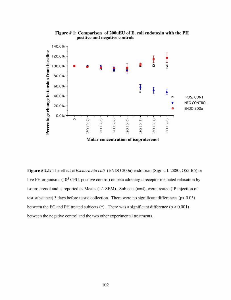
102
0
ISO
10(
-9)
ISO
10(
-8)
ISO
10(
-7)
ISO
10(
-6)
ISO
10(
-5)
ISO
10(
-4)
ISO
10(
-3)
0.0%
20.0%
40.0%
60.0%
80.0%
100.0%
120.0%
140.0%
POS. CONTNEG CONTROLENDO 200u
Figure # 1: Comparison of 200uEU of E. coli endotoxin with the PH positive and negative controls
Molar concentration of isoproterenol
Perc
enta
ge c
hang
e in
tens
ion
from
bas
elin
e
*
Figure # 2.1: The effect ofEscherichia coli (ENDO 200u) endotoxin (Sigma L 2880, O55:B5) or
live PH organisms (105 CFU, positive control) on beta adrenergic receptor mediated relaxation by
isoproterenol and is reported as Means (+/- SEM). Subjects (n=4), were treated (IP injection of
test substance) 3 days before tissue collection. There were no significant differences (p> 0.05)
between the EC and PH treated subjects (*). There was a significant difference (p < 0.001)
between the negative control and the two other experimental treatments.
103
There remains much work to do in this area. First, it remains to be shown that the
endotoxin from PH is equivalently virulent as the EC endotoxin with respect to the vascular beta
adrenergic receptors. There are four portions to the lipopolysaccharide molecule. It will become
important to identify which of these components or combination of these components are active in
the disruption of the beta adrenergic receptor. This information would allow the isolation and
elimination of the offending moiety while retaining the portions of the microbiological organism
that are important in immunostimulation aspects of the vaccine. Further experimentation using each
of the four virulence factors recognized for PH, with respect to the beta adrenergic receptor system
would result in additional useful information to the nature of the virulence mechanisms of the PH
organism.
CONCLUSIONS
1) The evidence presented here gives a strong suggestion that EC endotoxin alone,
administered parenterally is sufficient to cause vascular smooth muscle associated beta
adrenergic receptor disruption similar to that seen with exposure to PH organisms.
2) The definitive experiment using isolated purified PH lipopolysaccharide has yet to be
completed. The characterization of the PH endotoxin could be of significance in
determining the mode of action and toxicokinetics with respect to the disruption of the
BARC.
3) Similar experiments using the various isolated and purified components (e.g.the lipid A
inner core, outer core or o-specific polysaccharide chain) of the endotoxin would also be
enlightening.
104
REFERENCES
Bensard, D. D., A. Banerjee, R.C. McIntyre, R.L. Berens, A.H. Harken (1994). “Endotoxindisrupts beta-adrenergic signal transduction in the heart.” Archives of Surgery 129(2): 198-204.
Campbell, K. L., R.A. Forse (1993). “Endotoxin exposed atria exhibit G-protein based deficits ininotropic regulation.” Surgery 114: 471-9.
Cyanamid (1992). Presponse®- Advertising data.
Forse, R. A., R. Leibel, M. Gagner (1989). “The effects of Escherichia coli endotoxin on theadrenergic control of lipolysis in the human adipocyte.” Journal of Surgical Research 46(1): 41-8.
Lacroix, R., J.R. Duncan, R.P. Jenkins, R.A. Leitch, J.A. Perry, J.C. Richards (1993). “Structuraland serological specificities of Pasteurella haemolytica lipopolysaccharides.” Infection andImmunity 61(1): 170-81.
Maheswaran, S. K. (1995). Further evaluation of vaccines claiming to be endotoxin free hasrevealed endotoxin. Personal Communication, February 23,
Martin, S. W. (1983). “Vaccination; is it effective in preventing respiratory disease or influencingweight gains in feedlot calves.” Canadian Veterinary Journal 24: 10-19.
Rimsay, R. L., J.E. Coyle-Dennis, L.H. Lauerman, and P.G. Squire (1981). “Purification andbiological characterization of endotoxin fractions from Pasteurella haemolytica.” American Journalof Veterinary Research 42: 2134-38.
Romano, F. D., S.B. Jones (1985). “Beta adrenergic stimulation of myocardial cyclic AMP inendotoxic rats.” Circulatory Shock 17(3): 243-52.
Shepard, R. E., Lang, C.H., K.H. McDonough (1987). “Myocardial adrenergic responsivenessafter lethal and non-lethal doses of endotoxin.” American Journal of Physiology 252(2Pt2): h410-6.
Thorlarkson, B., W. Martin, D. Peters (1990). “A field trial to evaluate the efficacy of a commercialPasteurella haemolytica bacterial extract in preventing bovine respiratory disease.” CanadianJournal of Veterinary Research 31: 573-9.
van Heuven-Nolsen, D., G. Folkerts, D.J. Dewildt, F.P. Ninjkamp (1986). “The influence ofBordetella pertussis and its constituents on the beta-adrenergic receptor in the guinea pigrespiratory system.” Life Science 38(8): 677-85.
Weekley, L. B., P. Eyre, H.P. Veit, N. Sriranganathan (1991A). “Exposure of sheep to Pasteurellahaemolytica alters ex vivo pulmonary vascular smooth muscle alpha adrenergic responses: role ofvascular endothelium.” Research Communications in Chemical Pathology and Pharmacology74(1): 121-4.
Weekley, L. B., P.Eyre, H.P. Veit, N. Sriranganathan (1993A). “Morphological and functionaldisturbances in pulmonary vascular endothelium following exposure of sheep to an avirulent strainof Pasteurella haemolytica.” Journal of Comparative Pathology 108(1): 65-72.

105
Weekley, L. B., P.Eyre (1993B). “Vaccination with live Pasteurella haemolytica alters vascularadrenergic reactivity.” Research Communications in Chemical Pathology and Pharmacology 79(3):389-92.
Weekley, L. B., P. Eyre (1993E). “Disturbances in ex vivo vascular smooth muscle responsesfollowing exposure to Pasteurella haemolytica vaccines.” Journal of Veterinary Pharmacology andTherapeutics 16: 446-53.
Weekley, L. B. (1994). Personal Communication.
106
C). CHANGES IN BETA ADRENERGIC STIMULATED CYCLIC ADENOSINEMONOPHOPHATE LEVELS (CAMP), IN EX-VIVO AORTIC RINGS, FOLLOWINGIN-VIVO, INTRA-PERITONEAL EXPOSURE TO PASTEURELLA HAEMOLYTICA A-1IN RATS
The exact pathogenesis of pneumonic pasteurellosis is unclear. Weekley has demonstrated,
in rats, that a single intraperitoneal injection of 105 CFU of Pasteurella haemolytica A-1 in rats
can result in an elimination of the beta adrenergic mediated aortic smooth muscle relaxant response
to isoproterenol (Weekley 1993A). Similar results have been described for the pulmonary
vasculature of cattle (Weekley 1993B) and sheep (Weekley 1993E). Beta adrenergic receptors
(BAR) are intimately involved with vascular homeostasis. Beyond the effects of the modification ot
the release of oxidative enzymes elaborated by leukocytes, the beta adrenergic receptor may also act
in several key ways to inhibit the release of potentially severely destructive endogenous elements.
The significance of this to the establishment of pneumonic pasteurellosis in ruminants is not
understood. Evidence exists to support a relationship between the disruption of the beta adrenergic
receptor and development of the clinical and pathological signs of respiratory disease in cattle.
Beta adrenergic receptors associated with the vascular endothelium have a role in regulating
the fluid dynamics between the intravascular and extravascular microenvironments (Mizus 1985;
Minnear 1986). It is necessary to regulate the extravasation of vascular fluid, electrolytes and
osmotic factors to maintain health. Other beta adrenoceptors modulate the elaboration of tissue
factor, the first step of the extrinsic pathway of coagulation (Busso 1991). Tissue factor generation
causes the release of thromboplastin and prothrombin which is then converted to thrombin and
fibrinogen. The presence of fibrin attached to the luminal surface of the vessel is a characteristic of
the endothelium noted to occur in response to a PH challenge in rats and cattle (Majno 1961;
Weekley 1993A).
107
The beta adrenergic receptor is a critical factor in the control of leukocyte antibacterial
responses both in-vitro and in-vivo (Bourne 1971; Ignarro 1974; Zurier 1974; Hills 1975; Harlan
1981; Busse 1984; Engels 1985; Tecoma 1986; Warren 1987; Barnard 1992; Kubes 1993;
Granger 1994; Seccombe 1994; Al-Essa 1995). Increased pulmonary vascular trapping and
margination of leukocytes coupled with an interruption in the beta adrenoceptor mediated inhibition
of the neutrophil may lead to a catastrophic release of free radicals and proteases. This could result
in pulmonary and vascular tissue destruction. It has been demonstrated that neutrophil deficient
animals suffer less pulmonary tissue damage than neutrophil sufficient animals (Slocombe 1985;
Breider 1991). Similar findings have been reported in the kidney (Paller 1989) and in the liver
(Jaeschke 1990) of rats for various inflammatory disease states. Other evidence suggests that the
beta receptor on macrophages can inhibit the release of pro-inflamatory cytokine products such as
Tumor Necrosis Factor-alpha (Hu 1991; Severn 1992).
With respect to the bovine respiratory system, there are a number of unique anatomical
features that make the vascular smooth muscle relaxation effects of BARs necessary to maintain
optimum health. The bovine pulmonary vasculature has a thicker tunica muscularis compared to
other mammals (Robinson 1984; Breeze 1985; Weekley 1995; Wren 1995). The pulmonary
vasculature controls blood flow, at the capillary / acinus level, by manipulation of vascular
constriction and dilation. The mechanisms involved in the dynamic control of the vasculature are
varied and involve cholinergic, adrenergic, non-cholinergic non-adrenergic systems and humoral
mediators (Breeze 1985; Barnes 1986; Barnes 1995). The adrenergic system is involved with the
establishment of an optimal ventilation / perfusion match at the level of the pulmonary acinus
(Green 1982; Barnes 1995). The beta adrenergic receptor is one factor in the metabolic equation
that acts to mediate vascular relaxation and balance the vasoconstrictive effects of the alpha-1 and
alpha-2 beta adrenergic receptors.

108
Disruption of beta adrenoceptor vasodilation may result in a shift of the dynamic vascular
smooth muscle balance to result in an exaggerated alpha adrenergic mediated vasoconstriction. The
consequences to the pulmonary and vascular tissue may include tissue hypoxia further enhancing
vascular constrictive responses, metabolic acidosis, and increased trapping of circulating leukocytes,
especially neutrophils in the capillaries and post-capillary venules.
The importance of the regulatory role of beta adrenoceptor on leukocytes, in the pulmonary
vasculature smooth muscle and on the vascular endothelium, is well established (Engels 1985,
Ogunbiyi 1986, Borregaard 1988, Bensard 1996, Opdahl 1993, Secomb 1994, Ignano 1974,
Bourne 1971, Ignano 1974, Al Essa 1995). The devastating consequences of the uncoupling of this
receptor is a logical explanation for some of the signs of the pathogenesis of pneumonic
pasteurellosis. Evidence exists that suggests that the virulence factor, endotoxin, elaborated by
Gram negative bacteria may be involved in the uncoupling of beta receptors (van Heuven-Nolsen
1986). Endotoxin acts directly on the beta adrenergic receptor to decrease receptor expression (van
Heuven-Nolsen 1986; Forse 1989). There is a significant decrease in receptor density on cardiac
myocytes in the presence of endotoxemia in human patients (Shepard 1987). Still other studies
have demonstrated a diminished response of the beta-1 cardiac adrenoceptor distal to the agonist-
receptor interaction (Campbell 1993). This additional evidence indicates that the beta adrenergic
dysfunction, in the presence of endotoxin, may be responsible for the lack of cardiac response to
beta-adrenergic agonists in endotoxemia (Romano 1985; Bensard 1994). One may expect the
vascular system to be equally susceptible to endotoxin and beta adrenergic receptor dysfunction,
This may explain some of the pathological and clinical signs of pneumonic pasteurellosis. Given
these findings and the evidence presented by Weekley et al (1993E) it is important to explore the
beta adrenergic receptor dysfunction of aortic tissue, exposed in-vivo to Pasteurella haemolytica A-
1.

109
The beta adrenergic receptor is a G-protein linked receptor protein. Agonist ligand binding
activates a G-protein intracellular linkage which in turn initiates the conversion of ATP to cAMP by
the inducible enzyme adenylylate cyclase. cAMP then binds to regulatory subunits causing the
liberation of active catalytic subunits that phosphorylate protein substrates. In vascular smooth
muscle tissue, the result is that there is a relaxation and subsequent vasodilation of the affected
vessel.
It is not clear why the vascular smooth muscle fails to respond to isoproterenol stimulation
after PH administration. It is possible that the failure of the vascular smooth muscle to relax may
be due to the disruption of the intracellular cascade of events associated with the beta adrenergic
receptor. Numerous sites, within the post-receptor cascade, may be affected by slight biochemical,
pH and biomolecular changes resulting in the disruption of the beta adrenergic receptor's ability to
initiate vascular smooth muscle relaxation. Finally disruption of smooth muscle relaxation may
exist within the vascular smooth muscle itself, either with respect to the signal / muscle interface
(myosin light chain kinase) or within the smooth muscle proper (calcium flux and channel
disruptions). To date, none of these possibilities have been examined. This paper proposes to
examine the receptor and post-receptor cascade to establish if a defect exists between the receptor
and the elaboration of the second messenger cAMP.
MATERIALS AND METHODS
A) Treatments and Subjects
Adult male Sprague-Dawley rats (Harlan Sprague-Dawley Inc., Indianapolis, Indiana)
150-175 g were used in this series of experiments. The animals initially were housed in a
quarantine area for a 1 week for acclimatization to the new facility and observed for signs of
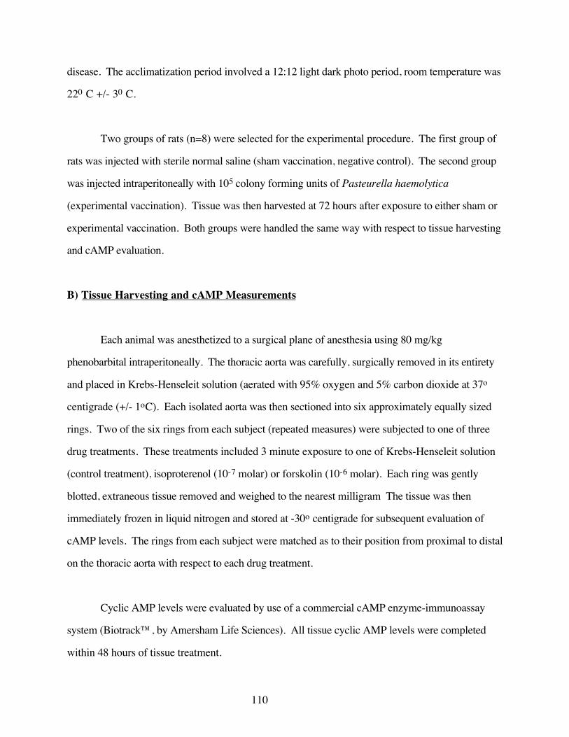
110
disease. The acclimatization period involved a 12:12 light dark photo period, room temperature was
220 C +/- 30 C.
Two groups of rats (n=8) were selected for the experimental procedure. The first group of
rats was injected with sterile normal saline (sham vaccination, negative control). The second group
was injected intraperitoneally with 105 colony forming units of Pasteurella haemolytica
(experimental vaccination). Tissue was then harvested at 72 hours after exposure to either sham or
experimental vaccination. Both groups were handled the same way with respect to tissue harvesting
and cAMP evaluation.
B) Tissue Harvesting and cAMP Measurements
Each animal was anesthetized to a surgical plane of anesthesia using 80 mg/kg
phenobarbital intraperitoneally. The thoracic aorta was carefully, surgically removed in its entirety
and placed in Krebs-Henseleit solution (aerated with 95% oxygen and 5% carbon dioxide at 37o
centigrade (+/- 1oC). Each isolated aorta was then sectioned into six approximately equally sized
rings. Two of the six rings from each subject (repeated measures) were subjected to one of three
drug treatments. These treatments included 3 minute exposure to one of Krebs-Henseleit solution
(control treatment), isoproterenol (10-7 molar) or forskolin (10-6 molar). Each ring was gently
blotted, extraneous tissue removed and weighed to the nearest milligram The tissue was then
immediately frozen in liquid nitrogen and stored at -30o centigrade for subsequent evaluation of
cAMP levels. The rings from each subject were matched as to their position from proximal to distal
on the thoracic aorta with respect to each drug treatment.
Cyclic AMP levels were evaluated by use of a commercial cAMP enzyme-immunoassay
system (Biotrack™, by Amersham Life Sciences). All tissue cyclic AMP levels were completed
within 48 hours of tissue treatment.

111
C) Preparation of Drugs and Solutions
Krebs-Henseleit buffer was used during the dissection of the ex-vivo aortic rings. The
composition of the Krebs-Henseleit solution (in one liter of sterile double distilled water in a sterile
5 liter Erlenmeyer flask as follows: 6.9 grams NaCl ; 0.35 grams KCl; 0.28 CaCl; 0.14 grams
MgSO4; 2.09 grams NaHCO3; 0.16 grams KH2PO4; 2.0 grams dextrose) as described by
Weekley (1994). This solution was aseptically mixed immediately before use.
Isoproterenol solution was prepared from stock powder (Sigma I5627) in sterile double
distilled water. Isoproterenol was mixed to result in a concentration of 10-7 molar in a volume of
30 milliliters of Krebs-Henseleit. Each aortic ring was then exposed to this final solution for
exactly 3 minutes. The solution was maintained at 370 centigrade (+/- 2oC) and aerated with 95%
oxygen / 5% carbon dioxide gas.
Forskolin, (Sigma, F6886) was mixed to give a final concentration of 10-6 molar
concentration in 30 milliliters of Krebs-Henseilet. Forskolin acts to induce the activity of cAMP in
the cell. The procedure for exposure of aortic rings to forskolin was identical to that of the
isoproterenol.
The control solution was Krebs-Henseleit alone. Control tissues were treated in an identical
manner to that described above.
D) Statistical Analysis of Data
This experiment is to evaluate the relative differences in cAMP concentration at three
different points of the receptor to smooth muscle relaxation cascade. Each treatment result (control,
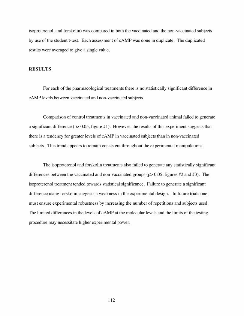
112
isoproterenol, and forskolin) was compared in both the vaccinated and the non-vaccinated subjects
by use of the student t-test. Each assessment of cAMP was done in duplicate. The duplicated
results were averaged to give a single value.
RESULTS
For each of the pharmacological treatments there is no statistically significant difference in
cAMP levels between vaccinated and non-vaccinated subjects.
Comparison of control treatments in vaccinated and non-vaccinated animal failed to generate
a significant difference (p> 0.05, figure #1). However, the results of this experiment suggests that
there is a tendency for greater levels of cAMP in vaccinated subjects than in non-vaccinated
subjects. This trend appears to remain consistent throughout the experimental manipulations.
The isoproterenol and forskolin treatments also failed to generate any statistically significant
differences between the vaccinated and non-vaccinated groups (p> 0.05, figures #2 and #3). The
isoproterenol treatment tended towards statistical significance. Failure to generate a significant
difference using forskolin suggests a weakness in the experimental design. In future trials one
must ensure experimental robustness by increasing the number of repetitions and subjects used.
The limited differences in the levels of cAMP at the molecular levels and the limits of the testing
procedure may necessitate higher experimental power.
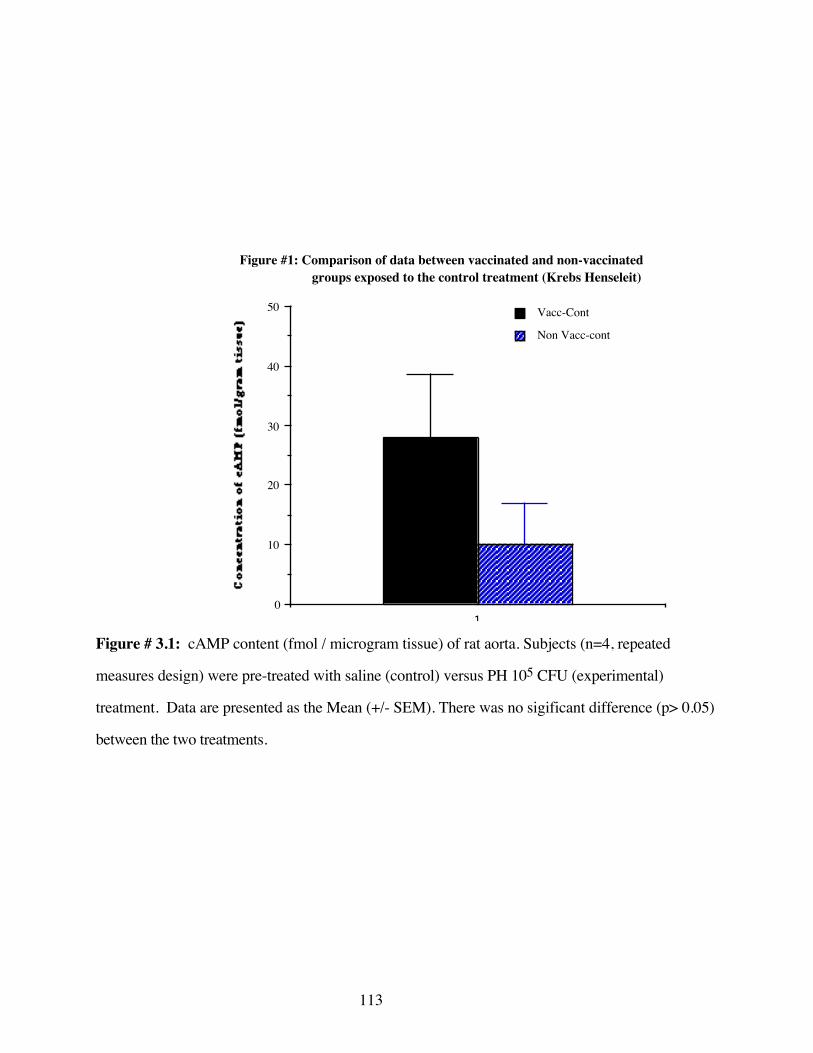
113
10
10
20
30
40
50 Vacc-Cont
Non Vacc-cont
Figure #1: Comparison of data between vaccinated and non-vaccinated groups exposed to the control treatment (Krebs Henseleit)
Con
cent
rati
on
of
cAM
P (f
mol
/gra
m
tiss
ue)
Figure # 3.1: cAMP content (fmol / microgram tissue) of rat aorta. Subjects (n=4, repeated
measures design) were pre-treated with saline (control) versus PH 105 CFU (experimental)
treatment. Data are presented as the Mean (+/- SEM). There was no sigificant difference (p> 0.05)
between the two treatments.
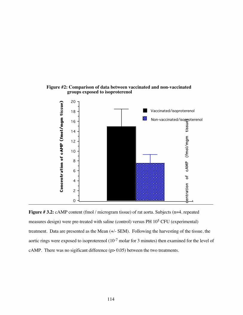
114
0
2
4
6
8
10
12
14
16
18
20
Vaccinated/isoproterenol
Non-vaccinated/isoproterenol
Figure #2: Comparison of data between vaccinated and non-vaccinated groups exposed to isoproterenol
Con
cent
rati
on
of
cAM
P (f
mol
/mgm
ti
ssue
)
Figure # 3.2: cAMP content (fmol / microgram tissue) of rat aorta. Subjects (n=4, repeated
measures design) were pre-treated with saline (control) versus PH 105 CFU (experimental)
treatment. Data are presented as the Mean (+/- SEM). Following the harvesting of the tissue, the
aortic rings were exposed to isoproterenol (10-7 molar for 3 minutes) then examined for the level of
cAMP. There was no sigificant difference (p> 0.05) between the two treatments.
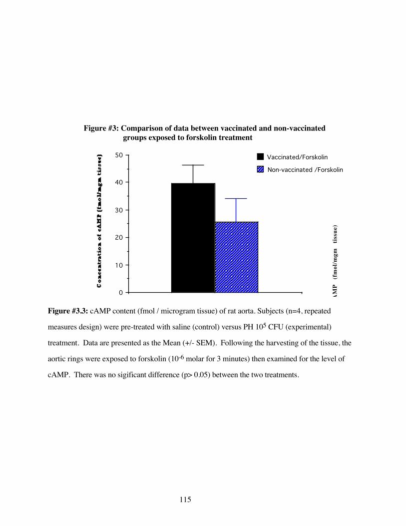
115
0
10
20
30
40
50 Vaccinated/Forskolin
Non-vaccinated /Forskolin
Figure #3: Comparison of data between vaccinated and non-vaccinated groups exposed to forskolin treatment
Con
cent
rati
on
of
cAM
P (f
mol
/mgm
ti
ssue
)
Figure #3.3: cAMP content (fmol / microgram tissue) of rat aorta. Subjects (n=4, repeated
measures design) were pre-treated with saline (control) versus PH 105 CFU (experimental)
treatment. Data are presented as the Mean (+/- SEM). Following the harvesting of the tissue, the
aortic rings were exposed to forskolin (10-6 molar for 3 minutes) then examined for the level of
cAMP. There was no sigificant difference (p> 0.05) between the two treatments.
116
DISCUSSION
Beta adrenergic receptors are integral to the homeostasis of the endothelium and vasculature
in general, and the pulmonary vasculature in particular. Disruption of the BAR can explain many of
the signs and symptoms associated with bovine pneumonic pasteurellosis. This line of
investigation has great potential to reveal solutions with respect to the prophylactic and clinical
treatment of bovine respiratory disease.
The degradation of cAMP is closely regulated. Generally cAMP is hydrolyzed to 5'-AMP
by the action of cAMP phosphodiesterase. The degradation or the inhibition of a cell's ability to
degrade cAMP could also lead to increased levels of cAMP.
Currently, two possible post cAPKs molecular mechanisms leading to vascular smooth
muscle relaxation are proposed. The first involves the redistribution of Ca++ which prevents the
contraction of the vascular smooth muscle (Walsh 1995; Miyashita 1997). The second mechanism
involves the calcium active sites in the myosin light chain kinase. In contraction, calcium release
activates the light chain myosin and initiates vascular smooth muscle contraction. The
phosphorylation of the calcium active sites in the myosin light chain kinase blocks the action of the
calcium molecule. This then occludes the active sites and prevents muscular contraction (Yamagishi
1994). There is a possibility that the disruption of the Ca++ sequestering ability of the cell is
responsible for the inhibition of vascular relaxation as noted by Weekley (1993A, 1993B).
Results of these experiments are inconclusive. Though the concept and execution has merit
there are likely too few subjects or repetitions to allow any conclusions to be drawn. Given the
current results, expanding the number of subjects and repeating the study would, at the very least,
allow for power calculations to suggest the appropriate number of subjects and repetitions.
117
CONCLUSIONS
1) There is not a statistical difference in the level of cAMP between vaccinated and non-
vaccinated subjects.
2) Further evaluation using a larger sample size is needed before any definitive conclusions
can be drawn about the level of cAMP with respect to exposure to PH specifically of
endotoxin in general.
3) The evaluation of post cAMP mechanisms may be beneficial in further research of this
type. Specifically evaluation of the activity of the tetramer cAMP dependent protein kinase,
the cAMP phosphodiesterase activity and the level of intracellular calcium is necessary to
further assess the disruption of beta adrenergic receptor mediated vascular smooth muscle
relaxation.
4) Insufficient information and statistical power prevents conclusions being drawn on the
data produced by this experiment.
118
REFERENCES
Al-Essa, L. Y., M. Niwa, K.I. Kohno, M. Nozaki, K. Tsurumi (1995). “Heterogeneity of circulatingand exudated polymorphonuclear leukocytes in superoxide-generating response to cyclic AMP andcAMP elevating agents.” Biochemical Pharmacology 49(3): 315-322.
Barnard, M. L., S. Matalon (1992). “Mechanisms of extracellular reactive oxygen species injury tothe pulmonary microvasculature.” Journal of Applied Physiology 72(5): 1724-9.
Barnes, P. J. (1986). “Nonadrenergic noncholinergic neural control of human airways" ArchivesInternationales de Pharmacodynamie, 280: 208-28.
Barnes, P. J., S. F. Liu (1995). “Regulation of pulmonary vascular tone.” PharmacologicalReviews 47(1): 87-131.
Bensard, D. D., A. Banerjee, R.C. McIntyre, R.L. Berens, A.H. Harken (1994). “Endotoxindisrupts beta-adrenergic signal transduction in the heart.” Archives of Surgery 129 (2): 198-204.
Bourne, H. R., R.I. Lehrer, M.J. Cline, K.L. Melmon (1971). “Cyclic 3'5'-adenosinemonophosphate in the human leukocyte:synthesis degradation, and effects on neutrophilcandidacidal activity.” Journal of Clinical Investigation 50: 920-27.
Borregaard, N. (1988). The respiratory burst. The Respiratory Burst and its PhysiologicalSignificance. R. R, Sbarra. New York, Plenum Press: 1-8.
Breeze, R. (1985). Structure, function and metabolism in the lung. Veterinary Clinics of NorthAmerica: Food Animal Practice. R. Breeze. Philadelphia, W.B. Saunders. 1: 219-235.
Breider, M. A., S. Kumar, R.E. Corstvet (1991). “Interaction of bovine neutrophils in Pasteurellahaemolytica mediated damage to pulmonary endothelial cells.” Veterinary Immunology andImmunopathology 27: 337-50.
Busse, W. W., J.M. Sosman (1984). “Isoproterenol inhibition of isolated human neutrophilfunction.” Journal of Allergy and Clinical Immunology 73: 404-12.
Busso, N., S. Huet, E. Nicodeme, J. Hiernaux, F. Hyafil (1991). “Refractory period phenomenonin the induction of tissue factor expression on endothelial cells.” Blood 78(8): 2027-35.
Campbell, K. L., R.A. Forse (1993). “Endotoxin exposed atria exhibit G-protein based deficits ininotropic regulation.” Surgery 114: 471-9.
Engels, F., R.S. Oosting, F.P. Nijkamp (1985). “Pulmonary macrophages induce deterioration ofguinea pig tracheal beta adrenergic function through release of oxygen free radicals.” EuropeanJournal of Pharmacology 111: 143-4.
Forse, R. A., R. Leibel, M. Gagner (1989). “The effects of Escherichia coli endotoxin on theadrenergic control of lipolysis in the human adipocyte.” Journal of Surgical Research 46(1): 41-8.
Granger, D. N., P. Kubes (1994). “The microcirculation and inflammation: modulation ofleukocyte endothelial cell adhesion.” Journal of Leukocyte Biology 55: 662-75.
Green, J. F. (1982). Fundamental Cardiovascular and Pulmonary Physiology. Philadelphia, Lea andFebiger.
119
Harlan, J. S., P.D. Killen, L.A. Harker, G.E. Striker (1981). “Neutrophil mediated endothelialinjury in-vitro.” Journal of Clinical Investigations 68: 1394-1403.
Hills, H. R., P.G. Estensen, G. Quie, N.A. Hogan, N.D. Goldberg (1975). “Modulation of humanneutrophil chemotactic responses by cyclic 3' 5'-guanosine monophosphate and cyclic 3' 5'-adenosine monophosphate.” Metabolism 24: 447-53.
Hu, X. X., E.A. Goldmuntz, C.F. Brosnan (1991). “The effect of norepinephrine on endotoxinmediated macrophage activation.” Journal of Neuroimmunology 31(1).
Ignarro, L. J., T.F. Lint, W.J. George (1974). “Hormonal control of lysosomal enzyme releasefrom human neutrophils. Effects of autonomic agents on enzyme release, phagocytosis, and cyclicnucleotides.” Journal of Experimental Medicine 139: 1395-1410.
Jaeschke, H., A. Farhood, C.W. Smith (1990). “Neutrophils contribute to ischemia / reperfusioninjury in the rat liver in-vivo.” FASEB-J 4: 3355-59.
Kubes, P., R. Smith, M.D. Grisham, D.N. Granger (1993). “Neutrophil mediated proteolysis,differential roles for cathepsin G and elastase.” Inflammation 17(3): 321-32.
Majno, G., G.E. Palade (1961). “Studies on inflammation: I. The effect of histamine and serotoninon vascular permeability. An electron microscopic study.” Journal of Biophysics and Biochemistryand Cytology 11: 571-83.
Minnear, F. L., A. Johnson, A.B. Malik (1986). “Beta adrenergic modulation of pulmonarytransvascular fluid and protein exchange.” Journal of Applied Physiology 60: 266-74.
Miyashita, Y., S.J. Sollott, L. Cheng, J.L. Kinsella, E. Koh, E.G. Lakatta, J.P. Froehlich (1997).“Redistribution of intracellular Ca2+ stores after beta adrenergic stimulation of rat tail artery.”American Journal of Physiology 272(1 Part 2): H244-55.
Mizus, I., W.Summer, I. Farrukh, J.R. Michael, G.H. Gurtner (1985). “Isoproterenol oraminophylline attenuate pulmonary edema after acid lung injury.” American Review of RespiratoryDisease 131: 256-9.
Ogunbiyi, P. O. (1986). Effects of Microbial Infections on Guinea Pig and Calf Homeostasis:Implications for Therapy. Doctoral Thesis, Faculty of Graduate Studies. Guelph, University ofGuelph: 280.
Opdahl, H., H.B. Benestad and G. Nicolayson (1993). “Effect of beta-adrenergic agents on humanneutrophil granulocyte activation with N-formyl-methionyl-leucyl-phenylalanine and phorbolmyristste acetate.” Pharmacology and Toxicology 72: 221-8.
Paller, M. S. (1989). “Effect of neutrophil depletion on ischemic renal injury in the rat.” Journal ofLaboratory and Clinical Medicine 113: 379-86.
Robinson, N. E., R.F. Slocomb, F.J. Derksen (1984). Physiology of the bovine lung. BovineRespiratory Disease : A Symposium. R. W. Loan. College Station, Texas, Texas A&M UniversityPress: 163-222.
Romano, F. D., S.B. Jones (1985). “Beta adrenergic stimulation of myocardial cyclic AMP inendotoxic rats.” Circulatory Shock 17(3): 243-52.
120
Seccombe, J. F., P.J. Pearson, H. V. Schaff (1994). “Oxygen radical mediated vascular injuryselectively inhibits receptor dependent release of nitric oxide from canine coronary arteries.”Journal of Thoracic and Cardiovascular Surgery 107: 505-9.
Severn, A., N.T. Rapson, C.A. Hunter, F.Y. Liew (1992). “Regulation of Tumor Necrosis Factorproduction by adrenaline and beta adrenergic agonists.” Journal of Immunology 148(11): 3441-5.
Shepard, R. E., Lang, C.H., K.H. McDonough (1987). “Myocardial adrenergic responsivenessafter lethal and non-lethal doses of endotoxin.” American Journal of Physiology 252(2Pt2): h410-6.
Slocombe, R. F., J. Malark, R. Ingersoll, F.J. Dirksen, N.E. Robinson (1985). “Importance ofneutrophils in the pathogenesis of acute pneumonic pasteurellosis in calves.” American Journal ofVeterinary Research 46(11): 2253-58.
Tecoma, E. S., H.J. Motulsky, A.E. Traynor, G.M. Omann, H.Muller, L.A. Sklar (1986).“Transient catecholamine modulation of neutrophil activation: kinetic and intracellular aspects ofisoproterenol action.” Journal of Leukocyte Biology 40: 629-44.
van Heuven-Nolsen, D., G. Folkerts, D.J. Dewildt, F.P. Ninjkamp (1986). “The influence ofBordetella pertussis and its constituents on the beta-adrenergic receptor in the guinea pigrespiratory system.” Life Science 38(8): 677-85.
Walsh, M. P., G.J. Kargacin, J. Kendrick-Jones, T.M. Lincoln (1995). “Intracellular mechanismsinvolved in the regulation of vascular smooth muscle tone.” Canadian Journal of Physiology andPharmacology May:73(5): 565-73.
Warren, J. S., P.A. Ward, K.J. Johnson (1987). Mechanisms of Endothelial Damage to PulmonaryEndothelium. Pulmonary Endothelium in Health and Disease. U. S. Ryan. New York, MarcelDekker. 32: 107-20.
Weekley, L. B., P.Eyre, H.P. Veit, N. Sriranganathan (1993A). “Morphological and functionaldisturbances in pulmonary vascular endothelium following exposure of sheep to an avirulent strainof Pasteurella haemolytica.” Journal of Comparative Pathology 108(1): 65-72.
Weekley, L. B., P.Eyre (1993B). “Vaccination with live Pasteurella haemolytica alters vascularadrenergic reactivity.” Research Communications in Chemical Pathology and Pharmacology 79(3):389-92.
Weekley, L. B., P. Eyre (1993E). “Disturbances in ex vivo vascular smooth muscle responsesfollowing exposure to Pasteurella haemolytica vaccines.” Journal of Veterinary Pharmacology andTherapeutics 16: 446-53.
Weekley, L. B. (1994). Personal Communication.
Weekley, L. B., H. P.Veit (1995). “Potential morphologic and physiologic factors that maypredispose the bovine lung to respiratory disease.” The Compendium 17(7): 974-983.
Wren, G. (1995). “Understanding the bovine respiratory system.” Bovine VeterinarianNovember: 6-9.
Yamagishi, T., Yanagisawa, T., K. Satoh, N. Taira (1994). “Relaxant mechanisms of cyclic AMPincreasing agents in porcine coronary artery.” European Journal of Pharmacology Jan 14: 251(2-3): 253-62.

121
Zurier, R. B., G. Weissman, S.Hoffstein, S. Kammerman, H.H. Tai (1974). “Mechanisms oflysosomal enzyme release from human leukocytes. II. Effects of cAMP and cGMP, autonomicagonists and agents which affect microtubule function.” Journal of Clinical Investigation 53: 297-303.
122
D). EXAMINATION OF TWO CURRENTLY USED PASTEURELLA
HAEMOLYTICA VACCINES IN THE ASSESSMENT OF THEIR POTENTIAL FOR
BETA ADRENERGIC UNCOUPLING ACTIVITY IN THE RAT AORTA
The Bovine Respiratory Disease Complex (BRDC) and specifically pneumonic
pasteurellosis (also known as shipping fever or transit fever) has been recognized in cattle. This
disease occurs as a series of events that are described by a complex multifactorial equation that, as
yet, is not completely understood. The disease is characterized by the finding of Pasteurella
haemolytica (PH) in the lower respiratory tract simultaneously with the clinical or pathological
diagnosis of fibrinopurulent bronchopneumonia. Exposure of the host to PH alone is not always
sufficient to induce the disease. To further complicate the issue, the bacterium Pasteurella
haemolytica A-1 (PH), is a commensal found on the mucosa of the nasal and pharyngeal areas of
most healthy domesticated ruminant species .
Many investigators have demonstrated that the action of respiratory viruses including:
Infectious Bovine Rhinotracheitis, Parainfluenza-3 virus, Bovine Viral Diarrhea virus, and Bovine
Syncytial Respiratory virus act synergistically with PH to promote pneumonic pasteurellosis. The
viral component appears to disrupt the physical, cytological and immunological pulmonary defense
mechanisms rendering the host more susceptible to bacterial invasion (Rosner 1971; Thomas 1974;
Yates 1982; Bielefeldt Ohmann 1985; Lopez 1986; Kershen 1987; Perino 1989; Olchowy 1994).
Other factors have also been found to enhance the likelihood of disease. For instance, physical
stressors including: vaccination, castration, crowding, starvation, dehydration, cold exposure, and
transportation may precipitate the disease (Olson 1980; Filion 1984; Slocombe 1984; Seigel 1987;
Vogel 1987; Edwards 1989; Binkhorst 1990; Anderson 1991). Stress, in general, also acts to
increase circulating cortisol and can result in a decrease in immunological and physical pulmonary
defenses (Roth 1982; Ballieux 1987; Deitch 1987; Fisher 1989; Shaw 1990; Murata 1991).
123
It has been reported that prophylactic vaccination for bovine respiratory disease, particularly
pneumonic pasteurellosis, is ineffective and may even compromise the vaccinated animals' health
(Martin 1983; Thorlarkson 1990) . Some preliminary studies of vascular physiology with respect
to vaccination sequelae were conducted by Weekley et al (1993E). The vaccines Precon® (live
vaccine) and Presponse® (leukotoxoid rich, endotoxin free vaccine) administered parenterally
causes both beta and alpha-2 adrenergic receptor dysfunction in ex-vivo vascular rings in rats.
Similar results have been described for the pulmonary vasculature in sheep and cattle (Weekley
1991A; 1993D). Disruption of the adrenergic mechanisms due to exposure to vaccines may be
associated with the perceived failure of prophylactic vaccination to meet the needs preventative
health for animals. This mechanism may also be of significance in the molecular pathogenesis of
the disease pneumonic pasteurellosis.
Adrenergic disruption in general and beta adrenergic uncoupling associated with the use of
monovalent Pasteurella vaccines may have devastating consequences with respect to the prevention
of this disease. The precise molecular mechanism and events by which vaccines act to disrupt beta
adrenergic receptors has not yet been described. The vaccines evaluated by Weekley et al are not
currently in use, having been replaced by new monovalent vaccines which have entered the bovine
biological / vaccine market. It is reasonable to evaluate the ability of some vaccines currently in use
to assess for their potential to precipitate beta receptor uncoupling. The two vaccines which were
evaluated were, One Shot ® (Smith Klein Beecham) and Once PMH® (Biocor).
METHODS AND MATERIALS
A) Treatments and Subjects
Rats, adult male Sprague-Dawley (Harlan Sprague-Dawley Inc, Indianapolis, Indiana)
approximately 150-175 g were used in this series of experiments. The animals initially were
housed in a quarantine area for a 1 week for acclimatization to the new facility and observed for
124
signs of disease. The acclimatization period involved a 12:12 light dark photo period, room
temperature was 220+/- 30 C.
Both One Shot® and Once PMH® were refrigerated until use as directed by the
manufacturers' storage instructions. The vaccines were obtained as a sealed commercial preparation
and stored at 40 F. Six groups of animals were used in this experiment (n=4). Three groups were
controls, diluent control Once PMH® (n=4), diluent control One Shot® (n=4) and a negative sterile
saline experimental control (n=4). There were also three experimental groups, One Shot® vaccine
(n=4), Once PMH® vaccine (n=4), and a PH (105CFU) positive control (n=4). The commercial
vaccines were not modified in any way prior to the use of either the diluent or vaccine.
The amount of vaccine administered for each experimental commercial vaccine was
approximately 0.5 milliliter intraperitoneally. For the PH positive control group the volume was
appropriate to give the necessary 105 CFU (approximately one milliliter). Each control animal was
administered one milliliter of the appropriate control intraperitoneally.
B) Preparation of Drugs and Solutions
Krebs-Henseleit buffer was used in the biophysical evaluation and dissection of the ex-vivo
aortic rings. The composition of the Krebs-Henseleit solution ( in one liter of sterile double distilled
water in a sterile 5 liter Erlenmeyer flask as follows: 6.9 grams NaCl ; 0.35 grams KCl; 0.28 CaCl;
0.14 grams MgSO4; 2.09 grams NaHCO3; 0.16 grams KH2PO4; 2.0 grams dextrose. as described
by Weekley (1994). This solution was aseptically mixed immediately before use.
Potassium chloride was used for initial smooth muscle contraction. This solution was
prepared in sterile, double distilled water (30 millimolar concentration when 200 microliters of stock
KCl solution is added to 30 milliliters of the bathing solution).

125
Isoproterenol, a beta adrenergic receptor agonist, was used to evaluate the beta receptor
functioning of isolated ex-vivo aortic vascular tissue. This solution was prepared in sterile, double
distilled water. Serial decreasing logarithmic concentrations of isoproterenol were made from the
initial 10-2 molar solution. The serial dilutions were made by taking 100 microliters of the initial
stock solution and diluting this in 900 microliters of sterile double distilled water sequentially to 10-
9 molar solution.
C) Tissue Collection and Measurement Techniques
Tissue was collected 72 hours after vaccination. Subjects were anesthetized by an intra-
peritoneal administration of 80 mg/kg pentobarbital followed be the anticoagulant (25 U) of sodium
heparin. Once at a deep surgical plane of anesthesia, the abdomen was opened to reveal the
diaphragm. The approach to the thorax and thoracic aorta was through the diaphragm. The
ventrum of the thorax was removed to completely expose the thoracic contents. Once exposed, the
aorta from the base of the heart to the last rib, was carefully removed ensuring that no damage or
stretching occurred. The entire vessel was then placed in warm aerated Krebs-Henseleit (37o+/-
2oC, 95% oxygen: 5% carbon dioxide) for further dissection and complete removal of extraneous
fat and superficial tissue. All aortic rings were obtained from the base of the heart. The rings were
carefully measured to be 3 mm in width. Exceptional care was taken to ensure that there was no
stretching or damage to the endothelium this segment was then used for biophysical testing.
The 3 mm aortic ring, removed for biophysical testing, was then placed between two glass
hooks. Biophysical testing involved the assessment of the change in tension (isometric contraction)
of the aortic ring due to the addition of known concentrations of various drugs. The ring was
placed between the two hooks in a thirty-five milliliter tissue bath. One hook was attached to a fixed
point at the bottom of the tissue chamber while the other hook was attached to an electronic
transducer that was calibrated to give output in grams tension. The tissue was placed in an insulated

126
heated tissue bath containing Krebs-Henseleit bicarbonate buffer . The buffer was aerated by
bubbling 95% oxygen and 5% carbon dioxide through each of four individually isolated tissue
baths. A constant tissue bath temperature of 37 O+/- 2oC was maintained by circulating water
through a temperature controlled water pump and sequentially through the outer heating jacket of
each tissue bath. Each tissue ring was placed in the tissue bath under 2 grams resting tension for
two hours and allowed to equilibrate. The Krebs-Henseleit buffer was replaced with fresh buffer
every 15-20 minutes throughout equilibration. Testing started immediately after the equilibration
period. The aortic rings, in the tissue bath, were exposed to 200 µl of potassium chloride to ensure
contraction (KCl, 30 millimolar) and allowed to equilibrate to this treatment over 30 minutes. After
equilibration and stabilization to KCl the recording was started and sequential increasing log molar
doses of isoproterenol were added. Isoproterenol hydrochloride was used to evaluate the response
of beta adrenergic receptors in mediating smooth muscle relaxation.
Electronically calibrated analog signals were transmitted from the transducer connected to
the glass hook through an analog digital converter (MacLab A/D signal converter, World Precision
Instruments). Digitized information was captured by MacLab data acquisition software (MacLab
2.51) on a Model M5011 Macintosh SE computer (1MB RAM, 800 K drive, 1.40 MB hard disk)
in real time. Records of aortic smooth muscle relaxation as a cumulative dose response to log
molar increases in the isoproterenol concentration were obtained and the results stored on a 3.5 inch
computer disk.
D) Statistical Analysis of Data
Data were analyzed in several ways. Initially the data were compared for each experimental
treatment at the isoproterenol concentration of 10-5 molar. In pilot studies, this concentration of
isoproterenol appeared to be in the middle of the generated dose response curve and so was selected
to be an appropriate point for comparison of different treatments for the experimental protocols.
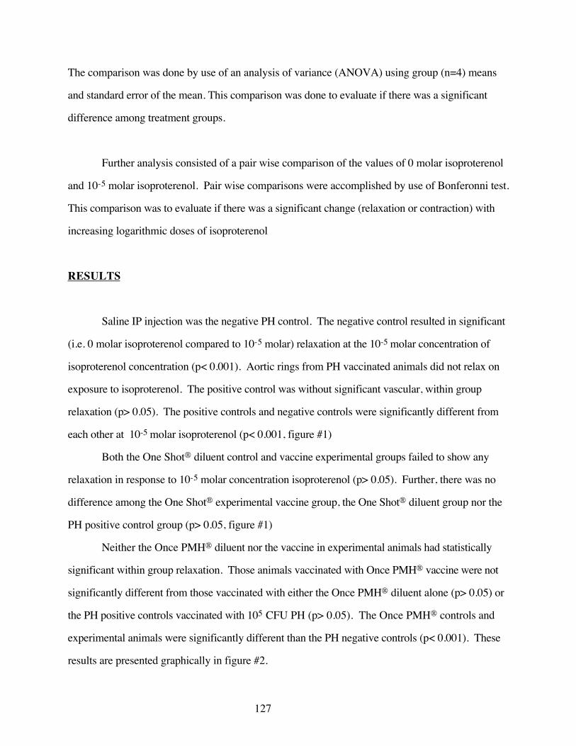
127
The comparison was done by use of an analysis of variance (ANOVA) using group (n=4) means
and standard error of the mean. This comparison was done to evaluate if there was a significant
difference among treatment groups.
Further analysis consisted of a pair wise comparison of the values of 0 molar isoproterenol
and 10-5 molar isoproterenol. Pair wise comparisons were accomplished by use of Bonferonni test.
This comparison was to evaluate if there was a significant change (relaxation or contraction) with
increasing logarithmic doses of isoproterenol
RESULTS
Saline IP injection was the negative PH control. The negative control resulted in significant
(i.e. 0 molar isoproterenol compared to 10-5 molar) relaxation at the 10-5 molar concentration of
isoproterenol concentration (p< 0.001). Aortic rings from PH vaccinated animals did not relax on
exposure to isoproterenol. The positive control was without significant vascular, within group
relaxation (p> 0.05). The positive controls and negative controls were significantly different from
each other at 10-5 molar isoproterenol (p< 0.001, figure #1)
Both the One Shot® diluent control and vaccine experimental groups failed to show any
relaxation in response to 10-5 molar concentration isoproterenol (p> 0.05). Further, there was no
difference among the One Shot® experimental vaccine group, the One Shot® diluent group nor the
PH positive control group (p> 0.05, figure #1)
Neither the Once PMH® diluent nor the vaccine in experimental animals had statistically
significant within group relaxation. Those animals vaccinated with Once PMH® vaccine were not
significantly different from those vaccinated with either the Once PMH® diluent alone (p> 0.05) or
the PH positive controls vaccinated with 105 CFU PH (p> 0.05). The Once PMH® controls and
experimental animals were significantly different than the PH negative controls (p< 0.001). These
results are presented graphically in figure #2.
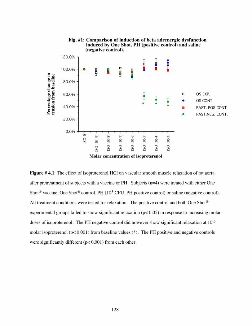
128
ISO
0
ISO
10(
- 9)
ISO
10(
-8)
ISO
10(
-7)
ISO
10(
-6)
ISO
10(
-5)
ISO
10(
-4)
ISO
10(
-3)
0.0%
20.0%
40.0%
60.0%
80.0%
100.0%
120.0%
OS EXP.OS CONT
PAST. POS CONTPAST.NEG. CONT.
Fig. #1: Comparison of induction of beta adrenergic dysfunction induced by One Shot, PH (positive control) and saline (negative control).
Molar concentration of isoproterenol
Perc
enta
ge c
hang
e in
te
nsio
n fr
om b
asel
ine
*
Figure # 4.1: The effect of isoproterenol HCl on vascular smooth muscle relaxation of rat aorta
after pretreatment of subjects with a vaccine or PH. Subjects (n=4) were treated with either One
Shot® vaccine, One Shot® control, PH (105 CFU, PH positive control) or saline (negative control).
All treatment conditions were tested for relaxation. The positive control and both One Shot®
experimental groups failed to show significant relaxation (p< 0.05) in response to increasing molar
doses of isoproterenol. The PH negative control did however show significant relaxation at 10-5
molar isoproterenol (p< 0.001) from baseline values (*). The PH positive and negative controls
were significantly different (p< 0.001) from each other.
129
ISO
0
ISO
10(
- 9)
ISO
10(
-8)
ISO
10(
-7)
ISO
10(
-6)
ISO
10(
-5)
ISO
10(
-4)
ISO
10(
-3)
0.0%
20.0%
40.0%
60.0%
80.0%
100.0%
120.0%
140.0%
PMH EXP.PMH CONTPAST. POS CONTPAST.NEG. CONT.
Fig #2: Comparison of induction of beta adrenergic dysfunction induced by Once PMH and PH (positive control) and saline (negative control)
Molar concentration of isoproterenol
Perc
enta
ge c
hang
e in
te
nsio
n fr
om b
asel
ine
*
Figure # 4.2: The effect of isoproterenol HCl on vascular smooth muscle relaxation of rat aorta
after pretreatment of subjects with a vaccine or PH. Subjects (n=4) were treated with either Once
PMH® vaccine, Once PMH® control, PH (105 CFU, PH positive control) or saline (negative
control). All treatment conditions were tested for relaxation. The positive control and both Once
PMH® experimental groups failed to show significant relaxation (p< 0.05) in response to
increasing molar doses of isoproterenol. The PH negative control did however show significant
relaxation at 10-5 molar isoproterenol (p< 0.001) from baseline values (*). The PH positive and
negative controls were significantly different (p< 0.001) from each other.
Overall, there was no vascular relaxation to increasing doses of isoproterenol in any group
with the exception of the negative PH control (saline). All experimental and diluent control groups
were statistically similar at 10-5 isoproterenol with respect to between group comparisons.
isoproterenol caused relaxation only in the negative PH control.
130
DISCUSSION
The aortic rings from the positive experimental control (Pasteurella haemolytica 105 CFU)
and negative experimental control (sterile saline) responded to increasing doses of isoproterenol as
described by Weekley (1993E). Exposure to the PH organism causes the disruption of the
vascular smooth muscle beta receptor and a failure of the tissue to respond to increasing molar
doses of isoproterenol. The negative experimental control (saline) doesn't appear to interfere with
the pharmacological functioning of the vascular beta adrenergic receptors, as defined by this testing
paradigm, indicating approximately 40% vasorelaxation.
The results of parenteral exposure of rats to One Shot ® indicates that there is a complete
failure of the vascular smooth muscle to respond to isoproterenol. Similarly parenteral exposure to
the diluent alone prevented the vascular beta receptors from responding to isoproterenol. This
seems to indicate that there is either a chemical or biological element associated with the diluent that
has the ability to inhibit beta receptors.
Once PMH® also caused disruption of the vascular beta adrenergic receptor both as the
whole vaccine and as the diluent control. However, in this experiment the diluent control appeared
to start to respond at higher doses of isoproterenol. At concentrations of 10-4 and 10-3 molar
isoproterenol there is a distinct initiation of vasorelaxation response in the diluent control. There is
statistically significant difference between the diluent and the whole vaccine at the higher doses of
isoproterenol. This indicates that while there is an element in the diluent that can disrupt beta
adrenergic receptors, it may not cause a complete disruption of these receptors or doesn't affect all
beta receptors equally.
The mechanism by which beta adrenergic receptors are disabled is currently unknown.
There is a suggestion that endotoxin may be integral to the process of beta receptor disruption.

131
There is however, no information concerning whether all beta receptors are affected simultaneously
or even if each beta receptor is disrupted to the same extent. The results of the Once PMH®
experiment suggests that, in this case, the disruption of the beta receptors may be overcome at
relatively high doses of beta agonist. This may be due to the induction of partially damaged
receptor mediated events or that at higher doses, undamaged receptors are recruited to result in a
partial smooth muscle response to isoproterenol. The One Shot® vaccine appeared to result in total
elimination of all isoproterenol mediated responses by the vascular smooth muscle. There was no
response to isoproterenol at any dose examined. This suggests that there is a relationship between
the dose of isoproterenol and the vascular response with respect to the pathology that initiates
receptor disruption.
There is a suggestion in the literature and in a previously completed experiment in this
laboratory that endotoxin may be of importance to these events (Romano 1985; Shepard 1987;
Forse 1989; Campbell 1993; Bensard 1994). Endotoxin is a constituent of Gram negative bacterial
cell wall that is released when the cell disintegrates. In most cases, endotoxin is easily detected by
the Limulus Ameobocyte Lysate (LAL) assay but very difficult to eliminate from vaccines of gram
negative organisms. In the case of commercial vaccines however, there maybe chemical entities that
result in a false negative indication in the LAL (Maheswaran 1995). It has not been definitively
demonstrated that endotoxin is the only constituent that can lead to vascular beta adrenergic receptor
dysfunction. Further, the exact mechanism of this disruption is yet to be described, therefore the
synergistic or antagonist chemical interaction can not be assessed.
Due to the proprietary nature of vaccines and biologicals it is unknown what chemical and
biological additives are in the diluent or whole vaccine. It would appear from these results that at
least at high isoproterenol concentrations there is a difference in the beta receptor functioning with
respect to Once PMH® and One Shot®. It is unclear, at this time, if these in-vitro results occur to
132
the same extent in-vivo. The evaluation of this difference may suggest the mechanism or
mechanisms that are involved with this phenomenon.
The vascular events associated with vaccine induced beta adrenergic receptor disruption, are
of unknown clinical consequence in-vivo due to redundant protective mechanisms of the lung
vasculature. Gross examination of the bovine lung tissue may indicate pulmonary congestion due
the decrease in pulmonary venous flow. Interstitial edema may be present if the beta adrenergic
receptors associated with mediating fluid extravasation are affected or due to increased
intrapulmonary pressures secondary to lack of vasodilation. Further, should the beta receptors
involved with inhibition of endothelial / coagulation events also be disrupted, one may see an
increase in fibrin formation. These events are likely to occur in the immediate post-vaccination
period with physiological consequences lasting up to three weeks.
The clinical consequences of the disruption of vascular beta adrenergic receptor dysfunction
has not been examined. The events associated with vaccine induced dysfunction may include
decreases in weight gain and food conversion, increased incidence of morbidity and mortality
associated with respiratory disease in cattle and respiratory distress. Epidemiological studies may
be of value in assessing these changes with respect to beta adrenergic receptor dysfunction.
The clinical value of a vaccine is its ability to protect the target population from a specific
disease. The commercial usefulness of a vaccine relies on its ability to economically protect
animals. It is clear that should a vaccine be effective in generating an immunoprotective response
but also induce untoward effects resulting in disruption in animal growth, that vaccine is less than
ideal both chemically and commercially. These results suggest that some currently used vaccines
may have the ability to disrupt normal pulmonary vascular functioning. It remains to be seen if the
vaccines have clinical and economic implications beyond the theoretical events presented here.
133
CONCLUSIONS
1) Both One Shot® and Once PMH® induce beta adrenergic receptor dysfunction. Though
the actual mechanism has not been elucidated the mechanism of action is likely to involve
endotoxin either in the vaccine component or in the diluent.
2) This disruption may be due to factors (e.g. endotoxin) contained in the diluent
component and possibly the active portion of the vaccine. An element associated with the
chemical or biological make-up of the diluent that can precipitate the disruption of vascular
beta adrenergic receptors.
3) The disruption of the adrenergic receptor appears to occur to an equal extent with respect
to the diluent and the whole vaccine preparation. Other vaccine preparations remain to be
evaluated and may have similar effects on beta adrenergic receptors.
134
References
Anderson, N. V., Y.D. Youanes, J.G. Vestwebber, C.A. King, R.D. Klemm, G.A. Kennedy (1991).“The effects of stressful exercise on leukocytes in cattle with experimental pneumonicpasteurellosois.” Veterinary Research Communications 15: 189-204.
Ballieux, R. E., C.J. Heijnen (1987). Stress and the immune system. Biology of Stress in FarmAnimals: An Integrative Approach. T. v. A. A. Weipkema: 29-39.
Bensard, D. D., A. Banerjee, R.C. McIntyre, R.L. Berens, A.H. Harken (1994). “Endotoxindisrupts beta-adrenergic signal transduction in the heart.” Archives of Surgery 129(2): 198-204.
Bielefeldt Ohmann, H., L.A. Babiuk (1985). “Viral bacterial pneumonia in calves: effect of bovineherpesvirus 1 on immunological functions.” The Journal of Infectious Diseases 151(5): 937-47.
Binkhorst, G. J., P.A.J. Henricks, T.S.G.A.M. Ingh, R. Hajer, F.P. Nijkamp (1990). “The effect ofstress on host defense system and on lung damage in calves experimentally infected withPasteurella haemolytica type A1.” Journal of Veterinary Medicine A 37: 525-36.
Campbell, K. L., R.A. Forse (1993). “Endotoxin exposed atria exhibit G-protein based deficits ininotropic regulation.” Surgery 114: 471-9.
Deitch, E. A., R.M. Bridges (1987). “Stress hormones modulate neutrophil and lymphocyteactivity in-vitro.” The Journal of Trauma 27(10): 1146-1153.
Edwards, A. J. (1989). “The effects of stressors like rumen overload and induced abortion on BRDin feedlot cattle.” Agri-Practice 10(2): 10-15.
Filion, L. G., P.J. Wilson, H. Beilefeldt-Ohmann, L.A. Babiuk, R.G. Thomson (1984). “Thepossible role of stress in the induction of pneumonic pasteurellosis.” Canadian Journal ofComparative Medicine 48: 268-74.
Fisher, L. A. (1989). “Corticotropin releasing factor: endocrine and responses to stress.” Trendsin Pharmaceutical Sciences 10: 189-93.
Forse, R. A., R. Leibel, M. Gagner (1989). “The effects of Escherichia coli endotoxin on theadrenergic control of lipolysis in the human adipocyte.” Journal of Surgical Research 46(1): 41-8.
Kershen, R. P., B.W. Bennett, D.E. Flack, R.L. Jensen, J.K.Collins (1987). “Bovine RespiratorySyncytial Virus infection in yearling feedlot cattle.” Agri-Practice April: 23-6.
Lopez, A., M.G. Maxie, L. Ruhnke, M. Savan, R.G. Thompson (1986). “Cellular inflammatoryresponse in the lungs of calves exposed to Bovine Viral Diarrhea Virus, Mycoplasma bovis andPasteurella haemolytica.” American Journal of Veterinary Research 47(6): 1283-6.
Maheswaran, S. K. (1995). Further evaluation of vaccines claiming to be endotoxin free hasrevealed endotoxin.
Martin, S. W. (1983). “Vaccination; is it effective in preventing respiratory disease or influencingweight gains in feedlot calves.” Canadian Veterinary Journal 24: 10-19.
Murata, H., H. Hirose (1991). “Suppression of bovine neutrophil function by sera from cortisoltreated calves.” British Veterinary Journal 147: 63-70.
135
Olchowy, T. W. J., T.R. Ames, T.W. Molitor (1994). “Interaction of Bovine Respiratory SyncytialVirus with bovine alveolar macrophages in-vivo: effects of virus infection upon selected cellfunctions.” Canadian Journal of Veterinary Research 58: 42-8.
Olson, D. P., C.J. Papasian, R.C. Ritter (1980). “The effects of cold stress on neonatal calves 1.clinical condition and pathological lesions.” Canadian Journal of Comparative Medicine 44: 11-18.
Perino, L. J. (1989). “Immunosuppressive effects of bovine viral pathogens: a review.” The BovinePractioner 24: 42-8.
Romano, F. D., S.B. Jones (1985). “Beta adrenergic stimulation of myocardial cyclic AMP inendotoxic rats.” Circulatory Shock 17(3): 243-52.
Rosner, S. F. (1971). “Bovine Parainfluenza type 3 virus infection and pasteurellosis.” Journal ofthe American Veterinary Medical Association 159(11): 1375-82.
Roth, J. A., M.L. Kaeberle (1982). “Effect of glucocorticoids on the bovine immune system.”Journal of the American Veterinary Medical Association 180(8): 894-901.
Seigel, H. S. (1987). Effects of behavioral and physical stress on immune response. Biology ofStress in Farm Animals: An Integrative Approach. T. v. A. A. Weipkema: 39-54.
Shaw, F. D., R.K. Tume (1990). “Beta endorphin and cortisol concentrations in the plasma ofcattle.” Australian Veterinary Journal 67(11): 423-4.
Shepard, R. E., Lang, C.H., K.H. McDonough (1987). “Myocardial adrenergic responsivenessafter lethal and non-lethal doses of endotoxin.” American Journal of Physiology 252(2Pt2): h410-6.
Slocombe, R. F., F.J. Derksen, N.E. Robinson (1984). “Interactions of cold stress and Pasteurellahaemolytica in the pathogenesis of pneumonic pasteurellosis in calves: changes in pulmonaryfunction.” American Journal of Veterinary Research 45(9): 1764-70.
Thomas, L. H., A.P. Collins (1974). “Virus infection and incidence of respiratory disease: aserological study of four micro-organisms at a large beef rearing farm.” Veterinary Record 94:506-9.
Thorlarkson, B., W. Martin, D. Peters (1990). “A field trial to evaluate the efficacy of a commercialPasteurella haemolytica bacterial extract in preventing bovine respiratory disease.” CanadianJournal of Veterinary Research 31: 573-9.
Vogel, W. H. (1987). “Stress-the neglected variable in experimental pharmacology andtoxicology.” Trends in Pharmaceutical Sciences 8: 35-8.
Weekley, L. B., P. Eyre, H.P. Veit, N. Sriranganathan (1991A). “Exposure of sheep to Pasteurellahaemolytica alters ex-vivo pulmonary vascular smooth muscle alpha adrenergic responses: role ofvascular endothelium.” Research Communications in Chemical Pathology and Pharmacology74(1): 121-4.
Weekley, L. B., P.Eyre, H.P. Veit, N. Sriranganathan (1993D). “Vaccination of calves withPasteurella haemolytica alters cholinergic responses of pulmonary circulation.” Abstract,American College of Veterinary Internal Medicine.

136
Weekley, L. B., P. Eyre (1993E). “Disturbances in ex-vivo vascular smooth muscle responsesfollowing exposure to Pasteurella haemolytica vaccines.” Journal of Veterinary Pharmacologyand Therapeutics 16: 446-53.
Weekley, L. B. (1994). Personal Communication.
Yates, W. D. G. (1982). “A review of Infectious Bovine Rhinotracheitis, Shipping Feverpneumonia and viral bacterial synergism in respiratory disease of cattle.” Canadian Journal ofComparative Medicine 46: 225-63.

137
E). POSSIBLE PHYSIOLOGICAL INTERACTION OF THE ALPHA-2ADRENERGIC SYSTEM AND NONADRENERGIC-NONCHOLINERGIC (NANC)MECHANISMS OF THE RAT AORTA.
Neural control of the vascular system is essential to the homeostasis of the body. The
vascular system is generally recognized to be modulated by three interactive systems that act in
concert to maintain the appropriate blood flow to various organs. These systems are the cholinergic
system, the adrenergic system and the nonadrenergic-noncholinergic system (NANC).
The NANC system is part of the autonomic nervous system. These neurons are described
as those that fail to have the histological features of either adrenergic or cholinergic neurons of the
autonomic nervous system. Many transmitter substances are associated with these neurons
however nitric oxide (NO) is recognized as a very important mediator and part of the family of
endothelial derived relaxing factors.
Epidemiological evidence suggests that vaccination in cattle against Pasteurella haemolytica
can result in increases in morbidity and decreased weight gain as noted by Thorlarkson
(Thorlarkson 1990) and increases in mortality noted by Martin (1980;1983). Weekley has
demonstrated that, after exposure of Sprague-Dawley rats to live PH organisms, changes in the
vascular smooth muscle response to alpha adrenergic agents are noted. In particular, in these
experimental animals, the alpha-2 adrenergic receptor mediated system results in an exaggerated
vasoconstriction response when exposed to the selective alpha-2 adrenergic agonist, clonidine.
Further, the beta adrenergic agonist isoproterenol fails to elicit vasorelaxation as occurs in the
untreated controls (Weekley 1993A). These deficits, as a result of the exposure of experimental
animals to live PH have been demonstrated in several species including cattle, and sheep (Weekley
1991A; 1991B; 1993D; 1993E; 1994). Taken together, these results suggest an increased
vasoconstriction, as a result of the disruption of the adrenergic based vasorelaxation mechanisms.
This can result in ischemia in pulmonary tissue associated with the affected blood vessels. One of
138
the consequences may result in compromised defenses leading to an individual's increased
susceptibility to respiratory disease. The implications of the experiments by Weekley et al offer a
possible explanation for the vascular response noted as sequalae to prophylactic vaccination with
monovalent PH vaccines in cattle.
The interaction of the vascular alpha-2 adrenergic receptor and nitric oxide of the NANC
system in the homeostatic regulation of vascular tone has recently been suggested by experiments
in this laboratory. In early experiments (results not presented here) it was noted that aortic rings
that had not been precontracted gave the expected constriction response when exposed to clonidine.
Ex-vivo aortic rings that had been exposed to the alpha-1 agonist, phenylephrine showed
vasorelaxation when exposed to clonidine. These results suggest a dual nature to the response of
the alpha-2 adrenergic receptor based on the predominant vascular tone. A suggestion, as a result
of the possible dual nature of the alpha-2 receptors, is that very rapid relaxation responses (e.g. on
the order of seconds) changes are mediated by the alpha-2 receptor. This fine vascular smooth
muscle motor control in aortic rings has not previously been described.
This paper will focus on a possible explanation for the excessive vascular constriction of the
alpha-2 receptors after exposure to PH A1 in-vivo, as noted by Weekley (1993B). Vascular alpha-
2 adrenergic receptors are generally recognized to exist in two functionally distinct locations in the
vessel. Some are directly associated with vascular smooth muscle others are associated with the
vascular endothelium. The alpha-2 receptors of the smooth muscle appear to mediate smooth
muscle contraction. The molecular mechanisms associated with the alpha-2 adrenergic system,
leading to vasoconstriction have been studied for some time. Alpha-2 receptors act through an G-
inhibitory-alpha protein that inhibits adenylylyl cyclase. This results in a decrease in intracellular
cAMP levels. The mechanism by which the inhibitory G protein leads to a decrease in cAMP is
unclear but is most likely associated with voltage gated calcium channels. The end result of alpha-2
adrenergic stimulation on vascular smooth muscle is vasoconstriction. More recent papers have
139
suggested that the mechanism, by which alpha-2 receptors mediate vasoconstriction, is dependent
on the influx of extracellular calcium through voltage operated calcium channels (Parkinson 1995).
Other vascular alpha-2 adrenergic receptors are associated with the vascular endothelium.
Most research has examined the functioning of these receptors with respect to the coronary vessels
(Ishibashi 1997). There exists some evidence that L-arginine, a precursor of nitric oxide is involved
in the vasorelaxation of the aorta in the rat (Schini 1991). The implication is that the smooth muscle
may mediate vasodilation via the NANC mediator, nitric oxide. Support for this theory is given in
recent papers where it been demonstrated that alpha-2 receptors can cause a nitric oxide mediated
vasorelaxation in the peripheral vessels in normal rats (Brockman 1996) and in the cerebral
vasculature of rats (Bryan 1995).
Nitric oxide (NO) is one of a series of molecules in the family of vascular endothelial
derived relaxing factors. Nitric oxide is very short lived, on the order of seconds (Palmer 1987;
Gryglewski 1988). Derived from L-arginine by the enzyme nitric oxide synthase (NOS), nitric
oxide is thought to be generated by the intact vascular endothelium (Moncada 1991; Moncada
1993). Other authors have suggested that the generation and action of NO on vascular smooth
muscle is independent of the need for an intact endothelium (Schini 1991). Nw-nitro-L-arginine
methyl ester (L-NAME) is a specific inhibitor of NOS and therefore would inhibit the actions of
NO in the vascular system and reduce vasodilation.
Nitric oxide released in response to the endothelial based alpha-2 adrenergic receptors
which act through guanylyl cyclase to generate cyclic guanylyl mono-phosphate (cGMP)
(Rapoport 1983). This cGMP then interacts with cytosolic calcium to decrease its concentration
intracellularly and results in vascular smooth muscle relaxation (Fiscus 1988).
140
A possible explanation for the results described by Weekley with respect to the enhanced
vasoconstriction observed after treatment with PH is that the NO mediated relaxation component of
the alpha-2 adrenergic receptor is disrupted. The disruption in the balance of the vasorelaxation and
vasoconstriction components results in an uncontrolled vasoconstriction in response to the alpha-2
adrenergic agonist clonidine. It is necessary to demonstrate that nitric oxide is mediated through
alpha-2 adrenergic receptors
This paper focuses on the interaction of the adrenergic and the NANC mediator, NO, as a
possible explanation for the findings reported by Weekley. This paper proposes to examine the
possibility that there is an interaction between the NANC and the alpha-2 adrenergic vascular
control mechanism of the rat aorta. I will examine the possibility that NO release can be mediated
by clonidine through the alpha-2 receptor.
MATERIALS AND METHODS
A) Treatments and Subjects
Rats, adult male Sprague-Dawley (Harlan Sprague-Dawley Inc., Indianapolis, Indiana)
approximately 150-175 g were used in this series of experiments. The animals initially were
housed in a quarantine area for a 1 week for acclimatization to the new facility and observe for signs
of disease. The acclimatization period involved a 12:12 light dark photo period, room temperature
was 220+/- 30 C.
Three groups of rats (n=4) were selected for the experimental procedure. The first group
was used to assess whether or not an alpha-1 adrenergic receptor mediated relaxation response
exists in the presence of high vascular tone (positive control). High vascular tone was initiated by
exposing the aortic rings in-vitro to phenylephrine a selective alpha-1 agonist. The second group
141
(30 minutes bathed in L-NAME) and the third group (120 minutes bathed in L-NAME) was used
to asses the effect of a decrease in nitric oxide has on the alpha-2 mediated relaxation response in
the presence of high alpha-1 vascular tone.
B) Preparation of Drugs and Solutions
Krebs-Henseleit buffer was used in the biophysical evaluation and dissection of the ex-vivo
aortic rings. The composition of the Krebs-Henseleit solution ( in one liter of sterile double distilled
water in a sterile 5 liter Erlenmeyer flask as follows: 6.9 grams NaCl ; 0.35 grams KCl; 0.28 CaCl;
0.14 grams MgSO4; 2.09 grams NaHCO3; 0.16 grams KH2PO4; 2.0 grams dextrose). as
described by Weekley (1994). This solution was aseptically mixed immediately before use.
Phenylephrine was mixed in sterile saline at the appropriate concentration to result in 10-6
molar in a 30 milliliter tissue bath. The phenylephrine was added to the tissue bath after tissue
stabilization for 60 minutes. Five minutes after addition of the phenylephrine testing with clonidine
was started. L-NAME was mixed in sterile water to result in a concentration of 10 molar in a thirty
milliliter tissue bath. The tissue was first exposed to L-NAME for thirty minutes then testing
initiated. The tissue was then allowed to recover and return to baseline over 20 minutes.
Phenylephrine was then added for a second time (incubation for five minutes) then incubation with
L-NAME for 120 minutes. The solution in the tissue bath was changed every 15 to 20 minutes pre
and post-incubation.
Isoproterenol, a beta adrenergic receptor agonist, was used to evaluate the beta receptor
functioning of isolated ex-vivo aortic tissue. This solution was prepared in sterile, double distilled
water. Isoproterenol was mixed to result in a concentration of 10-7 molar in a volume of 30
milliliters of Krebs-Henseleit . Each aortic ring was then exposed to this final solution for exactly 3

142
minutes. The solution was maintained at 370 centigrade (+/- 2oC) and aerated with 95% oxygen
5% carbon dioxide gas.
C) Tissue Collection and Measurement Techniques
Subjects were anesthetized with an intra-peritoneal injection of sodium pentobarbital (80
mg/kg), which was then followed by a separate but simultaneous administration of 25 U of sodium
heparin. Once at a deep surgical plane of anesthesia, the abdomen was opened to reveal the
diaphragm. The approach to the thorax and thoracic aorta was through the diaphragm. The
ventrum of the thorax was removed to completely expose the thoracic contents. Once exposed the
aorta, from the base of the heart to the last rib, was carefully removed ensuring that no damage or
stretching occurred. The entire vessel was the placed in warm aerated Krebs-Henseleit (370 C,
95% oxygen: 5% carbon dioxide) for further dissection and complete removal of extraneous fat
and superficial tissue. All aortic rings were obtained from the base of the heart. The rings were
carefully trimmed to 3 mm in width. Exceptional care was taken to ensure that there was no
stretching or damage to the endothelium. This segment was then used for biophysical testing.
The 3 mm aortic ring, removed for biophysical testing, was then placed between two glass
hooks. Biophysical testing involved the assessment of the change in tension (isometric contraction)
of the aortic ring due to the addition of known concentrations of various drugs. The ring was
placed between the two hooks in a thirty-five milliliter tissue bath. One hook was attached to a fixed
point at the bottom of the tissue chamber while the other hook was attached to an electronic
transducer that was calibrated to give output in grams tension. The tissue was placed in an insulated
heated tissue bath containing Krebs-Henseleit bicarbonate buffer . The buffer was aerated by
bubbling 95% oxygen and 5% carbon dioxide through each of four individually isolated tissue
baths. A constant tissue bath temperature of 37.0 +/- 2.0oC was maintained by circulating water
through a temperature controlled water pump and sequentially through the outer heating jacket of
143
each tissue bath. Each tissue ring was placed in the tissue bath under 2 grams resting tension for
two hours and allowed to equilibrate. The Krebs-Henseleit buffer was replaced with fresh buffer
every 15-20 minutes throughout equilibration and testing. Testing started immediately after the
equilibration period. The aortic rings, in the tissue bath, were exposed to 10-6 molar phenylephrine
and allowed to equilibrate to this treatment over 5 minutes. After equilibration to the phenylephrine
the recording was started and sequentially increasing log molar doses of clonidine were added to
evaluate the response of alpha-2 adrenergic receptors in causing smooth muscle relaxation. Aortic
rings were evaluated after precontraction with phenylephrine ( 10-6 molar) without the addition of
the specific NO antagonist L-NAME. This was to assess the other tissues were expressed to either
30 min L-NAME or 120 min L-NAME before assessment of the pharmacological response to
clonidine in alpha-1 mediated increased vascular tone.
Electronically calibrated analog signals were transmitted from the transducer to an analog
digital converter (MacLab A/D signal converter, World Precision Instruments). Digitized
information was captured by MacLab data acquisition software (MacLab 2.51) on a Model M5011
Macintosh SE computer (1MB RAM, 800 K drive, 20 sc hard disk) in real time. Records of
vascular smooth muscle activity, as a cumulative dose response to log molar increases in
isoproterenol were obtained and the results stored on a 3.5 inch computer disk.
D) Statistical Analysis of Data
Data were analyzed in several ways. Initially the data were compared for each experimental
treatment at the clonidine concentration of 10-5 molar. In pilot studies this appeared to be in the
middle of the generated dose response curve and so was selected to be an appropriate point for
comparison of different treatments for the experimental protocols. The comparison was done by
use of an analysis of variance (ANOVA) using group (n=4) means and standard error of the mean,
to evaluate for a significant difference among treatment groups.
144
Further analysis consisted of a pair wise comparison of the values of 0 molar clonidine and
10-5 molar clonidine. Pairwise comparisons were done by use of the Student "t" test. This was to
evaluate for a change (relaxation or contraction) with increasing logarithmic doses of clonidine
RESULTS
Results indicate that aortic rings, in the presence of alpha-1 adrenergic increased vascular
tone due to pre-contraction with phenylephrine, demonstrate vasorelaxation when an alpha-2
adrenergic selective agonist, clonidine, is introduced. The addition of clonidine, in the absence of L-
NAME, and at higher doses of clonidine (e.g.10-3 molar) reversed the relaxation trend to result in a
constriction response (figure #1).
In the presence of L-NAME, the clonidine mediated relaxation response was diminished.
The reduction in vascular relaxation was proportional to the length of time the aortic tissues were
exposed to L-NAME. The aortic segments treated with L-NAME for thirty minutes were
significantly different than either the control group ( p< 0.01) or the second experimental group
treated for 120 minutes (p< 0.001). The 30 minute L-NAME group demonstrated an intermediate
amount of reduction in relaxation compared to the other two groups. The group exposed to L-
NAME for 120 minutes was also significantly different, less relaxation, from the control group (p<
0.003). The relaxation response to clonidine, appears to be diminished if, nitric oxide synthetase
(NOS) and therefore nitric oxide (NO), is decreased.
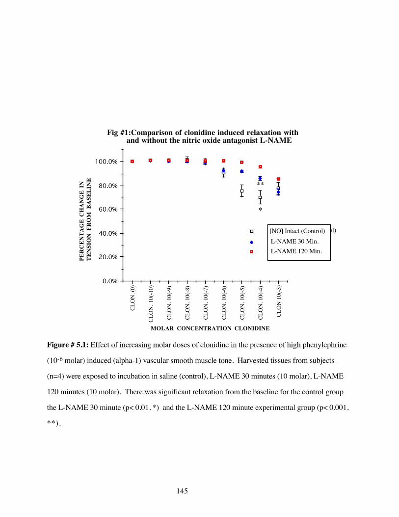
145
CLO
N. (
0)
CLO
N. 1
0(-1
0)
CLO
N. 1
0(-9
)
CLO
N. 1
0(-8
)
CLO
N. 1
0(-7
)
CLO
N. 1
0(-6
)
CLO
N. 1
0(-5
)
CLO
N. 1
0(-4
)
CLO
N 1
0(-3
)0.0%
20.0%
40.0%
60.0%
80.0%
100.0%
[NO] INTACT
L-Name 30MinL-Name 120 Min
Fig #1:Comparison of clonidine induced relaxation with and without the nitric oxide antagonist L-NAME
MOLAR CONCENTRATION CLONIDINE
PER
CEN
TAG
E C
HA
NG
E IN
TE
NSI
ON
FR
OM
BA
SELI
NE
(control)[NO] Intact (Control)L-NAME 30 Min.L-NAME 120 Min.
*
**
Figure # 5.1: Effect of increasing molar doses of clonidine in the presence of high phenylephrine
(10-6 molar) induced (alpha-1) vascular smooth muscle tone. Harvested tissues from subjects
(n=4) were exposed to incubation in saline (control), L-NAME 30 minutes (10 molar), L-NAME
120 minutes (10 molar). There was significant relaxation from the baseline for the control group
the L-NAME 30 minute (p< 0.01, *) and the L-NAME 120 minute experimental group (p< 0.001,
**).
146
The relaxation of the tissue for any single experimental condition (comparison of 0 molar
versus 10-5 molar) was significant for both the untreated controls (p< 0.001) and the 30 minute
treated experimental (p< 0.05). There was no significant relaxation, for a similar comparison, in the
experimental group treated with L-NAME for 120 minutes (p> 0.05).
In both cases where L-NAME was used there appeared to be a trend towards the initiation
of a relaxation response at higher concentrations of clonidine. There is a correlation between the
inhibition of NOS and NO and the ability of clonidine to elicit relaxation in the presence of alpha-1
adrenergic mediated aortic tone.
DISCUSSION
Previous studies have demonstrated that the nitric oxide precursor L-arginine is sufficient to
elicit relaxation of the rat aorta and that this relaxation was not dependent on the presence of an
intact endothelium (Schini 1991). There is an indication from previous experimentation that both
the smooth muscle cell and the endothelium can generate NO (Schini 1991). This study did not
examine the interaction, at the level of the aorta, of the endothelium, alpha-2 adrenergic receptor
complex and other alpha-2 adrenergic agonists. Other studies have made a connection between the
stimulation of the alpha-2 adrenergic agonist and the generation of NO in the peripheral vessel, the
superior mesenteric artery (Brockman 1996). In these studies, however, it was found that the
endothelium is a necessary and essential feature in the generation of NO as a response to alpha-2
adrenergic receptor stimulation (Brockman 1996).
The results of this experiment suggest that clonidine can mediate vasodilation through the
alpha-2 receptor resulting from an increase in nitric oxide in ex-vivo rat aorta. Nitric oxide is a very
labile molecule of short duration (Palmer 1987; Gryglewski 1988). This suggests that the activity
of NO is important to vascular tone on a second to second basis. The balancing of vascular tone is

147
a complex interaction of multiple systems. The adrenergic system has the ability to initiate
vasoconstriction by the alpha-1 receptor and vasorelaxation by the activation of beta adrenergic
receptors. These two adrenergic receptors work on the order of minutes. The interaction of the
vascular mechanisms associated with the alpha-2 adrenergic receptors has the ability to modify
vascular tone on a second to second basis.
Others have shown that there is a dynamic interaction between alpha-2 mediated
vasoconstriction and vasodilation in coronary arteries in-vivo (Ishibashi 1997). This interaction is
modified by the predominant vascular tone. It has yet to be demonstrated that in the in-vivo or
clinical situation, the maintenance of bovine pulmonary vascular (or rat aorta) tone is a balance
between the two actions of the alpha-2 receptor.
These results suggest that there is a dual nature associated with the alpha-2 adrenergic
receptor. The subtype of the alpha-2 receptor, that mediates the results, noted in this paper and
previous experiments, has not yet been identified and may be species and even tissue specific
(Brockman 1996). It remains to be investigated as to what the effects of parenteral Pasteurella
haemolytica A1 has on the alpha-2 adrenergic receptor mediated NO activity both as an ex-vivo
preparation and as an in-vivo model.
The results of enhanced aortic constriction are consistent with the elimination of the
balancing influence of alpha-2 mediated, endothelial based, NO release. There is some evidence to
indicate that endotoxin (LPS) is a key element in the disruption of adrenergic receptors as
demonstrated by Weekley et al (1993A; 1993B; 1994). Recent studies examining the effects of
endotoxin on the alpha-2 mediated release of NO are not in agreement. Examining the effects of
LPS on in-vitro cultured aortic smooth muscle cells, demonstrates that there is actually an induction
of NOS and an enhancement of L-arginine transportation (Wileman 1995). Other authors have
148
found that LPS induces NOS in rat cardiac myocytes in-vivo (Sulakhe 1996). It is however very
difficult to extend these results to the situation involving endothelial intact, ex-vivo aortic rings.
It is clear that the interactions of multiple control systems involved in the maintenance of
vascular tone are complex and redundant. Furthermore LPS has direct effects on the adrenergic
system, nitric oxide synthetase, nitric oxide and on the induction and regulation of the inflammatory
cytokines. The affects of LPS in general and live PH specifically, remain to be investigated with
respect to the alpha-2 mediated release of NO in-vivo.
CONCLUSIONS
1) The alpha-2 adrenergic receptor mediates nitric oxide relaxation that can be found in the
NANC system, and is active in the ex-vivo rat aorta segments.
2) The alpha-2 adrenergic system appears to have dual regulation mechanisms that can
cause both vasoconstriction and vasodilation.
3) The mechanism by which the alpha-2 adrenergic receptor causes vasodilation is
associated with the molecule nitric oxide and can be blocked by incubation with L-NAME, a
selective NOS antagonist.
149
References
Brockman, C., I. Gonzalez-Cabrera, P.W. Abel (1996). “Alpha-2 adrenoceptor subtype causingnitric oxide mediated vascular relaxation in rats.” The Journal of Pharmacology and ExperimentalTherapeutics 278: 1235-43.
Bryan, R. M., M.L. Steenberg, M.Y. Eichler, T.D. Johnson, M.W.G. Swafford, M.S. Suresh(1995). “Permissive role of NO in alpha-2 adrenoceptor mediated dilations in rat cerebralarteries.” American Journal of Physiology 269 (Heart Circulation Physiology)(38): H1171-H1174.
Fiscus, R. R. (1988). “Molecular mechanisms of endothelium mediated vasodilation.” Seminars inThrombosis and Hemostasis 14 (Supplement): 12-22.
Gryglewski, J., R.M. Botting, J.R. Vane (1988). “Mediators produced by the endothelial cell.”Hypertension 12: 530-48.
Ishibashi, Y. D. J. D., R. J. Bache (1997). “Endogenous nitric oxide masks alpha-2 adrenergiccoronary vasoconstriction during exercise in the ischemic heart.” Circulation Research 80: 196-207.
Martin, S. W., A.H. Meek, D.G. Davis, R.G. Thompson, J.A. Johnson, A. Lopez, L. Stephens, R.A.Curtis, J.F. Prescott, S. Rosendal, M.Savan, A.J. Zubaidy, M.R. Bolton (1980). “Factors associatedwith mortality in feedlot cattle: The Bruce County beef cattle project.” Canadian Journal ofComparative Medicine 44: 1-10.
Martin, S. W. (1983). “Vaccination; is it effective in preventing respiratory disease or influencingweight gains in feedlot calves.” Canadian Veterinary Journal 24: 10-19.
Moncada, S., E.A. Higgs, H.F. Hodson, R.G. Knowles, P. Lopez-Jaramillo, T. McCall, R>M>J>Palmer, M.W. Radomski, D.D. Rees, R. Schulz (1991). “The L-arginine: nitric oxide pathway.”Journal of Cardiovascular Pharmacology 17(suppl. 3): S1-S9.
Moncada, S., A. Higgs (1993). “The L-arginine nitric oxide pathway.” The New England Journalof Medicine 329(27): 2002-12.
Palmer, R., A.G. Ferrige, S. Moncada (1987). “Nitric oxide release accounts for the biologicalactivity of Endothelium-Derived Relaxing Factor.” Nature 327: 524-26.
Parkinson, N. A., A.D. Hughes (1995). “The mechanism of action of alpha-2 receptors in humanisolated subcutaneous resistance arteries.” British Journal of Pharmacology 115: 1463-68.
Rapoport, R. M., F. Murad (1983). “Agonist induced endothelium dependent relaxation in ratthoracic aorta may be mediated through cGMP.” Circulation Research 52: 352-7.
Schini, V. B., P.M. Vanhoutte (1991). “L-arginine evokes relaxations of the rat aorta in both thepresence and absence of endothelial cells.” Journal of Cardiovascular Pharmacology 17 (suppl. 3):S10-S14.
Sulakhe, P. V., L. Sandirasegarane, J.P. Davis, X.T. Vo, W. J. Costain, R.R. Mainra (1996).“Alterations in inotropy, nitric oxide and cyclic GMP synthesis, protein physphorylation and ADP-ribosylationin the endotoxin treated rat myocardium and cardiomyocytes.” Molecular and CellularBiochemistry 163/164: 305-18.
150
Thorlarkson, B., W. Martin, D. Peters (1990). “A field trial to evaluate the efficacy of a commercialPasteurella haemolytica bacterial extract in preventing Bovine Respiratory Disease.” CanadianJournal of Veterinary Research 31: 573-9.
Weekley, L. B., P. Eyre, H.P. Veit, N. Sriranganathan (1991A). “Exposure of sheep to Pasteurellahaemolytica alters ex vivo pulmonary vascular smooth muscle alpha adrenergic responses: role ofvascular endothelium.” Research Communications in Chemical Pathology and Pharmacology74(1): 121-4.
Weekley, L. B., P. Eyre, H.P. Veit, N. Sriranganathan (1991B). “Exposure of sheep to Pasteurellahaemolytica alters pulmonary vascular smooth muscle alpha adrenergic responses: role of vascularendothelium.” Research Communications in Chemical Pathology and Pharmacology 74(1): 121-4.
Weekley, L. B., P.Eyre, H.P. Veit, N. Sriranganathan (1993A). “Morphological and functionaldisturbances in pulmonary vascular endothelium following exposure of sheep to an avirulent strainof Pasteurella haemolytica.” Journal of Comparative Pathology 108(1): 65-72.
Weekley, L. B., P.Eyre (1993B). “Vaccination with live Pasteurella haemolytica alters vascularadrenergic reactivity.” Research Communications in Chemical Pathology and Pharmacology 79(3):389-92.
Weekley, L. B., P.Eyre, H.P. Veit, N. Sriranganathan (1993D). “Vaccination of calves withPasteurella haemolytica alters cholinergic responses, of pulmonary circulation.” Abstract,American College of Veterinary Internal Medicine.
Weekley, L. B., P. Eyre (1993E). “Disturbances in ex vivo vascular smooth muscle responsesfollowing exposure to Pasteurella haemolytica vaccines.” Journal of Veterinary Pharmacologyand Therapeutics 16: 446-53.
Weekley, L. B., P.Eyre (1994). “Altered endothelium dependent alpha adrenergic vascularreactivity following exposure to Pasteurella haemolytica.” Veterinary Research Communications18: 133-40.
Weekley, L. B. (1994). Personal Communication.
Wileman, S. M., G.E. Mann, A.R. Baydoun (1995). “Induction of L-aginine transport and nitricoxide synthase in vascular smooth muscle cells: synergistic action of proinflammatory cytokinesand bacterila lipopolysaccharide.” British Journal of Pharmacology 116: 3243-50.
151
F). BETA ADRENOCEPTOR ACTIVITY OF MICOTIL 300® IN BOVINEPULMONARY VASCULAR TISSUE, A POSSIBLE ADJUNCT TO THERAPY
Micotil 300® (tilcomisin, 20-dexo-20-(3,5-dimethylpiperidine-1-yl) desmycosin), is a new
macrolide antibiotic (Dow 1991). It has been developed to eliminate the need for multiple dosing in
the treatment of bovine respiratory disease complex involving Pasteurella haemolytica (Merrill
1989; Gorham 1990; Morck 1993). The use of this antibiotic leads to a significant improvement in
clinical signs in cattle with-in 24 hours of administration. It appears unlikely that the immediate
clinical improvement seen is strictly due only to the antibiotic activity of this drug. This paper is to
examine the hypothesis that Micotil 300® may have beta adrenergic activity to account for the rapid
clinical improvement in cattle with BRDC.
The bovine respiratory system has a number of anatomical features that may predispose
these animals to respiratory disease. The most important of these is the size of the lung in the
bovine compared to body weight or metabolic activity. The ratio of the size and therefore
physiological activity of the ruminant lung to gross body size is less than observed in other
mammals (Veit 1978). This suggests that the bovine lung must be more physiologically and
metabolically active than in other mammals to meet the same homeostatic demands (Veit, 1978).
The blood supply to this organ is therefore very critical to the health of the animal and the health
and homeostasis of the respiratory system. Another difference is that the bovine pulmonary
vasculature has a thicker tunica muscularis than that of other mammals. The ruminant tunica
muscularis can result in an enhanced constriction reaction in the presence of hypoxia to correct any
ventilation-perfusion mismatches (Robinson 1984; Weekley 1995; Wren 1995) . Another possible
consequence of enhanced vasoconstriction is the increased trapping or margination of leukocytes in
the pulmonary vasculature, though this has yet to be investigated.
152
As with all mammals, the pulmonary vasculature maintains blood flow, at the capillary /
alveolar level, by vasoconstriction and vasodilation. The control of the pulmonary vessels is varied
and involves several neural and hormonal systems. The vascular control mechanisms include: the
cholinergic, adrenergic and non-cholinergic non-adrenergic (NANC) systems among others
(Breeze 1985; Barnes 1986; Barnes 1995). Despite the redundancy, diversity and integration of the
systems impinging on the pulmonary vasculature, the actions of the adrenergic system appear to be
of primary importance to the adequate functioning of the whole animal.
Weekley has shown in several species that the beta adrenergic mechanisms associated with
the functioning of vascular smooth muscle are disrupted by exposure to parenteral Pasteurella
haemolytica A-1 (Weekley 1991; 1991A; 1991B; 1993A; 1993B; 1993E). These findings suggest
the importance of the beta receptor in mediating this disease. The distribution of beta adrenergic
receptors with respect to the metabolic and physiological functions of the pulmonary vasculature
suggest that the disruption of this receptor may be an integral event with respect to the pathological
changes found in bovine respiratory disease complex.
Beta adrenergic receptors modulate the action of several endogenous, potentially self-
injurious mechanisms at the level of the endothelium and the cell. The beta adrenergic system
associated with the vascular endothelium, has been demonstrated to have a role in regulating the
fluid dynamics between the intravascular and extracellular fluid compartments (Mizus 1985;
Minnear 1986). Unbalanced loss of fluid to the interstitium can result in edema and decreased lung
performance. Endothelial beta receptors also modify the elaboration of tissue factor, the first step
of the extrinsic pathway of coagulation Tissue factor generation results in the release of
thromboplastin and prothrombin which is then converted to thrombin and fibrinogen (Busso 1991).
The presence of fibrin attached to the endothelial luminal surface following exposure to PH was
described by Weekley and others (Majno 1961; Weekley 1993A).

153
The beta adrenergic receptor is an element in the modulation of leukocyte antibacterial
response both in-vitro and in-vivo (Bourne 1971; Ignarro 1974; Zurier 1974; Hills 1975; Harlan
1981; Busse 1984; Engels 1985; Tecoma 1986; Warren 1987; Barnard 1992; Kubes 1993;
Granger 1994; Seccombe 1994; Al-Essa 1995) . Disruption of this mechanism may have
significant implications in the pathogenesis of pneumonic pasteurellosis. There is evidence for the
role of neutrophils in tissue destruction. It has been demonstrated that neutrophil deficient animals
suffer less pulmonary tissue damage in respiratory disease than neutrophil sufficient animals
(Slocombe 1985; Breider 1991). Potentially increased pulmonary vascular margination of
leukocytes due to vasoconstriction coupled with an interruption in the beta adrenergic receptor
mediated inhibition of neutrophils could lead to a catastrophic release of free radicals and proteases
with subsequent pulmonary and vascular tissue destruction. A similar beta adrenergic suppressive
effect has been described in the macrophage with respect to the elaboration of the endogenous
cytokine, Tumor Necrosis Factor-alpha (Hu 1991; Severn 1992).
Disruption of the delicate balance between BARC induced vasodilation and alpha adrenergic
vasoconstriction may be important in BRDC. The consequences of beta adrenergic disruption to
the pulmonary and vascular tissue may include local tissue hypoxia, further enhancing the vascular
constrictive response. There would likely be extravasation of fluid, metabolic acidosis, increased
trapping of leukocytes in the capillaries and post-capillary venules, and increased elaboration of
cytokines, proteases and oxygen free radicals associated with neutrophils and macrophages.
Reversal or amelioration of this situation may require a functional beta adrenergic mechanism.
The importance of the pulmonary vascular beta mechanism in disease has been investigated.
It has been shown that the beta-2 adrenergic agonist, terbutaline, can reduce microvascular serum
leakage initiated by in-vivo exposure to endotoxins (Sigurdsson 1988). This action is likely to
occur by the activation of endothelial based beta receptors (Steinberg 1984; Mizus 1985; Minnear
1986; Sigurdsson 1988). There is also a reduction in the hemodynamic and respiratory effects of

154
endotoxin in sheep treated with terbutaline (Sigurdsson 1989). This suggests that treatment of an
endotoxemic animal with a beta-2 agonist may be beneficial in alleviating the tissue damage
associated with endotoxemia.
The regulatory role of beta adrenergic receptors on leukocytes in the pulmonary vasculature
smooth muscle and on the vascular endothelium is established. The evidence of Weekley et al
(1993E; 1994; 1995) suggests that the beta adrenergic receptor system associated with the vascular
smooth muscle, may be rendered non-functional by exposure to PH. The consequences of the
uncoupling of this receptor may be an important and essential step in the pathogenesis of
pneumonic pasteurellosis. Further, these results suggest deficits in other beta adrenergic receptors.
The pathological changes noted in bovine respiratory disease are compatible with the changes
expected by disruption of both pulmonary endothelial and leukocyte based beta adrenergic receptor
complexes.
The use of Micotil 300� appears to reverse the clinical respiratory signs associated with the
early stages of pneumonic pasteurellosis (Merrill 1989; Gorham 1990; Morck 1993). The concept
that the efficacy of Micotil 300® demonstrates some beta adrenergic activity is likely. This beta
activity may be a beneficial adjunct in the treatment of pneumonic pasteurellosis. As far as is
known to date beta adrenergic activity would be unique to Micotil 300® among the antibiotics
commonly used for the treatment of bovine respiratory disease complex.
This paper examines the possibility that beta adrenergic receptor activity exists as part of the
drug action profile of the antibiotic, Micotil 300®.
155
MATERIALS AND METHODS
A) Subjects
Bovine pulmonary vascular tissue was obtained from local abattoirs. The animals were of
mixed breeds and gender between the ages of 0.5 and 7.0 years of age, from local sources. All
animals were normal, without overt signs of disease, as assessed on antemortem and postmortem
inspection. Estimated ages and weights were noted for animals involved in this study (table #1).
None of the animals had received any treatments within 3 weeks of slaughter this was assessed by
examination of the carcass for injection sites postmortem and review of management records. Any
animals showing antemortem signs of disease, indications of medication administration or
postmortem signs of disease were eliminated from this study.
B) Tissue Removal and Storage
Animals were assessed by an antemortem examination in the holding pen immediately
adjacent to the abattoir. The animal was then led into a clean standing stall on the abattoir floor,
stunned, rendered unconscious and insensible to pain. The animal was then bled to death by
incision of the subclavian and carotid arteries and the jugular veins bilaterally. The animal was then
placed in a dorsal recumbancy.
The heart and lungs were removed within 15-20 minutes. The lungs were immediately
dissected for the tertiery level artery and veins. All vascular tissues were removed and placed in a
Krebs Henseleit solution with-in 25-35 minutes of the animals stunning. Pulmonary arteries and
veins were identified and removed. Care was taken to avoid stretching, twisting or rough handling
of the tissues.
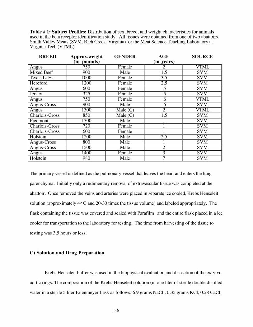
156
Table # 1: Subject Profiles: Distribution of sex, breed, and weight characteristics for animalsused in the beta receptor identification study. All tissues were obtained from one of two abattoirs,Smith Valley Meats (SVM, Rich Creek, Virginia) or the Meat Science Teaching Laboratory atVirginia Tech (VTML)
BREED Approx.weight(in pounds)
GENDER AGE(in years)
SOURCE
Angus 750 Female 2 VTMLMixed Beef 900 Male 1.5 SVMTexas L. H. 1000 Female 3.5 SVMHereford 1200 Female 2.5 SVMAngus 600 Female .5 SVMJersey 325 Female .5 SVMAngus 750 Female .6 VTMLAngus-Cross 900 Male .6 SVMAngus 1300 Male (C) 2 VTMLCharlois-Cross 850 Male (C) 1.5 SVMPiedmont 1300 Male 1 SVMCharlois-Cross 720 Female 1 SVMCharlois-Cross 600 Female 1 SVMHolstein 1200 Male 2.5 SVMAngus-Cross 800 Male 1 SVMAngus-Cross 1500 Male 2 SVMAngus 1400 Female 3 SVMHolstein 980 Male 7 SVM
The primary vessel is defined as the pulmonary vessel that leaves the heart and enters the lung
parenchyma. Initially only a rudimentary removal of extravascular tissue was completed at the
abattoir. Once removed the veins and arteries were placed in separate ice cooled, Krebs Henseleit
solution (approximately 4o C and 20-30 times the tissue volume) and labeled appropriately. The
flask containing the tissue was covered and sealed with Parafilm� and the entire flask placed in a ice
cooler for transportation to the laboratory for testing. The time from harvesting of the tissue to
testing was 3.5 hours or less.
C) Solution and Drug Preparation
Krebs Henseleit buffer was used in the biophysical evaluation and dissection of the ex-vivo
aortic rings. The composition of the Krebs-Henseleit solution (in one liter of sterile double distilled
water in a sterile 5 liter Erlenmeyer flask as follows: 6.9 grams NaCl ; 0.35 grams KCl; 0.28 CaCl;
157
0.14 grams MgSO4; 2.09 grams NaHCO3; 0.16 grams KH2PO4; 2.0 grams dextrose). as
described by Weekley (1994). This solution was aseptically prepared.
Potassium chloride was used for the initial smooth muscle contraction to baseline. This
solution was prepared in sterile, double distilled water (30 millimolar concentration when 200
microliters of stock KCl solution is added to 30 milliliters of the solution in the tissue bath).
Micotil 300® was prepared by dilution in propylene glycol. Serial dilutions of Micotil
300® were made by taking one hundred microliters of stock solution and diluting in 900 microliters
of propylene glycol. This resulted in a stock concentration of 1.66 x 10⁻³ molar which was then
serially diluted to give concentrations from 1.66x10⁻³ molar to 1.66x10⁻¹⁰ molar (when 100
microliters is added to 30 milliliters of the tissue bath).
Isoproterenol, a mixed beta adrenergic receptor agonist, was used to evaluate the beta
receptor functioning of isolated ex-vivo aortic tissue. This solution was prepared in sterile, double
distilled water. Serial decreasing logarithmic concentrations of isoproterenol were made from an
initial 10-2 molar solution. The serial dilutions were made by taking 100 microliters of the initial
stock solution and diluting this in 900 microliters of sterile double distilled water sequentially to 10-
9 molar solution.
Terbutaline, a selective beta-2 adrenergic agonist and dobutamine, a selective beta-1
adrenergic agonist, were prepared in sterile double distilled water for each experiment. The
appropriate quantity of drug was weighed out and prepared for use each day. Terbutaline and
dobutamine were prepared to give a final concentration of 10-3 molar when 100 microliters of the
stock solution is added to a 30 milliliter tissue bath. Serial dilutions of the stock solution were
made from 10-3 to 10-10 molar for each drug used in the experiment.
158
The antagonists were prepared in a similar manner as the other drugs Atenolol, a selective
beta-1 antagonist and ICI118,551, a selective beta-2 antagonist, were also prepared using sterile
double distilled water. Each drug was prepared to give a final concentration of 10-6 molar in a 30
milliliter tissue bath from the stock solution. Each drug was made freshly on the day of its use.
The vascular samples for the cumulative dose-response experiment were placed in one of
three experimental groups. One set of vascular rings, acting as a standard, (assayed with known
adrenergic agents, isoproterenol, terbutaline or dobutamine) was compared to the experimental
tissues (assayed with Micotil 300®). Non-specific beta receptor activation (isoproterenol) was then
compared to the relative contributions of the beta-1 (dobutamine) and beta-2 receptors (terbutaline).
Tissues were then evaluated in the presence of a selective beta-receptor blocking drugs propranolol
(mixed), atenolol HCl (beta-1) and ICI-118,551 HCl (beta -2).
D) Testing Protocol
Once in the laboratory, the vascular tissue samples were carefully removed from the
transportation/storage flasks. The tissue was allowed to warm to room temperature (approximately
22o C), then was placed in a tissue bath of warmed Krebs-Henseleit solution (37o +/- 2.0oC) that
had been aerated with oxygen and carbon dioxide (95% O2 / 5% CO2). Extravascular tissue was
removed and two 3 mm wide vascular rings were removed from each vessel. Two samples of each
pulmonary artery and vein from the same animal, were run in separate tissue baths under identical
conditions. The prepared vascular segments were mounted on glass hooks and suspended between
an electronic transducer (Grass Model #FT-103) and an anchor point in the tissue bath (30 mls)
aerated with 95% oxygen / 5% carbon dioxide gas. The tissues were allowed to equilibrate at 2
grams tension for 1.5 to 2 hours. A baseline reading was then obtained to ensure tissue stability.
The tissue was then pre-contracted with 30 millimolar potassium chloride and this level of
contraction was defined as baseline (100% contraction). Various standard agents, dobutamine,
159
terbutaline and isoproterenol and the experimental agent Micotil 300® were used to induce
relaxation, while propranolol HCl and selective beta antagonist agents (ICI 118,551 and atenolol)
were used to inhibit the various subtypes of beta adrenergic receptors. Cumulative dose response
curves were generated by the addition of sequentially increasing log doses of the appropriate
agonists in the absence and presence of respective antagonists. The cumulative dose response
information of vascular smooth muscle change in tension was collected using the digitally based
"MacLab" data collection system and stored on 3.5 disks. Each tissue bath was drained and the
Krebs-Henseleit was replaced every fifteen to twenty minutes throughout the experimental period to
ensure that there was no build up drugs and metabolic waste products.
E) Statistical Evaluation
Data were analyzed in several ways. Initially analysis consisted of a pair wise comparison
of the values of 0 molar and 10-5 molar within each treatment (for each agonist). These data
assessed whether any change was statistically significant. The data were then compared between
treatments at the drug concentration of 10-5 molar. The comparison between and within groups was
done by use of analysis of variance (ANOVA, n=4). This comparison was done to evaluate if there
was a significant difference among treatment groups. Post-ANOVA pairwise comparisons were
done by the Bonferonni test of specific selected groups. This comparison evaluated the difference
for a challenge with each agonist alone and in the presence of an antagonist.

160
RESULTS
Micotil 300®caused relaxation in the bovine pulmonary vessels (figures #1 and #2). The
change in tension, relaxation, from the baseline is significantly greater in the pulmonary vein than in
the artery (p< 0.01). The trend to relaxation from the baseline, in the artery, fails to reach statistical
significance (p> 0.05). The question as to whether relaxation is mediated by a beta receptor was
addressed by attempting to block the Micotil 300® induced relaxation by the addition of
propranolol. The dose of propranolol added to each tissue bath was 10-6 molar, which resulted in
an elimination of the relaxation response from baseline, in the case of the pulmonary vein (P> 0.05,
no significant change between the baseline and 10-5 molar Micotil 300®). A trend to antagonism of
the relaxation response was noted for the pulmonary artery, however, Micotil 300® failed to induce
vascular relaxation at a level significantly different from baseline. It is therefore difficult to assess
the true significance of the extent of the inhibition, by propranolol, of the relaxation in the
pulmonary artery. Comparison of the difference in the relaxation noted in the vein with Micotil
300® (at 10-5 molar Micotil 300®) in the presence of propranolol (at 10-5 molar Micotil 300®)
also yielded a significant differences between groups (p< 0.01)
To assess the subtypes of beta adrenergic receptors involved in this response, selective beta
receptor agonists and antagonists were employed. These drugs were used to elucidate the relative
contributions of each receptor subtype involved in the relaxation response for each tissue.
Assessment of the of beta-1 receptor component of the pulmonary vascular relaxation
response to Micotil 300® yielded some interesting results. The pulmonary artery responded with
significant relaxation to dobutamine (p< 0.001) which was not blocked by atenolol (figure #3). The
assessment of the ability of atenolol at 10-6 molar concentration to block the dobutamine response
suggests that the concentration of atenolol was insufficient to antagonize dobutamine. Other
explanations include that that there is a displacement of the antagonist by the agonist, or that the
161
number of subjects is too small. The vein did not relax in response to increasing molar
concentrations of dobutamine (p> 0.05, figure #4). A comparison of the relative relaxation induced
by dobutamine in both the artery and vein showed a greater relaxation response in the artery than in
the vein (figure #5). This difference in response to dobutamine between the two tissues was
statistically significant (p> 0.01).
Evaluation of the beta-2 adrenergic receptor in the relaxation response was examined.
Terbutaline failed to cause significant relaxation in the pulmonary artery (p> 0.05, figure #6).
Further, there was no significant difference between tissues with and without ICI 118,551 (p> 0.05).
There was however, significant relaxation induced by terbutaline in the pulmonary vein (p< 0.01,
figure #7). No significant relaxation occured in response to terbutaline in the presence of ICI
118,551 (p> 0.05). This indicates that the relaxation response to terbutaline was blocked by the
selective beta-2 antagonist, ICI-118,551 (at 10-6 molar concentration, p< 0.01 examined at 10-5
molar terbutaline with and without ICI 118,551).
Graphical comparison of the pulmonary artery and vein, suggests that the vein is more
responsive to the relaxation effect of Micotil 300® than the artery (figure #9)
The ability of the selective beta-2 antagonist, ICI 118,551, to block the relaxation response
of Micotil 300® in pulmonary venous tissue, was examined. The pulmonary vein did not relax to
Micotil 300® in the presence of ICI 118,551 (p> 0.05, figure #8). Examination of the effect of
Micotil 300® alone and Micotil 300® in the presence of ICI 118,551 suggests that there is a
significant difference when these groups are compared (p< 0.01, figure #9).
The assessment of the potential of the beta-1 antagonist to directly block the action of
Micotil 300® was examined. In the vein the atenolol failed to block relaxation induced by Micotil
300® (figure #10). There was a significant difference between the baseline and 10-5 molar Micotil
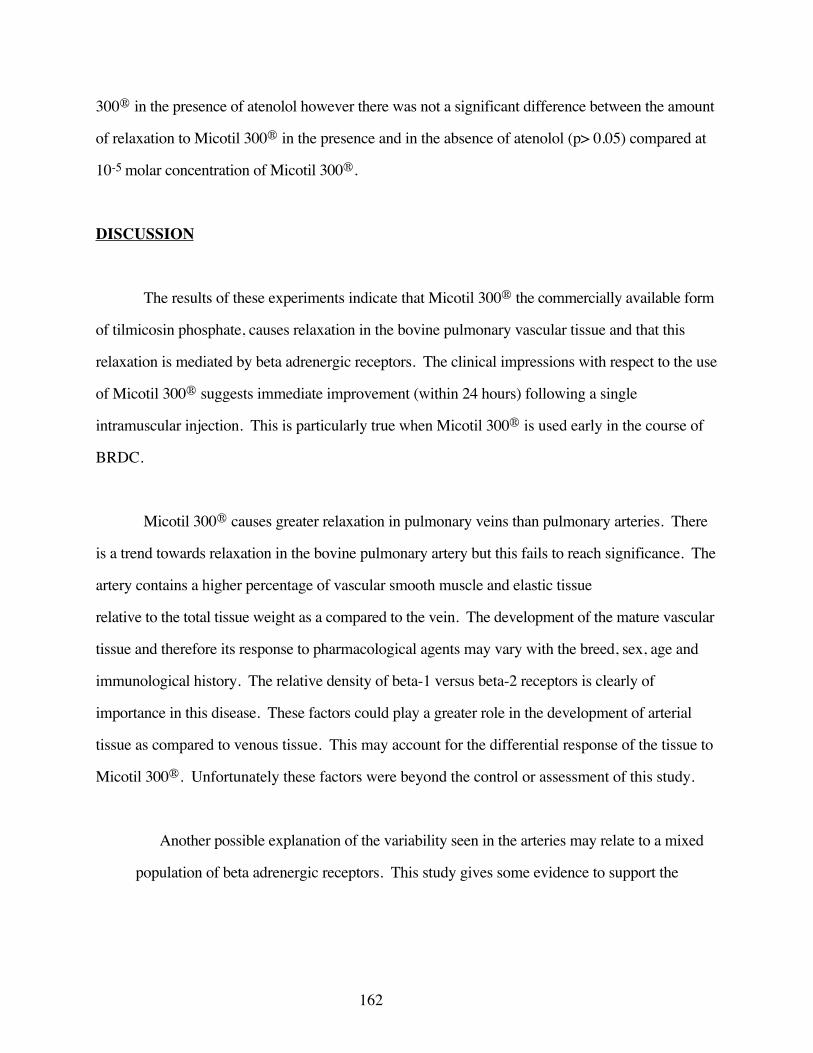
162
300® in the presence of atenolol however there was not a significant difference between the amount
of relaxation to Micotil 300® in the presence and in the absence of atenolol (p> 0.05) compared at
10-5 molar concentration of Micotil 300®.
DISCUSSION
The results of these experiments indicate that Micotil 300® the commercially available form
of tilmicosin phosphate, causes relaxation in the bovine pulmonary vascular tissue and that this
relaxation is mediated by beta adrenergic receptors. The clinical impressions with respect to the use
of Micotil 300® suggests immediate improvement (within 24 hours) following a single
intramuscular injection. This is particularly true when Micotil 300® is used early in the course of
BRDC.
Micotil 300® causes greater relaxation in pulmonary veins than pulmonary arteries. There
is a trend towards relaxation in the bovine pulmonary artery but this fails to reach significance. The
artery contains a higher percentage of vascular smooth muscle and elastic tissue
relative to the total tissue weight as a compared to the vein. The development of the mature vascular
tissue and therefore its response to pharmacological agents may vary with the breed, sex, age and
immunological history. The relative density of beta-1 versus beta-2 receptors is clearly of
importance in this disease. These factors could play a greater role in the development of arterial
tissue as compared to venous tissue. This may account for the differential response of the tissue to
Micotil 300®. Unfortunately these factors were beyond the control or assessment of this study.
Another possible explanation of the variability seen in the arteries may relate to a mixed
population of beta adrenergic receptors. This study gives some evidence to support the
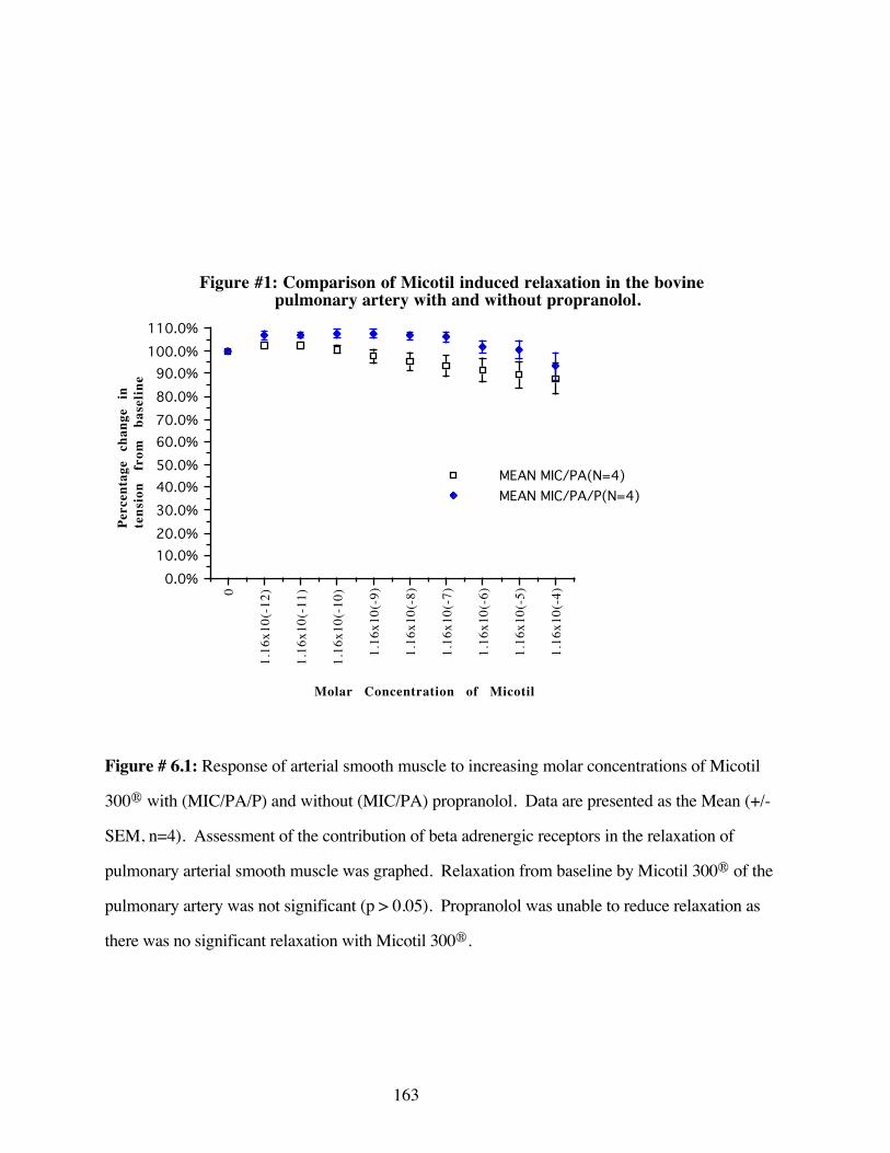
163
0
1.16
x10(
-12)
1.16
x10(
-11)
1.16
x10(
-10)
1.16
x10(
-9)
1.16
x10(
-8)
1.16
x10(
-7)
1.16
x10(
-6)
1.16
x10(
-5)
1.16
x10(
-4)0.0%
10.0%20.0%30.0%40.0%50.0%60.0%70.0%80.0%90.0%
100.0%110.0%
MEAN MIC/PA(N=4)MEAN MIC/PA/P(N=4)
Figure #1: Comparison of Micotil induced relaxation in the bovine pulmonary artery with and without propranolol.
Molar Concentration of Micotil
Perc
enta
ge
chan
ge
in
tens
ion
from
ba
selin
e
Figure # 6.1: Response of arterial smooth muscle to increasing molar concentrations of Micotil
300® with (MIC/PA/P) and without (MIC/PA) propranolol. Data are presented as the Mean (+/-
SEM, n=4). Assessment of the contribution of beta adrenergic receptors in the relaxation of
pulmonary arterial smooth muscle was graphed. Relaxation from baseline by Micotil 300® of the
pulmonary artery was not significant (p > 0.05). Propranolol was unable to reduce relaxation as
there was no significant relaxation with Micotil 300®.
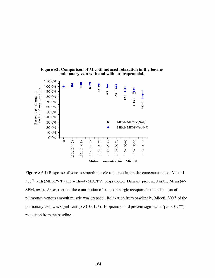
164
0
1.16
x10(
-12)
1.16
x10(
-11)
1.16
x10(
-10)
1.16
x10(
-9)
1.16
x10(
-8)
1.16
x10(
-7)
1.16
x10(
-6)
1.16
x10(
-5)
1.16
x10(
-4)0.0%
10.0%20.0%30.0%40.0%50.0%60.0%70.0%80.0%90.0%
100.0%110.0%
MEAN MIC/PV(N=4)MEAN MIC/PV/P(N=4)
Figure #2: Comparison of Micotil induced relaxation in the bovine pulmonary vein with and without propranolol.
Molar concentration Micotil
Perc
enta
ge
chan
ge
in
tens
ion
from
ba
selin
e
**
*
Figure # 6.2: Response of venous smooth muscle to increasing molar concentrations of Micotil
300® with (MIC/PV/P) and without (MIC/PV) propranolol. Data are presented as the Mean (+/-
SEM, n=4). Assessment of the contribution of beta adrenergic receptors in the relaxation of
pulmonary venous smooth muscle was graphed. Relaxation from baseline by Micotil 300® of the
pulmonary vein was significant (p > 0.001, *). Propranolol did prevent significant (p> 0.01, **)
relaxation from the baseline.
165
DOBU
T 0
DO
BUT
10(-1
0)
DO
BUT
10(-9
)
DO
BUT
10(-8
)
DO
BUT
10(-7
)
DO
BUT
10(-6
)
DO
BUT
10(-5
)
DO
BUT
10(-4
)0.0%10.0%20.0%30.0%40.0%50.0%60.0%70.0%80.0%90.0%
100.0%110.0%
PA-A/DPA-D
Figure #3: Comparison of dobutamine induced relaxation on the bovine pulmonary artery with and without atenolol.
Molar concentration of dobutamine
Perc
enta
ge
chan
ge
in
tens
ion
from
ba
selin
e
Figure # 6.3: Response of arterial smooth muscle to increasing molar concentrations of
dobutamine with (PA-A/D) and without (PA-D) atenolol. Data are presented as the Mean (+/-
SEM, n=4). Assessment of the contribution of beta-1 receptors in the relaxation of pulmonary
arterial smooth muscles was graphed. Relaxation from baseline by dobutamine of the pulmonary
artery was significant (p > 0.001, *). Atenolol did not prevent significant (p> 0.05) relaxation from
the baseline.
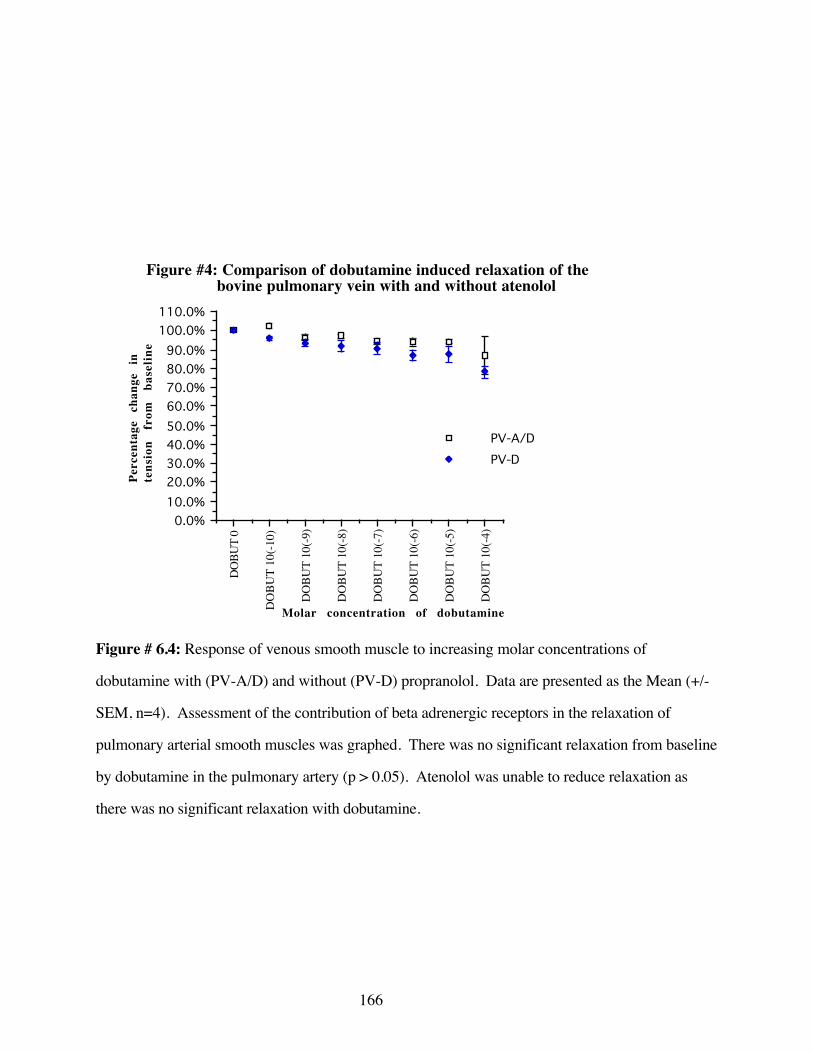
166
DOBU
T 0
DO
BUT
10(-1
0)
DO
BUT
10(-9
)
DO
BUT
10(-8
)
DO
BUT
10(-7
)
DO
BUT
10(-6
)
DO
BUT
10(-5
)
DO
BUT
10(-4
)0.0%10.0%20.0%30.0%40.0%50.0%60.0%70.0%80.0%90.0%
100.0%110.0%
PV-A/DPV-D
Figure #4: Comparison of dobutamine induced relaxation of the bovine pulmonary vein with and without atenolol
Molar concentration of dobutamine
Perc
enta
ge
chan
ge
in
tens
ion
from
ba
selin
e
Figure # 6.4: Response of venous smooth muscle to increasing molar concentrations of
dobutamine with (PV-A/D) and without (PV-D) propranolol. Data are presented as the Mean (+/-
SEM, n=4). Assessment of the contribution of beta adrenergic receptors in the relaxation of
pulmonary arterial smooth muscles was graphed. There was no significant relaxation from baseline
by dobutamine in the pulmonary artery (p > 0.05). Atenolol was unable to reduce relaxation as
there was no significant relaxation with dobutamine.
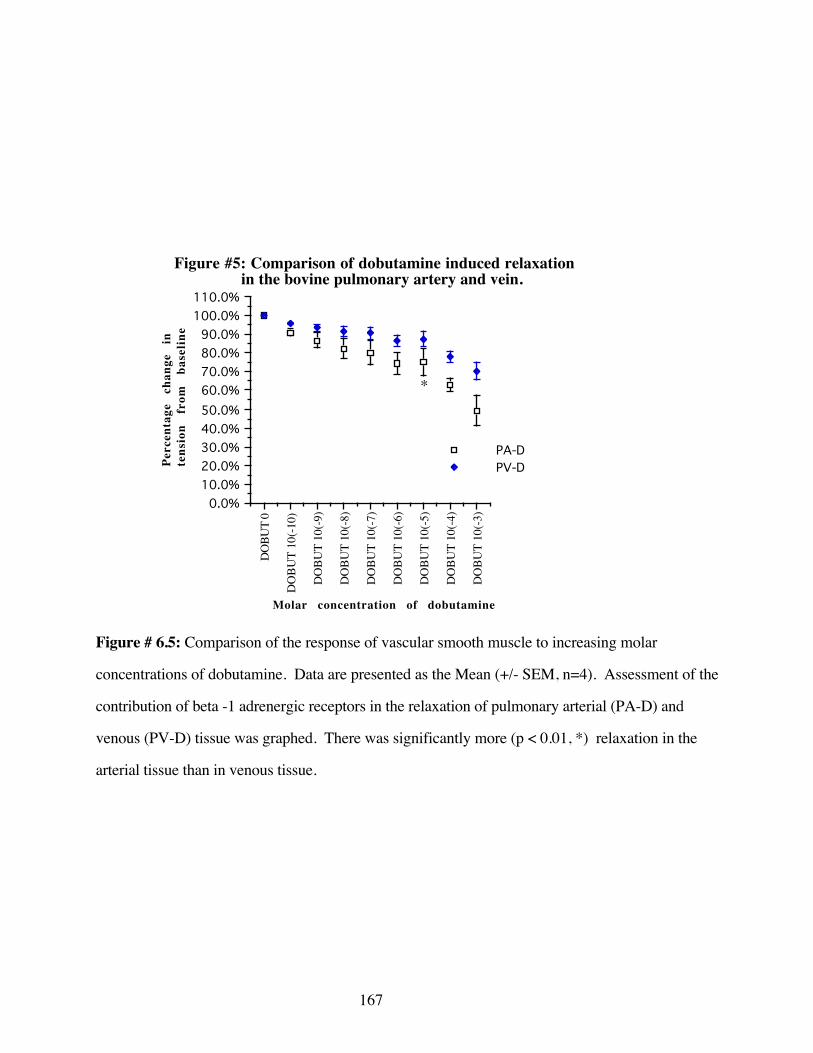
167
DOBU
T 0
DO
BUT
10(-1
0)
DO
BUT
10(-9
)
DO
BUT
10(-8
)
DO
BUT
10(-7
)
DO
BUT
10(-6
)
DO
BUT
10(-5
)
DO
BUT
10(-4
)
DO
BUT
10(-3
)0.0%10.0%20.0%30.0%40.0%50.0%60.0%70.0%80.0%90.0%
100.0%110.0%
PA-DPV-D
Figure #5: Comparison of dobutamine induced relaxation in the bovine pulmonary artery and vein.
Molar concentration of dobutamine
Perc
enta
ge
chan
ge
inte
nsio
n fr
om
base
line
*
Figure # 6.5: Comparison of the response of vascular smooth muscle to increasing molar
concentrations of dobutamine. Data are presented as the Mean (+/- SEM, n=4). Assessment of the
contribution of beta -1 adrenergic receptors in the relaxation of pulmonary arterial (PA-D) and
venous (PV-D) tissue was graphed. There was significantly more (p < 0.01, *) relaxation in the
arterial tissue than in venous tissue.

168
TERB
0
TERB
10(
-10)
TERB
10(
-9)
TERB
10(
-8)
TERB
10(
-7)
TERB
10(
-6)
TERB
10(
-5)
TERB
10(
-4)
0.0%10.0%20.0%30.0%40.0%50.0%60.0%70.0%80.0%90.0%
100.0%110.0%
Pul. Art/Terb.Pul. Art/ICI
Figure #6: Comparison of terbutaline induced relaxation in the bovine pulmonary artery with and without ICI 118,551.
Molar concentration of terbutaline
Perc
enta
ge
chan
ge
in
tens
ion
from
ba
selin
e
Figure # 6.6: Response of arterial smooth muscle to increasing molar concentrations of terbutaline
with (Pul Art/ICI) and without (Pul Art/Terb) the antagonist ICI 118,551. Data are presented as the
Mean (+/- SEM). Assessment of the contribution of beta-2 adrenergic receptors in the relaxation
of pulmonary arterial smooth muscles was graphed. There was no significant relaxation from
baseline by terbutaline (p> 0.05) in the pulmonary artery. ICI 118,551 was unable to reduce
relaxation as there was no significant relaxation with dobutamine.

169
TERB
0
TERB
10(
-10)
TERB
10(
-9)
TERB
10(
-8)
TERB
10(
-7)
TERB
10(
-6)
TERB
10(
-5)
TERB
10(
-4)
0.0%10.0%20.0%30.0%40.0%50.0%60.0%70.0%80.0%90.0%
100.0%110.0%
Pul. Vein/TerbPul. Vein/ICI
Figure #7: Comparison of terbutaline induced relaxation in the bovine pulmonary vein with and without ICI 118,551.
Molar concentration of terbutaline
Perc
enta
ge
chan
ge
in
tens
ion
from
ba
selin
e
**
*
Figure # 6.7: Response of venous smooth muscle to increasing molar doses of terbutaline with
(Pul Vein/ICI) and without (Pul Vein/Terb) the antagonist ICI 118,551. Data are presented as the
Mean (+/- SEM, n=4). Assessment of the contribution of beta-2 adrenergic receptors in the
relaxation of pulmonary venous smooth muscles was graphed. There was significant relaxation
from baseline by terbutaline (p< 0.01,*) in the pulmonary vein. There was no significant relaxation
in the presence of ICI 118,551 (p> 0.05, **) in the pulmonary vein.
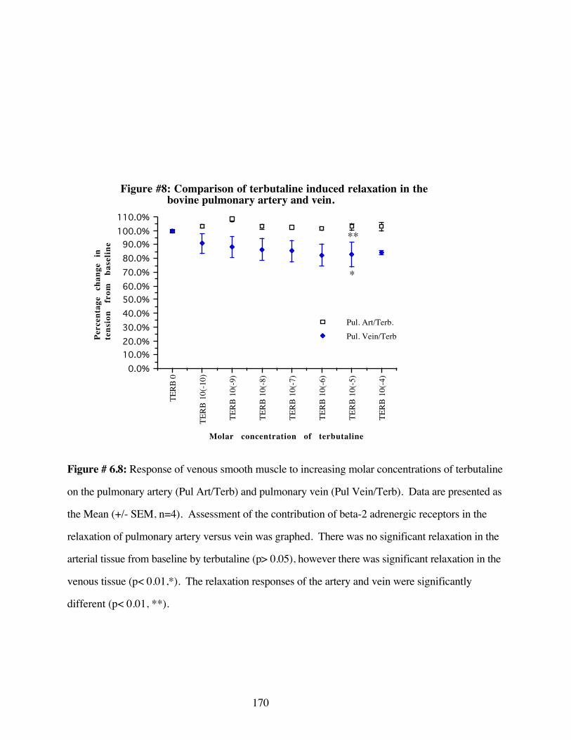
170
TERB
0
TERB
10(
-10)
TERB
10(
-9)
TERB
10(
-8)
TERB
10(
-7)
TERB
10(
-6)
TERB
10(
-5)
TERB
10(
-4)
0.0%10.0%20.0%30.0%40.0%50.0%60.0%70.0%80.0%90.0%
100.0%110.0%
Pul. Art/Terb.Pul. Vein/Terb
Figure #8: Comparison of terbutaline induced relaxation in the bovine pulmonary artery and vein.
Molar concentration of terbutaline
Perc
enta
ge
chan
ge
in
tens
ion
from
ba
selin
e
*
**
Figure # 6.8: Response of venous smooth muscle to increasing molar concentrations of terbutaline
on the pulmonary artery (Pul Art/Terb) and pulmonary vein (Pul Vein/Terb). Data are presented as
the Mean (+/- SEM, n=4). Assessment of the contribution of beta-2 adrenergic receptors in the
relaxation of pulmonary artery versus vein was graphed. There was no significant relaxation in the
arterial tissue from baseline by terbutaline (p> 0.05), however there was significant relaxation in the
venous tissue (p< 0.01,*). The relaxation responses of the artery and vein were significantly
different (p< 0.01, **).
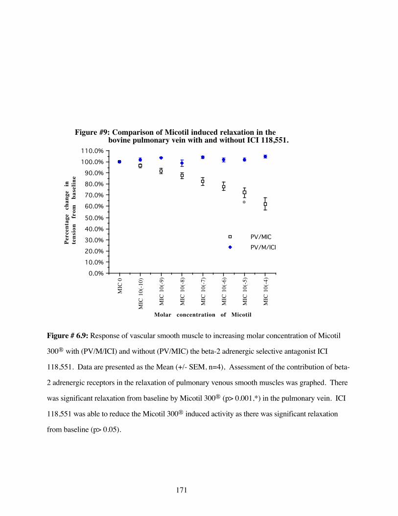
171
MIC
0
MIC
10(
-10)
MIC
10(
-9)
MIC
10(
-8)
MIC
10(
-7)
MIC
10(
-6)
MIC
10(
-5)
MIC
10(
-4)0.0%
10.0%
20.0%
30.0%
40.0%
50.0%
60.0%
70.0%
80.0%
90.0%
100.0%
110.0%
PV/MICPV/M/ICI
Figure #9: Comparison of Micotil induced relaxation in the bovine pulmonary vein with and without ICI 118,551.
Molar concentration of Micotil
Perc
enta
ge
chan
ge
in
tens
ion
from
ba
selin
e
*
Figure # 6.9: Response of vascular smooth muscle to increasing molar concentration of Micotil
300® with (PV/M/ICI) and without (PV/MIC) the beta-2 adrenergic selective antagonist ICI
118,551. Data are presented as the Mean (+/- SEM, n=4), Assessment of the contribution of beta-
2 adrenergic receptors in the relaxation of pulmonary venous smooth muscles was graphed. There
was significant relaxation from baseline by Micotil 300® (p> 0.001,*) in the pulmonary vein. ICI
118,551 was able to reduce the Micotil 300® induced activity as there was significant relaxation
from baseline (p> 0.05).
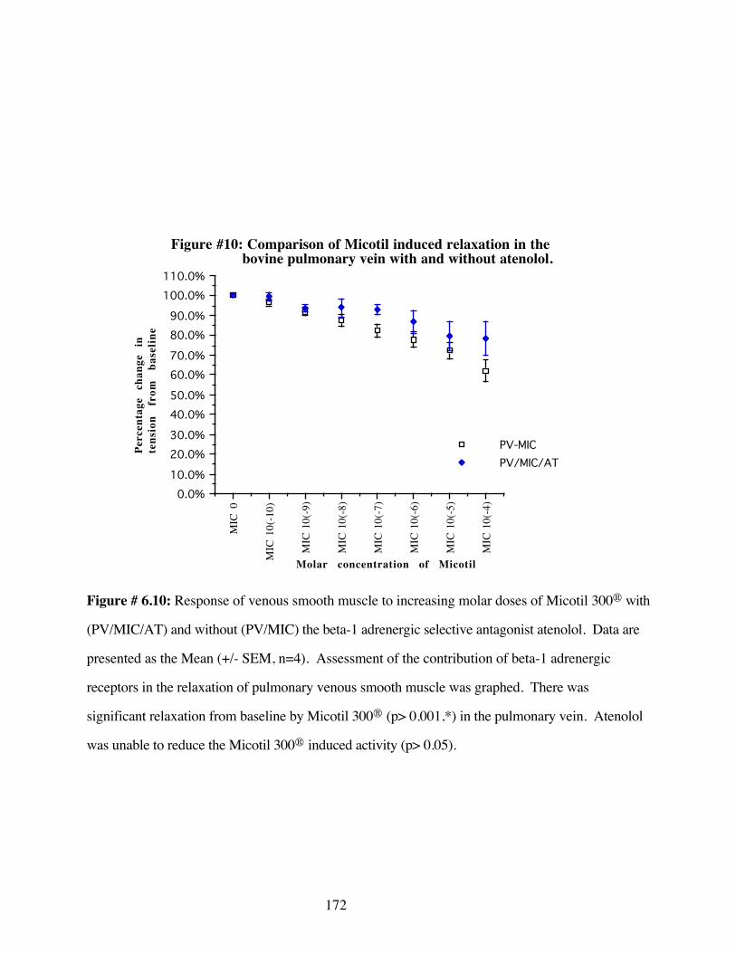
172
MIC
0
MIC
10(
-10)
MIC
10(
-9)
MIC
10(
-8)
MIC
10(
-7)
MIC
10(
-6)
MIC
10(
-5)
MIC
10(
-4)0.0%
10.0%
20.0%30.0%
40.0%50.0%
60.0%70.0%
80.0%90.0%
100.0%110.0%
PV-MICPV/MIC/AT
Figure #10: Comparison of Micotil induced relaxation in the bovine pulmonary vein with and without atenolol.
Molar concentration of Micotil
Perc
enta
ge
chan
ge
in
tens
ion
from
ba
selin
e
Figure # 6.10: Response of venous smooth muscle to increasing molar doses of Micotil 300® with
(PV/MIC/AT) and without (PV/MIC) the beta-1 adrenergic selective antagonist atenolol. Data are
presented as the Mean (+/- SEM, n=4). Assessment of the contribution of beta-1 adrenergic
receptors in the relaxation of pulmonary venous smooth muscle was graphed. There was
significant relaxation from baseline by Micotil 300® (p> 0.001,*) in the pulmonary vein. Atenolol
was unable to reduce the Micotil 300® induced activity (p> 0.05).
173
claim that the beta 1 and beta 2 adrenergic receptors exist in the bovine pulmonary artery with the
beta-1 receptor being predominant. The vein very likely also contains a mixed population of beta
receptors where the beta 2 receptor is the dominant. The evidence presented here is that a mixed
population of beta 1 and beta 2 receptors exist on both the venous and arterial sides of the
pulmonary vasculature. Further data suggest that Micotil 300® has greater activity at the beta 2
receptor. The specific differential activity among beta adrenergic receptors has yet to be quantified.
It is clear that the venous aspect of the pulmonary vasculature responds more vigorously to
Micotil 300® induced vasorelaxation than the arterial tissue. This could result in increased venous
drainage. The significance of these findings with respect to the overall disease process is only
conjecture at this time. The disruption of beta adrenergic vascular receptors after exposure to
Pasteurella haemolytica A1 appears to occur over a 24 hour period with only partial disruption in
the first 24 hours. The obvious consequence of deficient beta receptors in the balancing equation
between alpha receptor contraction and beta mediated relaxation is a predominence of the alpha
receptor mediated vasoconstriction. Therefore any decrease in pulmonary congestion, secondary to
an up-regulation of beta mediated relaxation may be of benefit to the animal afflicted with BRDC.
The significance of decreased venous drainage in the presence of a relatively unchanged
arterial supply is obvious. The pathogenesis of BRDC in general, and pneumonic pasteurellosis in
particular, is recognized as fibrinous, congested lung fields. The action of a specific venous dilator,
Micotil 300®, may be to activate those beta receptors that are not affected early in the disease
process. This may serve to recruit additional beta adrenergic receptors which would assist in the
reduction of the pulmonary congestion noted in bovine pasteurellosis. This may be the explanation
for the immediate relief of signs in cattle diagnosed early with BRDC following the use of Micotil
300®. This theory is not without foundation, as Sigurdsson (1988) has demonstrated that the use
of the beta-2 adrenergic receptor agonist, terbutaline is effective in relieving the symptoms of
respiratory distress in sheep exposed to endotoxin.

174
The beta adrenergic mechanism, though important in the maintenance of various pulmonary
vascular homeostatic mechanisms, has largely been ignored with respect to the etiology
andtreatment of BRDC. Weekley has shown that the activity of PH administered parenterally, in
multiple species can disrupt beta adrenoceptors associated with vascular smooth muscle (Weekley
1991A; 1993A; 1993E)..
The disruption of beta adrenergic receptors is unlikely to be limited to those receptors
associated with vascular smooth muscle. The maintenance of the pulmonary vascular integrity is
intimately involved with beta receptors associated with the endothelium. Other beta adrenergic
receptors act to modulate extravasation of intravascular fluid and osmotic elements (Mizus 1985;
Minnear 1986) and inhibit endothelial reactions with the coagulation cascade, thereby preventing the
generation of intravascular fibrin. Still other beta receptors act to modulate the release of
antibacterial enzymes and oxygen free radicals by neutrophils and cytokines by macrophages in the
presence of bacteria or bacterial products. Though not yet specifically investigated, the theoretical
consequence of the disruption of these beta receptors or a partial disruption of the populations of
beta receptors associated with the pulmonary vascular system may account for the majority of the
gross and microscopic pathological findings associated with pneumonic pasteurellosis.
The macrolide antibiotic, Micotil 300® has exceptional efficacy in the treatment of bovine
pneumonic pasteurellosis. This antibiotic also appears to have significant beta adrenoceptor activity
which may account in part for its clinical efficacy. It is apparent that the bovine pulmonary
vasculature examined contains a mixed population of beta receptors with predominant features of
the beta-1 receptor on the arterial side and beta-2 receptor on the venous side. It is likely that the
predominant beta-2 adrenergic receptor activity of Micotil 300® has a beneficial effect early in the
course of pneumonic pasteurellosis, to reduce the severity of pulmonary congestion. It may further
be speculated, based on the homeostatic activities of the vascular beta adrenergic receptors, that beta-
175
2 adrenergic activity will also reduce extravasation of fluid, decrease endothelial elaboration of
coagulation active products and inhibit release of enzymes and oxidative free radicals from
leukocytes.
CONCLUSIONS
1) Micotil 300® has the ability to cause vasorelaxation in pulmonary vasculature.
2) This vasorelaxation is more prominent in the pulmonary veins than in the pulmonary
arteries.
3) The veins appear to respond to beta-2 adrenergic receptor agonists while the arteries
appear to respond more vigorously to beta 1 adrenergic agonists in-vitro.
4) These results when combined with the differential sensitivity for vasorelaxation between
the pulmonary veins and arteries suggests that Micotil 300® has a predominant beta 2
adrenergic receptor activity.
176
REFERENCES
Al-Essa, L. Y., M. Niwa, K.I. Kohno, M. Nozaki, K. Tsurumi (1995). “Heterogeneity of circulatingand exudated polymorphonuclear leukocytes in superoxide-generating response to cyclic AMP andcAMP elevating agents.” Biochemical Pharmacology 49(3): 315-322.
Barnard, M. L., S. Matalon (1992). “Mechanisms of extracellular reactive oxygen species injury tothe pulmonary microvasculature.” Journal of Applied Physiology 72(5): 1724-9.
Barnes, P. J. (1986). “Nonadrenergic noncholinergic neural control of human airways.” ArchivesInternationales de Pharmacodynamie 280: 208-28.
Barnes, P. J., S. F. Liu (1995). “Regulation of pulmonary vascular tone.” PharmacologicalReviews 47(1): 87-131.
Bourne, H. R., R.I. Lehrer, M.J. Cline, K.L. Melmon (1971). “Cyclic 3'5'-adenosinemonophosphate in the human leukocyte:synthesis degradation, and effects on neutrophilcandidacidal activity.” Journal of Clinical Investigation 50: 920-27.
Breeze, R. (1985). Structure, function and metabolism in the lung. Veterinary Clinics of NorthAmerica: Food Animal Practice. R. Breeze. Philadelphia, W.B. Saunders. 1: 219-235.
Breider, M. A., S. Kumar, R.E. Corstvet (1991). “Interaction of bovine neutrophils in Pasteurellahaemolytica mediated damage to pulmonary endothelial cells.” Veterinary Immunology andImmunopathology 27: 337-50.
Busse, W. W., J.M. Sosman (1984). “Isoproterenol inhibition of isolated human neutrophilfunction.” Journal of Allergy and Clinical Immunology 73: 404-12.
Busso, N., S. Huet, E. Nicodeme, J. Hiernaux, F. Hyafil (1991). “Refractory period phenomenonin the induction of tissue factor expression on endothelial cells.” Blood 78(8): 2027-35.
Dow, S. W., M.G. Papich (1991). “Keeping current on developments in antimicrobial therapy.”Pet Practice June: 600-9.
Engels, F., R.S. Oosting, F.P. Nijkamp (1985). “Pulmonary macrophages induce deterioration ofguinea pig tracheal beta adrenergic function through release of oxygen free radicals.” EuropeanJournal of Pharmacology 111: 143-4.
Gorham, P. E., L.H. Carroll, J.W. McAskill, L.E. Watkins, E.E. Ose, L.V. Tonkinson, J.K. Merrill(1990). “Tilmicosin as a single injection treatment for respiratory disease of feedlot cattle.”Canadian Veterinary Journal 31: 826-9.
Granger, D. N., P. Kubes (1994). “The microcirculation and inflammation: modulation ofleukocyte endothelial cell adhesion.” Journal of Leukocyte Biology 55: 662-75.
Harlan, J. S., P.D. Killen, L.A. Harker, G.E. Striker (1981). “Neutrophil mediated endothelialinjury in-vitro.” Journal of Clinical Investigations 68: 1394-1403.
Hills, H. R., P.G. Estensen, G. Quie, N.A. Hogan, N.D. Goldberg (1975). “Modulation of humanneutrophil chemotactic responses by cyclic 3' 5'-guanosine monophosphate and cyclic 3' 5'-adenosine monophosphate.” Metabolism 24: 447-53.

177
Hu, X. X., E.A. Goldmuntz, C.F. Brosnan (1991). “The effect of norepinephrine on endotoxinmediated macrophage activation.” Journal of Neuroimmunology 31(1).
Ignarro, L. J., T.F. Lint, W.J. George (1974). “Hormonal control of lysosomal enzyme releasefrom human neutrophils. Effects of autonomic agents on enzyme release, phagocytosis, and cyclicnucleotides.” Journal of Experimental Medicine 139: 1395-1410.
Kubes, P., R. Smith, M.D. Grisham, D.N. Granger (1993). “Neutrophil mediated proteolysis,differential roles for Cathepsin G and Elastase.” Inflammation 17(3): 321-32.
Majno, G., G.E. Palade (1961). “Studies on inflammation: I. The effect of histamine and serotoninon vascular permeability. An electron microscopic study.” Journal of Biophysics and Biochemstryand Cytology 11: 571-83.
Merrill, J. K., L.V. Tonkinson (1989). “The effectiveness of Micotil for the treatment of bovinerespiratory disease.” The Bovine Practioner 24: 26-8.
Minnear, F. L., A. Johnson, A.B. Malik (1986). “Beta adrenergic modulation of pulmonarytransvascular fluid and protein exchange.” Journal of Applied Physiology 60: 266-74.
Mizus, I., W.Summer, I. Farrukh, J.R. Michael, G.H. Gurtner (1985). “Isoproterenol oraminophylline attenuate pulmonary edema after acid lung injury.” American Review of RespiratoryDisease 131: 256-9.
Morck, D. W. J. K. M., B.E. Thorlakson, M.E. Olson, L.V. Tonkinson, J.W. Costerton (1993).“Prophylactic efficacy of Tilmicosin for bovine respiratory tract disease.” Journal of the AmericanVeterinary Medical Association 202(2): 273-7.
Robinson, N. E., R.F. Slocomb, F.J. Derksen (1984). Physiology of the bovine lung. BovineRespiratory Disease : A Symposium. R. W. Loan. College Station, Texas, Texas A&M UniversityPress: 163-222.
Seccombe, J. F., P.J. Pearson, H. V. Schaff (1994). “Oxygen radical mediated vascular injuryselectively inhibits receptor dependent release of nitric oxide from canine coronary arteries.”Journal of Thoracic and Cardiovascular Surgery 107: 505-9.
Severn, A., N.T. Rapson, C.A. Hunter, F.Y. Liew (1992). “Regulation of Tumor Necrosis Factorproduction by adrenaline and beta adrenergic agonists.” Journal of Immunology 148(11): 3441-5.
Sigurdsson, G. H., J.T. Christenson (1988). “Influence of terbutaline on endotoxin induced lunginjury.” Circulatory Shock 23(3): 153-63.
Sigurdsson, G. H., J.T. Christenson, M. al-Mousawi, A. Owunwanne (1989). “Use of indium-111oxine to study pulmonary and hepatic leukocyte sequestration in endotoxin shock and effects of thebeta-2 receptor agonist terbutaline.” American Journal of Physiology- Imaging 4(4): 136-42.
Slocombe, R. F., J. Malark, R. Ingersoll, F.J. Dirksen, N.E. Robinson (1985). “Importance ofneutrophils in the pathogenesis of acute pneumonic pasteurellosis in calves.” American Journal ofVeterinary Research 46(11): 2253-58.
Steinberg, S. F., E.A. Jaffe, J.P. Bilezikian (1984). “Endothelial cells contain beta adrenergicadrenoreceptors.” Naunyn-Schmiedebergs Archives of Pharmacology 325: 310-3.
178
Tecoma, E. S., H.J. Motulsky, A.E. Traynor, G.M. Omann, H.Muller, L.A. Sklar (1986).“Transient catecholamine modulation of neutrophil activation: kinetic and intracellular aspects ofisoproterenol action.” Journal of Leukocyte Biology 40: 629-44.
Veit, H. P., R.l. Farrell (1978). “The anatomy and physiology of the bovine respiratory systemrelating to pulmonary disease.” Cornell Veterinarian 68: 555-581.
Warren, J. S., P.A. Ward, K.J. Johnson (1987). Mechanisms of endothelial damage to pulmonaryendothelium. Pulmonary Endothelium in Health and Disease. U. S. Ryan. New York, MarcelDekker. 32: 107-20.
Weekley, L. B., P. Eyre, H.Veit, N. Sriranganathan (1991A). “Endothelium dependent andendothelium independent changes in pulmonary vascular smooth muscle reactivity followingexposure to Pasteurella haemolytica.” Procedings of the 10th Annual Veterinary RespirationSociety p.S1
Weekley, L. B., P. Eyre, H.P. Veit, N. Sriranganathan (1991B). “Exposure of sheep to Pasteurellahaemolytica alters pulmonary vascular smooth muscle alpha adrenergic responses: role of vascularendothelium.” Research Communications in Chemical Pathology and Pharmacology 74(1): 121-4.
Weekley, L. B., P.Eyre, H.P. Veit, N. Sriranganathan (1993C). “Morphological and functionaldisturbances in pulmonary vascular endothelium following exposure of sheep to an avirulent strainof Pasteurella haemolytica.” Journal of Comparative Pathology 108(1): 65-72.
Weekley, L. B., P.Eyre (1993B). “Vaccination with live Pasteurella haemolytica alters vascularadrenergic reactivity.” Research Communications in Chemical Pathology and Pharmacology 79(3):389-92.
Weekley, L. B., P. Eyre (1993E). “Disturbances in ex vivo vascular smooth muscle responsesfollowing exposure to Pasteurella haemolytica vaccines.” Journal of Veterinary Pharmacologyand Therapeutics 16: 446-53.
Weekley, L. B., P.Eyre (1994). “Altered endothelium dependent alpha adrenergic vascularreactivity following exposure to Pasteurella haemolytica.” Veterinary Research Communications18: 133-40.
Weekley, L. B. (1994). Personal Communication.
Weekley, L. B., H. P.Veit (1995). “Potential morphologic and physiologic factors that maypredispose the bovine lung to respiratory disease.” The Compendium 17(7): 974-983.
Weekley, L. B., P.Eyre, H.P. Veit, N. Sriranganathan (1996). “Evidence that Pasteurellahaemolytica vaccines cause sub-clinical impairments in the bovine pulmonary circulation.”Personal Communication
Wren, G. (1995). “Understanding the bovine respiratory system.” Bovine Veterinarian November:6-9.
Zurier, R. B., G. Weissman, S.Hoffstein, S. Kammerman, H.H. Tai (1974). “Mechanisms oflysosomal enzyme release from human leukocytes. II. Effects of cAMP and cGMP, autonomicagonists and agents which affect microtubule function.” Journal of Clinical Investigation 53: 297-303.
179
IV. GENERAL DISCUSSION
The importance of beta adrenergic receptor activity to the pathogenesis of Bovine
Pneumonic Pasteurellosis, and possibly vaccine induced immunopathology have not yet been
demonstrated in the target species. The accumulating evidence is providing growing evidence that
the adrenergic system in general and the beta adrenergic receptors in particular are intimately
involved in the development of Bovine Respiratory Disease Complex and pneumonic pasteurellosis.
Intimate involvement of the adrenergic system associated with the pulmonary venous system
is physiologically well described. The beta adrenergic receptor has been demonstrated to be
involved with vascular smooth muscle control, endothelial control of thrombolic events and
extravasation of intravascular fluid and finally control of the elaboration of cytokines, elastase and
oxygen free radicals in cells. The pulmonary alveolar and intravascular macrophages as well as the
neutrophil are examples of immunologically important cells controlled by the BARC.
Beta adrenergic receptor mechanisms may be altered, as defined by its lack of response to
selective pharmacological agents, after exposure to PH. Previous researchers have demonstrated
that the exposure to PH as in the form of a monovalent vaccine can have deleterious effects on the
adrenergic vascular smooth muscle system. It remains to be seen what the affect of exposure to PH
would be in a method consistent to a field situation in the target species. However, the current
experiments suggest that the vascular smooth muscle associated beta adrenergic receptor /
transduction mechanism is deleteriously affected.
Experimental results indicate that exposure to parenteral PH, in the form of a vaccine, can
cause a pharmacologically defined beta adrenergic disruption for as long as twenty one days post -
exposure. This disruption appears to occur within 24-48 hours of exposure. Though this is not
180
representative of the classical field situation it speaks to the need for early intervention in the
treatment of pneumonic pasteurellosis in cattle.
The establishment of the affects of endotoxin on cardiac beta adrenergic receptor
mechanisms has been well established . There is now some evidence that endotoxin alone may also
affect rat aortic vascular associated beta adrenergic receptors mechanisms. The recognition that
endotoxin is the culprit gives some insight into future research in the development of vaccines to
prevent this economically devastating disease.
Evidence from experiments examining the level of cAMP following PH exposure fail to
meet scientific or experimental criteria to glean any conclusions. Perhaps increasing the number of
experimental units would add sufficiently to the experimental power as to allow conclusions to be
drawn.
Investigation of the alpha-2 receptor demonstrates that the receptor complex is associated
with vasorelaxation through the mechanism defined by cGMP as the second messenger and
mediated by nitric oxide. This suggests that cGMP of the alpha-2 receptor mechanism is likely
equally susceptible to the affects of vaccination with PH. Since adrenergic system mediated
vasorelaxation is thought to be predominantly through the beta adrenergic and alpha-2 adrenergic
receptors disruption of these systems would result in disruption of the ability of the vascular system
to vasodilate in response to local or systemic conditions.
The evaluation of two currently used vaccines suggests that there is disruption of the beta
adrenergic receptor mechanism after vaccination. Experiemental review of these vaccines is
hindered by the fact that they were not evaluated in the target species. However, in view of the
similarity of the effect of parenteral exposure to PH in rats and sheep, it is reasonable to expect that
there is beta adrenergic receptor disruption in cattle similar to that in the rat.
181
Micotil 300® has been demonstrated to have beta adrenergic activity at the vascular smooth
muscle associated beta adrenergic receptor. Given the physiological significance of the beta
adrenergic receptor to vascular physiology and the positive results of the use of terbutaline (a beta-
2, selective agonist) in the treatment of endotoxin induced lung injury, the beta adrenergic activity of
Micotil 300® suggests that this feature of this macrolide antibiotic serves as a positive adjunct in the
treatment of BRDC. Further the in-vitro levels of this drug used are comparable to the tissue
concentrations in lung tissue as described by the manufacturer.
The results presented above are limited in their application for several reasons. First, these
studies were completed in a non target species. The use of the rat aorta is not anatomically identical
to the pulmonary vasculature of the ruminant. Secondly, these studies have been completed on ex-
vivo tissue. A model that examines tissue in-vitro fails to recognize and address the dynamic
compensation of the vascular system in general and the pulmonary vasculature specifically.
Specific compensation in the organism for vascular associated adrenergic dysfunction may render
insignificant the findings described above. However there is some support for the biologic
significance of the vascular associated adrenergic receptor system in the form of the positive effect
of the beta agonist, terbutaline and clinical observations associated with Micotil 300®. Third, it is
scientifically reasonable to suggest that the pulmonary vascular associated beta adrenergic receptor
is intimately involved with the development of pneumonic pasteurellosis. It is unlikely that this is
the initiating factor or even the primary event in the development of the clinical disease. There are
very likely a number of events that occur before the disruption of the beta adrenergic receptors.
There is at least one event preceding the disruption of beta receptor mechanism disruption, in the
pathogenesis of pneumonic pasteurellosis, this is likely the elaboration of inflammatory cytokines.
Finally, the intact pulmonary system is not without redundant and multiple compensatory
mechanisms that may be able to ameliorate or even eliminate the consequences of BARC
182
disruption. These protective mechanisms cannot be reproduced in their complexity in the ex-vivo /
in-vitro system.
Relevant future studies must include the following. First, the evaluation of the
pathophysiology of the PH mediated disruption of beta adrenergic receptors to the bovine
pulmonary vascular system. This may be best accomplished by the assessment of pulmonary
vascular functioning by use of a Swan-Ganz catheter in the target species. Further, the clinical
significance of vascular dysfunction must be evaluated with respect to the in-vivo health and
morbidity of the target species.
Secondly the evaluation of the events preceding the vascular associated beta adrenergic
mechanism disruption must also be evaluated. The significance of the beta receptor of the
pulmonary alveolar macrophage with respect to the elaboration of cytokines is very likely, in my
opinion, to be an essential ingredient as to whether or not an individual animal either develops or
doesn't develop pneumonic pasteurellosis. It is clear that the immunopharmacology of stress is also
integral to this process and must be weighed in the assessment of these events.
Third, the evaluation of the beta receptors involved with the neutrophil per se, the endothelial
mediated thrombolic events, the extravasation of intravascular fluid and consequences of disruption
of these beta adrenergic receptors must be investigated. The nature of the disruption of the
neutrophil based beta adrenergic receptor associated mechanisms may be accomplished by use of
the Microphysiometer® (Molecular Devices, Sunnyvale California). Other techniques may be
available to evaluate the other beta adrenergic associated mechanisms.
Fourth, examination of the activities of the neutrophil in the development of oxygen free
radicals and other chemical species in the development of pneumonic pasteurellosis may suggest
ways of treatment of the consequences of pneumonic pasteurellosis.
183
Finally the evaluation of the affect of PH on vascular associated beta adrenergic receptor
mechanisms in the target species under field conditions may allow the correlation of clinical, gross
and histopathological signs of the disease with the status and level of the adrenergic system
functional pathology at the molecular level. This is likely to make the current research relevant to
the real world situation.
Based on these results and those of previous colleagues it is becoming more obvious that
the adrenergic system is intimately involved in the etiology and pathogenesis of the disease
pneumonic pasteurellosis in cattle and that this disease has profound vascular effects which have
previously been under-appreciated until now. Use of this information and manipulation of the
vascular parameters discussed in this dissertation may lead to insights for the reduction of a
significant bovine disease and the attending economic burden placed on the cattle producers.
Hopefully the information presented in this dissertation is one small step towards the resolution of
this devastating problem.
184
V. REFERENCES
Abraham, E. (2000). “NF-kapp-B activation.” Crit. Care Med. 20(4-supplement): 53-60.
Aderem, A. A., D.S. Cohen, S. Wright, Z.A. Cohn, (1986). “Bacterial lipopolysaccarides primemacrophages for enhanced release of arachidonic acid metabolites.” Journal of ExperimentalMedicine 164: 165-73.
Adnot, S., I. Cigarini, A. Harf (1989). “Pulmonary vasodilator responses to calcitonin gene relatedpeptide (CGRP) and Substance P (SP).” American Review of Respiratory Disease 139: A49.
Akira, S. T. K. (1997). “NF-IL1 and NF alpha-B in cytokine gene regulation.” Adv. Immunol 65:1-40.
Al-Essa, L. Y., M. Niwa, K.I. Kohno, M. Nozaki, K. Tsurumi (1995). “Heterogeneity of circulatingand exudated polymorphonuclear leukocytes in superoxide-generating response to cyclic AMP andcAMP elevating agents.” Biochemical Pharmacology 49(3): 315-322.
Anderson, A. L., P.F. Moore, D.M. Hyde, D.L. Dungworth (1986). “Bronchus associatedlymphoid tissue in the lungs of cattle: relationship to age.” Research in Veterinary Science 41: 211-20.
Angen, O. R. M., D.A. Caugant, J.E. Olsen Magne Bisgaard (1999). “Taxonomic relationships ofthe [Pasteurella] haemolytica complex as evaluated by DNA-DNA hybridizations and 16s rRNAsequencing with proposal of Mannheimia haemolytica gen. nov., comb. nov., Mannheimiaglucosida sp. nov., Mannheimia ruminalis sp. nov. and Mannheimia varigena sp. nov.”International Journal of Systematic Bacteriology 49: 67-86.
Arenzana-Seisdedos, F. P. T., M. Rodriquez (1997). “Nuclear localization of I-kappa B alphapromotes activation part of NF-Kappa-B.” J. Cell Sci 110: 369-378.
AVMA Council Report (1993). “Immunization Guidelines.” AVMA 203(2): 238-242.Baeuerle, P. A. V. R. B. (1997). “NF-kappa-B as a frequent target for immunosuppressive andanti-inflammatory molecules.” Adv. Immunol 65: 111-137.
Bahou, W. B. C., C. Pottere (1993). “The Thrombin receptor extracellular domain contains sitescrucial for peptide ligand induced activation.” J. Clin. Invest. 91: 1405-1413.
Baldwin, A. S. (1996). “The NF-kappa-B and I-kappa-B proteins: new discoveries and insights.”Annu. Rev. Immunol. 14: 649-681.
Barnard, M. L., S. Matalon (1992). “Mechanisms of extracellular reactive oxygen species injury tothe pulmonary microvasculature.” Journal of Applied Physiology 72(5): 1724-9.
Barnes, P. J. (1986). “Nonadrenergic noncholinergic neural control of human airways.” ArchivesInternationales de Pharmacodynamie 280: 208-28.
Barnes, P. J., S. F. Liu (1995). “Regulation of pulmonary vascular tone.” PharmacologicalReviews 47(1): 87-131.
Barnett, C. C., D.A. Patrick, C.C. Salliman (1997). “Beta-adrenergic stimulation down regulatesneutrophil priming for superoxide generation but not elastase release.” J. Surg. Res 70: 166-170.
185
Bayne, E. K., E.A. Rupp, G. Limjuco, J. Chin, J.A. Schmidt (1986). “Immunocytochemicaldetection of interleukin 1 with-in stimulated human monocytes.” Journal of Experimental Medicine163: 1267-80.
Benburnou, N., P. Matsiota-Bernard, C. Jolivet, P. Ougen, M, Guenounou (1992). “TumorNecrosis Factor, IL 1, and IL-6, in bronchoalveolar washings, and their in-vitro production duringNippostrongylus brasiliensis infection.” Clinical and Experimental Immunology 88: 264-8.
Bensard, D. D., A. Banerjee, R.C. McIntyre, R.L. Berens, A.H. Harken (1994). “Endotoxindisrupts beta-adrenergic signal transduction in the heart.” Archives of Surgery 129(2): 198-204.
Bergofsky, E. H. (1979). Active control of the normal pulmonary circulation. Pulmonary VascularDiseases. K. M. Moser. New Yory, Marcel Dekker. 14: 233-77.
Beutler, B., J. Mahoney, N.L. Trang, P. Pekala, A. Creami (1985). “Purification of cachetin, alipoprotein lipase suppressing hormone secreted by endotoxin-induced RAW 264.7 cells.” Journalof Experimental Medicine 161: 984-95.
Beutler, B. A., D. Greenwald, J. D. Holmes, M. Chang, Y. Pan et al (1985). “Identity of TumorNecosis Factor and the macrophage secreted factor cachectin.” Nature 316: 552-4.
Birnbaumer, L. (1992). “Receptor to effector signaling through G-protein: Roles for beta, gammadimers as well as subunits.” Cell 71: 1069-72.
Blackwell, T. S. J. W. C. (1997). “The role of Nuclear Factor kappa B in cytokine regulation.”Am. J. Respir. Cell Mol. Biol. 17: 3-9.
Blood, D. C., V. P. Stoddert (1988). Balliere's Comprehensive Veterinary Dictionary. London,Balliere Tindall.
Bohrer, H. F. Q., T. Zimmermen, Y. Zhang, T. Jillmer, D. Mannel, B.W. Bottigon, D.M. Stern, R.Waldherr, H.D. Saeyer, R. Ziegler, A. Bierhaus, J. Martin, P.P. Nawroth (1997). “Role of NF-kappa-B in the mortality of sepsis.” J. Clin. Invest. 100(5): 972-985.
Boorman, G. A., L.W. Schwartz, F.D. Wilson (1979). “In-vitro macrophage colony formation byfree lung cells during pulmonary injury.” Journal of the Reticuloendothelial Society 26: 867-74.
Borregaard, N. (1988). The respiratory burst. The Respiratory Burst and its PhysiologicalSignificance. R. R, Sbarra. New York, Plenum Press: 1-8.
Bosquet, J. P. K. J., W.W. Busse, M. Johnson, A.M. Vignola (2000). “Asthma, frombronchioconstriction to airway inflammation and remodeling.” Am J. Respir Crit Care Med 161:1720-1745.
Bourne, H. R., R.I. Lehrer, M.J. Cline, K.L. Melmon (1971). “Cyclic 3'5'-adenosinemonophosphate in the human leukocyte:synthesis degradation, and effects on neutrophilcandidacidal activity.” Journal of Clinical Investigation 50: 920-27.
Bowden, D. H., I.Y.R. Adamson (1980). “Role of and interstitial cells in the generation of alveolarmacrophages.” Laboratory Investigations 42: 518-24.
Bowden, D. H. (1984). “The alveolar macrophage.” Environmental and Health Perspectives 55:327-42.
186
Bowden, J. J., I. Sulakvelidze, D.M. McDonald (1994). “Inhibition of neutrophil and eosinophiladhesion to venules of rat trachea by the beta-2 adrenergic agonist formoterol.” Journal of AppliedPhysiology 77(1): 397-405.
Brass, L. F. D. R. M., A.G. Williams (1991). “Receptor and C Protein mediated reponses tothrombin in H.E.L. cells.” J. Biol. Chem. 266: 958-965.
Brass, L. F., D.R. Manning (1988). “Interactions in platelets between G-proteins and the agoniststhat stimulate phospholipase C and inhibit adenylyl cyclase.” J. Biol. Chem. 263: 5348-5455.
Brauer, R. B. C. G. B. N., M. Stadler, C.D. Heidecke, B. Holzmann (2000). “Endotoxin inducedlung inflammation is dependent of the complement membrane attack complex.” Infect. andImmunity 68(3): 1626-1632.
Brauer, R. B., F. Sanfilippo (1994). “Hepatic and extrahepatic biosynthesis of complement factorC6 in the rat.” J. Immunol. 153: 3168-3176.
Brauer, R., F. Sanfilippo (1995). “The contribution of the terminal compliment components toacute and hyperacute allograft rejection in rats.” Transplantation 59: 288-293.
Breeze, R. (1985). Structure, function and metabolism in the lung. Veterinary Clinics of NorthAmerica: Food Animal Practice. R. Breeze. Philadelphia, W.B. Saunders. 1: 219-235.
Breider, M. A., R.D. Walker, F.M. Hopkins, T.W. Schultz, T.L. Bowersock (1988). “Pulmonarylesions induced by Pasteurella haemolytica in neutrophil sufficient and neutrophil deficientcalves.” Canadian Journal of Veterinary Research 52: 205-9.
Breider, M. A., S.Kumar, R. Corstvet (1990). “Bovine pulmonary endothelial cell damage mediatedby Pasteurella haemolytica pathogenic factors.” Infection and Immunity 58: 1671-77.
Breider, M. A., S. Kumar, R.E. Corstvet (1991). “Interaction of bovine neutrophils in Pasteurellahaemolytica mediated damage to pulmonary endothelial cells.” Veterinary Immunology andImmunopathology 27: 337-50.
Breider, M. A., S. Kumar, R.E. Corstvet (1991). “Protective role of bovine neutrophils inPasteurella haemolytica mediated endothelial cell damage.” Infection and Immunity 59(12): 4570-5.
Brigham, K. L. B. M. (1986). “Endotoxin and lung injury.” Am Rev, Respir, Disease 133: 913-927.
Brogen, K. A., c. Adlam, H.D Lehmkuhl, R.C. Cutlip, J.M. Knights, R. L. Engen (1989). “Effectof Pasteurella haemolytica (A1) capsular polysaccharride on sheep lung in-vivo and on pulmonarysurfactant in-vitro.” American Journal of Veterinary Research 50(4): 555-8.
Busse, W. W., J.M. Sosman (1984). “Isoproterenol inhibition of isolated human neutrophilfunction.” Journal of Allergy and Clinical Immunology 73: 404-12.
Busso, N., S. Huet, E. Nicodeme, J. Hiernaux, F. Hyafil (1991). “Refractory period phenomenonin the induction of Tissue Factor expression on endothelial cells.” Blood 78(8): 2027-35.
Campbell, K. L., R.A. Forse (1993). “Endotoxin exposed atria exhibit G-protein based deficits ininotropic regulation.” Surgery 114: 471-9.
187
Carney, D. H. R. M., W.R. Redin (1992). “Enhancement of incisional wound healing andneovascularization in normal rats by thrombin and synthetic Thrombin receptor activatingpeptides.” J. Clin. Invest. 89: 1469-1477.
Cazin, M., D. Paluszezak,A. Bianchi, J.C. Cazin, C. Aerts, C. Voisin (1990). “Effects ofanaerobiosis upon morphology and energy metabolism of alveolar macrophages.” EuropeanRespiratory Journal 3(9): 1015-22.
Chae, C. H., M.J. Gentry, A.W. Confer, G.A. Anderson (1990). “Resistance to host immunedefense mechanisms afforded by capsular material of Pasteurella haemolytica.” VeterinaryMicrobiology 25(2-3): 241-51.
Chang, Y., R.Young, D. Post et al (1987). “Identification and characterization of the Pasteurellahaemolytica leukotoxin.” Infection and Immunity 55(10): 2348-54.
Chester, I. R., P.M. Meadow, T.L. Pitt (1973). “The relationship between O-antigeniclipopolysaccharide and serological specificity in strains of Pseudamonas aeruginosa of differentO-serotypes.” Journal of General Microbiology and Bacteriology 78: 305-18.
Christman, J. W. A. V., T.S. Blackwell, (2000A). “NF kappa-B binding activity in mixed BALcells from patients with SIRS and ARDS.” Sepsis 4: 35-41.
Christman, J. W. R. T. S., T. Blackwell (2000). “The role of Nuclear Factor kappa-B in pulmonarydiseases.” Chest 117: 1432-1437.
Chung, K. F. P. J. B. (1999). “Cytokines in asthma.” Thorax 54: 825-857.Clinkenbeard, K., C.R. Clarke,R.J. Morton, R.J. Panciera, A. W. Confer, D.A. Mosier (1992).“Role of Pasteurella haemolyica leukotoxin in virulence and immunity in shipping feverpneumonia.” The Compendium 14(9): 1249-61.
Clinkenbeard, K. D., A.W. Boon, D.A. Mosier, A.W. Confer (1989A). “Effects of Pasteurellahaemolytica leukotoxin on isolated bovine neutrophils.” Toxicon 27: 797-804.
Clinkenbeard, K. D., M.L. Upton (1991). “Lysis of bovine platelets by Pasteurella haemolyticaleukotoxin.” American Journal of Veterinary Research 52(3): 453-7.
Confer, A. W., K.R. Simons (1986). “Effects of Pasteurella haemolytica lipopolysaccharide onselected functions of bovine leukocytes.” American Journal of Veterinary Research 47(1): 154-7.
Confer, A. W., R.J. Panciera, D.A. Moiser (1986). “Serum antibodies to Pasteurella haemolyticalipopolysaccharide: relationship to experimental bovine pneumonic pasteurellosis.” AmericanJournal of Veterinary Research 47(5): 1134-8.
Confer, A. W., R.J. Panciera, K.D. Clinkenbeard, D.A. Mosier (1990). “Molecular aspects ofvirulence of Pasteurella haemolytica.” Canadian Journal of Veterinary Research 54: S48-S52.
Confer, A. W., R.J. Panciera, A.J. Mort, D.A. Mosier (1989). “Serum antibody response tocarbohydrate antigens of Pasteurella haemolytica serotypa 1: relation to experimentally inducedbovine pneumonic pasteurellosis.” American Journal of Veterinary Research 50(1): 98-105.
Corbeil, L. B., R.P. Gogolewski (1985). Mechanisms of bacterial injury. Veterinary Clinics ofNorth America: Food Animal Pratice. R. Breeze. Philadelphia, W. B. Saunders Company. 1: 367-75.
188
Cravens, R. (1993). “Complex disease requires sophisticated product, Part I.” Topic in VeterinaryMedicine 4(2): 16-24.
Cravens, R. (1993A). “Clinical trials, field use affirm efficacy and safety. Part II.” Topics inVeterinary Medicine 4(2): 26-30.
Cuatrecasas, P., G.P.E. Tell, V.Sika, I. Parikh, K.J. Chang (1974). “Noradrenaline binding and thesearch for the catecholamine receptor.” Nature 273: 471.
Czermak, B. J. A. B. L., N.M. Bless, H. Schmal, H.P. Friedl, P.A. Ward (1998). “Role ofcomplement in in-vitro and in-vivo lung inflammation reactions.” J. Leukocyte Biol. 64(1): 40-48.
Dargatz, D. A. (1996). Epidemiology of Bovine Respiratory Disease in the United States,.USGovernment Publication, USDA
Dashwood, M., M. Jacobs (1985). “Autoradiographic study of the alpha adrenoceptors on rat aortaand tail artery.” European Journal of Pharmacology 115: 129-30.
Davies, A. O. (1984). “Rapid desensitization and uncoupling of human beta adrenergic receptors inan in-vitro model of lactic acidosis.” Journal of Clinical Endocrinology and Metabolism 59(3):398-405.
Davies, A. O., W.M. Sameulson, (1986). “Non-equilibrium of two redox couplets in humanplasma: lactate-pyruvate and beta-hydroxybutyrate-acetoacetate.” Critical Care Medicine 14: 936-41.
Davies, A. O. (1988). “Exercise induced fall in coupling of human beta-2 adrenergic receptors.”Metabolism 37(10): 916-8.
Davies, R. L., R. Parton, J.G. Coote, H.A. Gibbs, J.H. Freer (1992). “Outer-membrane protein andlipopolysaccharide variation in Pasteurella haemolytica serotype A1 under different growthconditions.” Journal of General Microbiology 138(5): 909-22.
De la Cardena, R. A. S., A.F; Page, J.D. (1993). “Activation of the kallikrein kinin system afterendotoxin administration to normal human volunteers.” Blood 81: 3313-3317.
Deitch, E. A., R.M. Bridges (1987). “Stress hormones modulate neutrophil and lymphocyteactivity in-vitro.” The Journal of Trauma 27(10): 1146-1153.
Dixon, P. M. (1992). “Respiratory mucocilliary clearance in the horse in health and disease, itspharmacological modification.” Veterinary Record 131: 229-35.
Doebber, T. W., M.S. Wu, J. C. Robbins, B.M. Choy, M.N. Chang, T.Y. T.Y. Shen (1985).“Platelet Activating Factor (PAF) involvement in endotoxin induced hypotension in rats. Studieswith PAF receptor antagonist kadsurenone.” Biochemical and Biophysical ResearchCommunications 127: 799-812.
Drake, T. A. J. H. M., T.S. Edgington (1989). “Selective cellular expression of tissue factor inhuman tissues.” Am. J. Pathology 134(1887-1897).
Dungworth, D. L. (1983). The Respiratory System: Chapter 6 539-687.
Dyer, R. M. (1982). “The Bovine Respiratory Disease Complex: A complex interaction of host,environmental and infectious factors.” Compendium 4(7): S296-S307.
189
Emau, P., S.N. Giri, M.L.Bruss (1987). “Effects of smooth and rough Pasteurella haemolyticalipopolysaccharides on plasma cyclic nucleotides and free fatty acids in calves.” VeterinaryMicrobiology 15: 279-292.
Engels, F., R.S. Oosting, F.P. Nijkamp (1985). “Pulmonary macrophages induce deterioration ofguinea pig tracheal beta adrenergic function through release of oxygen free radicals.” EuropeanJournal of Pharmacology 111: 143-4.
Evermann, J. F. (1988). “Perspectives on vaccination programs for dairy cattle: factors thatinfluence immunization.” Agri-practice 9(5): 21-7.
Fei, H. J. A. B., E. Parhami, T.A. Drake (1993). “Regulation of endothelial cell tissue factorexpression by minimally oxidized LDL and lipopolysaccharide.” Arterio. Thromb. 13: 1711-1717.
Fisher, A. W. F. (1965). “The intrinsic innervation of the pulmonary vessels.” Acta Anatomica 60:481-496.
Foreman, K. E., B.K. Bonish, M.L. Jones, K.J. Johnson, M.M. Glovsky, S.M. Eddy, P.A. Ward(1994). “C-5A induced expression of P-selectin in endothelial cells.” J. Clin. Invest. 94: 1147-1155.
Forse, R. A., R. Leibel, M. Gagner (1989). “The effects of Escherichia coli endotoxin on theadrenergic control of lipolysis in the human adipocyte.” Journal of Surgical Research 46(1): 41-8.
Foster, A. P, M.J. Andrews, F.M. Cunningham (1992). “Effects of WEB 2086, an antagonist tothe receptor for Platelet Activating Factor (PAF) on PAF induced responses in the horse.” EquineVeterinary J. 24: 203-207.
Galant, S. P., S. Allred (1981). “Binding and functional characteristics of beta-adrenergic receptorsin the intact neutrophil.” Journal of Laboratory and Clinical Medicine 98: 227-37.
Galant, S. P., S. Britt (1984). “Uncoupling of the beta adrenergic receptor as a mechanism of in-vitro neutrophile desensitization.” Journal of Laboratory and Clinical Medicine 103: 322-32.
Getty, R. (1975). The Anatomy of Domestic Animals. Philadelphia, W.B. Saunders.
Geunter, C. A., M.H. Welch (1982). Pulmonary Medicine. Philadelphia, J.B. Lippincott Company.
Ghezzi, D. C. A. D., M. Bianchi, M.E. Rosendich, J.E. Repine, C.W. White (1991). “Hypoxiaincreases production of interleukin 1 and Tumor Necrosis Factor by human mononuclear cell.”Cytokine 3: 189-194.
Goldman, R. C., L. Leive (1980). “Heterogeneity of antigenic side chain length inlipopolysaccharide from E. coli 0111 andSalmonella typhimurium LT2.” European Journal ofBiochemistry 107: 145-153.
Goldblum, S.E. (1991). Chapter 8. "The role of cytokines in acute pulmonary vascular endothelialinjury", in Cytokines and Inflammation, E.S. Kimball, Editor p.191-234
Granger, D. N., P. Kubes (1994). “The microcirculation and inflammation: modulation ofLeukocyte Endothelial Cell Adhesion.” Journal of Leukocyte Biology 55: 662-75.
190
Green, J. F. (1982). Fundamental Cardiovascular and Pulmonary Physiology. Philadelphia, Lea andFebiger.
Grigorian, G. Y. U. S. R. (1987). “Platelet Activating Factor effects on bovine pulmonary arteryendothelial cells.” Circ. Res. 61: 389-395.
Grimm, S. P. A. B. (1993). “The inducible Transcription Factor NF-kappa B: structure-functionrelationship of its protein subunits.” Biochem. J 1(290(pt2)): 297-308.
Gustafsson, L. E., A.M. Leone, M.G. Persson, N.P. Wiklund, S. Moncada (1991). “Endogenousnitric oxide is present in the exhaled air of rabbits, guinea pigs and humans.” Biochemistry andBiophysics Research Communications 181: 852-7.
Hagmann, W., C. Denzlinger, D. Keppler (1985). “Production of leukotrienes in endotoxicshock.” FASAB Letters 180: 309-316.
Hales, J. R. S., J.D. Findley (1968). “Respiration of the ox: normal values and the effect ofexposure to hot environments.” Respiratory Physiology 4: 333-52.
Hamacher, J. S. S., J.A. Schifferli, J. Villard, L.D. Nicod (1998). “Soluble complement receptortype CD-35 in inflammatory lung diseases.” Eurp. Respir. J. 11(1): 112-119.
Hamblin, A. S. (1993). Cytokines and Cytokine Receptors. London, Oxford University Press.
Harding, F. A. M., J.G.; Gross, J.A.; Raulet, D.H.; Allison, J.P. (1992). “CD28-Mediatedsignaling co-stimulation murine T-cells and prevents induction of anergy in T-cell clones.” Nature356: 607-609.
Harlan, J. S., P.D. Killen, L.A. Harker, G.E. Striker (1981). “Neutrophil mediated endothelialinjury in-vitro.” Journal of Clinical Investigations 68: 1394-1403.
Hendricks, P. A. J., G.J. Binkhorst, F.P. Nijkamp (1986). “Stress influences phagocytic cellfunction in calves.” Agents and Actions 19(5/6): 355-6.
Hepler, J. R., A.G. Gilman (1992). “G-proteins.” Trends in Pharmaceutical Science 17: 383-387.
Hills, H. R., P.G. Estensen, G. Quie, N.A. Hogan, N.D. Goldberg (1975). “Modulation of humanneutrophil chemotactic responses by Cyclic 3' 5'-Guanosine Monophosphate and Cyclic 3' 5'-Adenosine Monophosphate.” Metabolism 24: 447-53.
Hjerpe, C. A. (1986). Bovine Respiratory Disease Complex. Current Veterinary Therapy: FoodAnimal Practice. J. L. Howard. Philadelphia, W.B. Saunders: 670-81.
Hoffman, B. B. (1995). Chapter. 9- Adrenoceptor-activating and other sympathomimetic drugs.Basic and Clinical Pharmacology. B. E. Katzung. Norwalk, Appleton and Lange: 115-46.
Hoffman, B. B., R.J. Lefkowitz, P. Taylor (1996). Neurotransmission: The autonomic and somaticmotor nervous system. Goodman and Gilman's The Pharmacological Basis of Therapeutics. L. E.L. J.E. Hardman. New York, McGraw Hill: 199-248.
Hu, X. X., E.A. Goldmuntz, C.F. Brosnan (1991). “The effect of norepinephrine on endotoxinmediated macrophage activation.” Journal of Neuroimmunology 31(1).
191
Huang, E. M., T.C. Detwiler (1987). “Thrombin induced phosphoinositide hydrolysis inplatelets.” Biochem J. 242: 11-18.
Humbert, H. G. M., S. Ying et al (1999). “The immunopathology of extrinsic (atopic) and intrinsic(non-atopic) asthma more simularities than differences.” Immunol. Today 20: 528-533.
Ignarro, L. J., T.F. Lint, W.J. George (1974). “Hormonal control of lysosomal enzyme releasefrom human neutrophils. Effects of autonomic agents on enzyme release, phagocytosis, and cyclicnucleotides.” Journal of Experimental Medicine 139: 1395-1410.
Irwin, M. R. (1975). Levamisole; its effect on host defenses against infection. ComparativePathology. Davis, CA, University of California, Davis: 56.
Jaeschke, H., A. Farhood, C.W. Smith (1990). “Neutrophils contribute to ischemia / reperfusioninjury in the rat liver in-vivo.” FASEB-J 4: 3355-59.
Jensen, R., R.E. Pierson, P.M. Braddy, D.A. Saari, L.H. Lauerman, J.J. England, H. Keyvanfar, J.R.Collier, D.P. Horton, A.E. McChesney, A. Benitez, R.M. Christie (1976A). “Shipping Feverpneumonia in yearling feedlot cattle.” Journal of the American Veterinary Medicial Association169(5): 500-6.
Jensen, R., R.E. Pierson, P.M. Braddy, D.A. Saari, L.H. Lauerman, J.J. England, D.P. Horton, A.E.McChesney, (1976B). “Diseases of yearling feedlot cattle in Colorado.” Journal of the AmericanVeterinary Medical Association 169(5): 497-9.
Johnson, A., T.J. Ferro (1992). “TNF alpha augments pulmonary vasoconstriction via theinhibition of nitrovasodilator activity.” Journal of Applied Physiology 73(6): 2483-92.
Joklik, W. K., H.P. Willett, D.B. Amos, C.M. Wilfert (1993). Zinsser Microbiology. Norwalk,Appleton & Lange.
Kelly, J. (1990). “Cytokines of the lung.” American Review of Respiratory Disease 141: 765-88.
Kilgore, K. S.(1995). “Enhancement by the complement membrane attack complex of TumorNecrosis Factor alpha induced endothelial cell expression of E-selectin and ICAM.” J. Immunol.155: 1434-1441.
Kips, J. C. P., R.A. (1999). “Airway wall remodeling, does it occur and what does it mean?” Clin.Exp. Allergy 29: 1457-1466.
Ku, G., N.S. Doherty, L.F. Schmidt, R.L. Jackson, R.J. Dinerstein (1990). “Ex-vivolipopolysaccharide-induced interleukin-1 secretion from murine peritoneal macrophages inhibitedby probucol, a hypocholesterolemic agent with anti-oxidant properties.” FASEB Journal 4: 1645-53.
Kubes, P., R. Smith, M.D. Grisham, D.N. Granger (1993). “Neutrophil mediated proteolysis,differential roles for cathepsin G and elastase.” Inflammation 17(3): 321-32.
Lacroix, R., J.R. Duncan, R.P. Jenkins, R.A. Leitch, J.A. Perry, J.C. Richards (1993). “Structuraland serological specificities of Pasteurella haemolytica lipopolysaccharides.” Infection andImmunity 61(1): 170-81.
Langer, S. Z., H. Schoemaker (1989). “Alpha adrenoceptor subtypes in blood vessels: physiologyand pharmacology.” Clinical and Experimental Theory and Practice A11(suppl. 1): 21-30.

192
Larrick, J. W., S.C. Wright (1990). “Cytotoxic mechanism of Tumor Necrosis Factor-alpha.”FASEB Journal 4: 3215-23.
Le Tuzlo, Y. R., P. Kaneko, G. Moine, C.A. Fantuzzi, G. Dinarello, E. Abraham (1997).“Hemorrhage increases cytokine expression in lung mononuclear cells in mice: invovlvement ofcatecholamines in Nuclear Factor (NF)-kappa B. Regulation and cytokine expression.” J. Clin.Invest. 99: 1516-1529.
Lee, J. I. (1998). “Nuclear Factor kappa B: Important transcription factor and therapeutic target.”J. Clin. Pharmacol. 38: 981-993.
Leeper-Woodford, S. K.(1992). “Phagocytosis and ATP levels in alveolar macrophages duringacute hypoxia.” Am. J. Resp. Cell Mol. Biol. 6: 320-334.
Lefkowitz, R. J. (1974). “Molecular pharmacology of beta adrenergic receptors. A status report.”Biochemical Pharmacology 23: 2069-79.
Lefkowitz, R. J., M.C. Caron (1988). “Adrenergic receptors: models for the study of receptorscoupled to guanine nucleotide regulatory proteins.” The Journal of Biological Chemistry 263(11):4993-6.
Lefkowitz, R. J., B.B. Hoffman, P. Taylor (1991). Neurohumeral Transmission: the Autonomic andSomatic Nervous Systems. New York, Pergamon Press.
Lekeux, P. (1993). Pulmonary Function in Healthy Exercising and Diseased Animals. Gent,Belgium, Flemish Veterinary Journal.
Lentsch, A. B., N.M. Bless, N. Van Rooijen, P.A. Ward (1999). “Essential role of alveolarmacrophages in intrapulmonary activation of NF-kappa-B.” Am J. Respir. Cell Mol. Biol. 20: 692-698.
Leuderitz, O., Galanos, C., Risse, H.J., Ruschman, E., S. Schlect, G. Schmidt, H. Schulte-Holthausen, R. Wheat, O. Westphal (1966). “Structural relationships of Salmonella O and Rantignes.” Annals of the New York Academy of Science 133: 349-74.
Levitzki, A. (1988). “From epinephrine to cyclic AMP.” Science 241: 800-806.
Lewis, J. S., J.C.E. Underwood, A.L. Harris, C.E. Lewis (1999). “Macrophage responses tohypoxia: relevance to disease mechanisms.” J. Leukocyte Biol. 66: 889-900.
Liggitt, H. D. (1985). Defense mechanisms of the bovine lung. Veterinary Clinics of NorthAmerica. R. Breeze. Philadelphia, W.B. Saunders. 1: 347-66.
Limbird, L. E., J.D. Sweatt (1985). Alpha-adrenergic receptors: apparent interaction with multipleeffector systems. The Receptors, New York NY, Academic Press Inc,. Vol. II: 281-305.
Lincoln, T. M., T.L. Cornwell (1991). “Towards an understanding of the mechanism of action ofcyclic AMP and cyclic GMP in smooth muscle relaxation.” Blood Vessels 28: 127-37.
Loan, R. W., M.G. Tigges, F.C. Faries, C.W. Purdy (1992). “On the farm management practicesfor preventing BRD.” Veterinary Medicine October: 1038-45.
193
Lodish, H., D. Baltimore, A. Berk, S. L. Zipursky, P. Matsudaira, J. Darnell (1995). Molecular CellBiology. Oxford, Scientific American.
Lopez, A., M.G. Maxie, L. Ruhnke, M. Savan, R.G. Thompson (1986). “Cellular inflammatoryresponse in the lungs of calves exposed to bovine viral diarrhea virus, Mycoplasma bovis and,Pasteurella haemolytica.” American Journal of Veterinary Research 47(6): 1283-6.
Mackman, N. K. B., T.S. Edgington (1991). “Lipopolysaccharide mediated transcription activationof the human tissue factor generation 1A THP-1 monocytic cells requires both activator protein 1and Nuclear Factor kappa binding sites.” J. Exp. Med. 1991 174: 1517-1526.
Maheswaran, S. K., M.S. Kannan, D.J. Weisse, K.R. Reddy, E.L. Townsend, H.S. Yoo, B,W. Lee,L. O. Whiteley (1993). “Enhancement of neutrophil mediated injury to bovine pulmonaryendothelial cells by Pasteurella haemolytica leukotoxin.” Infection and Immunity 61(6): 2618-25.
Majno, G., G.E. Palade (1961). “Studies on inflammation: I. The effect of histamine andserotonine on vascular permiability. An electron microscopic study.” Journal of Biophysics andBiochemstry and Cytology 11: 571-83.
Malik, A. B. (1986). “Thrombin induced endothelial injury.” Semin. Thromb. and Hemos. 12:184-196.
Marks, R. M., P.A. Ward (1989). “Rapid induction of neutrophil endothelial adhesion byendothelial complement fixation.” Nature 339: 314-317.
Martin, S. W., A.H. Meek, D.G. Davis, R.G. Thompson, J.A. Johnson, A. Lopez, L. Stephens, R.A.Curtis, J.F. Prescott, S. Rosendal, M.Savan, A.J. Zubaidy, M.R. Bolton (1980). “Factors associatedwith mortality in feedlot cattle: The Bruce County beef cattle project.” Canadian Journal ofComparative Medicine 44: 1-10.
Martin, S. W. (1983). “Vaccination; is it effective in preventing respiratory disease or influencingweight gains in feedlot calves.” Canadian Veterinary Journal 24: 10-19.
Martinotti, E., M.C. Breschi, C.Omni, M. Pani, M.A. Ciucci, P. Nieri (1991). “Comparative studyof postsynaptic alpha-adrenoceptors in aorta obtained from human and other mammalian species.”Pharmacological Research 23(1): 57-69.
Matthews, J. R. (1995). “Regulation of the DNA binding activity of NF-kappa B.” Intern. J.Biochem Cell Biology 27: 865-879.
Matthews, N., M.L. Neale, S.K. Jackson, J.M. Stark (1987). “Tumor killing by Tumor NecrosisFactor: inhibition by anaerobic conditions, free radical scavengers and inhibitors of arachadonatemetabolism.” Immunology 62(1): 153-5.
Maxwell, V. B. (1993). “Airway hyper-responsiveness.” Compendium of Continuing Education15: 1379-1387.
McAdams, B. P., J.F. Waterfall, (1988). “The effect of age on the sensitivity of pre- and post-synaptic alpha-adrenoceptors to agonists and antagonists in the rat.” Naunyn-Schmiedebergs-Arch-Pharmacologia 334(4): 430-5.
McClenahan, D. J., O.A. Evanson, D.J. Weiss (2000). “Role of Platelet Activating Factor inalveolar septal injury associated experimentally induced pneumonia pasteurellosis in calves.” Am.J. Vet. Res. 61(3).
194
McDermott, M. R, J. Bienenstock (1982). “The structural basis for immunity in the respiratorytract.” International Review of Experimental Pathology 23: 47-112.
McGuire, R. L., L.A. Babiuk (1982). “In-vitro culture characteristic of bovine alveolarmacrophage.” Journal of the Reticuloendothelial Society 31: 251-60.
McIntyre, T., V. Madur, S.M. Prescott, G.A. Zimmerman (1997). “Molecular mechanisms of earlyinflammation.” Thromb. Haem. 78(1): 302-305.
Minnear, F. L., A. Johnson, A.B. Malik (1986). “Beta adrenergic modulation of pulmonarytransvascular fluid and protein exchange.” Journal of Applied Physiology 60: 266-74.
Minnear, F. L. M. A. D. M., S. Leonhardt (1993). “Isoproterenol antagonises endothelialpermiability induced by Thrombin and Thrombin Receptor Peptide.” J. Appl. Physiol. 75: 1171-1179.
Mizus, I., W.Summer, I. Farrukh, J.R. Michael, G.H. Gurtner (1985). “Isoproterenol oraminophylline attenuate pulmonary edema after acid lung injury.” American Review of RespiratoryDisease 131: 256-9.
Moine, P. R. S., D. Kaneko (1997). “Systemic blood loss affects NF-kappa-B regulatorymechanisms in the lungs.” Am J. Physiol. 273: L185-L192.
Morck, D. W., T.C Watts, S.D. Acres, J.W. Costerton (1987). “Electron microscopic descriptionof glycocalyx and fimbriae on the surface of Pasteurella haemolytica A-1.” Canadian Journal ofVeterinary Research 51: 83-88.
Morck, D. W., M.E. Olson, S.D. Acres, P.Y. Daoust, J.W. Costerton (1989). “Presence ofbacterial glycocalyx and fimbriae on Pasteurella haemolytica in feedlot cattle with pneumonicpasteurellosis.” Canadian Journal of Veterinary Research 53: 167-171.
Morgan, J. P., K.G. Morgan (1984). “Alteration of cytoplasmic ionized calcium levels in smoothmuscle by vasodilators in the ferret.” Journal of Physiology 357: 539-46.
Morgan, J. P., C. L. Perreault, K.G. Morgan (1991). “The cellular basis of contraction andrelaxation in cardiac and vascular smooth muscle.” American Heart Journal 121: 961-968.
Morrison, D. C., R.J. Ulevitch (1978). “The effects of bacterial endotoxins on host mediationsystems.” American Journal of Pathology 93: 527-36.
Morrison, D. C., J.L. Ryan (1987). “Endotoxins and Disease.” Annual Review of Medicine 38:417-32.
Mosmann, T. R., M.A. Giedlin; R.L. Coffman (1986). “Two types of murine helper T-cell clone I.Definition according to profiles of lymphokine activities and secreted proteins.” J. Immunol. 136:2348.
Mosmann, T. R, N.E. Street; R. Budd; A. O'Gara; T.A.T. Fong; M.W. Bond; K.W.M. Moore; A.Sher; D.F. Fiorentino (1991). “Diversity of cytokine synthesis and function of mouse CD1 T-cells.” Immunol. Rev. 123: 209.
Motulsky, H. J., P.A. Insel (1982). “Adrenergic receptors in man.” New England Journal ofMedicine 307: 18-29.
195
Mueller, H., H.J. Motulsky, L.A. Sklar (1988). “The potency and kinetics of the beta adrenergicreceptors on human neutrophils.” Molecular Pharmacology 34: 347-53.
Muller, J. M. H. W. L. Z.-h., P.A. Baekerle (1993). “Nuclear Factor kappa B: a mediator oflipopolysaccharide effects.” Immunobiol. 187: 233-256.
Mutters, R., W.Mannheim, M. Bisgaard (1989).Chapter 1-Taxonomy of the Group, "Pasturellacae"Academic Press Limited.
Nawroth, P. P. (1986). “Interleukin 1 induces endothelial procoagulation while expressing cellsurface anticoagulant activity.” Proc. Natl. Acad. Sci. USA 83: 3460-3464.
Ngaiza, J. R., E.A. Jaffe (1991). “A 14 amino acid peptide derived from the amino terminase of thecleaved thrombin receptor elevates intracellular calcium and stimulates prostacyclin production inhuman endothelial cells.” Biochem. Biophysic. Res. Comm, 1991 179 (3): 1656-1661.
Ogunbiyi, P. O. (1986). Effects of Microbial Infections on Guinea Pig and Calf Homeostasis:Implications for Therapy. Doctoral Thesis, Faculty of Graduate Studies. Guelph, University ofGuelph: pp 280.
Ollivier, V. P., R.R. Cobb, D. deProst, N. Mackman (1996). “Elevated Cyclic AMP responseelement binding protein in Cyclic AMP inhibition of NF-alpha B mediated transcription.” J. Biol.Chem 271: 20828-20835.
Opdahl, H., H.B. Benestad and G. Nicolayson (1993). “Effect of beta-adrenergic agents on humanneutrophil granulocyte activation with N-formyl-methionyl-leucyl-phenylalanine and phorbolmyristate acetate.” Pharmacology and Toxicology 72: 221-8.
Orthner, C. L. G. M. R., L.A. Fitzgerald (1995). “Pyrrolidine di-thiocarbamate abrogates TissueFactor (TF) expression by endothelial cells. Evidence implicating Nuclear Factor kappa beta in TFinduction by diverse agents.” Blood 86: 436-443.
Ostrowski, J., M.A. Kjelsberg, M. G. Caron, R. J. Lefkowitz (1992). “Mutagenesis of the beta-2adrenergic receptor: How structure elucidates function.” Annual Review of Pharmacology andToxicology 32: 167-183.
Paller, M. S. (1989). “Effect of neutrophil depletion on ischemic renal injury in the rat.” Journal ofLaboratory and Clinical Medicine 113: 379-86.
Parke, A., D.V. Parke (1995). “The pathogenesis of inflammatory disease: surgical shock andmultiple system organ failure.” Inflammopharmacology 3: 149-68.
Parry, G. C. N. M., N (1997). “Role of cyclic AMP response element binding protein in CyclicAMP inhibition of NF-kappa B mediated transcription.” J. Immunology 159: 5450-5458.
Paulsen, D. B., D.A. Mosier, K.D. Clinkenbeard, A.W. Confer (1989). “Direct effects ofPasteurella haemolyica lipopolysaccharide on bovine pulmonary endothelial cells in-vitro.”American Journal of Veterinary Research 50(9): 1633-7.
Pepperl, D. J., J.W. Regan (1994). Chpt. 4. Adrenergic receptors. Handbook of Receptors andChannels, CRC Press Inc.: 45-78.

196
Pinkard, R. N., M. Halonen, D.J. Hanahan (1982). Biological activities of acetylglycerol etherphosphorycholine in experimental animals. Role of Chemical mediators in the Pathophysiology ofAcute Illness and Injury. R. McCorn. NY, Raven Press. 3rd Edition: 248-350.
Radostits, O. M., K.E. Leslie, J. Fetrow (1994). Health and Production Management in BeefFeedlots. Herd Health. O. M. Radostits, K.E. Leslie, J. Fetrow. Philadelphia, W.B. Saunders: 394-433.
Raetz, C. R. H. (1990). “Biochemistry of endotoxins.” Annual Review of Biochemistry 59: 129-70.
Ramer-Quinn, D. S. R. A. B., V.M. Sanders (1997). “Activated T helper 1 and T helper 2 cellsdifferentially express the beta-2 adrenergic receptor; a mechanism for selective modulation of T-helper 1 cell cytokine production.” J. Immunol. 159(10): 4857-4867.
Randriantsoa, A., C. Heitz, J.C. Stoclet (1981). “Functional characterization of post-junctionalalpha adrenoceptors in rat aorta.” European Journal of Pharmacology 75: 57-60.
Reece, W. O. (1991). Respiration and exercise physiology. Dukes' Physiology of DomesticAnimals. M. J., W. O. R. Swenson. Ithaca, Cornell University Press: 261-306.
Rehmtulla, A. J., R.G. Thompson (1981). “A review of the lesions in shipping fever of cattle.”Candian Veterinary Journal 22: 1-8.
Rice Conlon, J., P. E. Shewen (1993). “Clinical and serological evaluation of a Pasteurellahaemolytica A1 capsular polysaccharide vaccine.” Vaccine 11(7): 767-72.
Rimsay, R. L., J.E. Coyle-Dennis, L.H. Lauerman, and P.G. Squire (1981). “Purification andbiological characterization of endotoxin fractions from Pasteurella haemolytica.” AmericanJournal of Veterinary Research 42: 2134-38.
Robinson, N. E., R.F. Slocomb, F.J. Derksen (1984). Physiology of the bovine lung. BovineRespiratory Disease : A Symposium. R. W. Loan. College Station, Texas, Texas A&M UniversityPress: 163-222.
Romano, F. D., S.B. Jones (1985). “Beta adrenergic stimulation of myocardial Cyclic AMP inendotoxic rats.” Circulatory Shock 17(3): 243-52.
Ross, E. M. (1996). Chapter 2: Pharmacodynamics- mechanisms of drug action and therelationship between drug concentration and effect. Goodman and Gilman's The Pharmacologicalbasis of therapeutics. L. E. L. J.E. Hardman. New York, McGraw Hill: 29-41.
Ruffolo, R. R., A.J. Nichols, J.M. Stadel, J.P. Hieble (1991). “Structure and function of alphaadrenoceptors.” Pharmacological Revews 43(4): 475-505.
Ryan, J. W., U.S. Ryan (1982). “Metabolic functions of the pulmonary vascular endothelium.”Advances in Veterinary Science and Comparative Medicine 26: 79-98.
Sadikot, R. T. J. W. C., T.S. Blackwell (2000). “Chemokines and chemokine receptors inpulmonary disease.” Curr. Opin. Invest. Drugs 1(3): 314-320.
Sales, M. E., L. Sterin-Borda, E.S. Borda (1989). “Sympathetic control of specific and non-specific immune response.” International Journal of Immunopharmacology 11(8): 947-51.
197
Salman, M. D., M.E. King, K.G. Odde, R.G. Mortimer (1991A). “Annual costs associated withdisease incidence and prevention in Colorado cow calf herds participating in rounds 2 and 3 of thenational animal health monitoring system from 1986 to 1988.” Journal of the American VeterinaryMedical Association 198(6): 968-973.
Salman, M. D., M.E. King, K. G. Odde, R.G. Mortimer (1991B). “Annual disease incidence inColorado cow calf herds participating in rounds 2 and 3 of the national animal health monitoringsystem from 1986 to 1988.” Journal of the American Veterinary Medical Association 198(6): 962-967.
Salman, M. D., M.E. King, K.G. Odde, R.G. Mortimer (1991C). “Costs of veterinary services andvaccines / drugs used for prevention and treatment of diseases in 86 Colorado cow-calf operationsparticipating in the national animal monitoring system (1986-1988).” Journal of the AmericanVeterinary Medical Association 198(10): 1739-44.
Sanders, V.S. (1997). “Differential expression of the beta-2 adrenergic receptor by Th-1 and Th-2clones: implications for cytokine production and B-cell help.” J. Immunol 158: 4200-4209.
Scannell, G. (1999). “Leukocyte responses to hypoxic/ischemic conditions.” New Horizon 4:177-183.
Scannell, G. K. W., N.D. Vazir, J. Zhang, C.J. Kaupke, M. Jalall, C. Hect (1995). “Effects oftrauma and Leukocyte Intracellular Adhesion Molecule-1, CD-18 expressions.” J. Trauma 39(4):641-644.
Scarpeti, E. M. (1989). “Regulation of endothelial cell coagulant properties, Modulation of TissueFactor plasminogen activator inhibitors and Thrombomodulin by phorbol-12-myristate 13 acetateand Tumor Necrosis Factor.” J. Biol. Chem. 264: 20705-20713.
Schreurs, A. D. J. M., F.P. Nijkamp (1982). “Haemophilus influenza induced loss of lung betaadrenoceptor binding sites and modulation by changes in peripheral catecholaminergic input.”European Journal of Pharmacology 77: 95-102.
Schreurs, A. D. J. M., J. Verhoff, F.P. Ninjkamp (1983). “Bacterial cell wall components decreasethe number of guinea pig lung beta adrenoceptors.” European Journal of Pharmacology 87: 127-32.
Schummer, A., R.Nickel, W.O. Sack (1979). The Viscera of the Domestic Mammals. New York,Springer-Verlag.
Schwartz, M. D., E.E. Moore, F.A. Moore (1996). “Nuclear Factor kappa-B is activated in alveolarmacrophages from patients with Acute Respiratory Distress Syndrome.” Crit. Care Med. 24:1285-1292.
Seccombe, J. F., P.J. Pearson, H. V. Schaff (1994). “Oxygen radical mediated vascular injuryselectively inhibits receptor dependent release of nitric oxide from canine coronary arteries.”Journal of Thoracic and Cardiovascular Surgery 107: 505-9.
Sen, R. D. B. (1986). “Multiple nuclear factors interact with the immunoglobulin enhancersequences.” Cell 46: 705-716.
Severn, A., N.T. Rapson, C.A. Hunter, F.Y. Liew (1992). “Regulation of Tumor Necrosis Factorproduction by adrenaline and beta adrenergic agonists.” Journal of Immunology 148(11): 3441-5.

198
Sharma, S. A., T.W.J. Olchowy, Z. Yang, M.A. Breider (1992). “Tumor Necrosis Factor alpha andInterleukin 1 alpha enhance lipopolysaccharide mediated bovine endothelial cell injury.” Journal ofLeukocyte Biology 51: 579-85.
Shenkar, R. E. A. (1999). “Mechanisms of lung neutrophil activation after hemmorrhage orendotoxemia roles of reactive oxygen intermediates. NF-kappa-B and cyclic AMP responseelement binding protein.” J. Immunol. 16: 954-962.
Shenker, R., C.A. De La Motte, E.F. Peptic, P.E. Corleto (1994). “Thrombin receptor activatingpeptides differentially stimulate platelet derived growth factor production, monocytic cell adhesionand 2-selectin expression in human umbilical vein endothelium cells.” J. Biol. Chem. 269: 13936-13941.
Shewen, P. E., B.N. Wilkie (1982). “Cytotoxin of Pasteurella haemolytica acting on bovineleukocytes.” American Journal of Veterinary Research 46: 1212-14.
Shewen, P. E., B.N. Wilkie (1988A). “Vaccination of calves with leukotoxin culture supernatantfrom Pasteurella haemolytica.” Canadian Journal of Veterinary Research 52: 30-6.
Shewen, P. E., A. Sharp, B.N. Wilkie (1988B). “Efficacy testing a Pasteurella haemolytica extractvaccine.” Veterinary Medicine 83.
Shewen, P. E., J.A. Rice-Conlon (1993). Chapter 18 / Pasteurella. Ames, Iowa State UniversityPress.
Shoo, M. K. (1989). “Experimental bovine pneumonic pasteurellosis: a review.” VeterinaryRecord 124: 141-4.
Sigurdsson, G. H., J.T. Christenson (1988). “Influence of terbutaline on endotoxin induced lunginjury.” Circulatory Shock 23(3): 153-63.
Sigurdsson, G. H., J.T. Christenson, M. al-Mousawi, A. Owunwanne (1989). “Use of indium-111oxine to study pulmonary and hepatic leukocyte sequestration in endotoxin shock and effects of thebeta-2 receptor agonist terbutaline.” American Journal of Physiology Imaging 4(4): 136-42.
Simon, M. I., M.P. Strathmann, N. Gautam (1991). “Diversity of G-proteins in signaltransduction.” Science 252: 802-808.
Sklar, L. A. (1986). “Ligand receptor dynamics and signal amplification in the neutrophil.”Advances in Immunology 39: 95-143.
Slocombe, R. F., N.E. Robinson, F.J. Derksen (1982). “The effect of vagotomy on the respiratorymechanics and gas exchange in the neonatal calf.” American Journal of Veterinary Research 43:1168-71.
Slocombe, R. F., J. Malark, R. Ingersoll, F.J. Dirksen, N.E. Robinson (1985). “Importance ofneutrophils in the pathogenesis of acute pneumonic pasteurellosis in calves.” American Journal ofVeterinary Research 46(11): 2253-58.
Smedly, L. A., M.G. Tonnesen, R.A. Sandhaus, C. Haslett, L.A. Gutherie, R.B. Johnston, P.M.Henson, G.S. Worthen (1986). “Neutrophil mediated injury to endothelial cells. Enhancement byendotoxin and essential role of neutrophil elastase.” Journal of Clinical Investigation 77(4): 1233-43.

199
Sminia, T., G.J. van der Brugge-Gemelkoorn, S.H. M. Jeurissen (1989). “Structure and functionof bronchus associate lymphoid tissue (BALT).” Critical Reviews in Immunology 9(2): 119-50.
Smith, C. E. (1988). “The use of live cultures of Pasteurella haemolytica of Pasteurella multocidato immunize cattle against bovine respiratory disease.” The Bovine Practioner 23: 31-4.
Smith, R. A., J.W. Lynch (1992). “Preparing Holstein beef calves for the feedyard.” TheCompendium of Continuing Education: 1739-44.
Steinberg, S. F., E.A. Jaffe, J.P. Bilezikian (1984). “Endothelial cells contain beta adrenergicadrenoreceptors.” Naunyn-Schmiedebergs Archives of Pharmacology 325: 310-3.
Stirling, R. G. C., K.F. (2000). “Future treatments of allergic diseases and asthma.” Br. Med.Bull. 56(4): 1037-1053.
Storck, J. B. K., E.R. Zimmerman (1995). “The tethered ligand receptor is the responsible receptorfor the thrombin induced release of Von Willebrand Factor from endothelial cells.” Thromb. Res.770: 249-258.
Strader, C. D., T.M. Fong, M.R. Tota, D. Underwood (1994). “Structure and function of Gprotein-coupled receptors.” Annual Review of Biochemistry 63: 101-32.
Suquet, C. M., R.W. Leid (1983). “Aggregation of equine platelet by PAF (Platelet ActivatingFactor).” Inflammation 7: 197-203.
Tang, W. J., A. G. Gilman (1992). “Adenylyl cyclases.” Cell 70: 869-72.
Taylor, P. P. A. I. (1990). Molecular Basis of Drug Action. New York, Churchill Livingstone.
Tecoma, E. S., H.J. Motulsky, A.E. Traynor, G.M. Omann, H.Muller, L.A. Sklar (1986).“Transient catecholamine modulation of neutrophil activation: kinetic and intracellular aspects ofisoproterenol action.” Journal of Leukocyte Biology 40: 629-44.
Tenney, S. M., J.F Remmers (1963). “Comparative quatitative morphology of the mammalian lungdiffusing area.” Nature 197: 554-6.
Tesfamarium, B. G. T. A., K. Normandin, M.J. Antonaccio (1993). “Invovlvement of the tetheredligand receptor in thrombin induced endothelium mediated relaxation.” Am. J. Physiol. 265H:1744-1749.
Thompson, H. L., J.M. Wilton (1992). “Interaction and intracellular killing ofCandida albicansblastospores by human polymorphonuclear leukocytes, monocytes and monocyte-derivedmacrophages in aerobic and anaerobic conditions.” Clinical and Experimental Immunology 87(2):316-21.
Thompson, R. F., F. Gilka (1974). “A brief review of pulmonary clearance of bacterial aerosolsemphasizing aspects of particular relevance to Veterinary Medicine.” Canadian Veterinary Journal15: 99-107.
Thorlarkson, B., W. Martin, D. Peters (1990). “A field trial to evaluate the efficacy of a commercialPasteurella haemolytica bacterial extract in preventing Bovine Respiratory Disease.” CanadianJournal of Veterinary Research 31: 573-9.
Tizard, I. R. (2000). Veterinary Immunology: An Introduction. Philadelphia, W.B. Saunders.
200
Tran, K. K. M., D. Thomas (1997). “Distinct functional properties of I-kappa-B-alpha and I-kappa-B-beta.” Mol. Cell Biol 17: 5386-5399.
Trigo, E. H. D. L., R.G. Breeze, R.W. Leid, R.M. Silflow, (1984). “Bovine Pulmonary AlveolarMacrophages: antemortem recovery and in-vitro evaluation of bacterial phagocytosis and killing.”American Journal of Veterinary Research 45(9): 1842-1847.
Tucker, A., I.F. McMurtry, J.T. Reeves, A.F. Alexander, D.H. Will, R.F. Grover (1975). “Lungvascular smooth muscle as a determinant of pulmonary hypertension at high altitude.” AmericanJournal of Physiology 228: 762-7.
USDA (1992). Cattle and calves death loss 1991, National Agricultural Statistics Service.
van de Weerdt, M. L. J. C., C.H. Uystepruyst, G. Deby, P. Lekeux (1999). “Ketoprofen andphenylbutazone attenuation of PAF-induced lung inflammation in calves.” Veterinary. J. ofVeterinary Medicine 157: 39-49.
Van Furth, G. (1989). “Origin and turnover of monocytes and macrophages.” Curr. Top. Pathol.79: 125-150.
van Heuven-Nolsen, D., G. Folkerts, D.J. Dewildt, F.P. Ninjkamp (1986). “The influence ofBordetella pertussis and its constituents on the beta-adrenergic receptor in the guinea pigrespiratory system.” Life Science 38(8): 677-85.
Vandenbroucke-Grauls, C. M.,W.M. Thijssen, K.P.M. van Kessel, B.S. vanAsbeck, J.Verhoef(1987). “Injury to endothelial cells by phagocytosing polymorphonucear leukocytes andmodulatory role of lipoxygenase products.” Infection and Immunity 55(6): 1447-54.
VanHoutte, P. M., V.M. Miller, (1989). “Alpha 2 adrenoceptors and Endothelium DerivedRelaxing Factor.” The American Journal of Medicine 87(suppl. 3C): 3C-1S- 3C6S.
Vargaftig, G. G. M. C., J. Benveniste (1981). “Present concepts on the mechanisms of plateletaggregation.” Biochem. Pharm. 30: 263-271.
Varki, A. (1992). “Selectins and other mammalian sialic acid binding lectins.” Curr. Opin CellBiol. 4: 257-266.
Veit, H. P., R.l. Farrell (1978). “The anatomy and physiology of the bovine respiratory systemrelating to pulmonary disease.” Cornell Veterinarian 68: 555-581.
von Asmuth, E. S. U., W.A. Burman (1995). “Endothelial cell associated Platelet Activating Factor(PAF) a co-stimulatory intermediate in TNF-alpha induced peroxide release by adherent neutrophilleukocytes.” J. Immunol. 154: 1383-1390.
Vukajlovitch, S. W. J. H., D.C. Morrison (1987). “Activation of human serum complement bybacterial lipopolysaccharrides: structural requirements for antibody independent activation of theclassical and alternative pathways.” Molecular Immunology 24(4): 319-31.
Wadsworth, R. M. (1994). “Vasoconstrictor and vasodilator effects of hypoxia.” Trends inPharmaceutical Sciences, 15: 47-53.
Ward, P. A., M.S. Mulligan (1993). “Molecular mechanisms in acute lung injury.” Advances inPharmacology 24: 275-92.

201
Ward, P. A. (1989). “The acute inflammatory reaction.” Curr. Opin Immunol. 2: 5-9.
Warner, S. J. C., P. Libby (1989). “Human vascular smooth muscle cells, target for and source ofTNF.” Journal of Immunology 142: 100-9.
Warren, J. S., P.A. Ward, K.J. Johnson (1987). Mechanisms of endothelial damage to pulmonaryendothelium. Pulmonary Endothelium in Health and Disease.U.S. Ryan. New York, MarcelDekker. 32: 107-20.
Watson, S, S. Arkinstall (1994). The G-Protein Linked Receptor: Facts Book. London, AcademicPress.
Waurzyniak, B. J., K.D. Clinkenbeard, A.W. Confer (1994). “Enhancement of Pasteurellahaemolytica leukotoxin by bovine serum albumin.” American Journal of Veterinary Research55(9): 1267-74.
Weber, C. W. E. (2000). “Modulation of vascular cell activation function and apoptosis: Role ofantioxidants and Nuclear Factor kappa-B.” Curr. Topics in Cell Regulation 36: 217-235.
Weekley, L. B., P. Eyre, H.Veit, N. Sriranganathan (1991A). “Endothelium dependent andendothelium independent changes in pulmonary vascular smooth muscle reactivity followingexposure to Pasteurella haemolytica.” Procedings of the 10th Annual Veterinary RespirationSociety p.S1
Weekley, L. B., P. Eyre, H.P. Veit, N. Sriranganathan (1991A). “Exposure of sheep to Pasteurellahaemolytica alters ex vivo pulmonary vascular smooth muscle alpha adrenergic responses: role ofvascular endothelium.” Research Communications in Chemical Pathology and Pharmacology74(1): 121-4.
Weekley, L. B., P. Eyre, H.P. Veit, N. Sriranganathan (1991B). “Exposure of sheep to Pasteurellahaemolytica alters pulmonary vascular smooth muscle alpha adrenergic responses: role of vascularendothelium.” Research Communications in Chemical Pathology and Pharmacology 74(1): 121-4.
Weekley, L. B., P.Eyre, H.P. Veit, N. Sriranganathan (1993A). “Morphological and functionaldisturbances in pulmonary vascular endothelium following exposure of sheep to an avirulent strainof Pasteurella haemolytica.” Journal of Comparative Pathology 108(1): 65-72.
Weekley, L. B., P.Eyre (1993B). “Vaccination with live Pasteurella haemolytica alters vascularadrenergic reactivity.” Research Communications in Chemical Pathology and Pharmacology 79(3):389-92.
Weekley, L. B., P.Eyre, H.P. Veit, N. Sriranganathan (1993D). “Vaccination of calves withPasteurella haemolytica alters cholinergic responses, of pulmonary circulation.” Abstract,American College of Veterinary Internal Medicine.
Weekley, L. B., P. Eyre (1993E). “Disturbances in ex vivo vascular smooth muscle responsesfollowing exposure to Pasteurella haemolytica vaccines.” Journal of Veterinary Pharmacology andTherapeutics 16: 446-53.
Weekley, L. B., H. P.Veit (1995). “Potential morphologic and physiologic factors that maypredispose the bovine lung to respiratory diease.” The Compendium 17(7): 974-983.
202
Weekley, L. B., P.Eyre, H.P. Veit, N. Sriranganathan (1996). “Evidence that Pasteurellahaemolytica vaccines cause sub-clinical impairments in the bovine pulmonary circulation.”Personal Communication
Weiss, D. J., M.C. Bauer, L.O. Whiteley, S. K. Maheswaran, T.R. Ames (1991). “Changes inblood and brochoalveolar lavage fluid components in calves with experimentally inducedpneumonic pasteurellosis.” American Journal of Veterinary Research 52(2): 337-344.
Wessman, G. E. (1964). “Interrelationships of smooth and nonsmooth variants in the dissociationof Pasteurella haemolytica.” Journal of Bacteriology 88(2): 356-60.
Wiksie, S. E. (1985). Feedlot cattle pneumonias. The Veterinary Clinics of North America: FoodAnimal Practice. Philadelphia, W.B. Saunders. 1(#2): 289-310.
Wilson, C. F., A.D. Sutherland, L. Inglis, W. Donachie (1992). “Characterization and biologicalactivity of monoclonal antibodies specific for Pasteurella haemolytica A1 capsule andlipopolysaccharide.” Veterinary Microbiology 31: 161-8.
Wilson, S. H., T.L. Church, S.D. Acres (1985). “The influence of feedlot management on anoutbreak of bovine repiratory disease.” Canadian Veterinary Journal 26(11): 335-41.
Wittum, T. E., M.D. Salman, K.G.Odde (1993). “Causes and costs of calf mortality in Coloradobeef herds participating in the National Animal Health Monitoring System.” JAVMA 203(2): 238-242.
Wohlgemuth, K., J.B. Herrick (1987). “Bovine respiratory disease: an overview of costs, causesand control.” Nordon News 62: 32-6.
Wren, G. (1995). “Understanding the bovine respiratory system.” Bovine Veterinarian November:6-9.
Xing, Z., M.Jordana, H. Kirpalani, K.E. Driscoll, T.J. Schall, J.Gauldie (1994). “Cytokineexpression by neutrophils and macrophages in-vivo: endotoxin induces Tumor Necrosis Factoralpha, Macrophage Inflammatory Protein 2, Interleukin 1 beta, and Interlukin 6 but not RANTES orTansforming Growth Factor beta-1 mRNA experession in acute lung inflammation.” AmericanJournal of Respiratory, Cell and Molecular Biology 10: 148-153.
Yamamoto T.; O. Kayikawa, T. R. M., S.R. Sharar, J.M. Harlan, R.K. Winn (1998). “The role ofleukocyte emmigration and IL-8 on the development of the lipopolysacharide induced lung injury inrabbits.” J. Immunol. 161: 5704-5709.
Yoo, H. S., S. Maheswaran, G.Lin, E.L. Townsend, T.R. Ames (1995). “Induction of inflammatorycytokines in bovine alveolar macrophages following stimulation with Pasteurella haemolyticalipopolysaccharride.” Infection and Immunity 63(2): 381-8.
Zimmerman. B.J.; J.C. Paulson, T. S. A. (1994). “Thrombin receptor peptide mediated leukocyterolling in rat mesentric venules, roles of P-selectin and sialyl-lewis X.” Am. J. Physiol. 267(3 pt 2H): 1049-1053.
Zurier, R. B., G. Weissman, S.Hoffstein, S. Kammerman, H.H. Tai (1974). “Mechanisms oflysosomal enzyme release from human leukocytes. II. Effects of cAMP and cGMP, autonomicagonists and agents which affect microtubule function.” Journal of Clinical Investigation 53: 297-303.

203
VI. VITAE
Dr. Ernest Rogers was born in Montreal, Quebec, Canada. After graduation from high
school he accepted a technician's position in Physiological Psychology at McGill University and
worked in the areas of Learning Psychology and the Neurophysiology of Addictive Behaviors. Dr.
Rogers then moved to the University of Toronto where he continued his technical career in the
study of epileptiform activity (Kindling) of the limbic system. After a move to Vancouver, British
Colombia, he took on the position of Veterinary Technician. In May 1979, Dr. Rogers was
awarded the diploma, Laboratory Animal Technician from the Canadian Association of Laboratory
Animal Science.
In September 1979, he entered the University of Guelph. Dr. Rogers graduated with a
Bachelor of Arts (General, Major-Psychology) and a Bachelor of Science (Specialized Honors
Biology, Minor Biomedical Sciences) from the University of Guelph in 1985. In August of 1987
Dr. Rogers then entered the professional program of veterinary medicine at Tuskegee University,
School of Veterinary Medicine and graduated in May 1991.
Immediately after graduation Dr. Rogers worked in anesthesia at both Tuskegee University
and Auburn University. In April 1993 he came to Virginia Tech. to complete graduate studies in
immunopharmacology and immunotoxicology under Drs Peter Eyre and Hugo Veit. During his
stay at Virginia Tech he has had the opportunity to continue his interest in clinical veterinary
behavior by consulting and writing in this area.
Dr. Rogers hopes to advance his career in pharmacology, toxicology and veterinary
medicine. He will return to his other passions of SCUBA diving, photography, skiing and flying.
He currently lives with his canine companions, Tucker, CoCo, Punk and one feline companion,
Tutoro. Unfortunately, their companions Boo and Kodiak have died.