effects of model composition and number of image sources
TRANSCRIPT

Bulletin of the JSME
Journal of Biomechanical Science and EngineeringVol.16, No.3, 2021
© 2021 The Japan Society of Mechanical EngineersJ-STAGE Advance Publication date : 14 July, 2021Paper No.21-00105
[DOI: 10.1299/jbse.21-00105]
Effects of model composition and number of image sources on
the accuracy of model-based 3D/2D image registration
methods for measuring three-dimensional knee kinematics
Cheng Chung LIN*, Hsuan Yu LU** and Tung Wu LU**,***
1. Introduction
X-ray fluoroscopy and dynamic radiography systems have been increasingly used for noninvasive measurement of
three-dimensional (3D) kinematics of the joints of the lower limbs in recent decades (Gray et al., 2019; You et al., 2001;
Banks and Hodge, 1996; Anderst et al., 2009; Brainerd et al., 2010). To describe the 3D, dynamic motion of a joint, 3D
motion trajectories of adjacent bones are required and can be obtained by best-matching the bone geometry to the
corresponding successive two-dimensional (2D) images provided by the imaging systems. To this purpose, a number
of 3D/2D image registration algorithms were proposed (Flood and Banks, 2018; Haque et al., 2012; Akbari-Shandiz et
al., 2018; Markelj et al., 2012; Postolka et al., 2020; Lin et al., 2020; Lin et al., 2018; Lin et al., 2013), in which the
subject-specific bone geometry were obtained either using computed tomography (CT) (You et al., 2001; Tsai et al., 2010;
Postolka et al., 2020; Pitcairn et al., 2018), magnetic resonance (MR) imaging (Moro-Oka et al., 2007; Li, Van de Velde
and Bingham, 2008; Moewis et al., 2012) or statistical shape models (Tsai et al., 2015).
* Department of Electrical Engineering, Fu Jen Catholic University 510, Zhongzheng Rd., New Taipei City 24205, Taiwan, R.O.C.
** Department of Biomedical Engineering, National Taiwan University, National Taiwan University 1, Sec. 1, Jen-Ai Road, Taipei 100233, Taiwan, R.O.C.
*** Department of Orthopaedic Surgery, School of Medicine, National Taiwan University 1, Sec. 1, Jen-Ai Road, Taipei 100233, Taiwan, R.O.C.
E-mail: [email protected]
Abstract Model-based three-dimensional(3D)/two-dimensional(2D) image registration methods have been widely applied in measuring 3D kinematics of the knee during dynamic activities. However, the combined effects of bone model compositions (radiodensity vs. homogeneous-density) and the number of fluoroscopic views on the measurement accuracy remained unclear. The current study evaluated experimentally the accuracy of the four model-based 3D/2D image registration configurations on the accuracy of measured knee kinematics, namely homogeneous-density model/single-plane image (HS), radiodensity model/single-plane image (RS), homogeneous-density model/biplane images (HB), and radiodensity model/biplane images (RB). Computed tomography (CT) of the knee and asynchronous biplane fluoroscopic images of the simulated knee motions were collected from a cadaveric knee joint for the evaluation of the registration configurations. The results showed that the use of biplane fluoroscopic images ensured mean absolute errors (MAE) below 0.3 mm and 0.9° in each motion component regardless of the types of bone models. Application of radiodensity model could generate digitally reconstructed radiographs more similar to the fluoroscopic images, diminishing MAE in all motion components and measurement bias. As a result, the RS configuration was capable of reconstructing the 3D knee joint angles with MAE comparable to those obtained using the HB configuration. Among the four tested configurations, the RB configuration was most accurate and least affected by the fast skeleton motions. Keywords : Accuracy, Kinematics, 3D fluoroscopy, Image registration, Radiodensity, Knee, Multiple x-ray
images
Received: 15 March 2021; Revised: 8 June 2021; Accepted: 5 July 2021
1

2© 2021 The Japan Society of Mechanical Engineers
Lin, Hsuan Yu Lu and Tung Wu Lu, Journal of Biomechanical Science and Engineering, Vol.16, No.3 (2021)
[DOI: 10.1299/jbse.21-00105]
The geometric model obtained from the CT preserves the radiodensity of the bone, which allowed further generation
of a digitally reconstructed radiograph (DRR) through a ray-tracing technique (Siddon, 1985), a synthetic image showing
similar contrast to the real fluoroscopic images. Given the merit of the DRR, the CT-based bone models are commonly
used in the 3D/2D image registration tasks (You et al., 2001; Tsai et al., 2010; Postolka et al., 2020; Pitcairn et al., 2018).
In order to minimize the radiation exposure on the subjects, on the other hand, MR images may be a potential alternative
to the CT images. However, the appearance of the images obtained with standard MR imaging are notably different
from those with CT, which inherently limits its use in DRR generation. In situations that the radiodensity of the bones
are not available, the bone silhouette generated with flat-shading are used in the shape-matching process for the
reconstruction of 3D bone poses (Giphart et al., 2012; Smoger et al., 2017; Moewis et al., 2012; Moro-Oka et al., 2007;
Stentz-Olesen et al., 2017). Previous experimental evaluations have shown that while the flat shading is advantageous
in computational efficiency, the lack of interior information and non-attenuated bone edges of flat-shaded images
negatively affect the accuracy of the 3D/2D image registration (Fregly et al., 2005; Lin et al., 2014). To overcome the
limitation, treating voxel intensities interior to the bony surfaces as homogeneous radiodensity and generating synthetic
images with the ray-tracing may be a viable alternative to mimic CT-generated DRR.
Single-plane static or mobile fluoroscopy systems (List et al., 2017) provided successive real-time planar images of
the joint of interest during the subjects’ motions. While the single-plane imaging system allowed a less restricted field
of view for motion data acquisition, the 3D/2D image registration with single-plane images were often reported to be less
accurate especially in out-of-plane motion components (Postolka et al., 2020; Tsai et al., 2010; Flood and Banks, 2018).
Biplane imaging with two x-ray units was demonstrated to topically address the issue with compromises on the effective
field of view. Custom-built stereo radiography (Guan et al., 2016; Ivester et al., 2015; You et al., 2001), clinical
asynchronous biplane x-ray imaging system (Akbari-Shandiz et al., 2018; Lin et al., 2020; Lin et al., 2018) and the
configuration of two C-arm fluoroscopes (Barré and Aminian, 2018; Li, Van de Velde and Bingham, 2008) are commonly
adopted. To the best knowledge of the authors, the combined effects of the number of image sources (single-plane vs.
biplane) and the bone model compositions (homogeneous density vs. radiodensity) on the performance of the model-
based 3D/2D image registration remain unclear.
The current study aimed to evaluate the accuracy of four model-based 3D/2D image registration configurations with
different combinations of bone model types (homogeneous-density vs. radiodensity) and fluoroscopic image sources
(single-plane vs. biplane) for measuring the 3D kinematics of the knee joint. The same dataset acquired from a
validation experiment conducting on a cadaveric knee joint was used for all the evaluations.
2. Materials and Methods
2.1 Specimen preparation and simulated knee motion
A left knee specimen consisting of the femur, tibia, fibula, patella and surrounding soft tissues was obtained from a
male donor who had under-gone above-knee amputation procedures, and was stored at -70°C immediately after
harvest for use in the validation experiment. The knee joint was free from any diagnosed abnormalities. Prior to the
experiment, the knee specimen was defrosted for 24 hours at 4°C. The proximal end of the femur and distal end of the
tibia were attached with plastic-made bases (each with a mass of 150g) via bone cement. The femoral base was attached
to a fixed frame and the tibial base was free to move in space. This enabled the flexion/extension of the joint by applying
a small contact force to the tibial base with minimal interference to the natural motion of the knee (Wilson et al., 2000)
(Fig. 1A). To record the real-time 3D movement of the femur and tibia, clusters composed of four 7-mm infrared
retroreflective markers were rigidly implanted into respective bones. Overall, four clusters were affixed onto the femur
and three clusters were attached to the tibia. At a controlled room temperature of 19°C, four trials of isolated knee
flexion and extension motions were simulated by manually bending and extending the specimen at four different cadences
(ranging from 0.4 to 0.8 cycles/s) guided by a metronome while allowing variations from the specified cadence. This
enabled rotation speeds distributed over a speed range commonly found in daily activities. This study was approved by
the Institutional Review Board of the China Medical University Hospital (approval number: DMR101-IRB1-139).
2.2 Image acquisition
During each trial of the simulated knee motions, asynchronous biplane fluoroscopic images were acquired using a
2
2© 2021 The Japan Society of Mechanical Engineers
Lin, Hsuan Yu Lu and Tung Wu Lu, Journal of Biomechanical Science and Engineering, Vol.16, No.3 (2021)
[DOI: 10.1299/jbse.21-00105]
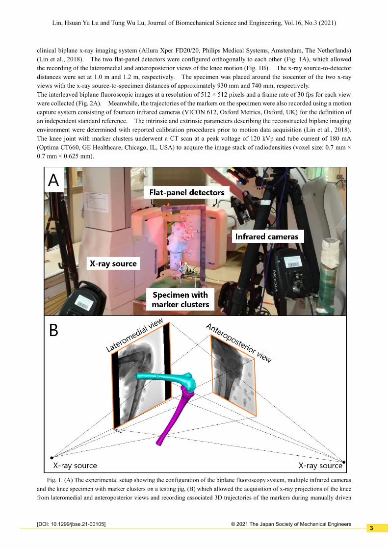
clinical biplane x-ray imaging system (Allura Xper FD20/20, Philips Medical Systems, Amsterdam, The Netherlands)
(Lin et al., 2018). The two flat-panel detectors were configured orthogonally to each other (Fig. 1A), which allowed
the recording of the lateromedial and anteroposterior views of the knee motion (Fig. 1B). The x-ray source-to-detector
distances were set at 1.0 m and 1.2 m, respectively. The specimen was placed around the isocenter of the two x-ray
views with the x-ray source-to-specimen distances of approximately 930 mm and 740 mm, respectively.
The interleaved biplane fluoroscopic images at a resolution of 512 × 512 pixels and a frame rate of 30 fps for each view
were collected (Fig. 2A). Meanwhile, the trajectories of the markers on the specimen were also recorded using a motion
capture system consisting of fourteen infrared cameras (VICON 612, Oxford Metrics, Oxford, UK) for the definition of
an independent standard reference. The intrinsic and extrinsic parameters describing the reconstructed biplane imaging
environment were determined with reported calibration procedures prior to motion data acquisition (Lin et al., 2018).
The knee joint with marker clusters underwent a CT scan at a peak voltage of 120 kVp and tube current of 180 mA
(Optima CT660, GE Healthcare, Chicago, IL, USA) to acquire the image stack of radiodensities (voxel size: 0.7 mm ×
0.7 mm × 0.625 mm).
Fig. 1. (A) The experimental setup showing the configuration of the biplane fluoroscopy system, multiple infrared cameras
and the knee specimen with marker clusters on a testing jig, (B) which allowed the acquisition of x-ray projections of the knee
from lateromedial and anteroposterior views and recording associated 3D trajectories of the markers during manually driven
3

2© 2021 The Japan Society of Mechanical Engineers
Lin, Hsuan Yu Lu and Tung Wu Lu, Journal of Biomechanical Science and Engineering, Vol.16, No.3 (2021)
[DOI: 10.1299/jbse.21-00105]
flexion and extension motions.
Fig. 2. (A) A fluoroscopic image extracted from the region of the femur; (B) an axial slice extracted from the original CT
image stack; (C) the same slice in (B) with voxel intensities within the region of the bone set to a constant; (D) a digitally
reconstructed radiograph (DRR) created using the CT radiodensity model (B); and (E) a pseudo-DRR created using the
homogeneous-density model (C).
2.3 CT-derived volumetric bone models
The CT slices of the cadaveric knee were semi-manually segmented to isolate the regions of the femur and tibia. A
sub-volume that covered the complete region of the target bone was extracted from the original data set. Within the
sub-volume, the radiodensity information of the bone was retained while the voxel intensities exterior to the bone were
set to -1000 (Fig. 2B). To mimic the situation that the radiodensity information of the bone is not available, a new sub-
volume was duplicated and the voxel intensities of the bone were set to a constant value obtained by averaging the
intensities of the bone (Fig. 2C). As a result, the original image sub-volume was taken as the volumetric bone model
containing radiodensity information while the processed sub-volume gave the bone model with homogeneous density.
The 3D geometric models of the femur and tibia were also created from the segmented regions with the marching cubes
algorithm (Lorensen and Cline, 1987). The geometric models were used to determine the anatomical coordinate system
(ACS) of the bone using an automated determination method (Miranda et al., 2010).
2.4 Model-based 3D/2D image registration
The pose parameters of the femur and tibia with respect to the fluoroscopy coordinate system (FCS) were determined
separately by best-matching the model-projections to the fluoroscopic images using the four model-based 3D/2D image
registration configurations. Each of the configurations was established with different bone model types and numbers
of fluoroscopic views, and was termed as homogeneous-density model/single-plane image (HS), radiodensity
model/single-plane image (RS), homogeneous-density model/biplane images (HB), and radiodensity model/biplane
images (RB).
For the radiodensity and homogeneous-density bone models, the original intensity values (i.e., Hounsfield scale)
were converted to the linear attenuation coefficients. To generate a DRR, virtual rays were cast from the x-ray source
4

2© 2021 The Japan Society of Mechanical Engineers
Lin, Hsuan Yu Lu and Tung Wu Lu, Journal of Biomechanical Science and Engineering, Vol.16, No.3 (2021)
[DOI: 10.1299/jbse.21-00105]
through the volume of the bone model to each pixel of the image plane. The attenuation coefficient of voxels of the
bone model went through by virtual rays were accumulated, representing the attenuation of the x-rays as they pass through
the bone (Siddon, 1985). The intensity values of the DRR were thereafter determined using the attenuated information.
To speed up the process of DRR generation, we implemented a ray-tracing with trilinear interpolation method (Otake et
al., 2012). Note that since the radiodensity and homogeneous-density bone models are different in compositions, the
appearances of the resulting DRRs were also visibly different (Figs. 2D and 2E).
The 3D/2D image registration was executed in a framework of an optimization procedure where the design variables
were six pose parameters of the bone and the objective function was the score representing the similarity between the
DRR and the fluoroscopic image. In the current study, if only the lateromedial view fluoroscopic images were used for
the image registration, the genetic algorithm (population size = 50; generation size = 90) (Goldberg, 1989) was used to
search for the optimal pose parameters of the bone model such that the generated DRR best-matched the fluoroscopic
image. In cases when the biplane fluoroscopic images were used, an image registration procedure termed model-based
interleaved biplane fluoroscopy image tracking (MIBFT) (Lin et al., 2020) was implemented to determine the pose
parameters of the bone models.
In order to evaluate objectively the performance of the four tested approaches (i.e., HS, RS, HB and RB), a consistent
similarity metrics Gradient Correlation (GC) (Penney et al., 1998) was used to quantify the image similarity in the current
evaluation. For the computation of GC values, the horizontal Sobel operator was applied on the DRR and the
fluoroscopic images to create gradient images. The normalized cross correlation (NCC) between the two gradient
images was then computed (Penney et al., 1998). In the same fashion, the NCC was also computed from gradient
images created using the vertical Sobel operator. The sum of the two NCCs then gave the final value of GC. A particle
filter-based 2D/2D template registration (Lin et al., 2020) was employed to update the initial pose parameters prior to the
3D/2D image registration.
2.5 Data analysis
The cluster markers were used to determine the standard reference, from which the markers presenting on the CT
were extracted and expressed in the corresponding ACS, and the markers obtained from the motion capture system were
expressed in FCS using a spatial transformation obtained from a calibration procedure (Lin et al., 2018). Co-registration
of the marker clusters in ACS ad FCS gave the reference poses of the bone (Söderkvist and Wedin, 1993). Reference
knee kinematics were obtained from the reference poses of the femur and tibia, from which the angles were decomposed
into flexion(+)/extension(-), abduction(+)/adduction(-) and external(+)/internal(-) rotations following the joint coordinate
system proposed by Grood and Suntay (Grood and Suntay, 1983).
The errors of the bone pose parameters were quantified by comparing the standard references and the registered
poses obtained from the four tested approaches. The errors in the knee kinematics were computed as the differences
between the standard reference kinematics and those derived from the registered bone poses. For each trial (between
41 to 86 frames per trial), mean absolute errors (MAE) and mean errors (ME) were determined for each registration
method, where the MAE represents the overall errors of each motion component and the ME represents the measurement
bias. The means and standard deviations of the MAE and ME across all trials and bones were obtained. The effects
of motion speeds in translation and rotation components on the deviations in the registered bone poses were evaluated.
3. Results
3.1 Bone pose MAE and ME
The four model-based 3D/2D registration configurations were evaluated by quantifying the MAE and ME of the
bone six-degree-of-freedom poses (Table 1 and 2). In general, utilization of biplane fluoroscopic images yielded
accurate spatial bone positions with both MAE and ME below 0.3 mm (Table 1 and 2). The configuration RS with
single-plane image and radiodensity bone model gave out-of-plane translation errors (Tz) up to 4.7 mm in MAE (Table
1) and -4.2 mm in ME (Table 2). The configuration HS yielded maximum MAE of 8.2 mm and 1.5° in translation and
rotation components, respectively (Table 1). When compared to the configurations using homogeneous-density model
for image registration, utilization of radiodensity model diminished translation MAE ranging from 0.1 mm to 3.5 mm
and rotation MAE ranging from 0.2° to 0.7° (Table 1). Similar tendencies were also found in the ME, and the bias of
bone poses was lowered with radiodensity models (Table 2).
5
2© 2021 The Japan Society of Mechanical Engineers
Lin, Hsuan Yu Lu and Tung Wu Lu, Journal of Biomechanical Science and Engineering, Vol.16, No.3 (2021)
[DOI: 10.1299/jbse.21-00105]
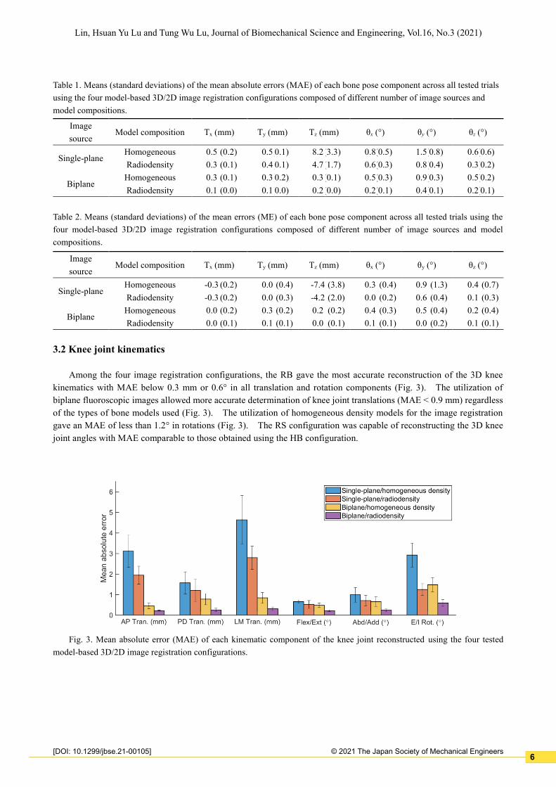
Table 1. Means (standard deviations) of the mean absolute errors (MAE) of each bone pose component across all tested trials
using the four model-based 3D/2D image registration configurations composed of different number of image sources and
model compositions.
Table 2. Means (standard deviations) of the mean errors (ME) of each bone pose component across all tested trials using the
four model-based 3D/2D image registration configurations composed of different number of image sources and model
compositions.
3.2 Knee joint kinematics
Among the four image registration configurations, the RB gave the most accurate reconstruction of the 3D knee
kinematics with MAE below 0.3 mm or 0.6° in all translation and rotation components (Fig. 3). The utilization of
biplane fluoroscopic images allowed more accurate determination of knee joint translations (MAE < 0.9 mm) regardless
of the types of bone models used (Fig. 3). The utilization of homogeneous density models for the image registration
gave an MAE of less than 1.2° in rotations (Fig. 3). The RS configuration was capable of reconstructing the 3D knee
joint angles with MAE comparable to those obtained using the HB configuration.
Fig. 3. Mean absolute error (MAE) of each kinematic component of the knee joint reconstructed using the four tested
model-based 3D/2D image registration configurations.
Image
source Model composition Tx (mm) Ty (mm) Tz (mm) θx (°) θy (°) θz (°)
Single-plane Homogeneous 0.5 (0.2) 0.5 (0.1) 8.2 (3.3) 0.8 (0.5) 1.5 (0.8) 0.6 (0.6)
Radiodensity 0.3 (0.1) 0.4 (0.1) 4.7 (1.7) 0.6 (0.3) 0.8 (0.4) 0.3 (0.2)
Biplane Homogeneous 0.3 (0.1) 0.3 (0.2) 0.3 (0.1) 0.5 (0.3) 0.9 (0.3) 0.5 (0.2)
Radiodensity 0.1 (0.0) 0.1 (0.0) 0.2 (0.0) 0.2 (0.1) 0.4 (0.1) 0.2 (0.1)
Image
source Model composition Tx (mm) Ty (mm) Tz (mm) θx (°) θy (°) θz (°)
Single-plane Homogeneous -0.3 (0.2) 0.0 (0.4) -7.4 (3.8) 0.3 (0.4) 0.9 (1.3) 0.4 (0.7)
Radiodensity -0.3 (0.2) 0.0 (0.3) -4.2 (2.0) 0.0 (0.2) 0.6 (0.4) 0.1 (0.3)
Biplane Homogeneous 0.0 (0.2) 0.3 (0.2) 0.2 (0.2) 0.4 (0.3) 0.5 (0.4) 0.2 (0.4)
Radiodensity 0.0 (0.1) 0.1 (0.1) 0.0 (0.1) 0.1 (0.1) 0.0 (0.2) 0.1 (0.1)
6

2© 2021 The Japan Society of Mechanical Engineers
Lin, Hsuan Yu Lu and Tung Wu Lu, Journal of Biomechanical Science and Engineering, Vol.16, No.3 (2021)
[DOI: 10.1299/jbse.21-00105]
Fig. 4. Scatter plots of absolute translation and rotation deviations of the bones against the translation and rotation speeds.
The solid lines represent the moving average of the deviations over speed intervals. The colors indicate the data obtained
using different model-based 3D/2D image registration configurations (HS: blue; RS: orange; HB: yellow; RB: purple).
3.3 Effects of movement speeds
The scatter plots and the moving averages of the absolute translation and rotation deviations against the movement
speeds showed that the RB configuration gave the lowest pose deviations, the HS showed the highest deviations, and the
performance of the RS and HB were in-between (Fig. 4). As indicated by curves of moving averages, the translation
deviations obtained with four configurations were not associated with translation speeds, but the rotation deviations
obtained with HS, RS and HB were increased with translation speeds (Fig. 4). Overall, the RB configuration was the
one least affected by the fast skeleton motions (Fig. 4), the deviation values of moving averages being consistently below
0.4 mm and 1° over translation speeds from 20 mm/s to 200 mm/s and rotation speeds from 20°/s to 150°/s.
4. Discussion
The current study compared the accuracy of four model-based 3D/2D image registration configurations with different
bone model types and number of fluoroscopic views in measuring 3D knee kinematics during dynamic activities. The
results showed that using single-plane fluoroscopic images for model-based image registration had the greatest errors in
out-of-plane translations whether the homogeneous-density or radiodensity model was used (Table 1), in agreement with
previous findings (Fregly et al., 2005; Postolka et al., 2020; Tsai et al., 2010). It is difficult to compare directly the
current findings with those reported in the literature considering the differences in the algorithms and experiment setup
used. For example, in comparison to previous static evaluations using cadaveric specimen (Tsai et al., 2010), the current
validation study conducting on fast dynamic motions of the knee definitely increased image artifacts associated with
motions and tissue overlapping. The abundant soft tissues covering the current knee specimen have also compromised
the contrast of the bone with respect to the neighboring regions on the fluoroscopic images (Fig. 2A), and thus the effects
were varying over different knee joint angles. Not well-distinguishable bone outlines at certain image frames affected
the measurement accuracy of the bone poses, especially in the out-of-plane translation component while the HS and RS
were used (Tables 1 and 2).
Configuration of stereo x-ray imaging can be achieved via biplane fluoroscopy, from which the pose parameters are
determined by simultaneously best-matching the pair of DRRs to the pair of fluoroscopic images. In such a way, the
out-of-plane motion components (e.g. Tz and θx) for one fluoroscopic view were practically the in-plane motion
components of the other view (e.g. Tx and θz) if two views were configured orthogonally. Given the benefit of the
stereo imaging, the bias (Table 2) and thus the MAE (Table 1) in Tz component (i.e., the original out-of-plane translation)
were largely compensated regardless of the models used for registration. Both the HB and RB methods were shown to
yield registration errors with similar magnitudes among six motion components of the bone (Table 1). These
characteristics enabled a more precise description of the knee joint kinematics especially in joint translation components
(Fig. 3).
7

2© 2021 The Japan Society of Mechanical Engineers
Lin, Hsuan Yu Lu and Tung Wu Lu, Journal of Biomechanical Science and Engineering, Vol.16, No.3 (2021)
[DOI: 10.1299/jbse.21-00105]
The composition of the model content (homogeneous-density vs. radiodensity) was found to affect the accuracy of
the 3D/2D image registration whether single-plane or biplane images were employed for analysis (Tables 1 and 2).
Since the homogeneous-density model neglects the differences of attenuation coefficients between the cortical and
cancellous bones, the DRRs created from the accumulation of such the attenuation information were thereafter visibly
different in details from DRRs generated using the radiodensity model (Figs. 2D and 2E). The radiodensity model-
derived DRRs were generally more similar to the fluoroscopic image as indicated by the lower negative gradient
correlations (Fig. 5), which were intuitively associative to the reduced errors (Table 1) and bias (Table 2). Comparing
the changes of the objective function values at various deviated bone poses, the use of the radiodensity model gave an
objective function more sensitive to the bone pose deviations in the x- and y-axis translations and z-axis rotation (Fig.
5). This feature may be beneficial to lowering the number of local minima in optimization, leading to a more reliable
image registration outcome. As a result, the use of the radiodensity model (RS) was shown to allow more accurate
determination of knee joint angles than those using the homogeneous-density model (HS) and even comparable to those
obtained with the HB (Fig. 3).
The homogeneous-density model implemented in the current study was considered an ideal bone model created from
a hypothetical scenario that the outer surface of the bone geometry was identical to that of the CT-derived radiodensity
model. In fact, in situations when the CT is not available, the models created using MR images (Moewis et al., 2012;
Moro-Oka et al., 2007) or statistical shape models (Tsai et al., 2015) possibly presented morphological discrepancies to
the CT-derived model. It should be noted that the small shape differences have also been demonstrated to affect notably
the registration accuracy especially in the out-of-plane translation component (Moewis et al., 2012).
The effects of the translation speeds and rotation speeds on the registration errors were quantified with the setup of
the current validation study as the fluoroscopic images were acquired at various rotation speeds of the knee joint motion.
The effects of the motion speeds on the registration accuracy were represented in a twofold aspect. The most direct
effect was on any of the image artifacts associated with the bone motions. Since the pulsed fluoroscopy system with a
short pulse interval was employed in the current study, the motion artifacts were considered minimal which might merely
have subtle effects on the registration errors. Another effect was on the inter-frame displacement of the bone, of which
the higher motion speeds would directly enlarge the displacement of the bone between successive image frames. The
more the initial pose parameters were away from the true poses, the lower probability of an optimization process might
converge to the global minimum. The phenomenon has been shown in previous capture range analysis studies (van de
Kraats et al., 2005; Lin et al., 2019; Lin et al., 2014). Although the template matching algorithm was used to diminish
the inter-frame displacement, some of the components of inter-frame displacement were not fully eliminated (Lin et al.,
2020). In general, increase of translation speeds of the bone did not consistently increase the overall translation errors
regardless of the methods used (Fig. 4). However, the HS, RS and HB methods didnot produce consistently low rotation
errors as the rotation speed increased (Fig. 4). It appeared that only the radiodensity model combined with the biplane
images (RB) allowed diminishing the negative effects associated with the fast bone movements.
8

2© 2021 The Japan Society of Mechanical Engineers
Lin, Hsuan Yu Lu and Tung Wu Lu, Journal of Biomechanical Science and Engineering, Vol.16, No.3 (2021)
[DOI: 10.1299/jbse.21-00105]
Fig. 5. Objective function values (negative of gradient correlation) determined with homogeneous-density model (solid
lines) and radiodensity model (dashed lines) at positions -5 mm to +5 mm and -5° to +5° away from the standard reference
bone pose.
5. Conclusions
The accuracy of four model-based 3D/2D image registration configurations with different bone model types and
numbers of fluoroscopic image sources on the determination of 3D kinematics of the knee joint was quantified. The
use of biplane fluoroscopic images improves the measurement accuracy of the six motion components of the bone and
thus the precise description of the six-degree-of-freedom kinematics of the knee. Application of radiodensity bone
models further diminished the measurement errors. The RS configuration enabled the accurate reconstruction of 3D
knee joint angles with errors comparable to those obtained using the HB. The current results suggest that of the four
configurations a biplane fluoroscopy combined with radiodensity models will produce the most accurate 3D bone poses
least influenced by the bone motion speeds, which will be preferred in clinical applications. Further studies with
increased sample size may help provide inferential statistics to confirm further the current findings.
Acknowledgment: The authors gratefully acknowledge Dr. Mei-Ying Kuo and Chien-Chun Kuo and the staff of China
Medical University Hospital for their assistance in the validation experiment.
Conflicts of interest: The authors declare no conflict of interest
Funding: None
References
Akbari-Shandiz, Mohsen, Joseph D. Mozingo, David R. Holmes Iii, and Kristin D. Zhao. 2018. 'An interpolation
technique to enable accurate three-dimensional joint kinematic analyses using asynchronous biplane fluoroscopy',
Medical engineering & physics, 60: 109-16.
Anderst, W., R. Zauel, J. Bishop, E. Demps, and S. Tashman. 2009. 'Validation of three-dimensional model-based tibio-
femoral tracking during running', Medical Engineering and Physics, 31: 10-16.
Banks, S. A., and W. A. Hodge. 1996. 'Accurate measurement of three-dimensional knee replacement kinematics using
single-plane fluoroscopy', IEEE Transactions on Biomedical Engineering, 43: 638-49.
Barré, Arnaud, and Kamiar Aminian. 2018. 'Error performances of a model-based biplane fluoroscopic system for
9

2© 2021 The Japan Society of Mechanical Engineers
Lin, Hsuan Yu Lu and Tung Wu Lu, Journal of Biomechanical Science and Engineering, Vol.16, No.3 (2021)
[DOI: 10.1299/jbse.21-00105]
tracking knee prosthesis during treadmill gait task', Medical & Biological Engineering & Computing, 56: 307-16.
Brainerd, Elizabeth L., David B. Baier, Stephen M. Gatesy, Tyson L. Hedrick, Keith A. Metzger, Susannah L. Gilbert,
and Joseph J. Crisco. 2010. 'X-ray reconstruction of moving morphology (XROMM): precision, accuracy and
applications in comparative biomechanics research', Journal of experimental zoology. Part A, Ecological genetics
and physiology, 313: 262-79.
Flood, Paris D. L., and Scott A. Banks. 2018. 'Automated Registration of 3-D Knee Implant Models to Fluoroscopic
Images Using Lipschitzian Optimization', IEEE Transactions on Medical Imaging, 37: 326-35.
Fregly, B. J., H. A. Rahman, and S. A. Banks. 2005. 'Theoretical accuracy of model-based shape matching for measuring
natural knee kinematics with single-plane fluoroscopy', Journal of Biomechanical Engineering, 127: 692-99.
Giphart, J. Erik, Christopher A. Zirker, Casey A. Myers, W. Wesley Pennington, and Robert F. LaPrade. 2012. 'Accuracy
of a contour-based biplane fluoroscopy technique for tracking knee joint kinematics of different speeds', Journal of
Biomechanics, 45: 2935-38.
Goldberg, D. E. 1989. Genetic Algorithms in Search, Optimization, and Machine Learning (Addison-Wesley: Boston).
Gray, Hans A., Shanyuanye Guan, Lucas T. Thomeer, Anthony G. Schache, Richard de Steiger, and Marcus G. Pandy.
2019. 'Three-dimensional motion of the knee-joint complex during normal walking revealed by mobile biplane x-
ray imaging', Journal of orthopaedic research : official publication of the Orthopaedic Research Society, 37: 615-
30.
Grood, E. S., and W. J. Suntay. 1983. 'A joint coordinate system for the clinical description of three-dimensional motions:
application to the knee', Journal of Biomechanical Engineering, 105: 136-44.
Guan, Shanyuanye, Hans A. Gray, Farzad Keynejad, and Marcus G. Pandy. 2016. 'Mobile Biplane X-Ray Imaging
System for Measuring 3D Dynamic Joint Motion During Overground Gait', IEEE Transactions on Medical Imaging,
35: 326-36.
Haque, Md A., W. Anderst, S. Tashman, and G. E. Marai. 2012. 'Hierarchical model-based tracking of cervical vertebrae
from dynamic biplane radiographs', Medical Engineering and Physics.
Ivester, John C., Adam J. Cyr, Michael D. Harris, Martin J. Kulis, Paul J. Rullkoetter, and Kevin B. Shelburne. 2015. 'A
Reconfigurable High-Speed Stereo-Radiography System for Sub-Millimeter Measurement of In Vivo Joint
Kinematics', Journal of Medical Devices, 9: 041009-09-7.
Li, G., S. K. Van de Velde, and J. T. Bingham. 2008. 'Validation of a non-invasive fluoroscopic imaging technique for the
measurement of dynamic knee joint motion', Journal of Biomechanics, 41: 1616-22.
Lin, Cheng-Chung, Chia-Lin Chang, Ming Lu, Tung-Wu Lu, and Ching-Ho Wu. 2018. 'Quantification of three-
dimensional soft tissue artifacts in the canine hindlimb during passive stifle motion', BMC Veterinary Research, 14:
389.
Lin, Cheng-Chung, Jia-Da Li, Tung-Wu Lu, Mei-Ying Kuo, Chien-Chung Kuo, and Horng-Chaung Hsu. 2018. 'A model-
based tracking method for measuring 3D dynamic joint motion using an alternating biplane x-ray imaging system',
Medical Physics, 45: 3637-49.
Lin, Cheng-Chung, Tung-Wu Lu, Jia-Da Li, Mei-Ying Kuo, Chien-Chung Kuo, and Horng-Chaung Hsu. 2020. 'An
automated three-dimensional bone pose tracking method using clinical interleaved biplane fluoroscopy systems:
application to the knee', Applied Sciences, 10: 8426.
Lin, Cheng-Chung, Tung-Wu Lu, Ting-Fang Shih, Tsung-Yuan Tsai, Ting-Ming Wang, and Shih-Jung Hsu. 2013.
'Intervertebral anticollision constraints improve out-of-plane translation accuracy of a single-plane fluoroscopy-to-
CT registration method for measuring spinal motion', Medical Physics, 40: 031912.
Lin, Cheng-Chung, Tung-Wu Lu, Ting-Ming Wang, Chao-Yu Hsu, and Ting-Fang Shih. 2014. 'Comparisons of surface
vs. volumetric model-based registration methods using single-plane vs. bi-plane fluoroscopy in measuring spinal
kinematics', Medical engineering & physics, 36: 267-74.
Lin, Cheng-Chung, Shuo Zhang, Chao-Yu Hsu, Jens Frahm, Tung-Wu Lu, and Ting-Fang Shih. 2019. 'Measuring three-
dimensional tibiofemoral kinematics using dual-slice real-time magnetic resonance imaging', Medical Physics, 46:
4588-99.
List, Renate, Barbara Postolka, Pascal Schütz, Marco Hitz, Peter Schwilch, Hans Gerber, Stephen J. Ferguson, and
William R. Taylor. 2017. "A moving fluoroscope to capture tibiofemoral kinematics during complete cycles of free
level and downhill walking as well as stair descent." In PLoS ONE, e0185952.
Lorensen, William E., and Harvey E. Cline. 1987. 'Marching Cubes: A High Resolution 3D Surface Construction
10

2© 2021 The Japan Society of Mechanical Engineers
Lin, Hsuan Yu Lu and Tung Wu Lu, Journal of Biomechanical Science and Engineering, Vol.16, No.3 (2021)
[DOI: 10.1299/jbse.21-00105]
Algorithm', Computer Graphics (ACM), 21: 163-69.
Markelj, P., D. Tomaževič, B. Likar, and F. Pernuš. 2012. 'A review of 3D/2D registration methods for image-guided
interventions', Medical Image Analysis, 16: 642-61.
Miranda, Daniel L., Michael J. Rainbow, Evan L. Leventhal, Joseph J. Crisco, and Braden C. Fleming. 2010. 'Automatic
determination of anatomical coordinate systems for three-dimensional bone models of the isolated human knee',
Journal of Biomechanics, 43: 1623-26.
Moewis, P., N. Wolterbeek, G. Diederichs, E. Valstar, M. O. Heller, and W. R. Taylor. 2012. 'The quality of bone surfaces
may govern the use of model based fluoroscopy in the determination of joint laxity', Medical engineering & physics.
Moro-Oka, T. A., S. Hamai, H. Miura, T. Shimoto, H. Higaki, B. J. Fregly, Y. Iwamoto, and S. A. Banks. 2007. 'Can
magnetic resonance imaging-derived bone models be used for accurate motion measurement with single-plane three-
dimensional shape registration?', Journal of Orthopaedic Research, 25: 867-72.
Otake, Yoshito, Mehran Armand, Robert S. Armiger, Michael D. Kutzer, Ehsan Basafa, Peter Kazanzides, and Russell
H. Taylor. 2012. 'Intraoperative image-based multiview 2D/3D registration for image-guided orthopaedic surgery:
incorporation of fiducial-based C-arm tracking and GPU-acceleration', IEEE Transactions on Medical Imaging, 31:
948-62.
Penney, Graeme P, Jürgen Weese, John A Little, Paul Desmedt, and Derek LG Hill. 1998. 'A comparison of similarity
measures for use in 2-D-3-D medical image registration', IEEE transactions on medical imaging, 17: 586-95.
Pitcairn, Samuel, Bryson Lesniak, and William Anderst. 2018. 'In vivo validation of patellofemoral kinematics during
overground gait and stair ascent', Gait & Posture, 64: 191-97.
Postolka, Barbara, Renate List, Benedikt Thelen, Pascal Schütz, William R. Taylor, and Guoyan Zheng. 2020. 'Evaluation
of an intensity-based algorithm for 2D/3D registration of natural knee videofluoroscopy data', Medical engineering
& physics.
Söderkvist, Inge, and Per-Åke Wedin. 1993. 'Determining the movements of the skeleton using well-configured markers',
Journal of Biomechanics, 26: 1473-77.
Siddon, R. L. 1985. 'Fast calculation of the exact radiological path for a three-dimensional CT array', Medical Physics,
12: 252-55.
Smoger, Lowell M., Kevin B. Shelburne, Adam J. Cyr, Paul J. Rullkoetter, and Peter J. Laz. 2017. 'Statistical shape
modeling predicts patellar bone geometry to enable stereo-radiographic kinematic tracking', Journal of
Biomechanics, 58: 187-94.
Stentz-Olesen, K., E. T. Nielsen, S. De Raedt, P. B. Jørgensen, O. G. Sørensen, B. L. Kaptein, M. S. Andersen, and M.
Stilling. 2017. 'Validation of static and dynamic radiostereometric analysis of the knee joint using bone models from
CT data', Bone & joint research, 6: 376-84.
Tsai, T. Y., J. S. Li, S. Wang, P. Li, Y. M. Kwon, and G. Li. 2015. 'Principal component analysis in construction of 3D
human knee joint models using a statistical shape model method', Comput Methods Biomech Biomed Engin, 18:
721-9.
Tsai, T. Y., T. W. Lu, C. M. Chen, M. Y. Kuo, and H. C. Hsu. 2010. 'A volumetric model-based 2D to 3D registration
method for measuring kinematics of natural knees with single-plane fluoroscopy', Medical Physics, 37: 1273-84.
van de Kraats, Everine B., Graeme P. Penney, Dejan Tomazevic, Theo van Walsum, and Wiro J. Niessen. 2005.
'Standardized evaluation methodology for 2-D-3-D registration', IEEE Transactions on Medical Imaging, 24: 1177-
89.
You, B. M., P. Siy, W. Anderst, and S. Tashman. 2001. 'In vivo measurement of 3-D skeletal kinematics from sequences
of biplane radiographs: Application to knee kinematics', IEEE Transactions on Medical Imaging, 20: 514-25.
Wilson, D.R., J.D. Feikes, A.B. Zavatsky, and J.J. O’Connor. 2000. 'The components of passive knee movement are
coupled to flexion angle', Journal of Biomechanics, 33: 465-473.
11