effects of hyperbaric oxygen therapy … thesis/laurie massey thesis.pdf · hyperbaric oxygen...
TRANSCRIPT
EFFECTS OF HYPERBARIC OXYGEN THERAPY
ON PHYSIOLOGIC CHANGES IN RATS
FOLLOWING SIMULATED
MICROGRAVITY
The members of the Committee approve the master’s Thesis of Laurie LaShonn Massey
Judy R. Wilson Supervising Professor ______________________________________
Abu B. Yilla ______________________________________
Amy M. Ables ______________________________________

Copyright © by Laurie LaShonn Massey 2005
All Rights Reserved

EFFECTS OF HYPERBARIC OXYGEN THERAPY
ON PSYSIOLOGIC CHANGES IN RATS
FOLLOWING SIMULATED
MICROGRAVITY
by
LAURIE L. MASSEY
Presented to the Faculty of the Graduate School of
The University of Texas at Arlington in Partial Fulfillment
of the Requirements
for the Degree of
MASTER OF SCIENCE IN EXERCISE PHYSIOLOGY
THE UNIVERSITY OF TEXAS AT ARLINGTON
August 2005

ACKNOWLEDGEMENTS
I would like to thank Dr. Wilson for allowing me to become a part of this very
important project and providing guidance throughout the process. Thank you Dr. Yilla
for helping me understand the right way and the wrong way to conduct statistical
analyses. And thank you Dr. Ables for your incite and advice, it was more valuable that
you may believe. I would also like to recognize all the faculty and staff in the
Kinesiology department for their dedication and commitment to their students and their
attention to quality.
Thank you Mom and Dad for giving me my life. You both are wonderful
parents.
Most importantly, thank you Jon for the sacrifices you have made over the last
two years so I could attend school full time and for your love and support throughout
this process.
July 20, 2005
iv

ABSTRACT
EFFECTS OF HYPERBARIC OXYGEN THERAPY
ON PSYSIOLOGIC CHANGES IN RATS
FOLLOWING SIMULATED
MICROGRAVITY
Publication No. ______
Laurie LaShonn Massey, MS
The University of Texas at Arlington, 2005
Supervising Professor: Judy R. Wilson, PhD.
The purpose of the present investigation was to examine the effects of
hyperbaric oxygen therapy on: 1) bone mineral density (BMD; cortical and cancellous)
of the mid-diaphysis of the femur, 2) mechanical characteristics (ultimate force (UF)
and fracture force (FF)) of the mid-diaphysis of the femur, 3) bone mineral resorption
and deposition, and 4) vasoconstrictive properties of the thoracic aorta following four
weeks of hind limb suspension in five-month-old, male Sprague-Dawley rats. Forty rats
were randomly divided into aging controls (AC, (n =10), aging control with hyperbaric
therapy (AC-HBO, n =10), hindlimb suspended (HLS, n =10), and hindlimb suspended v

with hyperbaric therapy (HLS-HBO, n =10) groups for 4 wks. Groups receiving HBO
were placed in a cage that was fitted for the animal hyperbaric chamber; HLS was
maintained during HBO treatments. HBO groups were treated for 90 minutes, 6 d/wk
(1×d) for a total of 24 treatments. After 28 d of HLS, animals were sacrificed under
isoflourane anesthesia, thoracic aorta segments were isolated, and the femurs were
removed for analysis. Cortical and cancellous BMD was determined using peripheral
quantitative computed tomography. Bone markers for resorption and deposition were
evaluated using commercially available assays to assess urinary content of
deoxypyridinoline (DPD) and serum content of osteocalcin (OC), respectively.
Mechanical testing of the femur was determined using a three-point bending test. Load-
displacement curves were constructed to determine UF and FF. Maximal
vasoconstriction (MAX) and the sensitivity (EC50) of aortic rings were determined in
response to increasing concentrations of phenylepherine (PHE) administered
cumulatively in vessel baths at 10×10-10-10×10-4 mol/l. While HLS and HBO appeared
to result in a decrease in BMD, these differences were not significant. In addition, HLS
and HBO did not have significant effects on biochemical bone markers or mechanical
strength. The maximal vasoconstriction response to PHE was decreased with HLS,
however, it was decreased to an even greater extent in the HLS-HBO group.
Hyperbaric oxygen therapy tended to decrease the levels of BMD and significantly
decreased the vasoconstrictive ability of the thoracic aorta suggesting at this treatment
level, HBO should not be used as an intervention during simulated spaceflight.
vi
TABLE OF CONTENTS
ACKNOWLEDGEMENTS....................................................................................... iv ABSTRACT .............................................................................................................. v LIST OF ILLUSTRATIONS..................................................................................... x LIST OF TABLES..................................................................................................... xi Chapter 1. INTRODUCTION……… ............................................................................. 1 1.1 Purpose……………… ............................................................................ 3
1.2 Hypothesis.………… .............................................................................. 3
1.3 Definition of Terms… ............................................................................. 4
1.4 Delimitations…..…….............................................................................. 5
1.5 Assumptions……………… .................................................................... 5
1.6 Limitations.………….............................................................................. 5
2. REVIEW OF LITERATURE........................................................................ 6 2.1 Bone Remodeling……………… ............................................................ 6
2.2 Regulation of Calcium Homeostasis……………… ............................... 7
2.3 Calcium Balance in Humans Following Microgravity............................ 7 2.4 Simulated Microgravity in Rats............................................................... 9
2.5 Bone Mineral Changes in Rats Following Simulated Microgravity ....... 10
vii
2.6 Bone Remodeling in Rats Following Simulated Microgravity ............... 11
2.7 Systemic and Local Factors Contributing to Bone Loss ......................... 12
2.8 Biochemical Markers of Bone Resorption .............................................. 12
2.9 Biochemical Markers of Bone Deposition .............................................. 13
2.10 Mechanical Properties of Bone.............................................................. 14
2.11 Vasoconstrictor Properties Following Simulated Microgravity ............ 15
2.11.1 Aorta………. .......................................................................... 16
2.11.2 Arterioles…. ........................................................................... 17
2.12 Body Mass…………. ............................................................................ 18
2.13 Hyperbaric Therapy…........................................................................... 18
2.13.1 Bone………............................................................................ 20
2.13.2 Vessels.…. .............................................................................. 21
3. METHODS………………….……………………………………...……..... 23
3.1 Animals……………................................................................................ 23 3.2 Hyperbaric Treatment.............................................................................. 25 3.3 Peripheral Quantitative Computed Tomography..................................... 27 3.4 Biochemical Bone Markers…. ................................................................ 27 3.5 Mechanical Testing….............................................................................. 28 3.6 Vascular Responses……. ........................................................................ 29 3.7 Statistical Analysis…............................................................................... 30 4. RESULTS……………….............................................................................. 33 4.1 Bone Mineral Density.............................................................................. 33 viii
4.2 Biochemical Markers of Bone Remodeling ............................................ 35 4.3 Mechanical Properties of Bone................................................................ 37 4.4 Vasoconstrictive Properties ..................................................................... 38 4.5 Body Mass…………. .............................................................................. 40 5. DISCUSSION……….………....................................................................... 42 5.1 Bone Mineral Density.............................................................................. 42 5.2 Biochemical Markers of Bone Remodeling ............................................ 43 5.3 Mechanical Properties of Bone................................................................ 44 5.4 Hyperbaric Therapy and Bone................................................................. 45 5.5 Vasoconstrictive Properties ..................................................................... 46 5.6 Body Mass…………. .............................................................................. 47 5.7 Conclusions………….............................................................................. 48 REFERENCES .......................................................................................................... 50 BIOGRAPHICAL INFORMATION......................................................................... 58
ix

LIST OF ILLUSTRATIONS
Figure Page 3.1 Hindlimb suspended rat in cage ...................................................................... 25 3.2 Hyperbaric treatment cage .............................................................................. 26 3.3 Experimental design scheme........................................................................... 31 4.1 Cancellous bone mineral density..................................................................... 34 4.2 Cortical bone mineral density ......................................................................... 35 4.3 Deoxypyridinoline........................................................................................... 36 4.4 Osteocalcin ...................................................................................................... 36 4.5 Ultimate force.................................................................................................. 37 4.6 Fracture force .................................................................................................. 38 4.7 EC50 ................................................................................................................. 39 4.8 Maximal vasoconstriction ............................................................................... 40 4.9 Body mass at day 28 of HLS........................................................................... 41
x

LIST OF TABLES
Table Page 3.1 Results for MANOVA Assumptions Testing ................................................. 32 4.1 Bone Mineral Density ..................................................................................... 34 4.2 Biochemical Bone Markers............................................................................. 35 4.3 Mechanical Properties ..................................................................................... 37 4.4 Vasoconstrictive Properties............................................................................. 39 4.5 Body Mass....................................................................................................... 41
xi

CHAPTER 1
INTRODUCTION
The prospect of sending a man to Mars is getting closer to reality, however
extreme duration space flight poses many physiological challenges. Exposure to
microgravity elicits several physical responses such as, fluid shifts from the lower
extremities to upper body, repositioning of certain organs, musculoskeletal unloading,
and a lack of stimulus to the vestibular system (40, 52). These adjustments can lead to
physiological adaptations that are detrimental to health such as decreases in mineral
density in weight bearing bones (43) and orthostatic intolerance (15, 16, 37, 41, 54).
Significant decreases in bone mineral density (BMD) have been observed
following spaceflight, however these changes have not been great enough to end a
mission (12). There is concern that missions over a year in length will increase the risk
of bone fractures later in life (12). Therefore, determining a means of reducing the
effect of weightlessness on BMD is paramount to the success of long term space flight.
It has been hypothesized that decreases in BMD are a result of a negative calcium
balance, neural, hormonal, and mechanical factors, changes of stress on bone, and
alterations in circulatory dynamics (43, 44, 55).
It is unclear which mechanism or mechanisms contribute to bone loss, thereby
making it difficult to determine a treatment that would attenuate this decrease.
Countermeasures that have been implemented to reduce the effects of microgravity are
1

calcium and phosphate supplementation, exercise regimens, and bisphosphonates.
These steps, however, have met with little or no success. Research (10, 49) has
suggested that the reduced blood supply to the bone may alter bone remodeling.
Increasing the blood supply by lower body negative pressure coupled with 40 minutes
of treadmill exercise six days a week has been shown to decrease bone resorption
following 30 days of bed rest (49), however more research is needed to determine its
effects on BMD.
Hyperbaric oxygen therapy (HBO) is a clinical treatment used most often in
healing chronic wounds. Two primary mechanisms of action are hyperoxygenation and
vasoconstriction. The mechanisms result from placing an individual under greater than
atmospheric pressure and by breathing 100 % oxygen. The data from numerous studies
have demonstrated the efficacy in healing chronic non-healing wounds of both soft
tissue and bone, however few studies have been conducted to determine the effects of
HBO on changes in bone mineral density and no research has been done to determine
the effects of HBO during simulated microgravity.
While on Earth, approximately 60% of bodily fluids are contained below the
waist. During space flight a cephalic fluid shift occurs causing fluids to redistribute
evenly throughout the body (41). This shifting of fluid causes the kidneys to increase
diuresis resulting in a reduced circulating blood volume. A change in the baroreceptor
reflex also occurs as a result of exposure to microgravity (37, 41, 54) which, along with
decreased vascular responsiveness to sympathetic output, have been considered as
mechanisms for decreased orthostatic responsiveness (15, 16). The combination of
2

these changes can lead to orthostatic intolerance and syncope when an astronaut returns
to a 1g environment. While standing, blood volume is redistributed to the lower body
and blood pooling occurs in the lower extremities. The resistance vessels are unable to
constrict creating inadequate venous return, which, along with the lower blood volume
and a decreased baroreceptor response results, in the inability to maintain blood
pressure in the upright position.
Vascular responsiveness to hyperoxia and hyperbaric oxygen therapy has been
investigated. Rousseau et al. (46) found that breathing 100% oxygen increases systemic
vascular resistance. Hyperbaric oxygen therapy in dogs produced a 30% increase in
systemic vascular resistance (4). No studies have examined vascular responsiveness
after exposure to microgravity or simulated microgravity, therefore the effects of
hyperoxia and hyperbaric oxygen therapy following microgravity and simulated
microgravity need to be examined.
1.1 Purpose
The purpose of this study was to determine the effects of hyperbaric oxygen
therapy on bone mineral loss, mechanical strength of bone, biochemical markers of
bone remodeling, and vascular contractility after four weeks of simulated microgravity
in adult rats.
1.2 Hypothesis
Hyperbaric oxygen therapy will attenuate the rate of bone mineral loss as a
result of simulated microgravity, therefore minimizing the effects on bone remodeling
and mechanical strength.
3

Hyperbaric oxygen therapy will enhance vasocontractility following simulated
microgravity.
1.3 Definition of Terms
Remodeling: A process in which bone mineral is removed and then replaced.
Osteocyte: Mature bone cell that maintains the bone matrix.
Osteoblast: Cell that produces new bone matrix by making and releasing organic
materials.
Osteoclast: Cell that removes bone matrix.
Resorption: The removal of bone matrix.
Deposition: The deposition of new bone matrix.
Cortical Bone: Dense outer layer of bone, also known as compact bone.
Cancellous bone: Inner layer of bone, adjacent to and less dense than cortical bone.
Microgravity: An environment where there is little gravitational force.
Hyperoxia: Oxygen breathing at levels greater than 21%.
Hyperbaric: Pressure greater than 1 atmosphere.
Ultimate Force: Highest load obtained at the instant of material damage.
Fracture Force: Load at which material experiences catastrophic failure or complete
fracture of the bone.
4

1.4 Delimitations
No control group for treatments of 100% oxygen at ambient pressure was used.
No control group for 21% oxygen at 2.5 ATA was used.
1.5 Assumptions
Hindlimb suspension mimics physiologic responses incurred within a
microgravity environment. Physiologic responses in HLS rats are similar to those
incurred by humans during space travel. Mechanism for bone loss is due to decreased
oxygen supply to the bone tissues. Aorta and femoral & carotid arteries responses are
the same as peripheral vessels.
1.6 Limitations
Gravity is still present during our experiment. Unloading of only the hindlimbs
will occur. Launch, landing, and transport to and from launching pad are not present.
Rats are frequently handled. Rats may be able to put pressure on hind limbs
intermittently while in the chamber cage.
5
CHAPTER 2
REVIEW OF LITERATURE
2.1 Bone Remodeling
Adult bone is a highly vascular, dynamic, and living tissue that is constantly
undergoing remodeling. The rate at which bone remodels varies with age, nutritional
status, activity level, and other environmental factors such as microgravity. The rate of
remodeling also varies regionally within the skeleton and locally within the bone. For a
young adult, approximately 1/5th of the total skeleton is recycled and replaced annually
(33).
During remodeling osteoclast and osteoblast actions are coupled, bone
resorption and deposition occur within the same area of the bone surface. Normally
remodeling does not cause a large change in bone structure because the rates of
resorption and deposition are close. However, after age 35 the osteoblast activity lags
behind osteoclast activity resulting in a slow decrease in bone mass (6).
Remodeling of bone occurs in distinct phases where the old bone is removed
then new bone is placed in the cavity. First, lining cells react to chemical signals which
expose the bone. Mononuclear osteoclast precursors are attracted to the area by
chemotaxis to congregate and fuse into multinuclear osteoclasts (6). Osteoclasts then
dissolve the old bone and disappear. Next osteoblasts enter the cavity and deposit new
bone matrix, which later mineralizes. After the new bone matrix is deposited some
6

osteoblasts die, some become buried in new bone and become osteocytes, while others
transform into lining cells (6). These processes occur within both cortical and
cancellous bone.
2.2 Regulation of Calcium Homeostasis
Calcium plays an important role in many physiological functions within the
body such as muscular contraction, transmission of signals across neural junctions,
blood clotting, and structural support (24). Serum levels of calcium lie within a narrow
range and are closely regulated through feedback mechanisms. The skeleton acts as a
reservoir where calcium can be stored or resorbed to maintain blood concentrations.
Absorption of calcium through the intestine and excretion by the kidneys is also
important mechanisms for maintaining the homeostatic balance of calcium. Moderation
of serum concentrations of calcium is achieved through secretion of parathyroid
hormone (PTH) and calcitonin. The parathyroid gland secretes PTH into the blood
stream when blood concentrations of calcium fall below normal. Parathyroid hormone
stimulates osteoclast activity, increases intestinal absorption of calcium, and decreases
the rate of calcium excretion by the kidneys (33). The thyroid secretes calcitonin into
the bloodstream when blood calcium concentrations are above normal. Calcitonin
inhibits osteoclast activity and increases the rate of calcium secretion through the
kidneys (33).
2.3 Calcium Balance in Humans Following Microgravity
Travel within the microgravity environment of space causes many physiological
responses that can be detrimental to astronauts’ health upon their return to earth. One
7

aspect that is of great concern is changes in bone deposition and resorption rates that
can lead to losses in bone mineral density (BMD). Early human spaceflight studies
have demonstrated increased calcium excretion through the urine (43, 44, 55) and feces
(43, 44). During the Skylab II mission, crewmen consumed a controlled diet and
collected urine and fecal excrement to determine changes in calcium balance during
spaceflight. Urinary calcium increased steadily to approximately double the pre-flight
values by day 28 while fecal calcium did not change significantly. The result was a
mean calcium loss of 50 mg/day during the last 16 days of flight (55). To further
demonstrate the changes in calcium metabolism, Rambaut et al. (44) combined data
from several orbital spaceflight missions occurring over 28, 59, and 84 days in which
diet was controlled and excrement was monitored. During the first 28 days calcium
excretion in the urine increased to approximately 100% of pre-flight levels then leveled
off for the remainder of the flight. Fecal calcium showed an initial decline at 14 days
then increased steadily throughout the 84 day period to approximately 300 mg/day
above pre-flight values (43). Calcium balance showed a steady decline throughout the
84 day period to a maximum deficit of approximately 300 mg/day (43).
It is hypothesized that a negative calcium balance is related to the decreases in
bone mineral density observed as a result of microgravity. Bone mineral density has
been shown to decrease at rates between approximately 1%-2% a month in specific
bones (27, 58). The decreases in bone mineral density have been observed primarily in
the weight bearing bones of the calcaneus, lumbar spine, and tibia (11, 43, 59).
8

2.4 Simulated Microgravity in Rats
Conducting research during space flight can pose many challenges. Space
missions are limited in number and duration, require many man hours, and are
expensive. The development of a ground-based model to simulate microgravity is
important in the study of the effects of the microgravity environment incurred in
humans during space travel. The benefits of ground based models are: (a) scheduling
times for experiments are not limited to the number of space missions, (b) not as many
precautions are needed during experimental manipulations on Earth, (c) ground-based
experiments are more economical, (d) experimental duration is not limited to mission
duration, (e) measurements can be made and tissue samples can be obtained at several
points during the experiment, and (f) experiments can be extended or repeated on a
regular basis (38).
The physiological responses to space travel include fluid shifts, repositioning of
certain organs, musculoskeletal unloading, and lack of stimulus to the vestibular system
(38). Human models have successfully simulated these effects by using bed rest and
bed rest with a head-down tilt. Human ground-based models have their own
limitations, therefore, animal models have been developed which successfully simulate
the effects of space flight. A common animal model is the hindlimb suspended (HLS)
rat model which has been approved by National Air and Space Administration (NASA).
The HLS model requires that the rat’s hindlimbs be suspended so the body axis creates
approximately a 30˚ angle with the floor. This allows for normal weight bearing on the
forelimbs (38). The HLS model presents minimal stress to the animal based upon
9

hormonal levels and body weight measured (38). Suspension is achieved by using a tail
traction harness which allows the rat freedom to move, eat, and groom.
2.5 Bone Mineral Changes in Rats Following Simulated Microgravity
Bone mineral loss during HLS in the rat model has been shown to occur in the
femur, lumbar vertebrae, and tibia. Increased bone mineral deposition has been
observed in the humerus, skull, and mandible. Arnaud et al. (1) suspended 6-month-old
rats for 4 weeks and measured bone mineral content in situ of the whole body and the
femur and skull separately using dual x-ray absorptometry (DEXA) technique. Bone
mineral content, when corrected for body weight, was 14% and 7% lower in whole
body and femur, respectively. There was no significant change in the skull bone
mineral content (1). Roer and Dillaman (45) calculated the differences in wet, dry, and
ash weights of the forelimbs, hindlimbs, and the skull during 1-, 2-, and 3-week HLS
studies on 28-day-old rats. For the 3-week experiment, when corrected for body
weight, the dry and ash weights of the femur and tibia were significantly lower (p <
0.05 for dry tibia; p < 0.001 for dry and ash femur, and ash tibia) in the HLS group.
The dry weights of the forelimbs were greater (p < 0.01) in the HLS group, however
there was no difference in ash weight, while the dry and ash weights of the skull and
mandible were greater (p < 0.001) in the HLS group (45).
Dehority et al. (14) measured the bone weight using regression analysis in 6-
month-old HLS and control rats in three separate experiments of 1-, 3-, and 5 week
durations. There were significant decreases (p < 0.05) in the regression slopes for the
fat-free weights of the femur and lumbar vertebrae but not for the humerus. Globus et
10

al. (21) measured changes in calcium in the humerus, mandible, tibia, and lumbar
vertebrae after 15 days of HLS. The tibia and lumbar vertebrae of the HLS rats
contained 86.2 ± 2.5% and 75.5 ± 3.5% (mean ± SD) calcium of the control values
respectively. There were no differences in calcium content in the mandible and
humerus between the HLS and control groups (21).
Bloomfield et al. (5) used peripheral computed tomography (pQTC) to compare
the differences in BMD of the mid-diaphysis and proximal metaphysis of the tibia in
six-month old rats after 7-, 14-, 21-, and 28-days of HLS. (Peripheral quantitative
tomography separates cortical and cancellous bone mineral compartments and provides
a real measure volumetric density (mg/cm3).) When compared to 0-day control both the
28-day HLS and 28-control exhibited a similar increase in cortical BMD of the
proximal tibia, +5.7 % and +4.0 % respectively. Cancellous BMD decreased by 21%
after 28-days HLS in proximal tibia when compared to 0-day control. Greater BMD
was observed at the tibial mid-diaphysis at days 14 – 28 of HLS. No significant
difference was observed when BMD values were compared to 28-day control.
2.6 Bone Remodeling in Rats Following Simulated Microgravity
Mean periostial apposition rates (MAR) in adult rats (6-months-old) after 2 and
4 weeks of HLS were reduced by 65% and 85% respectively (14). In the same study
mean periostial bone formation rate (BFR) of the tibiofibular junction (TFJ) and the
tibia mid-shaft were decreased by approximately 80% (p < 0.001), and there were no
significant changes in BFR in the humerus (14). In a separate study periostial MAR
and BFR were 61% and 90% lower at 21 days HLS at 21 days in the tibial mid-
11

diaphysis; no significant differences in MAR and BFR were observed in the mid-tibia
and humeral diaphysis (5). In cancellous bone, a decrease of 33% and 69% (p < 0.05)
in MAR and BFR respectively was observed after 2 weeks of HLS in adult rats (14).
The percent surface area occupied by cancellous bone occupied by osteoblasts
decreased by 66% (p < 0.05), there were no significant changes in osteoclast numbers or
cancellous bone volume (14). These results suggest that a decrease in BFR or
osteoblast activity may account for the net bone loss seen during simulated
microgravity.
2.7 Systemic and Local Factors Contributing to Bone Loss
The regional decreases in hindlimb mass and bone mineral density and the
increases or stabilization of the skull and forelimb mass and bone mineral density may
be attributed to an alteration in perfusion of bone as a result of the cephalad fluid shift
during HLS rather than systemic factors that regulate bone mineral homeostasis (1, 14,
21, 45). In a study by Dehority et al. (14), changes in PTH were not observed after 5
weeks of HLS, however there were regional changes in MAR and BFR, therefore the
data did not indicate that systemic factors contribute to bone loss (14). It is believed
that if systemic factors influenced bone loss during simulated microgravity, then
changes in BMD would appear in all areas of the body.
2.8 Biochemical Markers of Bone Resorption
Biochemical markers can be used as an indicator of the rate of bone turnover.
During bone formation type I collagen is laid down then bonded together by pyridinium
and pyrrolic cross-links for structural integrity (29). During resorption the collagen is
12

broken down by osteoclasts then pyridinoline (PYD) and deoxypyridinoline (DPD) are
released. Pyridinoline and DPD are neither metabolized nor absorbed from the diet (18)
and can be measured in the blood or urine. Low levels of resorption markers, such as
DPD, reflect low rate of bone resorption and high levels of DPD reflect a high rate of
bone resorption activity. A high rate of bone loss can lead to a decrease in bone mineral
density and osteoporosis. Moderate correlations have been observed between high
concentrations of bone resorption markers and bone mineral loss (18, 53). Caillot-
Agusseau et al. 1998 and 2000 (8, 9) observed a 50 % and 54 % increase in DPD in
cosmonauts during 21 and 180 days of spaceflight respectively. Kurokouchi et al. (30)
measured changes in tartrate-resistant acid phosphatase (TRAP), a bone resorption
marker, in five-week old rates during HLS. The results indicated a significant increase
in TRAP at days one and three, but returned to pre-HLS level at day five and remained
there throughout the duration of the study.
2.9 Biochemical Markers of Bone Deposition
Osteocalcin (OC) is a non-collagen protein excreted by exclusively osteoblasts
into the bone matrix during bone deposition (56). Some osteocalcin escapes the bone
matrix entering the bloodstream and is a good indicator of osteoblast activity
(IMMUTOPICS). The literature suggests that bone formation is reduced during space
travel and simulated microgravity. Caillot-Agusseau et al. (8, 9) measured changes in
OC during 21 and 180 day space missions in cosmonauts. Osteocalcin levels were
decreased by 18 % and 27 % during 21 and 180 days respectively when compared to
pre-flight levels. Kurokouchi et al. (30) observed a decrease in OC levels from day
13

three to day 14 in 5-week old HLS rats. Patterson-Buckendahl et al. (42) measured
weekly changes in OC levels for four weeks during HLS in 6.5 week-old rats.
Osteocalcin for the control rats gradually decreased throughout the four week period.
The HLS rats demonstrated a significantly greater decrease in OC than the control for
week one and leveled out at week two but remained significantly lower than the control
value. There was no significant difference in OC levels for weeks three and four and
the rate of decrease in OC decreased in parallel between the two groups for the
remaining two weeks of the experiment.
Biochemical markers for bone resorption or deposition should not be used to
predict the rate of bone mineral loss or bone mineral deposition (3, 32) however they
may be used to indicate whether bone loss is a result of excess resorption or decreased
deposition rates.
2.10 Mechanical Properties of Bone
Cortical and cancellous bone is found in all bony structures. When found
together, their structure is able to withstand high loads. Cortical bone is dense and
surrounds the outer surfaces of bone. The cellular structures of cortical bone called
lamella are oriented so that they run parallel to the bone surface enabling it to resist high
compressive loads (34). Cancellous bone is primarily located at the ends of long bones
and along the inner lining of bone. The structural framework of cancellous bone forms
an open network of struts and plates that give it strength to resist stress from all
directions and direct the stress towards the cortical bone (34). The amount of force a
bone is able to absorb is dependent upon the volume of bone matrix, the type of bone
14

matrix (woven or laminallar), the degree of mineralization, and the structural
arrangement (39). Disruptions in the remodeling of bone can lead to alterations in bone
architecture and the removal of structural elements which lead to the loss of mechanical
strength and fractures (6).
The deterioration of the struts and plates due to high rates of resorption weaken
cancellous bone making it more susceptible to fractures. Hogan et al. (26)measured
changes in the mechanical properties of cancellous bone in female rats that underwent
an ovariectomy to stimulate bone loss. After five weeks there was approximately a
60% reduction in stiffness, energy absorbed, and maximal force in the proximal tibia of
the ovariectomized versus sham rats. In the three point bending test of the femur a 10%
increase in maximal force was observed in the ovariectomized rats when compared to
sham. In a study by Bloomfield et al. (5) pQCT was used to measure changes in bone
mineral density and differentiate between losses in cortical and cancellous bone in HLS
rats. Mechanical testing was then performed to determine if there was a relationship
between changes in compartmental bone density and the structural integrity of bone.
The main finding was that there was an 18% increase in fracture force that
corresponded to a 5.6% increase in cortical density of the diaphysis of the tibia.
2.11 Vasoconstrictor Properties Following Simulated Microgravity
Alterations in vasoconstrictor properties following simulated microgravity have
been examined as a possible mechanism of orthostatic intolerance.
15

2.11.1 Aorta
Delp et al. (16) suspended Sprague-Dawley rats (age not reported) for 2 weeks
to examine the vasoreactivity of the abdominal and thoracic aorta and to determine
whether the influences were endothelium dependent or endothelium independent. The
vasoconstrictors, KCl, norepinephrine (NE), arginine vasopressin (AVP), and CaCl2
were used to test the responsiveness in vitro using force transducers. The vessel
characteristics before treatment were similar between the HLS and control groups
(P<0.05). Maximal contractile force in response to KCl and NE in vessels with the
endothelium in tact and denuded were lower in the HLS than control rats. Maximal
contractile response to AVP, phenylepherine (PHE), and CaCl2 of denuded vessels were
also lower in HLS rats. Sensitivity to KCl of intact and denuded vessels was greater in
HLS rats, however sensitivity to NE was not different in intact and denuded vessels. For
denuded vessels, sensitivity to AVP and PE were not different between the groups.
Sensitivity to CaCl2 could not be determined because a plateau in vasoconstrictor
response was not established. Results were similar between thoracic and abdominal
aortas.
In a similar study Delp et al. (15) suspended 6-month-old Fischer 344/Brown
Norway Rats for two weeks, then isolated the abdominal aorta and observed the
vasoconstrictive properties in response to KCl, NE, and AVP. Maximal force
production was significantly lower in HLS rats when induced by KCl, NE, and AVP.
Sensitivity to NE and AVP was not different between the two groups and in contrast to
the previous study, the sensitivity to KCl was lower in HLS rats.
16

Delp et al. concluded that HLS decreases in vascular force production were not
due to endothelium dependent factors because the changes in force production in
response to KCl still occurred with the endothelium removed. Delp et al. “attributes
these changes to alterations in smooth muscle resting membrane potential due to
alterations in regulating intracellular calcium concentration or the depressed ability of
the smooth muscle contractile apparatus to produce force” (15, 16). The exact
mechanisms of these changes are not fully understood. No explanation was given for
the difference in the increased sensitivity of KCl in study 1 and decreased sensitivity to
KCl in study 2.
2.11.2 Arterioles
Looft-Wilson et al. (31) suspended Sprague-Dawley rats (age unknown) for 4
weeks to study the effects of simulated microgravity on vasoconstriction in cannulated
2nd order mesenteric arteries in response to increasing concentrations of NE, KCl, and
serotonin. Mesenteric arteries were used because they are important regulators of blood
pressure through their effect on peripheral vascular resistance. The results of the first
test showed no differences (p > 0.05) between HLS and control groups in response to
PE, KCl, and serotonin. In a second test, cannulated 3rd order arteries were given
chemicals in increasing concentrations administered in series: PE (vasoconstrictor),
ACh (vasodilator), then KCl (vasoconstrictor). There were no significant differences
between groups for the series of reactions. The results indicated that two weeks of HLS
had no effect on VC response to PE, KCl, and serotonin.
17

2.12 Body Mass
It is not uncommon for astronauts to experience decreases in body mass during a
space mission. An average decreases in body mass of 6.29 pounds in humans have
occurred during space missions ranging from 9 to 30 days in duration (7, 17, 50). Stein
et al. (50) and Drummer et al. (17) attributed the weight loss to a negative caloric
balance.
Animal HLS studies have mixed results regarding changes in body mass.
Studies ranging from 1 – 4 weeks HLS have demonstrated no change in body mass (5,
45) while other studies ranging from 2 - 4 weeks report a significant decrease in body
mass (1, 15). Delp et al. reported a 56 ± 10 g (3 %) decrease in body mass in rats after
2 weeks HLS and Arnaud et al. reported a 4 % (actual values in grams where not
reported) decrease in body mass after 4 weeks HLS.
2.13 Hyperbaric Therapy
At sea level ambient air is 20.94% oxygen and 0.04% carbon dioxide, the
pressure of atmospheric air is 760 mmHg or one atmosphere absolute (ATA).
According to Dalton’s Law, the sum of the partial pressures of the gases in a mixture
equals the total pressure, therefore at 1 ATA the partial pressure of oxygen (PO2) of the
air we breathe is approximately 160 mmHg and the partial pressure of carbon dioxide
(PCO2) is approximately 0.304 mmHg. Oxygen is delivered to cells via the cardio-
circulatory system in combination with hemoglobin. The initial PO2 of air we breathe is
approximately 160 mmHg before it enters our bodies. Once inside the lungs, the
alveolar partial pressure (PAO2) decreases to 103 mmHg, due to the temperature and
18
humidification of the air within the lungs. The partial pressure of oxygen decreases to
98 mmHg in arterial circulation (PaO2) where it is bound mostly to hemoglobin (98%),
an oxygen binding compound within red blood cells. A small amount (2%) is dissolved
in the plasma. In the capillary region oxygen diffuses across the capillary wall into the
interstitial fluid where the partial pressure of oxygen decreases to 40 mmHg by the time
the blood reaches the end of the capillary. After diffusion into the interstitial fluid,
oxygen diffuses toward a nearby cell where it must cross the cellular membrane, move
through the cytoplasm and into the mitochondria, where is it utilized, reaching a final
partial pressure of 2-3 mmHg.
Our cells require oxygen to meet its metabolic demands to perform their
respective activities or functions. A reduction in oxygen supply can limit or inhibit
normal cellular activity and can even cause cell death or necrosis. Several conditions
can limit the amount of oxygen that reaches the tissues which include but are not
limited to a decrease in PO2 experienced at high altitudes, anemia, or ischemia.
Insufficient oxygen availability to tissues can inhibit cellular functions. Hyperbaric
oxygen therapy (HBO) has been used as a countermeasure in conditions where there is
low tissue oxygenation resulting from reduced or disrupted blood flow.
Hyperbaric oxygen therapy provides a way to increase the normal PaO2 of 100
mmHg to levels greater than 2000 mmHg. Breathing 100% O2 at increased pressures
increases the amount of O2 dissolved in the plasma. The O2 is carried in solution to the
tissues and provides a tremendous gradient from the capillaries to the tissues that
increases the perfusion distance of the oxygen two to four times above normal (48).
19

The use of hyperbaric therapy for certain clinical conditions to increase oxygen delivery
to tissues has been shown to restore or enhance cellular functions and speed healing.
To gain a better understanding of how much hyperbaric therapy can increase
the amount of oxygen dissolved in the blood consider that at 1 ATA approximately 197
ml of oxygen is bound to hemoglobin per liter of blood and 3.0 mL of oxygen per liter
of blood is dissolved in plasma. At 3.0 ATA with 100% the amount of oxygen bound to
hemoglobin remains essentially unchanged, but the amount of oxygen dissolved in
plasma increases to 60 ml·L-1 of blood (20).
2.13.1 Bone
Bone tissue responses to varying concentrations of oxygen have been examined
in vitro. Shaw et al. (47) examined the effects of 5, 20, 35, 65, and 95% oxygen on
embryonic cartilage explants. At oxygen concentrations of 5%, little or no formation of
osteoid occurred and at 35% osteogenesis was optimal. Osteogenesis at 95% oxygen
was less than what was observed at 35%, increased chondroclasia and osteoclasia at
65% and 95% oxygen were observed. Therefore, it appears that bone tissue is broken
down more rapidly at higher oxygen concentrations. Similar results have also been
reported in other studies that have looked at varying oxygen concentrations on bone
tissue in vitro. Gray et al. (23) and Goldhaber (22) both observed increased rate of bone
resorption with increasing concentrations of oxygen. It was suggested that the increase
in oxygenation increased cell activity by raising cellular metabolic activity. No studies
have looked at the effects of HBO oxygen therapy on bone remodeling following
simulated microgravity.
20

2.13.2 Vessels
Acute responses to hyperoxia and HBO therapy have been examined. Rousseau
et al. (46) measured the changes in systemic vascular resistance (SVR) using
echocardiography while breathing 100% oxygen compared to breathing room air.
Systemic vascular resistance was significantly increased when compared to air-
breathing (14.1 ± 1.1 to 18 ± 1.3 mmHg·L-1·min-1, p < 0.05). Changes in blood flow to
the calf were measured using venous occlusion plesmography, while breathing oxygen
at increasing intervals (11kPa (medical air), 20 kPa, 40 kPa, and 56 kPa (100%)). There
was a significant reduction (p < 0.05) in blood flow between each concentration when
compared with baseline and between 20 kPa and 56 kPa. Blood flow was 1.3 ± 0.4, 1.1
± 0.5, 1.0 ± 0.5, and 0.9 ± 0.5 ml·min-1·100 ml-1 tissue for 11kPa (medical air), 20 kPa,
40 kPa, and 56 kPa (100%), respectively. In their experiment, Rousseau et al. (46)
demonstrated that hyperoxia increased SVR in vivo, therefore it was concluded that
breathing increasing concentrations of oxygen reduces blood flow to the calf. The
physiologic mechanisms for these results could not be concluded.
Berry et al. (4) measured changes in SVR and blood flow in vivo during HBO
therapy using 100% oxygen at 2 ATA for 90 minutes in dogs. Systemic vascular
resistance was significantly elevated (p < 0.05) after 30 minutes and remained elevated
during the remainder of the treatment. After returning to room air SVR remained
elevated for up to 30 minutes post treatment. Blood flow to the vertebral, coronary,
renal, mesenteric, and hepatic arteries remained unchanged. Carotid blood flow
decreased from 96 ± 8 ml·min-1 to 79 ± 3 ml·min-1 and remained depressed (75 ± 3
21

ml·min-1) up to 30 minutes post treatment. Total organ blood flow remained unchanged
as well as regional vascular resistance. Berry et al. (4) suggested that blood flow is
redistributed from peripheral vascular beds to major organs during hyperbaric hyperoxia
and that this compensates for decreased cardiac output in order to preserve blood flow
to the major organs.
Tahepold et al. (51) measured vascular responsiveness in isolated rat thoracic
aorta immediately and 24 hours following 60 minutes of breathing 95% oxygen. These
values were compared to room air breathing (normoxic) controls. The contractile
response to PHE was attenuated in vessels harvested immediately post treatment, but
not in the vessels harvested 24 hours later. For vasodilation, the immediate group had
an earlier relaxation response to Ach and SNP and increased maximal relaxation when
compared to the normoxic group. The 24 hour post group had a more pronounced
relaxation response to SNP when compared to normoxic. It was suggested by the
authors that the mechanisms for these responses warrant further examination. No
studies have examined the effects of HBO oxygen therapy on vascular responsiveness
following simulated microgravity.
22

CHAPTER 3
METHODS
3.1 Animals
Five month old, male, Sprague-Dawley rats were separated into four groups
with similar mean weights. The experimental groups consisted of hindlimb-suspended
(HLS) (n = 10) and hindlimb-suspended undergoing hyperbaric oxygen therapy (HLS-
HBO) (n = 10). The controls groups consisted of aging controls (AC) (n = 10) and
aging controls undergoing hyperbaric oxygen therapy (AC-HBO) (n = 10). While all
groups were fed the same diet, Harlan Teklad (2018, 18 % protein) rat diet (Harlan
Holdin Inc., Wilmington, DE), the experimental groups were free fed and the control
groups were fed the previous day’s average consumption of the experimental groups.
Individual food and water consumption were measured and recorded daily. Body
weight was measured and recorded at the beginning of the experiment and once weekly
thereafter and on the 28th day. All HLS rats’ abdominal and genital areas were cleaned
daily and Vaseline (Chesegorough-Ponds USA Co., Greenwich CT) was applied to
prevent urine burn. All procedures were approved by the University’s Animal Care and
Use Committee prior to the initiation of the experiment.
Rats were given one week to acclimate to their new environment prior to the
beginning of the experiment. The cages were 18 inches cubed and made out of clear
acrylic and aluminum bracing. A swivel hook was suspended from a line that was
23

placed across the top at the middle of the cage. The tail harness of the rat was attached
to the swivel to allow the animal free range of the cage. After the acclimatization
period the experimental groups were fitted with their harness (Figure 1). A combination
injection of Ketamine (60 mg/Kg) and Tranquived (5 mg/Kg) drugs was given IP to
sedate the rats while fitting the tail harness.
The tail was prepared for the application of the harness by cleaning it with soap
and water and drying with a hairdryer. Then acetone was rubbed over the tail to remove
oils and scales and then it was sprayed with PDQ skin adhesive (Cramer Products, Inc.
Gardner, KA). The tail harness (Figure 3.1) consisted of cloth tape approximately 30
cm in length, folded along the long axis so the tacky areas were no longer exposed. The
tape was then trimmed along its length to approximately ¾ of its width. The tape was
then folded in half along the short axis and a paperclip was anchored with a staple at the
folded end and secured into place with a bobby pin and tape. Marine Goop (Eclectic
Products, Inc., Pineville LA) was applied along the insides of the cloth tape then
adhered along the lateral sides of the tail at approximately the middle 3/5 of the tail.
Zinc oxide ointment was applied to the base of the tail to deter chewing and Vaseline
was applied. After the rats awakened they were returned to their cage and their tail was
suspended from the swivel hook with the paperclip. All limbs were in contact with the
floor of the cage. The following day, day 1 of the experiment, the rats were suspended
so their hindlegs did not contact the floor and the long axis of their body created
approximately a 30˚ angle with the floor of their cage (Figure 3.1). The animals in the
24

HLS groups remained in that position for 28 days. Tail suspension was also maintained
during the HBO treatments.
Figure 3.1 Hindlimb suspended rat in cage.
At the end of the 28 days the rats were sedated using Isoflourane (1.0-1.5%).
While sedated, blood and urine samples were collected. The thoracic cavity was
opened, exposing the heart which was perfused externally with cold saline. The
thoracic aorta and femur were dissected, weighed, and then frozen to be used for further
analysis.
3.2 Hyperbaric Treatment
HLS-HBO and AC-HBO underwent hyperbaric oxygen therapy six days a week
for four weeks. The treatment was 1.5 hours in duration at a depth of 2.5 ATA (22.5 psi
gauge) with 100% oxygen. Rats were placed in a cage made out of acrylic and 25

aluminum bracing which allowed for continual HLS. The cage was designed to fit in
the hyperbaric chamber (Gulf Coast Hyperbarics, Panama City, FL) and accommodates
six rats (3 HLS, and 3 non-HLS) (Figure 3.2). Acrylic dividers were used to keep the
rats separated. The chamber was flushed for 30 seconds and pressurized at a rate of 2.5
psi/min to 22.5 psi gauge (2.5 ATA), using 100% oxygen, and maintained for 1.5 hours.
Compression and decompression time added 10 minutes to each side of the treatment.
Oxygen was allowed to vent at a rate of approximately 5-7 L/min, in order to prevent
carbon dioxide buildup in the chamber, while maintaining pressure. At the end of the
treatment the pressure was brought back to ambient level at a rate of 2.5 psi/minute.
Afterward the rats were removed from the chamber and returned to their cages.
Figure 3.2 Hyperbaric treatment cage.
26
3.3 Peripheral Quantitative Computed Tomography
Peripheral Quantitative Computed Tomographic (pQTC) scans were
performed ex vivo on the femur mid-diaphysis using a Stratec XCT Research-M device
(Norland Corp., Fort Atkinson, WI). This pQCT device has a scanning fan beam
thickness of 0.5mm and minimal voxel resolution of 70µm, minimizing the partial
volume effect common in earlier QCT models. Daily machine calibration was
performed using a hydroxyapatite standard cone phantom to ensure measurement
precision throughout the study. Thawed femurs were placed in a 1 mol/L PBS-filled
vial to maintain proper hydration during the course of the scan, after which time they
were returned to the -80˚C freezer. A single scan of each femur centered at 50% of the
bone length was used for analysis. Scan speed was set at 5.0 mm/sec with a voxel
resolution of 0.07 × 0.07 × 0.50mm.
Analyses were performed using STRATEC software (version 5.40B). The
analysis for mid-diaphyseal bone (contour mode 3, peel mode 1) was then applied to
each femur.
Peripheral Quantitative Computed Tomography (pQTC) protocols were
implemented at Texas A&M University, College Station, TX under the direction of
Susan Bloomfield, PhD.
3.4 Biochemical Bone Markers
The determination of bone deposition and bone resorption was conducted using
commercially available kits. Bone resorption rate was analyzed using a Metra, DPD
EIA kit (Quidel Corp., San Deigo, CA) to measure urinary DPD crosslinks. The DPD
27

kit utilized a competitive enzyme assay utilizing a monoclonal anti-DPD antibody to
capture DPD (REF METRA). DPD was corrected for urinary concentration of creatine.
Bone resorption rate was analyzed using an Immutopics, Rat Osteocalcin
Immuoradiometric (IRMA) kit (Immutopics, Inc., San Clemente, CA). Osteocalcin was
measured using a two-site immunoradiometric assay.
Bone marker analysis was conducted at the University of Texas Southwestern
Medical Center at Dallas, Texas under the direction of Dr. Joseph Zerwekh.
3.5 Mechanical Testing
Mechanical testing of the mid-diaphysis of the femur was determined using a
three-point bending test on an Instron 1125 machine. Sites of testing were matched to
pQCT sampling sites: femoral mid-diaphysis (50% of total bone length). Prior to
testing, anteroposterior (AP) and mediolateral (ML) surface diameters at each testing
site were measured. Femurs (thawed to room temperature) were placed lateral side
down on metal pin supports located ±9mm from the mid-diaphysis testing site. For all
tests, quasistatic loading was applied in displacement control at 2.54 mm/min to the
upper bone surface until fracture occurred. The small displacements of the servo-
controlled Instrin were monitored by a linear variable differential transformer, and the
applied force was measured with a 4.45 kilonewton (kN) load cell at 445 N maximum
load. Load and displacement outputs were digitized to a personal computer at 10Hz
using NOTEBOOK PRO software (version 8.01, Laboratory Technologies Corp.,
Wilmington, MA).
28

Load-displacement curves were later analyzed with TABLECURVE 2.0
(Jandel, San Rafael, CA). Fracture force (FF) was defined as the highest load obtained
at the instant of material damage. Ultimate force Load at which material experiences
catastrophic failure or complete fracture of the bone. The same investigator visually
checked all load-displacement curves to assure that consistent criteria were used in
designating ultimate load.
Mechanical testing protocols were implemented at Texas A&M University,
College Station, TX under the direction of Harry A. Hogan, PhD.
3.6 Vascular Responses
Vascular responsiveness to the vasoconstrictor PHE was examined. Rings of
the thoracic aorta were cut into 1-2 mm lengths and mounted on two stainless steel
wires that were passed through the vessel lumen. One wire was attached to a fixed end
and the other to a force transducer so that the vessels could be stretched in order to
produce 1.5 g of tension. The resting tensions of the vessels were allowed to stabilize
for 30-60 minutes before testing. The vessels suspended in a modified Krebs-Henseleit
tissue bath of the following composition (mM): NaCl 115, NaCO3 20, KCl 4.0, K2HPO4
0.9, MgSO2 1.1, and glucose C6H12O6 bubbled with 95% O2 and 5% CO2 to achieve a
pH of 7.4 and temperature was maintained at 37˚C. Constriction of arterial rings was
determined in response to increasing concentrations of PHE (10-10 - 10-4 mol/l).
Constriction response was expresses as a percentage of KCl-induced preconstriction.
Data was collected on Power Lab equipment (AD Instruments, Colorado Springs, CO)
using Chart 5 Software (AD Instruments, Colorado Springs, CO) and concentration-
29

response curves were produced using Table Curve software (AD Instruments, Colorado
Springs, CO) to obtain the values for EC50 and maximal vasoconstriction.
Vascular response testing was conducted at the University of North Texas
Health Science Center, Forth Worth, TX under the direction of Joan Carroll, Ph.D.
3.7 Statistical Analyses
Group differences were determined using four separate 2 × 2 multiple analysis of
variance (MANOVA) (HLS [with vs. without HLS] × HBO [with vs. without HBO]),
see figure 3.3 for the experimental design. A type I error level was pre-set at 5% (p ≤
0.05) and, because there were five analyses, a Bonferrioni adjustment was used to
protect for the inflation of alpha. The adjustment was p ≤ 0.01. The analyses were: a)
cortical BMD and cancellous BMD in the mid-diaphysis of the femur, b) ultimate load
and ultimate failure of the mid-diaphysis of the femur, c) osteocalcin and DPD, and d)
thoracic aorta EC50 and maximal vasoconstriction in response to PHE. Vascular data
were analyzed using four-parameter (minimum, maximum, EC50, slope) nonlinear
regression. If a significant effect was present follow-up ANOVAs were preformed.
Analyses were performed using SPSS Version 11.5 (SPSS Inc., Chicago, IL). Due to
the exploratory nature of this study, significant pairwise comparisons were
acknowledged when no significant effect occurred with the MANOVA.
30
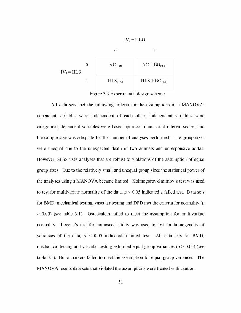
IV2 = HBO
0 1
0 AC(0,0) AC-HBO(0,1)
IV1 = HLS
1 HLS(1,0) HLS-HBO(1,1)
Figure 3.3 Experimental design scheme.
All data sets met the following criteria for the assumptions of a MANOVA;
dependent variables were independent of each other, independent variables were
categorical, dependent variables were based upon continuous and interval scales, and
the sample size was adequate for the number of analyses performed. The group sizes
were unequal due to the unexpected death of two animals and unresponsive aortas.
However, SPSS uses analyses that are robust to violations of the assumption of equal
group sizes. Due to the relatively small and unequal group sizes the statistical power of
the analyses using a MANOVA became limited. Kolmogorov-Smirnov’s test was used
to test for multivariate normality of the data, p < 0.05 indicated a failed test. Data sets
for BMD, mechanical testing, vascular testing and DPD met the criteria for normality (p
> 0.05) (see table 3.1). Osteocalcin failed to meet the assumption for multivariate
normality. Levene’s test for homoscedasticity was used to test for homogeneity of
variances of the data, p < 0.05 indicated a failed test. All data sets for BMD,
mechanical testing and vascular testing exhibited equal group variances (p > 0.05) (see
table 3.1). Bone markers failed to meet the assumption for equal group variances. The
MANOVA results data sets that violated the assumptions were treated with caution.
31

Table 3.1 Results for MANOVA Assumptions Testing.
Assumption BMD Bone Markers Mechanical Testing
Vascular Testing
Multivariate Normal Distribution (Kolmogorov-Smirnov)
0.20(Canc) 0.20(Cort)
0.002 (DPD)* 0.20 (OSTEO)
0.06 (FXF) 0.20 (FMX)
0.13 (MX) 0.15 (EC50)
Homoscedasticity (Levene)
0.65(Canc) 0.95 (Cort)
0.05 (DPD)* 0.04 (OSTEO)*
0.503 (FXF) 0.444 (FMX)
0.16 (MX) 0.15 (EC50)
p > 0.05 indicates the assumption is upheld. * Failed the assumption for corresponding test.
Changes in body mass pre- and post-28 day experimental period were compared
using a 2 × 2 repeated measures analysis of covariance (ANCOVA) (HLS [with vs.
without HLS] × HBO [with vs. without HBO]) design. A type I error level was pre-set
at 5% (p ≤ 0.05), because there five analyses a Bonferrioni adjustment was used to
protect for the inflation of alpha. The adjustment was p ≤ 0.01.
32

CHAPTER 4
RESULTS
One rat each from the HLS and HLS-HBO groups died during the experimental
period, therefore the total N was reduced by two (N = 38) and the n for the two HLS
groups were each reduced by one (HLS, n = 9 and HLS-HBO, n = 9). The group
sample sizes were further reduced during the experimental procedures for bone marker
testing due to inadequate dilution of samples resulting in data beyond the measurable
range. Group sample size was also reduced for vascular testing due to damage to the
vessel during isolation and preparation.
4.1 Bone Mineral Density
Bone mineral densities for the cancellous and cortical compartments of the mid-
diaphysis were measured. The average values for cancellous and cortical BMD are
located in table 4.1 and are shown graphically in figures 4.1 and 4.2. The 2 × 2
MANOVA on BMD (HLS [with vs. without HLS] × HBO [with vs. without HBO])
using Pillai’s Trace revealed no overall significant difference (F (6, 68) =1.54, p =
0.179). However, due to the exploratory nature of the analysis any significant pairwise
comparisons (p < 0.039) were of interest. The pairwise comparison for cortical BMD
between HLS and AC-HBO was significant (p = 0.014, =HLSx 1419.70 ± 14.67
mg/cm3, HBOACx − = 1437.07 ± 16.70 mg/cm3).
33

Table 4.1 Bone Mineral Density Cancellous (mg/cm3) Cortical(mg/cm3) AC (n = 10) 395.66 ± 44.59 1430.82 ± 12.55 AC-HBO (n = 10) 378.46 ± 63.08 1437.07 ± 16.70 HLS (n = 9) 370.13 ± 45.40 1419.70 ± 14.67* HLS-HBO (n = 9) 383.42 ± 65.32 1427.12 ± 14.03 Group mean ± SD for each dependent variable. * Significant pairwise comparison with AC-HBO, p = 0.014.
Figure 4.1 Cancellous bone mineral density.
34

Figure 4.2 Cortical bone mineral density.* Significant pairwise comparison with AC HBO, p = 0.014.
4.2 Biochemical Markers of Bone Remodeling
Deoxypydiridinoline and OC were measured to determine bone resorption and
formation rates respectively. The average values for DPD and OC are located in table
4.2 and are shown graphically in figures 4.3 and 4.4. The 2 × 2 MANOVA on bone
markers (HLS [with vs. without HLS] × HBO [with vs. without HBO]) using Pillai’s
Trace revealed no overall significant difference (F (6, 56) = 0.818, p = 0.561). There
were no significant pairwise comparisons for bone markers (α > 0.039).
Table 4.2 Biochemical Bone Markers DPD (nmole/mmol Cr) Osteocalcin (ng/mL) AC (n = 8) 18 ± 4 69.00 ± 24.77 AC-HBO (n = 10) 18 ± 4 72.50 ± 23.82 HLS (n = 8) 17 ± 3 93.50 ± 51.05 HLS-HBO (n = 6) 17 ± 2 91.50 ± 13.79 Group mean ± SD for each dependent variable.
35

Figure 4.3 Deoxypyridinoline (nmol/mmol Cr).
Figure 4.4 Osteocalcin (ng/mL).
36
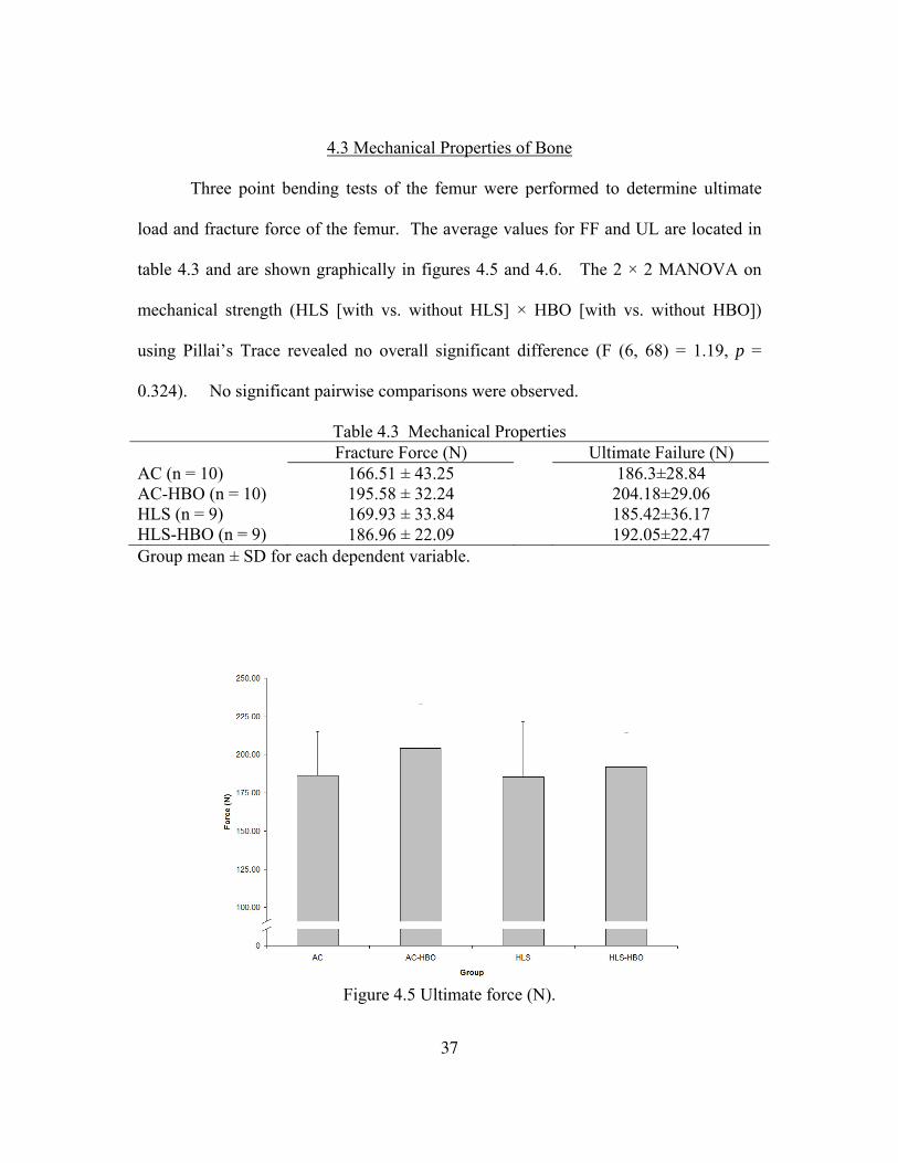
4.3 Mechanical Properties of Bone
Three point bending tests of the femur were performed to determine ultimate
load and fracture force of the femur. The average values for FF and UL are located in
table 4.3 and are shown graphically in figures 4.5 and 4.6. The 2 × 2 MANOVA on
mechanical strength (HLS [with vs. without HLS] × HBO [with vs. without HBO])
using Pillai’s Trace revealed no overall significant difference (F (6, 68) = 1.19, p =
0.324). No significant pairwise comparisons were observed.
Table 4.3 Mechanical Properties Fracture Force (N) Ultimate Failure (N) AC (n = 10) 166.51 ± 43.25 186.3±28.84 AC-HBO (n = 10) 195.58 ± 32.24 204.18±29.06 HLS (n = 9) 169.93 ± 33.84 185.42±36.17 HLS-HBO (n = 9) 186.96 ± 22.09 192.05±22.47 Group mean ± SD for each dependent variable.
Figure 4.5 Ultimate force (N).
37

Figure 4.6 Fracture force (N).
4.4 Vasoconstrictive Properties
Sensitivity (EC50) and maximal vasoconstriction were measured in response to
increasing concentrations of PHE. The average values for EC50 and MAX are located
in table 4.4 and are shown graphically in figures 4.7 and 4.8. The 2 × 2 MANOVA on
vascular responsiveness (HLS [with vs. without HLS] × HBO [with vs. without HBO])
revealed an overall significance (F (6, 58) =3.825, p = 0.003, power = 0.947). Follow-
up ANOVAs indicated no significant difference (F (3, 29) = 0.612, p = 0.613) among
EC50 and a significant difference among the maximal vasoconstriction (F (3, 29) =
10.935, p < 0.001, power = 0.997). Post hoc analysis revealed a significant difference
in maximal vasoconstriction between AC and HLS (p = 0.031, =ACx 97.66 ± 25.09 %,
=HLSx 59.97 ± 27.58 %), AC and HLS-HBO (p < 0.001, =ACx 97.66 ± 25.09 %,
=−HBOHLSx 32.99 ± 13.81 %), and AC-HBO and HLS-HBO (p = 0.008, =−HBOACx 72.95
± 25.93 %, =−HBOHLSx 32.99 ± 13.81 %).
38

Table 4.4 Vasoconstrictive Properties EC50 (mol) MAX (% constriction) AC (n = 9) 3.58×10⁻⁷ ± 2.42×10⁻⁷ 97.66 ± 25.09* AC-HBO (n = 10) 3.50×10⁻⁷ ± 1.18×10⁻⁷ 72.95 ± 25.93** HLS (n = 6) 4.74×10⁻⁷ ± 2.99×10⁻⁷ 59.97 ± 27.58 HLS-HBO (n = 8) 4.28×10⁻⁷ ± 1.71×10⁻⁷ 32.99 ± 13.81 Group mean ± SD for each dependent variable. * Significantly different compared with HLS and HLS-HBO, p < 0.039 for post hoc analysis. ** Significantly different compared with HLS-HBO, p = 0.008 for post hoc analysis.
Figure 4.7 EC50 (mol)
39

Figure 4.8 Maximal vasoconstriction (% maximal vasoconstriction). Expressed as a percentage of maximal preconstruction by KCl. * Significantly different compared with HLS and HLS-HBO, p < 0.039 for post hoc analysis. ** Significantly different compared with HLS-HBO, p = 0.008 for post hoc analysis.
4.5 Body Mass
Body mass on day one and on day 28 of the experimental protocol was
measured. The mean mass (g) for each experimental group is located in table 4.5 and is
shown graphically in figure 4.9. The repeated measures ANOVA revealed a significant
difference in post-experimental mass between groups (F (3, 33) = 12.456, p < 0.001,
power = 0.997). Post hoc analysis revealed that the mass of HLS and HLS-HBO were
significantly less than AC and AC-HBO rats (p < 0.01). There were no significant
differences between HLS and HLS-HBO or between AC and AC-HBO (p > 0.01)
40

Table 4.5 Body Mass (g) Day 1 Day 28 AC (n = 10) 423 ± 34 454 ± 41 AC-HBO (n = 10) 432 ± 27 460 ± 35 HLS (n = 9) 433 ± 35 415 ± 37 HLS-HBO (n = 9) 440 ± 31 409 ± 24
Group mean ± SD for each dependent variable.
Figure 4.9 Body mass (g) at day 28 of HLS. * Significantly different than AC and AC-HBO (p < 0.01).
41

CHAPTER 5
DISCUSSION
Effective countermeasures to the microgravitational alterations in BMD
decreases and orthostatic intolerance have yet to be identified. Ascertaining
countermeasures to eliminate or minimize these responses is essential to protect the
health and well-being of astronauts during and after their missions. The purpose of this
investigation was to determine if HBO therapy would attenuate or eliminate decreases
in BMD and orthostatic response resulting from simulated microgravity. To our
knowledge, this was the first study conducted to examine the effects of HBO therapy on
physiologic changes due to simulated microgravity.
5.1 Bone Mineral Density
An average decrease in BMD of 1-2 % per month in microgravity and
simulated microgravity environments has been reported in the literature (27, 58).
Previous studies have reported decreases in total BMD (1, 14, 21, 45), however they
neither separated the different types of bone into compartments nor isolated a specific
section of the bone. The BMD measurement technique, pQCT, is able to differentiate
between cortical and cancellous bone compartments which allow precise measurement
of the areas of bone mineral changes. It has been reported that the decreases in total
BMD in previous studies could be attributed to a decline in cancellous bone (5).
42

Bloomfield et al. (5) reported a significant decrease in cancellous BMD (by 21 %) at the
proximal tibia after 28 days HLS and a significant increase in cortical BMD (by 5.6 %)
at the metaphysis of the proximal tibia. The MANOVA results of the present
investigation indicate that HLS and HBO did not significantly affect cortical or
cancellous BMD at the mid-diaphysis of the femur following of the 28 day
experimental period. It appeared that HBO and weight bearing resulted in a slight
increase in cortical BMD. A pairwise comparison indicated that cortical BMD was
significantly reduced in the HLS group when compared to the AC-HBO group. Adding
HBO to HLS resulted in a slight increase in cortical BMD over HLS alone. Further
examination is required to confirm these results.
5.2 Biochemical Markers of Bone Remodeling
Bone continually undergoes a remodeling process where bone mineral is
removed or resorbed then new bone mineral is deposited within the cavity. The
uncoupling, increase in bone resorption, a decrease in bone deposition or the
combination of the two processes, can lead to a decrease in BMD. Deoxypyridinoline
and osteocalcin are commonly measured to determine alterations in bone resorption and
deposition respectively.
Previous space and land-based studies have observed increases in bone
resorption and decreases in bone formation markers. Caillot-Agusseauet al. (8, 9)
observed a 50 % and 54 % increase in DPD in cosmonauts during 21 and 180 days of
spaceflight respectively. Kurokouchi et al. (30) measured an increase in tartrate-
resistant acid phosphatase (TRAP) in five-week old rats at days one and three, but this
43
returned to pre-HLS levels at day five and remained within pre-HLS values during
remainder of the 14 of days HLS. Caillot-Agusseau et al. (8, 9) measured changes in
OC during 21 and 180 day space missions in cosmonauts. Osteocalcin levels were
decreased by 18% and 27 % during 21 and 180 days, respectively when compared to
pre-flight levels. Kurokouchi et al. (30) observed a decrease in OC levels from day
three to day 14 in 5-week old HLS rats.
There were no significant changes in PYD and OC among the groups in the
present study, therefore biochemical bone markers did not indicate that uncoupling of
bone formation and resorption had occurred. These results are consistent with the
observations that there were no significant changes in bone markers therefore any
significant changes BMD were not expected.
5.3 Mechanical Properties of Bone
Disruptions in the remodeling of bone can lead to alterations in bone
architecture and the removal of structural elements which lead to the loss of mechanical
strength and fractures (6). This is a primary concern when planning long term space
travel such as a mission to Mars where travel time to and from the planet will take about
three years. A fracture that occurs in space could compromise the health of the
individual, the crew, and the mission.
The deterioration of the struts and plates due to high rates of resorption weaken
cancellous bone making it more susceptible to fractures. Hogan et al. (26) measured
changes in the mechanical properties of cancellous bone in female rats that underwent
an ovariectomy to stimulate bone loss. In a three point bending test of the femur, a 10%
44

increase in maximal force was observed in the ovariectomized rats when compared to
sham. In a study by Bloomfield et al. (5) pQCT was used to measure changes in bone
mineral density and differentiate between losses in cortical and cancellous bone in six-
month old rats after 28 days of HLS. Mechanical testing was then performed to
determine if there was a relationship between changes in compartmental bone density
and the structural integrity of bone. The main finding was that there was an 18%
increase in fracture force that corresponded to a 5.6 % increase in cortical density of the
mid-diaphysis of the tibia. The results of the present study indicate HLS and HBO did
not alter the strength of the shaft of the femur as determined by the three point bending
test. These results correspond to the maintenance of BMD at the end of the
experimental period.
5.4 Hyperbaric Therapy and Bone
The rationale for the use of HBO as a countermeasure to reduce the rate of bone
demineralization was based on the research supporting the use of HBO in non-union
and delayed union fractures (13, 57). It was hypothesized that by increasing oxygen
delivery to the bone tissue with HBO, balance in the bone remodeling process would be
maintained. Within microgravity and simulated microgravity a cephalad fluid shift
occurs. It has been demonstrated that the perfusion pressure of blood to the femur is
compromised (10). Hyperbaric oxygen therapy increases the delivery of oxygen
through its dissolution in the plasma. On reaching the tissues, the increased
concentration gradient increases the perfusion distance of oxygen. It was hypothesized
45

that these two factors combined would compensate for the decreased perfusion pressure
to the hind limbs that occurs with HLS.
5.5 Vasoconstrictive Properties
The regulation of orthostasis upon arrival to Earth after space travel is
compromised and can lead to syncope. Buckey et al. (7) observed that nine of 14
astronauts were unable to complete a 10 minute stand test after returning from 9-14
days of space travel. Buckey et al. (7) found that there was a greater vasoconstrictive
response in the astronauts who were able to complete the stand test versus the non-
finishers. Research has suggested that the cephalad shift of bodily fluids may elicit
physiologic responses that alter the baroreceptor reflex (37, 41, 54). However,
decreased vascular responsiveness to sympathetic output has also been proposed as a
contributing factor to orthostatic intolerance (15, 16).
It has been demonstrated that hyperbaric therapy increases SVR. Berry et al. (4)
observed an increase in SVR following 90 minutes of HBO therapy using 100 % O2 at 2
ATA. The SVR continued even after returning to ambient air and remained elevated for
30 minutes. Oxygen acts as an alpha adrenergic agent resulting in vasoconstriction
therefore it was concluded that SVR would be increased during HBO treatments (35,
36). The vessel response post treatment was a matter of speculation. We hypothesized
that the intermittent vasoconstrictive response to HBO might result in a more
vasoconstrictive state between HBO treatments. The results indicate that the opposite
response occurred. Hindlimb suspension in the present study resulted in a decreased
maximal vasoconstrictive response to PHE of the isolated thoracic aorta when
46

compared to control rats, a 39 % and 54 % decrease in MAX was observed between AC
and HLS, and AC-HBO and HLS-HBO respectively. These results are consistent with
previous studies examining the effect of HLS on maximal vasoconstriction of the
abdominal and thoracic aortas (15, 16). A further reduction in maximal
vasoconstriction occurred in the aortas of the HLS-HBO group. A 66 % decrease in
MAX occurred between AC and HLS-HBO. Our results of decreased vasoconstriction
in response to PHE are supported by the findings of Tahepold et al. (51).
There were no differences in the sensitivity (EC50, the mean effective agonist
concentration that produces 50 % of maximal vasoconstrictive response) to PHE
between HLS and control rats. Previous studies have also reported no changes in
sensitivity to PHE after HLS (15, 16). Hyperbaric therapy had no affect on sensitivity
to PHE between any of the experimental groups.
5.6 Body Mass
The HLS model to simulate microgravity has been successfully used in
numerous studies. The HLS model presents minimal stress to the animal based upon
hormonal levels (14, 38) and body weight measures (5, 21, 38, 45). However, several
HLS studies ranging from two to five weeks in duration have demonstrated significantly
lower body weight in the HLS groups when compared to controls (14, 15, 25). Body
mass of the HLS and HLS-HBO rats in this study were significantly lower than both
control groups. The decreases in body mass may have been due to a normal
physiologic response to simulated microgravity, such as decreased appetite leading to a
47

decrease in food consumption, decreased muscle mass due to disuse and reduction in
blood volume.
5.7 Conclusions
Twenty-eight days of hindlimb suspension and 90 minutes of hyperbaric oxygen
therapy treatment at 2.5 ATA using 100 % oxygen did not have a significant effect on
cortical or cancellous bone mineral at the mid-diaphysis of the femur. Biochemical
bone markers for bone remodeling did not indicate significant changes with HBO.
Deoxypyridinoline and osteocalcin levels were maintained indicating that rates of bone
resorption and deposition were maintained. Furthermore no alterations in bone
mechanical strength were observed. The absence of a significant effect due to HLS in
the present study was not consistent with previous studies. No studies, however, have
examined the effects of HBO on bone parameters. Therefore, there are no bone data to
compare the HBO results with from this experiment. To confirm these results, future
research should to be conducted that examines the effects of HBO and HLS on bone.
Altering the HBO treatment frequency and duration, increased sample size, and the
addition of hyperoxia and pressure control groups should be considered in future
investigations.
Under the conditions of the current study we could not recommend the use of
HBO as an intervention to offset detrimental effects of microgravity on bone. However,
other pressure and oxygen doses might show positive results. When compared to
humans, rat’s arterial blood pO2 is approximately twice as great as humans (19).
Klemetti et al. (28) found that reducing HBO therapy treatment time in rats to 60
48

minutes from 90 minutes (the standard clinical treatment time in humans) for seven
days on surgically damaged mandibles significantly restored blood flow to the bony
surgical site. Barth et al. (2) found that once daily HBO therapy 5 day a week was more
effective at repairing surgically damaged bone tissue than a twice daily regimen.
Maximal vasoconstriction response to PHE was decreased with HLS and was
further reduced in both HBO groups. The effect of HBO on maximal vasoconstriction
could further attenuate the orthostatic response in astronauts returning from space.
Therefore, under the conditions of this study HBO is not recommended as a
countermeasure to the orthostatic intolerance experienced after space travel. However,
further studies that vary the dosage of the HBO treatment (duration and depth) may
have different results.
49

REFERENCES
1. Arnaud S. B., J. S. Harper, M. Navidi. Mineral distribution in rat skeletons after
exposure to a microgravity model. J Gravit Physiol. 2(1):P115-6, 1995.
2. Barth E., T. Sullivan, E. Berg. Animal model for evaluating bone repair with and
without adjunctive hyperbaric oxygen therapy (HBO): comparing dose schedules. J
Invest Surg. 3(4):387-392, 1990.
3. Bauer D. C., P. M. Sklarin, K. L. Stone, et al. Biochemical markers of bone turnover
and prediction of hip bone loss in older women: the study of osteoporotic fractures. J
Bone Miner Res. 14(8):1404-1410, 1999.
4. Berry J. M., M. F. Doursout, B. D. Butler. Effects of hyperbaric hyperoxia on cardiac
and regional hemodynamics in conscious dogs. Aviat Space Environ Med. 69(8):761-
765, 1998.
5. Bloomfield S. A., M. R. Allen, H. A. Hogan, M. D. Delp. Site- and compartment-
specific changes in bone with hindlimb unloading in mature adult rats. Bone. 31(1):149-
157, 2002.
50

6. Born G. V. R., P. Cuatrecasas, H. Herken, K. Melmon. Handbook of Experimental
Pharmacology. 107:748, 1993.
7. Buckey J. C.,Jr, L. D. Lane, B. D. Levine, et al. Orthostatic intolerance after
spaceflight. J Appl Physiol. 81(1):7-18, 1996.
8. Caillot-Augusseau A., M. H. Lafage-Proust, C. Soler, J. Pernod, F. Dubois, C.
Alexandre. Bone formation and resorption biological markers in cosmonauts during and
after a 180-day space flight (Euromir 95). Clin Chem. 44(3):578-585, 1998.
9. Caillot-Augusseau A., L. Vico, M. Heer, et al. Space flight is associated with rapid
decreases of undercarboxylated osteocalcin and increases of markers of bone resorption
without changes in their circadian variation: observations in two cosmonauts. Clin
Chem. 46(8 Pt 1):1136-1143, 2000.
10. Colleran P. N., M. K. Wilkerson, S. A. Bloomfield, L. J. Suva, R. T. Turner, M. D.
Delp. Alterations in skeletal perfusion with simulated microgravity: a possible
mechanism for bone remodeling. J Appl Physiol. 89(3):1046-1054, 2000.
11. Collet P., D. Uebelhart, L. Vico, et al. Effects of 1- and 6-month spaceflight on bone
mass and biochemistry in two humans. Bone. 20(6):547-551, 1997.
12. Convertino V. A., H. Sandler. Exercise countermeasures for spaceflight. Acta
Astronaut. 35(4-5):253-270, 1995.
51

13. Coulson D. B., A. B. Ferguson Jr, R. C. Diehl Jr. Effect of hyperbaric oxygen on the
healing femur of the rat. Surg Forum. 17:449-450, 1966.
14. Dehority W., B. P. Halloran, D. D. Bikle, et al. Bone and hormonal changes induced
by skeletal unloading in the mature male rat. Am J Physiol. 276(1 Pt 1):E62-9, 1999.
15. Delp M. D., M. Brown, M. H. Laughlin, E. M. Hasser. Rat aortic vasoreactivity is
altered by old age and hindlimb unloading. J Appl Physiol. 78(6):2079-2086, 1995.
16. Delp M. D., T. Holder-Binkley, M. H. Laughlin, E. M. Hasser. Vasoconstrictor
properties of rat aorta are diminished by hindlimb unweighting. J Appl Physiol.
75(6):2620-2628, 1993.
17. Drummer C., C. Hesse, F. Baisch, et al. Water and sodium balances and their
relation to body mass changes in microgravity. Eur J Clin Invest. 30(12):1066-1075,
2000.
18. Eastell R., A. Colwell, L. Hampton, J. Reeve. Biochemical markers of bone
resorption compared with estimates of bone resorption from radiotracer kinetic studies
in osteoporosis. J Bone Miner Res. 12(1):59-65, 1997.
19. Flecknell P. A. Laboratory Animal Anesthesia. 1996.
20. Gill A. L., C. N. Bell. Hyperbaric oxygen: its uses, mechanisms of action and
outcomes. QJM. 97(7):385-395, 2004.
52

21. Globus R. K., D. D. Bikle, E. Morey-Holton. Effects of simulated weightlessness on
bone mineral metabolism. Endocrinology. 114(6):2264-2270, 1984.
22. GOLDHABER P. The effect of hyperoxia on bone resorption in tissue culture. AMA
Arch Pathol. 66(5):635-641, 1958.
23. Gray D. H., J. M. Katz, K. Speak. The effects of varying oxygen tensions upon bone
resorption in vitro. J Bone Joint Surg Br. 60-B(4):575-578, 1978.
24. Heer M., N. Kamps, C. Biener, et al. Calcium metabolism in microgravity. Eur J
Med Res. 4(9):357-360, 1999.
25. Hefferan T. E., G. L. Evans, S. Lotinun, M. Zhang, E. Morey-Holton, R. T. Turner.
Effect of gender on bone turnover in adult rats during simulated weightlessness. J Appl
Physiol. 95(5):1775-1780, 2003.
26. Hogan H. A., S. P. Ruhmann, H. W. Sampson. The mechanical properties of
cancellous bone in the proximal tibia of ovariectomized rats. J Bone Miner Res.
15(2):284-292, 2000.
27. Holick M. F. Perspective on the impact of weightlessness on calcium and bone
metabolism. Bone. 22(5 Suppl):105S-111S, 1998.
28. Klemetti E., S. Rico-Vargas, P. Mojon. Short duration hyperbaric oxygen treatment
effects blood flow in rats: pilot observations. Lab Anim. 39(1):116-121, 2005.
53
29. Knott L., A. J. Bailey. Collagen cross-links in mineralizing tissues: a review of their
chemistry, function, and clinical relevance. Bone. 22(3):181-187, 1998.
30. Kurokouchi K., T. Ito, S. Ohmori, K. Kanda, Y. Murata, H. Seo. Changes in the
markers of bone metabolism following skeletal unloading. Environ Med. 39(1):21-24,
1995.
31. Looft-Wilson R. C., C. V. Gisolfi. Rat small mesenteric artery function after
hindlimb suspension. J Appl Physiol. 88(4):1199-1206, 2000.
32. Marcus R., L. Holloway, B. Wells, et al. The relationship of biochemical markers of
bone turnover to bone density changes in postmenopausal women: results from the
Postmenopausal Estrogen/Progestin Interventions (PEPI) trial. J Bone Miner Res.
14(9):1583-1595, 1999.
33. Martini F. H. Fundamentals of Anatomy and Physiology. Sixth Edition ed. San
Franscisco, CA: Pearson Education, 2004.
34. Martini F. H., M. J. Timmons, R. B. Tallitsch. Human Anatomy. Fourth Edition ed.
Upper Saddle River, New Jersey: Prentica Hall, 2003.
35. Messina E. J., D. Sun, A. Koller, M. S. Wolin, G. Kaley. Increases in oxygen
tension evoke arteriolar constriction by inhibiting endothelial prostaglandin synthesis.
Microvasc Res. 48(2):151-160, 1994.
54

36. Molenat F., A. Boussuges, A. Grandfond, et al. Haemodynamic effects of
hyperbaric hyperoxia in healthy volunteers: an echocardiographic and Doppler study.
Clin Sci (Lond). 106(4):389-395, 2004.
37. Money K. E. Biological effects of space travel. Can Aeronaut Space J. 27(3):195-
201, 1981.
38. Morey-Holton E. R., R. K. Globus. Hindlimb unloading rodent model: technical
aspects. J Appl Physiol. 92(4):1367-1377, 2002.
39. O'Connell MB FRCPC, John X. Bone Structure and Remodeling. 2005(April
17)2004.
40. Pace N. Weightlessness: a matter of gravity. N Engl J Med. 297:32-37, 1977.
41. Paskovaty A., E. A. O'Rangers. Health Consequences of Spaceflight. Journal of
Pharmacy Practice. 16(2):101--106, 2003.
42. Patterson-Buckendahl P., R. K. Globus, D. D. Bikle, C. E. Cann, E. Morey-Holton.
Effects of simulated weightlessness on rat osteocalcin and bone calcium. Am J Physiol.
257(5 Pt 2):R1103-9, 1989.
43. Rambaut P. C., R. S. Johnston. Prolonged weightlessness and calcium loss in man.
Acta Astronaut. 6(9):1113-1122, 1979.
44. Rambaut P. C., C. S. Leach, G. D. Whedon. A study of metabolic balance in
crewmembers of Skylab IV. Acta Astronaut. 6(10):1313-1322, 1979.
55
45. Roer R. D., R. M. Dillaman. Bone growth and calcium balance during simulated
weightlessness in the rat. J Appl Physiol. 68(1):13-20, 1990.
46. Rousseau A., Z. Bak, B. Janerot-Sjoberg, F. Sjoberg. Acute hyperoxaemia-induced
effects on regional blood flow, oxygen consumption and central circulation in man.
Acta Physiol Scand. 183(3):231-240, 2005.
47. Shaw J. L., C. A. Bassett. The effects of varying oxygen concentrations on
osteogenesis and embryonic cartilage in vitro. J Bone Joint Surg Am. 49(1):73-80, 1967.
48. Sheffeld P. J., N. T. Workman. Non-invasive tissue oxygen measurements in
patients administered normbaric and hyperbaric oxygen by mask. Hyperb Oxyg Rev.
6:47--62, 1985.
49. Smith S. M., J. E. Davis-Street, J. V. Fesperman, et al. Evaluation of treadmill
exercise in a lower body negative pressure chamber as a countermeasure for
weightlessness-induced bone loss: a bed rest study with identical twins. J Bone Miner
Res. 18(12):2223-2230, 2003.
50. Stein T. P., M. J. Leskiw, M. D. Schluter, et al. Energy expenditure and balance
during spaceflight on the space shuttle. Am J Physiol. 276(6 Pt 2):R1739-48, 1999.
51. Tahepold P., P. Elfstrom, I. Eha, et al. Exposure of rats to hyperoxia enhances
relaxation of isolated aortic rings and reduces infarc size of isolated hearts. Acta Physiol
Scand. 175:271-277, 2002.
56

52. Vernikos J. Human physiology in space. Bioessays. 18(12):1029-1037, 1996.
53. Weaver C. M., M. Peacock, B. R. Martin, et al. Quantification of biochemical
markers of bone turnover by kinetic measures of bone formation and resorption in
young healthy females. J Bone Miner Res. 12(10):1714-1720, 1997.
54. West J. B. Physiology in microgravity. J Appl Physiol. 89(1):379-384, 2000.
55. Whedon G. D., L. Lutwak, J. Reid, et al. Mineral and nitrogen balance study: results
of metabolic observations on Skylab II 28-day orbital mission. Acta Astronaut. 2(3-
4):297-309, 1975.
56. Woitge H. W., M. J. Seibel. Molecular markers of bone and cartilage metabolism.
Curr Opin Rheumatol. 11(3):218-225, 1999.
57. Wray J. B., L. S. Rogers. Effect of hyperbaric oxygenation upon fracture healing in
the rat. J Surg Res. 8(8):373-378, 1968.
58. Wronski T. J., E. R. Morey. Alterations in calcium homeostasis and bone during
actual and simulated space flight. Med Sci Sports Exerc. 15(5):410-414, 1983.
59. Wronski T. J., E. R. Morey. Skeletal abnormalities in rats induced by simulated
weightlessness. Metab Bone Dis Relat Res. 4(1):69-75, 1982.
57

BIOGRAPHICAL INFORMATION
The author received her Bachelor’s degree in exercise science in 1998 from The
University of Texas at Arlington. She began her graduate studies in exercise
physiology in the fall of 2003. While pursuing her degree she was a graduate teaching
assistant and participated in several research activities involving electromyography,
mechanomyography, stretching, and muscle strength. Laurie plans to pursue a career in
secondary or higher level education or work in a research laboratory. Laurie’s research
interest is in sport performance and spinal cord injury.
58