effects of fullerene (c60), multi-wall carbon nanotubes...
TRANSCRIPT

RESEARCH ARTICLE
Effects of fullerene (C60), multi-wall carbon nanotubes(MWCNT), single wall carbon nanotubes (SWCNT) and hydroxyland carboxyl modified single wall carbon nanotubes on riverinemicrobial communities
J. R. Lawrence1 & M. J. Waiser1 & G. D. W. Swerhone1 & J. Roy1 &
V. Tumber1 & A. Paule2 & A. P. Hitchcock3& J. J. Dynes4 & D. R. Korber5
Received: 17 November 2015 /Accepted: 2 February 2016# Springer-Verlag Berlin Heidelberg 2016
Abstract Commercial production of nanoparticles (NP) hascreated a need for research to support regulation of nanotech-nology. In the current study, microbial biofilm communitieswere developed in rotating annular reactors during continuousexposure to 500 μg L−1 of each nanomaterial and subjected tomultimetric analyses. Scanning transmission X-rayspectromicroscopy (STXM) was used to detect and estimatethe presence of the carbon nanomaterials in the biofilm com-munities. Microscopy observations indicated that the commu-nities were visibly different in appearance with changes inabundance of filamentous cyanobacteria in particular.Microscale analyses indicated that fullerene (C60) did notsignificantly (p<0.05) impact algal, cyanobacterial or bacte-rial biomass. In contrast, MWCNT exposure resulted in a sig-nificant decline in algal and bacteria biomass. Interestingly,the presence of SWCNT products increased algal biomass,
significantly in the case of SWCNT-COOH (p<0.05) buthad no significant impact on cyanobacterial or bacterialbiomass. Thymidine incorporation indicated that bacterialproduction was significantly reduced (p < 0.05) by allnanomaterials with the exception of fullerene. Biolog assess-ment of carbon utilization revealed few significant effects withthe exception of the utilization of carboxylic acids. PCA andANOSIM analyses of denaturing gradient gel electrophoresis(DGGE) results indicated that the bacterial communities ex-posed to fullerene were not different from the control, theMWCNT and SWNT-OH differed from the control but noteach other, whereas the SWCNT and SWCNT-COOH bothdiffered from all other treatments and were significantly dif-ferent from the control (p<0.05). Fluorescent lectin bindinganalyses also indicated significant (p<0.05) changes in thenature and quantities of exopolymer consistent with changes
Responsible editor: Philippe Garrigues
* J. R. [email protected]
M. J. [email protected]
G. D. W. [email protected]
A. P. [email protected]
J. J. [email protected]
D. R. [email protected]
1 Environment Canada, National Hydrology Research Centre, 11Innovation Blvd., Saskatoon, SK, Canada S7N 3H5
2 Global Institute for Water Security, University of Saskatchewan,Saskatoon, SK S7N 5A8, Canada
3 Brockhouse Institute for Materials Research, McMaster University,Hamilton, ON L8S 4M1, Canada
4 Canadian Light Source, University of Saskatchewan,Saskatoon, SK S7N 5A8, Canada
5 Food and Bioproduct Sciences, University of Saskatchewan,Saskatoon, SK S7N 5A8, Canada
Environ Sci Pollut ResDOI 10.1007/s11356-016-6244-x
in microbial community structure during exposure to allnanomaterials. Enumeration of protozoan grazers showed de-clines in communities exposed to fullerene or MWCNT but atrend for increases in all SWCNT exposures. Observationsindicated that at 500 μg L−1, carbon nanomaterials significant-ly alter aspects of microbial community structure and functionsupporting the need for further evaluation of their effects inaquatic habitats.
Keywords Carbon nanotubes . Fullerenes . Effects .
Microbial activity . Diversity . Metabolism
Introduction
The development and application of nanotechnology haveraised significant concerns about the adverse effects ofnanomaterials on human health and the environment.Manufactured nanomaterials have been reported as potentiallymore toxic than larger particles of the same composition be-cause of their large specific surface area and unique catalyticproperties. The commercial production of nanomaterials andtheir incorporation into a variety of products have generated aneed for research to support regulation of the nanotechnologysector (Handy et al. 2008).Worldwide CNT production valueswere estimated by Mueller and Nowak (2008) to range from350 to 500 tons/year. Recent estimates of demand for CNTsare in the range of 3700–4100 tons increasing to 10,500–12,000 tons by 2020. Nanoparticles produced in large amountsinclude carbon black, fullerenes and a range of carbonnanotubes. Carbon nanomaterials have a wide variety ofapplications due to their: high tensile strength, electronicconductance, semi-conductor potential, high surface area andpotential for sorption (Ajayan et al. 1999; Ajayan and Zhou2001; Ball 2001). These applications include aerospace, fibreproduction, semiconductors, sorbents and remediation(Ajayan and Zhou 2001; Lee et al. 2005). Carbon-basednanomaterials are also finding applications in water treatment,waste water treatment, drug delivery, as well as food packag-ing preservation (Theron et al. 2008). Their use in cosmeticshas been extensive and would likely contribute to aquaticenvironmental loading (Chae et al. 2010). Commercial scaleproduction and use with growing demand raises clear con-cerns regarding the potential health and environmental effectsof these materials (Dreher 2004).
The applications proposed for this technology suggest thatnanoparticles could enter aquatic systems through directdischarges and from industrial as well as domesticwastewater effluents. In their review, Petersen et al. (2011)note that most CNTs are part of a variety of composites thatmay weather or even be incinerated. The modelling exercisesof Gottchalk et al. (2009, 2010) and Mueller and Nowack(2008) assume that the bulk of CNTs manufactured is part of
a variety of polymer-containing products assumed to enter theenvironment through landfills. The fraction reaching waste-water treatment plants, which is largely unknown, will bederived from clothing and fabric manufacturing and other tex-tile applications (Kohler et al. 2008). Of greatest concern maybe their proposed use in pollution control and in situ remedi-ation where they may be released intentionally (Mauter andElimelech 2008; Apul et al. 2012).
Environmental concentrations of nanomaterials are largelyunknown, although modelling has permitted estimates, rang-ing from 620 kg year−1 for nanosilver and 47,300 kg year−1
for TiO2 entering the surface waters of Switzerland (Muellerand Nowack 2008). Releases of this magnitude would resultin nano to microgram levels in the receiving environment(Mueller and Nowack 2008). Modelling-based estimates ofCNTs in the aquatic environment are generally in the low ngL−1 (Gottshalk et al. 2009). Of course, hot spots may arise as aresult of manufacturing, transport and disposal. Although rel-atively little is known regarding actual environmental concen-trations, concerns have been raised about the potential toxicityand environmental impacts of CNTs (Handy et al. 2008;Petersen et al. 2011). Indeed, a number of studies have shownthat carbon nanomaterials have antimicrobial properties underpure culture conditions (e.g., Kang et al. 2007; Ghafari et al.2008; Kang et al. 2009; Neal 2008). Carbon nanomaterialsappear to require close contact and may disrupt membranesas a result of electrostatic, oxidative or physical puncturinginteractions and most notably by production of reactive oxy-gen or nitrogen species (Jackson et al. 2013). Complex micro-bial community studies have concentrated on impacts in sew-age effluents and sludges (Kang et al. 2009; Yin et al. 2009;Luongo and Zhang 2010; Goyal et al. 2010).While Tong et al.(2007) and Chung et al. (2011) examined effects of fullerene(C60) and multiwalled carbon nanotubes (MWCNT) respec-tively on a soil microbial community, they found no effect ondiversity based on DGGE, biomass or enzyme activity.Velzeboer et al. (2011) examined the effects of high levels ofMWCNTs in aquatic sediments with no effect on invertebratediversity and increased numbers during 3-month exposures. Incontrast, their long-term (15 months) study detected signifi-cant effects of 2000 μg L−1 MWCNTon sediment communitystructure (Velzeboer et al. 2013). However, conditions, such ashigh concentrations of organic matter found in complex envi-ronmental systems, including soil, anaerobic sludge andwastewater effluent, may mitigate carbon nanomaterial toxic-ity to varying degrees (Tong et al. 2007; Kang et al. 2009;Lawrence et al. 2016). In terms of environmental fate andeffects, it is apparent that these nanomaterials may be modi-fied upon entering the environment undergoing aggregation(Chen et al. 2004), coating with organic matter and cations, aswell as potential modification by oxidants or microorganisms(Hyung et al. 2007). Lawrence et al. (2016) describe the natureof the coatings developing on MWCNTs and SWCNTs in
Environ Sci Pollut Res
association with aquatic biofilms demonstrating that they arehighly complex and result in a reduction in toxicity throughsuppression of ROS production. Toxicity of nanomaterials inthe natural environment is intimately linked to environmentalparameters, including natural organic matter and solutionchemistry which may be far more important in dictating thetoxicology of the nanomaterials than the as manufactured state(Petersen et al. 2011).
As has been noted in a wide variety of studies (Haak andMcFeters 1982a, b; Lawrence et al. 2004; Battin et al. 2009),microbial communities represent the base of the food web,driving most biogeochemical cycles and so-called ecosystemservices. Therefore, it is essential to examine the fate andeffects of these materials under controlled but representativeconditions. Biofilms are already known as extensive naturalsinks for metals, pesticides, antimicrobial agents and a varietyof other environmental contaminants (Dynes et al. 2006a, b;Wolfaardt et al. 1994). Based on this and observations regard-ing apparent scavenging of nanoparticles by bacterial EPS(Liu et al. 2007), Battin et al. (2009) suggested that biofilmsmay be exposed to higher levels of nanoparticles than plank-tonic communities. It is suggested that exopolymeric sub-stances (EPS) represent the most likely point of interactionfor nanomaterials with the biological compartment ofaquatic ecosystems and that both direct and indirect effectsmay occur as a result of bioaccumulation of carbonnanomaterials in the EPS pool and their subsequent intro-duction into the food web. We have used complex riverbiofilm communities and multimetric analyses to assessthe fate and effects of a wide range of environmental con-taminants, including municipal wastewater effluents,metals, pharmaceuticals and personal care products(Lawrence et al. 2004, 2005, 2009, 2012). Rotating annularreactors have been used to generate communities on de-fined substrata that are then subjected to analyses that aremicroscopic, molecular, genomic, functional and activitybased. Here, we have applied this approach in conjunctionwith scanning transmission X-ray microscopy analyses toassess the exposure, fate and effects of a panel of carbonnanomaterials in these complex microbial communities.
Materials and methods
Microcosm operation
The experimental setup and reactor design for biofilm devel-opment has been described in detail previously (Lawrenceet al. 2000, 2004). Natural river water (South SaskatchewanRiver, Saskatoon, SK, Canada) was used as inoculum and as asource of carbon and nutrients. The nutrients and nanoparti-cles were added directly to the individual reactors using aperistaltic pump. Nutrient levels were assessed as described
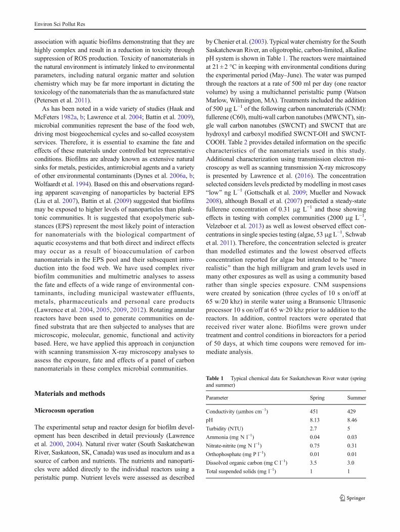
by Chenier et al. (2003). Typical water chemistry for the SouthSaskatchewan River, an oligotrophic, carbon-limited, alkalinepH system is shown in Table 1. The reactors were maintainedat 21±2 °C in keeping with environmental conditions duringthe experimental period (May–June). The water was pumpedthrough the reactors at a rate of 500 ml per day (one reactorvolume) by using a multichannel peristaltic pump (WatsonMarlow, Wilmington, MA). Treatments included the additionof 500 μg L−1 of the following carbon nanomaterials (CNM):fullerene (C60), multi-wall carbon nanotubes (MWCNT), sin-gle wall carbon nanotubes (SWCNT) and SWCNT that arehydroxyl and carboxyl modified SWCNT-OH and SWCNT-COOH. Table 2 provides detailed information on the specificcharacteristics of the nanomaterials used in this study.Additional characterization using transmission electron mi-croscopy as well as scanning transmission X-ray microscopyis presented by Lawrence et al. (2016). The concentrationselected considers levels predicted by modelling in most casesBlow^ ng L−1 (Gottschalk et al. 2009; Mueller and Nowack2008), although Boxall et al. (2007) predicted a steady-statefullerene concentration of 0.31 μg L−1 and those showingeffects in testing with complex communities (2000 μg L−1,Velzeboer et al. 2013) as well as lowest observed effect con-centrations in single species testing (algae, 53 μg L−1, Schwabet al. 2011). Therefore, the concentration selected is greaterthan modelled estimates and the lowest observed effectsconcentration reported for algae but intended to be Bmorerealistic^ than the high milligram and gram levels used inmany other exposures as well as using a community basedrather than single species exposure. CNM suspensionswere created by sonication (three cycles of 10 s on/off at65 w/20 khz) in sterile water using a Bransonic Ultrasonicprocessor 10 s on/off at 65 w·20 khz prior to addition to thereactors. In addition, control reactors were operated thatreceived river water alone. Biofilms were grown undertreatment and control conditions in bioreactors for a periodof 50 days, at which time coupons were removed for im-mediate analysis.
Table 1 Typical chemical data for Saskatchewan River water (springand summer)
Parameter Spring Summer
Conductivity (μmhos cm−1) 451 429
pH 8.13 8.46
Turbidity (NTU) 2.7 5
Ammonia (mg N l−1) 0.04 0.03
Nitrate-nitrite (mg N l−1) 0.75 0.31
Orthophosphate (mg P l−1) 0.01 0.01
Dissolved organic carbon (mg C l−1) 3.5 3.0
Total suspended solids (mg l−1) 1 1
Environ Sci Pollut Res

Confocal laser scanning microscopy and image analysis
All stained and control materials were analyzed by confocallaser microscopy (Nikon -C2, Confocal laser microscope) at-tached to a Nikon Eclipse 80i standard light microscope,equipped with 488/543/633 nm excitation as well as reflectionand transmission imaging (Nikon, Chiyoda, Tokyo, Japan)using fluorescent staining, Syto9 (Life Technologies,Burlington, ON, Canada) and Triticum vulgaris lectin-TRITC (Sigma, St. Louis, MI) to visualize bacterial cellsand exopolymer respectively as described in detail (Neuet al. 2001).
Exopolymer analyses
Lectins labeled with either fluorescein isothiocyanate orTRITC (Sigma, St. Louis, MO) or Cy5 (Research Organics,Cleveland, OH) were applied for exopolymer analyses. Thelectins T. vulgaris (β(1,4) N-acetyl glucosamine, N-acetylneuraminic acid), Arachis hypogaea (terminal β-galactose,N-acetyl galactosamine (associated with algal-cyanobacterialpolymers)), Canavalia ensiformis (α-linked mannose or glu-cose residues), Glycine max (terminal α- or β-linked N-acetylgalactosamine; associated with algal-cyanobacterialpolymers) and Ulex europaeus (α-L-fucose) were used aloneor in combination for in situ analyses of polymer composition.Staining, imaging, image analyses and calculations of lectinbinding volumes were carried out by using the equations ofNeu et al. (2001).
Protozoan and micrometazoan enumeration
Protozoa and micrometazoa were enumerated using phasecontrast microscopy. Samples were removed from the reactorson a weekly basis and the numbers of protozoa andmicrometazoa manually counted on replicate 2 cm2 subsam-ples using phase contrast microscopy.
Carbon utilization spectra
Carbon utilization spectra were determined for biofilm sam-ples using commercial Eco-plates (Biolog, Hayward, CA)(Lawrence et al. 2004).
Chemical analyses
All effluents from the reactors and carbon nanomaterialsworking solutions were assessed for the presence of metalcontaminants by subjecting waters and materials to ICP-MSanalyses. Samples were submitted to the SaskatchewanResearch Council Analytical Facility in Saskatoon SK forextraction and determination of metal levels by ICP-MS.
Scanning transmission X-ray microscopy and dataanalysis
STXMdata was measured on beamline 10ID1 at the CanadianLight Source (CLS, Saskatoon, SK, Canada) (Kaznatcheevet al. 2007). Further details of the X-ray fluorescence detectorand its operation are presented elsewhere (Hitchcock 2012).
All STXM samples were prepared by deposition of 1–5 μLof the biofilm solution material onto Si3N4 windows (1×1mm, thickness 100 nm on a 200-μm thick chip,5 mm × 5 mm, Norcada Inc., Edmonton, Canada) andanalysed as described in detail (Dynes et al. 2006a, b;Lawrence et al. 2016). STXM was used analytically by mea-suring image sequences at specific energies (Jacobsen et al.2000) or from image difference maps which are the differenceof on- and off-resonance images (Dynes et al. 2006a). Dataanalysis was performed using aXis2000 (Hitchcock 2014).
Molecular analyses
Total community DNA extraction For each treatment biore-actor, a frozen (−80 °C) polycarbonate strip was asepticallycut (2 cm2) and transferred to a 50-ml polypropylene tube
Table 2 Characterization of carbon nanomaterials
CNM Purity Dimensions SSA BD TD Functionalization
MWCNT >95 % OD>50 nm/ID 5-15 nm/lgth 10–20 μm >40 m2/g 0.05 g/cm3 2.1 g/cm3 0
SWCNT >90 % OD 1-2 nm/lgth 5–30 μm >490 m2/g 0.14 g/cm3 – 0(308068-56-6)
SWCNT-OH >90 % OD 1-2 nm/lgth 5–30 μm >490 m2/g 0.14 g/cm3 – 4 wt% (308068-56-6)
SWCNT-COOH >90 % OD1-2 nm/lgth 5–30 μm >490 m2/g 0.14 g/cm3 – 2.75 w%(308068-56-6)
Fullerene C60 >99 %
(CAS #)
SSA specific surface area, BD bulk density, TD true density
Supplier: M K Impex Canada (MKnano); 6382 Lisgar Drive; Mississauga, Ontario, L5N 6X1
Environ Sci Pollut Res

(Falcon, Becton Dickinson, Franklin Lanes, NJ). Bacterialcells from the frozen biofilm samples were removed fromthe polycarbonate strip with a sterile metal scraper and totalDNA was extracted by using the FastDNA spin kit for soil(Bio101 systems Qbiogene, Carlsbad, CA) according to themanufacturer’s instructions.
PCR amplification The bacterial 16S rRNA gene was ampli-fied using Buniversal^ primers to perform DGGE. The primerconsensus sequence forward was 5′- CCT ACG GGA GGCAGC AG -3′ (preceded by a GC clamp for DGGE (not forsequencing) = CGC CCG CCG CGC CCC GCG CCC GGCCCG CCG CCC CCG CCC G (40 nt)) and reverse, 5′- CCGTCA ATT CMT TTG AGT TT-3′ position (length) 341–357(17 nt) and 907–926 (20 nt), respectively, and the amplifiedPCR fragment size was 586 base pairs (Muyzer et al. 1993;Muyzer and Ramsing 1995). PCR amplification was conduct-ed in a 25-μl reaction volume containing 1 μl of DNA tem-plate, 10 pmol of each appropriate primer as described byMuyzer et al. (1993, Muyzer and Ramsing 1995), 1.25 UTaq DNA polymerase (New England Biolabs Ipswich,MA.), 1× PCR buffer, 2.5 mM MgCl2 and 200 μM dNTPs.A touchdown PCR program using the PTC-200 thermocycler(MJ Research, Inc. Waltham, MA) consisted of an initial de-naturation step of 94 °C for 5 min, followed by 10 cycles ofdenaturation at 94 °C for 1 min, annealing at 66 °C (decreas-ing in each cycle by 1 °C) for 1 min and an elongation step of72 °C for 1 min. Following these steps, another 20 cycles of95 °C for 1 min, annealing at 56 °C for 1 min and elongation at72 °C for 1 min, with a final elongation step of 72 °C for7 min, were performed. The correctly sized PCR productwas verified by electrophoresis on a 1.5 %w/v agarose gel in1.0× Tris-acetate-EDTA (TAE) buffer (40 mM Tris, 20 mMacetic acid and 1 mM EDTA) for 1.0 h at 100 V. Gels werestained using ethidium bromide and documented using theAlphaImager 3300 gel documentation and image analysis sys-tem (Alpha Innotech Corporation, San Leandro, CA).
Denaturing gradient gel electrophoresis analysis
After the specificity and size of the amplified products werechecked on agarose gels, the PCR product was separated bydenaturing gradient gel electrophoresis analysis (DGGE)(Muyzer et al. 1993; Muyzer and Ramsing 1995) using anIngeny phorU2 system (Ingeny, Leiden, The Netherlands).Aliquots (20 ul) of PCR product were mixed with 4 μL ofloading dye buffer and resolved on a 6 % (w/v) polyacryl-amide gel in 1.0× TAE buffer using denaturing gradients from45 to 65 % (100 % denaturant contains 7 M urea and 40 %deionized formamide). DGGE was carried out at 40 V for10min and then 100V for 18 h at 60 °C. After electrophoresis,the gel was stained with SYBR Green I (1:10,000 dilution;Molecular Probes, Eugene, OR) for 15 min, with gentle
agitation and photographed using the AlphaImager 3300 geldocumentation and image analysis system (Alpha InnotechCorporation, San Leandro, CA).
Experimental design and statistical analyses
The experimental design consisted of an untreated control andexposure to carbon-based nanoparticles at 500 μg L−1. Riverbiofilm communities were allowed to develop in the absenceof the nanomaterials or in their presence for 50 days. Eachtreatment had 3 identical replicate reactors randomly assignedto it on the reactor bench (replications). Each analysis wasdone on subsamples from randomly selected biofilm couponsfrom among the 12 identical coupons in each replicate reactor.The confocal laser scanning microscopy (CLSM) imagingwas done at 5 random locations across a transect on a1 cm2 piece of the biofilm coupon from each reactorn= 3. Subsampling for other analyses, protozoan counts,chlorophyll-a, thymidine incorporation, carbon utilizationanalyses as well as molecular DGGE analyses was alsocarried out using three randomly selected subsamples fromamong the 12 identical coupons in each replicate reactor(n= 3). Analysis of variance was used to detect significantdifferences among sample means at p < 0.05. Analyseswere carried out using the commercial package, MiniTab(State College, PA, USA).
Band detecting, matching and processing of DGGE gelswere completed with the GelCompare II software 4.6(Applied Maths, Kotrijk, Belgium). Fingerprint data wasprocessed by generating a band-matching table (Boonet al. 2002). The binary data was exported and comparedby principal component analysis (PCA) with PRIMER v6software (PrimerE-Ltd Lutton, UK). Statistical analyses ofPCA scores generated from the first two axes were runusing an analysis of similarity (ANOSIM) with PRIMERrv6 software (Clarke 1993). The inclusion of DGGE laddersallowed GelCompareII to normalize the position of bandsin all of the lanes under examination. PRIMER v6 was alsoused to perform PCA and cluster analyses on other datasets obtained from the analyses, i.e. carbon utilization, bio-mass, lectin binding, etc.
Results and discussion
Algae
Impacts on photosynthetic organisms may arise through a va-riety of direct and in direct mechanisms; however, it is gener-ally agreed that effects are largely driven by direct interactionswith the nanomaterials (Jackson et al. 2013). In the case ofalgae direct shading of the cells particularly as a result ofagglomeration of CNTs and algae cells appears to a possible
Environ Sci Pollut Res

source of inhibition. Schwab et al. (2011) exposed Chlorellavulgaris and Pseudokirchneriella subcapitata to either pris-tine or oxidized CNTs. They reported that for C. vulgaris thelowest observed effects concentration (LOEC) was0.53 mg L−1 for each type of CNT, whereas P. subcapitatawas much less sensitive with reduced growth only when ex-posed to dispersed pristine CNT at LOEC 5.5 mg L−1. Theydetermined that irreversible binding of CNTs to the cell sur-faces and formation of aggregates resulting in shading was themechanism (Schwab et al. 2011). In contrast, Bennet et al.(2013) examined interact ions of five CNTs withP. subcapitata concluding that shading was not a factor intoxicity which they ascribed to photoactivity of the CNTswhich reduced growth by up to 200 %. At the concentrationused in the current study and based on STXM imaging of thebiofilms (Fig. 3), it appears that shading did not play a role inthese complex communities (see also Lawrence et al. 2016).Long et al. (2012) carried out studies investigating the effectsof a panel of MWCNTs with diameters 10, 20–40, and 60–100 nm on C. vulgaris. These authors reported that growthinhibition ofC. vulgaris occurred at EC50 values of 41.0, 12.7and 12.4 mg L−1, corresponding to increasing diameter of theCNTs. Our observations suggest no effect of diameter giventhat bothMWCNTs and SWCNT had effects on algal biomass(Fig. 1). These authors also reported that placing the culturesin the dark markedly reduced toxicity (EC50 values of 62.2,36.8 and 46.3 mg L−1 respectively), concentrations far in ex-cess of 500 μg L−1. Thus, as is frequently the case in theliterature, light mediated toxicity is indicated for CNTs; how-ever, this was not addressed in the current study. Exposure ofthe algae Dunaliella tertiolecta to carboxylated MWCNT in-dicated that the LOEC for oxidative stress and photosynthesisoccurred at 10 mg L−1 (Wei et al. 2010), an LOEC well abovethe exposure concentrations achieved here. Our observationson river biofilm communities indicated that only two of theCNTs had a significant effect on algal biomass and no product
had an effect on the cyanobacterial biomass (Fig. 1).Velzeboer et al. (2008) similarly reported no effects of nomi-nal concentrations of up to 100 mg/L fullerene (C60) andSWCNTs. MWCNT exposures resulted in a significant(p<0.05) reduction in algae biomass, while SWCNT-COOHin the reactor and biofilm community resulted in a decline inalgal biomass (p<0.05) (Fig. 1). STXM observations wouldsuggest that CNTs were integrated into the biofilm communityand in intimate association with cells in the biofilm. The dis-tribution and quantities did not appear consistent with coatingand shading of the algae; therefore, other mechanisms maybeacting in this case. In general, high values for no observedeffects have been reported with the exception of C. vulgariswere the LOECwas 53 μg L−1 for each type of CNT (Schwabet al. 2011) and our observation of effects for MWCNT andSWCNT-COOH treatments at 500 μg L−1.
Bacteria
As outlined by Jackson et al. (2013) and others, there are anumber of recognized mechanisms for the antimicrobial prop-erties of CNTs, they: (i) disrupt membranes due to strongelectrostatic interactions, by oxidation of the membrane orphysically puncturing the cell envelop particularly duringcell-CNT agglomeration, (ii) production of reactive oxygenspecies has been observed for CNTs and suggested as a sig-nificant source of damage to the cell, DNA, proteins, etc. Ithas also been noted that: (iii) numerous impurities comingfrom manufacturing catalysts and suspension solutions maybe major sources of toxicity found in CNTs (Kang et al. 2007,2008; Luongo and Zhang 2010, Arias and Yang 2009, Yang etal. 2010; Nel et al. 2006; Musee et al. 2011). Fullerenes havebeen reported to exhibit antimicrobial properties by a numberof authors (Fortner et al. 2005; Lyon et al. 2006, 2008; Fanget al. 2007); however, there remains a lack of clarity withregard to the mechanisms involved. It has been suggested thatfullerene (C60) has a high affinity for electrons and therebydisrupts the electron transport chain of bacteria precipitatingcell death (Lyon et al. 2008). Our observations suggest thatwith the exception of observable effects on protozoa, othermetrics were not impacted by the presence of fullerene inthe river biofilm communities.
In this regard, Kang et al. showed that the main CNTmech-anism contributing to the inactivation of Escherichia coli wasdirect contact with the highly purified CNTs (Kang et al. 2007,2008). It was also apparent that size and length were criticalfactors in this interaction with the much smaller size ofSWCNTs apparently contributing to their increased toxicityrelative to MWCNTs (Kang et al. 2008). A number of inves-tigations have noted the relative toxicity SWCNT >MWCNT(Jia et al. 2005; Kang et al. 2007, 2008, 2009). Jia et al. (2005)examined a panel of SWNTs and MWNTs (with a diameter of10–20 nm, MWNT10), and fullerenes (C60) which were: all
Fig. 1 Results of ANOVA (n= 3, p< 0.05) using the results of bacterial,cyanobacterial and algae biomass determined by digital image analyses ofconfocal image stacks (representative 3D projections in Fig. 8).Parameters indicated by different letters are significantly different fromthe control at p< 0.05
Environ Sci Pollut Res
made of carbon, covered a range of surface areas, sizes andchemical properties. Using mammalian cells (guinea pig mac-rophages) and a phagocytosis assay they found that in terms oftoxicity SWNTs > MWNT10 > C60. The lowest observedeffects concentrations reported were 0.38 μg cm−2 forSWCNTs and 3.06 μg cm−2 MWCNT and C60. Similarly,other authors have found that for interactions with pure cul-tures, river water and effluents SWCNTs have exhibited thehighest toxicity relative to other CNTs (Kang et al. 2009). Ithas also been claimed that MWCNT may be sites where bac-teria proliferate (Akhavan et al. 2009). The difference in tox-icity between these two CNTs has been ascribed thenanometric size of SWCNTs which allows greater interactionwith the bacterial cell (Kang et al. 2008).
In contrast, we found that when bacterial biomass as deter-mined by confocal laser microscopy and digital image analy-ses was considered, only the MWCNT treatment resulted in asignificant (p<0.05) reduction (Fig. 1). It has been noted thatbiofilms respond differently relative to planktonic bacteria toexposure to CNTs. Rodrigues and Elimelech (2010) foundthat to have the same impact on a biofilm required 10× theconcentration of SWCNT relative to cells not protected by anEPS matrix. Similarly, Luongo and Zhang (2010) attributed adose-dependent relationship between MWCNT concentrationand respiration inhibition, where sheared mixed liquor dem-onstrated a greater degree of inhibition compared to unshearedmixed liquor. These authors suggested that the extracellularpolymeric substances (EPS) associated with biological flocswhich are intact in unsheared materials offer protection fromthe CNTs. It may be speculated that the EPSmatrix modulatesexposure to nanomaterials in biofilm systems contributing tovariable outcomes depending upon CNT access to the cellenvelop. The role of organic matter (Tong et al. 2012) inmitigating toxicity of CNTs in soils and aquatic environmentsparallels the effects of EPS in biofilm situations. These factorsare particularly relevant given that most effects of CNTs ap-pear to be extremely short range phenomena. As noted byKang et al. (2007), direct close contact between the CNTand bacteria is proposed to cause bacterial cell death. Thus,aggregation and interactions with EPS may both mitigate insitu toxicity of these nanomaterials.
Protozoa
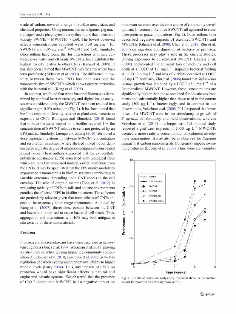
Protozoa and micrometazoans have been described as ecosys-tem engineers (Jones et al. 1994;Weerman et al. 2011) playinga critical role selective grazing impacting community compo-sition (Glucksman et al. 2010; Lawrence et al. 2002) as well asregulation of carbon cycling and nutrient availability to highertrophic levels (Parry 2004). Thus, any impacts of CNTs onprotozoa would have significant effects in natural andengineered aquatic systems. We observed that the presenceof C60 fullerene and MWCNT had a negative impact on
protozoan numbers over the time course of community devel-opment. In contrast, the three SWCNTs all appeared to stim-ulate protistan grazer populations (Fig. 2). Other authors havedescribed negative impacts of oxidized SWCNTs andMWCNTs (Ghafari et al. 2008; Chan et al. 2013; Zhu et al.2006) on ingestion and digestion of bacteria by protozoa.These processes may play a role in the current studies.During exposures to an oxidized SWCNT, Ghafari et al.(2008) documented the apparent loss of mobility and celldeath at a LOEC of 1.6 mg L−1, impaired bacterial feedingat LOEC 3.6 mg L−1 and loss of viability occurred at LOEC6.8 mg L−1. Similarly, Zhu et al. (2006) found that Stylonychiamytilus growth was inhibited by a LOEC of 1 mg L−1 of afunctionalized MWCNT. However, these concentrations aresignificantly higher than those predicted for aquatic environ-ments and substantially higher than those used in the currentstudy (500 μg L−1). Interestingly, and in contrast to ourobservations, Velzeboer et al. (2008, 2011) reported that lowerdoses of a MWCNT were in fact stimulatory to growth ofS. mytilus in laboratory and field observations, whereasVelzeboer et al. (2013) in a longer term (15 months) studyreported significant impacts of 2000 μg L−1 MWCNTsdeemed a more realistic concentration, on sediment inverte-brate communities. It may also be as observed for Daphniamagna that carbon nanomaterials (fullerenes) impede swim-ming behavior (Lovern et al. 2007). Thus, there are a number
Fig. 2 Results of protozoan analyses by treatment show the cumulativecounts for protozoa on a weekly basis (n= 3)
Environ Sci Pollut Res

of potential mechanisms that may contribute to in situ effects;it is also important to note that the diversity of protozoa activein natural biofilm communities is not reflected in those usedfor toxicological studies.
Community-level effects
The microbial community may be the most relevant level oforganization for assessing impacts of nanomaterials in aquaticenvironments, particularly given that biofilms are thought tobe major sinks for nanomaterials (Battin et al. 2009) and aremajor drivers of ecosystem function. A range of diversity andfunctional assessments may be used to determine effects ofCNTs on the microbial community. To date, community-focused studies in soils and water have not used more realisticconcentrations, i.e. reflecting modelled estimates (Gottschalket al. 2009, 2010; Mueller and Nowack 2008) which suggestCNT levels would be from high ng L−1 to low μg L−1 at most.Most frequently, exposures have been in the mg/L to low gL−1 or g/kg range; in the current study, we have attemptedachieve an exposure of 500 μg L−1 in a constantly mixedsystem (Lawrence et al. 2000). We have also applied a rangeof approaches to control and estimate exposure in the biofilmsystem, including ICP-MS analyses of water (Table 3) as wellas scanning transmission X-ray microscopy andspectromicroscopy (STXM) of biofilms (Fig. 3). Our STXManalyses would indicate that the Bin biofilm^ exposures are
very Bpatchy^ and may be essentially zero at many locationsbut may be locally very high in the biofilm SWCNTs. Thisphenomenon is evident in Fig. 3 which shows an example ofmapping of the aggregated SWCNT carbon nanomaterials inin the context of biofilm showing diatoms, bacteria and poly-meric substances using STXM. We have also analysed thenanomaterials for the presence ofmetals using STXManalysisat the Ni 2p edge on pure compounds and in the biofilm.Further, ICP-MS analyses were performed which did not re-veal elevated levels of any metals in the reactor waters duringthe experiment (Table 3). Exposure to CNTs in our experi-ments did not result in significant changes in broad measuressuch as biofilm thickness or chlorophyll-a content of the com-munity (Fig. 4). Although digital imaging indicated signifi-cant changes (p < 0.05), a reduction in algal biomass in
Fig. 3 STXM C1s images of biofilm exposed to SWCNT illustratingclumping, aggregation and localized distribution in the biofilm. Redindicates SWCNT materials, green indicates protein, bacterial cells andpolymeric materials, blue indicates non-specific optical density showingdiatoms, arrows indicate large and small aggregates in the context ofdiatoms, bacteria and polymeric substances
Table 3 Results of ICP-MS analyses of reactor water
Metal Cr Co Cu Mn Mo Ni Ag
Source
Control bld bld 0.9 bld 1.6 1.2 bld
Fullerene C60 bld bld 1 bld 1.6 1.2 bld
MWCNT bld bld 1 bld 1.6 1.5 bld
SWCNT bld bld 0.7 bld 1.6 1.2 bld
SWCNT-OH bld bld 0.6 bld 2.0 1.3 bld
SWCNT-COOH bld bld 0.8 bld 1.6 1.2 bld
Units = μg L−1
Bld below limit of detection
Fig. 4 Graphs of chlorophyll-a concentrations (top) and thickness inbiofilms exposed to the panel of carbon-based nanomaterials.Parameters indicated by different letters are significantly different fromthe control at (p < 0.05, n= 3)
Environ Sci Pollut Res

MWCNT treatment and an increase in algal biomass inSWCNT-COOH exposures (Fig. 1). Thymidine incorpora-tion, a measure of bacterial production was not significantlyaffected (p<0.05) by fullerene exposure but was reduced byMWCNT and all SWCNT exposures (Fig. 5). Soil andsediment-based studies have applied MWCNTs significantlyreducing microbial biomass carbon and nitrogen as well asenzymatic activity when applied at 5 g kg−1, but not at0.5 g kg−1 (Chung et al. 2011). While Velzeboer et al.(2011) found that when MWCNTs were added at 2 g kg−1,no impacts on invertebrate diversity were observed andmacroinvertebrate numbers increased. Authors working withwastewaters have similarly described CNT induced toxicityon the microbial community. For example, Kang et al.(2009) reported toxic effects of MWCNT and SWCNT corre-lated with increasing bacterial cell inactivation in wastewaters.When Yin et al. (2009) examined SWCNT containing efflu-ents, there was a reduction in chemical oxygen demand
removal by the microorganisms, which was confirmed byGoyal et al. (2010). Similarly, Luongo and Zhang (2010)found a decrease in microbial respiration correlated with in-creasing MWCNT concentration in a mixed liquor. Thus, ourobservations of negative impacts on bacterial production arein keeping with the literature.
Carbon utilization spectra generated using the BiologEcoplate system has proven to be a sensitive indicator of ef-fects of various stresses on microbial communities and hasbeen applied in a range of studies (Lawrence et al. 2004,2009). Here, we applied the system to assess the impact of apanel of CNTs on carbon utilization in river biofilm commu-nities. Curiously, of the 30 carbon substrates present in theassay, SWCNT-COOH did not suppress or increase utilizationin any case, SWCNT suppressed utilization of 4-hydroxbenzoic acid and L-phenylalanine (p<0.05) while ful-lerene increased utilization of glycogen and hydroxybutyricacid, MWCNT increased use of 4-hydroxybenzoic acid andhydroxybutryic acid utilization and SWCNT-OH treatmentssignificantly increased utilization of L-phenylalanine andhydroxybutyric acid (Fig. 6). Although there are no other di-rectly comparable studies in the literature, the narrow range ofimpacted carbon substrata appears unusual when compared tostudies of impacts of metals (Lawrence et al. 2004) or phar-maceuticals (Lawrence et al. 2005) where much broader ef-fects were detected.
To investigate the diversity and nature of the community,we applied fluorescent lectin binding analyses (Neu et al.2001) to determine the amount and nature of the exopolymericsubstances produced by the CNT exposed and reference com-munities. Figure 7 shows the results of these analyses, a shiftin total EPS can be seen with significant reductions apparent
Fig. 5 Graph of bacterial production based on thymidine incorporation.Parameters indicated by different letters are significantly differentp< 0.05 (n= 3, ANOVA)
Fig. 6 Results of carbon utilization assays using the Biolog Ecoplate system showing shifts in utilization of specific substrates classes relative to thereference community (n= 3, ANOVA, p < 0.05)
Environ Sci Pollut Res

in theMWCNTand SWCNT treatments in particular. It is alsoevident that each treatment results in a unique EPS fingerprintbased on the results from binding of the five lectin panel(Fig. 7). These patterns are reflective of the nature of theorganisms producing the polymeric substances and thus thebiodiversity of the community. Relative to reference condi-tions (Fig. 7), each community shows shifts in the amountsof terminal β-galactose, N-acetyl galactosamine residues (as-sociated with algal-cyanobacterial polymers), but no signifi-cant change inα-L-fucose residues, which are more in keeping
with bacterial EPS. Visual comparison of the confocal micros-copy images representative of those used for digital imageanalyses (Figs. 1, 4 and 7) demonstrate the unique appearanceof the communities (Fig. 8). That the bacterial diversity inthese communities has changed was confirmed by the PCAanalyses of the results of DGGE analyses (Fig. 9). Otherauthors have addressed the impact of CNTs on microbial di-versity using approaches such as DGGE (Muyzer andRamsing 1995; Muyzer et al. 1993). The application of dena-turing gradient gel electrophoresis (DGGE) did not identifyany changes in the microbial community structure in soil(Tong et al. 2007) or anaerobic biosolids (Nyberg et al.2008) when exposed to C60 fullerenes. These results parallelthose obtained in the current study where the C60 fullerenecommunity is not significantly different (ANOSIM p<0.05)from the control community based on its DGGE fingerprint.Tong et al. (2012) demonstrated that repeated applications ofBas manufactured^ SWCNTs affected microbial communitystructure as measured byDGGE and altered metabolic activityin a low organic matter soil. However, the authors concludedthat the SWCNT treatments did not produce Bsignificantlyaltered^ microbial communities. They did, however, detectchanges in specific organisms detected by sequencingDGGE bands but concluded that only minor changes wereobserved in community structure when SWNTs were appliedat 1000 μg g−1 soil weekly, accumulating to 6000 μg g−1 over
Fig. 7 Results of fluorescent lectin binding analyses of major sugarresidues in the exopolymers of the treatment communities. Bars withdifferent letters are significantly different from each other, the referencecommunity and other treatments (n= 3, p< 0.05, ANOVA)
Fig. 8 Representative CLSM photomicrographs of control, exposed toC60 fullerene, MWCNT, SWCNT, SWCNT-OH and SWCNT-COOHcontinuously for an 8-week experimental period. Bacteria (green),
Triticum vulgaris-TRITC lectin binding polymer (red), photosyntheticbiomass (blue/magenta)
Environ Sci Pollut Res

6 weeks (Tong et al. 2012). It is apparent in the current study(Fig. 9) that exposure to both MWCNT and SWCNT variantscan result in significant shifts in the nature of the eubacterialcommunity. ANOSIM analyses of the DGGE fingerprints in-dicated that the MWCNTcommunity was significantly differ-ent from the control and other CNT exposures. Similarly, theSWCNTexposed community was unique (p<0.05) relative toother communities, while the modified SWCNTs resulted incommunities similar to each other but significantly differentfrom the control as well as fullerene and SWCNT-exposedbiofilm communities. These differences may occur as a resultof direct impacts or toxicity such as selection for or againstspecific community members. However, the observed impactson protozoan grazing (Fig. 2) may also cause shifts in thebacterial community (see discussion above). CNTs and fuller-ene may also interfere with the tight linkages in terms of car-bon and energy flow in the community through impacts onparticular types of organisms or by shifting flows betweenbacteria and photosynthetic organisms that are vital to com-munity structure and function (Haack andMcFeters 1982a, b).It is also possible that their sorptive capacity may create lim-iting conditions by scavenging, metals, micronutrients or nu-trients. However, these effects require further investigation.
Conclusions
A multimetric approach allowed assessment of the impacts ofa panel of carbon-based nanomaterials on structure and func-tion of river biofilm communities. Although the tested levelwas lower than in many earlier studies, significant effects ofall materials could be detected on some aspect of communitystructure or function. It is useful to note that this occurreddespite the aggregation, transformations and coatings that oc-curred, altering CNT properties and reducing toxicity(Lawrence et al. 2016) Fullerene C60 appeared to have min-imal effects with the exception of protozoan grazing and
carbon utilization. Biomass measures such as thickness, chl-a, bacteria and cyanobacterial biomass were relatively insen-sitive to any changes induced by the CNT exposures. In con-trast, metabolic and molecular measures were more indicativeof effects in the community. STXM observations confirmedthat the nanomaterials integrated into the developing commu-nities with a patchy distribution, although estimates wouldsuggest that the biofilm may concentrate the CNTs to anequivalent local concentration of 200 ppm. Given that closecontact appears to be required for toxicity to occur, this im-plies that in situ exposures to the short range effects of CNTsand fullerene in the community are highly variable. BiofilmEPS have been viewed as a significant sink for nanomaterialsas well as a regulator of exposure, perhaps shielding cells fromdirect interaction with nanomaterials in the environment.Based on community level screening, it may be suggested thatSWCNT-OH>SWCNT-COOH>SWCNT>MWCNT>C60Fullerene.
Acknowledgments This work was funded through EnvironmentCanada’s Chemicals Management Plan. The Canadian Light Source(CLS) is supported by the Natural Sciences and Engineering ResearchCouncil of Canada, the National Research Council of Canada, theCanadian Institutes of Health Research, the Province of Saskatchewan,Western Economic Diversification Canada and the University ofSaskatchewan.
References
Ajayan PM, Zhou OZ (2001) Applications of carbon nanotubes. CarbonNanotubes 80:391–425
Ajayan PM, Charlier JC, Rinzler AG (1999) Carbon nanotubes: frommacromolecules to nanotechnology. Proc Natl Acad Sci U S A 96:14199–14200
Akhavan O, Abdolahad M, Abdi Y, Mohajerzadeh S (2009) Synthesis oftitania/carbon nanotubes heterojunction arrays for photoinactivationof E. coli in visible light irradiation. Carbon 47:3280–3287
Apul OG, Shao T, Zhang S, Karanfil T (2012) Impact of carbon nanotubemorphology on phenanthrene adsorption. Environ Toxicol Chem31(1):73–78
Arias LR, Yang L (2009) Inactivation of bacterial pathogens by carbonnanotubes in suspensions. Langmuir 25(5):3003–3012. doi:10.1021/la802769m
Ball P (2001) Roll up for the revolution. Nature 414:142–144Battin TJ, Kammer FVD, Weilhartner A, Ottofuelling S, Hofmann T
(2009) Nanostructured TiO2: transport behavior and effects onaquatic microbial communities under environmental conditions.Environ Sci Technol 43:8098–8104
Bennett SW, Adeleye A, Ji Z, Keller AA (2013) Stability, metal leaching,photoactivity and toxicity in freshwater systems of commercial sin-gle wall carbon nanotubes. Water Res 47:4074–4085. doi:10.1016/j.watres.2012.12.039
Boon N, Windt WD, Verstraete W, Top EM (2002) Evaluation of nestedPCR DGGE (denaturing gradient gel electrophoresis) with group-specific 16S rRNA primers for the analysis of bacterial communitiesfrom different wastewater treatment plants. FEMS Microbiol Ecol39:101–112
Boxall ABA, Chaudhry Q, Sinclair C, Jones A, Aitken R, Jefferson B,Watts C (2007) Current and future predicted environmental
Fig. 9 Analyses of DGGE analyses using ANOSIM where r= 0.78 andp < 0.001 confirmed a significant difference between communitiesexposed to the various CNTs and C60 fullerene. Each point representsthe average of 3 replicate gels, n= 3
Environ Sci Pollut Res

exposure to engineered nanoparticles; central science laboratory,department of the environment and rural affairs: London, UK
Chae So-R, Hotze EM, Xiao Y, Rose J, Wiesner MR (2010) Comparisonof methods for fullerene detection and measurements of reactiveoxygen production in cosmetic products. Environ Eng Sci 27:797–804. doi:10.1089/ees.2010.0103
Chan TS, Nasser F, St-Denis CH,Mandal HS, Ghafari P, Hadjout-Rabi N,Bols CN, Tang XS (2013) Carbon nanotube compared with carbonblack: effects on bacterial survival against grazing by ciliates andantimicrobial treatments. Nanotoxicology 7:251–258
Chen Q, Saltiel C, Manickavasagam S, Schadler LS, Siegel RW, YangHC (2004) Aggregation behavior of single-walled carbon nanotubesin dilute aqueous suspension. J Colloid Interface Sci 280:91–97
Chenier MR, Beaumier D, Roy R, Driscoll BT, Lawrence JR, Greer CW(2003) Impact of seasonal variations and nutrient inputs on the cy-cling of nitrogen and the degradation of hexadecane by replicatedriver biofilms. Appl Environ Microbiol 69:5170–5177
Chung H, Son Y, Yoon TK, Kim S, Kim W (2011) The effect of multi-walled carbon nanotubes on soil microbial activity. EcotoxicolEnviron Saf 74:569–575
Clarke KR (1993) Non-parametric multivariate analyses of changes incommunity structure. Aust J Ecol 18:117–143
Dreher KL (2004) Health and environmental impact of nanotechnology:toxicological assessment of manufactured nanoparticles. Toxicol Sci77:3–5
Dynes JJ, Lawrence JR, Korber DR, Swerhone GDW, Leppard GG,Hitchcock AP (2006a) Quantitative mapping of chlorhexidine innatural river biofilms. Sci Total Environ 369:369–383
Dynes JJ, Tyliszczak T, Araki T, Lawrence JR, Swerhone GDW, LeppardGG, Hitchcock AP (2006b) Speciation and quantitative mapping ofmetal species in microbial biofilms using scanning transmission X-ray microscopy. Environ Sci Technol 40:1556–1565
Fang JS, Lyon DY, Wiesner MR, Dong JP, Alvarez PJJ (2007) Effect of afullerene water suspension on bacterial phospholipids and mem-brane phase behavior. Environ Sci Technol 41:2636
Fortner JD, Lyon DY, Sayes CM, Boyd AM, Falkner JC, Hotze EM,Alemany LB, Tao YJ, Guo W, Ausman KD, Colvin VL, HughesJB (2005) C60 in water: nanocrystal formation and microbial re-sponse. Environ Sci Technol 39:4307–4316
Ghafari P, St-Denis CH, Power ME, Jin X, Tsou V, Mandal HS, BolsNC, Tang XS (2008) Impact of carbon nanotubes on the inges-tion and digestion of bacteria by ciliated protozoa. NatNanotechnol 3:347–351
Glucksman E, Bell T, Griffiths RI, Bass D (2010) Closely related protiststrains have different grazing impacts on natural bacterial commu-nities. Environ Microbiol 12:3105–3113. doi:10.1111/j.1462-2920.2010.02283.x
Gottschalk F, Sonderer T, Scholz RW, Nowack B (2009) Modeled envi-ronmental concentrations of engineered nanomaterials (TiO2, ZnO,Ag, CNT, Fullerenes) for different regions. Environ Sci Technol 43:9216–9222
Gottschalk F, Sonderer T, Scholz RW, Nowack B (2010) Possibilities andlimitations of modeling environmental exposure to engineerednanomaterials by probabilistic material flow analysis. EnvironToxicol Chem 29:1036–1048
Goyal D, Zhang XJ, Rooney-Varga JN (2010) Impacts of single-walledcarbon nanotubes on microbial community structure in activatedsludge. Lett Appl Microbiol 51:428–435
Haak SK, McFeters GA (1982a) Nutritional relationships among mi-croorganisms in an epilithic biofilm community. Microb Ecol 8:115–126
Haak SK, McFeters GA (1982b) Microbial dynamics of an epilithic matcommunity in a high alpine stream. Appl Environ Microbiol 43:702–707
Handy RD, OwenR, Valsami-Jones E (2008) The ecotoxicology of nano-particles and nanomaterials: current status, knowledge gaps, chal-lenges, and future needs. Ecotoxicology 17:315–325
Hitchcock AP (2012) Soft X-ray imaging and spectromicroscopy chapter22 in volume II of the handbook on nanoscopy, eds. Gustaaf VanTendeloo, Dirk Van Dyck and Stephen J. Pennycook (Wiley, 2012)pp 745–791
Hitchcock AP (2014) aXis2000 is written in Interactive Data Language(IDL). It is available free for non-commercial use from http://unicorn.mcmaster.ca/aXis2000.html
Hyung H, Fortner JD, Hughes JB, Kim J-H (2007) Natural organic matterstabilizes carbon nanotubes in the aqueous phase. Environ SciTechnol 41:179–184
Jackson P, Jacobsen NR, Baun A, Birkedal R, Kuhnel D, Jensen KA,Vogel U, Wallin H (2013) Bioaccumulation and ecotoxicity of car-bon Nanotubes. Chemistry Central J 7:154 http://journal.chemistrycentral.com/content/7/1/154
Jacobsen C, Wirick S, Flynn G, Zimba C (2000) Soft X-ray microscopyfrom image sequences with sub-100 nm spatial resolution. J Microsc197:173–184
Jia G, Wang HF, Yan L, Wang X, Pei RJ, Yan T, Zhao YL, Guo XB(2005) Cytotoxicity of carbon nanomaterials: single-wall nano-tube, multi-wall nanotube, and fullerene. Environ Sci Technol39:1378–1383
Jones CG, Lawton JH, Shackak M (1994) Organisms as ecosystem engi-neers. Oikos 69:373–386
Kang S, Pinault M, Pfefferle LD, Elimelech M (2007) Single-walledcarbon nanotubes exhibit strong antimicrobial activity. Langmuir23:8670–8673
Kang S, Herzberg M, Rodrigues DF, Elimelech M (2008)Antibacterial effects of carbon nanotubes: size does matter!Langmuir 24:6409–6413
Kang S, Mauter MS, Elimelech M (2009) Microbial cytotoxicity ofcarbon-based nanomaterials: implications for river water and waste-water effluent. Environ Sci Technol 43:2648–2653
Kaznatcheev KV, Karunakaran C, Lanke UD, Urquhart SG, Obst M,Hitchcock AP (2007) Soft X-ray spectromicroscopy beamline atthe CLS: commissioning results. Nucl Instrum Methods Phys ResSect A 582:96–99
Kohler AR, Som C, Helland A, Gottschalk F (2008) Studying the poten-tial release of carbon nanotubes throughout the application life cy-cle. J Clean Prod 16(8–9):927–937
Lawrence JR, Swerhone GDW, Neu TR (2000) Design and evaluation ofa simple rotating annular reactor for replicated biofilm studies. JMicrobiol Methods 42:215–224
Lawrence JR, Scharf B, Packroff G, Neu TR (2002) Microscale evalua-tion of the effects of grazing by invertebrates with contrasting feed-ing modes on river biofilm architecture and composition. MicrobEcol 44:199–207
Lawrence JR, Chenier M, Roy R, Beaumier D, Fortin N, SwerhoneGDW, Neu TR, Greer CW (2004) Microscale and molecular assess-ment of the impacts of nickel, nutrients and oxygen level on riverbiofilm communities. Appl Environ Microbiol 70:4326–4339
Lawrence JR, Swerhone GDW, Wassenaar LI, Neu TR (2005) Effects ofselected pharmaceuticals on riverine biofilm communities. Can JMicrobiol 51:655–669
Lawrence JR, Zhu B, Swerhone GDW, Roy J, Wassenaar LI, Topp E,Korber DR (2009) Comparative microscale analysis of the effects oftriclosan and triclocarban on the structure and function of river bio-film communities. Sci Total Environ 407:3307–3316
Lawrence JR, Dynes JJ, Korber DR, Swerhone GDW, Leppard GG,Hitchcock AP (2012) Monitoring the fate of copper nanoparticlesin river biofilms using scanning transmission X-ray microscopy(STXM). Chem Geol 329:18–25
Lawrence JR, Swerhone GDW, Dynes JJ, Hitchcock AP, Korber DR(2016) Complex organic corona formation on carbon nanotubes
Environ Sci Pollut Res

reduces microbial toxicity by suppressing reactive oxygen speciesproduction. Environ Sci Nano. doi:10.1039/C5EN00229J
Lee BI, Qi L, Copeland T (2005) Nanoparticles for materials design:present and future. J Ceram Process Res 6:31–40
Liu Y, Li J, Qiu X, Burda C (2007) Bactericidal activity of nitrogendoped metal oxide nanocatalysts and the influence of bacterialextracellular polymeric substances (EPS). J PhotochemPhotobiol A 190(1):94–100
Long Z, Ji J, Yang K, Lin D, Wu F (2012) Systematic and quantitativeinvestigation of the mechanism of carbon nanotubes' toxicity towardalgae. Environ Sci Technol 46:8458–8466
Lovern SB, Strickler JR, Klaper R (2007) Behavioral and physiologicalchanges in Daphnia magna when exposed to nanoparticle suspen-sions (titanium dioxide, nano-C60, and C60HxC70Hx). Environ SciTechnol 41:4465–4470
Luongo LA, Zhang XJ (2010) Toxicity of carbon nanotubes to the acti-vated sludge process. J Hazard Mater 178:356–362
Lyon DY, Adams LK, Falkner JC, Alvarez PJJ (2006) Antibacterial ac-tivity of fullerene water suspensions: effects of preparation methodand particle size. Environ Sci Technol 40:4360–4366. doi:10.1021/es0603655
Lyon DY, Brunet L, Hinkal GW, Wiesner MR, Alvarez PJJ (2008)Antibacterial activity of fullerene water suspensions (nC60) is notdue to ROS-mediated damage. Nano Lett 8:1539
Mauter MS, Elimelech M (2008) Environmental applications of carbon-based nanomaterials. Environ Sci Technol 42:5843–5859
Mueller NC, Nowack B (2008) Exposure modeling of engineered nano-particles in the environment. Environ Sci Technol 42:4447–4453
Musee N, Thwala M, Nota N (2011) The antibacterial effects ofengineered nanomaterials: implications for wastewater treatmentplants. J Environ Monit 13:1164–1183
Muyzer G, Ramsing NB (1995) Molecular methods to study the organi-zation of microbial communities. Water Sci Technol 32:1–9
Muyzer G, De Waal EC, Uitterlinden AG (1993) Profiling of complexmicrobial populations by denaturing gradient gel electrophoresisanalysis of polymerase chain reaction-amplified genes coding for16S rRNA. Appl Environ Microbiol 59:695–700
Neal A (2008) What can be inferred from bacterium–nanoparticle inter-actions about the potential consequences of environmental exposureto nanoparticles? Ecotoxicology 17(5):362–371
Nel A, Xia T, Madler L, Li N (2006) Toxic potential of materials at thenanolevel. Science 311:622–627
Neu TR, Swerhone GDW, Lawrence JR (2001) Assessment of lectin-binding-analysis for in situ detection of glycoconjugates in biofilmsystems. Microbiology 147:299–313
Nyberg L, Turco RF, Nies L (2008) Assessing the impact ofnanomaterials on anaerobic microbial communities. Environ SciTechnol 42:1938–1943
Parry JD (2004) Protozoan grazing of freshwater biofilms. Adv ApplMicrobiol 54:167–196
Petersen EJ, Zhang L, Mattison NT, O’Carroll DM, Whelton AJ, UddinN, Nguyen T, Huang Q, Henry TB, Holbrook RD, Chen KL (2011)Potential release pathways, environmental fate, and ecological risksof carbon nanotubes. Environ Sci Technol 45:9837–9856
Rodrigues DF, Elimelech M (2010) Toxic effects of single-walled carbonnanotubes in the development of E. coli biofilm. Environ SciTechnol 44(12):4583–4589. doi:10.1021/es1005785
Schwab F, Bucheli TD, Lukhele LP, Magrez A, Nowack B, Sigg L,Knauer K (2011) Are carbon nanotube effects on green algae causedby shading and agglomeration? Environ Sci Technol 45:6136–6144
Theron J, Walker JA, Cloete TE (2008) Nanotechnology and water treat-ment: applications and emerging opportunities. Crit Rev Microbiol34:43–69
Tong ZH, Bischoff M, Nies L, Applegate B, Turco RF (2007) Impact offullerene (C-60) on a soil microbial community. Environ SciTechnol 41:2985–2991
Tong Z, Bischoff M, Nies LF, Myerm P, Applegate B, Turco RF (2012)Response of soil microorganisms to as-produced and functionalizedsingle-wall carbon nanotubes (SWNTs). Environ Sci Technol 46:13471–13479. doi:10.1021/es303251r
Velzeboer I, Hendriks AJ, Ragas AMJ, Van de Meent D (2008) Aquaticecotoxicity tests of some nanomaterials. Environ Toxicol Chem 27:1942–1947
Velzeboer I, Kupryianchyk D, Peeters ETHM, Koelmans AA (2011)Community effects of carbon nanotubes in aquatic sediments.Environ Int 37:1126–1130
Velzeboer I, Peeters ETHM, Koelmans AA (2013) Multiwalled carbonnanotubes at environmentally relevant concentrations affect thecomposition of benthic communities. Environ Sci Technol 47:7475–7482. doi:10.1021/es400777j
Weerman E, Van der Geest HG, Van der Meulen MD, Manders EMM,Van de Koppel J, Herman PMJ, Admiraal W (2011) Ciliates asengineers of phototrophic biofilms. Freshw Biol 56:1358–1369
Wei LP, Thakkar M, Chen YH, Ntim SA, Mitra S, Zhang XY (2010)Cytotoxicity effects of water dispersible oxidized multiwalled car-bon nanotubes onmarine alga,Dunaliella tertiolecta. Aquat Toxicol100:194–201
Wolfaardt GM, Lawrence JR, Headley JV, Robarts RD, Caldwell DE(1994) Microbial exopolymers provide a mechanism for bioaccu-mulation of contaminants. Microb Ecol 27:279–291
Yang C, Mamouni J, Tang Y, Yang L (2010) Antimicrobial activity ofsingle-walled carbon nanotubes: length effect. Langmuir 26:16013–16019
Yin Y, Zhang X, Graham J, Luongo L (2009) Examination of purifiedsingle-walled carbon nanotubes on activated sludge process usingbatch reactors. J Environ Sci Health A Tox Hazard Subst EnvironEng 44:661–665
Zhu Y, Zhao Q, Li Y, Cai X, Li W (2006) The interaction and toxicity ofmultiwalled carbon nanotubes with Stylonychia mytilus. J NanosciNanotechnol 6:1357–1364
Environ Sci Pollut Res