a3williamcalvin.com/1970s/1973en5.pdfdeafferentation effects in lateral cuneate nucleus of the cat:...
TRANSCRIPT

EXPERIMENTAL NEURLOGY 39, 86-102 (1973)
Deafferentation Effects in Lateral Cuneate Nucleus ofthe Cat : Correlation of Structural Alterations with
Firing Pattern Changes
TERRELL D. KJERULF, JOHN T. O'NEAL, WILLIAM H. CALVIN,
JOHN D. LOESER AND LESNICK E. WESTRUM 1
Department of Neurological Surgery, University of Washington School ofMedicine, Seattle, Washington 98195
Received September 23, 1972
A correlated anatomical and physiological investigation of the effects ofunilateral cervicothoracic dorsal rhizotomies upon lateral cuneate nucleusof the cat (LCN) is reported. Pairs of adult cats with identical survivaltimes were selected to correlate structural and functional changes. Twophases are described in the development of alterations of neuronal firingpatterns . In the first phase, a relative silence within LCN was associatedwith depletion of round synaptic vesicles in the presynaptic profiles (LRboutons) of primary dorsal root afferents . The second phase was char-acterized by a development of spontaneous electrical hyperactivity whichcorresponded anatomically to the presence of denuded postsynaptic specializa-tions, transient increase of adjacent extracellular space and an apparent de-crease in the number of dendritic spines . There was a persistence of anunaltered population of small presynaptic boutons with flattened vesicles(SF boutons) . The LCN neuronal membrane is viewed as having an in-trinsic tendency for repetitive firing which is enhanced by the functionaleffects of denuded postsynaptic specialization . A marked similarity wasfound between some of the spontaneous firing patterns of normal animals(doublets) and the high frequency bursting firing pattern in deafferentedpreparation. Three models for repetitive spike production are consideredin our analysis : oscillator-produced spikes ; EP SP-produced spikes ; andspike-evoked spikes . The spike-evoked spikes model is considered to be the
1 The authors thank William Congdon, Jerrold Maddocks, Vladimir Uhlir and Pa-tricia Yadock for their assistance in these experiments. This work was supported inpart by Grants NS04053 (Epilepsy Research), NS05211 (Neurosurgery Training),NS09677 (Motoneuron Repetitive Firing Mechanisms) and NS09678 (Deafferenta-tion) from the National Institute of Neurological Diseases and Stroke . Dr. O'Neal'spresent address is Division of Neurosurgery, University of Texas Medical Branch,Galveston, Tex . Dr. Westrum holds a joint appointment with Department of BiologicalStructure.
86
Copyright © 1973 by A,
is Press, Inc .All rights of reproductions my form reserved .
A3
I
U)

DEAFFERENTATION EFFECTS
origin of normal doublet activity and a candidate for the deafferented burstactivity . Abnormal hyperactivity after deafferentation may be a function ofchanges in the membrane characteristics occurring at or near the denudedpostsynaptic specializations .
INTRODUCTION
Claude Bernard, in 1872, proposed the general concept that tissue excit-ability may be increased by isolation from the controlling nervous input(3) . Cannon and Rosenblueth further elaborated this concept and formu-lated a theory of "denervation supersensitivity" applicable to all excitablestructures from smooth muscle to the central nervous system (8) . In theensuing 30 years, a number of students have investigated partial deaffer-entation of the central nervous system as reflected in abnormalities ofmotoneuron output and muscular response . Extracellular recordings fromdeafferented spinal cord demonstrated not only alterations of spontaneousneuronal activity, but also abnormal evoked responses (17) . In additionextracellular recordings in the spinal trigeminal nucleus have shownsimilar altered firing patterns (1) .Deafferentation has produced comparable abnormalities in spontaneous
neuronal activity patterns in the lateral cuneate nucleus (LCN) of cats .Our previous papers (14, 19) have described in detail the neurophysio-logical and fine structural features of this form of partial deafferentation .In the present paper, we shall correlate these individual observations toestablish a background for discussion of the possible mechanisms ofneuronal hyperactivity and repetitive firing .
MATERIALS AND METHODS
This paper utilizes a portion of the anatomical and physiological datareported separately in two other papers (14, 19) . Adult cats (2-5 kg)with survival times of 24 and 45 hr, 6, 10 and 16 days, have been selectedto correlate the structural and functional changes observed after Ci-T7dorsal rhizotomv. Two animals for each survival time were used, one forphysiological and the other for anatomical studies . The surgical techniquesemployed to accomplish the deafferentation have been described in detail(14, 19) . They are the same for each member of a pair of animals exceptthat the T i root was preserved in the animals for physiological recordingto allow characterization of units retaining their peripheral receptive fieldin the deafferented nucleus . The methods of tissue fixation and handlingfor electron microscopy, as well as the techniques for physiological record-ing, have been described in detail in our previous papers (14, 19) .
The physiological data analysis techniques employed in this paper arebasically those described by Calvin (5) . The definition of a burst is the
87

88
KJERULF ET AL .
property of a computer program which scans the list of interspike intervalsstored on computer tape after sampling from the original data recordings .This program looks for interspike intervals longer than 30 cosec ; uponfinding such a long interval, it examines the very next interval to see if itis shorter than 10 cosec . If both criteria are satisfied in succession, theshort interval is considered to denote the beginning of a doublet or burst .The "long" and "short" interval criteria may be varied by the computeroperator ; while 30 and 10 msec were common values, 15 and 5 cosec werealso used for one illustration . This burst detection scheme is utilized bythe programs which make rasters and histograms . This scheme is notinfallible ; indeed, one can detect occasional examples of the inclusion ofnonbursts in the burst rasters in this paper and the previous paper (14) .Sampling time bins were 0 .2 cosec. i n duration .
RESULTS
A detailed description of the normal fine structural architecture and-physiological characteristics of the cat lateral cuneate nucleus (LCN)was included in the previous papers (14, 19) . As a basis for later dis-cussion, a description of the normal repetitive firing mechanisms is intro-duced here .
One form of normal spontaneous activity in LCN includes neurons witha very high firing rate capability, appearing as doublets and, occasionally,triplets . Calvin (6) distinguished three models for multiple spike produc-tion which are germane to analysis of repetitive firing in LCN . Mostcommonly, a large depolarizing wave will evoke multiple spikes by con-verting the depolarization magnitude into firing frequency . The spikesfollowing the initial spike arise out of the interaction of a sustained synapticdepolarization with an oscillator mechanism. These oscillator-producedspikes bear no relationship to the timing of the various inputs . As thedepolarizing current increases, the interspike interval decreases . Char-acteristic of this mechanism, then, is a fluctuating depolarizing drive re-flected in a variable interspike interval. The brevity of the doublet activityand its relative invariability in LCN make it doubtful that a depolariza-tion-to-frequency model can adequately account for the phenomenon .
A second model accounts for a repeated action potential response viaan EPSP-produced spike ; in this model, the EPSP from a single inputmay be almost large enough to fire the cell. Here, the timing of the inputand output spikes is correlated . This model may be applicable to theClarke's column cells where single IA fibers may produce EPSP in the2-5 my range (11) and, by analogy, to the LCN cell where presynapticprofiles are large and probably establish multiple contacts with postsynaptic
A)
,1

r UE.1FFERENTATION EFFECTS
89
elements. While such a large EPSP might produce the first spike, it wouldbe unlikely that a second subsequent EPSP could produce the second spikeand maintain the precise timing between the first and second spike of the
doublet. This would tend to rule out sequential EPSP from two differentafferent fibers, as they could not be expected to retain such synchrony . It
is also unlikely that multiple contacts from a single bifurcating afferent
fiber would produce two successive EPSP separated by an interval of
1-1 .6 cosec (which was the observed interspike interval separation in
I .CN) .The third model for repetitive firing is found in the depolarizing after-
potential theory (6, 13) . The depolarizing aftermath of the initial spikemay rise through threshold and cause an extra spike, even if the original
depolarizing influence has dissipated . The second spike is correlated with
the peak of the delayed depolarization (DD) from the first spike . If theDD peak occurs within 2 msec, one would expect the extra spike to occurbefore 2 cosec . Inspection of Fig . 1 of this paper and Fig . 1 of the precedingpaper (14) demonstrates the second spike occurring within 2 cosec of the
first spike . The few existing intracellular records from the LCN are notof sufficient quality to demonstrate this depolarizing afterpotential if it does
exist (9, 21) . It is interesting, however, that the homologous Clarke'scolumn cells invariably present a delayed depolarization, peaking about 2
msec following the initiation of the spike (10, 11) . Thus, for the present,
as in spinal motoneuron (7) and hippocampal pyramidal cell (13), it seemsplausible that the spike-evoked spikes mechanism is sometimes utilized by
the LCN cell under normal circumstances . Peripheral needle electrode
stimulation of LCN receptive fields yields a train of spikes from cells in thenormal nucleus . This artificial, synchronized input probably acts througha potent synaptic effect and oscillator-produced spikes (16) .
normal
one day
Tic . 1 . Spontaneous activity in LCN . Upper trace, normal cat ; middle trace, 24hr after ipsilateral dorsal rhizotomies of C,-T7, sparing T, . Note the absence of unitactivity : Lower trace, 6 days after ipsilateral dorsal rhizotomies of C-T7 sparingT,. Note the hyperactivity and burst firing patterns . Calibration : 10 cosec . Spikes areabout 200 µv. Gain in 1-day trace is half of others .
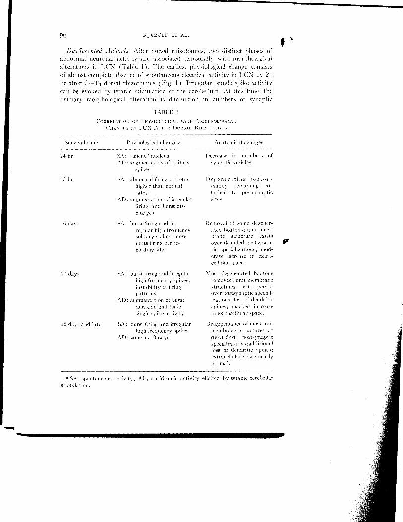
90 KJERtLF ET AL .
Deafferenied Animals . After dorsal rhizotomies, two distinct phases ofabnormal neuronal activity are associated temporally with morphologicalalterations in LCN (Table 1) . The earliest physiological change consistsof almost complete absence of spontaneous electrical activity in LCN by 24hr after C I-T7 dorsal rhizotomies (Fig. 1) . Irregular, single spike activitycan be evoked by tetanic stimulation of the cerebellum . At this time, theprimary morphological alteration is diminution in numbers of synaptic
TABLE 1
Cog RELATION OF PHYSIOLOGICAL v'rtI MORPftOLOGICALCHANLGS IS LCN ArrFe DORSAL RHIZOTOMnES
6 days
SA : burst firing and ir-regular high frequencysolitary spikes ; moreunits firing per re-cording site
10 days
SA: burst firing and irregularhigh frequency spikes ;instability of firingpatterns
AD : augmentation of burstduration and tonicsingle spike activity
16 days and later
SA: burst firing and irregularhigh frequency spikes
AD : same as 10 days
SA, spontaneous activity ; AD, antidromic activity elicited by tetanic cerebellarstimulation .
Removal of some degener-ated boutons ; unit mem-brane structure existsover denuded postsynap-tic specializations ; mod-crate increase in extra-cellular space .
Most degenerated boutonsremoved ; unit membranestructures still persistover postsynaptic special-izations ; loss of dendriticspines ; marked increasein extracelhilar space .
Disappearance of most unitmembrane structures atdenuded postsynapticspecializations ;additionalloss of dendritic spines ;extracellular space nearlynormal .
1
Survival time Physiological Changes" Anatomical changes
24 hr SA : "silent" nucleus Decrease in numbers ofAD : augmentation of solitary synaptic vesicles
spikes
45 hr SA : abnormal firing patterns, Degenerating boutonshigher than normal mainly remaining at-rates . tached to postsvnaptic
AD : augmentation of irregular sitesfiring, and burst dis-charges

DEAFFERENTATION EFFECTS 91
FIG . 2 . (a) A preparation from LCN of a normal cat showing the frequently oc-curring large boutons with round vesicles (LR) contacting a medium-sized dendrite(D) both directly and via a spine (s) . X11,700. (b) Twenty-four hours survival .Moderate vesicle depletion in swollen LR boutons (dLR) and an unaltered SFbouton contacts a dendrite (D) and spine (s) . X11,700 .

9 2 KJERULF LT AL .
vesicles in the large presynaptic profiles with round vesicles (LR boutons)(Fig. 2) . This diminution ranges from a slight decrease in numbers ofsynaptic vesicles to almost total depletion . In some boutons where vesicleloss is incomplete, the remaining vesicles are aggregated and pleomorphic .The majority of LR profiles in LCN seem to show these changes .
The second phase of abnormal neuronal activity develops between 45 hrand 10 days after dorsal rhizotomies . It is characterized initially by anaugmented response to tetanic cerebellar stimulation (2-day survival)which consists of repetitive, single spikes interspersed with burst firing .This evoked form of hyperactivity is succeeded by a similar, but spon-taneously occurring, form of rhythmically recurrent bursting by 6 daysafter deafferentation . As previously described (14), burst recurrence ratestend to vary from unit to unit (6-12/sec) but are remarkably regular foreach individual unit . Cessation of each burst is typically abrupt with onlya slight decline in frequency prior to termination (Fig . 1) .
The structural changes which occur between 2 and 6 (lays after dorsalrhizotomies are characterized by a sequential progression of degenerationof essentially all 1 .R boutons from electron-lucent to electron-dense forms(Fig. 3) . A few denuded poAsynaptic specializations (as in Fig . 5) areidentifiable by 6 (lays ; however, many of the degenerating boutons remainattached to postsynaptic elements at this time . The small presynaptic pro-
Fic. 3. Six days survival . Dense degenerated boutons (dB) are attached to adendrite (D) . SF boutons contact the dendrite and a spine (s) . X12,150 .
IR
1

1EAFFERENTATION EFFECTS
T
KF11-11
~ II- lOmsec
0 5 10 spikes per burst
- -- ---------
T2
93
T2--
Fin. 4 . Augmented burst response in LCN . Oscilloscope trace T r shows a burst (3-4spikes/burst) recurring at 6/sec following tetanic cerebellar stimulation . Raster Tcorresponds to the oscilloscope trace. Note the end of tetanic stimulation at the topof the raster. At upper right is the spikes per burst histogram for the T i activity .Oscilloscope trace Tz, immediately following second application of tetanic stimulus,shows a burst (4-9 spikes/burst) recurring at 7/sec . Raster T2 corresponds to the T_oscilloscope trace . Note the end of tetanic stimulation at the top of the raster and thefollowing immediate increase in spikes/burst with no alteration of the interspike in-terval . The T2 histogram demonstrates the corresponding spike/burst node . The 10-msec calibration mark applies to both oscilloscope traces. The vertical rows of dotson the histograms mark 0, 5 . and 10 spikes/burst. Spike amplitude 350 µv .
files with flattened synaptic vesicles (SF boutons) in LCN apparently do,not undergo any morphologic alterations .
By 10 days after dorsal rhizotomies the spontaneous activity patternin LCN, which was rather stereotyped for any given unit at earlier survivals,now manifests unstable firing patterns . The previously characteristic highfrequency bursts alternate with shorter bursts and a tonic pattern ofsolitary spikes . This spontaneous cycling activity seems to be unrelated toextraneous factors such as blood pressure or respiration. In addition the

94KJERULF ET AL .
1e
-1 1)-synaptic special i zation s (arrows) and in-FIG . 5 . Ten days survival .
creased extracellular space (asterisks) are indicated . Unaltered SF boutons persist .
M2,150 .
FIG. 6 . Ten days survival . Denuded postsynapticsites retain unit membrane
structures (arrows) and there are apposed glial profiles (G) . X44,100-

r
r
DEAFFERENTATION EFFECTS
augmented response to tetanic cerebellar stimulation first discernable in the45 hr survival is still present (Fig . 4) . Associated with these physiologicalalterations is an increase in the numbers of denuded postsynaptic specializa-tious previously apposed by degenerating LR profiles (Fig . 5) . After re-moval of these L .R boutons the extracellular space adjacent to the denudedpostsynaptic specializations is larger than normal and, at least transiently,often measures up to about 0 .2 µin . This localized increase in extracellularspace is most pronounced around medium to large dendrites 10 days afterdeafferentation (Fig . 5) .
Concomitant with (glial) removal of degenerated LR profiles and as-sociated temporally NN"ith the physiological changes of the 6- and 10-daysurvivals, two further anatomical alterations occur . A unit membrane-likestructure ( Fig. 6) is visible in the extracellular space directly over denudedpostsynaptic specializations . On its other side, i .e ., away from the post-synaptic specialization, this structure may be apposed by a variety ofelements which include glia, dendrites, axons or SF boutons (Fig . 6) .Secondly, the number of dendritic spines seems to decrease . Those spinesthat remain are contacted by SF boutons as in Fig . 3 .
Physiologically, the abnormal neuronal firing patterns summarized aboveseem firmly established by 10 days after dorsal rhizotomies . No additionalalterations in spike activity could be demonstrated in animals with longersurvivals . It must be emphasized that those units in LCN which respondedto peripheral stimulation (T i root was spared in those animals utilizedfor physiological study) never manifested the abnormalities seen in unitswhich had lost their afferent drive (14) . They retained all the featuresof the normal response to cerebellar and peripheral stimulation (Fig . 8) .The morphological picture is also essentially stable by this time. It con-sists of those features described for 10 days survival with the addition oftwo new alterations : an apparent loss of many of the unit membrane-likestructures in the extracellular space over denuded specializations ; andan apparent decrease in the size of the enlarged extracellular space seenadjacent to denuded specializations in the 10-day survival (Fig . 7) . Bothalterations take place between 10 and 16 days after dorsal rhizotomy .
DISCUSSION
The two clear-cut phases in the development of neuronal firing changesseem to be correlated with the concomitant fine structural alterations . Theearliest physiological change is relative electrical silence within LCN which
95
FIG . 7. Sixteen days survival . A denuded postsynaptic specialization (arrow) is ap-posed by glial lamellae (G) and appears to have lost the unit membrane structure .X46,800 .

96
SA
KJERULF ET AL.
KF12-7
I1
SA
SA
Fic. 8 . Normal spontaneous and evoked activity of unit with intact peripheralreceptive field . Oscilloscope trace KF12-7 illustrates a 400 µv unit with doublet firing
mode recurring at 18/sec . The combined spontaneous activity-natural stimulation
raster (SA, NS) shows the prestimulation doublet and then the increase of ac-tivity secondary to foot-pad pressure . The time scale at the top of the raster showsa dot every 1 msec for 20 msec, then one dot for each subsequent 10 msec . The T 1 ,NS raster (combination of activity immediately following tetanic cerebellar stimula-tion and peripherally evoked activity during this period) has the same time scale . At
the top left of the raster is the end of the tetanic stimulation period . Note the failure
of tetanic stimulation to alter firing frequency . There is a spread of activity mid-raster subsequent to natural stimulation . The SA raster at upper right demonstratesthe spontaneous activity before tetanic cerebellar stimulation at an expanded time
scale. The time scale at top marks each 1 msec . The SA histogram shows the
probability density of spikes . The oscilloscope trace T~ (immediately following tetanic
cerebellar stimulation) shows a 400 µv unit recurring at 22/sec . The 10-cosec calibra-
tion applies to both kymograph traces. Unit KF12-7 was a cutaneous unit (C-CCT)responding to foot pad pressure .

f DEAFFERENTATION EFFECTS 97
is presumably a manifestation of the loss of afferent drive . Previous in-vestigations in both the peripheral and central nervous systems have sug-gested an association of transmission failure with depletion of synapticvesicles. At the neuromuscular junction, Birks, Katz and Miledi foundsynaptic vesicle reduction at a time when failure of transmission and lossof spontaneous end plate activity were seen (4) . Hunt and Nelson alsodemonstrated a similar association early in the course of degeneration indenervated frog sympathetic ganglion (12) . In the CNS an identical as-sociation has been documented for transmission failure in the cat lateralgeniculate nucleus (20, 22) . The decreased activity in LCN, while notspecifically indicative of synaptic transmission failure, does correspondto a marked reduction of synaptic vesicles in degenerating LR boutons .Transmission failure may he the basis for the decreased neuronal activity,although it is unlikely that axonal conduction failure has occurred at thistime (18) .
The second phase of the reaction to deafferentation commences between2 and 10 clays after dorsal rhizotomies . Marked alterations of restingactivity make their appearance during this period . These are usuallycharacterized by the development of spontaneous hyperactivity in the formof burst firing, and unstable firing patterns which involve alternating orcycling modes of firing . During this period, electron microscopy showsdegeneration of essentially the entire population of LR boutons and theprogressive glial removal of these elements from their postsynaptic special-izations. By 10 days, when unstable firing patterns are observed, the mostcharacteristic anatomical alteration is denudation of postsynaptic special-izations accompanied by an increase in the extracellular space near thesesites and an apparent reduction in the number of dendritic spines . Thedenuded postsynaptic specializations are apposed by a unit membrane-likestructure during this period . Similarly in the trigeminal nucleus followingdeafferentation, numerous denuded postsynaptic specializations were seenat a time concurrent with neuronal hyperactivity (24) . The trigeminalstudy did not describe spine loss or increased extracellular space . The ob-servations common to these two regions supports the conclusion thatdenuded postsynaptic special izations may form at least one anatomical con-comitant to neuronal hyperactivity after deafferentation .The increased postsynaptic neuronal activity which follows dorsal
rhizotomies may be due to increased synaptic currents (perhaps due tothe proliferation of chemically sensitive sites) or to alteration in therepetitive firing mechanisms of the postsynaptic cell (perhaps due to changesin electrical excitability) . Certainly, a definite correlation between theobserved morphological alterations and resting activity changes awaitsgood intracellular records from such a deafferented population of cells .

98
zUJ
E
KjERUL1: ET AL .
S178-II
Smsec
A
B
C
D
FIG . 9 . Normal doublet and deafferented burst activity compared by raster displays .(A) Raster of normal LCN neuron which fired in doublet firing pattern . Computeraligns first spikes beneath one another producing solid line at left . (B) Probabilitydensity for (A), showing probability of finding a spike at various times after an initialspike (see methods) . (C and D) Probability density and raster for bursts from a 10-day deafferented neuron from a different animal . Note the similarity in the initialinterspike intervals of the burst when compared to the normal doublet firing patterns .

r There is suggestive extracellular data, however, of multiple types ofrepetitive firing mechanisms operating in this nucleus. With the populationof the LR boutons degenerating, the EPSP-produced spike response pre-sumably would disappear . Therefore, by exclusion, either the spike-evokedspikes mechanism or the oscillator-produced spikes model might be ex-pected to operate on the deafferented postsynaptic membrane and give anincreased number of spikes per burst . The source of the depolarizationrequired to initiate spikes via either mechanism is, however, probematical .The remaining SF boutons could modify the membrane potential . Pace-maker mechanisms (23) might develop .The burst firing in deafferented LCN neurons may not be due ex-
clusively to one mechanism, just as the doublet firing patterns in normalmotoneurons are produced by a mixture of the spike-evoked spike mechanismand the oscillator mechanism (7) . The labile firing patterns seen 10 daysafter dorsal rhizotomy suggest such a mixture : while the firing patternwas indeed bursting some of the time, it gradually changed and thenstopped bursting altogether (Fig . 6 in Ref . 14) . Such behavior is sug-gestive of variations in the synaptic drive and hence some involvement ofthe graded depolarization-to-frequency oscillator mechanism for producingspikes .
The best evidence for the spike-evoked spike hypothesis in the presentdata comes from regularly bursting units (as depicted in Figs . 1 and 8) .The doublet firing patterns seen in the normal LCN neurons suggest thespike-evoked spike mechanism . If the mechanisms which limit the secondspike from evoking a third spike, the third from evoking a fourth, etc .are altered, one might expect a burst instead of a doublet . Figure 9 com-pares the doublet firing pattern from a normal animal with the burstpatterns from an animal 10 (lays after dorsal rhizotomy . It is evident thatthe initial interspike intervals are similar . The occasional third spike(triplets) in the normal neuron suggests that the spike-evoked spikesmechanism is not limited to two spikes as tightly as it seems to be in catspinal motoneurons (7) .The problem of burst limitation remains to be discussed . The regularly
recurring, high frequency bursts have an abrupt cessation with terminalfiring rates of 500/sec . Comparison of this latter mode of burst terminationwith that of injured units (decremental amplitude, increasing interspike in-terval and spike duration) suggests a physiological mechanism for burst ces-sation . The residual SF bouton population might be related to this termina-
Note also the frequent triplets in the normal neuron's raster (A) . This similarity sug-gests that deafferentation may alter a normal repetitive firing mechanism (such as thespike-evoked spikes model discussed in the text) .
JEAFFERExT:\TrO EFFECTS 99

100 KJERULF ET AL .
tion process . Of course, cessation of the burst need not involve a synapticrelationship . It may simply be a feature of the postsynaptic membranethe delayed depolarization may be labile and become smaller with thesuccessive spikes, or an afterhyperpolarization may build up after severalspikes (7, 16) .
Spikes are thought to be initiated near the beginning of the axon oftypical central nervous system neurons . Then they antidromically invadethe soma-dendritic region while the axon spike is propagating ortho-dromically . It is this normal antidromic invasion which probably gives riseto both the afterhyperpolarization and the delayed depolarization (7) . Thedelayed depolarization apparently involves dendritic invasion . The afterhy-perpolarization involves both somatic and dendritic invasion . The after-hyperpolarization would seem essential for the oscillator mechanism,e.g., the extent of the antidromic invasion might control the oscillator'sfiring rate. Thus, both oscillator-produced spike and spike-evoked spiketheories involve the electrical properties of the soma-dendritic membrane .The present investigation provides no direct evidence to support an
increased synaptic current developing secondary to the spread of chemicallysensitive sites, as seen at the neuromuscular junction (2) and the post-ganglionic parasympathetic cell (15) . The increased extracellular spacewhich seems to develop concomitant with removal of degenerating LRboutons may act as a reservoir for various chemical substances whose neteffect on the postsynaptic element could be excitation . Furthermore, shouldtransmitter-like substance occur in this space, the mechanisms of their re-moval may be inadequate, with the consequent possibility of prolongedtransmitter action . However, the observed increase in extracellular spaceat 10 days seems to be a transient phenomenon . Since the alterations inresting activity are persistent beyond 10 days, it is difficult to attribute theobserved neuronal firing abnormalities solely to this observation .
The spontaneous high-frequency bursts seen after dorsal rhizotomiesalso can be triggered by a single stimulus to the cerebellar cortex (Fig. 2in Ref. 14) . These triggered bursts retain the characteristic interspikeintervals, again suggesting that spike-evoked spike model might be opera-tive. In addition, tetanic cerebellar stimulation leads to the augmentationof burst length (Fig . 4) with little or no change in the initial interspikeintervals .
In the normal LCN, tetanic cerebellar stimulation occasionally leads toa suppression of spontaneous activity . Presumably, the apparent inhibitoryinfluence is mediated by the SF boutons, since all LR boutons seem tocome from dorsal root fibers . In the deafferented LCN, tetanic cerebellarstimulation should activate this inhibitory mechanism ; yet the postsynapticresponse is augmentation of bursting activity . This implies that the post-

r
0
DEAFFERENTATION EFFECTS 101
synaptic membrane is the locus of the repetitive firing which characterizesthe activity in LCN after dorsal rhizotomies . The fact that tetanic cerebellarstimulation 24 lhr after dorsal rhizotomies produced irregular, solitaryspikes at a rate faster than the resting level, at a time when no apparentpostsynaptic changes with the electron microscope are seen, may suggestearly functional postsynaptic membrane changes . However, since a singleantidromic stimulus occasionally yields a monophasic spike following theantidromic spike by about 2.5 cosec, it is also possible that these solitaryspikes represent an ortllodromic transsynaptic phenomenon (21) . However,there is apparently no anatomical data to suggest such a direct cerebellarcortical projection to LCN .
REFERENCES
1 . ANDERSON, L. S ., R . G. BLACK, J . ABRAHAM, and A. A . WARD, JR. 1971 . Neuronalhyperactivity in experimental trigeminal deafferentation . J. Neurosurg. 35 :444-452 .
2. AXELSSON, J ., and S. A . THESLEFF. 1959 . Study of supersensitivity in denervatedmammalian skeletal muscle. J . Pli siol . 147 : 178-193 .
3. BERNARD, C . 1872. "Lecons de pathologic experimentale (et lecons sur les prop-rietes de la moelle epiniere) ." J. B . Bailliere et Fils, Paris .
4. BnRFs, R., B. KATz, and R. MILEDI . 1960 . Physiological and structural changes atthe amphibian myoneural junction, in the course of nerve degeneration . J .Physiol. 150 : 145-168 .
5 . CALVIN, AV . I1 . 1968 . Evaluating membrane potential and spike patterns byexperimenter-controlled computer displays . Exp. Neural . 21 : 512-534.
6. CALVIN, WV . H., 1972 . Synaptic potential summation and repetitive firing mech-anisms : input-output theory for the recruitment of neurons into epilepticbursting firing patterns . Brain Res . 39 : 71-94 .
7 . CALVIN, AV' . H ., and P. C . SCHWINDT. 1972 . Steps in production of motoneuronspikes during rhythmic firing . J . Neurophysiol . 35 : 297-310.
8. CANNON, `V. B ., and A . ROSENBLUETH . 1949 . "The Supersensitivity of DenervatcdStructures ." MacMillan, New York.
9. COOKE, J . D., B. LARSON, O. OSCARSSON, and B . SJOLUND. 1971 . Organizationof afferent connections to cuneocerebellar tract. Exp . Brain Res . 13 : 359-377 .
10 . EIDE, E., L. FEDINA, J. JANSEN, A . LUNDBERG, and L. VVKLICKY. 1969. Proper-ties of Clarke's column neurones . Acta Physiol. Scand. 77 : 125-144 .
11 . EIDE, E ., L. FEDINA, J. JANSEN, A . LuNDBERC, and L . VVKLICxf. 1969 . Unitarycomponents in the activation of Clarke's column neurons . Acta Physiol. Scand .77 : 145-158 .
12. HUNT, C. C., and P. G . NELsoN, 1965 . Structural and functional changes in thefrog sympathetic ganglion following cutting of the presynaptic nerve fibers .J . Physiol . 177 : 1-20 .
13. KANDEL, E. R., and \V. A . SPENCER . 1961 . Electrophysiology of hippocampalneurons II . After potential and repetitive firing . J . Neurophysiol . 24 : 243-259.
14. KJERULF, T . D ., and J. 1). LOESER . 1973 . Neuronal hyperactivity followingdeafferentation of the lateral cuneate nucleus . Exp . Neural . 39 : 70-85 .

102 KJERULF ET AL .
15 . KUFFLER, S . W., M . J . DENNIS, and A . J . HARRIS . 1971 . The development ofchemosensitivity in extrasynaptic areas of the neuronal surface after denerva-tion of parasympathetic ganglion cells in the heart of the frog . Proc. Roy. Soc .Loud. Ser . B 177 : 555-563 .
16, KUNG, M., and J. T . MIYAIIARA . 1968 . Factors responsible for multiple dischargeof neurons in Clarke's column . J. Neurophysiol . 31 : 624-638.
17 . LOESER, J . D., and A. A . WARD, JR . 1967 . Some effects of deafferentation onneurons of the cat spinal cord . Arch Neural. 17 : 629-636 .
18 . McDONALD, AV . I . 1972 . The time course of conduction failure during degenerationof a central tract . Exp. Braise lies. 14 : 550-556.
19 . O'NEAI., J . T ., and L . E. WESTRUM . 1972 . The fine structural synaptic organiza-tion of the lateral cuneate nucleus . A study of sequential alterations in de-generation . Brain Res. (In press) .
20 . PFCCI-SAAVEDRA, J., O . L . VACCAREZZA, T . A. READER, and E . PAQUALINI . 1970 .Synaptic transmission in the degenerating lateral geniculate nucleus . Anultrastructural and electrophysiological study . Erp. Neural . 26 : 607-620.
21 . RAUSH, J. 1969 . "Electrophysiology of the External Cuneate Nucleus of Cat ."Doctoral Dissertation, University of Washington, Seattle, Wash .
22 . VACCAREZZA, O. L., T . A. READER, E . PASQUALINI, and J . PECCI-SAAVEDRA. 1970 .Temporal course of synaptic degeneration in the lateral geniculate nucleus .Its dependence on axonal stump length . Exp. Neural . 28 : 277-285 .
23 . WACHTEL, H ., and W. A . WILSON . 1971. Slow hyperpolarizing waves in burstingneurons are produced by a regenerative, nonreversible, current source . Fed.Proc. 30B : 711 .
24 . `VESTRUM, L . E., and R. G . BLACK . 1971 . Fine structural aspects of the synapticorganization of the spinal trigentinal nucleus (pars interpolaris) of the cat .Brain Res . 25 : 265-287 .