effect of tyrosine kinase inhibitors on wound healing and tissue repair: implications for surgery in...
TRANSCRIPT
REVIEW ARTICLE
Effect of Tyrosine Kinase Inhibitors on Wound Healingand Tissue Repair: Implications for Surgery in Cancer Patients
Devron R. Shah • Shamik Dholakia •
Rashmi R. Shah
Published online: 14 February 2014
� Springer International Publishing Switzerland 2014
Abstract Small-molecule tyrosine kinase inhibitors (TKIs)
represent a major advance in the treatment of certain forms of
cancer. Unexpectedly, however, their use is associated with
serious toxic effects on many vital organs and functions. Some
of these effects, such as venous thromboembolism, haemor-
rhage, gastric perforation and a potential for impaired tissue
healing, have direct implications for the safety of surgery in
cancer patients. A number of currently approved TKIs are
suspected or have been reported to impair wound healing but,
understandably, there have been no formal pre- or post-
approval clinical trials to evaluate the extent of the risk.
Consequently, drug labels typically recommend discontinu-
ation of the TKI concerned prior to elective surgery. In
patients with gastric perforation, permanent discontinuation is
advised. These recommendations, which are based on a pre-
cautionary principle, raise a dilemma, especially in patients
with TKI-responsive tumours. This review focuses on the
labelled potential of these novel antineoplastic agents to
impair tissue repair and wound healing, and the evidence
concerning the likely mechanisms involved. At present,
because of the lack of formal clinical data, there are no evi-
dence-based guidelines on the management of surgery in
patients treated with TKIs. There is a need for a central registry
of clinical outcomes following emergency surgery in cancer
patients receiving TKIs and TKI-naıve matched controls.
Analysis of outcomes data from such registries will assist in
formulating guidelines on the management of elective surgery
in TKI-treated patients. If TKIs are shown to significantly
impair wound healing, patients receiving TKI therapy will
require special monitoring and a collaborative approach
between oncologists and surgeons for individualized reap-
praisal of the risk/benefit of the TKI treatment.
Key Points
A number of tyrosine kinase inhibitors (TKIs),
predominantly those targeting angiogenesis, are
recommended to be discontinued pre-surgery or in
the event of impaired wound healing and/or gastric
perforation. The labelled recommended courses of
action are highly heterogeneous.
Molecular evidence linking angiogenesis with
impaired wound healing and gastric perforations is
less than secure. Rather, the evidence suggests a
concerted role for a number of growth factors.
Discontinuation of a TKI in a patient with a TKI-
responsive tumour presents a dilemma, since this course
of action can adversely affect the disease control.
There is a pressing need for systematic collection of
data, preferably by establishment of a registry of TKI
recipients undergoing emergency surgery, in order
that any causal association can be investigated and
evidence-based guidelines can be formulated with
respect to continued use of TKIs in the elective
perioperative period.
The views expressed in this paper are those of the authors and do not
necessarily reflect the views or opinions of their affiliates, any
regulatory authorities or any of their advisory bodies.
D. R. Shah
Royal Berkshire Hospital, Reading, UK
S. Dholakia
University Hospital of Wales, Heath Park, Cardiff, UK
R. R. Shah (&)
Rashmi Shah Consultancy Ltd, 8 Birchdale, Gerrards Cross,
Buckinghamshire SL9 7JA, UK
e-mail: [email protected]
Drug Saf (2014) 37:135–149
DOI 10.1007/s40264-014-0139-x
1 Introduction
The past decade has witnessed the approval of a number
of small-molecule tyrosine kinase inhibitors (TKIs) for
treatment of a variety of cancers (Table 1). As of 30
November 2013, a total of 22 antineoplastic TKIs had
been approved by the US Food and Drug Administra-
tion (FDA), 19 of which had also been approved by the
European Medicines Agency (EMA) [1, 2]. While these
agents are generally well tolerated, they are also asso-
ciated with serious toxic effects on the heart, lungs,
liver, kidneys, thyroid, skin, blood coagulation, gastro-
intestinal tract and nervous system [3]. We have pre-
viously reviewed these novel agents with regard to their
cardiovascular safety [4], hepatotoxicity [2] and on-
target toxicities, which could serve as biomarkers of
their efficacy [3]. An adverse safety profile of these
agents is expected, since tyrosine kinases are widely
distributed throughout the body, with specific and
diverse functional roles in different organs. As a result,
promiscuous inhibition of tyrosine kinases can be
expected to give rise to undesirable effects at off-target
sites.
Many uncommon but serious and potentially fatal
adverse drug reactions associated with TKIs have been
only poorly documented in clinical trials, and treatment of
a larger number of less selected patients in routine
oncological practice increases the likelihood of detecting
toxicity [5–10]. For example, in October 2013, the FDA
recommended (temporary) suspension of the marketing of
ponatinib following an increasing frequency of reports of
serious and life-threatening cardiovascular events in
patients taking this drug [11]. The first TKI, imatinib, was
approved in 2001 but, of the remaining 21 TKIs approved
as of 30 November 2013, 13 (62 %) were approved during
a brief period dating from April 2011 to November 2013
(see Shah et al. [1] for a partial list). Not surprisingly,
therefore, even the postmarketing experience with their
use also remains limited, especially in the setting of
surgery.
In this review, we focus on the likelihood of the effects
of TKIs on tissue repair and wound healing. First, we
briefly summarize the prescribing information on the cur-
rently approved TKIs in this respect and then consider
whether the molecular and biochemical evidence is robust
enough to support the labelled recommendations. In view
of the associated co-morbidities, patients with cancer are
often frail and already face difficult recovery from surgery,
compared with their otherwise healthy counterparts. Con-
current treatment with a TKI may further complicate their
postoperative course and outcome if TKIs do indeed impair
wound healing.
2 Impaired Wound Healing and Gastric Perforation
Attributed to TKIs
2.1 Prescribing Information
For convenience, the prescribing information we typically
refer to in this review is the FDA-approved labels for the
TKIs, because these are the most detailed in their contents
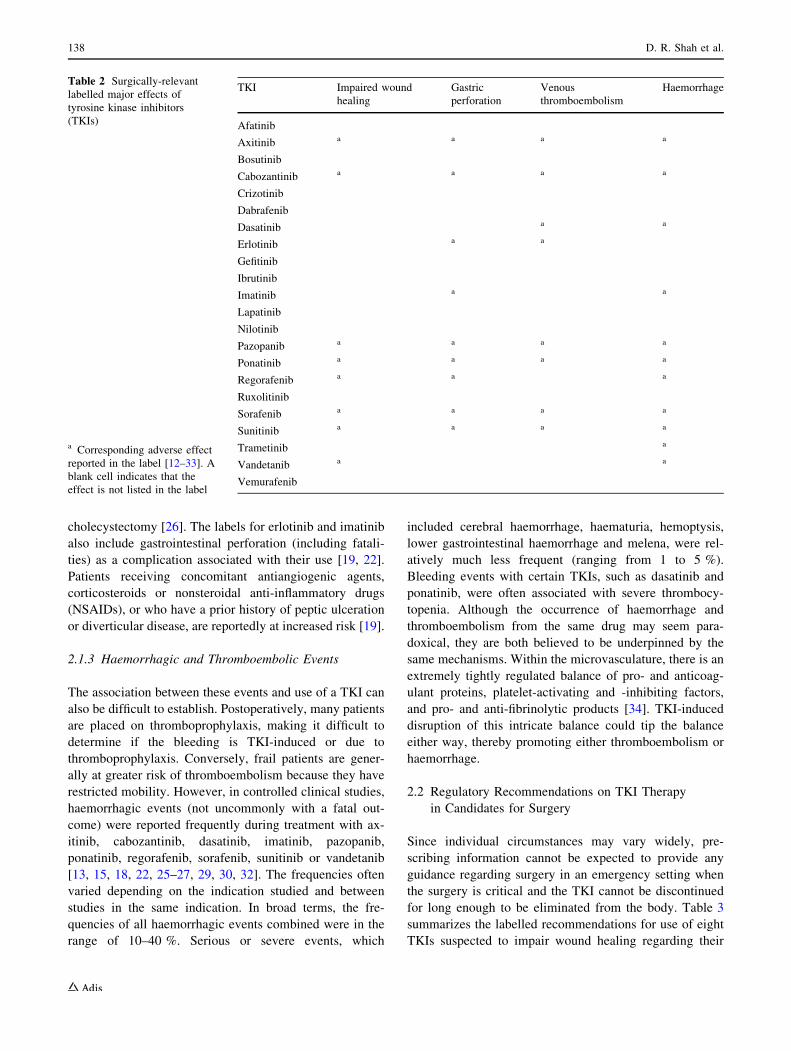
[12–33]. As shown in Table 2, the approved TKIs have
been variously labelled as causing a number of adverse
effects that are directly relevant in surgery. These include
impaired wound healing, venous thromboembolism and
haemorrhage, gastric perforation and fistulas. The rates of
these effects are not precisely known and are not strictly
comparable between different agents, because of differ-
ences in populations and the methodology used to assess
the rates. Nevertheless, they are uncommon enough for the
TKIs to be given regulatory approval for marketing.
2.1.1 Wound Healing
According to the drug labels, impaired wound healing is a
potential risk following treatment with axitinib, cabozan-
tinib, pazopanib, ponatinib, regorafenib, sorafenib, suniti-
nib or vandetanib [13, 15, 25–27, 29, 30, 32]. In terms of
frequency, there are no data on most of these agents, since
no formal studies have been conducted to investigate the
effect of any TKI on wound healing. However, compli-
cated or impaired wound healing was not uncommon
during clinical trials with axitinib (2 %) and cabozantinib
(2 %). There was no indication that wound healing was
affected by sorafenib monotherapy, but only 70 patients on
sorafenib underwent surgical procedures, mainly minor. In
clinical studies with vandetanib, a small number of patients
had surgery while receiving the drug, but there were no
reported wound healing complications.
2.1.2 Gastric Perforation
Spontaneous gastric perforation has been reported during
clinical trials in patients treated with axitinib (1 %), cab-
ozantinib (3 %), pazopanib (1 %), regorafenib (0.6 %) or
sorafenib (1 %) [13, 15, 25, 27, 29]. A number of these
patients developed fistulas (such as gastrointestinal and
tracheal-oesophageal) and some had a fatal outcome. The
label for cabozantinib carries a boxed warning regarding its
potential to cause gastric perforation, fistulas and haem-
orrhage [15]. Non-gastrointestinal fistulas, including tra-
cheal-oesophageal fistulas, were reported in 4 % of
cabozantinib-treated patients. Two (1 %) of these were
fatal. Serious gastrointestinal perforation (with a fistula)
occurred in one ponatinib-treated patient 38 days post-
136 D. R. Shah et al.
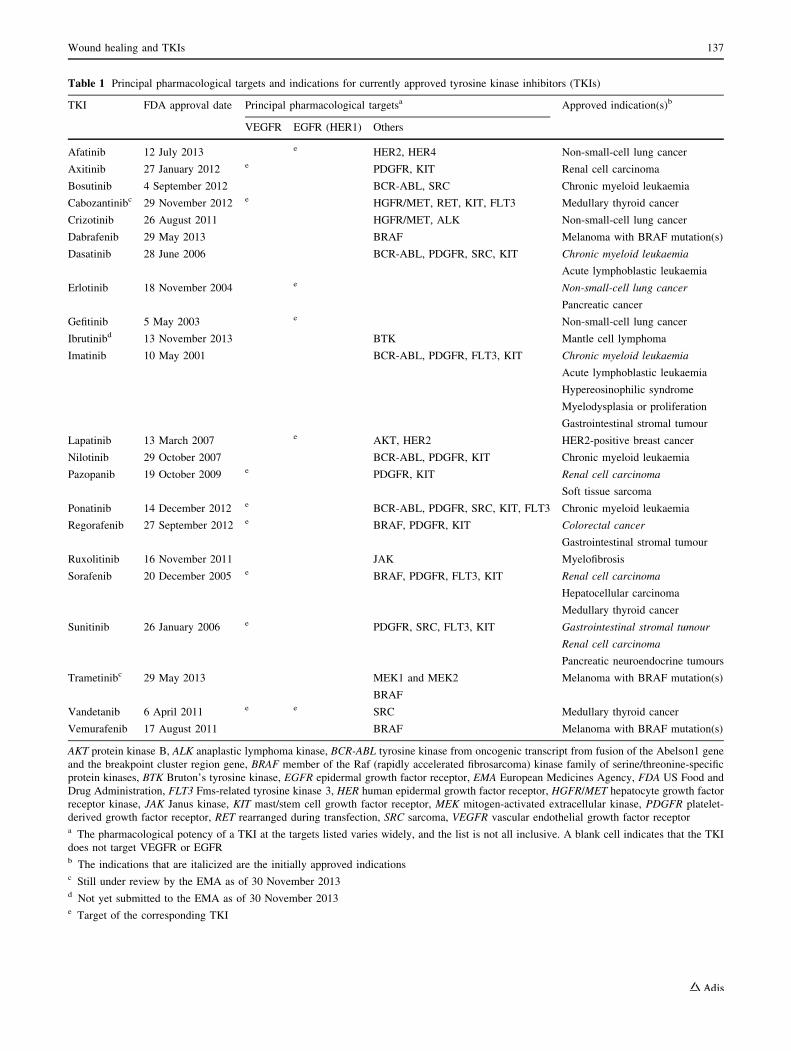
Table 1 Principal pharmacological targets and indications for currently approved tyrosine kinase inhibitors (TKIs)
TKI FDA approval date Principal pharmacological targetsa Approved indication(s)b
VEGFR EGFR (HER1) Others
Afatinib 12 July 2013 e HER2, HER4 Non-small-cell lung cancer
Axitinib 27 January 2012 e PDGFR, KIT Renal cell carcinoma
Bosutinib 4 September 2012 BCR-ABL, SRC Chronic myeloid leukaemia
Cabozantinibc 29 November 2012 e HGFR/MET, RET, KIT, FLT3 Medullary thyroid cancer
Crizotinib 26 August 2011 HGFR/MET, ALK Non-small-cell lung cancer
Dabrafenib 29 May 2013 BRAF Melanoma with BRAF mutation(s)
Dasatinib 28 June 2006 BCR-ABL, PDGFR, SRC, KIT Chronic myeloid leukaemia
Acute lymphoblastic leukaemia
Erlotinib 18 November 2004 e Non-small-cell lung cancer
Pancreatic cancer
Gefitinib 5 May 2003 e Non-small-cell lung cancer
Ibrutinibd 13 November 2013 BTK Mantle cell lymphoma
Imatinib 10 May 2001 BCR-ABL, PDGFR, FLT3, KIT Chronic myeloid leukaemia
Acute lymphoblastic leukaemia
Hypereosinophilic syndrome
Myelodysplasia or proliferation
Gastrointestinal stromal tumour
Lapatinib 13 March 2007 e AKT, HER2 HER2-positive breast cancer
Nilotinib 29 October 2007 BCR-ABL, PDGFR, KIT Chronic myeloid leukaemia
Pazopanib 19 October 2009 e PDGFR, KIT Renal cell carcinoma
Soft tissue sarcoma
Ponatinib 14 December 2012 e BCR-ABL, PDGFR, SRC, KIT, FLT3 Chronic myeloid leukaemia
Regorafenib 27 September 2012 e BRAF, PDGFR, KIT Colorectal cancer
Gastrointestinal stromal tumour
Ruxolitinib 16 November 2011 JAK Myelofibrosis
Sorafenib 20 December 2005 e BRAF, PDGFR, FLT3, KIT Renal cell carcinoma
Hepatocellular carcinoma
Medullary thyroid cancer
Sunitinib 26 January 2006 e PDGFR, SRC, FLT3, KIT Gastrointestinal stromal tumour
Renal cell carcinoma
Pancreatic neuroendocrine tumours
Trametinibc 29 May 2013 MEK1 and MEK2
BRAF
Melanoma with BRAF mutation(s)
Vandetanib 6 April 2011 e e SRC Medullary thyroid cancer
Vemurafenib 17 August 2011 BRAF Melanoma with BRAF mutation(s)
AKT protein kinase B, ALK anaplastic lymphoma kinase, BCR-ABL tyrosine kinase from oncogenic transcript from fusion of the Abelson1 gene
and the breakpoint cluster region gene, BRAF member of the Raf (rapidly accelerated fibrosarcoma) kinase family of serine/threonine-specific
protein kinases, BTK Bruton’s tyrosine kinase, EGFR epidermal growth factor receptor, EMA European Medicines Agency, FDA US Food and
Drug Administration, FLT3 Fms-related tyrosine kinase 3, HER human epidermal growth factor receptor, HGFR/MET hepatocyte growth factor
receptor kinase, JAK Janus kinase, KIT mast/stem cell growth factor receptor, MEK mitogen-activated extracellular kinase, PDGFR platelet-
derived growth factor receptor, RET rearranged during transfection, SRC sarcoma, VEGFR vascular endothelial growth factor receptora The pharmacological potency of a TKI at the targets listed varies widely, and the list is not all inclusive. A blank cell indicates that the TKI
does not target VEGFR or EGFRb The indications that are italicized are the initially approved indicationsc Still under review by the EMA as of 30 November 2013d Not yet submitted to the EMA as of 30 November 2013e Target of the corresponding TKI
Wound healing and TKIs 137
cholecystectomy [26]. The labels for erlotinib and imatinib
also include gastrointestinal perforation (including fatali-
ties) as a complication associated with their use [19, 22].
Patients receiving concomitant antiangiogenic agents,
corticosteroids or nonsteroidal anti-inflammatory drugs
(NSAIDs), or who have a prior history of peptic ulceration
or diverticular disease, are reportedly at increased risk [19].
2.1.3 Haemorrhagic and Thromboembolic Events
The association between these events and use of a TKI can
also be difficult to establish. Postoperatively, many patients
are placed on thromboprophylaxis, making it difficult to
determine if the bleeding is TKI-induced or due to
thromboprophylaxis. Conversely, frail patients are gener-
ally at greater risk of thromboembolism because they have
restricted mobility. However, in controlled clinical studies,
haemorrhagic events (not uncommonly with a fatal out-
come) were reported frequently during treatment with ax-
itinib, cabozantinib, dasatinib, imatinib, pazopanib,
ponatinib, regorafenib, sorafenib, sunitinib or vandetanib
[13, 15, 18, 22, 25–27, 29, 30, 32]. The frequencies often
varied depending on the indication studied and between
studies in the same indication. In broad terms, the fre-
quencies of all haemorrhagic events combined were in the
range of 10–40 %. Serious or severe events, which
included cerebral haemorrhage, haematuria, hemoptysis,
lower gastrointestinal haemorrhage and melena, were rel-
atively much less frequent (ranging from 1 to 5 %).
Bleeding events with certain TKIs, such as dasatinib and
ponatinib, were often associated with severe thrombocy-
topenia. Although the occurrence of haemorrhage and
thromboembolism from the same drug may seem para-
doxical, they are both believed to be underpinned by the
same mechanisms. Within the microvasculature, there is an
extremely tightly regulated balance of pro- and anticoag-
ulant proteins, platelet-activating and -inhibiting factors,
and pro- and anti-fibrinolytic products [34]. TKI-induced
disruption of this intricate balance could tip the balance
either way, thereby promoting either thromboembolism or
haemorrhage.
2.2 Regulatory Recommendations on TKI Therapy
in Candidates for Surgery
Since individual circumstances may vary widely, pre-
scribing information cannot be expected to provide any
guidance regarding surgery in an emergency setting when
the surgery is critical and the TKI cannot be discontinued
for long enough to be eliminated from the body. Table 3
summarizes the labelled recommendations for use of eight
TKIs suspected to impair wound healing regarding their
Table 2 Surgically-relevant
labelled major effects of
tyrosine kinase inhibitors
(TKIs)
a Corresponding adverse effect
reported in the label [12–33]. A
blank cell indicates that the
effect is not listed in the label
TKI Impaired wound
healing
Gastric
perforation
Venous
thromboembolism
Haemorrhage
Afatinib
Axitinib a a a a
Bosutinib
Cabozantinib a a a a
Crizotinib
Dabrafenib
Dasatinib a a
Erlotinib a a
Gefitinib
Ibrutinib
Imatinib a a
Lapatinib
Nilotinib
Pazopanib a a a a
Ponatinib a a a a
Regorafenib a a a
Ruxolitinib
Sorafenib a a a a
Sunitinib a a a a
Trametinib a
Vandetanib a a
Vemurafenib
138 D. R. Shah et al.
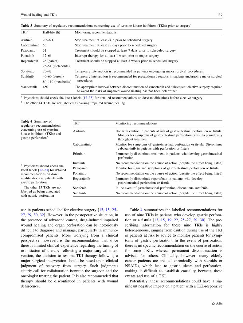
use in patients scheduled for elective surgery [13, 15, 25–
27, 29, 30, 32]. However, in the postoperative situation, in
the presence of advanced cancer, drug-induced impaired
wound healing and organ perforation can be notoriously
difficult to diagnose and manage, particularly in immuno-
compromised patients. More worrying from a clinical
perspective, however, is the recommendation that since
there is limited clinical experience regarding the timing of
re-initiation of therapy following a major surgical inter-
vention, the decision to resume TKI therapy following a
major surgical intervention should be based upon clinical
judgment of recovery from surgery. Such judgments
clearly call for collaboration between the surgeon and the
oncologist treating the patient. It is also recommended that
therapy should be discontinued in patients with wound
dehiscence.
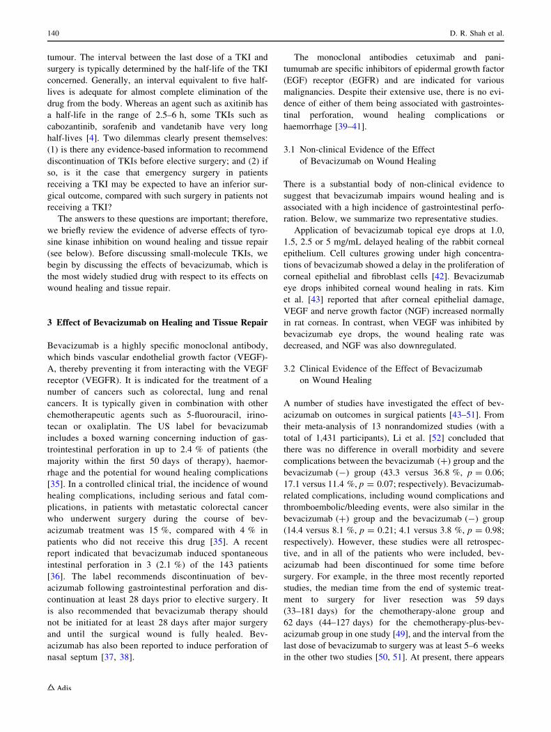
Table 4 summarizes the labelled recommendations for
use of nine TKIs in patients who develop gastric perfora-
tion or a fistula [13, 15, 19, 22, 25–27, 29, 30]. The pre-
scribing information for these nine TKIs is highly
heterogeneous, ranging from caution during use of the TKI
in patients at risk to advice to monitor patients for symp-
toms of gastric perforation. In the event of perforation,
there is no specific recommendation on the course of action
for some TKIs, whereas permanent discontinuation is
advised for others. Clinically, however, many elderly
cancer patients are treated chronically with steroids or
NSAIDs, which lead to gastric ulcers and perforation,
making it difficult to establish causality between these
events and use of a TKI.
Potentially, these recommendations could have a sig-
nificant negative impact on a patient with a TKI-responsive
Table 3 Summary of regulatory recommendations concerning use of tyrosine kinase inhibitors (TKIs) prior to surgerya
TKIb Half-life (h) Monitoring recommendations
Axitinib 2.5–6.1 Stop treatment at least 24 h prior to scheduled surgery
Cabozantinib 55 Stop treatment at least 28 days prior to scheduled surgery
Pazopanib 31 Treatment should be stopped at least 7 days prior to scheduled surgery
Ponatinib 12–66 Interrupt therapy for at least 1 week prior to major surgery
Regorafenib 28 (parent)
25–51 (metabolite)
Treatment should be stopped at least 2 weeks prior to scheduled surgery
Sorafenib 25–48 Temporary interruption is recommended in patients undergoing major surgical procedures
Sunitinib 40–60 (parent)
80–110 (metabolite)
Temporary interruption is recommended for precautionary reasons in patients undergoing major surgical
procedures
Vandetanib 450 The appropriate interval between discontinuation of vandetanib and subsequent elective surgery required
to avoid the risks of impaired wound healing has not been determined
a Physicians should check the latest labels [12–33] for detailed recommendations on dose modifications before elective surgeryb The other 14 TKIs are not labelled as causing impaired wound healing
Table 4 Summary of
regulatory recommendations
concerning use of tyrosine
kinase inhibitors (TKIs) and
gastric perforationa
a Physicians should check the
latest labels [12–33] for detailed
recommendations on dose
modifications in patients with
gastric perforationb The other 13 TKIs are not
labelled as being associated
with gastric perforation
TKIb Monitoring recommendations
Axitinib Use with caution in patients at risk of gastrointestinal perforation or fistula.
Monitor for symptoms of gastrointestinal perforation or fistula periodically
throughout treatment
Cabozantinib Monitor for symptoms of gastrointestinal perforation or fistula. Discontinue
cabozantinib in patients with perforation or fistula
Erlotinib Permanently discontinue treatment in patients who develop gastrointestinal
perforation
Imatinib No recommendation on the course of action (despite the effect being listed)
Pazopanib Monitor for signs and symptoms of gastrointestinal perforation or fistula
Ponatinib No recommendation on the course of action (despite the effect being listed)
Regorafenib Permanently discontinue regorafenib in patients who develop
gastrointestinal perforation or fistula
Sorafenib In the event of gastrointestinal perforation, discontinue sorafenib
Sunitinib No recommendation on the course of action (despite the effect being listed)
Wound healing and TKIs 139
tumour. The interval between the last dose of a TKI and
surgery is typically determined by the half-life of the TKI
concerned. Generally, an interval equivalent to five half-
lives is adequate for almost complete elimination of the
drug from the body. Whereas an agent such as axitinib has
a half-life in the range of 2.5–6 h, some TKIs such as
cabozantinib, sorafenib and vandetanib have very long
half-lives [4]. Two dilemmas clearly present themselves:
(1) is there any evidence-based information to recommend
discontinuation of TKIs before elective surgery; and (2) if
so, is it the case that emergency surgery in patients
receiving a TKI may be expected to have an inferior sur-
gical outcome, compared with such surgery in patients not
receiving a TKI?
The answers to these questions are important; therefore,
we briefly review the evidence of adverse effects of tyro-
sine kinase inhibition on wound healing and tissue repair
(see below). Before discussing small-molecule TKIs, we
begin by discussing the effects of bevacizumab, which is
the most widely studied drug with respect to its effects on
wound healing and tissue repair.
3 Effect of Bevacizumab on Healing and Tissue Repair
Bevacizumab is a highly specific monoclonal antibody,
which binds vascular endothelial growth factor (VEGF)-
A, thereby preventing it from interacting with the VEGF
receptor (VEGFR). It is indicated for the treatment of a
number of cancers such as colorectal, lung and renal
cancers. It is typically given in combination with other
chemotherapeutic agents such as 5-fluorouracil, irino-
tecan or oxaliplatin. The US label for bevacizumab
includes a boxed warning concerning induction of gas-
trointestinal perforation in up to 2.4 % of patients (the
majority within the first 50 days of therapy), haemor-
rhage and the potential for wound healing complications
[35]. In a controlled clinical trial, the incidence of wound
healing complications, including serious and fatal com-
plications, in patients with metastatic colorectal cancer
who underwent surgery during the course of bev-
acizumab treatment was 15 %, compared with 4 % in
patients who did not receive this drug [35]. A recent
report indicated that bevacizumab induced spontaneous
intestinal perforation in 3 (2.1 %) of the 143 patients
[36]. The label recommends discontinuation of bev-
acizumab following gastrointestinal perforation and dis-
continuation at least 28 days prior to elective surgery. It
is also recommended that bevacizumab therapy should
not be initiated for at least 28 days after major surgery
and until the surgical wound is fully healed. Bev-
acizumab has also been reported to induce perforation of
nasal septum [37, 38].
The monoclonal antibodies cetuximab and pani-
tumumab are specific inhibitors of epidermal growth factor
(EGF) receptor (EGFR) and are indicated for various
malignancies. Despite their extensive use, there is no evi-
dence of either of them being associated with gastrointes-
tinal perforation, wound healing complications or
haemorrhage [39–41].
3.1 Non-clinical Evidence of the Effect
of Bevacizumab on Wound Healing
There is a substantial body of non-clinical evidence to
suggest that bevacizumab impairs wound healing and is
associated with a high incidence of gastrointestinal perfo-
ration. Below, we summarize two representative studies.
Application of bevacizumab topical eye drops at 1.0,
1.5, 2.5 or 5 mg/mL delayed healing of the rabbit corneal
epithelium. Cell cultures growing under high concentra-
tions of bevacizumab showed a delay in the proliferation of
corneal epithelial and fibroblast cells [42]. Bevacizumab
eye drops inhibited corneal wound healing in rats. Kim
et al. [43] reported that after corneal epithelial damage,
VEGF and nerve growth factor (NGF) increased normally
in rat corneas. In contrast, when VEGF was inhibited by
bevacizumab eye drops, the wound healing rate was
decreased, and NGF was also downregulated.
3.2 Clinical Evidence of the Effect of Bevacizumab
on Wound Healing
A number of studies have investigated the effect of bev-
acizumab on outcomes in surgical patients [43–51]. From
their meta-analysis of 13 nonrandomized studies (with a
total of 1,431 participants), Li et al. [52] concluded that
there was no difference in overall morbidity and severe
complications between the bevacizumab (?) group and the
bevacizumab (-) group (43.3 versus 36.8 %, p = 0.06;
17.1 versus 11.4 %, p = 0.07; respectively). Bevacizumab-
related complications, including wound complications and
thromboembolic/bleeding events, were also similar in the
bevacizumab (?) group and the bevacizumab (-) group
(14.4 versus 8.1 %, p = 0.21; 4.1 versus 3.8 %, p = 0.98;
respectively). However, these studies were all retrospec-
tive, and in all of the patients who were included, bev-
acizumab had been discontinued for some time before
surgery. For example, in the three most recently reported
studies, the median time from the end of systemic treat-
ment to surgery for liver resection was 59 days
(33–181 days) for the chemotherapy-alone group and
62 days (44–127 days) for the chemotherapy-plus-bev-
acizumab group in one study [49], and the interval from the
last dose of bevacizumab to surgery was at least 5–6 weeks
in the other two studies [50, 51]. At present, there appears
140 D. R. Shah et al.
to be a lack of consensus on the optimal time interval
between discontinuation of bevacizumab and surgery.
Some investigators have reported higher rates of postop-
erative complications in patients who received bev-
acizumab within 8 weeks preceding surgery [45, 53],
whereas others [46, 47] have failed to confirm this. All of
the investigators have emphasized the need for confirma-
tory prospective studies before any firm conclusions are
drawn.
The risk of impaired wound healing seems to be limited
to preoperative use of bevacizumab. Since the FDA
approved bevacizumab for recurrent glioblastoma, its use
has increased in this patient population. Clark et al. [54]
reported that significantly more patients who received
preoperative bevacizumab developed healing complica-
tions than patients who did not receive it (35 versus
10.0 %, p = 0.004). In contrast, postoperative bev-
acizumab (started after a median of 43 days [range
22–65 days] after the second or third craniotomy) was
associated with a 6 % rate of impaired healing, which was
not significantly different from the rate in non-bev-
acizumab-treated controls (p = 1.0).
The duration of preoperative bevacizumab treatment (in
weeks) did not influence healing (odds ratio 0.98,
p = 0.55). More healing complications occurred in patients
receiving preoperative bevacizumab than in non-bev-
acizumab-treated controls before the third craniotomy (44
versus 9 %, p = 0.03).
4 Effect of Small-Molecule TKIs on Healing and Tissue
Repair
4.1 Non-clinical Evidence of the Effect of TKIs
on Wound Healing
Although there are hardly any published clinical studies on
the effects of small-molecule TKIs on wound healing and
tissue repair, a few non-clinical studies have been reported.
Roman et al. [55] reported that semaxanib, an inhibitor
of VEGFR, did not impair wound healing despite
decreasing tissue perfusion and microvascular density in a
wound-healing model in rats. Studies in knockout mice and
preclinical toxicology studies showed that the major targets
of toxicity following inhibition of EGFR are the skin and
gastrointestinal tract. This suggests that EGFR inhibitors
may also influence wound healing and gastrointestinal
ulcer healing rates. In an elegant study, Kaftan et al. [56]
investigated the effect of topical erlotinib, an EGFR
inhibitor, on healing of experimental bilateral tympanic
membrane perforation in rats. A solution of erlotinib
(10 mg/mL) was applied to one tympanic membrane of
each animal, and the vehicle only (in the control group)
was applied to the other side daily for 12 consecutive days,
followed by weekly observation for a total of 30 days. The
mean healing period was found to be 12.1 days in the
group with erlotinib and 6.4 days in the control group.
There were corresponding differences in the histological
parameters between the erlotinib group and the control
group. In another study, the same investigators showed that
both erlotinib (11.8 days) and cetuximab (9 days) pro-
longed healing latencies, compared with a reference value
(7 days), and differences were observed in the histological
parameters of the two groups [57]. The investigators con-
cluded that although inhibition of EGFR does not lead to a
persistent perforation, spontaneous perforation in patients
receiving long-term treatment with EGFR inhibitors may
be possible in patients with pre-existing tympanic mem-
brane pathology. Indeed, there is a report of bilateral
tympanic membrane perforation following long-term
treatment with erlotinib [58]. The same group reported that
inhibition of fibroblast growth factor (FGF) receptor
(FGFR)-1 by its specific inhibitor, SU5402, also inhibited
healing of tympanic membranes in a dose-dependent
manner [59]. This observation suggests a role of multiple
growth factors in wound healing.
We reviewed the pre-approval pharmaco-toxicological
evaluation reports prepared by the FDA [60] and the EMA
[61] for information on non-clinical evidence concerning
the effects of approved TKIs on wound healing and tissue
repair. There was no information available in the regula-
tory evaluations of axitinib, cabozantinib, pazopanib, po-
natinib, regorafenib and sorafenib, despite the warnings in
the prescribing information. For sunitinib, its potential
effect on wound healing was evaluated in female SKH1
mice orally administered 40 and 80 mg/kg for up to five
consecutive weeks [62]. On day 6, a full-thickness incision
was made on the back of each mouse, which was then
sealed with nylon sutures. Subsequently, the strength of the
healed wounds was evaluated on days 13, 20 and 34. Mice
treated with 40 mg/kg/day had wound tensile strength
comparable to control, at all time points evaluated. Tran-
sient treatment-related effects were observed in mice
treated with 80 mg/kg/day, corresponding to a 40 %
decrease in wound tensile strength on day 20.
For vandetanib, the regulatory evaluation report inclu-
ded a study by Ko et al. [63], which had determined the
effects of vandetanib on wound healing in Balb/c mice by
measuring breaking strength in a murine model of cuta-
neous wound healing. Mice were administered 0, 50 or
100 mg/kg/day of the drug by oral gavage once daily,
starting 7 days before wounding. The wound consisted of
two (2 cm) full-thickness horizontal incisions made
through the dorsal skin of the mouse through the pannic-
ulus carnosus. Treatment with vandetanib or the vehicle
was continued for a total of 14 or 35 days until 7 or
Wound healing and TKIs 141
28 days after wounding, respectively, when the breaking
strength of the wounded skin was measured. The results
indicated that the wound breaking strength was dose-
dependently decreased in mice treated with vandetanib
compared with controls at both 7 and 28 days after
wounding. Histological examinations showed that vande-
tanib-treated mice had a qualitative reduction in the degree
of fibrosis and epithelial proliferation at the wound site,
compared with controls; however, vandetanib had no effect
on microvasculature density.
The non-clinical evidence reviewed above suggests that
EGFR may have a greater role than VEGFR in influencing
wound healing, and that multiple growth factors, rather
than any one single factor, may be involved. On balance,
however, the evidence is not conclusive enough and at best
points to an effect at very high exposures.
4.2 Clinical Evidence of the Effect of TKIs on Wound
Healing
The clinical evidence is largely anecdotal, consisting of
case reports from pre-approval clinical trials and post-
approval clinical use. An exhaustive search of the PubMed
database, using a variety of search terms, revealed a
remarkable paucity of published clinical reports on the
effect of TKIs on wound healing. What little evidence
exists is sufficient to raise question marks concerning any
improvement in the risk/benefit of discontinuing a TKI
before surgery, especially in a patient with a TKI-respon-
sive tumour.
Investigators reporting a small, retrospective study in
patients with metastatic renal cell cancer (mRCC) sug-
gested that discontinuation of sunitinib or sorafenib therapy
may actually be risky in terms of progression with new
metastases and potential complications [64]. In a later
study, the same investigators showed that following dis-
continuation of TKI therapy in 36 patients with mRCC,
recurrence was observed in 24 patients (66.7 %). Re-
exposure to the TKI was effective in 86.9 % of these cases
[65]. In another small study in patients with mRCC, dis-
continuation of TKI therapy 2 weeks before surgery, fol-
lowing a median treatment period of 17 weeks, was
associated with an increased incidence and severity of
intraoperative adhesions [66]. Govindan et al. [67] descri-
bed four instances in which patients underwent surgical
procedures (emergency laparotomy, internal fixation of
bony metastasis and drainage of a labial abscess followed
by laparotomy) while receiving gefitinib 250 mg once
daily for management of advanced non-small-cell lung
cancer. There was no evidence of an adverse effect on
wound healing. Apart from this single report, we were
unable to locate any other published reports concerning the
effects of TKIs on non-ophthalmic surgical complications.
Johnson et al. [68] reported the case of a 79-year-old
woman who presented with a persistent corneal epithelial
defect while undergoing treatment with erlotinib for lung
cancer. Within 2 weeks of her discontinuing erlotinib
treatment, the abrasion healed and had no recurrence. I-
brahim et al. [69] also reported a case of a 60-year-old
female, who presented with sudden onset of painless loss of
vision in one eye due to a perforated corneal ulcer, fol-
lowing 3 months of treatment with gefitinib for metastatic
adenocarcinoma of the lung. Gefitinib was stopped, and the
patient went on to have a corneal graft surgery but post-
operatively developed corneal graft melting. Animal stud-
ies have suggested that the corneal effects of gefitinib may
be irreversible [70]. Saint-Jean et al. [71] reported on ten
eyes in five patients during treatment with systemic EGFR
inhibitors; four patients were receiving erlotinib for end-
stage lung carcinoma, and one patient was receiving pa-
nitumumab for end-stage colorectal cancer. Multiple epi-
thelial defects were observed in all ten eyes, corneal
melting and thinning were observed in three eyes in two
patients, two eyes in one patient presented with lower lid
ectropion, and two eyes in two patients presented with
corneal perforation, both requiring penetrating
keratoplasty.
5 Evidence of the Roles of VEGF and EGF in Wound
Healing
The potential role of growth factors in wound healing is
best illustrated by recombinant platelet-derived growth
factor (PDGF). This agent has been approved by the FDA
for treatment of lower-extremity diabetic neuropathic
ulcers with adequate blood supply and which have exten-
ded into the subcutaneous tissue or beyond [72].
However, examination of Tables 1 and 2 reveals that
nine of the ten TKIs (imatinib being the exception) that are
labelled as being potentially associated with impaired
wound healing and/or gastrointestinal perforation are
active at VEGFR and/or EGFR. The suspected association
is stronger for TKIs that are active at VEGFR than for
those that are active at EGFR. Below, therefore, we sum-
marize the pathophysiology of wound healing and the
evidence of a role of growth factors, especially VEGF and
EGF, in wound healing and tissue repair.
5.1 Pathophysiology of Wound Healing
The process of wound healing consists of three overlapping
phases, which consist of blood clotting and inflammation,
formation and proliferation of new tissue and, finally, tis-
sue remodelling. Each of these three phases is associated
with a complex cascade of cellular and biochemical
142 D. R. Shah et al.
responses, resulting in release of growth factors, cytokines,
hormones and low molecular weight mediators from the
serum and from degranulating platelets. Briefly, normal
wound healing is characterized by formation of fibrovas-
cular granulation tissue, which contains fibroblasts, colla-
gen and blood vessels.
The first phase is characterized by invasion, within
minutes, by various inflammatory cells. Platelets and neu-
trophils arrive first because of their abundance in the cir-
culation, followed by monocytes and lymphocytes. These
cellular elements secrete a broad spectrum of cytokines and
growth factors, which attract cells from the wound edge
and from the circulation. The second phase of wound
healing, the formation of new tissue, is initiated by
migration of keratinocytes of the injured epidermis and hair
follicles, followed by proliferation of these cells at the
wound edge. Depending on the effectiveness of angio-
genesis and new vessel formation, the vascular component
appears as early as day 3 after wounding. The third phase
of wound repair is the tissue remodelling phase, when the
epidermis returns to its normal thickness through re-dif-
ferentiation of keratinocytes. Most of the invading cellular
components, including most of the inflammatory cells,
undergo apoptosis, but persistence of fibroblasts may result
in formation of hypertrophic scars and keloids. The entire
process ultimately results in healing of the wound, typically
within 2 weeks. In the vast majority of surgical procedures,
nearly all acute wounds heal by an orderly and timely
process, with recovery of strength and integrity similar to
those of normal skin. Prolonged inflammation such as that
observed following bacterial infection frequently results in
severe tissue damage, which delays the healing process and
may cause excessive scarring or even malignant transfor-
mation of cells at the wound site. Impaired healing in
patients with risk factors such as old age, diabetes or
inadequate circulation often results in a chronic wound.
5.2 Evidence of the Role of VEGF in Wound Healing
The VEGF family comprises at least four members,
including VEGF-A, VEGF-B, VEGF-C and VEGF-D. Of
these, VEGF-A is highly specific for the growth of new
blood vessels in adult physiological and pathological pro-
cesses, and it has previously been found to be sufficient on
its own to stimulate angiogenesis in quiescent vasculature.
These ligands bind to and activate the tyrosine kinase
receptors VEGFR-1 (also known as FLT1), VEGFR-2
(KDR) and VEGFR-3 (FLT4). Of these, VEGFR-2 is
known to be the major signalling receptor, whereas VEG-
FR-1 is believed to function largely as a decoy receptor
binding VEGF. Although VEGFR-1 binds VEGF-A ten-
fold more tightly than does VEGFR-2, this interaction
alone has not been found to be sufficient to induce
angiogenesis in blood vessels. Cellular responses to a
wound result in release of VEGF, which induces angio-
genesis. VEGF is unique for its effects on multiple com-
ponents of the wound healing cascade, including collagen
deposition and epithelialization. Transcription and secre-
tion of VEGF are elevated in all forms of acute skin
wounds, and it is produced by many cell types that par-
ticipate in wound healing, including endothelial cells,
fibroblasts, platelets, neutrophils and macrophages. VEGF
also plays a role in mediating corneal nerve repair [73].
There is abundant evidence from animal studies to support
a role of VEGF in wound healing, and some key features,
as reviewed by Muller et al. [74], can be briefly summa-
rized as follows:
• Expression of VEGF-A strongly increases upon skin
injury—in particular, in keratinocytes and macro-
phages—but this is much less pronounced in animal
models of poorly healing wounds.
• When antibodies neutralizing VEGF-A were applied to
porcine wounds, there was impaired wound angiogen-
esis and formation of granulation tissue.
• There was a delay in wound healing in mice with
keratinocyte-specific knockout of VEGF-A, and inhi-
bition of VEGFR-2 function strongly reduced wound
angiogenesis and granulation tissue formation. How-
ever, wound closure was not affected, demonstrating
that a reduction in angiogenesis can be tolerated in
normally healing animals.
• VEGF-B seems to be dispensable for wound healing,
since incisional wounds generated in VEGF-B–defi-
cient mice healed normally and angiogenesis was not
affected.
• VEGF-D is expressed in the skin and upregulated
following injury, but its loss in mice did not obviously
affect the wound healing process, possibly because of
compensation by VEGF-C.
For a detailed account of the role of VEGF in wound
healing, the reader is referred to the reviews by Muller
et al. [74] and Bao et al. [75].
5.3 Evidence of the Role of EGF in Wound Healing
In addition to the VEGF signalling system, a normally
functioning EGF signalling system is also necessary in
many tissues, including the skin and its appendages, for
proper development and tissue homeostasis. The biological
activities of EGF are mediated through four tyrosine kinase
receptors, designated EGFR (HER1 or ErbB1), HER2
(ErbB2), HER3 (ErbB3) and HER4 (ErbB4). EGFR
(HER1) is highly expressed in different cell types of the
healing wound—in particular, the keratinocytes. The latter
also express HER2 and HER3, indicating that different
Wound healing and TKIs 143
members of the EGF/EGFR families are involved in re-
epithelialization. Current evidence indicates that injury
induces several signalling pathways that involve EGF. For
example, phosphoinositide-3 kinase (PI3K) is activated by
the binding of ligands to cognate tyrosine kinase receptors.
One of the downstream mediators of the activated PI3K
signalling cascade is activated AKT (also known as protein
kinase B). Using AKT1-/- and AKT2-/- mice, Somanath
et al. [76] demonstrated that deficiency of AKT1, but not
AKT2, results in impaired assembly of collagen in skin
wounds and around the blood vessels.
EGF signalling also appears to modulate angiogenesis
via upregulation of angiogenic factors, such as VEGF.
Inhibition of the EGFR pathway has been shown to inhibit
angiogenesis, tumour growth and metastasis [77]. A dys-
functional EGF system results in defective cellular prolif-
eration and differentiation, one consequence of which is
impaired wound healing.
Evidence of the role of EGF in wound healing is also
derived primarily from animal studies of skin and corneal
wounds. Some key elements from a wealth of evidence
supporting the role of EGF in wound healing can be briefly
summarized as follows:
• EGFR ligands are growth factors for keratinocytes,
playing a central role in controlling proliferation of
these cells.
• EGFR ligands also activate mesenchymal cells and
stimulate fibroblast proliferation and angiogenesis.
• Diverse EGFR ligands are detected in wound fluid.
• Expression of EGFR transiently increases after wound-
ing, indicating a role of the EGF network in healing of
skin wounds.
• EGFR-knockout mice display a marked delay in wound
repair as a result of impaired re-epithelialization and
wound contraction.
In one multicentre study of the safety, ocular tolerance
and efficacy of an ophthalmic solution of EGF for treat-
ment of traumatic corneal epithelial defects in 104 patients,
the mean epithelial healing time was significantly shorter in
the EGF-treated group than in the placebo-treated group
(44.17 versus 61.05 h; p \ 0.01) [78]. The number of
epithelial defects that were completely healed at 24, 48 and
72 h after the onset of treatment was significantly greater in
the EGF-treated group. This was confirmed recently in a
much larger study, which reported that EGF eye drops were
very effective in treating acute heterogeneous corneal
diseases, without significant adverse effects and with
86.8 % clinical efficacy [79].
For a more detailed account of the role of EGF in wound
healing, the reader is referred to the reviews by Muller
et al. [74], Schneider et al. [80], Yu et al. [81] and Marquez
et al. [82].
5.4 Evidence of the Role of VEGF and EGF in Gastric
Ulcers
Gastrointestinal ulcers can be viewed conceptually as
internal wounds that resist normal healing processes.
Tyrosine kinase ligands also play a role in induction and
healing of gastric ulcers. Physiological activity of tyrosine
kinases is 20- to 40-fold greater in the gastric mucosa than
in the liver or pancreas, and age-associated changes in
gastric mucosal proliferative activity are accompanied by
parallel alterations in tyrosine kinase activity [83].
Increased tyrosine phosphorylation of proteins is necessary
for physiological and pathological regeneration [84].
Regeneration of the gastric mucosa following a wound or
an injury is controlled by a number of growth factors (such
as EGF, PDGF and hepatocyte growth factor [HGF]),
which coordinate proliferation and migration of cells after
binding to specific receptors on the cell surface [85, 86].
During ulcer healing, SRC kinase is activated by the EGF
cascade and regulates cell migration [87]. Animal studies
have shown significantly increased phosphorylation of
EGFR after ulcer induction and an increase in EGFR
expression in the early stages of ulcer healing, localized in
the epithelial cells of the ulcer margins and regenerating
glands [88]. Use of NSAIDs and/or infection with Heli-
cobacter pylori is a known risk factor for a gastric ulcer.
NSAIDs reduce both basal and EGF-induced re-epitheli-
alization by various molecular mechanisms, including
reduced SRC activity [87]. These findings suggest that
NSAIDs can directly affect the signalling pathways and
cell cytoskeleton essential for re-epithelialization.
H. pylori has been reported to upregulate tyrosine kinase
signal transduction pathways, including SRC, EGFR and
other factors following infection [89–91]. Reduction in
EGF and EGFR levels in gastric tissues has been observed
following eradication of H. pylori [92].
5.5 Relevance of TKI-Induced Clinical Inhibition
of VEGFR and EGFR
Although the evidence summarized above is highly sug-
gestive of a role of VEGF and/or EGF in wound and ulcer
healing, there are certain inconsistencies with regard to the
effects of pharmacological inhibition of these pathways on
wound healing. Given the molecular pharmacology of
wound healing, and the non-clinical evidence of the roles
of VEGF and EGF in this process, it would be reasonable
to expect clearer evidence of the adverse effects of TKIs on
wound healing in clinical trials and following their routine
clinical use. One explanation for the observed discrepancy
may be the marked differences in exposure to the TKI in
non-clinical studies and in patients in the clinical setting.
Duan et al. [93] evaluated the effects of SU6668, an
144 D. R. Shah et al.
inhibitor of VEGFR, PDGF receptor (PDGFR) and FGFR,
on the healing of skin wounds in a murine incisional wound
model and concluded that SU6668 at a fully efficacious
dose of 100 mg/kg/day had no significant effect on the
healing process and that inhibition of the receptors for
VEGF, PDGF and FGF at levels necessary to inhibit
tumour growth in mouse xenograft models does not affect
the healing of incisional wounds in mice.
While use of bevacizumab is associated with a signifi-
cant increase in the rates of impaired wound healing and
gastric perforation [35], there is no evidence of this being
the case with small-molecule TKIs (such as axitinib, paz-
opanib, ponatinib, regorafenib, sorafenib, sunitinib and
vandetanib), which inhibit the VEGF/VEGFR signalling
pathway, albeit via different mechanisms. Nevertheless,
these TKIs are labelled as carrying a risk of impaired
wound healing. It seems reasonable to conclude that the
label warnings have been guided, as a form of class
labelling based on a precautionary principle, by the effects
that are known to be associated with bevacizumab.
Likewise, there is also a similar discrepancy with regard
to the EGFR pathway. Although cetuximab, a highly spe-
cific inhibitor of the EGFR pathway, was shown to delay
healing of the tympanic membrane in animal studies [57],
extensive clinical experience with this protein or pani-
tumumab (another inhibitor of EGFR) has not led to any
concerns regarding an adverse effect on wound healing.
Nor has the extensive use of erlotinib and gefitinib, both
also active inhibitors of the EGF signalling pathway, been
noteworthy for similar effects. Indeed, there was no evi-
dence of impaired wound healing in gefitinib-treated
patients requiring emergency laparotomy [67]. There are
only a handful of isolated reports of an adverse effect on
the cornea, and the possibility of a tissue-specific effect
cannot be discounted.
Thus, the possibilities (1) that the roles of VEGF and
EGF are only peripheral or supportive; and (2) that other
growth factor(s) may have a more pivotal role in wound
healing merit further consideration.
6 Potential Role of Other Growth Factors in Wound
Healing
Wound healing is clearly a more complex process than is
currently understood, and it is now known that a whole
range of signalling systems coordinate the process. This is
demonstrated by analysis of growth factors, their receptors
and downstream signalling components. Interestingly, and
more importantly, similar molecular and cellular mecha-
nisms appear to underlie tissue repair and oncogenesis, and
experimental studies have provided strong evidence for the
hypothesis that cancer may be viewed as an over-healing
wound [94]. Ceelen et al. [95] have also drawn attention to
close similarities between wound healing, inflammation
and tumour growth, by tabulating the roles of various
growth factors that are potentially involved in these pro-
cesses. It is therefore intuitive that interventions that inhibit
tumour growth may be expected to inhibit wound healing.
Chmielowiec et al. [96] demonstrated that HGF/c-Met
signalling is essential for generation of the hyperprolifer-
ative epithelium in skin wounds. However, in the context
of TKIs, it is interesting to note that both crizotinib and
cabozantinib are inhibitors of HGF/c-Met signalling, yet
only cabozantinib is associated with impaired wound
healing and gastric perforation. Luo et al. [97] reported that
HGF, HGF receptor (HGFR), EGF, VEGF and cyclooxy-
genase (COX)-2 are activated in the injured mucosa of
patients with erosive oesophagitis, and speculated whether
their activation might be involved in mucosal repair and
ulcer healing of erosive oesophagitis. Most recently, Meyer
et al. [98] showed that wound repair is severely delayed in
mice lacking FGFR-1 and FGFR-2 in their keratinocytes.
Notably, however, the FGF pathway does not seem to be
the principal target of the currently approved TKIs that are
labelled as inhibiting wound healing. Demidova-Rice et al.
[99] have also emphasized the importance of platelets and
various platelet-derived factors in wound healing. No doubt
there are many other factors involved, and the reader is
referred to more detailed reviews [74, 100]. Having
reviewed the evidence of the roles of various factors such
as PDGF, EGF, FGF, HGF/c-Met, insulin growth factor
(IGF), VEGF, NGF and others in wound healing, Muller
et al. [74] concluded that in most cases, the consequences
of the loss of a single growth factor were relatively mild,
suggesting redundancy or compensation, and that some
growth factors seem to exert unique functions—the most
remarkable example being HGF, since loss of its receptor
on keratinocytes completely inhibited migration and pro-
liferation of the receptor-deficient cells in wounded skin.
They conclude that although extensive knowledge has been
gained on the role of certain growth factors and their
receptors in wound healing, there is still little information
on the function of some other growth factors in this regard.
Cazander et al. [101] have reviewed the evidence of the
role of complement, whereas Miyamoto et al. [102] have
highlighted a potential role of stem cell factor in wound
healing.
7 Discussion
As stated earlier, TKIs that are active at VEGFR and/or
EGFR are suspected to impair wound healing, and the
suspicion is stronger for TKIs that are active at VEGFR
than for those that are active at EGFR. However, it is
Wound healing and TKIs 145
evident that no one single candidate pharmacological target
stands out that can satisfactorily and consistently explain
the current labelling of TKIs with respect to their effect on
wound healing and, by inference, support the current rec-
ommendation to discontinue a TKI prior to surgery.
Temporarily withholding a medication, such as warfarin
or a b-blocker, before surgery in otherwise healthy patients
is a common practice and is guided by risk/benefit analysis
based on sound pharmacological principles and clinical
evidence.
The majority of TKIs are prescribed in a setting of
advanced cancers in patients with limited life expectancy,
and they have been approved for clinical use because they
are effective, often highly so in a subset of patients.
Clearly, different risk/benefit considerations apply when
contemplating temporary or permanent discontinuation of a
TKI in a patient with a TKI-responsive tumour. A direct
corollary of the common mechanisms that underlie tissue
repair and oncogenesis [93, 94] is that impaired wound
healing is either confined to, or more likely in, those
patients whose tumours appear to be responsive to treat-
ment with TKIs. If so, the practice of stopping a TKI before
surgery raises an even more acute dilemma. Wound healing
is a complex process involving a cascade of various equally
important growth factors, other than simply VEGF and
EGF. The clinical evidence of an adverse effect of small-
molecule TKIs on wound healing at clinical doses is at
present tenuous, making it difficult to make an informed
decision on whether it is advisable to discontinue their use
before surgery.
8 Conclusions
A large number of TKIs are in development or under
regulatory review, and their use is expected to increase
markedly in the future. Recommendations in prescribing
information are based on small sample sizes and often
anecdotal cases without any formal clinical trials. As
TKIs become used more widely, with expanding indi-
cations for each, their postmarketing safety profile will
require careful monitoring, and their risk/benefit and
prescribing recommendations may require regular re-
assessment.
In the absence of any reliable and definite evidence at
present, surgeons have to be guided by the recommenda-
tions in the prescribing information when elective surgery
is contemplated. However, the situation is different when a
patient on a TKI requires emergency surgery. In this set-
ting, the long half-life of many TKIs would preclude
waiting until the TKI is eliminated from the body. We
believe that a registry should be set up to gather more
reliable evidence concerning the outcomes of emergency
surgery in patients treated with TKIs. Ultimately, analysis
of outcomes data from such registries may facilitate the
evolution of informed guidelines on the management of
elective surgery in TKI-treated patients.
Conflicts of Interest No sources of funding were used in the
preparation of this review. Devron Shah and Shamik Dholakia are
doctors under training at district general hospitals and have no con-
sultancy relationships. Rashmi Shah provides expert consultancy
services on the development and safety of new drugs to a number of
pharmaceutical companies. Devron Shah, Shamik Dholakia and
Rashmi Shah have no other conflicts of interest that are directly rel-
evant to the content of this review.
References
1. Shah RR, Roberts SA, Shah DR. A fresh perspective on com-
paring the FDA and the CHMP/EMA: approval of antineoplastic
tyrosine kinase inhibitors. Br J Clin Pharmacol. 2013;76:
396–411.
2. Shah RR, Morganroth J, Shah DR. Hepatotoxicity of tyrosine
kinase inhibitors: clinical and regulatory perspectives. Drug Saf.
2013;36:491–503.
3. Shah DR, Shah RR, Morganroth J. Tyrosine kinase inhibitors:
their on-target toxicities as potential indicators of efficacy. Drug
Saf. 2013;36:413–26.
4. Shah RR, Morganroth J, Shah DR. Cardiovascular safety of
tyrosine kinase inhibitors: with a special focus on cardiac
repolarization (QT interval). Drug Saf. 2013;36:295–316.
5. Johnson JR, Ning Y-M, Farrell A, et al. Accelerated approval of
oncology products: the Food and Drug Administration experi-
ence. J Natl Cancer Inst. 2011;103:636–44.
6. Seruga B, Sterling L, Wang L, et al. Reporting of serious
adverse drug reactions of targeted anticancer agents in pivotal
phase III clinical trials. J Clin Oncol. 2011;29:174–85.
7. Niraula S, Seruga B, Ocana A, et al. The price we pay for
progress: a meta-analysis of harms of newly approved anticancer
drugs. J Clin Oncol. 2012;30:3012–9.
8. Jonsson B, Bergh J. Hurdles in anticancer drug development
from a regulatory perspective. Nat Rev Clin Oncol.
2012;9:236–43.
9. Broglio KR, Berry DA. Detecting an overall survival benefit that
is derived from progression-free survival. J Natl Cancer Inst.
2009;101:1642–9.
10. Sherrill B, Kaye JA, Sandin R, et al. Review of meta-analyses
evaluating surrogate endpoints for overall survival in oncology.
Onco Targets Ther. 2012;5:287–96.
11. Food and Drug Administration. FDA drug safety communica-
tion (5 November 2013): FDA asks manufacturer of the leuke-
mia drug Iclusig (ponatinib) to suspend marketing and sales.
http://www.fda.gov/drugs/drugsafety/ucm373040.htm. Acces-
sed 9 Nov 2013.
12. Food and Drug Administration. Gilotrif (afatinib) label
approved on 12 July 2012. http://www.accessdata.fda.gov/
drugsatfda_docs/label/2013/201292s000lbl.pdf. Accessed 10
Sep 2013.
13. Food and Drug Administration. Inlyta (axitinib) label approved
on 27 January 2012. http://www.accessdata.fda.gov/drugsatfda_
docs/label/2012/202324lbl.pdf. Accessed 10 Sep 2013.
14. Food and Drug Administration. Bosulif (bosutinib) label
approved on 4 September 2012. http://www.accessdata.fda.gov/
drugsatfda_docs/label/2012/203341lbl.pdf. Accessed 10 Sep
2013.
146 D. R. Shah et al.
15. Food and Drug Administration. Cometriq (cabozantinib) label
approved on 29 November 2012. http://www.accessdata.fda.
gov/drugsatfda_docs/label/2012/203756lbl.pdf. Accessed 10
Sep 2013.
16. Food and Drug Administration. Xalkori (crizotinib) label
approved on 24 February 2012. http://www.accessdata.fda.gov/
drugsatfda_docs/label/2012/202570s003lbl.pdf. Accessed 10
Sep 2013.
17. Food and Drug Administration. Tafinlar (dabrafenib) label
approved on 29 May 2013. http://www.accessdata.fda.gov/
drugsatfda_docs/label/2013/202806s000lbl.pdf. Accessed 10
Sep 2013.
18. Food and Drug Administration. Sprycel (dasatinib) label
approved on 7 October 2011. http://www.accessdata.fda.gov/
drugsatfda_docs/label/2011/021986s009s010lbl.pdf. Accessed
10 Sep 2013.
19. Food and Drug Administration. Tarceva (erlotinib) label
approved on 17 April 2012. http://www.accessdata.fda.gov/
drugsatfda_docs/label/2012/021743s017lbl.pdf. Accessed 10
Sep 2013.
20. Food and Drug Administration. Iressa (gefitinib) label approved
on 17 June 2005. http://www.accessdata.fda.gov/drugsatfda_
docs/label/2005/021399s008lbl.pdf. Accessed 10 Sep 2013.
21. Food and Drug Administration. Imbruvica (ibrutinib) label
approved on 13 November 2013. http://www.accessdata.fda.
gov/drugsatfda_docs/label/2013/205552s000lbl.pdf. Accessed
18 Nov 2013.
22. Food and Drug Administration. Gleevec (imatinib) label
approved on 31 January 2012. http://www.accessdata.fda.gov/
drugsatfda_docs/label/2012/021588s035lbl.pdf. Accessed 10
Sep 2013.
23. Food and Drug Administration. Tykerb (lapatinib) label
approved on 14 February 2012. http://www.accessdata.fda.gov/
drugsatfda_docs/label/2012/022059s013lbl.pdf. Accessed 10
Sep 2013.
24. Food and Drug Administration. Tasigna (nilotinib) label
approved on 1 May 2012. http://www.accessdata.fda.gov/
drugsatfda_docs/label/2012/022068s012lbl.pdf. Accessed 10
Sep 2013.
25. Food and Drug Administration. Votrient (pazopanib) label
approved on 15 November 2012. http://www.accessdata.fda.
gov/drugsatfda_docs/label/2012/022465s013lbl.pdf. Accessed
10 Sep 2013.
26. Food and Drug Administration. Iclusig (ponatinib) label
approved on 14 December 2012. http://www.accessdata.fda.gov/
drugsatfda_docs/label/2012/203469lbl.pdf. Accessed 10 Sep
2013.
27. Food and Drug Administration. Stivarga (regorafenib) label
approved on 27 September 2012. http://www.accessdata.fda.
gov/drugsatfda_docs/label/2012/203085lbl.pdf. Accessed 10
Sep 2013.
28. Food and Drug Administration. Jakafi (ruxolitinib) label
approved on 21 June 2012. http://www.accessdata.fda.gov/
drugsatfda_docs/label/2012/202192s001lbl.pdf. Accessed 10
Sep 2013.
29. Food and Drug Administration. Nexavar (sorafenib) label
approved on 14 October 2011. http://www.accessdata.fda.gov/
drugsatfda_docs/label/2011/021923s012lbl.pdf. Accessed 10
Sep 2013.
30. Food and Drug Administration. Sutent (sunitinib) label approved
on 16 November 2012. http://www.accessdata.fda.gov/
drugsatfda_docs/label/2012/021938s021s022s023lbl.pdf.
Accessed 10 Sep 2013.
31. Food and Drug Administration. Mekinist (trametinib) label
approved on 29 May 2013. http://www.accessdata.fda.gov/
drugsatfda_docs/label/2013/204114s000lbl.pdf. Accessed 10
Sep 2013.
32. Food and Drug Administration. Caprelsa (vandetanib) label
approved on 9 October 2012. http://www.accessdata.fda.gov/
drugsatfda_docs/label/2012/022405s003lbl.pdf. Accessed 10
Sep 2013.
33. Food and Drug Administration. Zelboraf (vemurafenib) label
approved on 17 August 2011. http://www.accessdata.fda.gov/
drugsatfda_docs/label/2011/202429s000lbl.pdf. Accessed 10
Sep 2013.
34. Elice F, Rodeghiero F, Falanga A, et al. Thrombosis associated
with angiogenesis inhibitors. Best Pract Res Clin Haematol.
2009;22:115–28.
35. Food and Drug Administration. Avastin (bevacizumab) label
approved on 26 January 2013. http://www.accessdata.fda.gov/
drugsatfda_docs/label/2013/125085s267lbl.pdf. Accessed 10
Sep 2013.
36. Borzomati D, Nappo G, Valeri S, et al. Infusion of bevacizumab
increases the risk of intestinal perforation: results on a series of
143 patients consecutively treated. Updates Surg. 2013;65:
121–4.
37. Fakih MG, Lombardo JC. Bevacizumab-induced nasal septum
perforation. Oncologist. 2006;11:85–6.
38. Traina TA, Norton L, Drucker K, et al. Nasal septum perforation
in a bevacizumab-treated patient with metastatic breast cancer.
Oncologist. 2006;11:1070–1.
39. Food and Drug Administration. Erbitux (cetuximab) label
approved on 4 March 2013. http://www.accessdata.fda.gov/
drugsatfda_docs/label/2013/125084s242lbl.pdf. Accessed 10
Sep 2013.
40. Food and Drug Administration. Vectibix (panitumumab) label
approved on 28 March 2013. http://www.accessdata.fda.gov/
drugsatfda_docs/label/2013/125147s169lbl.pdf. Accessed 10
Sep 2013.
41. Fuloria J. Safety profiles of current antiangiogenic therapies for
metastatic colorectal cancer. Onco Targets Ther. 2012;5:
133–42.
42. Kim TI, Chung JL, Hong JP, et al. Bevacizumab application
delays epithelial healing in rabbit cornea. Invest Ophthalmol Vis
Sci. 2009;50:4653–9.
43. Kim EC, Lee WS, Kim MS. The inhibitory effects of bev-
acizumab eye drops on NGF expression and corneal wound
healing in rats. Invest Ophthalmol Vis Sci. 2010;51:4569–73.
44. Scappaticci FA, Fehrenbacher L, Cartwright T, et al. Surgical
wound healing complications in metastatic colorectal cancer
patients treated with bevacizumab. J Surg Oncol. 2005;91:
173–80.
45. D’Angelica M, Kornprat P, Gonen M, et al. Lack of evidence for
increased operative morbidity after hepatectomy with perioper-
ative use of bevacizumab: a matched case-control study. Ann
Surg Oncol. 2007;14:759–65.
46. Kesmodel SB, Ellis LM, Lin E, et al. Preoperative bevacizumab
does not significantly increase postoperative complication rates
in patients undergoing hepatic surgery for colorectal cancer liver
metastases. J Clin Oncol. 2008;26:5254–60.
47. Mahfud M, Breitenstein S, El-Badry AM, et al. Impact of pre-
operative bevacizumab on complications after resection of
colorectal liver metastases: case-matched control study. World J
Surg. 2010;34:92–100.
48. Tamandl D, Gruenberger B, Klinger M, et al. Liver resection
remains a safe procedure after neoadjuvant chemotherapy
including bevacizumab: a case-controlled study. Ann Surg.
2010;252:124–30.
49. Constantinidou A, Cunningham D, Shurmahi F, et al. Periop-
erative chemotherapy with or without bevacizumab in patients
Wound healing and TKIs 147
with metastatic colorectal cancer undergoing liver resection.
Clin Colorectal Cancer. 2013;12:15–22.
50. Dede K, Mersich T, Besznyak I, et al. Bevacizumab treatment
before resection of colorectal liver metastases: safety, recovery
of liver function, pathologic assessment. Pathol Oncol Res.
2013;19:501–8.
51. Lubezky N, Winograd E, Papoulas M, et al. Perioperative
complications after neoadjuvant chemotherapy with and without
bevacizumab for colorectal liver metastases. J Gastrointest Surg.
2013;17:527–32.
52. Li DB, Ye F, Wu XR, et al. Preoperative administration of
bevacizumab is safe for patients with colorectal liver metastases.
World J Gastroenterol. 2013;19:761–8.
53. Reddy SK, Morse MA, Hurwitz HI, et al. Addition of bev-
acizumab to irinotecan- and oxaliplatin-based preoperative
chemotherapy regimens does not increase morbidity after
resection of colorectal liver metastases. J Am Coll Surg.
2008;206:96–106.
54. Clark AJ, Butowski NA, Chang SM, et al. Impact of bev-
acizumab chemotherapy on craniotomy wound healing. J Neu-
rosurg. 2011;114:1609–16.
55. Roman CD, Choy H, Nanney L, et al. Vascular endothelial
growth factor-mediated angiogenesis inhibition and postopera-
tive wound healing in rats. J Surg Res. 2002;105:43–7.
56. Kaftan H, Reuther L, Miehe B, et al. Delay of tympanic mem-
brane wound healing in rats with topical application of a tyro-
sine kinase inhibitor. Wound Repair Regen. 2008;16:364–9.
57. Kaftan H, Reuther L, Miehe B, et al. The influence of inhibition
of the epidermal growth factor receptor on tympanic membrane
wound healing in rats. Growth Factors. 2010;28:286–92.
58. Lee SM, Buchler T, Joseph T, et al. Bilateral eardrum perfora-
tion after long-term treatment with erlotinib. J Clin Oncol.
2008;26:2582–4.
59. Kaftan H, Reuther L, Miehe B, et al. Inhibition of fibroblast
growth factor receptor 1: influence on tympanic membrane wound
healing in rats. Eur Arch Otorhinolaryngol. 2012;269:87–92.
60. Food and Drug Administration. Product reviews and labels.
http://www.accessdata.fda.gov/scripts/cder/drugsatfda/index.cfm.
Accessed 10 Sep 2013.
61. European Medicines Agency. European public assessment
reports: assessment history and product information. http://www.
emea.europa.eu/ema/index.jsp?curl=pages/medicines/landing/epar_
search.jsp&mid=WC0b01ac058001d124. Accessed 10 Sep 2013.
62. European Medicines Agency. European public assessment
report for Sutent (sunitinib) (10 January 2007). http://www.
emea.europa.eu/docs/en_GB/document_library/EPAR_-_Scientific_
Discussion/human/000687/WC500057733.pdf. Accessed 10 Sep
2013.
63. Ko J, Ross J, Awad H, et al. The effects of ZD6474, an inhibitor
of VEGF signaling, on cutaneous wound healing in mice. J Surg
Res. 2005;129:251–9.
64. Johannsen M, Florcken A, Bex A, et al. Can tyrosine kinase
inhibitors be discontinued in patients with metastatic renal cell
carcinoma and a complete response to treatment? A multicentre,
retrospective analysis. Eur Urol. 2009;55:1430–9.
65. Johannsen M, Staehler M, Ohlmann CH, et al. Outcome of treat-
ment discontinuation in patients with metastatic renal cell carci-
noma and no evidence of disease following targeted therapy with or
without metastasectomy. Ann Oncol. 2011;22:657–63.
66. Harshman LC, Yu RJ, Allen GI, et al. Surgical outcomes and
complications associated with presurgical tyrosine kinase inhi-
bition for advanced renal cell carcinoma (RCC). Urol Oncol.
2013;31:379–85.
67. Govindan R, Behnken D, Read W, et al. Wound healing is not
impaired by the epidermal growth factor receptor-tyrosine
kinase inhibitor gefitinib. Ann Oncol. 2003;14:1330–1.
68. Johnson KS, Levin F, Chu DS. Persistent corneal epithelial
defect associated with erlotinib treatment. Cornea. 2009;28:
706–7.
69. Ibrahim E, Dean WH, Price N, et al. Perforating corneal
ulceration in a patient with lung metastatic adenocarcinoma
treated with gefitinib: a case report. Case Rep Ophthalmol Med.
2012;2012:379132.
70. Yano S, Kondo K, Yamaguchi M, et al. Distribution and func-
tion of EGFR in human tissue and the effect of EGFR tyrosine
kinase inhibition. Anticancer Res. 2003;23(5A):3639–50.
71. Saint-Jean A, Sainz de la Maza M, Morral M, et al. Ocular adverse
events of systemic inhibitors of the epidermal growth factor
receptor: report of 5 cases. Ophthalmology. 2012;119:1798–802.
72. Food and Drug Administration. Regranex (becaplermin) label
approved on 11 March 2011. http://www.accessdata.fda.gov/
drugsatfda_docs/label/2011/103691s5095lbl.pdf. Accessed 10
Sep 2013.
73. Yu CQ, Zhang M, Matis KI, et al. Vascular endothelial growth
factor mediates corneal nerve repair. Invest Ophthalmol Vis Sci.
2008;49:3870–8.
74. Muller AK, Meyer M, Werner S. The roles of receptor tyrosine
kinases and their ligands in the wound repair process. Semin
Cell Dev Biol. 2012;23:963–70.
75. Bao P, Kodra A, Tomic-Canic M, et al. The role of vascular
endothelial growth factor in wound healing. J Surg Res.
2009;153:347–58.
76. Somanath PR, Chen J, Byzova TV. Akt1 is necessary for the
vascular maturation and angiogenesis during cutaneous wound
healing. Angiogenesis. 2008;11:277–88.
77. Perrotte P, Matsumoto T, Inoue K, et al. Anti-epidermal growth
factor receptor antibody C225 inhibits angiogenesis in human
transitional cell carcinoma growing orthotopically in nude mice.
Clin Cancer Res. 1999;5:257–65.
78. Pastor JC, Calonge M. Epidermal growth factor and corneal
wound healing: a multicenter study. Cornea. 1992;11:311–4.
79. Lou-Bonafonte JM, Bonafonte-Marquez E, Bonafonte-Royo S,
et al. Posology, efficacy, and safety of epidermal growth factor
eye drops in 305 patients: logistic regression and group-wise
odds of published data. J Ocul Pharmacol Ther. 2012;28:
467–72.
80. Schneider MR, Werner S, Paus R, et al. Beyond wavy hairs: the
epidermal growth factor receptor and its ligands in skin biology
and pathology. Am J Pathol. 2008;173:14–24.
81. Yu FX, Yin J, Xu K, et al. Growth factors and corneal epithelial
wound healing. Brain Res Bull. 2010;81:229–35.
82. Marquez EB, De Ortueta D, Royo SB, et al. Epidermal growth
factor receptor in corneal damage: update and new insights from
recent reports. Cutan Ocul Toxicol. 2011;30:7–14.
83. Majumdar AP, Edgerton EA, Arlow FL. Gastric mucosal tyro-
sine kinase activity during aging and its relationship to cell
proliferation in rats. Biochim Biophys Acta. 1988;965:97–105.
84. Lin W, Kao HW, Robinson D, et al. Tyrosine kinases and gastric
cancer. Oncogene. 2000;19:5680–9.
85. Tarnawski AS. Cellular and molecular mechanisms of gastro-
intestinal ulcer healing. Dig Dis Sci. 2005;50(Suppl 1):S24–33.
86. Tarnawski AS, Ahluwalia A. Molecular mechanisms of epithe-
lial regeneration and neovascularization during healing of gas-
tric and esophageal ulcers. Curr Med Chem. 2012;19:16–27.
87. Pai R, Szabo IL, Giap AQ, et al. Nonsteroidal anti-inflammatory
drugs inhibit re-epithelialization of wounded gastric monolayers
by interfering with actin, Src, FAK and tensin signaling. Life
Sci. 2001;69:3055–71.
88. Choi GH, Park HS, Kim KR, et al. Increased expression of
epidermal growth factor receptor and betacellulin during the
early stage of gastric ulcer healing. Mol Med Rep. 2008;1:
505–10.
148 D. R. Shah et al.
89. Elitsur Y, Majumdar AP, Tureaud J, et al. Tryosine kinase and
ornithine decarboxylase activation in children with Helicobacter
pylori gastritis. Life Sci. 1999;65:1373–80.
90. Meyer-ter-Vehn T, Covacci A, Kist M, et al. Helicobacter pylori
activates mitogen-activated protein kinase cascades and induces
expression of the proto-oncogenes c-fos and c-jun. J Biol Chem.
2000;275:16064–72.
91. Wong BC, Wang WP, So WH, et al. Epidermal growth factor
and its receptor in chronic active gastritis and gastroduodenal
ulcer before and after Helicobacter pylori eradication. Aliment
Pharmacol Ther. 2001;15:1459–65.
92. Coyle WJ, Sedlack RE, Nemec R, et al. Eradication of Heli-
cobacter pylori normalizes elevated mucosal levels of epidermal
growth factor and its receptor. Am J Gastroenterol. 1999;94:
2885–9.
93. Duan WR, Patyna S, Kuhlmann MA, et al. A multitargeted
receptor tyrosine kinase inhibitor, SU6668, does not affect the
healing of cutaneous full-thickness incisional wounds in SKH-1
mice. J Invest Surg. 2006;19:245–54.
94. Schafer M, Werner S. Cancer as an overhealing wound: an old
hypothesis revisited. Nat Rev Mol Cell Biol. 2008;9:628–38.
95. Ceelen W, Pattyn P, Mareel M. Surgery, wound healing, and
metastasis: recent insights and clinical implications. Crit Rev
Oncol Haematol. 2014;89:16–26.
96. Chmielowiec J, Borowiak M, Morkel M, et al. c-Met is essential
for wound healing in the skin. J Cell Biol. 2007;177:151–62.
97. Luo JC, Lin HY, Lu CL, et al. Growth factors expression in
patients with erosive esophagitis. Transl Res. 2008;152:81–7.
98. Meyer M, Muller AK, Yang J, et al. FGF receptors 1 and 2 are
key regulators of keratinocyte migration in vitro and in wounded
skin. J Cell Sci. 2012;125(Pt 23):5690–701.
99. Demidova-Rice TN, Wolf L, Deckenback J, et al. Human
platelet-rich plasma- and extracellular matrix-derived peptides
promote impaired cutaneous wound healing in vivo. PLoS ONE.
2012;7(2):e32146.
100. Werner S, Grose R. Regulation of wound healing by growth
factors and cytokines. Physiol Rev. 2003;83:835–70.
101. Cazander G, Jukema GN, Nibbering PH. Complement activation
and inhibition in wound healing. Clin Dev Immunol.
2012;2012:Article ID 534291.
102. Miyamoto K, Kobayashi T, Hayashi Y, et al. Involvement of
stem cell factor and c-kit in corneal wound healing in mice. Mol
Vision. 2012;18:1505–15.
Wound healing and TKIs 149