effect of platelet-rich plasma on sinus lifting: a randomized-controlled clinical trial
TRANSCRIPT

Effect of platelet-rich plasma onsinus lifting: a randomized-controlled clinical trial
Torres J, Tamimi F, Martinez P-P, Alkhraisat MH, Linares R, Hernandez G, Torres-Macho J, Lopez-Cabarcos E. Effect of platelet-rich plasma on sinus lifting: arandomized-controlled clinical trial. J Clin Periodontol 2009; 36: 677–687.doi: 10.1111/j.1600-051X.2009.01437.x.
AbstractObjective: The combination of anorganic bovine bone (ABB) with platelet-richplasma (PRP) has been widely used in bone regeneration procedures although itsbenefits are still unclear. The purpose of this study was to evaluate whether or not PRPimproves the efficacy of ABB in sinus floor augmentation. In addition, we haveinvestigated the effect of residual bone height and tobacco on implant survival in sinusaugmentation procedures.
Patient and Methods: Eighty-seven patients recruited for this study underwent 144sinus floor augmentation procedures using ABB alone or ABB plus PRP (ABB1PRP)in a randomized clinical trial. A total of 286 implants were placed in the augmentedbone, and their evolution was followed up for a period of 24 months. In order toinvestigate on a histological level and any adjunctive effects, we performed anancillary study in five edentulous patients with a symmetrical severely resorbedmaxilla. In these patients, a bilateral sinus augmentation was randomly performedusing ABB or ABB1PRP in a split-mouth design, and after 6 months, bone biopsieswere taken from the implant sites for histological and histomorphometric analysis.
Results: Overall, 96.2% of ABB and 98.6% of ABB1PRP implant success wereobtained during the monitoring period and differences were not found between sitesgrafted with and without PRP in the 87 patients studied. Densitometry assessments andgraft resorption were similar in both experimental groups. However, the histologicaland histomorphometrical analysis in the five edentulous patients revealed that boneaugmentation was significantly higher in sites treated with ABB1PRP (p40.05).Another outcome from our study is that the lack of initial bone support (p40.05) andsmoking (p 5 0.05) appeared to have a negative effect on the treatment success, whichwas accentuated when both circumstances coincided.
Conclusions: PRP is not a determining factor for implant survival in sinus liftingprocedures. However, this study revealed that PRP can improve the osteoconductiveproperties of ABB by increasing the volume of new bone formed. Moreover, in sinusaugmentation procedures the implant’s survival rate appears to be more influenced bythe residual bone height or by tobacco than by the type of bone graft.
Key words: anorganic bovine bone; boneregeneration; platelet-rich plasma; sinus flooraugmentation
Accepted for publication 10 May 2009
Jesus Torres1,2, Faleh Tamimi2,3,Pedro-Pablo Martinez2, MohammadHamdan Alkhraisat4, Rafael Linares1,Gonzalo Hernandez5, JuanTorres-Macho6 and EnriqueLopez-Cabarcos4
1Ciencias de la Salud III, Universidad Rey
Juan Carlos, Alcorcon, Spain; 2Private
practice, Clinica Dental Alcala, Madrid,
Spain; 3Faculty of Dentistry, McGill
University, Montreal, QC, Canada; 4Faculty
of Pharmacy, Universidad Complutense,
Madrid, Spain; 5Faculty of Dentistry,
Universidad Complutense, Madrid, Spain;6Hospital Infanta Cristina, Parla,
Madrid, Spain
Dental implants’ reconstruction of eden-tulous jaws with adequate bone volumeand density has achieved a high levelof reliability and a considerable rate ofsuccess (Adell et al. 1990). However, thequality of bone in the posterior maxillaryregions often shows a low level of miner-alization (type IV), and severe bone
Conflict of interest and source offunding statement
There is no conflict of interests.This work was supported by the Minis-try of Science and Technology (grantMAT2006-13646-C03-01), the UCM
Programme for Research Groups, andthe Spanish Agency of InternationalCooperation (AECI/A011152/07). Dr.F. Tamimi acknowledges a postdoctoralgrant from the Spanish Foundation forScience and Technology (FECYT).
J Clin Periodontol 2009; 36: 677–687 doi: 10.1111/j.1600-051X.2009.01437.x
677r 2009 John Wiley & Sons A/S
resorption, two factors that render theinitial stability of dental implants difficult(Misch 1990, McCarthy et al. 2003).Besides, the volume of bone in this areais usually limited by the maxillary sinuspneumatization, hindering implant place-ment (Branemark et al. 1984, Blomqvistet al. 1996).
The maxillary sinus-lift surgical tech-nique increases the bone height in theposterior area, and enables the place-ment of implant-supported prostheses(Tatum 1986). This procedure involvesthe detachment of Shneider’s membranefrom the maxillary sinus floor, creating aspace that is filled with a grafting mate-rial to promote a vertical bone augmen-tation into the maxillary sinus cavity.
Different grafting materials havebeen used to fill the space createdbetween the superiorly repositionedsinus membrane and the floor of themaxillary sinus. Early studies advocatedthe use of autogenous bone in the aug-mented space (Boyne & James 1980).However, its availability is restricted bythe limited amount of intra-oral grafts,the morbidity associated with the secondsurgery at the donor site and the highcost for bone harvesting from extra-oralsites. Therefore, alternative graft mate-rials have been developed, includingdemineralized (human) bone matrix(Groeneveld et al. 1999) anorganicbovine bone (ABB) (Valentini & Aben-sur 2003), tricalcium phosphate (Zijder-veld et al. 2005) or bioactive glassparticles (Tadjoedin et al. 2000).
Bio-Osss (Geistlich Biomaterials) isa biocompatible and osteoconductive(Rosen et al. 2002) ABB that providesan ideal scaffold for new bone formation(Hamerle et al. 1998, Piattelli et al. 1999).It has been extensively used in maxillarysinus floor augmentation (Valentini &Abensur 2003, Wallace et al. 2005) withhigh clinical success rates (Carmagnola etal. 2003). However, the lack of osteo-inductive properties encouraged research-ers to find ways of improving its perfor-mance in vivo, and the addition of platelet-rich plasma (PRP) has been assayed. PRPis an autologous fibrin adhesive with ahigh platelet concentration easily obtainedfrom whole blood by centrifugation (Anto-naides 1981, Marx et al. 1998, Anitua1999). Furthermore, PRP has a high con-centration of angiogenic and mitogenicgrowth factors implicated in bone healing,such as transforming growth factor (TGF)(Wikesjo et al. 1998), platelet-derivedgrowth factor (PDGF) and insulin-likegrowth factor (Giannobile et al. 1996),
and has been used in sinus floor elevationrecently (Philippart et al. 2005, Consoloet al. 2007, Galindo-Moreno et al. 2007).Nevertheless, preliminary case reportsusing PRP as an adjuvant in bone regen-eration procedures have been controversial(Anitua 1999, Roldan et al. 2004, Wiltfanget al. 2004, Kassolis & Reynolds 2005,Philippart et al. 2005, Klongnoi et al.2006a, b, Consolo et al. 2007, Galindo-Moreno et al. 2007) and no definitiveconclusions have been drawn from them.
The ultimate procedure for evaluatingthe clinical advantages of a biomaterialis a controlled clinical trial. However,very few controlled clinical trials havebeen performed to evaluate the use-fulness of combining PRP withosteoconductive materials for bone aug-mentation procedures. The aim of thisstudy was to evaluate the effect of differ-ent variables such as a combination ofPRP with ABB, residual bone height andsmoking on bone augmentation in sinuslift procedures. For this, two randomized-controlled clinical trials were carried out:an inter-patient clinical trial in a group of87 patients and a split-mouth clinical trialconducted in a group of five patients. Thecontrol group consisted of maxillary sinuslifting with Bio-Osss alone while theexperimental group included sinus liftingwith Bio-Osss1PRP. The results wereobtained by means of clinical investiga-tion, radiographs, histologic and histo-morphometric analysis.
Patient and Methods
Patients
Inter-patient randomized-controlledclinical trial
Patients were enrolled in the study onthe basis of having insufficient boneheight (o7 mm) in the posterior maxilla.In these patients, sinus floor augmenta-
tion was planned to allow rehabilitationwith fixed implant-supported prosthesis.Smokers were included in the study,while patients with severe systemicdisease [American Society of Anaesthe-siology (ASA) III or IV] and a previoushistory of chronic sinusitis wereexcluded. Informed written consent toparticipate in this study was obtainedfrom all patients, in particular explain-ing the objectives and protocol of thestudy, and possible side effects. Beforecommencing this study, approval wasobtained from the ethical committee forclinical trials of the ‘‘Hospital San Car-los’’ (Madrid, Spain), to carry out a pilotclinical study in the dental clinic ‘‘Clin-ica Dental Alcala’’ (Madrid, Spain).
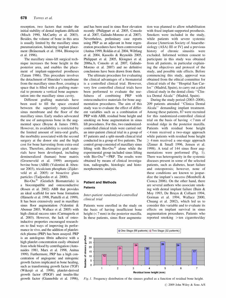
During the study period (2003–2008),209 patients attended ‘‘Clinica DentalAlcala’’ demanding implant treatment.Among these patients, 87 were recruitedfor this randomized-controlled clinicaltrial on the basis of having o7 mm ofresidual ridge in the posterior maxilla.Patients with residual bone heighto4 mm received a two-stage approachwhile patients with residual bone height� 4 mm received a one-stage approach(Zinner & Small 1996, Jensen et al.1998). A total of 144 sinus floor aug-mentations were performed (Fig. 1).There was heterogeneity in the systemicdiseases present in some of the selectedpatients, such as diabetes, heart failureand osteoporosis; however, none ofthese conditions are known to jeopar-dize the implant’s success (Mombelli &Cionca 2006). On the other hand, thereare several authors who associate smok-ing with dental implant failure (Bain &Moy 1993, De Bruyn & Collaert 1994,Gorman et al. 1994, Wallace 2000,Chuang et al. 2002), which led us toconsider this variable and to evaluate itseffects on implant survival in sinusaugmentation procedures. Patients whoreported smoking 4ten cigarettes/day
Fig. 1. Frequency distribution of the sinuses grafted as a function of residual bone height.
678 Torres et al.
r 2009 John Wiley & Sons A/S

were defined as smokers (Tonetti et al.1995), while patients who reportedsmoking only occasionally were notconsidered to be smokers.
Split-mouth randomized-controlledclinical trial
In addition to the above-mentioned cri-teria, the inclusion criteria for the split-mouth study were patients with bilateralloss of teeth in the maxillary premolarand molar areas, and Cadwood classifi-cation V or VI (Cawood & Howell1991) that required a bilateral, two-stage, sinus floor-lifting procedure (firststage, bone grafting; second stage, pla-cement of implants) (Boyne & James1980).
Radiography (orthopantomography)and computed tomography (CT) wereperformed 6 months post-operatively onsurgical sites, before and after treatment,and the bone density was quantifiedin both PRP1ABB and ABB groupsusing SIMPlant 7 (Columbia Scientific,Columbia, MD, USA) software. Sixselected zones in each maxillary sinuswere chosen for a standardized evalua-tion (Rao & Alfidi 1981). All fivepatients were edentulous for 7–10 yearsin the maxilla, which had insufficientretention for their upper denture with aresidual alveolar ridge between 1 and3 mm diagnosed by CT (see Fig. 2).
Randomization
The allocation of participants to inter-vention groups in a truly unpredictable,randomized sequence was performed bya computerized random number gener-ated using GraphPadQuickCalc software(GraphPad Software Inc., La Joya, CA,USA), including the concealment of the
allocation schedule until the assignmentwas made. Subject numbers wereassigned at the baseline examination inconsecutive order by the principal inves-tigator. The sample size used has beenusual in previous studies for this type ofclinical evaluation. Patients included inthe inter-patient clinical trial were allo-cated by a blinded assistant into twogroups: the first was to be treated withABB alone, and the second withABB1PRP. On the other hand, each ofthe patients included in the split-mouthstudy was treated with ABB alone in themaxilla of one side and with ABB1PRPin the contralateral one. The graft mate-rials were randomly allocated for eitherside in each patient.
Blinding
The surgeon was blinded to the graftmaterial applied to each patient beforegraft implantation. An assistant handledthe PRP–ABB or the ABB group afterthe surgeon had already accessed thesinus and elevated the membrane. Thehistologist was blinded to the samples’groups throughout the histomorphome-trical analysis.
PRP
PRP was prepared according to Anitua’smethod15 (Anitua 1999). Blood wascollected from all patients 30 min beforestarting the surgery to ensure that theblinding of the surgeon was maintained.In the ABB1PRP group, between 10and 20 cm3 of blood was withdrawn viavenous aspiration into 4.5 cm3 test tubesand mixed with a 3.8% sodium citratesolution at a 5/1 (v/v) ratio, achievinganticoagulation through calcium bind-ing. The blood was then centrifugedusing a Btis PRGF System II centrifuge(Bti Biotechnology Institute S.L., Vitor-ia, Spain) into three basic components:red blood cells (RBCs), PRP and platelet-poor plasma (PPP). Because of the dif-ferent densities of the components,the RBC layer forms at the bottom ofthe tube, the PRP layer in the middle andthe PPP layer at the top. A pipette (GilsonInc., Middleton, WI, USA) was usedto separate the layers, from the less denseto the denser. Therefore, PPP was sepa-rated first (about 2.25 cm3), followed byPRP (about 0.9 cm3), leaving the RBClayer as a residual (about 2.25 cm3).
Flow cytometry (ADVIA 120, Hema-tology system, Bayer, Leverkusen, Ger-many) was used for platelet counting.
Platelet counts were 2.97 � 0.7-fold overperipheral blood, confirming that the PRPpreparation technique used in this studyproduced a source of highly concentratedplatelets. The average peripheral bloodplatelet count was 275.000 � 58.000/ml.Before its surgical application, PRP(0.2 ml) was activated with a 30% CaCl2solution, and a PRP gel was obtained andmixed with ABB (�1.5 g).
Surgical protocol
Because of the insufficient bone heightfor implant stabilization, in all cases, thesinus floor augmentation was preparedfollowing the method described byBoyne & James (1980) and Tatum(1986). The decision to place simulta-neous or delayed implants depended onwhether or not the residual crest hadenough bone height to permit primaryimplant stability (Zinner & Small 1996,Jensen et al. 1998).
The osteotomy of the lateral wall of themaxilla was performed under local anaes-thesia, and the entire buccal plate wasremoved before the elevation of the sinusmembrane and the implantation of theassigned graft material. No membraneswere used to cover the lateral wall defectafter the bone substitute was placed. In thetwo-stage sinus augmentation procedure,a healing time of 6 months was allowedbefore implant placement (Osseotite, Bio-met 3i Inc., Palmbeach, FL, USA).
Post-operatively, antibiotics, anti-in-flammatory and antiseptics were pre-scribed, and sutures were removed 1week after surgery. Patients wereinstructed not to wear their prosthesisfor 2–3 weeks after surgery. For the next4 months, the existing upper prosthesiswas adapted to the maxilla for aestheticsand relined periodically with a softtissue conditioner. All patients wererequired to follow a soft diet and nomastication was permitted.
Histological and histomorphometric
procedures
The histological and histomorphometicanalysis was performed in the fivepatients recruited for the split-mouthrandomized clinical trial. In thesepatients, bone biopsies were obtainedfrom both treated sites. After a healingperiod of 6 months, these patients werecalled for implant placement and biop-sies were retrieved from dental-implantsites using a trephine burr (Ø 5 3.0 mm� 10.0 mm in length) (see Fig. 3).
Fig. 2. Scheme of the split-mouth rando-mized method used in the controlled clinicaltrial. The upper panel shows a computedtomography of a patient before surgery.Randomly, one sinus was grafted with anor-ganic bovine bone (ABB) alone and theother sinus was grafted with ABB plusplatelet-rich plasma (lower panel).
Effect of platelet-rich plasma on sinus lifting 679
r 2009 John Wiley & Sons A/S

Subsequently, the biopsies were fixed in10% formaldehyde (pH 7.4) and storedat 41C. After dehydration in ascendingseries of alcohol (60–100%), biopsieswere embedded in 2-hydroxy-ethyl-methacrylate (Technovit, Leica Micro-systems GMBH, Wetzlar, Germany),then photopolymerized for 6 h withultraviolet light, 2 h with white lightand 6 h with blue light into ready-to-cut sample blocks.
A saw microtome Exakts (LeicaMicrosystems GmbH) was used to cut200-mm-thick coronal sections from thecylinders. Thereafter, the sections wereground to a thickness of 40–50mm bymeans of a grinder Exakts (EXAKT,Norderstedt, Germany) to achieve betterhistological visualization without risk-ing the loss of the samples. Afterwards,surface staining was performed withbasic fuchsine and methylene blue(Donath & Breuner 1982). The histologi-cal evaluation of bone neoformation wascarried out by means of optical micro-scopy. The histomorphometric analysiswas performed using light micrographs(at magnification � 6) of the biopsyslices, which were captured with a digitalcamera and analysed with the histomor-phometric software MIP-4 (Digital ImageSystem, Barcelona, Spain). The sectionswere analysed by a single examinerblinded to the bone graft material andthe following measurements were takenfrom each sample: newly formed bonevolume (NB), soft tissue volume (ST) andresidual graft volume (RG). The measure-ments were expressed as percentages ofthe total sample volume.
NBð%Þ ¼ Newly formed bone volume
Total sample volume
� 100
STð%Þ ¼ Soft tissue volume
Total sample volume� 100
RGð%Þ ¼ Residual graft volume
Total sample volume� 100
Statistical analysis
The distribution of the patients’ sys-temic conditions (diabetes, smoking,etc.) among clinical treatments’ groupswas assessed using the w2 test, in orderto evaluate comparability betweengroups. Moreover, two-way analysis ofvariance tests, in the univariate analysis,were used to find any associationbetween patients’ (i) systemic condi-tions and (ii) treatment on the one
hand and (i) implant survival and (ii)patient treatment success on the other. Amultiple stepwise logistic regressionanalysis was performed to assess thejoint contribution of the smoking beha-viour and residual bone height toimplant survival. Histomorphometricmeasurements of the biopsies takenfrom the grafted sites were comparedwithin each patient using a t-test forpaired samples. For all univariate ana-lyses, a p40.05 was chosen. A statis-tical software package (SPSS 17.0,SPSS, Chicago, IL, USA) was used forthe statistical analysis.
Results
Inter-patient-controlled clinical trial
This study started in January 2003 andended by June 2005. The group of 87patients selected for the inter-patientstudy consisted of 47 females and 40males with an age range between 52and 78 years (see Table 1). The presence
of systemic disorders was registeredamong the study groups. The distribu-tion of systemic disorders was balancedamong the different treatment groups(Fig. 4).
Fig. 3. The upper panel shows a computedtomography of the augmented sinus. The low-er panel shows the harvesting of bone biopsies6 months after sinus floor augmentation with atrephine bur of 3 mm diameter for histologicand histomorphometric examination.
Table 1. Distribution of patients by systemic disorder into assigned treatment groups
Total numberof patients
Residual bone height Type of graft
one stage(4–7 mm)
two stage(o4 mm)
ABB1PRP ABB
Male 40(46) 24 (60%) 16 (40%) 31 (50%) 31 (50%)Female 47(54) 31 (66%) 16 (34%) 43 (52.4%) 39 (47.6%)Smoker 31(35) 18 (58%) 13 (42%) 22 (50%) 22 (50%)Non-smoker 56(65) 38 (67.8%) 18 (32.2%) 51 (52%) 47 (48%)Diabetes type I 4(4.5) 3 (75%) 1 (25%) 4 (57%) 3 (43%)Diabetes type II 5(6) 2 (50%) 2 (50%) 4 (44.4%) 5 (55.6%)Osteoporosis 11(12.6) 5 (45.4%) 6 (54.6%) 11 (55%) 9 (45%)Ischaemic heart disease 7(8) 3 (43%) 4 (57%) 5 (45.4%) 6 (54.6%)Hypertension 14(16) 8 (57.2%) 6 (42.8%) 10 (50%) 10 (50%)
Data are presented in absolute number (percentage).
Fig. 4. Distribution of patients’ demography as a function of residual bone height and type ofgraft. M, male; F, female; S, smoker; NS, non smoker; DT I, diabetes type I; DT II, diabetestype II; OST, osteoporosis; HTA, hypertension; and IHD, ischaemic heart disease.
680 Torres et al.
r 2009 John Wiley & Sons A/S
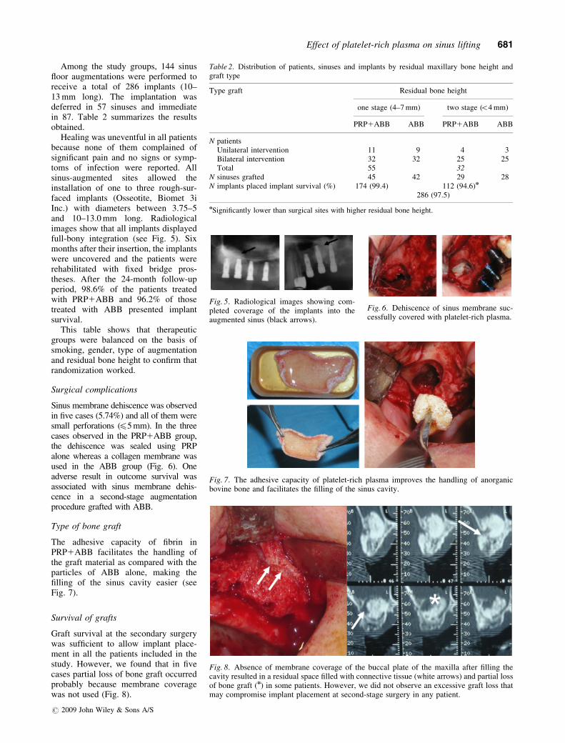
Among the study groups, 144 sinusfloor augmentations were performed toreceive a total of 286 implants (10–13 mm long). The implantation wasdeferred in 57 sinuses and immediatein 87. Table 2 summarizes the resultsobtained.
Healing was uneventful in all patientsbecause none of them complained ofsignificant pain and no signs or symp-toms of infection were reported. Allsinus-augmented sites allowed theinstallation of one to three rough-sur-faced implants (Osseotite, Biomet 3iInc.) with diameters between 3.75–5and 10–13.0 mm long. Radiologicalimages show that all implants displayedfull-bony integration (see Fig. 5). Sixmonths after their insertion, the implantswere uncovered and the patients wererehabilitated with fixed bridge pros-theses. After the 24-month follow-upperiod, 98.6% of the patients treatedwith PRP1ABB and 96.2% of thosetreated with ABB presented implantsurvival.
This table shows that therapeuticgroups were balanced on the basis ofsmoking, gender, type of augmentationand residual bone height to confirm thatrandomization worked.
Surgical complications
Sinus membrane dehiscence was observedin five cases (5.74%) and all of them weresmall perforations (45 mm). In the threecases observed in the PRP1ABB group,the dehiscence was sealed using PRPalone whereas a collagen membrane wasused in the ABB group (Fig. 6). Oneadverse result in outcome survival wasassociated with sinus membrane dehis-cence in a second-stage augmentationprocedure grafted with ABB.
Type of bone graft
The adhesive capacity of fibrin inPRP1ABB facilitates the handling ofthe graft material as compared with theparticles of ABB alone, making thefilling of the sinus cavity easier (seeFig. 7).
Survival of grafts
Graft survival at the secondary surgerywas sufficient to allow implant place-ment in all the patients included in thestudy. However, we found that in fivecases partial loss of bone graft occurredprobably because membrane coveragewas not used (Fig. 8).
Table 2. Distribution of patients, sinuses and implants by residual maxillary bone height andgraft type
Type graft Residual bone height
one stage (4–7 mm) two stage (o4 mm)
PRP1ABB ABB PRP1ABB ABB
N patientsUnilateral intervention 11 9 4 3Bilateral intervention 32 32 25 25Total 55 32
N sinuses grafted 45 42 29 28N implants placed implant survival (%) 174 (99.4) 112 (94.6)n
286 (97.5)
nSignificantly lower than surgical sites with higher residual bone height.
Fig. 5. Radiological images showing com-pleted coverage of the implants into theaugmented sinus (black arrows).
Fig. 6. Dehiscence of sinus membrane suc-cessfully covered with platelet-rich plasma.
Fig. 7. The adhesive capacity of platelet-rich plasma improves the handling of anorganicbovine bone and facilitates the filling of the sinus cavity.
Fig. 8. Absence of membrane coverage of the buccal plate of the maxilla after filling thecavity resulted in a residual space filled with connective tissue (white arrows) and partial lossof bone graft (n) in some patients. However, we did not observe an excessive graft loss thatmay compromise implant placement at second-stage surgery in any patient.
Effect of platelet-rich plasma on sinus lifting 681
r 2009 John Wiley & Sons A/S

Survival rates
Implant survival is defined as theimplant remaining in situ during theentire observation period. In this study,out of over 286 implants placed, 279remained in situ during the entire obser-vation period, being tested indepen-dently for absence of mobility, andseven failed, which yielded 97.5%implant survival. We found differencesbetween the one- and two-stage surgerygroups, indicating that the residualheight could affect the survival rate.Furthermore, six implants were lost atsites with residual bone height o4 mm,while only one was lost at sites withresidual bone height between 4 and7 mm. This result indicates that theamount of residual bone height signifi-cantly influenced the implant survivalafter sinus augmentation (p40.05);however, the bone graft type seemed tohave no effect on implant survival.Implant failure was higher before pros-thetic loading, and in smokers (seeTable 3). Indeed, five out of the sevenfailed implants occurred in four smok-ing patients (p40.05). The details of theimplant’s failure in the inter-patientstudy are given in Table 4.
Treatment success rate was defined asthe rate of patients that presented com-plications during the observation period.According to this definition, 92.8% ofthe patients’ treatments were successful.Moreover, treatments’ success seemedto be higher in patients with moreresidual bone height (p 5 0.05), and innon-smokers (p40.05).
Moreover, a o4 mm residual boneheight and smoking had a combinednegative effect on the treatment successrate, although these differences did notreach statistical significance in the uni-variate and multivariate analyses (seeTable 5).
Both the patient-based and theimplant-based statistical analysis of thetreatment groups agreed on the negativeeffect that both smoking and reducedresidual bone height had on the im-plant survival and treatment success.Although there was no significant inter-action between the two effects, the datasuggested that the negative effect on thetreatment success was accentuated whenboth circumstances coincided.
Split-mouth-controlled clinical trial
The five patients included in the split-mouth-controlled clinical trial received
10 sinus floor augmentations with a totalof 26 implants. A summary of the resultsobtained from these patients is presentedin Table 6.
After 6 months, CT showed no sig-nificant differences between ABB- andPRP1ABB-treated sites (see Fig. 9). Inboth cases, the augmented bone pre-
Table 3. Implant distribution, failure and survival as a function of the residual maxillary boneheight, bone substitute material and smoking behaviour
Type graft Residual bone height
one stage (4–7 mm) two stage (o4 mm)
PRP1
ABBABB implant
survival (%)PRP1
ABBABB implant
survival (%)
Implants placed in smokingpatients
34 (1) 32(0) 98.4 24 (1) 20 (3) 90.9
Implants placed in non-smokingpatients
61(0) 47(0) 100 34(0) 30 (2) 96.9n
Total n implants placed 95(1) 79(0) 99.4 58(1) 50(5) 95.0Implant survival (%) 98.9 100 99.4 98.2 90.7nn 95.0
(), number of failed implants;nSignificantly different from smokers (p40.05);nnImplant survival significantly lower than in sinuses treated with PRP1ABB (p40.05).
Table 4. Details of patients with failed implants in the inter-patient study
Gender Age(years)
Surgicalstages
Bonegraft
Membrane’sdehiscence
Smokingbehaviour
Failure implant’sposition
Time offailure
M 67 1 PRP1ABB ND Yes 25 BLF 65 2 ABB ND Yes 16 FYLM 71 2 ABB D Yes 26, 27 BLF 63 2 PRP1ABB ND Yes 17 FYLF 75 2 ABB ND No 15, 16 BL
BL, before prosthetic loading; FYL, failure during first year of prosthetic loading; ND, no
dehiscence; D, dehiscence.
Table 5. Distribution of patients by residual maxillary bone height and smoking behaviour
Smoking Non- smoking Total
One stage (4–7 mm) 19 (94.8) 36 (100) 55 (98.2)Two stage (o4 mm) 12 (75) 20 (95) 32 (87.5)nn
Total 31 (87.1)n 56 (98.2) 87 (92.8)
(), treatment success rate in %;nSignificantly lower (p40.05) than non-smokers;nnSignificantly lower (p40.05) than one-stage treated patients.
Table 6. Patient, implants and graft data for the split mouth clinical trial
Patient Gender Age(years)
Preoperativebone height
(mm)
Healing time(months)
Postoperativebone height
(mm)
Implants’position
PRP1ABB ABB PRP1ABB ABB
1 M 55 2 3 6 13 13 14, 16, 25, 262 F 67 1 1 6 11 10 14, 15, 16
24, 26, 273 M 62 2 2 6 11 10 14,16,17
25, 26, 274 F 58 1 3 6 12 13 15,17
16, 175 F 64 3 2 6 14 12 15, 16, 17
25, 26, 27
682 Torres et al.
r 2009 John Wiley & Sons A/S
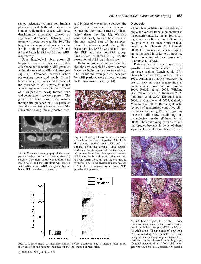
sented adequate volume for implantplacement, and both sites showed asimilar radiographic aspect. Similarly,densitometric assessment showed nosignificant differences between bothtreatment modalities (see Fig. 10). Theheight of the augmented bone was simi-lar in both groups: 10.4 � 0.7 and9.4 � 0.7 mm in PRP1ABB and ABB,respectively.
Upon histological observation, allbiopsies revealed the presence of trabe-cular bone and remaining ABB granuleswithin the treated maxillary sinuses (seeFig. 11). Differences between nativepre-existing bone and newly formedbone were clearly observed because ofthe presence of ABB particles in thewhole augmented area. On the surfacesof ABB particles, newly formed boneand connective tissue were present. Thegrowth of bone took place mainlythrough the guidance of ABB particlesfrom the pre-existing bone surface of thesinus floor along the augmented area,
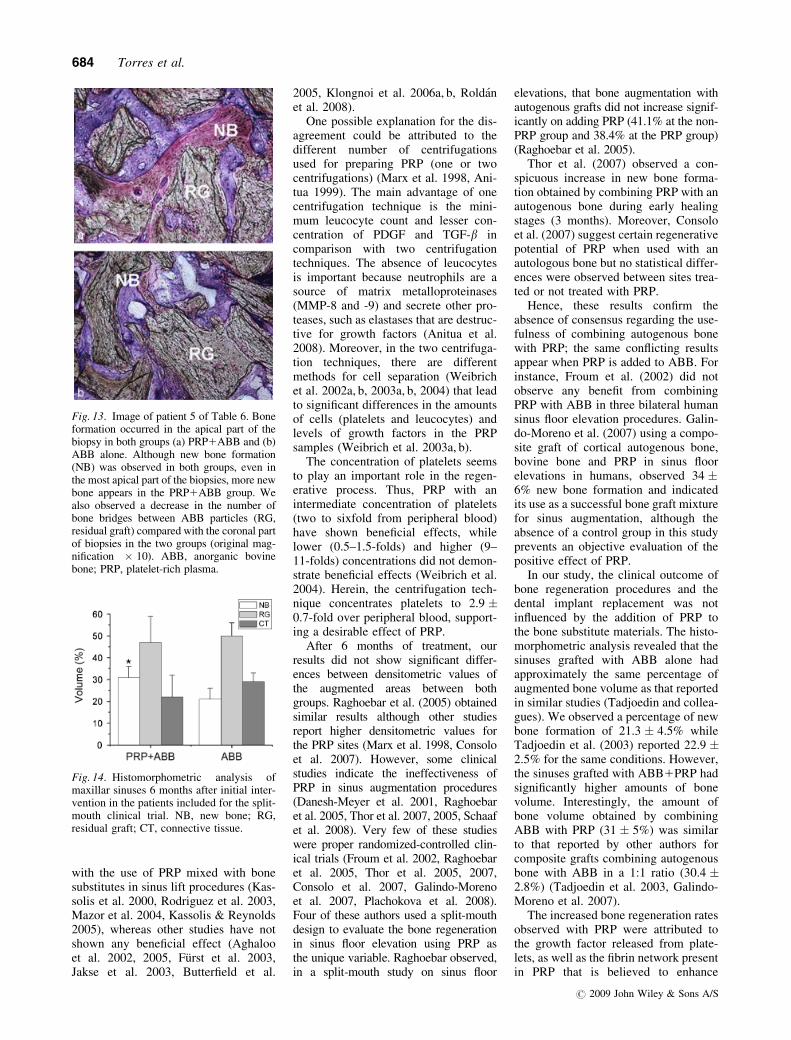
and bridges of woven bone between thegrafted particles could be observed,connecting them into a mass of miner-alized tissue (see Fig. 12). We alsoobserved newly formed bone even inthe most apical part of the samples.Bone formation around the graftedbone particles (ABB) was seen in boththe PRP and the non-PRP group.Furthermore, as shown in Fig. 13, theresorption of ABB particles is low.
Histomorphometric analysis revealedthat the area occupied by newly formedbone was higher in the sites treated withPRP, while the average areas occupiedby ABB particles were almost the samein the two groups (see Fig. 14).
Discussion
Although sinus lifting is a reliable tech-nique for vertical bone augmentation inthe posterior maxilla, implant loss is stillregistered as often as in 17% of thepatients with less than 6 mm residualbone height (Tonetti & Hammerle2008). For this reason, bioactive agentsare being tested in order to improve theclinical outcome of these procedures(Palmer et al. 2008).
Platelets are a natural source ofgrowth factors with beneficial effectson tissue healing (Lynch et al. 1991,Giannobile et al. 1996, Wikesjo et al.1998, Anitua et al. 2004); however, theuse of PRP in bone regeneration inhumans is a moot question (Anitua1999, Roldan et al. 2004, Wiltfanget al. 2004, Kassolis & Reynolds 2005,Philippart et al. 2005, Klongnoi et al.2006a, b, Consolo et al. 2007, Galindo-Moreno et al. 2007). Recent systematicreviews of randomized-controlled clin-ical trials combining PRP with graftingmaterials still show conflicting andinconclusive results (Palmer et al.2008). The controversy extends to ani-mal studies because in some of them,significant benefits have been reported
Fig. 9. Computed tomography of the samepatient before (a) and 6 months after (b)surgery. The right sinus was grafted withPRP1ABB, and the left sinus was graftedwith ABB alone. ABB, anorganic bovinebone; PRP, platelet-rich plasma.
Fig. 10. Densitometry of maxillary sinuses before treatment, and 6 months after initialintervention in the patients included for the split-mouth clinical trial.
Fig. 11. Histological overview of biopsiestaken from the sinus of patient 2 in Table6, showing residual bone (RB) and twosquares delimiting coronal (dark square)and apical (white square) sites of the sample,where new bone formation appears betweenABB particles in both groups: the one trea-ted with ABB alone (a) and the one treatedwith PRP1ABB (b). (Original magnification� 2.5.) ABB, anorganic bovine bone; PRP,platelet-rich plasma.
Fig. 12. Image of patient 3 of Table 6. Boneformation took place in the coronal part ofthe biopsy in both groups (a) PRP1ABB and(b) ABB alone. The presence of new bone(NB) surrounding ABB particles (RG, resi-dual graft) and creating bridges between ABBparticles can be observed in both groups.(Original magnification � 20.) ABB, anor-ganic bovine bone; PRP, platelet-rich plasma.
Effect of platelet-rich plasma on sinus lifting 683
r 2009 John Wiley & Sons A/S
with the use of PRP mixed with bonesubstitutes in sinus lift procedures (Kas-solis et al. 2000, Rodriguez et al. 2003,Mazor et al. 2004, Kassolis & Reynolds2005), whereas other studies have notshown any beneficial effect (Aghalooet al. 2002, 2005, Furst et al. 2003,Jakse et al. 2003, Butterfield et al.
2005, Klongnoi et al. 2006a, b, Roldanet al. 2008).
One possible explanation for the dis-agreement could be attributed to thedifferent number of centrifugationsused for preparing PRP (one or twocentrifugations) (Marx et al. 1998, Ani-tua 1999). The main advantage of onecentrifugation technique is the mini-mum leucocyte count and lesser con-centration of PDGF and TGF-b incomparison with two centrifugationtechniques. The absence of leucocytesis important because neutrophils are asource of matrix metalloproteinases(MMP-8 and -9) and secrete other pro-teases, such as elastases that are destruc-tive for growth factors (Anitua et al.2008). Moreover, in the two centrifuga-tion techniques, there are differentmethods for cell separation (Weibrichet al. 2002a, b, 2003a, b, 2004) that leadto significant differences in the amountsof cells (platelets and leucocytes) andlevels of growth factors in the PRPsamples (Weibrich et al. 2003a, b).
The concentration of platelets seemsto play an important role in the regen-erative process. Thus, PRP with anintermediate concentration of platelets(two to sixfold from peripheral blood)have shown beneficial effects, whilelower (0.5–1.5-folds) and higher (9–11-folds) concentrations did not demon-strate beneficial effects (Weibrich et al.2004). Herein, the centrifugation tech-nique concentrates platelets to 2.9 �0.7-fold over peripheral blood, support-ing a desirable effect of PRP.
After 6 months of treatment, ourresults did not show significant differ-ences between densitometric values ofthe augmented areas between bothgroups. Raghoebar et al. (2005) obtainedsimilar results although other studiesreport higher densitometric values forthe PRP sites (Marx et al. 1998, Consoloet al. 2007). However, some clinicalstudies indicate the ineffectiveness ofPRP in sinus augmentation procedures(Danesh-Meyer et al. 2001, Raghoebaret al. 2005, Thor et al. 2007, 2005, Schaafet al. 2008). Very few of these studieswere proper randomized-controlled clin-ical trials (Froum et al. 2002, Raghoebaret al. 2005, Thor et al. 2005, 2007,Consolo et al. 2007, Galindo-Morenoet al. 2007, Plachokova et al. 2008).Four of these authors used a split-mouthdesign to evaluate the bone regenerationin sinus floor elevation using PRP asthe unique variable. Raghoebar observed,in a split-mouth study on sinus floor
elevations, that bone augmentation withautogenous grafts did not increase signif-icantly on adding PRP (41.1% at the non-PRP group and 38.4% at the PRP group)(Raghoebar et al. 2005).
Thor et al. (2007) observed a con-spicuous increase in new bone forma-tion obtained by combining PRP with anautogenous bone during early healingstages (3 months). Moreover, Consoloet al. (2007) suggest certain regenerativepotential of PRP when used with anautologous bone but no statistical differ-ences were observed between sites trea-ted or not treated with PRP.
Hence, these results confirm theabsence of consensus regarding the use-fulness of combining autogenous bonewith PRP; the same conflicting resultsappear when PRP is added to ABB. Forinstance, Froum et al. (2002) did notobserve any benefit from combiningPRP with ABB in three bilateral humansinus floor elevation procedures. Galin-do-Moreno et al. (2007) using a compo-site graft of cortical autogenous bone,bovine bone and PRP in sinus floorelevations in humans, observed 34 �6% new bone formation and indicatedits use as a successful bone graft mixturefor sinus augmentation, although theabsence of a control group in this studyprevents an objective evaluation of thepositive effect of PRP.
In our study, the clinical outcome ofbone regeneration procedures and thedental implant replacement was notinfluenced by the addition of PRP tothe bone substitute materials. The histo-morphometric analysis revealed that thesinuses grafted with ABB alone hadapproximately the same percentage ofaugmented bone volume as that reportedin similar studies (Tadjoedin and collea-gues). We observed a percentage of newbone formation of 21.3 � 4.5% whileTadjoedin et al. (2003) reported 22.9 �2.5% for the same conditions. However,the sinuses grafted with ABB1PRP hadsignificantly higher amounts of bonevolume. Interestingly, the amount ofbone volume obtained by combiningABB with PRP (31 � 5%) was similarto that reported by other authors forcomposite grafts combining autogenousbone with ABB in a 1:1 ratio (30.4 �2.8%) (Tadjoedin et al. 2003, Galindo-Moreno et al. 2007).
The increased bone regeneration ratesobserved with PRP were attributed tothe growth factor released from plate-lets, as well as the fibrin network presentin PRP that is believed to enhance
Fig. 13. Image of patient 5 of Table 6. Boneformation occurred in the apical part of thebiopsy in both groups (a) PRP1ABB and (b)ABB alone. Although new bone formation(NB) was observed in both groups, even inthe most apical part of the biopsies, more newbone appears in the PRP1ABB group. Wealso observed a decrease in the number ofbone bridges between ABB particles (RG,residual graft) compared with the coronal partof biopsies in the two groups (original mag-nification � 10). ABB, anorganic bovinebone; PRP, platelet-rich plasma.
Fig. 14. Histomorphometric analysis ofmaxillar sinuses 6 months after initial inter-vention in the patients included for the split-mouth clinical trial. NB, new bone; RG,residual graft; CT, connective tissue.
684 Torres et al.
r 2009 John Wiley & Sons A/S

osteoconduction of the bone substitutematerial (Marx et al. 1998). Althoughsome authors suggest that PRP induceshigher graft resorption rates in autolo-gous bone grafts (Fennis et al. 2004,Wiltfang et al. 2004), in this study,PRP did not show any effect on theresorption of the ABB particles. More-over, the type of bone graft did notinfluence either implant survival ortreatment success.
There are several reports associatingsmoking with dental implant failure(Chuang et al. 2002, Schwartz-Aradet al. 2002). Tobacco contains severalnoxious substances, resulting in vaso-constriction and decreased tissue perfu-sion, which produces pathologicalischaemia impairing wound healing. Inthis study, implant survival and treat-ment success was significantly lower insmoking patients than in non-smokers,smoking thus being an important riskfactor in sinus augmentation procedures.This finding confirms previous reportsthat associate smoking habits with fail-ure of implants placed in augmentedsinuses (Olson et al. 2000, Kan et al.2002, Schwartz-Arad et al. 2002).
The implant survival in augmentedmaxillary sinuses observed in this studywas significantly higher in patients withhigher amounts of residual bone height.This result is in agreement with previousstudies (Geurs et al. 2001). On the otherhand, the prevalence of membrane perfora-tion observed was similar to that reportedby other authors (5.74%) (Pjetursson et al.2008, Tonetti & Hammerle 2008). Further-more, the lateral wall of the maxilla wasnot covered with any membrane after graftplacement, and this did not affect the finaloutcome of the treatments.
Conclusions
PRP is not a determining factor forimplant survival in sinus lifting proce-dures. However, this study revealed thatPRP could improve the osteoconductiveproperties of ABB by increasing thevolume of new bone formed, and alsothe handling properties of bone substi-tute particles. Moreover, in sinus aug-mentation procedures implant survivalrate appears to be more influenced bythe residual bone height or by tobaccothan by the type of bone graft.
Acknowledgements
The authors also thank Mrs. AscensionMarin, the staff of ‘‘Clinica Dental
Alcala’’ and Santiago Cano Alsua fortheir help and support of this study.
References
Adell, R., Eriksson, B., Lekholm, U., Brane-
mark, P. I. & Jemt, T. (1990) Long-term
follow-up study of osseointegrated implants
in the treatment of totally edentulous jaws.
International Journal of Oral and Maxillofa-
cial Implants 5, 347–359.
Aghaloo, T., Moy, P. K. & Freymiller, E. G.
(2002) Investigation of platelet-rich plasma
in rabbit cranial defects: a pilot study. Jour-
nal of Oral Maxillofacial Surgery 60, 1176–
1181.
Aghaloo, T., Moy, P. K. & Freymiller, E. G.
(2005) Evaluation of platelet-rich plasma in
combination with freeze-dried bone in the
rabbit cranium. A pilot study. Clinical Oral
Implants Research 16, 250–257.
Anitua, E. (1999) Plasma rich in growth factors:
Preliminary results of use in the preparation
of future sites for implants. International
Journal of Oral and Maxillofacial Implants
14, 429–535.
Anitua, E., Aguirre, J. J., Algorta, J., Ayerdi, E.,
Cabezas, A.I, Orive, G. & Andia, I. (2008)
Effectiveness of autologous preparation rich
in growth factors for the treatment of chronic
cutaneous ulcers. Journal of Biomedical
Material Research B Applied Biomaterials
84, 415–421.
Anitua, E., Andia, I., Ardanza, B., Nurden, P. &
Nurden, A. T. (2004) Autologous platelets as
a source of proteins for healing and tissue
regeneration. Thrombosis and Haemostasis
91, 4–15.
Antonaides, H. N. (1981) Human platelet-
derived growth factor (PDGF): Purification
of PDGF-I and PDGF-II and separation of
their reduced subunits. Proceedings of the
National Academy of Sciences of USA 78,
7314–7317.
Bain, C. A. & Moy, P. K. (1993) The associa-
tion between the failure of dental implants
and cigarette smoking. International Journal
of Oral and Maxillofacial Implants 8, 609–
615.
Blomqvist, J. E., Alberius, P. & Isaksson, S.
(1996) Retrospective analysis of one-stage
maxillary sinus augmentation with endoss-
eous implants. International Journal of Oral
and Maxillofacial Implants 11, 512–521.
Boyne, P. J. & James, R. A. (1980) Grafting of
the maxillary sinus floor with autogenous
marrow and bone. Journal of Oral Surgery
38, 613–616.
Branemark, P. I., Adell, R., Albrektsson, T.,
Lekholm, U., Lindstrom, J. & Rockler B, .
(1984) An experimental and clinical study of
osseointegrated implants penetrating the
nasal cavity and maxillary sinus. Journal of
Oral and Maxillofacial Surgery 42, 497–505.
Butterfield, K. J., Bennett, J., Gronowicz, G. &
Adams, D. (2005) Effect of platelet-rich
plasma with autogenous bone graft for max-
illary sinus augmentation in a rabbit model.
Journal of Oral Maxillofacial Surgery 63,
370–376.
Carmagnola, D., Adriaens, P. & Berglundh, T.
(2003) Healing of human extraction sockets
filled with Bio-Oss. Clinical Oral Implants
Research 14, 137–143.
Cadwood, J. I. & Howell, R. A. (1991) Recon-
structive preprosthetic surgery. I. Anatomical
considerations. International Journal of Oral
and Maxillofacial Surgery 20, 75–82.
Chuang, S. K., Wei, L. J., Douglass, C. W. &
Dodson, T. B. (2002) Risk factors for dental
implant failure: a strategy for the analysis of
clustered failure-time observations. Journal
of Dental Research 81, 572–577.
Consolo, U., Zaffe, D., Bertoldi, C. & Ceccher-
elli, G. (2007) Platelet-rich plasma activity
on maxillary sinus floor augmentation by
autologous bone. Clinical Oral Implants Res-
earch 18, 252–262.
Danesh-Meyer, M. J., Filstein, M. R. & Shana-
man, R. (2001) Histological evaluation of
sinus augmentation using platelet rich plasma
(PRP): a case series. Journal of the Interna-
tional Academy of Periodontology 3, 48–56.
De Bruyn, H. & Collaert, B. (1994) The effect
of smoking on early implant failure. Clinical
Oral Implants Research 5, 260–264.
Donath, K. & Breuner, G. (1982) A method for
the study of undecalcified bones and teeth
with attached soft tissues. The Sage–Schliff
(sawing and grinding) technique. Journal of
Oral Pathology 11, 318–326.
Fennis, J. P., Stoelinga, P. J. & Jansen, J. A.
(2004) Mandibular reconstruction: a histolo-
gical and histomorphometric study on the use
of autogenous scaffolds, particulate cortico-
cancellous bone grafts and platelet rich plas-
ma in goats. International Journal of Oral
Maxillofacial Surgery 33, 48–55.
Froum, S. J., Wallace, S. S., Tarnow, D. P. &
Cho, S. C. (2002) Effect of platelet-rich
plasma on bone growth and osseointegration
in human maxillary sinus grafts: three
bilateral case reports. International Journal
of Periodontics Restorative Dentistry 22,
45–53.
Furst, G., Gruber, R., Tangl, S., Zechner, W.,
Haas, R., Mailath, G., Sanroman, F. & Wat-
zek, G. (2003) Sinus grafting with autoge-
nous platelet-rich plasma and bovine
hydroxyapatite. A histomorphometric study
in minipigs. Clinical Oral Implants Research
14, 500–508.
Galindo-Moreno, P., Avila, G., Fernandez-Bar-
bero, J. E., Aguilar, M., Sanchez-Fernandez,
E., Cutando, A. & Wang, H. L. (2007)
Evaluation of sinus floor elevation using a
composite bone graft mixture. Clinical Oral
Implants Research 18, 376–382.
Geurs, N. C., Wang, I. C., Shulman, L. B. &
Jeffcoat, M. K. (2001) Retrospective radio-
graphic analysis of sinus graft and implant
placement procedures from the Academy of
Osseointegration Consensus Conference on
Sinus Grafts. International Journal of Perio-
dontics Restorative Dentistry 21, 517–523.
Giannobile, W. V., Hernandez, R. A., Finkel-
man, R. D., Ryan, S., Kiritsy, C. P.,
D’Andrea, M. & Lynch, S. E. (1996)
Effect of platelet-rich plasma on sinus lifting 685
r 2009 John Wiley & Sons A/S

Comparative effects of platelet-derived
growth factor-BB and insulin-like growth
factor-I, individually and in combination, on
periodontal regeneration in Macaca fascicu-
laris. Journal of Periodontal Research 31,
301–312.
Gorman, L. M., Lambert, P. M., Morris, H. F.,
Ochi, S. & Winkler, S. (1994) The effect of
smoking on implant survival at second-stage
surgery: DICRG Interim Report No. 5. Dental
Implant Clinical Research Group. Implant
Dentistry 3, 165–168.
Groeneveld, E. H., van den Bergh, J. P., Holz-
mann, P., ten Bruggenkate, C. M., Tuinzing,
D. B. & Burger, E. H. (1999) Histomorpho-
metrical analysis of bone formed in human
maxillary sinus floor elevations grafted with
OP-1 device, demineralized bone matrix or
autogenous bone. Comparison with non-
grafted sites in a series of case reports.
Clinical Oral Implants Research 10, 499–
509.
Hamerle, C. H., Chiantella, G. C., Karring, T. &
Lang, N. P. (1998) The effect of a deprotei-
nized bovine bone material on bone regen-
eration around titanium dental implants.
Clinical Oral Implants Research 9, 151–162.
Jakse, N., Tangl, S., Gilli, R., Berghold, A.,
Lorenzoni, M., Eskici, A., Haas, R. & Pertl,
C. (2003) Influence of PRP on autogenous
sinus grafts. An experimental study on sheep.
Clinical Oral Implants Research 14, 578–
583.
Jensen, O. T., Shulman, L. B., Block, M. S. &
Iacono, V. J. (1998) Report of the Sinus
Consensus Conference of 1996. International
Journal of Oral and Maxillofacial Implants
13 (Suppl.), 11–45.
Kan, J. Y., Rungcharassaeng, K., Kim, J.,
Lozada, J. L. & Goodacre, C. J. (2002)
Factors affecting the survival of implants
placed in grafted maxillary sinuses: a clinical
report. Journal of Prosthetic Dentistry 87,
485–489.
Kassolis, J. D. & Reynolds, M. A. (2005)
Evaluation of the adjunctive benefits of plate-
let-rich plasma in subantral sinus augmenta-
tion. Journal of Craniomaxillofacial Surgery
16, 280–287.
Kassolis, J. D., Rosen, P. S. & Reynolds, M. A.
(2000) Alveolar ridge and sinus augmenta-
tion utilizing platelet-rich plasma in combi-
nation with freeze-dried bone allograft:
case series. Journal of Periodontology 71,
1654–1661.
Klongnoi, B., Rupprecht, S., Kessler, P., Thor-
warth, M., Wiltfang, J. & Schlegel, K. A.
(2006a) Influence of platelet-rich plasma on a
bioglass and autogenous bone in sinus aug-
mentation. An explorative study. Clinical
Oral Implants Research 17, 312–320.
Klongnoi, B., Rupprecht, S., Kessler, P., Zim-
mermann, R., Thorwarth, M., Pongsiri, S.,
Neukam, F. W., Wiltfang, J. & Schlegel, K.
A. (2006b) Lack of beneficial effects of
platelet-rich plasma on sinus augmentation
using a fluorohydroxyapatite or autogenous
bone: an explorative study. Journal of Clin-
ical Periodontology 33, 500–509.
Lynch, S. E., de Castilla, G. R., Williams, R. C.,
Kiritsy, C. P., Howell, T. H., Reddy, M. S. &
Antoniades, H. N. (1991) The effect of short-
term application of a combination platelet
derived growth factors on periodontal
wound healing. Journal of Periodontology
62, 458–467.
Marx, R. E., Carlson, E. R., Eichstaedt, R. M.,
Schimmele, S. R., Strauss, J. E. & Georgeff,
K. R. (1998) Platelet-rich plasma. Growth
factors enhancement for bone grafts. Oral
Surgery Oral Medicine Oral Pathology Oral
Radiology and Endodontics 85, 638–646.
Mazor, Z., Peleg, M., Garg, A. K. & Luboshitz,
J. (2004) Platelet-rich plasma for bone graft
enhancement in sinus floor augmentation
with simultaneous implant placement: patient
series study. Implant Dentistry 13, 65–72.
McCarthy, C., Patel, R. R., Wragg, P. F. &
Brook, I. M. (2003) Sinus augmentation bone
grafts for the provision of dental implants:
report of clinical outcome. International
Journal of Oral and Maxillofacial Implants
18, 377–382.
Misch, C. E. (1990) Density of bone: effect on
treatment plans, surgical approach, healing,
and progressive bone loading. International
Journal of Oral Implantology 6, 23–31.
Mombelli, A. & Cionca, N. (2006) Systemic
diseases affecting osseointegration therapy.
Clinical Oral Implants Research 17 (Suppl.
2), 97–103.
Olson, J. W., Dent, C. D., Morris, H. F. & Ochi,
S. (2000) Long-term assessment (5 to 71
months) of endosseous dental implants
placed in the augmented maxillary sinus.
Annals of Periodontology 5, 152–156.
Palmer, R. M., Cortellini, P., Bosshardt, D.,
Cairo, F., Christgau, M., De Sanctis, M.,
Etienne, D., Fourmousis, I., Hughes, F., Jep-
sen, S., Sculean, A., Sicilia, A., Trombelli, L.,
Van der Velden, U. & Yilmaz, S. (2008)
Periodontal tissue engineering and regenera-
tion: Consensus Report of the Sixth European
Workshop on Periodontology. Journal of
Clinical Periodontology 35 (Suppl. 8),
83–86.
Philippart, P., Daubie, V. & Pochet, R. (2005)
Sinus grafting using recombinant human tis-
sue factor, platelet-rich plasma gel, autolo-
gous bone, and anorganic bovine bone
mineral xenograft: histologic analysis and
case reports. International Journal of Oral
and Maxillofacial Implants 20, 274–281.
Piattelli, M., Favero, G. A., Scarano, A., Orsini,
G. & Piattelli, A. (1999) Bone reactions to
anorganic bovine bone (Bio-Oss) used in
sinus augmentation procedures: a histologic
long-term report of 20 cases in humans.
International Journal of Oral and Maxillofa-
cial Implants 14, 835–840.
Pjetursson, B. E., Tan, W. C., Zwahlen, M. &
Lang, N. P. (2008) A systematic review of the
success of sinus floor elevation and survival
of implants inserted in combination with
sinus floor elevation. Journal of Clinical
Periodontology 35 (Suppl. 8), 216–240.
Plachokova, A. S., Nikolidakis, D., Mulder, J.,
Jansen, J. A. & Creugers, N. H. (2008) Effect
of platelet-rich plasma on bone regeneration
in dentistry: a systematic review. Clinical
Oral Implants Research 19, 539–545.
Raghoebar, G. M., Schortinghuis, J., Liem, R.
S., Ruben, J. L., van der Wal, J. E. & Vissink,
A. (2005) Does platelet-rich plasma promote
remodeling of autologous bone grafts
used for augmentation of the maxillary sinus
floor? Clinical Oral Implants Research 16,
349–356.
Rao, P. S. & Alfidi, R. J. (1981) The environ-
mental density artifact: a beam-hardening
effect in computed tomography. Radiology
141, 223–227.
Rodriguez, A., Anastassov, G. E., Lee, H.,
Buchbinder, D. & Wettan, H. (2003) Max-
illary sinus augmentation with deproteinated
bovine bone and platelet rich plasma with
simultaneous insertion of endosseous
implants. Journal of Oral Maxillofacial Sur-
gery 61, 157–163.
Roldan, J. C., Jepsen, S., Miller, J., Freitag, S.,
Rueger, D. C., Acil, Y. & Terheyden, H.
(2004) Bone formation in the presence of
platelet-rich plasma vs. bone morphogenetic
protein-7. Bone 34, 80–90.
Roldan, J. C., Knueppel, H., Schmidt, C., Jep-
sen, S., Zimmermann, C. & Terheyden, H.
(2008) Single-stage sinus augmentation with
cancellous iliac bone and anorganic bovine
bone in the presence of platelet-rich plasma
in the miniature pig. Clinical Oral Implants
Research 19, 373–378.
Rosen, B. V., Hobbs, L. W. & Spector, M.
(2002) The ultarestructure of anorganic
bovine bone and selected synthetic hydroxia-
patites used as bone graft substitute materials.
Biomaterials 23, 921–928.
Schaaf, H., Streckbein, P., Lendeckel, S., Hei-
dinger, K., Gortz, B., Bein, G., Boedeker, R.
H., Schlegel, K. A. & Howaldt, H. P. (2008)
Topical use of platelet-rich plasma to influ-
ence bone volume in maxillary augmentation:
a prospective randomized trial. Vox Sanguinis
94, 64–69.
Schwartz-Arad, D., Samet, N., Samet, N. &
Mamlider, A. (2002) Smoking and complica-
tions of endosseous dental implants. Journal
of Periodontology 73, 153–157.
Tadjoedin, E. S., de Lange, G. L., Bronckers, A.
L., Lyaruu, D. M. & Burger, E. H. (2003)
Deproteinized cancellous bovine bone (Bio-
Oss) as bone substitute for sinus floor eleva-
tion. A retrospective, histomorphometrical
study of five cases. Journal of Clinical Perio-
dontology 30, 261–270.
Tadjoedin, E. S., de Lange, G. L., Holzmann, P.
J., Kulper, L. & Burger, E. H. (2000) Histo-
logical observations on biopsies harvested
following sinus floor elevation using a
bioactive glass material of narrow size
range. Clinical Oral Implants Research 11,
334–344.
Tatum, H. Jr. (1986) Maxillary and sinus
implant reconstructions. Dental Clinics of
North America 30, 207–229.
Thor, A., Franke-Stenport, V., Johansson, C. B.
& Rasmusson, L. (2007) Early bone forma-
tion in human bone grafts treated with plate-
let-rich plasma: preliminary histomorpho-
686 Torres et al.
r 2009 John Wiley & Sons A/S

metric results. International Journal of Oral
Maxillofacial Surgery 36, 1164–1171.
Thor, A., Wannfors, K., Sennerby, L. & Ras-
musson, L. (2005) Reconstruction of the
severely resorbed maxilla with autogenous
bone, platelet-rich plasma, and implants: 1-
year results of a controlled prospective 5-year
study. Clinical Implant Dental Related
Research 7, 209–220.
Tonetti, M. S. & Hammerle, C. H. (2008)
European Workshop on Periodontology
Group C Advances in bone augmentation to
enable dental implant placement: Consensus
Report of the Sixth European Workshop on
Periodontology. Journal of Clinical Perio-
dontology 35 (Suppl. 8), 168–172.
Tonetti, M. S., Pini-Prato, G. & Cortellini, P.
(1995) Effect of cigarette smoking on perio-
dontal healing following GTR in infrabony
defects. A preliminary retrospective study.
Journal of Clinical Periodontology 22, 229–
234.
Valentini, P. & Abensur, D. J. (2003) Maxillary
sinus grafting with anorganic bovine bone: a
clinical report of long-term results. Interna-
tional Journal of Oral and Maxillofacial
Implants 18, 556–560.
Wallace, R. H. (2000) The relationship between
cigarette smoking and dental implant failure.
The European Journal of Prosthodonthic and
Restorative Dentistry 8, 103–106.
Wallace, S. S., Froum, S. J., Cho, S. C., Elian,
N., Monteiro, D., Kim, B. S. & Tarnow, D. P.
(2005) Sinus augmentation utilizing anorgan-
ic bovine bone (Bio-Oss) with absorbable and
nonabsorbable membranes placed over the
lateral window: histomorphometric and clin-
ical analyses. International Journal of Perio-
dontics Restorative Dentistry 25, 551–559.
Weibrich, G., Hansen, T., Kleis, W., Buch, R. &
Hitzler, W. E. (2004) Effect of platelet con-
centration in platelet-rich plasma on peri-
implant bone regeneration. Bone 34, 665–
671.
Weibrich, G., Kleis, W. K., Buch, R., Hitzler,
W. E. & Hafner, G. (2003a) The harvest
smart PreP system versus the Friadent-
Schultze platelet-rich plasma kit. Clinical
Oral Implants Research 14, 233–239.
Weibrich, G., Kleis, W. K. & Hafner, G.
(2002a) Growth factor levels in the platelet-
rich plasma produced by 2 different methods:
Curasan-type PRP versus PCCS System.
International Journal of Oral and Maxillofa-
cial Implants 17, 184–190.
Weibrich, G., Kleis, W. K., Hafner, G. &
Hitzler, W. E. (2002b) Growth factor levels
in platelet-rich plasma and correlations with
donor age, sex, and platelet count. Journal of
Craniomaxillofacial Surgery 30, 97–102.
Weibrich, G., Kleis, W. K., Hafner, G., Hitzler,
W. E. & Wagner, W. (2003b) Comparison of
platelet, leukocyte, and growth factor levels
in point-of-care platelet-enriched plasma,
prepared using a modified Curasan kit, with
preparations received from a local blood
bank. Clinical Oral Implants Research 14,
357–362.
Wikesjo, U. M., Razi, S. S., Sigurdsson, T. J.,
Tatakis, D. N., Lee, M. B., Ongpipattanakul,
B., Nguyen, T. & Hardwick, R. (1998) Perio-
dontal repair in dogs: effect of recombinant
human transforming growth factor-beta1 on
guided tissue regeneration. Journal of Clin-
ical Periodontology 25, 475–481.
Wiltfang, J., Kloss, F. R., Kessler, P., Nkenke,
E., Schultze-Mosgau, S., Zimmermann, R. &
Schlegel, K. A. (2004) Effects of platelet-rich
plasma on bone healing in combination with
autogenous bone and bone substitutes in
critical-size defects. An animal experiment.
Clinical Oral Implants Research 15, 187–
193.
Zijderveld, S. A., Zerbo, I. R., van den Bergh, J.
P., Schulten, E. A. & ten Bruggenkate, C. M.
(2005) Maxillary sinus floor augmentation
using a beta-tricalcium phosphate (Cerasorb)
alone compared to autogenous bone grafts.
International Journal of Oral and Maxillofa-
cial Implants 20, 432–440.
Zinner, I. D. & Small, S. A. (1996) Sinus-lift
graft: using the maxillary sinuses to support
implants. Journal of the American Dental
Association 127, 51–57.
Supporting Information
Additional Supporting Information maybe found in the online version of thisarticle:
Table S1. Supporting information inaccordance with the CONSORT State-ment 2001 checklist used in reportingrandomized trials.
Please note: Wiley-Blackwell is notresponsible for the content or function-ality of any supporting materials suppliedby the authors. Any queries (other thanmissing material) should be directed tothe corresponding author for the article.
Address:
Jesus Torres Garcıa-Denche
Department of Health Sciences III;
Faculty of Health Sciences,
Rey Juan Carlos University,
28922 Alcorcon,
Madrid
Spain
E-mail: [email protected]
Clinical Relevance
Scientific rationale for the study:Although platelets are a naturalsource of growth factors with bene-ficial effects on tissue healing, theuse of PRP in bone regeneration is amoot question. In this study, wecompare the bone augmentationcapacity of ABB alone and ABB
combined with PRP in sinus floorelevation procedures.Principal findings: PRP can improvethe osteoconductive properties ofABB by increasing the bone volumeinside the sinuses even though thisdoes not affect the final success ofthe treatment. On the contrary, resi-dual bone height and smoking beha-
viour are relevant factors fortreatment success.Practical implications: PRP is not adetermining factor for implant survi-val; however, it improves both thehandling and the osteoconductiveproperties of the graft in sinus aug-mentation procedures.
Effect of platelet-rich plasma on sinus lifting 687
r 2009 John Wiley & Sons A/S