effect of nusa protein on expression of the nusa, infb operon in e. coli
TRANSCRIPT

volume 13 Number 9 1985 Nucleic Acids Research
Effect of NusA protein on expression of the nusA,infB operon in E.eoH
J.A.Plumbridge*, J.Dondon, Y.Nakamura1 and M.Grunberg-Manago
Insthut de Biologic Physico-Chimique, 13 nie Pierre et Marie Curie, 75005 Paris, France, and'Institute of Medical Science, University of Tokyo, P.O. Takanawa, Tokyo 108, Japan
Received 2 January 1985; Revised 8 April 1985; Accepted 9 April 1985
ABSTRACTProtein and operon fusions between jacZ and various genes of the nysA.lnfB operonhave been constructed on A bacterlophages and used to show that the operon Isnegatively regulated by the level of NusA protein. Overproducing NusA (but not IF2)from a multicopy plasmld reduces the level of p-galactosldase from the fusionsIndicating repression of the operon. Introducing the A carrying the fusions into nusAmutant strains produces a higher level of 0-galactosldase-lndlcatlve of derepresslon ofthe operon. In particular, a larger form of the NusA protein which does not affectbacterial growth per se causes a derepresslon of the operon. As both protein andoperon fusions respond equivalently. we conclude that the nusA protein Is acting atthe transcriptional level to regulate expression of the nusA. infB operon.
INTRODUCTION
The NusA protein seems to be plelotroplc In the domain of transcription
termination. Its precise role jn vivo has still to be determined. The gene was first
identified by a mutation nusAI which prevented the growth of N dependent A
bacterlophage ( 1 ) . As AN protein Is required to overcome early transcription
termination during the lytlc cycle of A development, this phenotype Implied that the wild
type NusA protein was acting as an antltermlnatlon factor. The NusA protein was
subsequently Identified as the "L* factor necessary for the synthesis of p-galactosldase
In vitro (2) again Implying an antltermlnatlon function. As well as binding specifically
to AN protein (3) It Is also bound stolchlometrlcally by RNA polymerase core enzyme
( 4 ) . NusA protein bound to RNA polymerase can be replaced by slgma. The mutually
exclusive binding of sigma and NusA Implies that NusA acts at some point after
initiation.
Numerous jri vitro experiments support the Idea that NusA Is a transcription
termination factor. NusA was shown to enhance termination or pausing at the A tR2 site
( 5 ) . at the trpt terminator (0) and within the rrnB operon ( 7 ) . Evidence for a
termination function jn vivo was somewhat weaker ( 8 ) . Recently, however, amber
mutations In NusA have been Isolated on the basis that they allowed AN" phages to
grow ( 9 ) .
C IR L Press Limited, Oxford, England. 3371Downloaded from https://academic.oup.com/nar/article-abstract/13/9/3371/2381742by gueston 04 April 2018
Nucleic Acids Research
A reasonable interpretation of all these results Is that NusA Is primarily a
termination factor for RNA polymerase which is used by antltermlnatlon factors ( e . g .
xN) as a coupling protein to fix them to the transcribing polymerase complex. The
mechanism used by NusA to promote termination Is still obscure but It has been shown
to reduce the transcription rate In a sequence specific manner (10) and there is now
strong evidence that a particular DNA sequence called 'box A' is necessary for NusA
function during bacterlophage x transcription (11) and possibly for E. coll rRNA
operons (12 ) .
The nusA gene has been localised adjacent to InfB. the gene for translational
Initiation factor IF2 within a complex operon located at 69 min on the E. coll map (13 ) .
The first gene of the operon Is metY coding for a minor form of tRNAm* This gene Is
immediately preceded by a promoter shown Jn vitro (14) and |ri vivo (15) to be the
main promoter for the operon. The metY gene Is followed by DNA sequences
characteristic of two tandem rho-independent terminators (here called t-jtg) which
function in vitro (14 ) . After these terminators there is a series of structural genes, not
all of which have been identified with known proteins. A protein of sequence molecular
weight 15000 but with a mobility on SDS polyacrylamide gels of 21000 will be referred to
here as 15/21K and a second protein of molecular weight => 15000 will be referred to as
15k. The order of genes Is met Y, t i t P . 15 /21 . nusA. InfB. _15. (14. 15. 16. 17).
Further downstream, but transcribed in the same direction are the genes for ribosomal
protein S15 (rpsO) and polynucleotlde phosphorylaae (pnp) (18) . These genes seem
to have their own transcription signals (18 ) .
As nusA affects the transcription of various other bacterlophage and E. coli
operons It was pertinent to ask if NusA affected the operon of which It Is Itself a
member. As an approach to answer this question we have used the method of gene
fusions to make hybrid operons between nusA. InfB and lacZ using the vectors created
by Casadaban and coworkers (19) . We have used these fusions to Investigate the
effect of excess NusA on the operon and also to study the effect of a modified form of
the protein ; a hybrid between NusA and chloramphenicol transacetylase
(NusA: : cm). NusA does Indeed effect the level of expression of the whole operon and
In a manner characteristic of transcrlptlonal autoregulatlon.
MATERIALS AND METHODS
General
Standard procedures for recomblnant DNA work were taken from Manlatls et al
(20) or Davis et al ( 21 ) . Restriction enzymes and other DNA modifying enzymes were
all from commercial suppliers and used as recommended. General bacteriological
3372
Downloaded from https://academic.oup.com/nar/article-abstract/13/9/3371/2381742by gueston 04 April 2018

Nucleic Acids Research
Table 1 E.
Kane
a) E.coli
IBPCS321
IBPC3341
IBPC535S
IBPC5357
IBPC181
rBPC183
JC3560
IBPC242
IBFC241
IBPC244
IBPC246
IBPC252
IBPC251
IBPC2S4
IBFC2S6
coll and Bacterlophage x strains used
Relevant Genotype
F~ thi-1, argG6, argE3, hls-4, «tl-l,
xyl-5, tax-297, rpaL, AlacX74
IBPC3321 recA
IBPC5321 nusAiten
IBFC53S5 recA
IBPCS321 arot; , nusA( tal)
IBPC183 his4, recA
F~ argG6, natBl, hia-1, leu-6, mtl-2,xyl-7, malAl, gal-6, lacTJ, tonA2,
tsx-l,supE44, pnpiiTn5, X
JC3560 ar^G , nusAiicm, pnp
JC356O nusAiten, pnp
JC3560 argG .nusAticn
JC3560 nusAiicn
IBPC242 mal+, X°
IBPC241 mal+, x"
IBPC244 «al+, X*
IHPC246 mal+, x"
b) Bacterlophage x
AHM540
XSEH
X512
X512Lac~
Xnav8-5
XE2
XF7
XD7
UBB21, cl+, nlnR
CI857, nlnS, W4O3 , E11OO ,3100
A(exo, bet, gam, lnt), CI857, lac
A<exo, bet, gam, lnt), CI6S7
1DB21, cl , nlnR, pheS,T-lac fusion
lm21, cl , nlnR, ln£B-lacZ protein fusion
Isn21, cl , nlnR, nusA-lacZ protein fusion
lnB21,cI,nlnR,15/21-lacZXX operon fusion
Origin orReference
pheS , argG
derivativeOf IBPCSB01 (23)
This work
•
m
M
Pt
(24)
nils work
la
M
M
-
PI
m
m
(25)
(26)
(27)
nils work
(28)
This work (Figl)
nils work
mis work (Flg2)
techniques are mostly described In Miller 122) as Is the method for assaying 0 -
galactosldase.
The E, coll and bacterlophage x strains used In this work are listed In Table I.
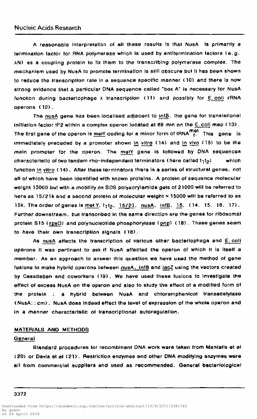
pBP280 (24) Is shown in Figure I. it carries a 10 kb insert Including argQ, the njjsA.
3373
Downloaded from https://academic.oup.com/nar/article-abstract/13/9/3371/2381742by gueston 04 April 2018

Nucleic Acids Research
A 18 21kA ar«Q matY I nu»A 11
\
rp«O pnp
TflH E
psnsoft lac'Z Y A Sa
PMC1403
lm*rt E-Bfl ol p8P2S0 Into pWC1403 batwaan E and B $ DMAF2 (Lac*)E Bo/B Sa . E
pMAFl
nutAHacZ hybrid (Lac«)
tnaart E-Sm ol pBP280 Mo pMC1403 batwa«n E and Sma$pMAE4 (Lac*)E Sm B 8a E
I „,„„ K I I P*IA1!4
Waaat with Bam HL fffl In with Hanow, r»fioata^pMAE4-B (Lac*)E Sm 8a EI lnr./»y/»uiiJ ~1 I
mm-tacZ hybrid (Lac*)M HSS.7 M
J I I |att | Imm21
Sa
lacZE Sac
PMAE4-S
Xna*S-S
\512Lac"
DMAE4-Bz - W B nu»A Otga«t \na«8-5 wtth M«tD, AS12 Lac'wtth EcoR1hybrid J | and PMAE4-B wHh EcoR1 and Mat D, Koala, In
H E B vitro packagaI t:i!!iy/V//HI 1 attl N CI857
E(S3.8%)Sac/ WA»7d j ltt] H CI8S7
lacZ' 'lacz;tE
±Am
Sa
'lacZHnfB nu«A|m«tY c)gOhybrid IS/21k
In vivo Croat with ANMMOdmmZD
- f alt N knmti \E2
1kb
F IQ . 1 Derivation of xE2 carrying an InfB-lacZ protein fusion.Tho looatlon of genes Identified on pBP280 Is Indicated. Only the relevant restrictionsites are shown. Details of the construction are given In Materials and Method. %refers to the position of the equivalent sites as percentage of wild type A. A primepreceding or following a gene means that the gene Is Incomplete on that side.E = EcoRI. H = Hlndlll. B = BamHI. Bg = BgJII. 8m «• Smal. Sa = SaJI. M » Mstll.Sao = Saol
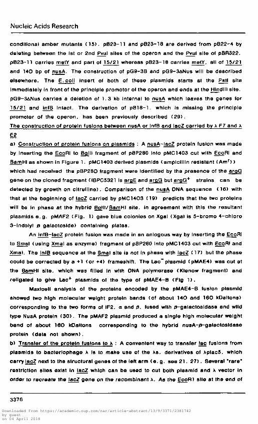
InfB operon and rpaO and pnp. Plasmlds of the nusA. InIB region are listed In Table 2.
and shown In Figure 3 together with the genes oarried. pB19-l Is the IO. 2 Kb left hand
part of pBP280 from EcoRI to jHlndlll. pB2O-l carries the 5. O kb Sail to BgJII
fragment. The nusA gene is not complete on this plasmld but the slightly truncated
protein Is stable and functional. (pB2O-1 is equivalent to the pYN87 ( 9 ) ) . pB22-4
3374
Downloaded from https://academic.oup.com/nar/article-abstract/13/9/3371/2381742by gueston 04 April 2018

Nucleic Acids Research
Table 2 Plasmlds of the nusA.lnfB region and the relative Intracellularconcentration of NusA and IF2 In strains carrying them.
Plaomld
pACYC184
PG9-3B
pG9-3ARuBA
PBR322
JDB19-1
PB20-1
PB22-4
PB23-18
PB23-11
pB18-l
Genes of the(1)
nusA,infB region
P, B>etY,t 1.15/21 ,nuaA,in£B
P, metY,t,t_,15/21,lnfB
argG, p, metY, t , t,,, 15/21, nusA, lnfB
argG,P,metY,tt ,15/21,nuflA1
P,netY,t,t,,,15/21,nuflA'
P,metY,t,t,,15/21
P.metY.t t ,
nusA,infB
Relative
NUBA
1
5-8
0.9-1.5
1
6-11
(3)3-6
n.d.
n.d.
n.d.
1-1 .5
( 2 )Levels of Protein
IP2a
1
7-10
7-12
1
9-20
0.6-1
n.d.
n.d.
n.d.
1.5-3
IP20
1
6-10
6-10
1
6-16
0.6-1
n.d.
n.d.
n.d.
2 - 3
(1) The ONA Insert carried by these plasmids Is shown in Figure 3. P refers to themajor promoter for the operon and t^g to the tandem rho-lndependant terminators.(2) The cellular levels of NusA and IF2 were measured in exponentially growingcultures of IBPC5321 XF7. IBPC5321 xE2 and IBPC5341 AD7 carrying the variousplasmlds. Extracts were analysed by electrophoresis on duplicate SDS poly-acrylamide gels which were blotted onto nitrocellulose and treated with antl-NusA orantl-IF2 followed by « i | protein A as described (34) . The treated blots wereautoradlographed and the radioactive bands were cut from the blot and counted toquantltate the amount of NusA or IF2. These values were normalised to the proteinconcentration In the extracts. The amount of crude extract analysed on the gelsvaried from 1 to 10 /ig. Relative levels of NusA and IF2 were calculated by comparingwith the strains carrying only the vector plasmld. The numbers show the range ofvalues obtained over the three cultures.(3) Mixture of wt protein and slightly smaller truncated proteinn.d. = not determined
carries tho 1. 9 kb Pstl fragment from the beginning of the nusA. lnfB operon. (and Is
equivalent to pKUl (14) and pSMll (15) ) . This plasmid produces a smaller NusA
fragment than pB2O-l. which still complements the nusAI mutation (14) but not the
3375Downloaded from https://academic.oup.com/nar/article-abstract/13/9/3371/2381742by gueston 04 April 2018
Nucleic Acids Research
conditional amber mutants (15 ) . pB23-l l and pB23-18 are derived from pB22-4 by
deleting between the 1st or 2nd Pvul sites of the operon and the Pvul site of pBR322.
pB23-11 carries metY and part of 15/21 whereas pB23-18 carries metY. all of 15/21
and 14O bp of nusA. The construction of pQ9-3B and pG9-3ANus will be described
elsewhere. The E. coll Insert of both of these plasmlds starts at the Pstl site
Immediately In front of the principle promotor of the operon and ends at the Hjndlll site.
pG9-3ANus carries a deletion of 1.3 kb Internal to nusA which leaves the genes for
15/21 and InfB Intact. The derivation of pB18-l . which Is missing the principle
promoter of the operon. has been previously described (29 ) .
The construction of protein fusions between nusA or InfB and lacZ carried by x F7 and x
E2
a) Construction of protein fusions on plasmids : A nusA-lacZ protein fusion was made
by Inserting the EcoRI to BgJII fragment of pBP280 Into pMC1403 cut with EcoRI and
BamHI as shown In Figure 1. pMC1403 derived plasmlds (amplcillln resistant (Am r ) )
which had received the pBP28O fragment were identified by the presence of the argG
gene on the cloned fragment (IBPC5321 Is argE and arqG but arqQ+ strains can be
detected by growth on cltrulllne). Comparison of the nusA DNA sequence (16) with
that at the beginning of lacZ carried by pMC14O3 (19) predicts that the two proteins
will be In phase at the hybrid Bglll/BamHI site. In agreement with this the resultant
plasmlds e .g . pMAF2 (Fig. 1) gave blue colonies on Xgal (Xgal is 5-bromo 4-chloro
3-indolyt p galactoslde) containing plates.
An InfB-lacZ protein fusion was made in an anlogous way by Inserting the EcoRI
to Smal (using Xmal as enzyme) fragment of pBP280 into pMC1403 cut with EcoRI and
Xmal. The InfB sequence at the Smal site is not in phase with lacZ (17) but the phase
could be corrected by a +1 (or +4) frameshlft. The Lac~ plasmid (pMAE4) was cut at
the BamHI site, which was filled In with DNA polymerase (Klenow fragment) and
rellgated to give Lac+ plasmlda of the type of pMAE4-B (Fig 1) .
Maxloell analysis of the proteins encoded by the pMAE4-B fusion plaamld
showed two high molecular weight protein bands (of about 14O and 16O kDaltons)
corresponding to the two forms of IF2. a and 0. fused with p-galactosldase and wild
type NusA protein (30 ) . The pMAF2 plasmid produced a single high molecular weight
band of about 160 kOaltons corresponding to the hybrid nusA-p-galactosldase
protein (data not shown).
b) Transfer of the protein fusions to x : A convenient way to transfer lac fusions from
plasmlds to bacterlophage x Is to make use of the xs. derivatives of xplac5. which
carry lacZ next to the structural genes of the left arm (e. g. see 2 1 . 27) . Several "rare"
restriction sites exist In lacZ which can be used to cut both plasmid and x vector In
order to recreate the lacZ gene on the recomblnant x. As the EcoRI site at the end of
3376
Downloaded from https://academic.oup.com/nar/article-abstract/13/9/3371/2381742by gueston 04 April 2018

Nucleic Acids Research
lacZ Is mutated In pMC14O3 (19) we used instead the Mstll site near the beginning of
lacZ. This process is diagrammmed In Figure IB for pMAE4-B. The pMAF2 fusion was
transferred to x In exactly the same way.
The EcoRl to Mstll fragment carrying the fusion was Inserted between the left
arm of Anav8-5. a Lac*. I m m " phage (28) up to the Mstll site In lacZ and the right
arm of A512 (27) up to the EooRl site at 53. 8 %. A512 Is a Lac* cl857 phage carrying
a deletion between BamHI sites at 57. 7 and 71. 0 % A. To facilitate distinction between
the A512 starting phage and the required Lac+ fusion. A512 was made Lac" by
Inverting the Sac I fragment which covers the lacZ-A Junction to give A512 Lac~. pMAF2
or pMAE4-B were digested with EcoRl and Mstll. Anav8-5 with Mstll. and A512 Lac~
with EcoRl. The llgated mixtures were packaged In vitro and Lac+ Imm* cl857 phages
selected by plating on IBPC532KANM540) on X gal containing plates at 37°C. LacZ*
(blue) phages were purified on IBPC5321. Phages which carried arqO ware detected
since they formed lysogens of IBPC5321 at 3O°C which grew on minimal plates
supplemented with citrulllne and histldlne. Small scale liquid lysates (23) of argQ+.
Lac'1' phages were grown and phage ONA extracted. Phages of the type xpMAE4 (Fig
IB) and ApMAF2 were characterized.
These thermosensltlve lmmA cl857 ]nt phages were ohanged to I m m " . c l * . int+
phages by In vivo recombination with ANM540(25). A permissive host. IBPC5321. was
Infected with both phages at an mol of 2-3 . allowed to grow for 2hr and then plated on
IBPC5321 (A+> on Xgal containing plates to select for Lao+ I m m " phages. Such
phages were purified on IBPC5321 and LacZ*. arqG+ lysogens selected. These were In
turn purified and the phages Induced by U. V. Phage ONA was prepared and AE2 (InfB-
lacZ fusion) (Fig IB) and AF7 (nusA-lacZ fusion) Identified, by restriction enzyme
analysis.
Construction of a promotor proximal operon fusion carried by AD7
a) Construction of an operon fusion vector with a Pstl site : The BamHI to Sajl Jac
operon cassette of pMC871 (19) was cloned between the BamHI and Sail sites of
pGA39 (31) (chloramphenlcol resistant (Cm r ) ) to give pQMl as shown In Figure 2.
This produced Cmr colonies which were blue on Xgal. despite the absence of a
functional Lac promotor (pMC871 in also blue). The extraneous DNA between the Saml
and BamHI sites of pGMl was eliminated by digesting with Xmal and BamHI. filling In
the sites with DNA polymerase. Klenow fragment, and rellgatlng to give pGMA. This
process recreates both the Smal and BamHI sites which now overlap by one base pair.
The presence of this plasmid causes colonies to be blue on Xgal containing plates and
medium red on MacConkey lactose plates (350 units of p-galactostdase)
b) Insertion of a promotor proximal fragment of the nusA-lnfB operon : The 570 bp
Pstl-Xholl fragment of pB23-11 (Fig 3) was Inserted between the Pstl and BamH 1 sites
3377
Downloaded from https://academic.oup.com/nar/article-abstract/13/9/3371/2381742by gueston 04 April 2018

Nucleic Acids Research
I trp'BA lacZ Y A I* Km i p M C 8 7 1
j ..-•'' ln»art BamHI-Sall ot pWCB71 Into
pGA39
P Sm BCut pGM1 with B«mHIand Xmal,
S» E P fin In wtthKlanow, Bgata^pGalA
l™* r t P l t I " X h ° n o l P 8 * ' 1 1 woPB23-11 pOtlA<8fl««te<! wtthP«tIandBamHI
htrp'BA lacZ Y A
Sa E
EIA43.8-S3.8) So<67.5, 68.5%)I «tl | | N CI857
Ugaat \SEW with Sail,
\NMS40 with Psti, and
PQM01 with Psti and Sail.
UO«t«, In vitro packaga p sB
S^oct Lac'lmm21 I
* \D7
«t t
\SEW
P<66.S%)I N Imm21 \ N M 5 4 0
| , c A Y Z trpA B1 j P
PQM01
E(A43.8-53.8)
I attSa
11C A Y Z I
hybf Id oparon) Xn^y
lmn.21 XD7
'15/21k
1kb
FIQ. 2 Derivation of XO7 carrying a promoter proximal operon fusion(see legend to Fig. 1) . In addition P ° Psti. Xh = Xholl
of DGMA. This fragment carries the major promotor of the operon. metY. the two
terminator structures and half of the gene for 15/21K. pQMA derivatives which had
acquired a promotor containing fragment could be identified as those which were now
very strongly red on MacConkoy lactose plates. Such plasmids were analysed and
pQMOl was Identified (Fig 2 ) .
c) Transfer of the operon fusion to x : The Pstl-Sall fragment of pGMDl was Inserted
into the clll region of X between the Psti site at 06. S % and the Sail site at 67. 5 %. In
XNM540 the I m m " substitution removes the Psti site at 76. 3 % x . . leaving the Psti site
at 60. 5 % as the last site, x SEW (20) was digested with Sail. xNMMO with Psti and
pQMDl with Psti and Sajl. The DNAa were Iigated and packaged In vitro and then plated
on IBPC532KX*) on Xgal containing plates at 37°C. Blue plaques were purified on
3378
Downloaded from https://academic.oup.com/nar/article-abstract/13/9/3371/2381742by gueston 04 April 2018
Nucleic Acids Research
15/21
E
II
matv/ nusA tnfBA. I /?9 I
I f^j^^
AntibioticReaistanc*
Am
AmCmCm
TcTcTcAm
parts of the
Replicon
PBR322
PBR322pACVCpACVCPBR322D6R322PBR322PBR322
nusA-.Fig. 3 Structure of a series of plaamlds carrying parts of the nusA-. infB operon.The location of identified genes of pB19-l Is Indicated. The fragments of ONAsubcloned in the other plasmids Is shown. These subclones are further described InMaterials and Methods. The genes carried and the level of NusA and IF2 producedaro described In Table 2. Replicon pACYC means that these plasmlds carry thesame replicon as pACYC184 whloh is present in the vector plasmid pGA39(31) usedfor trto cloning.
IBPC5321 and lyaogens. picked from the center of the plaques, were purified and
Induced by U. V. Phage DNA was prepared and AQMD7 Identified. Experiments with this
phage were subsequently conducted in recA strains because the construction includes
a small duplication of about 1 % A ONA on either side of the insert which could be lost
by In vivo recombination.
Construction and characterization of a nusA-cat fused gene
Close and Rodriguez (32) have described the construction of chloramphenlcol
resistant DNA cartridges carrying the structural gene for chloramphenlcol trans-
acetylase (cat) but missing the natural promoter. The cat cartridge with Bam HI sites
at the ends was inserted into the unique BpJII site at the end of nusA of pBP280 (Fig 1)
so that the cat gene is transcribed In the same sense as nusA, This plasmid now
confers Cmr to the host bacteria. In addition to the vector encoded Tc r .
This plasmid (pBP280 Bojll: : cm) was introduced Into strain JC3560. which
is argQ and carries a Tn5 (Kanamycln resistant. Kmr ) transposon within pnp
(24 ) . The transformants were purified once and then grown through several cycles of
serial dilutions In L8 and then In LB containing chloramphenlcol. The saturated
cultures ware plated on the Tc8 selection plates of Maloy and Nunn (33) . After 2 days
growth at 37°C. Isolated colonies were picked and screened for ArgQ. Cm r . Kmr and
Tc r. Analysis of the Tc8 bacteria. I .e. those which had lost the plasmid. showed a
large number which had acquired Cmr with or without the adjacent Arg and Km
markers. One example of each pattern of recombination was further analysed : -
IBPC 241. 242. 244. 246 (see Table I ) .
3379
Downloaded from https://academic.oup.com/nar/article-abstract/13/9/3371/2381742by gueston 04 April 2018

Nucleic Acids Research
1 2 3 4 5 6
w.t.NusA-*~
Fig. 4 Immunoblott analysis of Cmr recomblnants.Freshly saturated cultures were lysed by boiling In 3DS sample buffer and analysedon a 10 % 8D8 polyacrylamlde gel (35 ) . Proteins were electrophoretlcally blottedonto nitrocellulose, treated with antlNusA and then protein A labelled with « i | (34)Lane I. JC3S60 : Lane 2. IBPC244 : Lane 3. IBPC246 : Lane 4. IBPC241 : Lane 5.IBPC244 : Lane 6. control bacteria (maxlcell strain CSR 603).
JC 3500 Is xr due to a malA mutation but It and the Cmr derivatives were
converted to \a by phage PI tranoductlon selecting for growth on maltose. The
recomblnants all grew normally and plated K* (an N dependent phage) normally at
42°C I. e. There was no phenotype like the nusAI mutation of Friedman and Baron
t l J ,
Samples of each recombinant were analysed by immunoblottlng (34) with antl
NusA as shown in Figure 4. All (our recomblnants showed a band reacting with antl
NusA antibodies of a molecular weight greater than wild type NusA. Maxlcell analysis of
the pBP280 BoJII: : cm plasmld similarly shows a band of molecular weight about BO
kDaltons. (data not shown). Comparison of the ONA sequence of nusA at the BoJII site
(18) with that of the beginning of the cat cartridge (32) shows that nusA Is fused in
phase with the cat gene such that the 30 nucleotldes before the natural Initiation codon
of cat are translated as 10 additional aminoaclds. The 80 000 daltons molecular weight
protein band is thus NusA (up to the BgJII site) plus 10 amino acids plus the 24 000
daltons of the chloramphenlcol transaoetylase protein. Maxlcell analysis of pBP280
Bp.Hl:: cm also showed considerable synthesis of wild type molecular weight chloram-
phenlcol transacetylase. thus we cannot say whether the hybrid NusA:: cm protein has
CAT activity or whether the Cm r derives from the wild type protein. The hybrid protein
would appear to have NusA activity since the recombinants are perfectly viable. The
Immunoblott (Fig 4) does show two faint bands somewhat smaller than wild type NusA.
so It is. in theory, possible that It Is these fragments rather than the hybrid NusA: : cm
which is permitting growth.
3380
Downloaded from https://academic.oup.com/nar/article-abstract/13/9/3371/2381742by gueston 04 April 2018

Nucleic Acids Research
The nuaA: : cm mutation was introduced Into IBPC5321 by phage PI trans-
ductfon of argG* or cm r from IBPC 252 (direct selection for Cm r was very Inefficient
compared to selection for argQ*) IBPC5355 was a rare transductant obtained by
selecting directly for Cm r which Is still argQ. It was converted to recA by conjugation
with an Hfr strain carrying recAl and selecting for His'*'.
The rujsA(tsl) mutation was introduced Into IBPC5321 using Pi grown on
YN2351 (Nakamura. Misusawa. Tsugawa. Zubar. Court. Imai. manuscript in prepa-
ration) and selecting for ArgQ+ at 30°C to give IBPC181. The recAl allele
was subsequently introduced by conjugation as for IBPC5355. to give IBPC183.
RESULTS
1. Description of nusA-lnfB fusions with lacZ
Two types of fusions between genes of the nusA-JofB operon and jac have
been constructed and transferred to Integration proficient >. bacterlophage. The first
type are protein fusions between nusA and lacZ (XF7) or InfB and lacZ (AE2) where
an N terminal fragment of NusA or IF2 is fused In phase with 0-galactosldase at the
level of the 8th amlno acid of the wild type 0-galactosidase protein to produce hybrid
NusA-0-galactosidase or IF2-0-galactosldase proteins. In the case of the InfB-lacZ
fusion carried by XE2 (Fig 1 ) . two hybrid proteins corresponding to the two forms of
IF2. a and p. are synthesized (30 ) . The 0-galactosldase activity of these fusions is
a measure of the level of transcription of the operon and of the translation of the
nusA or InfB mRNA. These fusions carry the major promoter of the nusA. InfB
operon plus several Kb. Including argQ. upstream of the operon.
The second type of fusion constructed Is an operon fusion carried by xD7
(Fig 2 ) . A short promoter proximal part of the nusA-lnfB operon. lacking any part of
the nuaA or infB structural genes, has been cloned before a lacZ complete with its
own translatlonal initiation signals but missing a functional promoter. In this case
wild type 0-galactosldase Is synthesized from the transcription signals at the
beginning of the nuaA-lnfB operon.
2. Overproduced NusA protein decreases expression of the nusA-infB operon
The effect of a series of plasmlds (Table 2 and Fig. 3) covering various
genes of the nusA-lnfB operon has been tested on the level of 0-gaiactosldase
expressed from the three fusions. The results are shown In Table 3. At the same
time the level of NusA and IF2 produced by these plasmids was measured by
quantitative Immunoblottlng and the results are shown in Table 2. Only plasmlds
which cause an appreciable overproduction of NusA protein (pQ9-3B. pB19- l .
pB20-l) have any effect on the level of p-galactosldase from any of the fusions.
They reduce the 0-galactosldase activity to about 50 % of the wild type activity. On
3381
Downloaded from https://academic.oup.com/nar/article-abstract/13/9/3371/2381742by gueston 04 April 2018

Nucleic Acids Research
Table 3 Effect of NusA and IF2 overproduction on the level of expressionfrom different nusA. InfB-lac fusion
Fusion type
Plaamid
PACTC194
PG9-3B
PG9-3ANUSA
(SR322
PB19-1
PB2O-1
JB22-4
PB23-18
PB23-11
PB18-1
XD7
15/21-lac operon
1 0 0
32HO
8314
1 0 0
3014
5712
7 0
108
1 0 5
9416
D
( 2 )
( 2 )
( 2 )
( 2 )
( 2 )
( 2 )
( 1 )
( 1 )
( 1 )
( 2 )
• 4 "
42
6 0
4 5
3 6
6 5
6 0
32
35
35
35
XP7
NuaA-Lac protein
. * «
100
55*17
9615
100
4314
5316
8812
10314
94
93*6
)
( 3 )
( 4 )
( 3 )
( 4 )
( 3 )
( 3 )
( 3 )
( 2 )
( 1 )
( 2 )
• 4 "
36
42
40
37
SO
48
33
34
33
33
IP2-Lac
' * "
1OO
72110
9614
1 0 0
6218
7612
8812
9812
9 1
97118
XE2
protein
)
( 4 )
( 4 )
( 3 )
( 4 )
( 4 )
( 2 )
( 2 )
( 2 )
( 1 )
( 2 )
35
40
33
35
48
33
40
33
40
33
Monolysogens of IBPC9321 with XE2 or XF7 and IBPC5341 with X07 were transformedwith the series of plasmids listed. Cultures were grown at 37°C In MOPS mediumenriched with all amino acids (36) and containing the relevant antibiotic : Cm (25/igml"1). Tc (10 Mgml"1). Am (500 MOml"1). Further Am was added during growthto maintain the presence of the plasmids.(1) 0-galactosldase activities were measured for each culture at. at least, fourpoints between A e 5 0 values of 0.2 and 0. 6 and are expressed as a percentage ofthe strain carrying the vector plasmld. The numbers in parenthesis are the numberof Independantly grown cultures tested. For IBPC534KXD7) 100 % = 1540 i 50units, for IBPC 5321 (XF7) 100 * = 360 i 40 unite and for IBPC5321 (XE2) 100 % =325 i 20 units.(2) D j Is the average doubling time In mln for each culture.
the other hand, plasmids which overproduce IF2 in the absence of NusA have no
effect on the fusions, compare pQ9-3B and pG9-3ANus. The plasmld pB18-1.
which carries the genes for both nusA and InfB but without the major promoter
causes a barely measureable Increase In NusA concentration (Table 2) and has no
effect on j3-galactosldase expression from the fusions. Qualitatively similar results
are observed for all three fusions tested. Quantitatively the IF2-Lac protein fusion
carried by xE2 seems to respond less than the promoter proximal operon fusion
3382
Downloaded from https://academic.oup.com/nar/article-abstract/13/9/3371/2381742by gueston 04 April 2018

Nucleic Acids Research
Fin. 5 Effect of growth rate on £-galactosldase expression from xE2 and AF7.IBPC 3321 lysogenlsed with AE2 or AF7 was grown In minimal MOPS mediumsupplemented with different carbon sources (36) to produce a range of growth rates: 1) Acetate : 2) Pyruvate : 3) Qlucose : 4) Qlucose enriched with all the amlnoaolds (36) n Is the number of doublings per hour. The results are the mean of sixmeasurements of 9-galactosldase activity between Agjo values of 0 .2 and 0.5 (orA 0.1 - 0. 25 for acetate).
(XD7) or the NusA-Lac protein fusion. (XF7) this point Is considered In the
Discussion.
As the promoter proximal fusion responds In the same way to the effect of NusA
as does the nusA-lac protein fusion It Is probable that the main effect of NusA is
transcriptlonal. Overproducing a large N terminal fragment of NusA from pB2O-1
causes almost as much repression as does wild type NusA. The somewhat smaller
fragment synthesized from pB22-4 seems also to exert some represser effect. The
plasmlds carrying the promoter proximal genes metY and 15/21 have no effect on the
level of 0-galactosidase. We. however, have no proof at the moment that these
plasmlds cause an augmentation In the cellular level of their gene products. Thus any
effect might be masked If their expression from the plaamids Is already severely
repressed.
As shown in Table 3. the presence of the plasmlds which overproduce NusA and
IF2 do reduce the doubling time of the rysogenlc bacteria. It was thus possible that the
decrease In 9-galactosldase seen was due to a decrease In growth rate rather than a
specific repressor effect. The level of 0-galactosldase under different growth rates was
measured and Is shown in Figure 5. Increasing the doubling time does decrease the
level of 0-galactosldase. However increasing the doubling time from 30 to 60 mln (the
range observed In Table 3) produces only a 15 % decrease In p-galactosldase activity
and not the 50 % decrease observed. In the experiments described above.
We also tested the effect of the plasmlds which overproduce NusA on wild type
9-galactosldase synthesized from the chromosome and saw none. Chromosomal lacZ*
strains carrying pQ9-3 or pG9-3 ANusA. induced with Isopropyl thlogalactoslde (IPTQ)
3383Downloaded from https://academic.oup.com/nar/article-abstract/13/9/3371/2381742by gueston 04 April 2018
Nucleic Acids Research
Table 4 The effect of mutant alleles on the level of expression from differentnusA. InfB-lac fusions
Fusion type
Strain
IBPCS341
IBPCS357
IBPC183
Genotype
w.t.
nusAitcm
nusA( t s l )
XD7
15/21-lac op
100 ( 2 )
169*2 ( 3 )
183*5 ( 3 )
eron
°T 2 >
82
80
88
X
nusA-lac
1 0 0
160*1
176*16
Yl
Z protein
)
( 3 )
( 3 )
( 3 )
7 0
7 0
8 0
XE2
infB-lacZ
1 0 0
120*2
133*3
protein
D
( 3 )
( 3 )
( 3 )
r2>
72
7 0
7 0
Isogenlc derivatives of IBPC5341 carrying the nusA: : cm or nusA(tsl) allele werelysogenlsed at low m.o. l . with the fusion phages. Lysogens were grown In MOPSmedium supplemented with arglnlne and hlstldlne at 30°C.(1) 0-galactosldase measurements were made for each culture at four pointsbetween A^SQ = 0.2 to 0.5 and are expressed as a percentage of the wild typestrain. The number In brackets Is the number of Independent monolysogens tested.For IBPC5341 (XF) 100 % Is 325 t 55 units : for xE2 100 % = 345 ± 40 units and forXD7 (TOO <*>1 1700 * 65 units.(2) Or Is the average doubling time In minutes for each culture.
gave £-galactosldase values of 106 % and 112 % compared to strain carrying
pACYC184. We can thus eliminate the possibility that overproduced NusA non-
speclflcally reduces gene expression. We conclude therefore that the repression is
specific although the magnitude is not very large.
3. Mutated NusA proteins derepress the nusA-lnfB operon
Two mutant forms of NusA protein were testsd. The modification of NusA protein
by addition of the 24 kDaltons of protein corresponding to the gene for chloramphenlcol
transacetylase Is described In Materials and Methods. The gene for this hybrid protein
was Introduced by homologous recombination, onto the E. coll chromosome. It
seemed to exert no appreciable effect on the bacteria except that the cells are now
chloramphenlcol resistant. The second mutated nusA gene Is the nusA(tsl) mutation.
This thermosensltlve mutant. Is defective In transcription termination at both 30°C and
42°C (Nakamura et al. . manuscript In preparation).
The two mutants IBPC5357 (nusA: : cm) and IBPC183 (nusA(tsl)) and the
Isogenlc parental (IBPC5341) were lysogenlsed at low m.o. l . with the three fusion
phages XE2. XF7 and XD7. ^galactosldase measurements were made on several
Independent lysogens to Identify monotysogens. Table 4 shows that both the nusA: : cm
and nusA(tsl) mutations cause a dereprasslon of all three fusions. The magnitude of
the effect Is very similar for the xO7 and xF7 fusions (160-180 %) but Is somewhat less
for the XE2 fusion (120-130 % ) . This Is not surprising though, since xE2 carries a wild
3384
Downloaded from https://academic.oup.com/nar/article-abstract/13/9/3371/2381742by gueston 04 April 2018
Nucleic Acids Research
type nusA gene which can complement the nusA mutation. Measurements In Table 4
were made at 30°C because of the thermosensltlvlty of the nysA(tsl) mutation. The
strain was also tested at 37°C. where It still grows normally, but no greater
derepression was observed.
DISCUSSION
The results reported here show that the level of functional NusA protein in the
cell affects the level of expression of the operon. Such a result is characteristic of an
autoregulated protein. The phenomenon has been Investigated using both operon
fusions and protein fusions with lacZ. Numerically similar results were obtained with
both types of probes and as the operon fusion used carried only the proximal part of the
nusA. InfB operon and none of the structural gene for nusA it seems very likely that the
autoregulation Is occurlng at the transcrlptlonal level.
The structure of the nusA. InfB operon suggests at least one possible
mechanism for how this autoregulation might occur. The first gene of the operon metY.
coding for a structural RNA. tRNA ftf . is followed by sequences characteristic of two
rho-independant terminators (14 ) . It is only after these terminators that the genes for
the structural proteins of the operon ( 1 5 / 2 1 . nusA. InfB. 15) are found. One role of
these terminators is presumably to reduce the level of transcription of the proteinMetgenes which follow, compared to the amount of tRNAj required. Thus NusA
could be regulating the amount of readthrough of these terminators. Several other
terminators have been shown to be sensitive to NusA protein. (5. 6. 7) . Alternatively
NusA has been shown to slow down transcription or promote pausing at sites not
recognisable from their sequence as terminators (7 . 10) and so it is possible that the
effect of NusA on Its own operon acts outside the terminators, but within the first 570 bp
of the operon cloned In AO7.
In any case the effect of NusA. whether direct or Indirect. Is felt throughout the
operon as far as the InfB gene. The InfB protein fusion does, however, seem to be less
sensitive to the overproduction of NusA protein than the earlier fusions. One possible
explanation for this Is that the minor promoter detected just In front of InfB (15. 29) Is
functioning In this fusion to enhance InfB expression when upstream transcription Is
reduced.
The magnitude of the repression effect Is not very great. A 7-10 fold
enhancement In NusA concentration produces only a 50 % decrease In gene
expression. It was thus possible that the results observed were due to other effects
e. g. : an effect of NusA on Initiation of transcription cannot be eliminated at this point.
One hypothesis tested was that the presence of the plasmlds causes a reduction In
growth rate and hence a reduction In the level of 0-galactosldase synthesized. The
3385
Downloaded from https://academic.oup.com/nar/article-abstract/13/9/3371/2381742by gueston 04 April 2018

Nucleic Acids Research
data of Figure 5 shows that reducing growth rate does decrease expression from the
fusion but not as much as the decrease observed with the plasmlds which cause NusA
overproduction. Another hypothesis, for which there Is no evidence, would be that
NusA controls the expression of something else which acts In trans to regulate the
transcription of the nusA. InfB operon.
Looked at from the other point of view, the fact that any change in the
expression of 9-galactosidase from the fusions Is observed on changing the growth
rates suggests that the nusA.lnfB operon is subject to "so-called" metabolic
regulation. The change observed is not very great though. An eight-fold Increase In
growth rate produced only a 60 % increase In p-galactosldase activity from x£2 or XF7.
Comparable experiments using genes fused to 0-galactosldase or galac-
toklnaae have shown for rrnE about lOO % increase for a less than three fold
Increase In growth rate (37) : for tufB a 600 % Increase In 0-galactosldase for a
fourfold increase In growth rate (38) and for trpS a 250 % Increase for a four fold
Increase In growth rate (39 ) . For these latter experiments, it was concluded that the
gene under study was subject to metabolic regulation. The much smaller effect seen
for the nusA-lnfB operon might imply that although basically subject to metabolic
regulation, the effect Is reduced by other regulatory factors coming Into play.
The nusA: : cm mutation described here Is interesting In the sense that the
protein does not appear to have any detectable phenotype on cell growth. However It
does exert a regulatory effect in enhancing Its own expression, and that of IF2
(Nakamura. Plumbrldge. Oondon. Qrunberg-Manago - manuscript In preparation)
and that of the nuaA-lacZ fusions described here. The magnitude of the derepresslon
seen is comparable to that observed with the nusA(tsi) mutation whioh has both a
strong thermosensltlve phenotype and Is defective In termination (Nakamura et al. .
manuscript In preparation). The nusA: : cm mutation has not been tested for
termination efficiency. It is possible that the protein is sufficiently defective In
termination to enhance expression of the operon but not to affect cell growth.
One Interpretation of the results described here is that the C terminal part of
NusA could be the region of the protein Important for regulation. The fact that from the
Bglll site onward 37 amino acids can be replaoed by protein sequences of
chloramphenlcol transacetylase shows that these sequences are not necessary for
NusA function, at least as far as permitting normal cell growth. However, this region
could be important for regulation as the effect of this mutation on the fusions is
comparable to that observed with a defective nusA mutation (nusA(ts l ) ) .
An alternative and maybe preferable hypothesis Is that the regulatory part Is
associated with the N terminal region. The protein synthesized from pB20- l . which Is
mlislng the same 37 amino acid* that are replaced in nusA:: cm. represses nusA. InfB
3386
Downloaded from https://academic.oup.com/nar/article-abstract/13/9/3371/2381742by gueston 04 April 2018

Nucleic Acids Research
operon expression almost as effectively as wild type NusA. The distinctly smaller
protein synthesized from pB22-4 (missing 150 amlno acids) also seems to exert some
repressor effect. These observations are not necessarily contradictory with the result
that the nusA: : cm mutation derepresses the operon, since It Is quite conceivable that
adding 24 kOaltons to the C terminal end of the protein causes a staple modification In
the N terminal region.
IF2 synthesis Is co-regulated with that of NusA. The precise reason for this is
still obscure. IF2 synthesis Is co-ordinated with that of the other translational Initiation
factors IF1 and IF3 and also to ribosomes (40 ) . How this co-ordination is achieved Is
not known. For IF3 at least two transcription start sites have been Identified (41 ) . For
IF2 a second minor promoter In front of the InfB gene has been claimed (15, 29) . This
second promoter could be functioning In the case of repression of the nusA.lnfB
operon (see above). These multlmodal transcription units could thus be involved In the
co-ordinated synthesis observed, but much more work Is needed to unravel this
particular aspect of cellular regulation.
To summarize, the data reported here strongly suggest that the NusA protein
autogenously regulates Its own transcription and that of the neighbouring genes. There
is as yet no definite evidence for a mechanism but It is tempting though to postulate that
NusA regulates transcription through the tandem terminators near the beginning of the
operon.
ACKNOWLEDGEMENTSWe thank Martin Schmidt for the gift of antl-NusA antibody. We thank Scott Butler forcritical reading of the manuscript and Mathias Springer for constructive discussions.This work was supported by grants to Doctor Qrunberg-Manago from the "C. N. R. 8."(Grant 18) and from the I .N .8 . E.R. M. (823.008. 831.003 and a short termfellowship granted to Y. Nakamura). from the ' M . R . I ' (82 V 1289). from the'FONDATION POUR LA RECHERCHE MEDICALE' and "E. I. du Pont' (to M. Q . - M . ) .
"Present address: Department of Molecular Biophysics and Biochemistry, Yale University, P.O. Box6666, 260 Whitney Ave., New Haven, CT 06511, USA
REFERENCES1. Friedman. D . I . . and Baron L.S. (1974) Virology 58 141-1482. Greenblatt. J. . U. J. . Adhya. S. . Friedman. D . I . . Baron. L. 8. .
Redfield. B.. Kung. H . . and Welssbach. H. . (1980) Proc. Natl. Acad.8cl. USA. 77 1991-1994
3. Greenblatt. J. . and U. J. , (1981) J. Mol. Biol. J47 11-234. Qreenblatt. J. . and LI. J. . (1981) Cell 24 421-4285. Qreenblatt. J. . Umont. M. . and Hanley. 8 . . (1981) Nature (London)
282. 215-2200. Farnham. P. . Qreenblatt. J. . and Plart. T. . (1982) Cell 29 945-9517. Kingston. R.E. . and Chamberlln. M . J . . (1981) Cell 27 523-5318. Ward. D. F. . and Qottesman. M.E. . (1981) Nature (London) 292 212-2159. Nakamura. Y. . and Uchida. H. . (1983) Mol. Gen. Qenet. JjJO 189-195
10. Schmidt. M.C. . and Chamberlln M.J. . (1984) Blochem. 23 197-203
3387
Downloaded from https://academic.oup.com/nar/article-abstract/13/9/3371/2381742by gueston 04 April 2018
Nucleic Acids Research
1). Friedman. D . I . . and Olson. E.R. . (1983) Cell 34 143-14912. LI. S .C . . Squires. C. L. . and Squires. C. . (1984) Cell 38 851-88013. Plumbrldge. J.A. . Howe. J . Q . . Springer. M. . Touatl-Sohwartz. D. .
Hershey. J.W. B. . and Qrunberg-Manago. M. . (1982) Proc. Natt. Acad.Scl. U8A. 79 5033-5037
14. Ishli. 8 . . Kurokl. K. . and Imamoto. F. . (1984) Proc. Natl. Acad. 8c.USA. 8J 409-413
15. Nakamura. Y. . and Mlzusawa. S. . (1985) EMBO J. In press16. Ishll. 8. . lhara. M. . Maekawa. T. . Nakamura. Y. . Uchlda. H. . and
Imamoto. F. . (1984) Nucl. Acl. Res. J2 3333-334217. Sacerdot. C. . Oessen. P.. Hershey. J.W. B. . Plumbrldge. J.A. . and
Qrunberg-Manago. M. . (1984) Proc. Natl. Acad. 8c. USA.tM. 7787-779118. Portier. C. . and Regnier. P. . (1984) Nuol. Acids. RSB. J2 6091-810219. Casadaban. M.J . . Chou. J. . and Cohen. 8 . N . . (1980) J. Bocterlol. 43
971-98020. Manlatls. T. . Frltsch. E. F.. and Sambrook. J. . (1982) Molecular cloning.
Cold Spring Harbor Laboratory. Cold Spring Harbor. New-York21. Davis. R.W. . Botsteln. P.. and Roth. J.R. . (1980) Advanced Bacterial
Qenetlcs. Cold Spring Harbor Laboratory. Cold Spring Harbor. New-York22. Miller. J. . (1972) Experiments In Molecular Genetics. Cold 8pring Harbor
Laboratory. Cold Spring Harbor. New-York23. Springer. M. . Trudel. M. . Giraffe. M. . Plumbrldge. J. . Fayat. Q. .
Mayaux. J. F. . Sacerdot. C. . Blanquet. S. . and Qrunberg-Manago. M. .(1983) J. Mol. Blol. V7J 263-279
24. Portier. C. . Mlgot. C. . and Qrunberg-Manago. M. . (1981) Mol. Gen.Genet. J83 298-305
25. Borck. K. . Beggs. J. D. . Brammar. W . J . . Hopkins. A. 8 . . and Murray.N.E . . (1976) Mol. Gen. Genet. V46 199-207
26. Enqulst. L. . Tiermeir. D. . Leder. P. . Welsberg. R. . and Sternberg. N. .(1976) Nature (London) 259 596-598
27. Kourllsky. P. . Perrlcaudet. M. . Qros. D. . Garapln. A. . Gottesman. M. .Frltsch. A. , and Tlollals. P. . (1978) Blochlmle 60 183-187
28. Springer. M. . Mayaux. J.F. . Fayat. Q. . Plumbrldge. J.A. . Qratio. M. .Blanquet. S. . and Grunberg-Manago. M. . (1985) J. Mol. Biol. (in press)
29. Plumbridge. J.A. . and Springer. M. . (1983) J. Mol. Biol.. 167 227-24330. Plumbridge. J.A. . Deville. F. . Sacerdot. C. . Petersen. H. U. .
Cenatlempo. Y. . Cozzone. A. . Grunberg-Manago. M. . and Hershey.J.W. B. . (1985) EMBO J. . 4. 223-229
31. An. Q. . and Frlesen. J.D. . (1980) J. Bacteriol. 140 400-40732. Close. T . J . . and Rodriguez. R. L. . (1982) Gene 20 305-31633. Maloy. 8. R.. and Nunn. W. D. (1981) J. Baoterlol. V45 1110-111234. Howe. J . G . . and Hershey. J.W. B. . (1981) J. Blol. Chem. . 256 12.836-
12.83935. Laemmll. U.K.. (1970) Nature (London) 227 680-68530. Neldhardt. F .C . . Bloch. P. L. . Pedersen. 8 . . and Reeh. 8 . . (1977) J.
Bacteriol. J29 378-38737. Mlura. A. . Kreuger. J. H.. Itoh. 8 . . de Boer. H.A. . and Nomura. M. .
(1981) Cell 25 773-78238. Takebe. Y. . and Kazlro. Y. . (1982) Mol. Qen. Qenet. 187 355-36339. Hall. C.V. and Yanofsky. C. . (1982) J. Bacteriol. V5J 918-92340. Howe. J . G . . and Hershey. J.W. B. . (1983) J. Biol. Chem. 258 1954-
195941. Mayaux. J . F . . Fayat. Q. . Fromant. M. . Springer. M . . Grunberg-
Manago. M. . and Blanquet. 8. . (1983) Proc. Natl. Acad. 8c. U8A 800152-6156
3388
Downloaded from https://academic.oup.com/nar/article-abstract/13/9/3371/2381742by gueston 04 April 2018