effect of nanolayering of calcium salts of phosphoric acid ... · 1. introduction dentin bonding...
TRANSCRIPT

Acta Biomaterialia 38 (2016) 190–200
Contents lists available at ScienceDirect
Acta Biomaterialia
journal homepage: www.elsevier .com/locate /ac tabiomat
Full length article
Effect of nanolayering of calcium salts of phosphoric acid estermonomers on the durability of resin-dentin bonds
http://dx.doi.org/10.1016/j.actbio.2016.04.0341742-7061/� 2016 Acta Materialia Inc. Published by Elsevier Ltd. All rights reserved.
⇑ Corresponding authors at: State Key Laboratory of Military Stomatology & National Clinical Research Center for Oral Diseases & Shaanxi Key Laboratory of OralDepartment of Prosthodontics, School of Stomatology, The Fourth Military Medical University, Xi’an, Shaanxi, China (L.-n. Niu). The Dental College of Georgia,University, Augusta, GA 30912-1129, USA (F.R. Tay).
E-mail addresses: [email protected] (L.-n. Niu), [email protected] (F.R. Tay).1 Equal contributors.
Fu-cong Tian a,1, Xiao-yan Wang a,1, Qi Huang b, Li-na Niu c,⇑, Jan Mitchell d, Zheng-yi Zhang e,Chandrani Prananik f, Lu Zhang a, Ji-hua Chen c, Lorenzo Breshi g, David H. Pashley d, Franklin R. Tay d,⇑aDepartment of Cariology and Endodontology, School and Hospital of Stomatology, Peking University, Beijing, PR ChinabDepartment of Operative Dentistry and Endodontics, Guanghua School of Stomatology, Sun Yat-sen University, Guangzhou, Chinac State Key Laboratory of Military Stomatology & National Clinical Research Center for Oral Diseases & Shaanxi Key Laboratory of Oral Diseases, Department of Prosthodontics,School of Stomatology, The Fourth Military Medical University, Xi’an, Chinad The Dental College of Georgia, Augusta University, Augusta, GA, USAeDepartment of Prosthodontics, School & Hospital of Stomatology, Zhejiang University, Hangzhou, Zhejiang, ChinafDepartment of Chemical and Biological Engineering, University of Colorado, Boulder, CO, USAgDepartment of Biomedical and Neuromotor Sciences, DIBINEM, University of Bologna, Italy
a r t i c l e i n f o
Article history:Received 27 January 2016Received in revised form 13 April 2016Accepted 19 April 2016Available online 27 April 2016
Keywords:10-MDPAnalogBond durabilityNanolayeringOrganic-inorganic hybrid calcium saltPhosphoric acid ester resin monomer
a b s t r a c t
To investigate the contribution of nanolayering on resin-dentin bond durability, two phosphoric acidester resin monomers, 10-methacryloyloxy-decyl-dihydrogen-phosphate (10-MDP) or its analog, methacryloyloxy-penta-propyleneglycol-dihydrogen-phosphate (MDA), were examined for their affinity formineralized dentin powder in a column chromatography setup. Hydroxyapatite (HA) powder was dis-persed in experimental primers consisting of 10-MDP or MDA solvated in ethanol/water and examinedwith FTIR, 31P MAS-NMR and XPS. Light-curable 10-MDP or MDA primers were used for bonding to den-tin, and examined after 24 h or one-year of water-aging by TEM for evidence of nanolayering, and formicrotensile bond strength evaluation. Primer-bonded dentin was examined by thin-film XRD to identifyshort-range order peaks characteristic of nanolayering of resin monomer-Ca salts. Although 10-MDP hadbetter affinity for mineralized dentin than MDA, both monomers completely eluted from the mineralizeddentin powder column using ethanol-water as mobile phase, indicating that the adsorption processeswere reversible. This finding was supported by chemoanalytic data. XRD of 10-MDP-bonded dentinshowed three diffraction peaks hat were absent from MDA-bonded dentin. Nanolayering was identifiedby TEM in 10-MDP-bonded dentin, but not in MDA-bonded dentin. Significant drop in bond strength (inMPa) was observed for both groups after one-year of water-aging compared with 24-h: 10-MDP groupfrom 48.3 ± 6.3 to 37.4 ± 4.6; MDA group from 50.7 ± 5.0 to 35.7 ± 3.8 (P < 0.05), with no significant dif-ference between the two groups at the same time-point. Because both functional monomer-primed,resin-bonded dentin exhibited similar bond strength decline after water-aging, presence of nanolayeringis unlikely to contribute to the overall resin-dentin bond durability.
Statement of Significance
The durability of resin-dentin bonds in 10-MDP containing self-etching adhesives has been anecdotallyattributed to the presence of nanolayering of 10-MDP-calcium salts in the resin-dentin interface.Results of the present work indicate that such a claim cannot be justified. Complete elution of the phos-phoric acid ester monomer from mineralized dentin powder in the column chromatography experimentsusing ethanol-water mobile phase to simulate the solvent mixture employed in most 10-MDP-containingdentin adhesives further challenges the previously proposed adhesion-decalcification concept that uti-lizes chemical bonding of phosphoric acid ester monomers to apatite as a bonding mechanism in 10-MDP containing dentin adhesives.
� 2016 Acta Materialia Inc. Published by Elsevier Ltd. All rights reserved.
Diseases,Augusta

F.-c. Tian et al. / Acta Biomaterialia 38 (2016) 190–200 191
1. Introduction
Dentin bonding interfaces degrade with time. Electron micro-scopy and other in vitro tests have provided ultrastructural evi-dence of degradation in hybrid layers, and decline in resin-dentinbond strengths that resulted from resin hydrolysis and collagendegradation [1,2]. Loss of micromechanical retention betweenadhesive and dentin eventually leads to clinical restoration failure[3]. Apart from micromechanical interlocking, chemical adhesionbetween specific functional resin monomers and tooth mineralshas been reported as an alternative mechanism for adhesion ofmethacrylate resins to tooth structures. Chemoanalytic methodshave identified prospective chemical reactions that occur in theresin-dentin interface, including adsorption of functional resinmolecules on the apatite surface and the formation of resinmonomer-calcium salts [4–6].
Due to challenges in quantifying chemical reactions in theresin-dentin interface, a direct link between chemical bondingand resin-dentin bond durability was difficult to be established[7,8]. The water solubility of 10-methacryloyloxydecyl dihydrogenphosphate (10-MDP)-calcium salt was the lowest among salts pro-duced by the reaction between phosphoric acid ester monomersand apatite [5,9,10]. This feature was used to account for the betterin vivo and in vitro dentin bonding results achieved by 10-MDP-containing commercial adhesives [11,12]. In the absence of directevidence, those long-term in vivo and in vitro bonding results wereused anecdotally as indirect evidence for the contribution of chem-ical bonding to the overall bonding performance [13]. Becausemany confounding factors are involved in studies on dentin adhe-sives, it is taxing to attribute overall bonding performance to thepresence of phosphoric acid ester monomer-calcium salts in theresin-dentin interface [14–16].
In 10-MDP primer-treated resin-tooth interfaces, 10-MDP-Casalts self-assemble into nanolayers, with hypothetical structuresconsisting of the methacrylate groups of two 10-MDP moleculesfacing each other, and the functional hydrogen phosphate groupsdirected away from each other [17]. Ultrastructural manifestationof 10-MDP-Ca nanolayering was corroborated with the appearanceof three characteristic peaks in the 2h range of 2–8� in thin-film X-ray diffraction (XRD) scans of adhesive-coated dentin [18]. Thesethree peaks represent short-range order of the precipitated salts[19]. Based on this finding, several claims have been made forthe function of nanolayering in dentin bonding, including protect-ing collagen fibrils from water-induced degradation due to theirhydrophobicity, increasing the resistance of residual apatite crys-tallites to acidic dissolution, and creating a more gradual transitionbetween the inorganic bonding substrate and the biomaterial [17].These claims, however, were not supported by experimentalevidence.
According to the literature, the propensity of nanolayering for-mation in resin-dentin adhesives containing phosphoric acid estermonomers is affected by the presence of 2-hydroxyethylmethacrylate (HEMA), agitation, application time, monomer impu-rity and molecule structure of the phosphoric acid ester monomers[20–22]. These factors, alone or in combination, could haveaccounted for the paucity of nanolayering in resin-dentin inter-faces created by commercial 10-MDP-containing adhesives [19].Other functional resin monomers with similar structure as 10-MDP have also been investigated [10,23,24]. These monomers con-tain different hydrocarbon or fluorocarbon chains as spacer groupbetween the methacrylate group and the phosphate group. Thespacer group is known to influence monomer characteristics suchas flexibility, solubility, hydrophobicity, viscosity, and wettingbehavior. Long spacers are used to avoid steric hindrance duringpolymerization and to enhance mechanical properties [25]. Thesefunctional resin monomers were tested for their calcium salt solu-
bility, chemical shifts after reacting with hydroxyapatite or pow-dered enamel and dentin with nuclear magnetic resonance(NMR) spectroscopy, bond strength and ultrastructural examina-tion of the resin-dentin interface. Many of them produce nanolay-ering patterns with varying interlayer thickness. However, none ofthese studies was able to elucidate whether the presence ofnanolayering was responsible for the durability of resin-dentinbonds.
To circumvent the problem of interference of other methacry-late resin monomers on nanolayering formation, 10-MDP and anew 10-MDP analog with a different spacer group were respec-tively used as the only resin monomer for creating experimentalself-etching dentin adhesives. The analog member had been testedin a pilot study to confirm the absence of nanolayering whenbonded to dentin, and served as the control analog for 10-MDP.This enabled the authors to evaluate the contribution of nanolayer-ing to the longevity of resin-dentin bonds. The interactionsbetween the two resin monomers and the dentin mineral phasewere first investigated, followed by their influences on dentinbonding performance. The first null hypothesis tested was thatthere is no difference in the affinity of 10-MDP and the analog resinmonomer for mineralized dentin. The second null hypothesistested was that there is no difference in the capability of bothphosphoric acid ester monomers to produce nanolayering on thedentin surface. The third null hypothesis tested was that nanolay-ering of phosphoric acid ester monomer-calcium salts is unstableafter water aging and does not contribute to the resin-dentin bonddurability.
2. Materials and methods
2.1. Materials
Sixty non-carious human third molars were used in the presentstudy. The use of human teeth for research was approved by theHuman Assurance Committee of the Augusta University, Georgia.The teeth were refrigerated at 4 �C in 0.9% NaCl that contained0.02% sodium azide to prevent bacterial growth.
The two phosphoric acid ester monomers examined were: 10-MDP (molar mass: 322.35 g/mol) and methacryloyloxy-penta-propyleneglycol-dihydrogen-phosphate (10-MDP analog, designatedas ‘‘MDA”; molar mass: 456.48 g/mol). The molecule structures ofthe two resin monomers are illustrated in Fig. 1. Both resin mono-mers were obtained from DM Healthcare Products, Inc. (San Diego,CA, USA). 2-hydroxyethyl methacrylate (HEMA; molar mass:130.14 g/mol) and hydroxyapatite (HA; particle size �200 nm)were obtained from MilliporeSigma (St Louis, MO, USA).
Two experimental primers were prepared by blending 10-MDPor MDA with ethanol and water in the ratio of 15:45:40 wt% [20].For primers used in dentin bonding as part of the experimentaladhesive systems, camphorquinone (CQ; 1 wt%) and ethyl-4-dimethylamino benzoate (EDMAB; 0.4 wt%), both from Milli-poreSigma, were added to render the primers light-curable. ThepH values of the experimental primer solutions (measured with apH meter, Orion Star A211, ThermoScientific, Waltham, MA, USA)were 2.65 for the primer containing 10-MDP, and 2.38 for the pri-mer containing 10-MDP analog.
2.2. Affinity of the two phosphoric acid ester monomers formineralized dentin
A modified hydroxyapatite column chromatography procedure[26] was adopted in the present study to examine the elution char-acteristics of the two phosphoric acid ester monomers in the pres-ence of mineralized dentin powder, as a measure of the affinity of
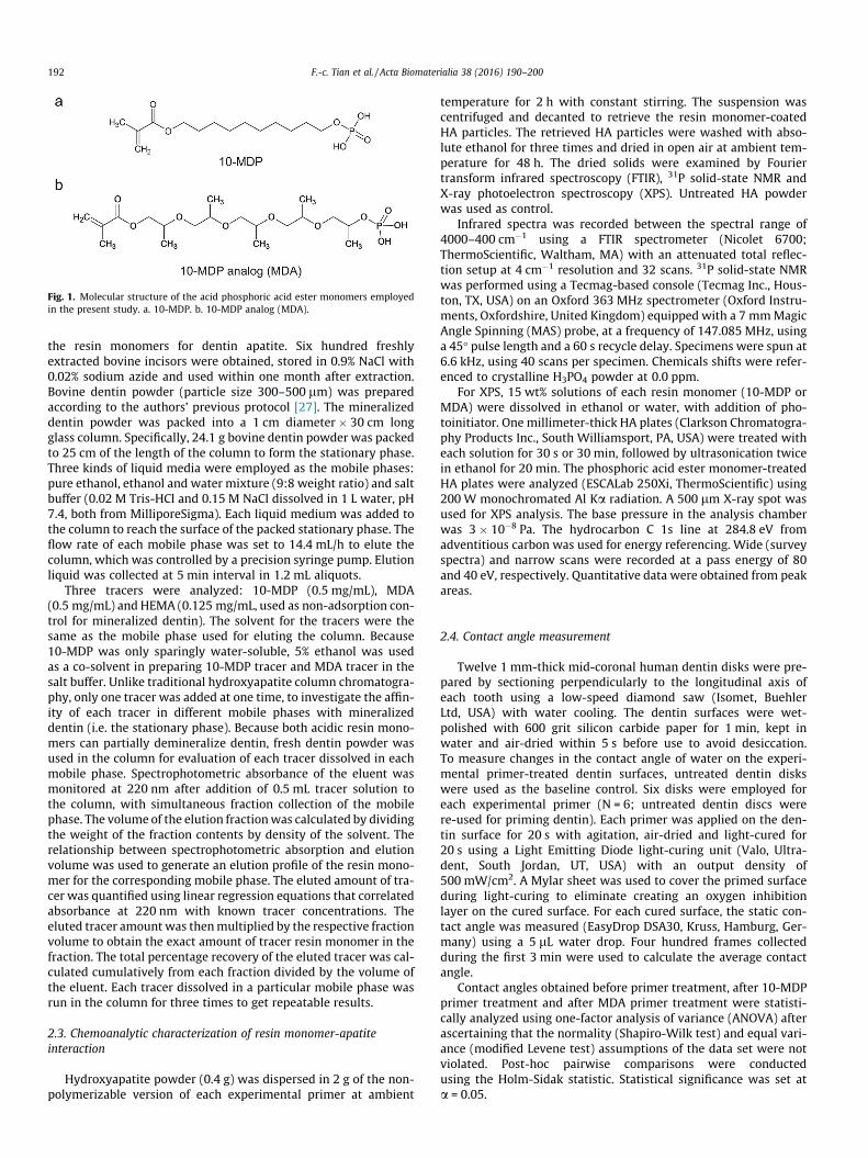
Fig. 1. Molecular structure of the acid phosphoric acid ester monomers employedin the present study. a. 10-MDP. b. 10-MDP analog (MDA).
192 F.-c. Tian et al. / Acta Biomaterialia 38 (2016) 190–200
the resin monomers for dentin apatite. Six hundred freshlyextracted bovine incisors were obtained, stored in 0.9% NaCl with0.02% sodium azide and used within one month after extraction.Bovine dentin powder (particle size 300–500 lm) was preparedaccording to the authors’ previous protocol [27]. The mineralizeddentin powder was packed into a 1 cm diameter � 30 cm longglass column. Specifically, 24.1 g bovine dentin powder was packedto 25 cm of the length of the column to form the stationary phase.Three kinds of liquid media were employed as the mobile phases:pure ethanol, ethanol and water mixture (9:8 weight ratio) and saltbuffer (0.02 M Tris-HCl and 0.15 M NaCl dissolved in 1 L water, pH7.4, both from MilliporeSigma). Each liquid medium was added tothe column to reach the surface of the packed stationary phase. Theflow rate of each mobile phase was set to 14.4 mL/h to elute thecolumn, which was controlled by a precision syringe pump. Elutionliquid was collected at 5 min interval in 1.2 mL aliquots.
Three tracers were analyzed: 10-MDP (0.5 mg/mL), MDA(0.5 mg/mL) and HEMA (0.125 mg/mL, used as non-adsorption con-trol for mineralized dentin). The solvent for the tracers were thesame as the mobile phase used for eluting the column. Because10-MDP was only sparingly water-soluble, 5% ethanol was usedas a co-solvent in preparing 10-MDP tracer and MDA tracer in thesalt buffer. Unlike traditional hydroxyapatite column chromatogra-phy, only one tracer was added at one time, to investigate the affin-ity of each tracer in different mobile phases with mineralizeddentin (i.e. the stationary phase). Because both acidic resin mono-mers can partially demineralize dentin, fresh dentin powder wasused in the column for evaluation of each tracer dissolved in eachmobile phase. Spectrophotometric absorbance of the eluent wasmonitored at 220 nm after addition of 0.5 mL tracer solution tothe column, with simultaneous fraction collection of the mobilephase. The volume of the elution fractionwas calculated by dividingthe weight of the fraction contents by density of the solvent. Therelationship between spectrophotometric absorption and elutionvolume was used to generate an elution profile of the resin mono-mer for the corresponding mobile phase. The eluted amount of tra-cer was quantified using linear regression equations that correlatedabsorbance at 220 nm with known tracer concentrations. Theeluted tracer amount was thenmultiplied by the respective fractionvolume to obtain the exact amount of tracer resin monomer in thefraction. The total percentage recovery of the eluted tracer was cal-culated cumulatively from each fraction divided by the volume ofthe eluent. Each tracer dissolved in a particular mobile phase wasrun in the column for three times to get repeatable results.
2.3. Chemoanalytic characterization of resin monomer-apatiteinteraction
Hydroxyapatite powder (0.4 g) was dispersed in 2 g of the non-polymerizable version of each experimental primer at ambient
temperature for 2 h with constant stirring. The suspension wascentrifuged and decanted to retrieve the resin monomer-coatedHA particles. The retrieved HA particles were washed with abso-lute ethanol for three times and dried in open air at ambient tem-perature for 48 h. The dried solids were examined by Fouriertransform infrared spectroscopy (FTIR), 31P solid-state NMR andX-ray photoelectron spectroscopy (XPS). Untreated HA powderwas used as control.
Infrared spectra was recorded between the spectral range of4000–400 cm�1 using a FTIR spectrometer (Nicolet 6700;ThermoScientific, Waltham, MA) with an attenuated total reflec-tion setup at 4 cm�1 resolution and 32 scans. 31P solid-state NMRwas performed using a Tecmag-based console (Tecmag Inc., Hous-ton, TX, USA) on an Oxford 363 MHz spectrometer (Oxford Instru-ments, Oxfordshire, United Kingdom) equipped with a 7 mmMagicAngle Spinning (MAS) probe, at a frequency of 147.085 MHz, usinga 45� pulse length and a 60 s recycle delay. Specimens were spun at6.6 kHz, using 40 scans per specimen. Chemicals shifts were refer-enced to crystalline H3PO4 powder at 0.0 ppm.
For XPS, 15 wt% solutions of each resin monomer (10-MDP orMDA) were dissolved in ethanol or water, with addition of pho-toinitiator. One millimeter-thick HA plates (Clarkson Chromatogra-phy Products Inc., South Williamsport, PA, USA) were treated witheach solution for 30 s or 30 min, followed by ultrasonication twicein ethanol for 20 min. The phosphoric acid ester monomer-treatedHA plates were analyzed (ESCALab 250Xi, ThermoScientific) using200W monochromated Al Ka radiation. A 500 lm X-ray spot wasused for XPS analysis. The base pressure in the analysis chamberwas 3 � 10�8 Pa. The hydrocarbon C 1s line at 284.8 eV fromadventitious carbon was used for energy referencing. Wide (surveyspectra) and narrow scans were recorded at a pass energy of 80and 40 eV, respectively. Quantitative data were obtained from peakareas.
2.4. Contact angle measurement
Twelve 1 mm-thick mid-coronal human dentin disks were pre-pared by sectioning perpendicularly to the longitudinal axis ofeach tooth using a low-speed diamond saw (Isomet, BuehlerLtd, USA) with water cooling. The dentin surfaces were wet-polished with 600 grit silicon carbide paper for 1 min, kept inwater and air-dried within 5 s before use to avoid desiccation.To measure changes in the contact angle of water on the experi-mental primer-treated dentin surfaces, untreated dentin diskswere used as the baseline control. Six disks were employed foreach experimental primer (N = 6; untreated dentin discs werere-used for priming dentin). Each primer was applied on the den-tin surface for 20 s with agitation, air-dried and light-cured for20 s using a Light Emitting Diode light-curing unit (Valo, Ultra-dent, South Jordan, UT, USA) with an output density of500 mW/cm2. A Mylar sheet was used to cover the primed surfaceduring light-curing to eliminate creating an oxygen inhibitionlayer on the cured surface. For each cured surface, the static con-tact angle was measured (EasyDrop DSA30, Kruss, Hamburg, Ger-many) using a 5 lL water drop. Four hundred frames collectedduring the first 3 min were used to calculate the average contactangle.
Contact angles obtained before primer treatment, after 10-MDPprimer treatment and after MDA primer treatment were statisti-cally analyzed using one-factor analysis of variance (ANOVA) afterascertaining that the normality (Shapiro-Wilk test) and equal vari-ance (modified Levene test) assumptions of the data set were notviolated. Post-hoc pairwise comparisons were conductedusing the Holm-Sidak statistic. Statistical significance was set ata = 0.05.

F.-c. Tian et al. / Acta Biomaterialia 38 (2016) 190–200 193
2.5. Microtensile bond strength evaluation
Forty human third molars were used for this part of the study(N = 10 for each experimental primer and each of the two agingperiods). Using the procedures described in Section 2.4, 3-mmthick coronal dentin blocks were prepared with the bonding sur-face residing in mid-coronal dentin. Each experimental primerwas applied on dentin surface for 20 s with agitation, air-dried,coated with a layer of unfilled, phosphoric acid ester monomer-free resin adhesive (DE Bonding Resin, Bisco, Inc., Schaumburg,IL, USA) and light-cured for 20 s. Two 2-mm thick layers of a hybridresin composite (Clearfil AP-X, Kuraray, Tokyo, Japan) were placedover the bonded surface. Each layer was light-cured individuallyfor 40 s. After storing in 37 �C water for 24 h or one year (with stor-age media changed weekly), each bonded tooth was vertically sec-tioned into 0.9 mm thick slabs. The center slab was saved fortransmission electron microscopy (TEM), as described in the subse-quent section. The two adjacent slabs were sectioned into0.9 mm � 0.9 mm beams; the central beams with enough dentinlength for testing from each slab were used for microtensile bondstrength testing. Thus, for each experimental primer and eachaging period, twenty beams containing the resin-dentin interface(one beam per slab � two slabs per tooth � ten teeth) were usedfor microtensile bond testing. Because beams obtained from thesame tooth cannot be considered independent specimens withoutincreasing variation in adhesive resistance, each tooth should beconsidered a statistical unit and mean values obtained from allspecimens derived from the same tooth should be analyzed [28].Accordingly, bond strength values derived from the two beams ofthe same tooth was averaged and the resultant value was usedto represent the bond strength of that tooth. For each group, tenvalues obtained from ten teeth were used for determining themean bond strength representative of that group (N = 10). Eachbeam was glued to a testing jig and stressed to failure under ten-sion using a universal testing machine (Vitrodyne V1000, LivecoInc, Burlington, VT, USA) at cross-head speed of 1 mm/min. Failuremodes were examined under a stereoscopic microscope and classi-fied as adhesive failure (A, failure along the adhesive interface),mixed failure (M, failure within the adhesive joint together withfailure within the resin composite or dentin), or cohesive failure(C, failure within the resin composite or dentin).
Statistical analysis was conducted with two-factor analysis ofvariance to examine the effects of the two experimental primersand the two aging periods, and the interaction of those two factorson tensile bond strength. The two strength values from one toothwere treated as statistically-dependent. Post-hoc pairwise compar-isons were performed using the Holm-Sidak statistic. Parametricstatistical methods were employed under the premise that the nor-mality and equal variance assumptions of the data sets were notviolated. Statistical significance was set at a = 0.05.
2.6. Transmission electron microscopy of the resin-dentin interface
Six specimens were randomly selected from the ten centralslabs of each group (N = 6), as described in the previous section.A 0.9 mm � 0.9 mm beam was prepared from each selected slab.The beams were dehydrated in an ascending ethanol series (50–100%), immersed in propylene oxide as transition medium, andembedded in epoxy resin. Ninety nanometer-thick sections wereprepared using an ultramicrotome and examined without staining,using a JEM-1230 TEM (JEOL, Tokyo, Japan) at 110 kV.
2.7. X-ray diffraction of primed dentin
Eight human teeth (four for each experimental primer) wereused for thin-film X-ray diffraction (XRD). Each primer was applied
to a 1-mm thick dentin surface for 20 s with agitation, air-driedand left uncured. The specimens were examined with thin-filmXRD (CuKa XRD; d/max2500, Rigaku, Tokyo, Japan) at 40 kV and200 mA. The incident beam angle was kept low and fixed at 1.0�.A scanning time of 0.02�/sec was employed for 2h scan from 0.6�to 40�.
3. Results
3.1. Affinity for mineralized dentin
Irrespective of the mobile phase, the elution profiles of the con-trol HEMA tracer exhibited maximal elution at around 10 mL, witha retention of about 40 min. The HEMA tracer was almost com-pletely recovered from the mineralized dentin column (Fig. 2a).For the 10-MDP tracer, double peaks were observed in the elutionprofiles of resin monomer in ethanol-water or salt buffer. The firstpeak appeared at around 12 mL, and the second peak appeared ataround 24 mL. Appearance of the first peak in the elution profilesof all 3 mobile phases is indicative of the presence of low moleculeweight impurities in the commercial source of 10-MDP; this impu-rity had no affinity for mineralized dentin and were rapidly eludedfrom the dentin column. Appearance of the second peak with dif-ferent peak heights is indicative of different extents of irreversibleaffinity of 10-MDP in different solvents with mineralized dentin.The recovery rate of 10-MDP from the mineralized dentin columnwere 100%, 36.5% and 18.9% for the ethanol-water mixture, saltbuffer and ethanol mobile phases, respectively (Fig. 2b). Similarto HEMA, the elution profiles of the MDA resin monomer alsoexhibited peak elution at approximately 10 mL. When dissolvedin ethanol-water, the elution profile was skewed toward the right,with prolonged, reversible affinity of the resin monomer for miner-alized dentin. Compared to 100% elution of MDA when the resinmonomer was dissolved in the ethanol-water mixture or salt buf-fer, only one third of the MDA eluded from dentin column when itwas dissolved in ethanol, which is indicative of irreversible affinityof ethanol-solvated MDA with mineralized dentin (Fig. 2c).
3.2. Chemoanalytic characterization of resin monomer-apatiteinteraction
Although absorbance peaks characteristic of the phosphatefunctional group in 10-MDP and MDA were detected by FTIR, thosepeaks overlapped with the HA phosphate peaks [29] in 10-MDP-treated HA and MDA-treated HA. Hence, definitive conclusionscould not be established regarding the affinity of those resin mono-mers for HA. By contrast, the C@O stretching vibration (1716 cm�1)of the methacryloxy carbonyl group [10] was detected from thetwo neat resin monomers (Fig. 3a for 10-MDP; Fig. 3b for MDA)but not from untreated HA. After reaction of the respective resinmonomer with HA and rigorous rinsing with ethanol, themethacryloxy carbonyl peak was retained in 10-MDP-treated HAbut disappeared from MDA-treated HA.
31P MAS NMR of HA (Fig. 3c) showed an intense peak at2.676 ppm that is assigned to the PO4
3� group of hydroxyapatite[30]. After the HA was treated with MDA for 2 h, a new peakemerged at 1.498 ppm, that may be assigned to CaHPO4�2H2O[29]. The aforementioned peak was absent when HA was treatedwith 10-MDP for 2 h. Instead, a low-intensity peak appeared at0.6 to �0.8 ppm that may be assigned to 10-MDP-Ca salt formation[17]. The peak at �5.22 to �7.68 ppm may be attributed to the for-mation of pyrophosphate on the HA surface after its interactionwith 10-MDP [31]. The hydroxyapatite PO4
3� peak shifted slightlyto the right (2.516 ppm) for the 10-MDP-treated specimen, andto the left (2.842 ppm) for the MDA-treated specimen.

Fig. 2. Elution profile of different tracers from mineralized dentin columns. a. Elution profile of 0.125 mg/mL HEMA (control). Table showed recovery rate and elution volumeof HEMA from the 3 mobile phases ethanol-water mixture, salt buffer and pure ethanol. b. Elution profile of 0.5 mg/mL 10-MDP showed double peaks (may be caused byimpurities) and prolonged elution of the 2nd peak compared to the control. Table showed recovery rate and elution volume of 10-MDP from different mobile phases (*numberin parenthesis indicate recovery rate of the first peak containing impurities other than 10-MDP; **numbers indicate elution volumes of first and second peaks). c. Elutionprofile of 0.5 mg/mL MDA showed asymmetrical peak in ethanol-water mixture. Table showed recovery rate and elution volume of MDA from different mobile phases.
194 F.-c. Tian et al. / Acta Biomaterialia 38 (2016) 190–200
For XPS, quantitative data of the atom% were obtained frompeak areas derived from the O 1s, Ca 2p, P 2p and C 1s, from whichthe Ca/P, O/Ca and C/Ca ratios were calculated (Table 1). The Ca/Pratios were substantially lower for 10-MDP treated HA (both 30 sand 30 min treatment) that may be attributed to the increase inP percentage after resin monomer adsorption to HA; only minimallowering the Ca/P ratios were identified fromMDA-treated HA. TheC/Ca and O/Ca ratios increased for 10-MDP treated HA as early as30 s when compared to the untreated HA, suggesting that therewas adsorbed10-MDP on HA. The C/Ca and O/Ca ratios showedslight drop for 10-MDP and slight increase for MDA after 30 minreaction compared to the 30 s spectra. The Ca 2p (Ca 2p1/2 at350.67 eV and Ca 2p3/2 at 347.12 eV) and P 2p (133.22 eV) narrowscans of HA did not exhibit significant shifts after the HA was trea-ted with 10-MDP or MDA. The C1 s narrow scan spectrum was alsoidentified from the commercial source of HA (Fig. 3d), with a majordeconvoluted CAC peak at 284.80 eV, a CAO peak at 286.2 eV, aACOO peak at 288.1 eV and a small hydrocarbonate peak289.5 eV [32], that may be attributed to the adsorption of adventi-tious hydrocarbon impurities and incorporation of CO2 in the airand solutions during synthesis [33]. These carbon-related peaksare frequently identified from commercially available synthetichydroxyapatite [34]. The CAC and CAO peaks in HA were almostidentical with similar peaks identified from the C 1s narrow scans
of 10-MDP [5] and MDA monomer. In addition, a ACOO ester peakwas present at �288.7 eV for the 10-MDP [5] and MDA monomer(not shown). This ACOO peak appeared in the C 1s narrow scansof HA that had been treated with 10-MDP for either 30 s or30 min (Fig. 3e). A similar ACOO ester peak associated with MDAadsorption was initially observed when HA was treated withMDA for 30 s; however, this peak was no longer observed afterHA was treated with MDA for 30 min (Fig. 3f).
3.3. Contact angles
Significant difference was detected from the contact angle data(P < 0.05). Contact angle of water on dentin surface before primertreatment was 25.03� ± 6.12�, which was significantly lower thanthe 45.32� ± 2.58� obtained for 10-MDP primed dentin (P < 0.05),and the 32.23� ± 6.07� obtained for MDA-primed dentin(P < 0.05). The 10-MDP primed dentin surface was significantlymore hydrophobic than the MDA-primed dentin surface (P < 0.05).
3.4. Microtensile bond strengths
Tensile bond strengths (means ± standard mediations) for den-tin bonded with the 10-MDP-containing experimental primer were48.3 ± 6.3 MPa after 24 h of water storage, and 37.4 ± 4.6 MPa after

Fig. 3. Chemoanalytic characterization of the interaction between the two phosphoric acid ester monomers with hydroxyapatite (HA). a. FTIR spectra of HA, neat 10-MDPprimer and 10-MDP primer-treated HA after ethanol washing. b. FTIR spectra of HA, neat MDA primer and MDA primer-treated HA after ethanol washing. For both a and b,arrows indicate emergence of new phosphate peaks following treatment of HA with the respective resin monomer. c. 31P MAS-NMR. Superimposition of the major P peak inhydroxyapatite (HA), 10-MDP primer-treated HA (HA – 10-MDP) and MDA primer-treated HA (HA – MDA). d. XPS narrow-scan spectrum of the C 1s region in untreated HA. e.Superimpositions of XPS narrow-scan spectrum of the C 1s regions of untreated HA and HA treated with 10-MDP primer for 30 s or 30 min (peak deconvolutions not shown).f. Superimpositions of XPS narrow-scan spectrum of the C 1s regions of untreated HA and HA treated with MDA primer for 30 s or 30 min (peak deconvolutions not shown).
Table 1Atom percentage ratio of hydroxyapatite (HA) treated with 10-MDP or 10-MDPanalog (MDA) resin monomer derived from XPS analysis.
Atomic% ratio Untreated HA 10-MDP MDA
30 s 30 min 30 s 30 min
Ca/P 1.35 1.06 1.08 1.27 1.26C/Ca 1.79 4.73 4.35 1.94 2.67O/Ca 3.41 4.16 3.88 3.13 3.49
F.-c. Tian et al. / Acta Biomaterialia 38 (2016) 190–200 195
one year of water-aging. Those for dentin bonded with the MDA-containing experimental primer were 50.7 ± 5.0 MPa after 24 hand 35.7 ± 3.8 MPa after one year. Significant difference was iden-tified for the factor ‘‘aging period” (P < 0.001) but not for the factor‘‘experimental primer” (P = 0.791). The interaction of those twofactors was not statistically significant (P = 0.070). Post-hoc pair-wise comparisons indicated that for the factor ‘‘aging period”, sig-
nificant differences in the bond strength data between 24 h andone year was detected for MDA (P < 0.001) and 10-MDP(P < 0.001). For the factor ‘‘experimental primer”, no significant dif-ference in the bond strength data between MDA and 10-MDP wasdetected after 24 h (P = 0.141) or after one year (P = 0.269). Failuremode distribution (in the order: adhesive failure/mixed failure/co-hesive failure) was 1/15/4 for the MDP immediate group, 2/15/3 forthe MDA immediate group, 2/18/0 for the MDP one-year group and4/15/1 for the MDA one year group. For all subgroups, mixed fail-ure was the predominant failure mode.
3.5. Transmission electron microscopy
Unstained, non-demineralized sections of the 10-MDP primer-treated dentin interface after 24 h of water storage showed a0.5–1 lm thick hybrid layer with a gray layer produced by the
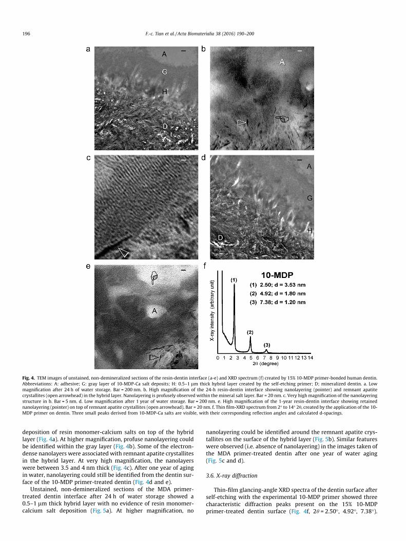
Fig. 4. TEM images of unstained, non-demineralized sections of the resin-dentin interface (a-e) and XRD spectrum (f) created by 15% 10-MDP primer-bonded human dentin.Abbreviations: A: adhesive; G: gray layer of 10-MDP-Ca salt deposits; H: 0.5–1 lm thick hybrid layer created by the self-etching primer; D; mineralized dentin. a. Lowmagnification after 24 h of water storage. Bar = 200 nm. b. High magnification of the 24-h resin-dentin interface showing nanolayering (pointer) and remnant apatitecrystallites (open arrowhead) in the hybrid layer. Nanolayering is profusely observed within the mineral salt layer. Bar = 20 nm. c. Very high magnification of the nanolayeringstructure in b. Bar = 5 nm. d. Low magnification after 1 year of water storage. Bar = 200 nm. e. High magnification of the 1-year resin-dentin interface showing retainednanolayering (pointer) on top of remnant apatite crystallites (open arrowhead). Bar = 20 nm. f. Thin film-XRD spectrum from 2� to 14� 2h, created by the application of the 10-MDP primer on dentin. Three small peaks derived from 10-MDP-Ca salts are visible, with their corresponding reflection angles and calculated d-spacings.
196 F.-c. Tian et al. / Acta Biomaterialia 38 (2016) 190–200
deposition of resin monomer-calcium salts on top of the hybridlayer (Fig. 4a). At higher magnification, profuse nanolayering couldbe identified within the gray layer (Fig. 4b). Some of the electron-dense nanolayers were associated with remnant apatite crystallitesin the hybrid layer. At very high magnification, the nanolayerswere between 3.5 and 4 nm thick (Fig. 4c). After one year of agingin water, nanolayering could still be identified from the dentin sur-face of the 10-MDP primer-treated dentin (Fig. 4d and e).
Unstained, non-demineralized sections of the MDA primer-treated dentin interface after 24 h of water storage showed a0.5–1 lm thick hybrid layer with no evidence of resin monomer-calcium salt deposition (Fig. 5a). At higher magnification, no
nanolayering could be identified around the remnant apatite crys-tallites on the surface of the hybrid layer (Fig. 5b). Similar featureswere observed (i.e. absence of nanolayering) in the images taken ofthe MDA primer-treated dentin after one year of water aging(Fig. 5c and d).
3.6. X-ray diffraction
Thin-film glancing-angle XRD spectra of the dentin surface afterself-etching with the experimental 10-MDP primer showed threecharacteristic diffraction peaks present on the 15% 10-MDPprimer-treated dentin surface (Fig. 4f, 2h = 2.50�, 4.92�, 7.38�).

Fig. 5. TEM images of unstained, non-demineralized sections of the resin-dentin interface (a-d) and XRD spectrum (e) created by 15% MDA primer-bonded human dentin.Abbreviations: A: adhesive; H: 0.5–1 lm thick hybrid layer; D; mineralized dentin. T: dentinal tubule. a. Lowmagnification after 24 h of water storage. No gray deposits couldbe identified on top of the hybrid layer. Bar = 200 nm. b. High magnification of the 24-h resin-dentin interface showing absence of nanolayering. Open arrowheads: remnantapatite crystallites. Bar = 20 nm. c. Low magnification after 1 year of water storage. Bar = 500 nm. d. High magnification of the 1-year resin-dentin interface showing absenceof nanolayering in the resin-dentin interface. Open arrowhead: remnant apatite crystallites. Bar = 20 nm. e. Thin film-XRD spectrum from 2� to 14� 2h, created by theapplication of the MDA primer on dentin. No peaks characteristic of short-range order can be identified.
F.-c. Tian et al. / Acta Biomaterialia 38 (2016) 190–200 197
These peaks were absent in the XRD spectrum of the MDA primer-treated dentin surface (Fig. 5e).
4. Discussion
Ideally, studies on the contribution of nanolayering of Ca-resinmonomer salts to the longevity of resin-dentin bonds should becontemplated using commercial self-etching adhesives [35].Because HEMA is present in most 10-MDP-containing self-etching adhesives [19] and HEMA inhibits interfacial nanolayeringof the functional monomer 10-MDP [21], nanolayering is rarelyidentified from the resin-dentin interface of bonds created by com-
mercialized 10-MDP-containing self-etching adhesives [19].Hence, the authors have to resort to using experimental primerscontaining only the functional resin monomers to accomplish theirobjectives. The use of similar experimental primers had beenshown to produce immediate resin-dentin bond strength thatwas similar to the use of commercial 10-MDP-containing adhe-sives [20].
Based on the present results, the first null hypothesis that‘‘there is no difference in the affinity of 10-MDP and the analogresin monomer for mineralized dentin” has to be rejected. In thecolumn chromatography experiments with ethanol-water mixtureas the mobile phase, the retention time of 10-MDP in the mineral-

198 F.-c. Tian et al. / Acta Biomaterialia 38 (2016) 190–200
ized dentin columnwas about 2 h vs 1 h for the 10-MDP analog (i.e.MDA) and HEMA. This implies that 10-MDP has better affinity forapatite than its analog. The use of ethanol-water mixture as themobile phase was to simulate the solvent vehicle in most commer-cially available 10-MDP-containing adhesives. Because bothethanol-water solvated 10-MDP and MDA were almost completelyrecovered from the column after 3 h of elution, the interactionsbetween these phosphoric acid ester monomers and apatite aremost likely to be reversible in nature.
The mineralized bovine dentin powder column chromatogra-phy setup in the present work is a modification of the hydroxyap-atite column chromatography technique [26]. This techniqueinvolves nonspecific interactions between positively-charged cal-cium ions and negatively-charged phosphate ions on the stationaryphase HA resin with negatively-charged carboxyl groups andpositively-charged amino groups in the analytes. Multi-modalinteraction such as electrostatic adsorption, hydrophobic adsorp-tion, ion exchange or hydrogen bonding may occur between theanalytes and HA [36,37]. In addition, a single analyte was addedat one time in the present work to compare the interaction equilib-rium with other analytes, instead of using the technique for purifi-cation purposes. The double peaks shown in the 10-MDP elutionprofile provides an illustration on how impurities in the commer-cial 10-MDP (<10% [22]); may be identified. The work presentedhere suggests that the low molecular weight impurity componentof the commercial 10-MDP may be removed by hydroxyapatitecolumn chromatography to produce 10-MDP with a higher degreeof purity for fabricating better quality self-etch adhesives. Researchin this area is in order.
The performance of column chromatography may be influencedby salts and solvents in the mobile phase. Generally, salts will sup-press the electrostatic interactions but enhance hydrophobic inter-actions. Conversely, organic solvents will suppress hydrophobicinteractions but enhance electrostatic interactions [38,39]. Ana-lytes that are retained solely by adsorption are eluted by a suitableorganic solvent, while those retained by an ion-exchange mecha-nism are best desorbed using an acidic or basic eluent that convertsthe analyte ions back to their molecular form [40]. As reflected bythe chromatography results (Fig. 2), when salt buffer was used asthe mobile phase, the elution profile of MDA was symmetricaland MDA was 100% recovered. Conversely, the elution profile of10-MDP (second peak) was asymmetrical and only 36.5% of the10-MDP eluded from the column. When ethanol was used as themobile phase, limited 10-MDP (18.9%) or its analog, MDA (33.5%)were recovered. These elution profiles provide hints with respectto the very complicated, mobile-phase dependent interactionsbetween the tested phosphoric acid ester monomers and the sta-tionary phase (i.e. mineralized dentin).
The chromatography results are not in contradiction with thechemoanalytic results. The FTIR, 31P MAS-NMR and XPS specimenswere prepared with 15% solution of the phosphoric acid estermonomers, which are much more concentrated than the tracersemployed in the chromatography part of the experiments. Afterreaction, the HA specimens were further washed with ethanol,which may not be able to desorb all the reaction products or unre-acted resin monomers. Emergence of new FTIR absorbance peaks at958 cm�1 for the 10-MDP-treated HA spectrum (Fig. 3a), and982 cm�1 on the MDA-treated HA spectrum (Fig. 3b) probably rep-resent m3/1(PO4) PAO stretch of the newly-formed phosphate salts,although definitive assignments of these new peaks were difficult.The weak peak at �730 cm�1 on the 10-MDP-treated HA spectrum(Fig. 3a) may be assigned to PAOAP linear bond formation (con-firmed by unpublished NMR examination data).
The FTIR results were also validated by the XPS results (Table 1).The Ca/P ratio was lower for 10-MDP-treated HA compared withMDA-treated HA because of the increase in P percentage caused
by monomer adsorption. The C/Ca and O/Ca ratios increased for10-MDP-treated HA compared to untreated HA because of theadsorption of 10-MDP on HA. High resolution C 1s scans of HAtreated with 10-MDP for 30 s or 30 min show that in the absenceof a displacement agent that displaces the adsorbed 10-MDP, thephosphoric acid ester monomer remained strongly attached tothe HA surface. Although there was initial adsorption of MDA toHA, this interaction is unstable because of the disappearance ofthe ACOO peak characteristic of MDA after 30 min of treatment.Based on the 31P MAS NMR results, disappearance of the ester peakon prolonged interaction of MDA with HA may be caused by MDAdesorption during formation of the CaHPO4�2H2O salt.
The second hypothesis that ‘‘there is no difference in the capa-bility of both phosphoric acid ester monomer to produce nanolay-ering on the dentin surface” has to be rejected. As reflected by thecontact angle result and pH measurement, the analog moleculeMDA is more hydrophilic and more soluble in water than 10-MDP, and the former is more acidic when solvated in theethanol-water mixture. The structure of the spacer group mayaccount for the absence of nanolayering in MDA primer-treateddentin. The poly-(propylene glycol) spacer group in MDA is lesshydrophobic than the methylene spacer in 10-MDP. Thus, theamphiphilic property of the MDA is not as evident as the 10-MDP monomer. The properties of the resin monomers werereflected in the TEM images resin-dentin interfaces; similar hybridlayer thickness (about 0.5–1.0 lm) was observed. Nanolayeringwas identified in the gray layer formed above the hybrid layer cre-ated by 10-MDP priming. The gray layer was believed to be 10-MDP-calcium salt deposition [19]; this layer was absent in theresin-dentin interface created by MDA. The TEM findings were fur-ther supported by the thin-film XRD results. The 10-MDP primeddentin specimens exhibited characteristic peaks of 10-MDP-calcium salt while the MDA primed dentin surface did not exhibitsuch peaks.
The third hypothesis that ‘‘nanolayering of phosphoric acidester monomer-calcium salts is unstable after water aging anddoes not contribute to the resin-dentin bond durability” cannotbe rejected. Although nanolayering features were ubiquitouslyidentified in the 10-MDP primer-treated dentin interface afterone year of water-aging, its bond strength decreased significantlyafter water aging. There was no significant difference in tensilebond strength values between 10-MDP primed dentin and MDA-primed dentin, both at 24 h and after one year of water-aging. Fail-ure modes of both monomer-bonded samples were predominantlymixed failure. Because the nanolayering structures were protectedby a comparatively hydrophobic resin coating (DE Bonding Resin),its resistance to dissolution after water aging is easy to compre-hend. Nevertheless, the contribution of nanolayering to resin-dentin bond stability is not as obvious as what was previously pro-posed [17]. Nanolayering is sparsely identified in resin-dentininterfaces produced by commercial 10-MDP-containing adhesives(e.g. Clearfil SE Bond 2, Kuraray Medical Inc., Tokyo, Japan) [19].However, the durability of these adhesives had been shown to beexcellent, which serves as indirect evidence of limited contributionof nanolayering to bond durability. Aged Clearfil SE Bond 2 speci-mens showed negligible bond strength drop under the same exper-imental conditions after one year of water aging (unpublisheddata). A possible explanation for the decline in bond strength asso-ciated with primers containing only 10-MDP may lie in the weakconnection between the 10-MDP-Ca salt deposits and the dentinsurface. Conceptually, one may consider 10-MDP-Ca salt depositsas a specific type of organic-inorganic hybrid fillers produced in-situ during dentin bonding. In resin composites, silica and glass fil-lers are silanized with methacryloxy silanes to enable them tobond to the methacrylate resin matrix. In the case of 10-MDP-Casalts, the inward facing of the methacrylate groups of two 10-

F.-c. Tian et al. / Acta Biomaterialia 38 (2016) 190–200 199
MDP molecules may drastically reduce the number of freelyavailable methacryloxy functionalities for coupling to the resinmatrix.
Hydroxyapatite is the major mineral phase of dentin. The apa-tite surface is covered with a highly-ordered bound water layer[41]. This surface water layer provides an efficient proton poolfor various adsorbed ions and terminal chemical groups derivedfrom the crystalline phase, such as PAOH (protonated or unproto-nated) and CaAOH groups [42–44]. When mineralized dentin istreated with an acidic primer, apatite dissolution occurs. Althoughthe step-by-step reaction mechanism remains unclear, 8 modelshave been proposed to explain the processes involved. Dissolutionkinetics may be influenced by concentration of acid, ionic strength,solution undersaturation, pH, temperature or crystal dimensions[45]. Generally, the reaction occurs on a diffusion basis: diffusionof chemical reagents (hydronium ions and anions of acid An�) frombulk solution to the solid/liquid interface, and diffusion of reactionproduct (Ca2+, PO43� and OH�) back into the bulk solution [46].Interaction between calcium cations and adsorbed anions of acidprobably happens on the apatite surface as an intermediate stageof the reaction, which results in breaking of surface „OACa bondsand detachment of calcium-anion salt followed by their diffusionaway from mineral surface [46]. In the present situation, adsorp-tion of phosphate anions on HA may involve an ion exchange pro-cess (with Ca2+) or hydrogen bonding (with PAOH group), andpossibly covalent pyrophosphate formation [47,48].
Based on the adsorption behavior of some carboxylic acids (e.g.oxalic acid) on the apatite surface, the adhesion-decalcificationconcept (AD concept) was developed and expanded to the inter-pretation of the effects of other phosphate ester functional mono-mers on HA [4,5]. The AD concept emphasizes the importance ofionic bond formation between the acid anion and lattice Ca2+ ionson the apatite surface [4,49]. As shown by the ability of adsorbed10-MDP to be slowly eluted from mineralized dentin in the chro-matography experiment, the extent and stability of this ion-exchange adsorption require further investigations before the ADconcept of chemical bonding may be justified as a mechanism forachieving long-term stable adhesion to dentin. As a matter of fact,oxalic acid can etch the dentin surface and reacts with calcium ionsin the smear layer to produce an insoluble calcium oxalate salt onthe dentin surface, which replaces the original smear layer with anartificial smear layer [50,51]. This may also be true for 10-MDP-Casalt because the nanolayering structures were mostly identifiedabove the hybrid layer and form a more or less distinctive gray saltlayer in the TEM images.
In acidic solutions, oxalate or phosphate anion would probablyreact with the released Ca2+ ions rather than adsorbing onto theapatite surface, because of non-equivalent adsorption of H+ andanion caused by differences in size and mobility [52]. Salt deposi-tion after acid-etching reaction is dependent upon the pH changeof the aqueous environment and the solubility product constant(ksp) of the formed salt species. Loose deposition does not neces-sarily imply the existence of chemical affinity on a hard tissue sur-face. Although several kinds of calcium salts have been proposedafter 10-MDP reacted with dentin or enamel powder, the stabilityand solubility of these calcium salts have yet to be identified.Because of these issues, the contribution of phosphoric acid estermonomer-calcium salt deposition in the hybrid layer on the dura-bility of resin-dentin bonds remains controversial.
Although the contribution of chemical bonding to the overallbonding durability requires further investigation, recent findingsindicate that 10-MDP has relatively stable interaction with colla-gen [53]. In specific experimental setups, immobilized 10-MDPwas found to induce limited extrafibrillar and intrafibrillar miner-alization of collagen fibrils [54]. These findings shed light on thepotential role of 10-MDP-calcium salt formation on remineraliza-
tion of poorly-infiltrated collagen scaffold in the hybrid layer andmerit more in-depth investigations.
5. Conclusion
Within the limits of the present study, it may be concluded thatthe claim that nanolayering of 10-MDP-Ca salts is responsible forthe durability of resin-dentin bonds created by self-etching 10-MDP-containing dentin adhesives cannot be justified. The 10-MDP-Ca salts may be viewed upon as a highly unique type oforganic-inorganic hybrid fillers created in-situ during the applica-tion of 10-MDP to dentin, in the absence of other resinous compo-nents that interfere with the phenomenological expression ofnanolayering. Complete elution of the phosphoric acid ester mono-mer from mineralized dentin powder in the column chromatogra-phy experiments using ethanol-water mobile phase to simulate thesolvent mixture employed in most 10-MDP-containing dentinadhesives further challenges the previously proposed adhesion-decalcification concept that utilizes chemical bonding of phospho-ric acid ester monomers to apatite as a bonding mechanism in 10-MDP containing dentin adhesives.
Acknowledgments
This work was supported by grant 2015AA020942 fromNational High Technology Research and Development Program ofChina, grant 81400555 from National Nature Science Foundation,grant Z14110000514016 from Beijing Municipal Science & Tech-nology Commission Project, and program IRT13051 from Chang-jiang Scholars and Innovative Research Team in University. Allauthors declared no conflict of interest associated with this work.
References
[1] D.H. Pashley, F.R. Tay, L. Breschi, L. Tjäderhane, R.M. Carvalho, M. Carrilho, A.Tezvergil-Mutluay, State of the art etch-and-rinse adhesives, Dent. Mater. 27(2011) 1–16.
[2] B. Van Meerbeek, K. Yoshihara, Y. Yoshida, A. Mine, J. De Munck, K.L. VanLanduyt, State of the art of self-etch adhesives, Dent. Mater. 27 (2011) 17–28.
[3] R.M. Carvalho, A.P. Manso, S. Geraldeli, F.R. Tay, D.H. Pashley, Durability ofbonds and clinical success of adhesive restorations, Dent. Mater. 28 (2012) 72–86.
[4] Y. Yoshida, B. Van Meerbeek, Y. Nakayama, M. Yoshioka, J. Snauwaert, Y. Abe, P.Lambrechts, G. Vanherle, M. Okazaki, Adhesion to and decalcification ofhydroxyapatite by carboxylic acids, J. Dent. Res. 80 (2001) 1565–1569.
[5] Y. Yoshida, K. Nagakane, R. Fukuda, Y. Nakayama, M. Okazaki, H. Shintani, S.Inoue, Y. Tagawa, K. Suzuki, J. De Munck, B. Van Meerbeek, Comparative studyon adhesive performance of functional monomers, J. Dent. Res. 83 (2004) 454–458.
[6] D. Fukegawa, S. Hayakawa, Y. Yoshida, K. Suzuki, A. Osaka, B. Van Meerbeek,Chemical interaction of phosphoric acid ester with hydroxyapatite, J. Dent. Res.85 (2006) 941–944.
[7] Z. Zhang, X. Wang, L. Zhang, B. Liang, T. Tang, B. Fu, M. Hannig, Thecontribution of chemical bonding to the short- and long-term enamel bondstrengths, Dent. Mater. 29 (2013) e103–e112.
[8] V.P. Feitosa, S. Sauro, F.A. Ogliari, A.O. Ogliari, K. Yoshihara, C.H. Zanchi, L.Correr-Sobrinho, M.A. Sinhoreti, A.B. Correr, T.F. Watson, B. Van Meerbeek,Impact of hydrophilicity and length of spacer chains on the bonding offunctional monomers, Dent. Mater. 30 (2014) e317–e323.
[9] K. Yoshihara, Y. Yoshida, S. Hayakawa, N. Nagaoka, Y. Torii, A. Osaka, K. Suzuki,S. Minagi, B. Van Meerbeek, K.L. Van Landuyt, Self-etch monomer-calcium saltdeposition on dentin, J. Dent. Res. 90 (2011) 602–606.
[10] V.P. Feitosa, F.A. Ogliari, B. Van Meerbeek, T.F. Watson, K. Yoshihara, A.O.Ogliari, M.A. Sinhoreti, A.B. Correr, G. Cama, S. Sauro, Can the hydrophilicity offunctional monomers affect chemical interaction, J. Dent. Res. 93 (2014) 201–206.
[11] M. Sarr, A.W. Kane, J. Vreven, A. Mine, K.L. Van Landuyt, M. Peumans, P.Lambrechts, B. Van Meerbeek, J. De Munck, Microtensile bond strength andinterfacial characterization of 11 contemporary adhesives bonded to bur-cutdentin, Oper. Dent. 35 (2010) 94–104.
[12] M. Peumans, J. De Munck, K.L. Van Landuyt, B. Van Meerbeek, Thirteen-yearrandomized controlled clinical trial of a two-step self-etch adhesive in non-carious cervical lesions, Dent. Mater. 31 (2015) 308–314.

200 F.-c. Tian et al. / Acta Biomaterialia 38 (2016) 190–200
[13] S. Inoue, K. Koshiro, Y. Yoshida, J. De Munck, K. Nagakane, K. Suzuki, H. Sano, B.Van Meerbeek, Hydrolytic stability of self-etch adhesives bonded to dentin, J.Dent. Res. 84 (2005) 1160–1164.
[14] H. Iwai, N. Nishiyama, Effect of calcium salt of functional monomer on bondingperformance, J. Dent. Res. 91 (2012) 1043–1048.
[15] H. Takahashi, Effect of calcium salt of 10-methacryloyloxydecyl dihydrogenphosphate produced on the bond durability of one-step self-etch adhesive,Dent. Mater. J. 33 (2014) 394–401.
[16] Y. Yokota, N. Nishiyama, Determination of molecular species of calcium salts ofMDP produced through decalcification of enamel and dentin by MDP-basedone-step adhesive, Dent. Mater. J. 34 (2015) 270–279.
[17] K. Yoshihara, Y. Yoshida, N. Nagaoka, D. Fukegawa, S. Hayakawa, A. Mine, M.Nakamura, S. Minagi, A. Osaka, K. Suzuki, B. Van Meerbeek, Nano-controlledmolecular interaction at adhesive interfaces for hard tissue reconstruction,Acta Biomater. 6 (2010) 3573–3582.
[18] Y. Yoshida, K. Yoshihara, N. Nagaoka, S. Hayakawa, Y. Torii, T. Ogawa, A. Osaka,B. Van Meerbeek, Self-assembled nano-layering at the adhesive interface, J.Dent. Res. 91 (2012) 376–381.
[19] F. Tian, L. Zhou, Z. Zhang, L. Niu, L. Zhang, C. Chen, J. Zhou, H. Yang, X. Wang, B.Fu, C. Huang, D.H. Pashley, F.R. Tay, Paucity of nanolayering in resin-rentininterfaces of MDP-based adhesives, J. Dent. Res. 95 (2016) (2015) 380–387.
[20] K. Yoshihara, Y. Yoshida, S. Hayakawa, N. Nagaoka, M. Irie, T. Ogawa, K.L. VanLanduyt, A. Osaka, K. Suzuki, Nanolayering of phosphoric acid ester monomeron enamel and dentin, Acta Biomater. 7 (2011) 3187–3195.
[21] Y. Yoshida, K. Yoshihara, S. Hayakawa, N. Nagaoka, T. Okihara, T. Matsumoto, S.Minagi, A. Osaka, K.L. Van Landuyt, B. Van Meerbeek, HEMA inhibits interfacialnano-layering of the functional monomer MDP, J. Dent. Res. 91 (2012) 1060–1065.
[22] K. Yoshihara, N. Nagaoka, T. Okihara, M. Kuroboshi, S. Hayakawa, Y. Maruo, G.Nishigawa, J. De Munck, Y. Yoshida, B. Van Meerbeek, Functional monomerimpurity affects adhesive performance, Dent. Mater. 31 (2015) 1493–1501.
[23] K. Yoshihara, Y. Yoshida, N. Nagaoka, S. Hayakawa, T. Okihara, J. De Munck, Y.Maruo, G. Nishigawa, S. Minagi, A. Osaka, B. Van Meerbeek, Adhesiveinterfacial interaction affected by different carbon-chain monomers, Dent.Mater. 29 (2013) 888–897.
[24] K. Yoshihara, Y. Yoshida, S. Hayakawa, N. Nagaoka, S. Kamenoue, T. Okihara, T.Ogawa, M. Nakamura, A. Osaka, B. Van Meerbeek, Novel fluoro-carbonfunctional monomer for dental bonding, J. Dent. Res. 93 (2014) 189–194.
[25] N. Moszner, U. Salz, J. Zimmermann, Chemical aspects of self-etching enamel-dentin adhesives: a systematic review, Dent. Mater. 21 (2005) 895–910.
[26] S. Hjerten, O. Levin, A. Tiselius, Protein chromatography on calcium phosphatecolumns, Acta Biochem. Biophys. 65 (1956) 132–155.
[27] M. Takahashi, M. Nakajima, J. Tagami, D.L. Scheffel, R.M. Carvalho, A. Mazzoni,M. Cadenaro, A. Tezvergil-Mutluay, L. Breschi, L. Tjäderhane, S.S. Jang, F.R. Tay,K.A. Agee, D.H. Pashley, The importance of size-exclusion characteristics oftype I collagen in bonding to dentin matrices, Acta Biomater. 9 (2013) 9522–9528.
[28] A.D. Loguercio, L.P. Barroso, R. Grande, A. Reis, Comparison of intra andintertooth resin-dentin bond strength variability, J. Adhes. Dent. 7 (2005) 151–158.
[29] N. Charadram, C. Austin, P. Trimby, M. Simonian, M.V. Swain, N. Hunter,Structural analysis of reactionary dentin formed in response to polymicrobialinvasion, J. Struct. Biol. 181 (2013) 207–222.
[30] Z.R. Hinedi, S. Goldberg, A.C. Chang, J.P. Yesinowski, A 31P and 1H MAS NMRstudy of phosphate sorption on calcium carbonate, J. Colloid Interface Sci. 152(1992) 141–160.
[31] Z.Q. He, W. Honeycutt, B.S. Xing, R.W. McDowel, P.J. Pellechia, T.Q. Zhang,Solid-state Fourier transform infrared and 31P nuclear magnetic resonancespectral features of phosphate compounds, Soil Sci. 172 (2007) 501–515.
[32] Y. Yoshida, B. Van Meerbeek, Y. Nakayama, J. Snauwaert, L. Hellemans, P.Lambrechts, G. Vanherle, K. Wakasa, Evidence of chemical bonding atbiomaterial-hard tissue interfaces, J. Dent. Res. 79 (2000) 709–714.
[33] H.B. Lu, C.T. Campbell, D.J. Graham, B.D. Ratner, Surface characterization ofhydroxyapatite and related calcium phosphates by XPS and TOF-SIMS, Anal.Chem. 72 (2000) 2886–2894.
[34] G.N. Raikar, J.L. Ong, L.C. Lucas, Hydroxyapatite characterized by XPS, Surf. Sci.Spectra 4 (1996) 9–13.
[35] A. Frassetto, L. Breschi, G. Turco, G. Marchesi, R. Di Lenarda, F.R. Tay, D.H.Pashley, M. Cadenaro, Mechanisms of degradation of the hybrid layer inadhesive dentistry and therapeutic agents to improve bond durability – aliterature review, Dent. Mater. 32 (2015) e41–e53.
[36] E. Schröder, T. Jönsson, L. Poole, Hydroxyapatite chromatography: altering thephosphate-dependent elution profile of protein as a function of pH, Anal.Biochem. 313 (2003) 176–178.
[37] L.J. Cummings, M.A. Snyder, K. Brisack, Protein chromatography onhydroxyapatite columns, Methods Enzymol. 463 (2009) 387–404.
[38] B.K. Glód, Ion exclusion chromatography: parameters influencing retention,Neurochem. Res. 22 (1997) 1237–1248.
[39] T. Arakawa, Y. Kita, D. Ejima, P. Gagnon, Solvent modulation of columnchromatography, Protein Pept. Lett. 15 (2008) 544–555.
[40] J.S. Frit, M. Macka, Solid-phase trapping of solutes for further chromatographicor electrophoretic analysis, J. Chromatogr. A 902 (2000) 137–166.
[41] W. Zhao, Z. Xu, Y. Yang, N. Sahai, Surface energetics of the hydroxyapatitenanocrystal-water interface: a molecular dynamics study, Langmuir 30 (2014)13283–13292.
[42] S. Cazalbou, C. Combes, D. Eicherta, C. Reya, Adaptive physico-chemistry ofbio-related calcium phosphates, J. Mater. Chem. 14 (2004) 2148–2153.
[43] M. Jarlbring, D.E. Sandström, O.N. Antzutkin, W. Forsling, Characterization ofactive phosphorus surface sites at synthetic carbonate-free fluorapatite usingsingle-pulse 1H, 31P, and 31P CP MAS NMR, Langmuir 22 (2006) 4787–4792.
[44] J. Kolmas, A. Slósarczyk, A. Wojtowicz, W. Kolodziejski, Estimation of thespecific surface area of apatites in human mineralized tissues using 31P MASNMR, Solid State Nucl. Magn. Reson. 32 (2007) 53–58.
[45] S.V. Dorozhkin, Dissolution mechanism of calcium apatites in acids: a reviewof literature, World J. Methodol. 2 (2012) 1–17.
[46] S.V. Dorozhkin, Surface reactions of apatite dissolution, J. Colloid Interface Sci.191 (1997) 489–497.
[47] D.N. Misra, Adsorption from solutions on synthetic hydroxyapatite:nonaqueous vs. aqueous solvents, J. Biomed. Mater. Res. 48 (1999) 848–855.
[48] C.D. Susan, Y.F. Alexander, Covalent surface modification of calciumhydroxyapatite ysing n-alkyl- and n-fluoroalkylphosphonic acids, Langmuir19 (2003) 7904–7910.
[49] M. Yoshioka, Y. Yoshida, S. Inoue, P. Lambrechts, G. Vanherle, Y. Nomura, M.Okazaki, H. Shintani, B. Van Meerbeek, Adhesion/decalcification mechanismsof acid interactions with human hard tissues, J. Biomed. Mater. Res. 59 (2002)56–62.
[50] D.H. Pashley, S.E. Galloway, The effects of oxalate treatment on the smear layerof ground surfaces of human dentine, Arch. Oral Biol. 30 (1985) 731–737.
[51] C.K. Yiu, N.M. King, B.I. Suh, L.J. Sharp, R.M. Carvalho, D.H. Pashley, F.R. Tay,Incompatibility of oxalate desensitizers with acidic, fluoride-containing total-etch adhesives, J. Dent. Res. 84 (2005) 730–735.
[52] A.E. Nielsen, Transport control in crystal growth from solution, Croat. Chem.Acta 53 (1980) 255–279.
[53] N. Hiraishi, N. Tochio, T. Kigawa, M. Otsuki, J. Tagami, Monomer-collageninteractions studied by saturation transfer difference NMR, J. Dent. Res. 92(2013) 284–288.
[54] H. Nurrohman, S. Nakashima, T. Takagaki, A. Sadr, T. Nikaido, Y. Asakawa, M.Uo, S.J. Marshall, J. Tagami, Immobilization of phosphate monomers oncollagen induces biomimetic mineralization, Biomed. Mater. Eng. 25 (2015)89–99.