effect of hydrothermal aging on the optical properties of
TRANSCRIPT
RESEARCH AND EDUCATION
This study waClinical AssobProfessor, D
676
Effect of hydrothermal aging on the optical properties ofprecolored dental monolithic zirconia ceramics
Hee-Kyung Kim, DDS, MSD, PhDa and Sung-Hun Kim, DDS, PhDbABSTRACTStatement of problem. The long-term color stability of precolored monolithic zirconia has notbeen thoroughly investigated.
Purpose. The purpose of this in vitro study was to evaluate the effect of hydrothermal aging on theoptical properties, phase transformation, and surface topography of precolored monolithic zirconiaceramics.
Material and methods. Precolored monolithic zirconia specimens (17.0×17.0×1.5 mm, n=50) andlithium disilicate glass-ceramic specimens (16.0×16.0×1.5 mm, n=50) were artificially aged in anautoclave at 134�C under 0.2 MPa for 0, 1, 3, 5, or 10 hours (n=10). CIELab color parameters wereobtained from spectral measurements. The translucency parameter (TP) and CIEDE2000 colordifferences (DE00) were calculated. The microstructural and surface properties were analyzed byX-ray diffraction (XRD), atomic force microscope (AFM), and scanning electron microscope (SEM).Data were analyzed with 2-way ANOVA and pairwise comparison (a=.05).
Results. Significant interactions were found between aging time and ceramic material on L*, a*,b*, and TP (P<.001) as follows: b* partial eta squared [hp
2]=0.689; L* hp2=0.186; a* hp
2=0.176;and TP hp
2=0.137. The b* values significantly decreased after aging for zirconia (P<.001),whereas TP increased after aging for zirconia (P<.014) except at 10 hours (P=.389) and forlithium disilicate (P<.001). The DE00 values relative to baseline ranged from 2.03 to 2.52 foraged zirconia and from 0.07 to 0.23 for aged lithium disilicate. XRD analysis revealed thathydrothermal aging promoted an increase in m-phase contents. AFM and SEM demonstratedsurface alterations after aging.
Conclusions. Optical properties and microstructures of precolored monolithic zirconiaceramics were affected by hydrothermal aging, and translucency increased slightly with agingtime. (J Prosthet Dent 2019;121:676-82)
Monolithic zirconia ceramics canprovide excellent mechanicalproperties without the risk ofchipping of veneering porcelain.However, low translucency andsusceptibility to hydrothermalaging of yttria-stabilized tetrag-onal zirconia polycrystal (Y-TZP)have been reported.1 When themonolithic design of Y-TZP isdirectly exposed to the oralenvironment, the absorption ofwater radicals can yield tensilestresses between tetragonal andmonoclinic phases, and the en-ergy difference between the 2phases further activates tetrag-onal to martensitic (t/m)transformation.2,3 Therefore,microstructural changes throughsuperficial alterations resultingin surface microcracks androughening can occur.4-8 Theresistance of Y-TZP to low-temperature degradation (LTD)
can depend on the dwell time,9-11 temperature,10 grain size,9residual stress inducedby surface treatments,5,6,12,13 sinteringprocesses,14 stabilizing content,9,11 and addition of silica.11
The effects of hydrothermal aging on the mechanicalproperties of Y-TZP have been investigated.15-18 Theflexural strength of zirconia ceramics significantlydecreased after 200 hours at 134�C under 0.2 MPa.15 Thebiaxial strength and toughness of Y-TZP decreased
as supported by the research grant no. M-2017-C0460-00057 from the Ajciate Professor, Department of Dentistry, Ajou University School of Medicepartment of Prosthodontics and Dental Research Institute, School of Den
depending on the amount of t/m phase trans-formation.16 In addition, Y-TZP could be decreased infatigue strength by about a factor of 2 to 3 under criticalcyclic loading in the presence of water.17 However, Coteset al18 reported that LTD did not affect the flexuralstrength of Y-TZP ceramics, although t/m phasetransformations were induced by an autoclave agingprocess.
ou University Medical Center Research Fund.ine, Suwon, Gyeong Gi-do, Republic of Korea.tistry, Seoul National University, Seoul, Republic of Korea.
THE JOURNAL OF PROSTHETIC DENTISTRY

Clinical ImplicationsSome color changes may be evident in precoloredmonolithic zirconia restorations with intraoral use,and thus, an esthetically unacceptable mismatchmight be expected approximately 3 to 4 years afterdelivery of the prostheses.
April 2019 677
The clinical LTD of Y-TZP has been simulated bysteam autoclave treatments at increased temperatures(120�C to 140�C) because the t/m phase transformationis thermally activated and accelerated by the presence ofwater.9,12,16,19 One hour of artificial aging treatment in anautoclave at 134�C has been reported to be equivalent to3 to 4 years in vivo at 37�C.9 Chevalier et al20,21 designedaccelerated aging in an autoclave at 134�C under 0.2 MPaover a period of 10 hours. In other studies,22,23 the arti-ficial aging protocol was performed in an autoclave at134�C under 0.2 MPa for 20 hours.
For the long-term clinical success of esthetic restora-tions, the color should be stable to ensure reliable colormatching. However, the color and translucency of dentalceramics is affected by hydrothermal aging.1,24-29 Dentalzirconia ceramics have been successively developed withimproved translucency and esthetics similar to those oflithia-based glass-ceramics, making them suitable for usein the anterior region.30 Precolored monolithic zirconiaceramics have been introduced with enhanced colorreproduction. Although precolored anatomic contourzirconia restorations bring esthetic and biomechanicaladvantages, information on how their optical propertiesare affected by LTD is lacking. Therefore, the purpose ofthis in vitro study was to evaluate the effect of hydro-thermal aging on the optical properties, phase trans-formation, and surface topography of precoloredmonolithic zirconia ceramics. In addition, the long-termcolor stability of precolored zirconia was compared withthat of precolored lithium disilicate. The null hypothesiswas that no significant differences would be found in theoptical properties of zirconia specimens subjected toaccelerated artificial aging with increasing aging time.
MATERIAL AND METHODS
Fifty square-shaped precolored monolithic zirconiaceramic specimens (17.0×17.0×1.5 mm; ZrO2, Y2O3
7.12% to 7.16%, and so forth.; Katana ML A Light;Kuraray Noritake) and 50 IPS e.max CAD lithium dis-ilicate glass-ceramic specimens (16.0×16.0×1.5 mm;SiO2, Other oxides; LT A2; Ivoclar Vivadent AG) as areference group were prepared. The final thicknesses ofthe specimens were adjusted to 1.5 ±0.01 mm, which isthe manufacturer-recommended occlusal wall thicknessfor posterior monolithic zirconia crowns, using a
Kim and Kim
horizontal grinding machine (HRG-150; AM Technol-ogy). Katana ML A Light (corresponding to Vita A 1.5 to2 as specified by the manufacturer) is a precoloredmultilayered zirconia, and in an attempt to get specimenswith the same color values, the specimens were cut fromthe lowermost layers of the blocks, which has been re-ported to exhibit higher chroma values than those of theupper part.31
Subsequently, the bottom surface of the specimenwas polished by using a multistep protocol (coarse,medium-coarse, and super-fine grit; Edenta) followed bydiamond paste (Legabril Diamond; Metalor Dental). Allmeasurements were carried out on the polished surfacesof the specimens. The specimens were further heat-treated at 930�C with 1 hour of holding time, followedby slow cooling to 500�C in a furnace, and then cooledslowly in air which could lead to the m/t phase trans-formation after annealing.32 Before the test, the speci-mens were ultrasonically cleaned in an isopropyl alcoholbath for 5 minutes. The specimens were artificially agedin an autoclave (SAC-230G; Shinhung) at 134�C under0.2 MPa and divided into 5 subgroups according to theaging time (n = 10), namely baseline (control), 1, 3, 5, and10 hours.
The spectral reflectance was measured with a spec-trophotometer (Color iControl; X-Rite) from 360 to 750nm at 10-nm intervals against a white polytetrafluoro-ethylene background (GM29010330; X-Rite; CommissionInternationale de l’Eclairage (CIE) L*=93.886, a*=e0.008,and b*=2.474), a black glass-ceramic tile (CM-A101B;Konica Minolta; CIE L*=0.11, a*=e0.014, and b*=0.018),and an A2 glass-ceramic tile (IPS e.max Press MO; Ivo-clar Vivadent AG; CIE L*=75.632, a*=e1.242, andb*=11.232). With a 6-mm diameter aperture and a 6-mmdiameter measurement area, diffuse/8-degree geometryand specular componenteexcluded (de:8�) geometriccondition were used. Five measurements per specimenwere made, and so 50 data points were collected for eachgroup. The reflectance data were then converted to colorparameters according to the CIELab color space relativeto D65 using the 2-degree standard observer (CIE, 1931).The mean color values against an A2 background wereused for the color difference calculation. CIEDE2000 colordifferences (DE00)33,34 between specimen pairs for Katanaand e.max were calculated as:
DE00=
"�DL’
KLSL
�2
+�
DC’
KCSC
�2
+�
DH ’
KHSH
�2
+RT
�DC’
KCSC
��DH’
KHSH
�#12
The translucency parameters (TPs) of the specimensfor each period were obtained by calculating the DE00
THE JOURNAL OF PROSTHETIC DENTISTRY
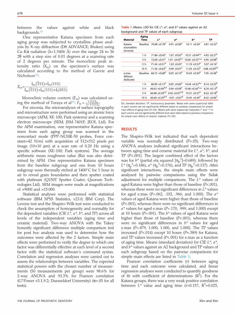
Table 1.Means ±SD for CIE L*, a*, and b* values against an A2background and TP values of each subgroup
MaterialAgingTime L* a* b* TP
Katana(monolithiczirconia)
Baseline 70.68 ±0.78a 0.91 ±0.06a 14.11 ±0.26c 4.81 ±0.22a
1 h 71.86 ±0.43c 1.07 ±0.03d 10.51 ±0.44a,* 4.93 ±0.27b
3 h 72.00 ±0.41c 1.01 ±0.05b,c 10.60 ±0.33a,** 4.95 ±0.08b
5 h 71.44 ±0.51b 1.03 ±0.03c 11.10 ±0.52b 5.07 ±0.16c
10 h 71.56 ±0.63b 0.99 ±0.01b 11.03 ±0.52b 4.88 ±0.09a,b
e.max(lithiumdisilicate)
Baseline 66.72 ±0.68A 0.05 ±0.10A 10.43 ±0.50A 7.95 ±0.28A
1 h 66.99 ±0.13A 0.05 ±0.06A 10.44 ±0.52A,* 8.14 ±0.25B
3 h 66.62 ±0.49A,B 0.04 ±0.09A 10.48 ±0.56A,** 8.24 ±0.13B
5 h 66.98 ±0.29A,C 0.02 ±0.07A,B 10.31 ±0.37A 8.22 ±0.18B
10 h 66.69 ±0.29A,B e0.01 ±0.07B 10.45 ±0.44A 8.42 ±0.06C
SD, standard deviation; TP, translucency parameter. Means with same superscript letterin each column are not significantly different based on pairwise comparisons for simplemain effects of aging time (P>.05). Means with same superscript characters (* and **) ineach column are not significantly different from each other based on pairwise comparisonsfor simple main effects of ceramic material (P>.05).
678 Volume 121 Issue 4
between the values against white and blackbackgrounds.35
One representative Katana specimen from eachaging group was subjected to crystalline phase anal-ysis by X-ray diffraction (D8 ADVANCE; Bruker) usingCu-Ka radiation (l=1.5406 Å) over the range 24 to 362q with a step size of 0.01 degrees at a scanning rateof 2 degrees per minute. The monoclinic peak in-tensity ratio (Xm) on the specimen’s surface wascalculated according to the method of Garvie andNicholson36:
Xm=Im�111
�+Imð111Þ
Im�111
�+Imð111Þ+Itð011Þ
Monoclinic volume content (Fm) was calculated us-ing the method of Toraya et al37: Fm = 1:311Xm
1+ 0:311Xm.
For zirconia, the microanalyses of surface topographyand microstructure were performed using an atomic forcemicroscope (AFM; XE-100; Park systems) and a scanningelectron microscope (SEM; JSM-7401F; JEOL Ltd). Forthe AFM examination, one representative Katana spec-imen from each aging group was scanned in thenoncontact mode (PPP-NCHR-50 probes, Force con-stant=42 N/m) with acquisition of 512×512 pixels perimage (10×10 mm) at a scan rate of 0.20 Hz using aspecific software (XE-100; Park systems). The averagearithmetic mean roughness value (Ra) was also deter-mined by AFM. One representative Katana specimenfrom the baseline subgroup and one from 10 hourssubgroup were thermally etched at 1400�C for 1 hour inair to reveal grain boundaries and then sputter coatedwith platinum (Q150T Sputter Coater; Quorum Tech-nologies Ltd). SEM images were made at magnificationsof ×9000 and ×15 000.
Statistical analyses were performed with statisticalsoftware (IBM SPSS Statistics, v23.0; IBM Corp). TheLevene test and the Shapiro-Wilk test were conducted tocheck the assumption of homogeneity and normality forthe dependent variables (CIE L*, a*, b*, and TP) across alllevels of the independent variables (aging time andceramic material). Two-way ANOVA with the Tukeyhonestly significant difference multiple comparison testfor post hoc analysis was used to determine how theoutcomes were affected by the 2 factors. Simple maineffects were performed to verify the degree to which onefactor was differentially effective at each level of a secondfactor with the statistical software’s command syntax.Correlation and regression analyses were carried out toassess the relationships between variables. The expectedstatistical powers with the chosen number of measure-ments (50 measurements per group) were 98.6% for2-way ANOVA and 93.3% for Pearson correlation(G*Power v3.1.9.2; Duesseldorf University) (a=.05 for alltests).
THE JOURNAL OF PROSTHETIC DENTISTRY
RESULTS
The Shapiro-Wilk test indicated that each dependentvariable was normally distributed (P>.05). Two-wayANOVA analyses indicated significant interactions be-tween aging time and ceramic material for L*, a*, b*, andTP (P<.001). The largest combined effect of the factorswas for b* (partial eta squared [hp
2]=0.689), followed byL* (hp
2=0.186), a* (hp2=0.176), and TP (hp
2=0.137). Aftersignificant interactions, the simple main effects wereanalyzed by pairwise comparisons using the Sidakadjustment for multiple comparisons. The L* values ofaged Katana were higher than those of baseline (P<.001),whereas there were no significant differences in L* valuesfor aged e.max (P=.062, .102, .966, and 1.000). The a*values of aged Katana were higher than those of baseline(P<.001), whereas there were no significant differences ina* values for aged e.max (P=.170, .999, and 1.000) exceptat 10 hours (P=.001). The b* values of aged Katana werehigher than those of baseline (P<.001), whereas therewere no significant differences in b* values for agede.max (P=.879, 1.000, 1.000, and 1.000). The TP valuesincreased (P<.014) except 10 hours (P=.389) for Katana,and TP values increased (P<.001) for e.max as a functionof aging time. Means (standard deviation) for CIE L*, a*,and b* values against an A2 background and TP values ofeach subgroup based on the pairwise comparisons forsimple main effects are listed in Table 1.
Pearson correlation coefficients (r) between agingtime and each outcome were calculated, and linearregression analyses were conducted to quantify goodnessof fit with coefficient of determinations (R2). For theKatana groups, there was a very weak positive correlationbetween L* value and aging time (r=0.157, R2=0.025,
Kim and Kim
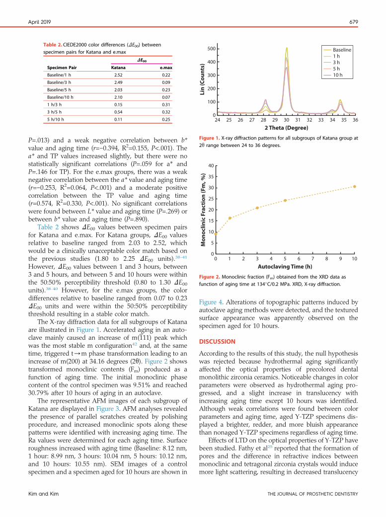
Table 2. CIEDE2000 color differences (DE00) betweenspecimen pairs for Katana and e.max
Specimen Pair
DE00
Katana e.max
Baseline/1 h 2.52 0.22
Baseline/3 h 2.49 0.09
Baseline/5 h 2.03 0.23
Baseline/10 h 2.10 0.07
1 h/3 h 0.15 0.31
3 h/5 h 0.54 0.32
5 h/10 h 0.11 0.25
2 Theta (Degree)
Lin
(Cou
nts)
3534333231
Baseline
30292827262524
400
300
200
100
0
500
36
1 h3 h5 h10 h
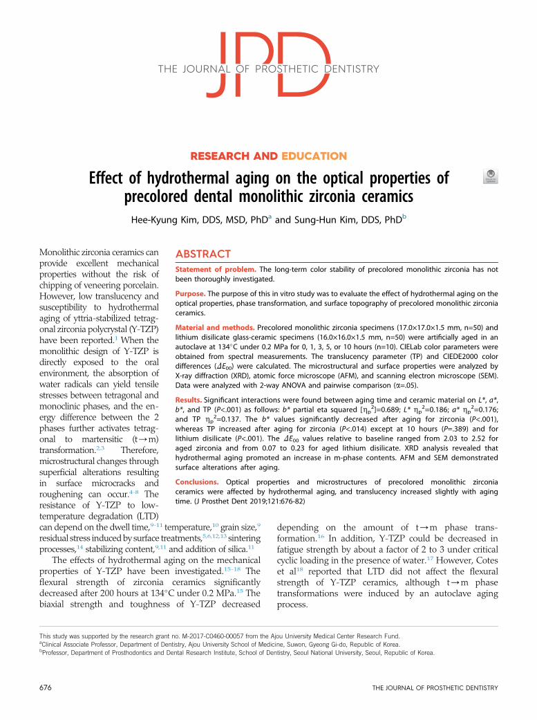
Figure 1. X-ray diffraction patterns for all subgroups of Katana group at2q range between 24 to 36 degrees.
Autoclaving Time (h)
Mon
oclin
ic F
ract
ion
(Fm
, %)
00
40
35
30
25
20
15
10
5
10987654321
Figure 2. Monoclinic fraction (Fm) obtained from the XRD data asfunction of aging time at 134�C/0.2 MPa. XRD, X-ray diffraction.
April 2019 679
P=.013) and a weak negative correlation between b*value and aging time (r=e0.394, R2=0.155, P<.001). Thea* and TP values increased slightly, but there were nostatistically significant correlations (P=.059 for a* andP=.146 for TP). For the e.max groups, there was a weaknegative correlation between the a* value and aging time(r=e0.253, R2=0.064, P<.001) and a moderate positivecorrelation between the TP value and aging time(r=0.574, R2=0.330, P<.001). No significant correlationswere found between L* value and aging time (P=.269) orbetween b* value and aging time (P=.890).
Table 2 shows DE00 values between specimen pairsfor Katana and e.max. For Katana groups, DE00 valuesrelative to baseline ranged from 2.03 to 2.52, whichwould be a clinically unacceptable color match based onthe previous studies (1.80 to 2.25 DE00 units).38-41
However, DE00 values between 1 and 3 hours, between3 and 5 hours, and between 5 and 10 hours were withinthe 50:50% perceptibility threshold (0.80 to 1.30 DE00units).38-40 However, for the e.max groups, the colordifferences relative to baseline ranged from 0.07 to 0.23DE00 units and were within the 50:50% perceptibilitythreshold resulting in a stable color match.
The X-ray diffraction data for all subgroups of Katanaare illustrated in Figure 1. Accelerated aging in an auto-clave mainly caused an increase of m(111) peak whichwas the most stable m configuration42 and, at the sametime, triggered t/m phase transformation leading to anincrease of m(200) at 34.16 degrees (2q). Figure 2 showstransformed monoclinic contents (Fm) produced as afunction of aging time. The initial monoclinic phasecontent of the control specimen was 9.51% and reached30.79% after 10 hours of aging in an autoclave.
The representative AFM images of each subgroup ofKatana are displayed in Figure 3. AFM analyses revealedthe presence of parallel scratches created by polishingprocedure, and increased monoclinic spots along thesepatterns were identified with increasing aging time. TheRa values were determined for each aging time. Surfaceroughness increased with aging time (Baseline: 8.12 nm,1 hour: 8.99 nm, 3 hours: 10.04 nm, 5 hours: 10.12 nm,and 10 hours: 10.55 nm). SEM images of a controlspecimen and a specimen aged for 10 hours are shown in
Kim and Kim
Figure 4. Alterations of topographic patterns induced byautoclave aging methods were detected, and the texturedsurface appearance was apparently observed on thespecimen aged for 10 hours.
DISCUSSION
According to the results of this study, the null hypothesiswas rejected because hydrothermal aging significantlyaffected the optical properties of precolored dentalmonolithic zirconia ceramics. Noticeable changes in colorparameters were observed as hydrothermal aging pro-gressed, and a slight increase in translucency withincreasing aging time except 10 hours was identified.Although weak correlations were found between colorparameters and aging time, aged Y-TZP specimens dis-played a brighter, redder, and more bluish appearancethan nonaged Y-TZP specimens regardless of aging time.
Effects of LTD on the optical properties of Y-TZP havebeen studied. Fathy et al25 reported that the formation ofpores and the difference in refractive indices betweenmonoclinic and tetragonal zirconia crystals would inducemore light scattering, resulting in decreased translucency
THE JOURNAL OF PROSTHETIC DENTISTRY
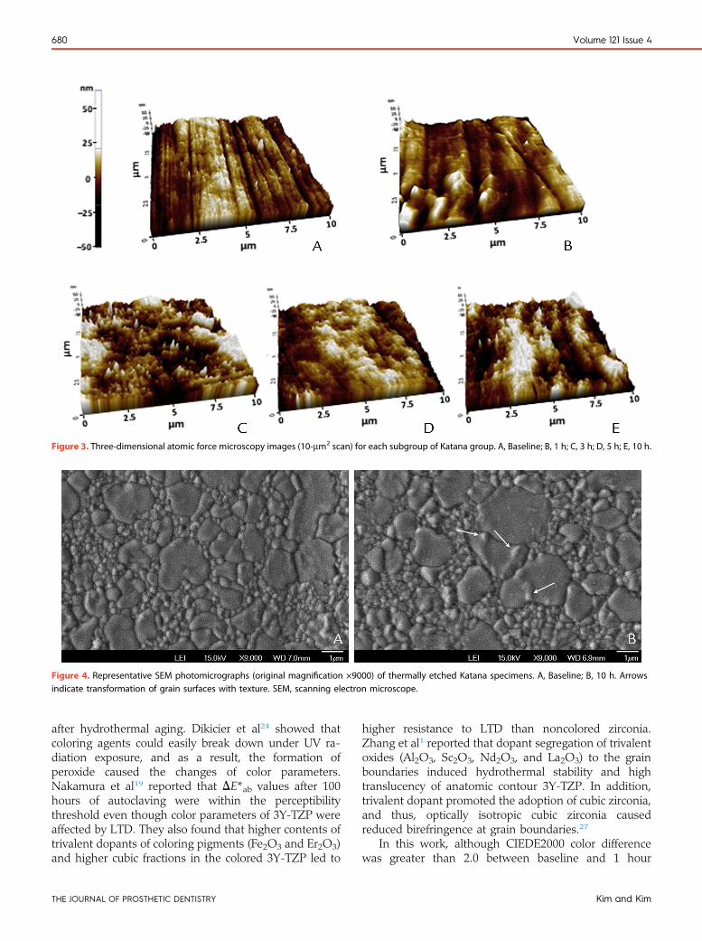
Figure 3. Three-dimensional atomic force microscopy images (10-mm2 scan) for each subgroup of Katana group. A, Baseline; B, 1 h; C, 3 h; D, 5 h; E, 10 h.
Figure 4. Representative SEM photomicrographs (original magnification ×9000) of thermally etched Katana specimens. A, Baseline; B, 10 h. Arrowsindicate transformation of grain surfaces with texture. SEM, scanning electron microscope.
680 Volume 121 Issue 4
after hydrothermal aging. Dikicier et al24 showed thatcoloring agents could easily break down under UV ra-diation exposure, and as a result, the formation ofperoxide caused the changes of color parameters.Nakamura et al19 reported that DE*ab values after 100hours of autoclaving were within the perceptibilitythreshold even though color parameters of 3Y-TZP wereaffected by LTD. They also found that higher contents oftrivalent dopants of coloring pigments (Fe2O3 and Er2O3)and higher cubic fractions in the colored 3Y-TZP led to
THE JOURNAL OF PROSTHETIC DENTISTRY
higher resistance to LTD than noncolored zirconia.Zhang et al1 reported that dopant segregation of trivalentoxides (Al2O3, Sc2O3, Nd2O3, and La2O3) to the grainboundaries induced hydrothermal stability and hightranslucency of anatomic contour 3Y-TZP. In addition,trivalent dopant promoted the adoption of cubic zirconia,and thus, optically isotropic cubic zirconia causedreduced birefringence at grain boundaries.27
In this work, although CIEDE2000 color differencewas greater than 2.0 between baseline and 1 hour
Kim and Kim

April 2019 681
(DE00=2.52), the difference was negligible thereafter forzirconia. In a similar way, Table 1 shows that changes incolor values, especially a decrease in b* values, occurredat 1 hour, and color values seemed to remain stableafterward for zirconia. However, the color parameters oflithium disilicate were relatively stable under agingtreatments. As might be deduced from the results, largercolor difference relative to baseline for zirconia thanlithium disilicate mainly resulted from the changes in b*values. Therefore, specific colorants might affect agingkinetics with regard to the behavior of oxygen vacanciesin the first 3 to 4 years of intraoral aging. In addition,early thermal or electrochemical degradations of specificdye cations within Y-TZP ceramics may occur. Furtherrelevant study is required.
In the present study, TP values increased withincreasing aging time for both zirconia and lithium dis-ilicate. Certain metal oxides such as coloring pigmentsmight enhance the formation of cubic zirconia, resultingin higher TP values with increasing aging time because ofthe reduced light scattering from the grain boundariesof cubic zirconia. IPS e.max CAD, as the original versionof lithium disilicate glass-ceramic, includes up to 4 wt%ZrO2 with additives for color and opalescence.30 Therecould be any ionic interactions within the glass-ceramiccrystalline phase leading to increased translucency afterhydrothermal aging. For Y-TZP, a small decrease in thetranslucency at 10 hours was detected and explainedperhaps by the increased light scattering caused bychanges in surface morphology. Further research for alonger period of aging time is needed to verify themagnitude and direction of shifts in the translucency.
According to ISO standard 13 356:2008, themaximum amount of monoclinic phase of Y-TZP afterautoclaving aging at 134�C under 0.2 MPa for a period of5 hours should not exceed 25% for a suitable biomedicaluse.2 In Pereira et al systematic review,3 m-phase con-tents were about 0% to 14% before aging but increasedto between 11% and 40% after accelerated aging for 10hours. They also showed that flexural strength decreasedonly when more than 50% of m-phase was noted. In thisstudy, increased t/m transformation behavior wasfound with increasing aging time, resulting in 24.37% ofm-phase at 5 hours and 30.79% at 10 hours of artificialaging. Hence, the time needed to reach a 25% mono-clinic fraction could be estimated at 20 years in the oralenvironment for precolored dental monolithic zirconiaceramics.
In the present study, polishing procedures were per-formed on the zirconia specimens and are recommendedas an essential clinical procedure to enhance the outcomeof the restorations.5 Pereira et al6 reported that asmoother surface could reduce the area for water inter-action, resulting in lower susceptibility to hydrothermalaging. However, Cattani-Lorente et al5 showed that mild
Kim and Kim
surface grinding increased aging resistance. Therefore,the presence of compressive stress due to surface treat-ments on the zirconia surface increased resistance againsthydrothermal degradation.13
In this study, aging caused increased roughness ofthe zirconia specimens, which is consistent with theprevious studies,5,7 showing about a 30% increase in Ravalue after artificial aging for 10 hours. Furthermore,previous studies5,7 found that t/m transformationinduced grain growth, which was associated with surfaceirregularities. Such irregular grain growth was alsodetected in AFM and SEM images of this study (Figs. 3and 4). Volume expansion of approximately 4%induced by t/m phase transformations would lead toan increase in surface roughness, causing wear to theopposing dentition.8
In this study, hydrothermal treatment was applied toclarify the effects of aging on the optical properties ofzirconia ceramics. This artificial aging technique can serveto predict the long-term degradation aging behavior ofY-TZP. However, in the oral environment, dental resto-rations will be exposed to different stimuli, includingmechanical aging, thermocycling, and chemical aging.Therefore, to reflect the clinical situation, more studiesare required to assess the changes in the optical prop-erties of dental ceramics under various aging conditions.In addition, in the present study, aging effects on theoptical properties of a single brand of higher translucencymonolithic zirconia ceramic, but not indicated for use inesthetically demanding zones, were compared with thoseof a representative glass-ceramic. Further studies shouldevaluate aging effects on the optical properties of recentlyintroduced esthetic dental ceramics with differentcompositions.
CONCLUSIONS
Within the limitations of this in vitro study, the followingconclusions were drawn:
1. Optical properties of precolored dental monolithiczirconia ceramics were affected by hydrothermalaging.
2. Translucency increased slightly with increasingaging time for Y-TZP and lithium disilicate glass-ceramics.
3. Increased phase transformations and surface alter-ations as a function of aging time were found on thesurfaces of precolored monolithic zirconia ceramics.
REFERENCES
1. Zhang F, Vanmeensel K, Batuk M, Hadermann J, Inokoshi M, vanMeerbeek B, et al. Highly-translucent, strong and aging-resistant 3Y-TZPceramics for dental restoration by grain boundary segregation. Acta Biomater2015;16:215-22.
2. International Organization for Standardization, ISO 13356:2008 Implants forsurgery - Ceramic materials based on yttria-stabilized tetragonal zirconia
THE JOURNAL OF PROSTHETIC DENTISTRY

682 Volume 121 Issue 4
(Y-TZP). Geneva: 2008. Available at, http://www.iso.ch/iso/en/prods-services/ISOstore/store.html.
3. Pereira GKR, Venturini AB, Silvestri T, Dapieve KS, Montagner AF,Soares FZM, et al. Low-temperature degradation of Y-TZP ceramics: asystematic review and meta-analysis. J Mech Behav Biomed Mater 2016;55:151-63.
4. Schubert H, Frey F. Stability of Y-TZP during hydrothermal treatment:neutron experiments and stability considerations. J Eur Ceram Soc 2005;25:1597-602.
5. Cattani-Lorente M, Durual S, Amez-Droz M, Wiskott HW, Scherrer SS.Hydrothermal degradation of a 3Y-TZP translucent dental ceramic: acomparison of numerical predictions with experimental data after 2 years ofaging. Dent Mater 2016;32:394-402.
6. Pereira GKR, Amaral M, Cesar PF, Bottino MC, Kleverlaan CJ, Valandro LF.Effect of low-temperature aging on the mechanical behavior or groundY-TZP. J Mech Behav Biomed Mater 2015;45:183-92.
7. Lucas TJ, Lawson NC, Janowski GM, Burgess JO. Effect of grain size on themonoclinic transformation, hardness, roughness, and modulus of agedpartially stabilized zirconia. Dent Mater 2015;31:1487-92.
8. Lucas TJ, Lawson NC, Janowski GM, Burgess JO. Phase transformation ofdental zirconia following artificial aging. J Biomed Mater Res B Appl Biomater2015;103:1519-23.
9. Chevalier J, Cales B, Drouin JM. Low-temperature aging of Y-TZP ceramics.J Am Ceram Soc 1999;82:2150-4.
10. Papanagiotou HP, Morgano SM, Giordano RA, Pober R. In vitro evaluationof low-temperature aging effects and finishing procedures on the flexuralstrength and structural stability of Y-TZP dental ceramics. J Prosthet Dent2006;96:154-64.
11. Kohorst P, Borchers L, Strempel J, Stiesch M, Hassel T, Bach FW, et al.Low-temperature degradation of different zirconia ceramics for dentalapplications. Acta Biomater 2012;8:1213-20.
12. Amaral M, Valandro LF, Bottino MA, Souza RO. Low-temperaturedegradation of a Y-TZP ceramic after surface treatments. J Biomed Mater ResB Appl Biomater 2013;101:1387-92.
13. Inokoshi M, Vanmeensel K, Zhang F, de Munck J, Eliades G, Minakuchi S,et al. Aging resistance of surface-treated dental zirconia. Dent Mater 2015;31:182-94.
14. Chevalier J. What future for zirconia as a biomaterial? Biomaterials 2006;27:535-43.
15. Flinn BD, deGroot DA, Mancl LA, Raigrodski AJ. Accelerated agingcharacteristics of three yttria-stabilized tetragonal zirconia polycrystallinedental materials. J Prosthet Dent 2012;108:223-30.
16. Ban S, Sato H, Suehiro Y, Nakanishi H, Nawa M. Biaxial flexure strength andlow temperature degradation of Ce-TZP/Al2O3 nanocomposite and Y-TZP asdental restoratives. J Biomed Mater Res B Appl Biomater 2008;87:492-8.
17. Zhang Y, Sailer I, Lawn BR. Fatigue of dental ceramics. J Dent 2013;41:1135-47.
18. Cotes C, Arata A, Melo RM, Bottino MA, Machado JP, Souza RO. Effectsof aging procedures on the topographic surface, structural stability, andmechanical strength of a ZrO2-based dental ceramic. Dent Mater 2014;30:e396-404.
19. Nakamura K, Harada A, Ono M, Shibasaki H, Kanno T, Niwano Y, et al.Effect of low-temperature degradation on the mechanical and microstructuralproperties of tooth-colored 3Y-TZP ceramics. J Mech Behav Biomed Mater2016;53:301-11.
20. Chevalier J, Gremillard L, Deville S. Low-temperature degradation of zirconiaand implications for biomedical implants. Annu Rev Mater Res 2007;37:1-32.
21. Chevalier J, Grandjean S, Kuntz M, Pezzotti G. On the kinetics and impact oftetragonal to monoclinic transformation in an alumina/zirconia composite forarthroplasty applications. Biomaterials 2009;30:5279-82.
22. Pereira GKR, Silvestri T, Camargo R, Rippe MP, Amaral M, Kleverlaan CJ,et al. Mechanical behavior of a Y-TZP ceramic for monolithic restorations:effect of grinding and low-temperature aging. Mater Sci Eng C Mater BiolAppl 2016;63:70-7.
THE JOURNAL OF PROSTHETIC DENTISTRY
23. Pereira GKR, Muller C, Wandscher VF, Rippe MP, Kleverlaan CJ,Valandro LF. Comparison of different low-temperature aging protocols: itseffects on the mechanical behavior of Y-TZP ceramics. J Mech Behav BiomedMater 2016;60:324-30.
24. Dikicier S, Ayyildiz S, Ozen J, Sipahi C. Effect of varying core thicknesses andartificial aging on the color difference of different all-ceramic materials. ActaOdontol Scand 2014;72:623-9.
25. Fathy SM, El-Fallal AA, El-Negoly SA, El-Bedawy AB. Translucency ofmonolithic and core zirconia after hydrothermal aging. Acta BiomaterOdontol Scand 2015;1:86-92.
26. Korkmaz-Ceyhan Y, Ontiveros JC, Powers JM, Paravina RD. Acceleratedaging effects on color and translucency of flowable composites. J EsthetRestor Dent 2014;26:272-8.
27. Khan MS, Islam MS, Bates DR. Cation doping and oxygen diffusion inzirconia: a combined atomistic simulation and molecular dynamics study.J Mater Chem 1998;8:2299-307.
28. Sato T, Shimada M. Control of the tetragonal to monoclinic phasetransformation of yttria partially stabilized zirconia in hot water. J Mater Sci1985;20:3988-92.
29. Li LF, Watanabe R. Influence of a small amount of Al2O3 addition on thetransformation of Y2O3-partially stabilized ZrO2 during annealing. J MaterSci 1997;32:1149-53.
30. Zhang Y, Lawn BR. Novel zirconia materials in dentistry. J Dent Res 2018;97:140-7.
31. Kim HK, Kim SH. Optical properties of pre-colored dental monolithiczirconia ceramics. J Dent 2016;55:75-81.
32. Kosma�c T, Oblak C, Jevnikar P, Funduk N, Marion L. Strength and reliability ofsurface treated Y-TZP dental ceramics. J Biomed Mater Res 2000;53:304-13.
33. Commission Internationale de l’Eclairage (CIE), Colorimetry, Technical reportCIE publication no. 15:2004, 3rd ed. Vienna: Central Bureau of the CIE; 2004.
34. Luo MR, Cui G, Rigg B. The development of the CIE 2000 color differenceformula: CIEDE2000. Color Res Appl 2001;26:340-50.
35. Johnston WM, Ma T, Kienle BH. Translucency parameter of colorants formaxillofacial prostheses. Int J Prosthodont 1995;8:79-86.
36. Garvie RC, Nicholson PS. Phase analysis in zirconia systems. J Am CeramSoc 1972;55:303-5.
37. Toraya H, Yoshimura M, Somiya S. Calibration curve for quantitative analysisof the monoclinic-tetragonal ZrO2 system by X-ray diffraction. J Am CeramSoc 1984;67:C119-21.
38. Ghinea R, Pérez MM, Herrera LJ, Rivas MJ, Yebra A, Paravina RD. Colordifference thresholds in dental ceramics. J Dent 2010;38S:e57-64.
39. Xu BT, Zhang B, Kang Y, Wang YN, Li Q. Applicability of CIELAB/CIEDE2000 formula in visual color assessments of metal ceramic restorations.J Dent 2012;40S:e3-9.
40. Paravina RD, Ghinea R, Herrera LJ, Bona AD, Igiel C, Linninger M, et al.Color difference thresholds in dentistry. J Esthet Restor Dent 2015;27:S1-9.
41. Pérez MM, Ghinea R, Herrera LJ, Ionescu AM, Pomares H, Pulgar R, et al.Dental ceramics: a CIEDE2000 acceptability thresholds for lightness, chromaand hue differences. J Dent 2011;39S:e37-44.
42. Christensen A, Carter EA. First-principles study of the surfaces of zirconia.Phys Rev B 1998;58:8050-64.
Corresponding author:Dr Sung-Hun KimDepartment of Prosthodontics and Dental Research InstituteSchool of DentistrySeoul National University275-1, Yeongeon-dong, Jongno-gu, SeoulREPUBLIC OF KOREAEmail: [email protected]
Copyright © 2018 by the Editorial Council for The Journal of Prosthetic Dentistry.https://doi.org/10.1016/j.prosdent.2018.06.021
Kim and Kim