effect of cryoprotective solutions, ethylene glycol...
TRANSCRIPT

www.academicjournals.com

OPEN ACCESS Asian Journal of Animal and Veterinary Advances
ISSN 1683-9919DOI: 10.3923/ajava.2016.608.619
Research ArticleEffect of Cryoprotective Solutions, Ethylene Glycol,Dimethyle-sulfoxide and Ficoll 70 with Different CombinationRatios on Vitrification of Bovine Oocytes and Embryos Producedin vitro1E.M.M. Abdel-Gawad, 1B.R. Abdel-Halim, 2N.A. Helmy and 1A.F. Badr
1Department of Theriogenology, Faculty of Veterinary Medicine, Beni-Suef University, Egypt2Department of Physiology, Faculty of Veterinary Medicine, Beni-Suef University, Egypt
AbstractObjective: The objective of the present study was to compare cryoprotective solutions such as Ethylene Glycol (EG), dimethyle-sulfoxide(DMSO) and ficoll 70 with different combination ratios for vitrification of mature bovine oocytes and embryos produced in vitro. Inaddition to the demonstration of the effect of the straw diameter on post thawing viability of the thawed matured oocytes and embryos.Materials and Methods: A total of 509 oocytes were collected from 175 ovaries by slicing technique. Matured oocytes frozen in solutionscontaining 20% EG+20% DMSO+0.3 M trehalose had mean survival rate of (44.43±4.98%). Mature oocytes frozen in solutions containing40% EG and 18% ficoll 70 by a ratio of 1:1, 2:1 and 3:2 in volume had a mean survival rate of 49.22±1.66, 54.33±3.11 and 62.00±3.71%,respectively. Results: The present study revealed that blastocysts cryopreserved in media containing EG+ficoll (3:2) had a significantly(p<0.01) higher recovery rate (79.28±13.08) compared to 45.00±16.24 blastocyst in embryos cryopreserved in DMSO, respectively.Moreover, recovery rates of blastocysts cryopreserved in media containing EG+ficoll (2:1) and in media containing EG+ficoll (3:1) werenumerically higher than those cryopreserved in DMSO group (50.00±3.74 and 63.49±6.83, respectively). Also using of ministraw for thecryopreservation of mature bovine oocytes had post-thawing viability significantly higher (<10%) than using midistraw.Conclusion: By this study it can be concluded that using of ministraw for the cryopreservation of mature bovine oocytes had post-thawingviability significantly higher (<10%) than using midistraw. So, combination of 40% EG+18% ficoll 70 by the ratio of 3:2 act as a goodcryoprotectant combination for vitrification of mature bovine oocytes.
Key words: In vitro maturation, cryopereservation, bovine, oocytes, ficoll 70, EG, DMSO, vitrification, embryo transfer
Received: April 27, 2016 Accepted: August 02, 2016 Published: September 15, 2016
Citation: E.M.M. Abdel-Gawad, B.R. Abdel-Halim, N.A. Helmy and A.F. Badr, 2016. Effect of cryoprotective solutions, ethylene glycol, dimethyle-sulfoxideand ficoll 70 with different combination ratios on vitrification of bovine oocytes and embryos produced in vitro. Asian J. Anim. Vet. Adv., 11: 608-619.
Corresponding Author: B.R. Abdel-Halim, Department of Theriogenology, Faculty of Veterinary Medicine, Beni-Suef University, Egypt
Copyright: © 2016 E.M.M. Abdel-Gawad et al. This is an open access article distributed under the terms of the creative commons attribution License, whichpermits unrestricted use, distribution and reproduction in any medium, provided the original author and source are credited.
Competing Interest: The authors have declared that no competing interest exists.
Data Availability: All relevant data are within the paper and its supporting information files.

Asian J. Anim. Vet. Adv., 11 (10): 608-619, 2016
INTRODUCTION
Vitrification is the rapid cooling of cells in liquidmedium in the absence of ice crystal formation. Vitrificationcan be achieved when the intracellular concentration ofcryoprotective agents (CPAs) is higher1 than 6 mol LG1. Thebenefits of a two-step vitrification method are that it allowsestablishment of a relatively complete equilibrium, whilereducing exposure of the oocyte to potential toxic effects ofCPAs. Previously, oocytes or embryos were first exposed tonon-vitrifying solutions containing permeating CPAs2. Next,the oocytes were exposed for a short time (45-60 sec) to aVitrifying Solution (VS) containing high concentrations ofpenetrating (4.8-6.4 mol LG1) and non-penetrating(0.5-0.75 mol LG1) CPAs before being plunged into liquidnitrogen (LN2)3. Since, the 1st successful vitrification of mouseembryos by Rall and Fahy1, this method has been usedwidely for oocyte and embryo cryopreservation. Numerousstudies have focused on CPA permeability and the rate atwhich it enters cells4. Other studies have investigatedincubation times in both the pre-treatment and vitrificationsolutions and found that the temperature used duringthe handling procedure is also important for successfulvitrification5. Open Pulled Straw (OPS) method originallydescribed by Vajta et al.6 allows for faster heat transferbetween the solution and the environment, achievingcooling/warming rates on the order of 20,000EC minG1. Inother studies, compared three approaches (standard0.25 mL straw, OPS and micro drop) for cooling a vitrificationsolution containing bovine oocytes, the highest cleavage ratewas achieved with the traditional straw7. These variationsmake the vitrification method seem difficult to master, whichhas limited the application of this technology in the fieldof reproductive biology8. Cells react to changes in extracellularosmolarity by altering their volume. Cells exposed tohypotonic or hypertonic solutions initially react either byswelling (hypotonic solutions) or shrinking (hypertonicsolutions) due to water exchange but later recover aspermanent solutes equilibrate across the cell membrane9. Thefinal intracellular concentration of cryoprotectant (ICCP) afterincubation in vitrification solutions by exposing cells tosucrose solutions with defined molarities was estimated byVanderzwalmen et al.3. The ICCP was calculated from thesucrose concentration that produced no change in cellvolume, i.e., when intra and extracellular osmolarities wereequivalent. Mouse oocytes were successfully cryopreserved10.Bovine oocytes were also vitrified and remained viable foroffspring production after in vitro fertilization and embryotransplantation11. Vitrified buffalo oocytes with 51.1% glycerolvia the straw method, obtained a maturation rate of
23.5% after thawing11. When glycerol was used with EG, whichincreased permeability of the cell membrane during oocytesvitrification and maturation rates of 30 ses exposure groupsdid not differ from those of controls12. Additionally, theOpen Pulled Straw (OPS) method results in a better survivalrate during cryopreservation than the straw method13.However, unlike other methods, the straw method is safer foroocytes vitrification because the oocytes are free of bacterialcontamination due to a lack of direct contact with liquidnitrogen. Oocytes were exposed to the cryoprotectantcomposed of 40% (v/v) ethylene glycol, 18% (w/v) ficoll 70 and0.3 M sucrose (EFS40) in three stepwise dilutions. Thawing wasconducted with a series of 0.5, 0.25 and 0.125 M sucrosedilutions in 20% Fetal Bovine Serum (FBS). Thawing resulted in98.9% morphological survival with intact cumulus cells in bothpopulations of oocytes14. The present study was designated tocompare cryoprotective solutions, such as Ethylene Glycol(EG), dimethyle-sulfoxide (DMSO) and ficoll 70 with differentcombination ratios for vitrification of mature bovine oocytes.In addition to the demonstration of the effect of the strawdiameter on post thawing viability of the thawed maturedoocytes.
MATERIALS AND METHODS
Animals and slaughterhouse materials: A total of509 oocytes were collected from 175 ovaries of maturecows (3-9 years age). Ovaries were transported in containercontaining saline 0.9% at 25 approximately 45 min after theanimal was slaughtered at a local abattoir in Beni-Suefgovernorate. Ovaries were rinsed 3 times with PBS to makethe ovaries neat in the washing solution15.
Chemicals and cryoprotectant agents: Ficoll 70, DMSO andmineral oil were purchased from Sigma Aldrich, while TissueCulture Medium-199 (TCM-199), Fetal Calf Serum (FCS) andBovine Serum Albumin (BSA) were purchased from Biomed.Phosphate Buffer Saline (PBS) was used freshly prepared at thelab.
Collection of oocytes: Ova were collected through slicingtechnique of the ovaries in sterile 9 cm petri dishes containPBS supplemented with 10% BSA16.
Selection of oocytes for maturation: Under stereomicroscopethe oocytes were washed 3 times with TCM-199supplemented with 50 mg mLG1 gentamycin sulfate.
According to Ganguli et al.17, the recovered oocyteswere classified based upon their morphological criteria into4 categories:
609

Asian J. Anim. Vet. Adv., 11 (10): 608-619, 2016
Grade I: Oocytes with evenly granulated cytoplasm andcompletely surrounded by multiple layers of cumulus cells“cCOCs”.
Grade II: Oocytes which were surrounded by scanty layers ofcumulus cells “sCOCs”.
Grade III: Nude oocytes that were devoid of cumulus cells.This grade was excluded from culturing, while grade I and IIoocytes were included to be cultured.
Grade IV: Mature bovine oocytes.
In vitro maturation (IVM) of selected oocytes: Formaturation of oocytes, COCs were washed twice in TCM-199supplemented with 10% FCS, 50 mg mLG1 gentamycinsulfate18 then transported to 50 µL droplets of the maturationmedium supplemented with 0.2 IU Follicular StimulatingHormone (FSH), 2.0 IU human chorionic gomadotropin(hCG) per milliliter19,20. The oocytes-containing-droplets(10 cells per droplet) were covered with 4 mL sterile mineral oilto prevent evaporation. The cells were incubated formaturation in CO2 incubator at temperature of 38.5EC, 5% CO2
tension for 24 h18. After which the oocytes were examinedunder stereomicroscope for evaluation of cumulus cellexpansion21. Accordingly, the oocytes were classified into3 classes:
C Excellent which have evenly granulated ooplasm withmultiple expanded layers of undegenerated cumulus cells
C Good oocytes showing evenly granulated ooplasmsurrounded by some layers of expanded cumulus cellsleaving 2 or 3 inner more unexpanded cumulus layers
C Poorly mature oocytes in which the cell exhibited unevencytoplasm with unexpanded and/or degeneratedcumulus cells. Excellent and good mature COCs wereused for cryopreservation in different cryopreservationmedia while poorly mature oocytes were excluded
Cryopreservation and ultra-rapid freezing: Cryoprotectantsfor vitrification were DMSO, EG, ficoll 70 and trehalose andthese were applied to make up various kinds of freezingmedia. All cryoprotectants were stocked by a version ofPBS containing 18% FCS except the control group, which wasstocked by TCM-199. Four kinds of cryoprotectant solutionswere used in the present study as follows: The DMSO group(control group)16: 20% DMSO+20% EG+0.3 M trehalose.The ficoll 70 group22: 40% EG+18% ficoll 70 (volume ratio of1:1 mL), 40% EG+18% ficoll 70 (volume ratio of 2:1 mL) and40% EG+18% ficoll 70 (volume ratio of 3:2 mL).
In vitro capacitation of cattle spermatozoa: Motilespermatozoa were selected using swim-up technique23. Forthis purpose, 2 straws of frozen bull semen received fromArtificial Insemination Centre, Beni-Suef were thawed in awater bath at 37EC for 30 sec then the semen was pooled in asterile warm tube. Six conical sterile eppendorf tubes eachcontained 1.0 mL S-TALP medium was prepared. In eachconical tube, 50 µL of semen was layered under 1.0 mL ofthe medium. The tubes held at 45EC angle for 1 h at39EC after which 200 µL of the upper most supernatant ofeach tube (that contain highly motile spermatozoa) werepooled in centrifugation tube. The pooled semen wascentrifuged at 1800 rpm for 10 min after which thesupernatant was discarded and the sperm pellet wasresuspended in 1.0 mL of F-TALP medium for 10-15 min forcapacitation24. Evaluation of sperm capacitation can bedetermined under the microscope to detect thehypermotility and clumping of sperms by head due toacrosomal reaction. Sperm concentration was measuredby haemocytometer and a sufficient medium was addedto yield the final concentration of 2×10⁶ sperm mLG1
(4000 sperm cell per droplets).
In vitro fertilization of mature oocytes: Followingmaturation, excellent and good matured COCs were washedthrice by F-TALP medium then placed in 50 µL droplets of thesame medium then incubated in CO2 incubator for 1 h afterwhich oocytes were inseminated with sperm suspension(2 µL per droplet). If some cumulus cells were still surroundingthe oocytes, it is necessary to nude the cells by repeatedgentle pipetting 24 h post-fertilization. Twenty four hourslater, the inseminated oocytes were washed 3 times using H-TCM-199 medium s upplemented with 10% FCS,50 µg mLG1 gentamycin sulfate and 5 µL mLG1 L-glutaminethen transferred to droplets of the same supplementedmedium (5 oocytes/100 µL) and incubated in theCO2 incubator. Fertilization was detected by the appearanceof the peripherally located second polar body and confirmedby cleavage of oocytes25.
In vitro culture: The oocytes were cultured in the supplementH-TCM-199 and placed in the CO2 incubator with change ofthe medium every 48 h for 7 successive days26.
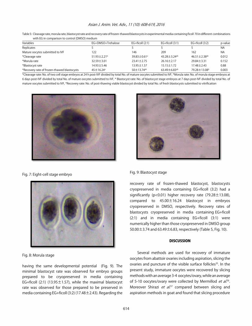
Embryo identification and evaluation: Cleaved embryoswere classified according to their cell number during aspecific time as 1-cell on day 1, 2-cells on day 2, 4-cells on day3, 8-cells on day 4 and 16-cells embryo on day 5 as well asmorula and blastocyst stages on days 6 and 7, respectively27.
610

Asian J. Anim. Vet. Adv., 11 (10): 608-619, 2016
Cryopreservation of mature oocytes and embryos:Vitrification method was used for cryopreservation of cattleoocytes and embryos. For this purpose excellent and goodmature COCs as well as morphologically normal compactmorula and blastocysts were used16.
Vitrification of mature oocytes and embryos by EG andDMSO (control group): Base Medium (BM) that used during vitrification was H-TCM-199 supplemented with 20%FCS and 50 µL mLG1 gentamycin sulfate28,29. The 1stVitrification Solution (VS1) consisted of 10% (v/v) EG+10% (v/v)DMSO as permeating cryoprotectants in BM, while the2nd Vitrification Solution (VS2) contained 20% (v/v) EG+20%(v/v) DMSO+0.3 M trehalose in the BM16.
Loading and thawing of straws: Straws of 0.25 and0.5 mL were preloaded to give the following configuration,150 µL BM, 5 µL air, 2×10 µL of the VS2 separated by 5 µL airthen matured oocytes and embryos were loaded in the final3rd column of the vitrification solution (10 µL) followed by 5 µL air then the reminder of the straw was filled withBM. Five matured oocytes or embryos were loaded in eachstraw that was heat-sealed and immediately dipped verticallyin liquid nitrogen (-196EC)28. After the appropriate vitrificationperiod (5-7 days), the mature oocytes and embryos (at morulaand blastocyst stages) were thawed by gentile agitation of thestraws for 10 sec in air followed by 20 sec at 35EC using awater bath. Immediately, the straw contents were expelled in6 cm diameter petri dishes for examination30.
Vitrification of mature oocytes and embryos by EG andficoll 70: The cryoprotectants were stocked by a version ofPBS medium containing 18% fetal calf serum. The EG wasprepared with DPBS into a fraction of 40%. Ficoll 70 wasprepared to an 18% stocking solution. Three ratios ofcryoprotectant solutions were used in the present study asfollows: 40% EG+18% ficoll 70 (volume ratio of 3:1 mL) and40% EG+18% ficoll 70 (volume ratio of 3:2 mL). Matured oocytes were pipetted out from 18% FCS in PBS and werethen immersed in the freezing medium for 10 min. Maturedoocytes were then packed into 0.25 and 0.5 mL straws and the sealed-labeled straws were plunged directly into liquidnitrogen for preservation. Straws were removed from theliquid nitrogen tank and put directly into the 25EC water bathfor 1 min for thawing. Approximately 1 min later, water onthe surface of thawed straw was wiped away to avoid cell
collapsing due to an imbalance of water electrolytes.One end of thawed straw was then cut and let thawed oocytes be squeezed into 0.5 M sucrose solution foreluting out cryoprotectants. Ten minutes later, ova were put into 18% FCS in PBS for observing the integrality of frozen-thawed oocytes22.
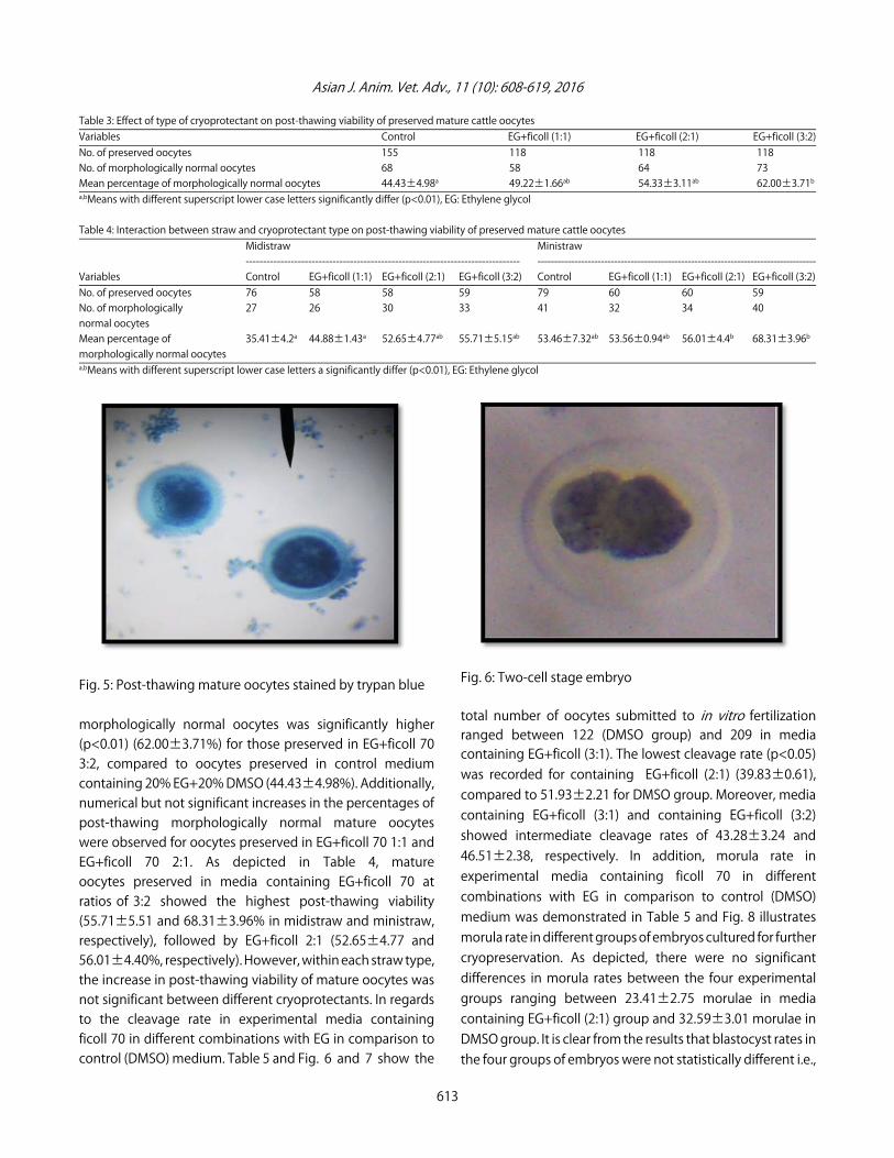
Evaluation of post-thawing viability of mature oocytesand embryos: The oocytes and embryos weremorphologically examined under stereomicroscope (X100)and the cells were considered normal if they have sphericaland symmetrical shape with no signs of lysis, membranedamage, swelling, degeneration or leakage of the cellularcontent. On the other hand, abnormal cells form appeared tohave a rupture zona pellucida or fragmented cytoplasm withsigns of degeneration31. The post-thawing viability of matureoocytes and embryos can also determine by the use of trypanblue stain (0.05% in PBS) for 2 min to differentiate the live anddead cells without adverse effects on the cells32. It is worthmentioning that in vitro survival rate is defined as the ratio ofviable cells after thawing to the total vitrified cell number33.
Statistical analysis: The obtained data were subjected tostatistical analysis as outlined by Snedecor et al.34.
RESULTS
The obtained results showed that, a total of 175 ovarieswere sliced to yield 728 oocytes which were recovered andgraded into 4 different grades. The mean percentages ofrecovered grade I and II oocytes related to total recoveredoocytes per replicate were 38.73±2.92 and 33.51±2.42%,respectively. Moreover, the average maturation rate ofrecovered grade I and II oocytes was 80.62±3.14 with a rangeof 66.66-96.15% (Table 1, Fig. 1, 2). Regarding the effect ofstraw type on post-thawing viability of cryopreservedmature oocytes, data presented in Table 2 show that out of251 oocytes which were preserved in midistraws, 116 oocytesappeared morphologically normal post-thawing representinga mean percentage of 47.16±2.62%. A significantly higher(p<0.01) percentage of post-thawing morphologically normaloocytes (57.83±2.59%, 147/258) was recorded for matureoocytes cryopreserved in ministraws (Fig. 3-5). Concerningeffects of type of cryoprotectant on post-thawingviability of preserved mature oocytes, results obtained inTable 3 showed that the mean percentage of post-thawing
611

Asian J. Anim. Vet. Adv., 11 (10): 608-619, 2016
Table 1: Descriptive statistics of slaughterhouse ovaries, recovered and matured oocytes in different trials of experiment oneRecovered immature oocytes (N = 728)------------------------------------------------------------------------------------------------------------------------------------------------------Grade I oocytes Grade II oocytes Grade III oocytes Grade IV oocytes---------------------------------- ---------------------------------- ---------------------------------- ----------------------------------
Percentage of Percentage of Percentage of Percentage of Statistics Ovaries No. total oocytes No. total oocytes No. total oocytes No. total oocytes Maturation rate* (%)Total 175 295 256 128 49Minimum 9 10 24.39 8 21.05 9 11.3 0 0 66.67Maximum 24 55 50.0 43 45.74 19 42.86 9 14.63 96.15Mean 17.5 29.5 38.73 25.6 33.51 12.8 20.49 4.9 7.26 80.62SE 1.78 5.15 2.92 4.31 2.42 1.1 3.17 0.77 1.41 3.14*Maturation rate: Number of recovered grade I and II immature oocytes, which was successfully matured divided by total number of recovered grade I and II immatureoocytes, SE: Standard error
Table 2: Effect of straw type on post-thawing viability of preserved mature cattle oocytesVariables Midistraw MinistrawNo. of preserved oocytes 251 258No. of morphologically normal oocytes 116 147Mean percentage of morphologically normal oocytes 47.16±2.62a 57.83±2.59b
a,bMeans with different superscript lower case letters significantly differ (p<0.01)
Fig. 1: Grade I and II immature bovine oocytes ready formaturation
Fig. 2: Mature bovine oocytes show excellent cumulus cellsexpantion
Fig. 3: Post-thawing mature oocytes
Fig. 4: Post-thawing degenerated cattle oocyte
612
Grade I
Grade II
Asian J. Anim. Vet. Adv., 11 (10): 608-619, 2016
Table 3: Effect of type of cryoprotectant on post-thawing viability of preserved mature cattle oocytesVariables Control EG+ficoll (1:1) EG+ficoll (2:1) EG+ficoll (3:2)No. of preserved oocytes 155 118 118 118No. of morphologically normal oocytes 68 58 64 73Mean percentage of morphologically normal oocytes 44.43±4.98a 49.22±1.66ab 54.33±3.11ab 62.00±3.71b
a,bMeans with different superscript lower case letters significantly differ (p<0.01), EG: Ethylene glycol
Table 4: Interaction between straw and cryoprotectant type on post-thawing viability of preserved mature cattle oocytesMidistraw Ministraw------------------------------------------------------------------------------- ------------------------------------------------------------------------------------
Variables Control EG+ficoll (1:1) EG+ficoll (2:1) EG+ficoll (3:2) Control EG+ficoll (1:1) EG+ficoll (2:1) EG+ficoll (3:2)No. of preserved oocytes 76 58 58 59 79 60 60 59No. of morphologically 27 26 30 33 41 32 34 40normal oocytesMean percentage of 35.41±4.2a 44.88±1.43a 52.65±4.77ab 55.71±5.15ab 53.46±7.32ab 53.56±0.94ab 56.01±4.4b 68.31±3.96b
morphologically normal oocytesa,bMeans with different superscript lower case letters a significantly differ (p<0.01), EG: Ethylene glycol
Fig. 5: Post-thawing mature oocytes stained by trypan blue
morphologically normal oocytes was significantly higher(p<0.01) (62.00±3.71%) for those preserved in EG+ficoll 703:2, compared to oocytes preserved in control mediumcontaining 20% EG+20% DMSO (44.43±4.98%). Additionally,numerical but not significant increases in the percentages ofpost-thawing morphologically normal mature oocyteswere observed for oocytes preserved in EG+ficoll 70 1:1 andEG+ficoll 70 2:1. As depicted in Table 4, matureoocytes preserved in media containing EG+ficoll 70 atratios of 3:2 showed the highest post-thawing viability(55.71±5.51 and 68.31±3.96% in midistraw and ministraw,respectively), followed by EG+ficoll 2:1 (52.65±4.77 and56.01±4.40%, respectively). However, within each straw type,the increase in post-thawing viability of mature oocytes wasnot significant between different cryoprotectants. In regardsto the cleavage rate in experimental media containingficoll 70 in different combinations with EG in comparison tocontrol (DMSO) medium. Table 5 and Fig. 6 and 7 show the
Fig. 6: Two-cell stage embryo
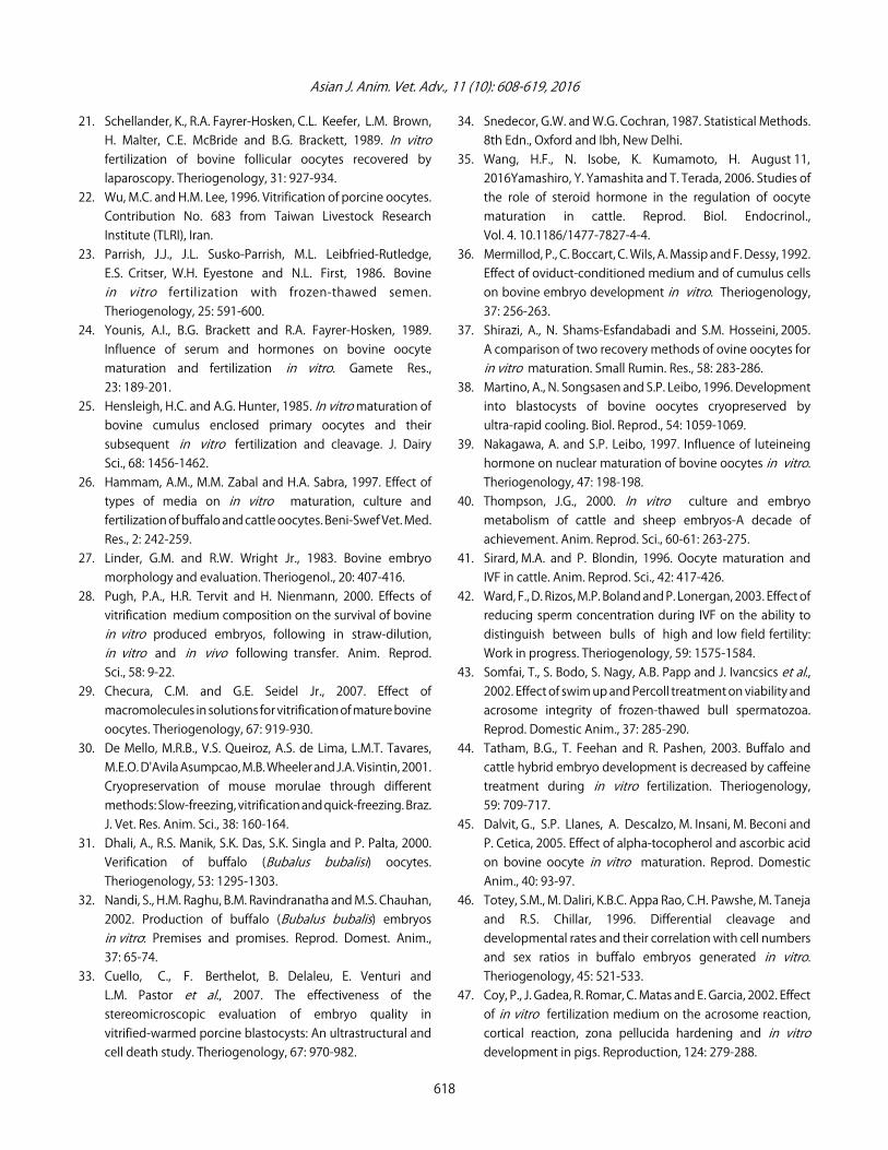
total number of oocytes submitted to in vitro fertilizationranged between 122 (DMSO group) and 209 in mediacontaining EG+ficoll (3:1). The lowest cleavage rate (p<0.05)was recorded for containing EG+ficoll (2:1) (39.83±0.61),compared to 51.93±2.21 for DMSO group. Moreover, mediacontaining EG+ficoll (3:1) and containing EG+ficoll (3:2)showed intermediate cleavage rates of 43.28±3.24 and46.51±2.38, respectively. In addition, morula rate inexperimental media containing ficoll 70 in differentcombinations with EG in comparison to control (DMSO)medium was demonstrated in Table 5 and Fig. 8 illustratesmorula rate in different groups of embryos cultured for furthercryopreservation. As depicted, there were no significantdifferences in morula rates between the four experimentalgroups ranging between 23.41±2.75 morulae in mediacontaining EG+ficoll (2:1) group and 32.59±3.01 morulae inDMSO group. It is clear from the results that blastocyst rates inthe four groups of embryos were not statistically different i.e.,
613
Asian J. Anim. Vet. Adv., 11 (10): 608-619, 2016
Table 5: Cleavage rate, morula rate, blastocyst rate and recovery rate of frozen-thawed blastocysts in experimental media containing ficoll 70 in different combinationswith EG in comparison to control (DMSO) medium
Variables EG+DMSO+Trehalose EG+ficoll (2:1) EG+ficoll (3:1) EG+ficoll (3:2) p-valueReplicates 5 5 5 5 NAMature oocytes submitted to IVF 122 146 209 162 NA*Cleavage rate 51.93±2.21b 39.83±0.61a 43.28±3.24ab 46.51±2.38ab 0.012*Morula rate 32.59±3.01 23.41±2.75 26.16±2.17 29.84±3.31 0.152*Blastocyst rate 14.93±5.46 13.95±1.57 15.15±1.72 17.48±2.43 0.88*Recovery rate of frozen-thawed blastocysts 45±16.24a 50±13.74ab 63.49±6.83ab 79.28±13.08b 0.003*Cleavage rate: No. of two cell stage embryos at 24 h post-IVF divided by total No. of mature oocytes submitted to IVF, *Morula rate: No. of morula stage embryos at6 days post-IVF divided by total No. of mature oocytes submitted to IVF, * Blastocyst rate: No. of blastocyst stage embryos at 7 days post-IVF divided by total No. ofmature oocytes submitted to IVF, *Recovery rate: No. of post-thawing viable blastocyst divided by total No. of fresh blastocysts submitted to vitrification
Fig. 7: Eight-cell stage embryo
Fig. 8: Morula stage
having the same developmental potential (Fig. 9). Theminimal blastocyst rate was observed for embryo groupsprepared to be cryopreserved in media containingEG+ficoll (2:1) (13.95±1.57), while the maximal blastocystrate was observed for those prepared to be preserved inmedia containing EG+ficoll (3:2) (17.48±2.43). Regarding the
Fig. 9: Blastocyst stage
recovery rate of frozen-thawed blastocyst, blastocystscryopreserved in media containing EG+ficoll (3:2) had asignificantly (p<0.01) higher recovery rate (79.28±13.08), compared to 45.00±16.24 blastocyst in embryoscryopreserved in DMSO, respectively. Recovery rates ofblastocysts cryopreserved in media containing EG+ficoll(2:1) and in media containing EG+ficoll (3:1) werenumerically higher than those cryopreserved in DMSO group50.00±3.74 and 63.49±6.83, respectively (Table 5, Fig. 10).
DISCUSSION
Several methods are used for recovery of immatureoocytes from abattoir ovaries including aspiration, slicing theovaries and puncture of the visible surface follicles35. In thepresent study, immature oocytes were recovered by slicingmethods with an average 3-4 oocytes/ovary, while an averageof 5-10 oocytes/ovary were collected by Mermillod et al.36.Moreover Shirazi et al.37 compared between slicing andaspiration methods in goat and found that slicing procedure
614

Asian J. Anim. Vet. Adv., 11 (10): 608-619, 2016
Fig. 10: Post-thawing blastocyst showing live and deadembryos
yielded significantly higher percentage of grade I oocytesthan aspiration (52 versus 22%). These findings come indisagreement of the earlier study of Martino et al.38 reporteda lower recovery rate of good quality oocytes by slicingcompared to aspiration. Thus, it could be concluded from thepresent study that slicing procedure in bovine predominatesover other ways for retrieval of a higher recovery rate andgrade I oocytes. The IVM utilized in the present study wasTCM-199 supplemented with FCS, FSH and E2 which found toyield the highest maturation rates. These findings agree withthe results of Younis et al.24 and Nakagawa and Leibo39,revealed that addition of LH singly or in combination withFSH and E2 had a significant enhancing influence on theIVM rate in bovine. Serum added to the oocytes culture mediaprovided a source of albumin that balance the osmolarity andact as a free radical scavenger40. In the present study, onlygrade I and II were utilized for IVM and subsequent IVEP aswell as vitrification as recommended by Sirard and Blondin41
as cumulus cells provide nutrients to oocytes during theirgrowth. The IVM of oocytes were performed in incubator inthe presence of 5% CO2 tension at 39EC for 24 h as describedby Atef20 and Ward et al.42. In the present stydy separation ofmorphologically normal and high motile sperms were selectedusing swim up separation technique. In this respect,Somfai et al.43 have recorded a lower recovery with a better quality of spermatozoa using swim up Percoll separationresulted in a higher recovery rate. The sperm capacitationwas performed by heparin (200 IU mLG1). However,Tatham et al.44 reported that treatment of spermatozoa by 5 mM caffeine and 5 µg heparin mLG1 resulted in a higher fertilization rate than did treatment with 5 µg heparin mLG1 alone. Therefore, the sperm concentration of
the present study was 2×10⁶ spermatozoa mLG1 as describedby Dalvit et al.45. The reduction of sperm concentration into(0.016-0.125×10⁶) leading to lowering the cleavage rate42. Thefertilization medium used in this study was TALP medium, in this concern, many studies recorded better results withTALP medium in comparison with BO medium46. Moreover, Coy et al.47 reported that the percentage ofcleavage were higher in oocytes cultured in TCM-199 and TBM than TALP. This study revealed that cleaved oocytes aswell as morula and blastocysts developmental rates/maturecells were 45.39±2.11, 28.00±2.81 and 15.29±2.79%,respectively. Previously, Anderiesz et al.48 obtained a higher blastocyst rates (25.00%) following addition of FSH and LHinto maturation media in bovine. In addition, Choi et al.19 by adding FSH, LH and E2 recorded 84.3±2.1, 48.6±4.5 and15.3±2.2% for cleavage, morula and blastocyst development,respectively. It was shown that the most important stepaffecting the quality of blastocyst is post-fertilizationculture condition49. Therefore, suboptimal in vitro cultureenvironment can seriously affect the developmental potentialof in vitro produced embryos. In the present study, accordingDalvit et al.45, the fertilized oocytes were cultured in thesupplemented H-TCM-199 and placed in the CO2 incubatorwith change of the medium every 48 h for 7 successive days.Therefore, it is worth denoting that the success of in vitroembryo production in bovine is a multifactorial process thatdepends upon choice of convenient cell type undergoingfurther development, type of the selective media, incubationcondition as well as providing the environment with additivesparticularly hormones and sera to enhance cellulardevelopmental activities. The present article studied the effectof sugars including trehalose and macromolecules as ficoll 70additions to cryoprotectant solutions on the post-thawingviability of matured cattle matured oocytes and embryos.As well as determined the effect of the straw diameteron the post-thawing viability of mature cattle oocytes.Two procedures are well known to induce cellularcryopreservation. The conventional slow freezing, whichexposed the cells at various phases of freezing and thawingto physical, chemical and biological hazard particularlyintracellular ice crystals formation50. On the other hand,vitrification is a method that suppresses both intra andextracellular ice formation thus producing instead a glass-likestate51. Throughout the present study, the method used forcryopreservation of bovine oocytes and embryos wasvitrification which has been successfully applied in severalmammalian species. Despite the importance of penetratingcryoprotectants to avoid intracellular ice crystals formation,the high concentration of these substances is toxic and may
615
Dead
Live

Asian J. Anim. Vet. Adv., 11 (10): 608-619, 2016
cause osmotic injury52. So using of less toxic substances,association of different cryoprotectants, gradual exposure andreduction of exposure time to cryoprotectant solution arerequired53. In the present experment, a combination of20% EG and 20% DMSO was used for cells vitrification.Incorporating of DMSO and EG-containing medium has atleast to advantages, vitrification is facilitated because of thegreater glass forming characteristics of DMSO54 as well as thepermeability of each of the cryoprotectants is enhanced bythe presence of second55. Similarly, Yamada et al.12 foundthat in vitro maturation rate of bovine oocytes vitrified inEG+DMSO (29.2%) was significantly higher than that aftervitrification in EG+glycerol (4.30%). The addition of trehaloseto embryo vitrification medium containing DMSO offeredsignificantly better results than those obtained with sucrose56.In this respect, trehalose has greater stabilizing effects oncell membrane than sucrose57. In relation to trehaloseconcentration, it was found that the best concentration thatachieved the best results was 11.3% (0.3 M)58. This studyshowed that the mean percentage of post-thawingmorphologically normal oocytes was significantly higher(p<0.01) (62.00±3.71%) for those preserved in EG+ficoll70 3:2, compared to oocytes preserved in control mediumcontaining 20% EG+20% DMSO+0.3 M trehalose(44.43±4.98%). Additionally, numerical but not significantincreases in the percentages of post-thawing morphologicallynormal mature oocytes were observed for oocytespreserved in EG+ficoll 70 (2:1) and EG+ficoll 70 (2:1). While, theblastocysts cryopreserved in media containing EG+ficoll(3:2) had a significantly (p<0.01) higher recovery rate(79.28±13.08), compared to 45.00±16.24 blastocyst inembryos cryopreserved in DMSO, respectively. Recovery ratesof blastocysts cryopreserved in media containing EG+ficoll(1:1) and in media containing EG+ficoll (2:1) were numericallyhigher than those cryopreserved in DMSO group. These resultsrun with previous studies, which concluded that the EG andficoll 70 cryoprotectants combination would maintain thesurvival rate of frozen-thawed porcine oocytes at 81% or more.In addition, the survival rate of oocytes in thecryoprotectant medium of 3.5 M DMSO plus 40% EG and18% ficoll 70 was 75%, although the exposure time was10 min. Oocytes frozen in solutions containing of 40% EG and18% ficoll 70 by a ratio of 2:1, 3:1 or 3:2 had a mean survivalrate of 81, 82 and 97%, respectively22. The addition oftrehalose in the cryoprotectant medium containing of EG andficoll 70 increase the intact rate to almost 100% but did notimprove the survival rate. Results of the present studysupported the concept of McWilliams et al.59 in their study
non-permeating solutes, such as ficoll 70 and sucrose servedas osmotic buffers for the recovery of cryopreserved oocytesas EG or DMSO permeating cryoprotectant was used.Polysaccharides like ficoll 70 could influence the viscosity ofthe vitrification solution and reduce the toxicity of thecryoprotectant through lowered concentration to preventcells from cryoinjury by reducing mechanical stress whichoccurs during cryopreservation60.
A typical low toxicity vitrification solution, EFS40 containsthree cryoprotectants: 40% EG-rapidly permeating, lowtoxicity agent, ficoll 70 at 18%, a macromolecule andsucrose-a non-permeating hexose sugar as described61. Thecattle embryos were incubated in the EFS40 for 1 min beforeplunging into LN2. A stepwise pre-equilibration procedure, inwhich the amount of penetrating cryoprotectant wasgradually increased was very effective for human oocytes62
and bovine oocytes6,63. According to the results of the presentstudy, regarding the effect of straw type on post-thawingviability of cryopreserved mature oocytes, show that out of251 oocytes which were preserved in midistraws, 116 oocytesappeared morphologically normal post-thawing representinga mean percentage of 47.16±2.62%. A significantly higher(p<0.01) percentage of post-thawing morphologicallynormal oocytes (57.83±2.59%, 147/258) was recorded formature oocytes cryopreserved in ministraws. Using0.25 mL straws was previously applied in studies concernedwith cryopreservation of mature oocytes and embryos ofdifferent species11,64-66. In this respect, Dattena et al.64
suggested that by using traditional 0.25 mL straws, themaximum cooling rate was 2500EC min which allows embryosto pass through certain critical temperature zones quicklyand decreases the chilling injuries. Several new techniqueshave been developed recently including Open Pulled Straw(OPS), electron microscope grids, cryoloop as well as cryotopmethods67,68. All these devices achieved 10-fold faster coolingrates than those obtained in standard straws. However, themajor limit for application of these vitrification techniques isthe direct contact between the medium containing cells andliquid nitrogen which may introduce infections69. A possiblesolution to the problem is the minimum volume cooling method whereas the embryos are loading in an extremelylow volume (20 µL) into 0.25 mL insemination straw, which issealed before cooling70.
CONCLUSION
It can be concluded that using of ministraw for the cryopreservation of mature bovine oocytes hadpost-thawing viability significantly higher (<10%) than using
616

Asian J. Anim. Vet. Adv., 11 (10): 608-619, 2016
midistraw. As well as, it was proved that matured oocytesfrozen in solutions containing 20% EG+20% DMSO+0.3 Mtrehalose had mean survival rate of (44.43±4.98%). Whilemature oocytes frozen in solutions containing 40% EG and18% ficoll 70 by a ratio of 1:1, 2:1 and 3:2 in volume had amean survival rate of 49.22±1.66, 54.33±3.11 and62.00±3.71%, respectively. So, combination of 40%EG+18% ficoll 70 by the ratio of (3:2) act as a goodcryoprotectant combination for vitrification of mature bovineoocytes.
ACKNOWLEDGMENT
Sincere gratitude is to Department of Physiology, Facultyof Veterinary Medicine, Beni-Suef University for their kindsupervision and his helpfulness during this study and for theirkind cooperation, faithful advice and careful revision of thisstudy.
REFERENCES
1. Rall, W.F. and G.M. Fahy, 1985. Ice-free cryopreservation ofmouse embryos at -196EC by vitrification. Nature,313: 573-575.
2. Vanderzwalmen, P., F. Ectors, L. Grobet, Y. Prapas andY. Panagiotidis et al., 2009. Aseptic vitrification of blastocystsfrom infertile patients, egg donors and after IVM. Reprod.BioMed. Online, 19: 700-707.
3. Vanderzwalmen, P., D. Connan, L. Grobet, B. Wirleitner andB. Remy et al., 2013. Lower intracellular concentration ofcryoprotectants after vitrification than after slow freezingdespite exposure to higher concentration of cryoprotectantsolutions. Hum. Reprod., 28: 2101-2110.
4. Wang, L., J. Liu, G.B. Zhou, Y.P. Hou, J.J. Li and S.E. Zhu, 2011.Quantitative investigations on the effects of exposuredurations to the combined cryoprotective agents on mouseoocyte vitrification procedures. Biol. Reprod., 85: 884-894.
5. Campos-Chillon, L.F., D.J. Walker, J.F. de la Torre-Sanchez andG.E. Seidel, Jr., 2006. In vitro assessment of a direct transfervitrification procedure for bovine embryos. Theriogenology,65: 1200-1214.
6. Vajta, G., P. Holm, M. Kuwayama, P.J. Booth, H. Jacobsen,T. Greve and H. Callesen, 1998. Open Pulled Straw (OPS)vitrification: A new way to reduce cryoinjuries of bovine ovaand embryos. Mol. Reprod. Dev., 51: 53-58.
7. Le Gal, F., R. De Roover, B. Verhaeghe, D. Etienne andA. Massip, 2000. Development of vitrified matured cattleoocytes after thawing and culture in vitro. Vet.Rec., 146: 469-471.
8. Zhou, Y., X. Fu, G. Zhou, B. Jia, Y. Fang, Y. Hou and S. Zhu,2014. An efficient method for the sanitary vitrification ofbovine oocytes in straws. J. Anim. Sci. Biotechnol.,Vol. 5. 10.1186/2049-1891-5-19.
9. Wang, X., A. Al Naib, D.W. Sun and P. Lonergan, 2010.Membrane permeability characteristics of bovine oocytes anddevelopment of a step-wise cryoprotectant adding anddiluting protocol. Cryobiology, 61: 58-65.
10. Whittingham, D.G., 1977. Fertilization in vitro anddevelopment to term of unfertilized mouse oocytespreviously stored at-196°C. J. Reprod. Fertil., 49: 89-94.
11. Wani, N.A., A.K. Misra and S.N. Maurya, 2004. Maturation ratesof vitrified-thawed immature buffalo (Bubalus bubalis)oocytes: Effect of different types of cryoprotectants. Anim.Reprod. Sci., 84: 327-335.
12. Yamada, C., H.V.A. Caetano, R. Simoes, A.C. Nicacio,W.B. Feitosa, M.E.O. D'Avila Assumpcao and J.A. Visintin, 2007.Immature bovine oocyte cryopreservation: Comparison ofdifferent associations with ethylene glycol, glycerol anddimethylsulfoxide. Anim. Reprod. Sci., 99: 384-388.
13. Sharma, G.T., P.K. Dubey and V. Chandra, 2010. Morphologicalchanges, DNA damage and developmental competence ofin vitro matured, vitrified-thawed buffalo (Bubalus bubalis)oocytes: A comparative study of two cryoprotectants andtwo cryodevices. Cryobiology, 60: 315-321.
14. Anchamparuthy, V., 2007. Vitrification of bovine oocytes.Ph.D. Thesis, The Faculty of the Virginia Polytechnic Instituteand State University, Blacksburg, Virginia.
15. Lim, K.T., G. Jang, K.H. Ko, W.W. Lee and H.J. Park et al., 2008.Improved cryopreservation of bovine preimplantationembryos cultured in chemically defined medium. Anim.Reprod. Sci., 103: 239-248.
16. Atef, N., 2008. Studies on cryopreservation of in vitroproduced bovine embryos. Ph.D. Thesis, Faculty Of VeterinaryMedicine, Benisuef University, Egypt.
17. Ganguli, G., A. Indra and P. Gupta, 1998. Suitability of thefollicular oocytes obtained from slaughtered buffaloovaries and assessment of their nuclear maturation. BuffaloJ., 14: 217-228.
18. Nedambale, T.L., F. Du, J. Xu, S.A. Chaubal andA. Dinnyes et al., 2006. Prolonging bovine sperm-oocyteincubation in modified medium 199 improves embryodevelopment rate and the viability of vitrified blastocysts.Theriogenology, 66: 1951-1960.
19. Choi, Y.H., E.M. Carnevale, G.E. Seidel Jr. and E.L. Squire, 2001.Effects of gonadotropins on bovine oocytes matured inTCM-199. Theriogenology, 56: 661-670.
20. Atef, N., 2005. Some studies on in vitro fertilization inbuffaloes. MVSc. Thesis, Faculty Of Veterinary Medicine,Benisuef University, Egypt.
617
Asian J. Anim. Vet. Adv., 11 (10): 608-619, 2016
21. Schellander, K., R.A. Fayrer-Hosken, C.L. Keefer, L.M. Brown,H. Malter, C.E. McBride and B.G. Brackett, 1989. In vitrofertilization of bovine follicular oocytes recovered bylaparoscopy. Theriogenology, 31: 927-934.
22. Wu, M.C. and H.M. Lee, 1996. Vitrification of porcine oocytes.Contribution No. 683 from Taiwan Livestock ResearchInstitute (TLRI), Iran.
23. Parrish, J.J., J.L. Susko-Parrish, M.L. Leibfried-Rutledge,E.S. Critser, W.H. Eyestone and N.L. First, 1986. Bovinein vitro fertilization with frozen-thawed semen.Theriogenology, 25: 591-600.
24. Younis, A.I., B.G. Brackett and R.A. Fayrer-Hosken, 1989.Influence of serum and hormones on bovine oocytematuration and fertilization in vitro. Gamete Res.,23: 189-201.
25. Hensleigh, H.C. and A.G. Hunter, 1985. In vitro maturation ofbovine cumulus enclosed primary oocytes and theirsubsequent in vitro fertilization and cleavage. J. DairySci., 68: 1456-1462.
26. Hammam, A.M., M.M. Zabal and H.A. Sabra, 1997. Effect oftypes of media on in vitro maturation, culture andfertilization of buffalo and cattle oocytes. Beni-Swef Vet. Med.Res., 2: 242-259.
27. Linder, G.M. and R.W. Wright Jr., 1983. Bovine embryomorphology and evaluation. Theriogenol., 20: 407-416.
28. Pugh, P.A., H.R. Tervit and H. Nienmann, 2000. Effects ofvitrification medium composition on the survival of bovinein vitro produced embryos, following in straw-dilution,in vitro and in vivo following transfer. Anim. Reprod.Sci., 58: 9-22.
29. Checura, C.M. and G.E. Seidel Jr., 2007. Effect ofmacromolecules in solutions for vitrification of mature bovineoocytes. Theriogenology, 67: 919-930.
30. De Mello, M.R.B., V.S. Queiroz, A.S. de Lima, L.M.T. Tavares,M.E.O. D'Avila Asumpcao, M.B. Wheeler and J.A. Visintin, 2001.Cryopreservation of mouse morulae through differentmethods: Slow-freezing, vitrification and quick-freezing. Braz.J. Vet. Res. Anim. Sci., 38: 160-164.
31. Dhali, A., R.S. Manik, S.K. Das, S.K. Singla and P. Palta, 2000.Verification of buffalo (Bubalus bubalisi) oocytes.Theriogenology, 53: 1295-1303.
32. Nandi, S., H.M. Raghu, B.M. Ravindranatha and M.S. Chauhan,2002. Production of buffalo (Bubalus bubalis) embryosin vitro: Premises and promises. Reprod. Domest. Anim.,37: 65-74.
33. Cuello, C., F. Berthelot, B. Delaleu, E. Venturi andL.M. Pastor et al., 2007. The effectiveness of thestereomicroscopic evaluation of embryo quality invitrified-warmed porcine blastocysts: An ultrastructural andcell death study. Theriogenology, 67: 970-982.
34. Snedecor, G.W. and W.G. Cochran, 1987. Statistical Methods.8th Edn., Oxford and Ibh, New Delhi.
35. Wang, H.F., N. Isobe, K. Kumamoto, H. August 11,2016Yamashiro, Y. Yamashita and T. Terada, 2006. Studies ofthe role of steroid hormone in the regulation of oocytematuration in cattle. Reprod. Biol. Endocrinol.,Vol. 4. 10.1186/1477-7827-4-4.
36. Mermillod, P., C. Boccart, C. Wils, A. Massip and F. Dessy, 1992.Effect of oviduct-conditioned medium and of cumulus cellson bovine embryo development in vitro. Theriogenology,37: 256-263.
37. Shirazi, A., N. Shams-Esfandabadi and S.M. Hosseini, 2005.A comparison of two recovery methods of ovine oocytes forin vitro maturation. Small Rumin. Res., 58: 283-286.
38. Martino, A., N. Songsasen and S.P. Leibo, 1996. Developmentinto blastocysts of bovine oocytes cryopreserved byultra-rapid cooling. Biol. Reprod., 54: 1059-1069.
39. Nakagawa, A. and S.P. Leibo, 1997. Influence of luteineinghormone on nuclear maturation of bovine oocytes in vitro.Theriogenology, 47: 198-198.
40. Thompson, J.G., 2000. In vitro culture and embryometabolism of cattle and sheep embryos-A decade ofachievement. Anim. Reprod. Sci., 60-61: 263-275.
41. Sirard, M.A. and P. Blondin, 1996. Oocyte maturation andIVF in cattle. Anim. Reprod. Sci., 42: 417-426.
42. Ward, F., D. Rizos, M.P. Boland and P. Lonergan, 2003. Effect ofreducing sperm concentration during IVF on the ability todistinguish between bulls of high and low field fertility:Work in progress. Theriogenology, 59: 1575-1584.
43. Somfai, T., S. Bodo, S. Nagy, A.B. Papp and J. Ivancsics et al.,2002. Effect of swim up and Percoll treatment on viability andacrosome integrity of frozen-thawed bull spermatozoa.Reprod. Domestic Anim., 37: 285-290.
44. Tatham, B.G., T. Feehan and R. Pashen, 2003. Buffalo andcattle hybrid embryo development is decreased by caffeinetreatment during in vitro fertilization. Theriogenology,59: 709-717.
45. Dalvit, G., S.P. Llanes, A. Descalzo, M. Insani, M. Beconi andP. Cetica, 2005. Effect of alpha-tocopherol and ascorbic acidon bovine oocyte in vitro maturation. Reprod. DomesticAnim., 40: 93-97.
46. Totey, S.M., M. Daliri, K.B.C. Appa Rao, C.H. Pawshe, M. Tanejaand R.S. Chillar, 1996. Differential cleavage anddevelopmental rates and their correlation with cell numbersand sex ratios in buffalo embryos generated in vitro.Theriogenology, 45: 521-533.
47. Coy, P., J. Gadea, R. Romar, C. Matas and E. Garcia, 2002. Effectof in vitro fertilization medium on the acrosome reaction,cortical reaction, zona pellucida hardening and in vitrodevelopment in pigs. Reproduction, 124: 279-288.
618

Asian J. Anim. Vet. Adv., 11 (10): 608-619, 2016
48. Anderiesz, C., A.P. Ferraretti, C. Magli, A. Fiorentino andD. Fortini et al., 2000. Effect of recombinant humangonadotrophins on human, bovine and murine oocytemeiosis, fertilization and embryonic development in vitro.Hum. Reprod., 5: 1140-1148.
49. Rinaudo, P. and R.M. Schultz, 2004. Effects of embryo cultureon global pattern of gene expression in preimplantationmouse embryos. Reproduction, 128: 301-311.
50. Matsuoka, K., S. Sakata, K. Ichino, S. Shimaya, T. Katagiharaand T. Suzuki, 1995. Ultra-rapid freezing of in vitro producedbovine embryos. Theriogenology, 43: 274-286.
51. Liebermann, J., J. Dietl, P. Vanderzwalmen and M.J. Tucker,2003. Recent developments in human oocyte, embryo andblastocysts vitrification: Where are we now? Reprod. Biomed.Online, 7: 623-633.
52. Arav, A., D. Shehu and M. Mattioli, 1993. Osmotic andcytotoxic study of vitrification of immature bovine oocytes.J. Reprod. Fertility, 99: 353-358.
53. Vajta, G., 2000. Vitrification of the oocytes and embryos ofdomestic animals. Anim. Reprod. Sci., 60-61: 357-364.
54. Ali, J. and J.N. Shelton, 1993. Vitrification of preimplantationstages of mouse embryos. J. Reprod. Fertility, 98: 459-465.
55. Vicente, J.S. and F. Garcia-Ximenez, 1994. Osmotic andcryoprotective effects of a mixture of DMSO and ethyleneglycol on rabbit morulae. Theriogenology, 42: 1205-1215.
56. Robertson, J.L., B.S. Minhas, G.W. Randall, M.G. Dodson,T.V. Palmer and D.D. Ricker, 1989. Ultrarapid freezing ofmouse embryos with DMSO and trehalose. Theriogenology,31: 250-261.
57. Smorag, Z., Y. Heyman, V. Garnier and B. Gajda, 1990. Theeffect of sucrose and trehalose on viability of one-andtwo-cell rabbit embryos. Theriogenology, 33: 741-747.
58. Dobrinsky, J.R., S.L. Stice, P.E. Phillips, R.T. Duby and J.M. Robl,1992. Development of IVM-IVF bovine embryos followingvitrification dilution treatments. Theriogenology, 37: 202-202.
59. McWilliams, R.B., W.E. Gibbons and S.P. Leibo, 1995. Osmoticand physiological responses of mouse zygotes andhuman oocytes to mono- and disaccharides. Hum. Reprod.,10: 1163-1171.
60. Dumoulin, J.C.M., J.M. Bergers-Janssen, M.H.E.C. Pieters,M.E. Enginsu, J.P.M. Geraedts and J.L.H. Evers, 1994. Theprotective effects of polymers in the cryopreservation ofhuman and mouse zonae pellucidae and embryos. Fertil.Steril., 62: 793-798.
61. Kasai, M., 1996. Simple and efficient methods for vitrificationof mammalian embryos. Anim. Reprod. Sci., 42: 67-75.
62. Kuleshova, L.L., D.R. MacFarlane, A.O. Trounson andJ.M. Shaw, 1999. Sugars exert a major influence on thevitrification properties of ethylene glycol-based solutions andhave low toxicity to embryos and oocytes. Cryobiology,38: 119-130.
63. Abe, Y., K. Hara, H. Matsumoto, J. Kobayashi andH. Sasada et al., 2005. Feasibility of a nylon-mesh holder forvitrification of bovine germinal vesicle oocytes in subsequentproduction of viable blastocysts. Biol. Reprod., 72: 1416-1420.
64. Dattena, M., C. Accardo, S. Pilichi, V. Isachenko, L. Mara,B. Chessa and P. Cappai, 2004. Comparison of differentvitrification protocols on viability after transfer of ovineblastocysts in vitro produced and in vivo derived.Theriogenology, 62: 481-493.
65. Martins, R.D., E.P. Costa, J.S.C. Chagas, F.S. Ignacio,C.A.A. Torres and C. McManus, 2005. Effects of vitrification ofimmature bovine oocytes on in vitro maturation. Anim.Reprod., 2: 128-134.
66. Cetin, Y. and A. Bastan, 2006. Cryopreservation of immaturebovine oocytes by vitrification in straws. Anim. Reprod.Sci., 92: 29-36.
67. Vajta, G., P.J. Booth, P. Holm, T. Greve and H. Calleson, 1997.Successful vitrification of early stage bovine in vitro producedembryos with the Open Pulled Straw (OPS) method. CryoLett., 18: 191-195.
68. Kuwayama, M., 2007. Highly efficient vitrification forcryopreservation of human oocytes and embryos: TheCryotop method. Theriogenology, 67: 73-80.
69. Fountain, D., M. Ralston, N. Higgins, J.B. Gorlin and L. Uhl et al.,1997. Liquid nitrogen freezers: A potential source of microbialcontamination of hematopoietic stem cell components.Transfusion, 37: 585-591.
70. Hamawaki, A., M. Kuwayama and S. Hamano, 1999. Minimumvolume cooling method for bovine blastocyst vitrification.Theriogenology, 51: 165-174.
619