effect of coupling agents on the local mechanical properties of bioactive dental composites by the...
TRANSCRIPT
Effect of coupling agents on the local mechanicalproperties of bioactive dental composites by thenano-indentation technique
Emily Ho, Michele Marcolongo*
Department of Materials Science and Engineering, Drexel University, Lebow Rm. 336, 32 Chestnut Streets,Philadelphia, PA 19104, USA
Received 19 February 2004; received in revised form 25 August 2004; accepted 16 September 2004
01
89
KEYWORDSCoupling agent;Hydroxyapatite;Composite;Nano-indentation;Polymethyl-
methacrylate;Surface treatment;Mechanical
properties;Dental implant
material
09-5641/$ - see front matter Q 2005
* Corresponding author. Tel.: C1 215 6760.E-mail address: marcolms@drexel.
Summary Objectives. Toexaminetheuseofnano-indentationasamethodofdeterminingthe interfacial mechanics of bioactive composites for mandibular bone substitutes.
Methods. Three coupling agents (PMMA-MAA, PMMA-MA and silane) were used totreat hydroxyapatite (HA) particles before incorporation into a polymethylmetha-crylate (PMMA) matrix. Nano-indentation was used to determine the hardness andYoung’s modulus on the HA particle surface, at the HA/PMMA interface and in thePMMA matrix region for each of the four groups. In addition bulk four-point bendingtests were conducted on each of the four groups as a comparison.
Results. The findings resulted in significant differences in the local interfacialYoung’s modulus between the polymer-treated composites and the uncoupledcontrol specimens with a marked improvement (50%) in modulus with eitherpolymertreated group. Similarly, the bending modulus of the polymer-treated groupswas significantly higher than the un-treated control group; however, thesedifferences were not as pronounced (approximately 15%).
Significance. The co-polymer-treated composites resulted in improved interfacialmodulus as compared to the un-treated controls and that the nano-indentationtechnique is a powerful tool for understanding the local interfacial mechanics ofbioactive composites.Q 2005 Academy Dental Materials. Published by Elsevier Ltd. All rights reserved.
Introduction
The present field of nano-indentation research grewfrom a desire to measure the mechanical properties
Academy Dental Materials. P
5 895 2329; fax: C1 215
edu (M. Marcolongo).
of hard thin films and other near surface treatmentsin the early 1980s [1–5]. Nano-indentation is a depth-sensing technique that can accurately characterizethe mechanical properties of almost all types of solidmaterials at a small scale [2,6–8]. If plastic defor-mation occurs, then there is a residual impressionleft in the surface of the specimen. In some cases,the permanent deformation or residual impression is
Dental Materials (2005) 21, 656–664
www.intl.elsevierhealth.com/journals/dema
ublished by Elsevier Ltd. All rights reserved.
Effect of coupling agents on the local mechanical properties of bioactive dental composites 657
not the result of plastic flow but may involvecracking, phase changes or structural changes withinthe specimen. In addition, nano-indentation cancontinuously record the penetration depth of thespecimen during dynamic loading and unloading atthe indentation tip while providing information onthe degree of energy absorbed, the Young’s modulusand hardness of the specimens. Several groups haveconducted nano-indentation testing on ceramics todetermine hardness and Young’s modulus from theanalysis of the load–displacement unloading curves[8–13]. They found that both Young’s modulus andhardness exhibit significant peak-load-dependence.Further, indentation size effect (ISE), while aconcern in the analysis of metals [2], is still underdebate in the testing of ceramics. ISE may influencethe calculation of the hardness and Young’s mod-ulus, which is determined in part from the esti-mation of the indentation projected area. However,none of these studies have fully analyzed thecharacteristics of the resulting nano-indentationmarks. The imprint of the indentations providesdetails on whether the indentations fracture orbehave differently with various peak-loads.
Recently, Xu et al. [14–16] applied nano-indenta-tion to the investigation of the effect of differentwhiskers on the reinforcement of dental resincomposites. In these studies, the materials experi-enced significant changes in hardness and Young’smodulus values as the indenter shifted from matrixto interface and from interface to fillers. While thisstudy gave new insights into interfacial mechanics,additional information on indentation size, shapeand local fracture mechanism in the system mightfurther help to elucidate the interfacial behavior.Indentation sizes are on the order of a micron,which may provide challenges in optically imagingthe indentations at the interface.
During the development of a load-bearing dentalimplant material, it was observed that there was aneed to promote the interfacial bonding of abioactive ceramic/polymer composite by introdu-cing a coupling agent capable of advancing chemi-cal bonding between the two materials of thecomposite. The modified interface surface wasintended to reduce debonding at the filler–matrixinterface leading to stronger, higher modulusmaterials [17–19]. The aim of this study was toexamine the mechanical properties of hydroxyapa-tite (HA) particulates reinforced polymethylmetha-crylate (PMMA) composites as a function of couplingagents. It was hypothesized that the global modulusof the materials would be affected by the incor-poration of various coupling agents at the particle/matrix interface and such interfacial differencescould be elucidated using nano-indentation.
Materials and methods
Material processing
The authors investigated hydroxyapatite (HA)/polymethylmethacrylate (PMMA) composite sys-tems with a volume ratio of 55:45 as dental implantmaterials. The HA scaffolds were fabricatedthrough a patented three-dimensional printingprocess (3DP) called Theriforme [20]. Scaffoldswere produced from HA powders of 20G5 mmaverage particle size (Ceramed Dental LLC, Lake-wood, CO, USA). A binder was added to bind theparticles for better control of the porosity. Thisprocess provided a multi-layer stacking feature thatcould be applied to complex shaped designs withthickness variations. The resulting scaffolds haddimensions of 10 mm by 10 mm by 4 mm. Sub-sequently, the HA scaffolds were sintered at 1250 8Cfor 1 h and the polymer binder was melted. Pure HAblocks were then infiltrated with molecular weight120,000 g/mol methylmethacrylate monomer(MMA) with benztroyl as the initiator. The PMMAadded blocks were pressurized in vacuum-sealedPVA bags at 60 psi, and were followed with a curingprocess at 60 8C then at 120 8C in a stepwise fashion.
The sintered pure HA scaffolds were immersedwith one of the three coupling agent solutions (seeTable 1) prior to PMMA infiltration and curingprocess. The uncoupled composites served ascontrols. The specimens were polished with 1 mmand 0.5 mm wet alumina polishing paste, to anoptical finish. Each polished composite surface wasmarked with three 2 mm by 2 mm grids by adiamond scriber to indicate the test position ofeach nano-indentation array.
Bulk material bending tests
Four point bending was performed on the controlsand each of the three treatment groups todetermine the bulk bending strength and Young’smodulus of the HA/PMMA composite. This test wasselected to simulate, in part, the load bearingaction of a reconstructed mandible during mastica-tion. The four point bending test setup wasprepared in accordance with ASTM C1161-95.Testing was performed on an Instron mechanicaltest machine Model 1331 (Canton, MA, USA) using aconstant load rate of 2.5 mm/min until failureoccurred. Load–displacement (N/mm) curves weregenerated on the data-acquisition device of thetesting machine. The deflection value was esti-mated from the displacement value at the failureload.
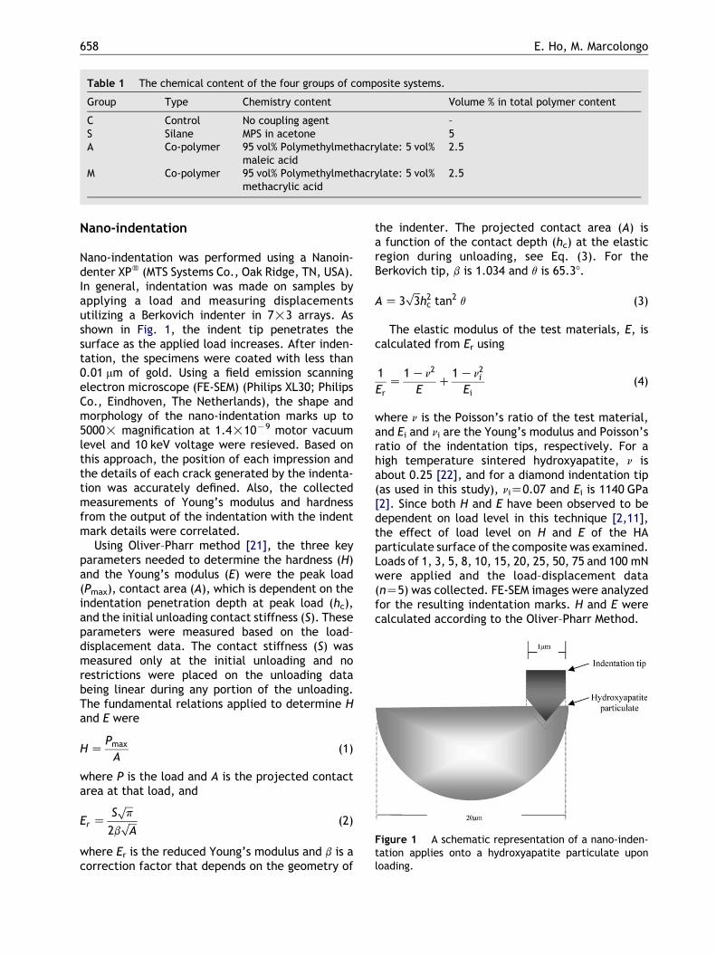
Table 1 The chemical content of the four groups of composite systems.
Group Type Chemistry content Volume % in total polymer content
C Control No coupling agent –S Silane MPS in acetone 5A Co-polymer 95 vol% Polymethylmethacrylate: 5 vol%
maleic acid2.5
M Co-polymer 95 vol% Polymethylmethacrylate: 5 vol%methacrylic acid
2.5
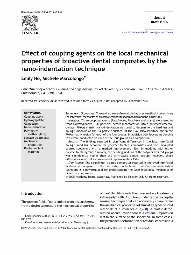
Figure 1 A schematic representation of a nano-inden-tation applies onto a hydroxyapatite particulate uponloading.
E. Ho, M. Marcolongo658
Nano-indentation
Nano-indentation was performed using a Nanoin-denter XPw (MTS Systems Co., Oak Ridge, TN, USA).In general, indentation was made on samples byapplying a load and measuring displacementsutilizing a Berkovich indenter in 7!3 arrays. Asshown in Fig. 1, the indent tip penetrates thesurface as the applied load increases. After inden-tation, the specimens were coated with less than0.01 mm of gold. Using a field emission scanningelectron microscope (FE-SEM) (Philips XL30; PhilipsCo., Eindhoven, The Netherlands), the shape andmorphology of the nano-indentation marks up to5000! magnification at 1.4!10K9 motor vacuumlevel and 10 keV voltage were resieved. Based onthis approach, the position of each impression andthe details of each crack generated by the indenta-tion was accurately defined. Also, the collectedmeasurements of Young’s modulus and hardnessfrom the output of the indentation with the indentmark details were correlated.
Using Oliver–Pharr method [21], the three keyparameters needed to determine the hardness (H)and the Young’s modulus (E) were the peak load(Pmax), contact area (A), which is dependent on theindentation penetration depth at peak load (hc),and the initial unloading contact stiffness (S). Theseparameters were measured based on the load–displacement data. The contact stiffness (S) wasmeasured only at the initial unloading and norestrictions were placed on the unloading databeing linear during any portion of the unloading.The fundamental relations applied to determine Hand E were
H ZPmax
A(1)
where P is the load and A is the projected contactarea at that load, and
Er ZS
ffiffiffi
pp
2bffiffiffi
Ap (2)
where Er is the reduced Young’s modulus and b is acorrection factor that depends on the geometry of
the indenter. The projected contact area (A) isa function of the contact depth (hc) at the elasticregion during unloading, see Eq. (3). For theBerkovich tip, b is 1.034 and q is 65.38.
A Z 3ffiffiffi
3p
h2c tan2 q (3)
The elastic modulus of the test materials, E, iscalculated from Er using
1
ErZ
1 Kn2
EC
1 Kn2i
Ei(4)
where n is the Poisson’s ratio of the test material,and Ei and ni are the Young’s modulus and Poisson’sratio of the indentation tips, respectively. For ahigh temperature sintered hydroxyapatite, n isabout 0.25 [22], and for a diamond indentation tip(as used in this study), niZ0.07 and Ei is 1140 GPa[2]. Since both H and E have been observed to bedependent on load level in this technique [2,11],the effect of load level on H and E of the HAparticulate surface of the composite was examined.Loads of 1, 3, 5, 8, 10, 15, 20, 25, 50, 75 and 100 mNwere applied and the load–displacement data(nZ5) was collected. FE-SEM images were analyzedfor the resulting indentation marks. H and E werecalculated according to the Oliver–Pharr Method.
Table 2 The bending properties of various compo-site systems.
Group Bending strength[MPa] (SD), (nZ5)
Bend stiffness [GPa](SD), (nZ5)
C 53.6 (4.8) 15.5 (1.0)S 55.2 (7.2) 15.3 (2.7)A 63.4 (2.9) 16.4 (0.7)M 67.9 (3.7) 17.9 (1.6)
Effect of coupling agents on the local mechanical properties of bioactive dental composites 659
The second analysis employed a load at theoptimal range (10 mN) to created 3!7 arrays ofthe indentations on the control and samples fromeach of three treatment groups. Load–displacementdata was correlated to the FE-SEM image of theindent mark. E and H were calculated for positionson the HA particles, ‘at the interface’ (defined as2 mm from the interface) and on the PMMA surfaceof the composite. Five indentation readings weremade for each location for each treatment group(including controls).
Data analysis
Utilizing a one way ANOVA, the authors evaluatedwhether any of the three applied coupling agentshas statistical significance at 95% confidence level(p values less than 0.05) in increasing the bulkbending strength, bulk bending modulus, localYoung’s modulus and local hardness in comparisonwith the uncoupled HA/PMMA composites.Also, one way ANOVA was applied to determine
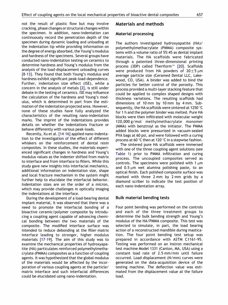
Figure 2 Local Young’s modulus measurements at the centFE-SEM images of indented hydroxyapatite at 5000! after 1indentation in hydroxyapatite at 2500! after 25 mN loading.
the statistical significance of each coupling agentamong other treatments based on the test outputsof the four point bending test and the nano-indentation study.
Results
Effect of coupling agent on bulk bendingbehavior
This study examined the bending properties ofthe bulk HA/PMMA composites with and withoutcoupling agents, see Table 2. The test resultsrevealed that the bending strengths of both co-polymer coupled specimens (group M and groupA) are statistically greater than the controls. Thebending strength of group M is 27% higher thanthe controls with pZ0.001, while the bendingstrength of group A is 18% higher with pZ0.004.However, there was no statistical significance inbending properties between A and M co-polymergroup (pZ0.06). Neither bending strength normodulus of group S (silane coupled composites)showed significant difference from the controls(pZ0.69 and pZ0.89, respectively). With theaddition of polymethylmethacrylate-methacrylicacid co-polymer coupling agent (group M), thebending modulus of the composites obtained a15% increase in comparison with the untreatedcontrols (pZ0.02). But the other co-polymeradded system, polymethylmethacrylate–maleic
er of hydroxyapatite as a function of peak load; with (a):0 mN loading, and (b): FE-SEM images of cracks induced
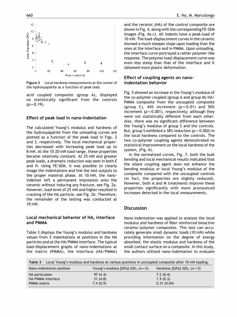
Figure 3 Local hardness measurements at the center ofthe hydroxyapatite as a function of peak load.
E. Ho, M. Marcolongo660
acid coupled composite (group A), displayedno statistically significant from the controls(pZ0.19).
Effect of peak load in nano-indentation
The calculated Young’s modulus and hardness ofthe hydroxyapatite from the unloading curves areplotted as a function of the peak load in Figs. 2and 3, respectively. The local mechanical proper-ties decreased with increasing peak load up to8 mN. At the 10–20 mN load range, these propertiesbecame relatively constant. At 25 mN and greaterpeak loads, a dramatic reduction was seen in both Eand H. Using FE-SEM, it was possible to clearlyimage the indentations and link the test outputs tothe proper material phase. At 10 mN, the nano-indenter left a permanent impression onto theceramic without inducing any fracture, see Fig. 2a.However, load level of 25 mN and higher resulted incracking of the HA particle, see Fig. 2b. Therefore,the remainder of the testing was conducted at10 mN.
Local mechanical behavior of HA, interfaceand PMMA
Table 3 displays the Young’s modulus and hardnessvalues from 5 indentations at positions in the HAparticles and at the HA/PMMA interface. The typicalload–displacement graphs of nano-indentations atthe matrix (PMMA), the interface (HA/PMMA)
Table 3 Local Young’s modulus and hardness at various p
Nano-indentation position Young’s modulus [G
HA particulate 97 (4.4)HA/PMMA interface 31 (4.8)PMMA matrix 7.4 (0.9)
and the ceramic (HA) of the control composite areshown in Fig. 4, along with the corresponding FE-SEMimages (Fig. 4a–c). All indents have a peak-load of10 mN. The load–displacement curves in the ceramicshowed a much steeper slope upon loading than theones at the interface and in PMMA. Upon unloading,the interface curve portrayed a rather polymer-likeresponse. The polymer load–displacement curve waseven less steep than that of the interface and itobtained more plastic deformation.
Effect of coupling agents on nano-indentation behavior
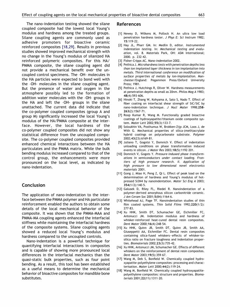
Fig. 5 showed an increase in the Young’s modulus ofthe co-polymer coupled (group A and group M) HA/PMMA composite from the uncoupled composite(group C), 44% increment (pZ0.01) and 50%increment (pZ0.001), respectively; although theywere not statistically different from each other.Also, there was no significant difference betweenthe Young’s modulus of group S and the controls.But, group S exhibited a 38% reduction (pZ0.002) inthe local hardness compared to the controls. Thetwo co-polymer coupling agents demonstrated nostatistical improvement on the local hardness of thesystem, (Fig. 6).
In the normalized curves, Fig. 7, both the bulkbending and local mechanical results indicated thatthe silane coupling agent does not enhance thebending modulus or local Young’s modulus of thecomposite compared with the uncoupled controls(in fact, the properties are slightly reduced).However, both A and M treatments improve theseproperties significantly with more pronouncedincreases detected in the local measurements.
Discussion
Nano-indentation was applied to analyze the localmodulus and hardness of fiber reinforced bioactiveceramic/polymer composites. This test can accu-rately generate small dynamic loads (10 mN) whileproviding information on the degree of energyabsorbed, the elastic modulus and hardness of thesmall contact surface on a composite. In this study,the authors utilized nano-indentation to evaluate
ositions in uncoupled composite after 10 mN loading.
Pa] (SD), (nZ5) Hardness [GPa] (SD), (nZ5)
7.2 (0.4)1.9 (0.3)0.31 (0.04)
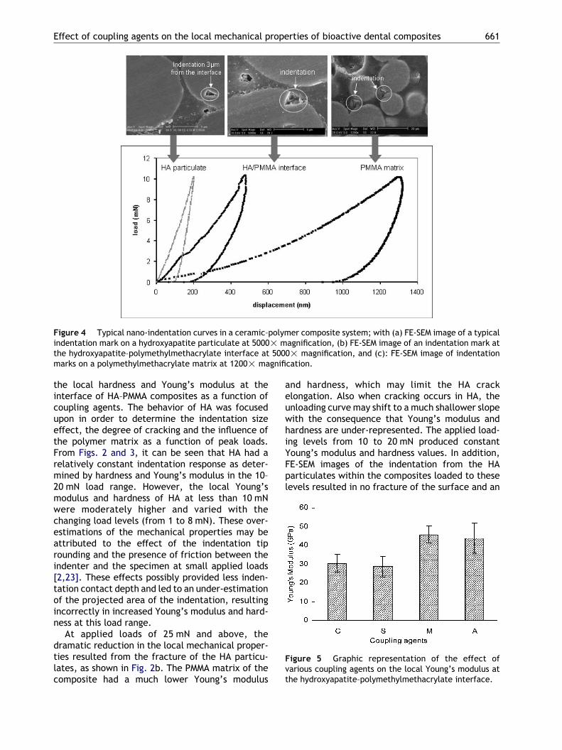
Figure 4 Typical nano-indentation curves in a ceramic–polymer composite system; with (a) FE-SEM image of a typicalindentation mark on a hydroxyapatite particulate at 5000! magnification, (b) FE-SEM image of an indentation mark atthe hydroxyapatite–polymethylmethacrylate interface at 5000! magnification, and (c): FE-SEM image of indentationmarks on a polymethylmethacrylate matrix at 1200! magnification.
Figure 5 Graphic representation of the effect ofvarious coupling agents on the local Young’s modulus atthe hydroxyapatite–polymethylmethacrylate interface.
Effect of coupling agents on the local mechanical properties of bioactive dental composites 661
the local hardness and Young’s modulus at theinterface of HA–PMMA composites as a function ofcoupling agents. The behavior of HA was focusedupon in order to determine the indentation sizeeffect, the degree of cracking and the influence ofthe polymer matrix as a function of peak loads.From Figs. 2 and 3, it can be seen that HA had arelatively constant indentation response as deter-mined by hardness and Young’s modulus in the 10–20 mN load range. However, the local Young’smodulus and hardness of HA at less than 10 mNwere moderately higher and varied with thechanging load levels (from 1 to 8 mN). These over-estimations of the mechanical properties may beattributed to the effect of the indentation tiprounding and the presence of friction between theindenter and the specimen at small applied loads[2,23]. These effects possibly provided less inden-tation contact depth and led to an under-estimationof the projected area of the indentation, resultingincorrectly in increased Young’s modulus and hard-ness at this load range.
At applied loads of 25 mN and above, thedramatic reduction in the local mechanical proper-ties resulted from the fracture of the HA particu-lates, as shown in Fig. 2b. The PMMA matrix of thecomposite had a much lower Young’s modulus
and hardness, which may limit the HA crackelongation. Also when cracking occurs in HA, theunloading curve may shift to a much shallower slopewith the consequence that Young’s modulus andhardness are under-represented. The applied load-ing levels from 10 to 20 mN produced constantYoung’s modulus and hardness values. In addition,FE-SEM images of the indentation from the HAparticulates within the composites loaded to theselevels resulted in no fracture of the surface and an
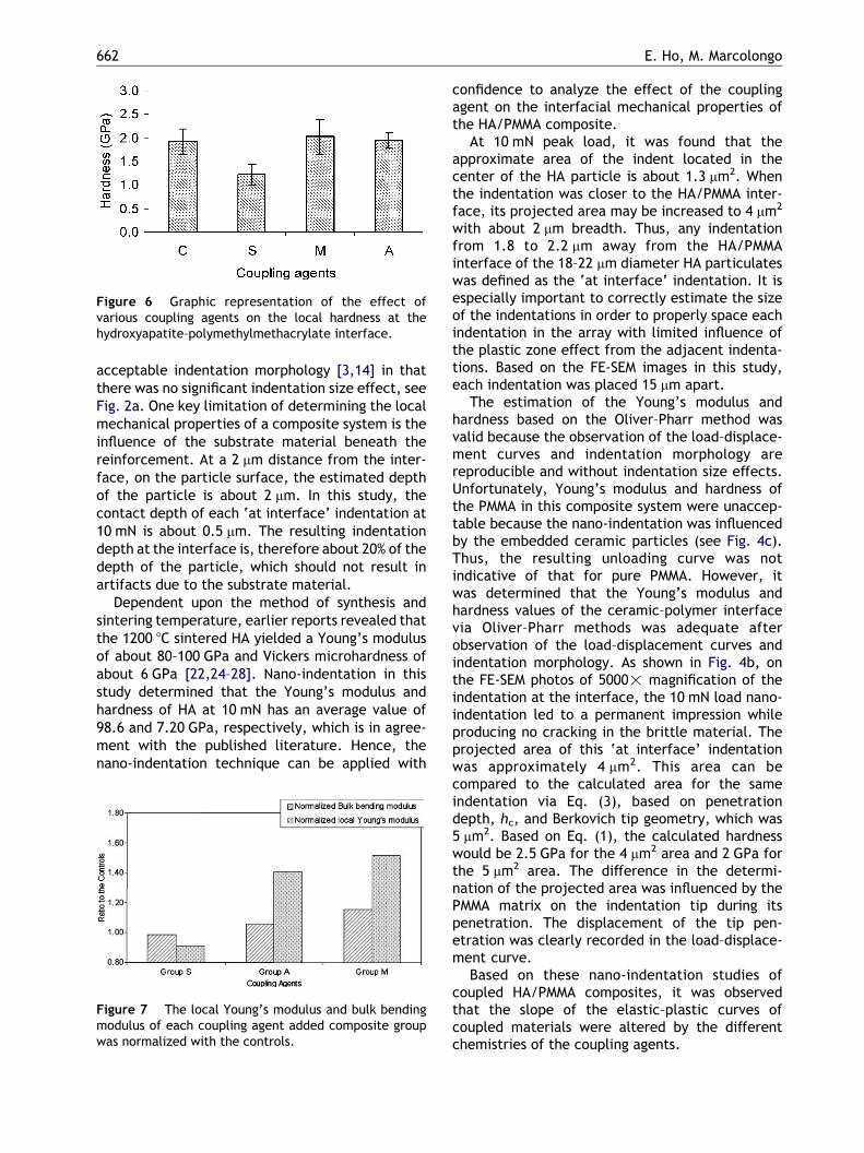
Figure 6 Graphic representation of the effect ofvarious coupling agents on the local hardness at thehydroxyapatite–polymethylmethacrylate interface.
E. Ho, M. Marcolongo662
acceptable indentation morphology [3,14] in thatthere was no significant indentation size effect, seeFig. 2a. One key limitation of determining the localmechanical properties of a composite system is theinfluence of the substrate material beneath thereinforcement. At a 2 mm distance from the inter-face, on the particle surface, the estimated depthof the particle is about 2 mm. In this study, thecontact depth of each ‘at interface’ indentation at10 mN is about 0.5 mm. The resulting indentationdepth at the interface is, therefore about 20% of thedepth of the particle, which should not result inartifacts due to the substrate material.
Dependent upon the method of synthesis andsintering temperature, earlier reports revealed thatthe 1200 8C sintered HA yielded a Young’s modulusof about 80–100 GPa and Vickers microhardness ofabout 6 GPa [22,24–28]. Nano-indentation in thisstudy determined that the Young’s modulus andhardness of HA at 10 mN has an average value of98.6 and 7.20 GPa, respectively, which is in agree-ment with the published literature. Hence, thenano-indentation technique can be applied with
Figure 7 The local Young’s modulus and bulk bendingmodulus of each coupling agent added composite groupwas normalized with the controls.
confidence to analyze the effect of the couplingagent on the interfacial mechanical properties ofthe HA/PMMA composite.
At 10 mN peak load, it was found that theapproximate area of the indent located in thecenter of the HA particle is about 1.3 mm2. Whenthe indentation was closer to the HA/PMMA inter-face, its projected area may be increased to 4 mm2
with about 2 mm breadth. Thus, any indentationfrom 1.8 to 2.2 mm away from the HA/PMMAinterface of the 18–22 mm diameter HA particulateswas defined as the ‘at interface’ indentation. It isespecially important to correctly estimate the sizeof the indentations in order to properly space eachindentation in the array with limited influence ofthe plastic zone effect from the adjacent indenta-tions. Based on the FE-SEM images in this study,each indentation was placed 15 mm apart.
The estimation of the Young’s modulus andhardness based on the Oliver–Pharr method wasvalid because the observation of the load–displace-ment curves and indentation morphology arereproducible and without indentation size effects.Unfortunately, Young’s modulus and hardness ofthe PMMA in this composite system were unaccep-table because the nano-indentation was influencedby the embedded ceramic particles (see Fig. 4c).Thus, the resulting unloading curve was notindicative of that for pure PMMA. However, itwas determined that the Young’s modulus andhardness values of the ceramic–polymer interfacevia Oliver–Pharr methods was adequate afterobservation of the load–displacement curves andindentation morphology. As shown in Fig. 4b, onthe FE-SEM photos of 5000! magnification of theindentation at the interface, the 10 mN load nano-indentation led to a permanent impression whileproducing no cracking in the brittle material. Theprojected area of this ‘at interface’ indentationwas approximately 4 mm2. This area can becompared to the calculated area for the sameindentation via Eq. (3), based on penetrationdepth, hc, and Berkovich tip geometry, which was5 mm2. Based on Eq. (1), the calculated hardnesswould be 2.5 GPa for the 4 mm2 area and 2 GPa forthe 5 mm2 area. The difference in the determi-nation of the projected area was influenced by thePMMA matrix on the indentation tip during itspenetration. The displacement of the tip pen-etration was clearly recorded in the load–displace-ment curve.
Based on these nano-indentation studies ofcoupled HA/PMMA composites, it was observedthat the slope of the elastic–plastic curves ofcoupled materials were altered by the differentchemistries of the coupling agents.
Effect of coupling agents on the local mechanical properties of bioactive dental composites 663
The nano-indentation testing showed the silanecoupled composite had the lowest local Young’smodulus and hardness among the treated groups.Silane coupling agents are commonly used asadhesive promoters for bioactive ceramicreinforced composites [18,29]. Results in previousstudies showed improved mechanical strength withno change in the Young’s modulus of silanized HAreinforced polymeric composites. For this HA/PMMA composite, the silane coupling agent didnot provide a mechanical benefit over the un-coupled control specimens. The –OH- molecules inthe HA particles were expected to bond well withthe –OH- molecules in the silane coupling agent.But the presence of water and oxygen in theatmosphere possibly led to the formation ofaddition water molecules with the –OH- groups inthe HA and left the –OH- groups in the silaneunattached. The current data did indicate thatthe co-polymer coupled composites (group A andgroup M) significantly increased the local Young’smodulus of the HA/PMMA composite at the inter-face. However, the local hardness of theco-polymer coupled composites did not show anystatistical difference from the uncoupled compo-site. The co-polymer coupled composites providedenhanced chemical interactions between the HAparticulates and the PMMA matrix. While the bulkbending modulus increased compared to that of thecontrol group, the enhancements were morepronounced on the local level, as indicated bynano-indentation.
Conclusion
The application of nano-indentation to the inter-face between the PMMA polymer and HA particulatereinforcement enabled the authors to obtain somedetails of the local mechanical behavior of thecomposite. It was shown that the PMMA-MAA andPMMA-MA coupling agents enhanced the interfacialstiffness while maintaining the interfacial hardnessof the composite systems. Silane coupling agentsshowed a reduced local Young’s modulus andhardness compared to the uncoupled controls.
Nano-indentation is a powerful technique forquantifying interfacial interactions in compositesand is capable of showing more pronounced localdifferences in the interfacial mechanics than thequasi-static bulk properties, such as four pointbending. As a result, this tool has been establishedas a useful means to determine the mechanicalbehavior of bioactive composites for mandible bonesubstitutes.
References
[1] Newey D, Wilkens M, Pollock H. An ultra low loadpenetration hardness tester. J Phys E: Sci Instrum 1982;15:119–22.
[2] Hay JL, Pharr GM. In: Medlin D, editor. Instrumentedindentation testing. In: Mechanical testing and evalu-ation, vol. 8. Materials Park, OH: ASM International;2000. p. 232–42.
[3] Fisher-Cripps AC. Nano-indentation 2002.[4] Pethica J. Microhardness tests with penetration depths less
than ion implanted layer thickness in ion implantation intometals. Third international conference on modification ofsurface properties of metals by ion-implantation. Man-chester/England: Pegammon Press/Oxford UniversityPress; 1981.
[5] Pethica J, Hutchings R, Oliver W. Hardness measurementsat penetration depths as small as 20nm. Philos Mag A 1983;48(4):593–606.
[6] Hinoki T, Zhang W, Kohyama A, Sato S, Nado T. Effect offiber coating on interfacial shear strength of SiC/SiC bynano-indentation technique. J Nucl Mater 1998;258–263(2):1567–71.
[7] Roop Kumar R, Wang M. Functionally graded bioactivecoatings of hydroxyapatite/titanium oxide composite sys-tem. Mater Lett 2002;55(3):133–7.
[8] Soloukhin VA, Posthumus W, Brokken-Zijp JCM, Loos J, deWith G. Mechanical properties of silica-(meth)acrylatehybrid coatings on polycarbonate substrate. Polymer2002;43(23):6169–81.
[9] Juliano T, Gogotsi Y, Domnich V. Effect of indentationunloading conditions on phase transformation inducedevents in silicon. J Mater Res 2003;18(5):1192–201.
[10] Domnich V, Gogots Y. Pressure-induxed phase transform-ations in semiconductors under contact loading. Fron-tiers of high pressure research. II. Application ofhigh pressure to low dimensional novel electronicsmaterials 2001.
[11] Gong J, Miao H, Peng Z, Qi L. Effect of peak load on thedetermination of hardness and Young’s modulus of hot-pressed Si3N4 by nanoindentation. Mater Sci Eng A 2003;354(1/2):140–5.
[12] Galusek D, Riley FL, Riedel R. Nanoindentation of apolymer-derived amorphous silicon carbonitride ceramic.J Am Ceram Soc 2001;5(84):1164–6.
[13] Whitehead AJ, Page TF. Nanoindentation studies of thinfilm coated systems. Thin Solid Films 1992;220(1/2):277–83.
[14] Xu HHK, Smith DT, Schumacher GE, Eichmiller FC,Antonucci JM. Indentation modulus and hardness ofwhisker-reinforced heat-cured dental resin composites.Dent Mater 2000;16(4):248–54.
[15] Xu HHK, Quinn JB, Smith DT, Quinn JB, Smith AA,Giuseppetti AA, Eichmiller FC. Dental resin compositescontaining silica-fused whiskers—effects of whisker-to-silica ratio on fracture toughness and indentation proper-ties. Biomaterials 2002;23(3):735–42.
[16] Xu HHK, Antonucci JM, Schumacher GE. Effects of differentwhiskers on the reinforcement of dental resin composites.Dent Mater 2003;19(5):359–67.
[17] Wang M, Deb S, Bonfield W. Chemically coupled hydro-xyapatite–polyethylene composites: processing and charac-terisation. Mater Lett 2000;44(2):119–24.
[18] Wang M, Bonfield W. Chemically coupled hydroxyapatite–polyethylene composites: structure and properties. Bioma-terials 2001;22(11):1311–20.
E. Ho, M. Marcolongo664
[19] Liu Q, de Wijn JR, de Groot K, van Blitterswijk CA. Surfacemodification of nano-apatite by grafting organic polymer.Biomaterials 1998;19(11/12):1067–72.
[20] Ho E, Marconlogo M. The effect of coupling agent onhydroxyapatite/polymethylmethacrylate composite.Drexel University Research Day 2003.
[21] Oliver WC, Pharr GM. An improved technique and determin-ing hardness and elastic modulus using load–displacement.J Mater Res 1992;7:1555–64.
[22] Akao M, Aoki H, Kato K. J Mater Sci 1981;16:809.[23] WenJ, LengY, ChenJ, ZhangC. Chemical gradient in plasma-
sprayed HA coatings. Biomaterials 2000;21(13):1339–43.[24] Roop Kumar R, Wang M. Modulus and hardness evaluations
of sintered bioceramic powders and functionally gradedbioactive composites by nano-indentation technique. MaterSci Eng A 2002;338(1/2):230–6.
[25] Habelitz S, Marshall SJ, Marshall Jr GW, Balooch M.Mechanical properties of human dental enamel on thenanometre scale. Arch Oral Biol 2001;46(2):173–83.
[26] Muralithran G, Ramesh S. The effects of sintering tempera-ture on the properties of hydroxyapatite. Ceram Int 2000;26(2):221–30.
[27] Hoepfner TP, Case ED. The influence of the microstructureon the hardness of sintered hydroxyapatite. Ceram Int 2003;29(6):699–706.
[28] Tancred DC, McCormack BAO, Carr AJ. A quantitative studyof the sintering and mechanical properties of hydroxyapa-tite/phosphate glass composites. Biomaterials 1998;19(19):1735–43.
[29] Labella R, Braden M, Deb S. Novel hydroxyapatite-based dental composites. Biomaterials 1994;15(15):1197–200.