effect of benzanthrone on the urinary bladder of guinea-pig
TRANSCRIPT

15.6.1973 Speeialia 683
Effect of B e n z a n t h r o n e on the U r i n a r y B l a d d e r of G u i n e a - P i g
I n dyes tu f f i ndus t r i e s i t has been obse rved t h a t work- m e n who come in c o n t a c t w i t h t he dye b e n z a n t h r o n e (an a n t h r a q u i n o n e der iva t ive ) d u r i n g i ts m a n u f a c t u r e , pu lver iza t ion , a n d s torage deve lop i tching, b u r n i n g sensat ion , e r y t h e m a a n d skin p i g m e n t a t i o n 1-a. A n t h r a - qu inone de r iva t i ve s are k n o w n to h a v e some tox ic effect on t h e u r i n a r y b l a d d e r mucosa~. I n t h e p r e s e n t p re l imi - n a r y inves t iga t ion , therefore , an a t t e m p t has been m a d e to s t u d y t he effect of b e n z a n t h r o n e on t h e u r i n a r y b l a d d e r of guinea-pig.
Mater ials and methods, I .T.R.C. Colony b red guinea- pigs (body we igh t 350-400 g) were used in th i s inves t iga - t ion. The an ima l s were d iv ided in to 3 groups of 40 each. 20 an ima l s of each group were a d m i n i s t e r e d b e n z a n t h r o n e da i ly b y local appl ica t ion , oral a n d i. p. rou tes respect ively .
Group I ( topical appl ica t ion) . The sk in of 20 an ima l s was p a i n t e d w i t h a 2 % b e n z a n t h r o n e suspens ion in i sotonic sal ine b y t he m e t h o d of SINGH et al 1. The 20 con t ro l an ima l s were p a i n t e d w i t h isotonic sal ine only.
Group I I (oral ad mi n i s t r a t i o n ) . 20 an ima l s were fed b y s t o m a c h t u b e w i t h t h e dye (25 m g / k g of body-weigh t ) suspended in 2 ml isotonic saline. 20 cont ro l s received on ly 2 ml of i sotonic saline.
Group I I I (i.p. in jec t ion) . 20 an ima l s were in jec ted w i t h t h e dye (25 m g / k g b o d y weight) suspended in 2 ml ster i le i sotonic saline. 20 con t ro l an i ma l s were i n j ec t ed on ly 2 ml of steri le i sotonic saline.
All an i ma l s were fed rou t ine l a b o r a t o r y d ie t (Hind Leve r Ltd. , Ind ia ) an d k e p t u n d e r u n i fo rm h u s b a n d r y cond i t ions t h r o u g h o u t t h e e x p e r i m e n t a l period. I n each group, 10 f rom b e n z a n t h r o n e a d m i n i s t e r e d an i ma l s an d 10 controls , were sacr i f iced a t t h e i n t e r v a l of 7 an d 15 days.
R o u t i n e a u t o p s y was pe r fo rmed on all an imals . A b d o m - inal v iscera were inspec ted for a n y gros s les ions . U r i n a r y b l a d d e r f rom each a n i m a l was r e m o v e d an d f ixed in f resh ly p r e p a r e d 1 0 % n e u t r a l formal in . Serial pa r a f f i n sect ions of 5 ~ m th i ckness were s t a ined w i t h h a e m a t o x y - l in an d eosin.
Gross e x a m i n a t i o n of v iscera l o rgans showed n o r m a l a p p e a r a n c e in an ima l s of all t h e groups a t 7 an d 15 days. A few smal l l igh t b r o w n par t ic les , wh ich a p p e a r e d to be b e n z a n t h r o n e , were seen e n t a n g l e d in t h e o m e n t u m a n d d i a p h r a g m a t i c surface of b e n z a n t h r o n e in jec ted an i ma l s a t 7 an d 1:5 days.
Microscopic e x a m i n a t i o n of u r i n a r y b l ad d e r e p i t h e l i u m of all an i ma l s in groups I, I I an d con t ro l of g roup I I I a t 7 an d 15 days showed n o r m a l a p p e a r a n c e (Figure 1). All b e n z a n t h r o n e in j ec t ed guinea-pigs of g roup I I I showed mi ld vascu la r conges t ion in t h e l a m i n a p ro p r i a a n d sub- m u c o s a a t 7 days (Figure 2). E p i t h e l i u m a n d m u s c u l a r layers showed n o r m a l appea rance . A t 15 days 6 an i ma l s showed m a r k e d vascu la r conges t ion in l a m i n a p rop r i a a n d submucosa . 4 an ima l s showed ev idence of mucosa l lesion
Fig. 1. Normal appearance of the urinary bladder wall. HE, • 28.
t G. B. SINGIt, S. N. SHARNA and S. H. ZAIDI. Indian J. reed. Sci. 21, 727 (1967).
2 D. H. TRIVEDI and A. K. NIYOGI, Indian J. ind. Med. 7d, 13 (1968). G. ]3. SINGH and S. H. ZAIDI, J. Indian reed. Ass. 52, 558 (1969).
4 G. B. SINGH, Indian J. ind. Med. 76, 122 (1970). 5 D. HIJ~TE~, The disease o/ occupations, 5th edn. (The English
Universities Press Ltd., London 1969), p. 827.
Fig. 2. Vascular congestion more marked in the submucosal layer. Fig. 3. Localised damage of the epithelial layer. HE, • 40. HE, • 28.
684 Specialia ]~XPI~RIENTIA 29/6
(Figure 3). Large n u m b e r of acute i n f l a m m a t o r y cells were p resen t in the lamina propr ia and submucosa . Mus- cular layers p resen ted normal appearance . Vascular congest ion was severe. No prol i fera t ive ac t iv i ty of the epi thel ial mucosa was observed.
6 K. P. PANDYA, G. B. SINGH and N. C. JOSHI, Acta pharmac, tox. 28, 499 (1970).
7 K.P. PANDYA, under communication (1972). s Authors are grateful to Dr. S. H. ZAiDI, Director of the Centre for
his keen interest in the work. Technical assistance of Messrs. MULKRAJ and V. G. MISRA is highly appreciated. Mr. M. AHMAD is responsible for the preparation of photomicrographs.
9 Present address: Department of Surgery, Institute of Medical Sciences, Banaras Hindu University, Varanasi (India).
B e n z a n t h r o n e caused s ignif icant decrease of ascorbic acid level in blood and o ther body organs ~. PANDYA 7 no ted the presence of benzan th rone or its possible meta - boi i tes in guinea-pig urine af ter i .p. ad mi n i s t r a t i o n of benzan throne . Lowered body ascorbic ac id level combined wi th t he effect of b e n z a n t h r o n e or its possible metabo l i t es excre ted in the urine m a y be responsible for epithelial damage. Mucosal lesion was no t discernible on dermal appl ica t ion or oral admin i s t r a t i on of benzan th rone 8.
G. t3. SINGH and V . N . P . TRIPATHI 9
Indus t r ia l Toxicology Research Centre, Post B o x No 80, Lucknow (India), 79 A p r i l 1972.
E f f e c t o f L i n d a n e 1 o n t h e S k i n o f A l b i n o R a t s
Lindane is being used ex tens ive ly as dusts , emulsions and vapours to contro l insects under d i f ferent condi- t ions 2, 3 Occupat ional poisoning among workers engaged in the synthesis , fo rmula t ion and appl ica t ion of l indane is also repor ted by d i f ferent workers 4-~. L i t e ra tu re on the h is topathologica l changes in the skin of individuals repea t - edly exposed to l indane is no t adequate . Phys icochemica l factors such as par t ic le size, the per iod of exposure and the vehicles are known to influence the degree of toxic i ty . In t he l ight of such var ia t ions , de rmal appl ica t ion of l indane under t ropical condit ions, as in th is country , mer i t s fu r ther s tudy. This repor t , which is tile cont inua- t ion of our earlier observations~, 8, deals witt l the histo- pathological changes in the skin of albino ra ts af ter t h e y are exposed d i rec t ly to the act ion of l indane.
Materials and Methods. 50 female albino ra ts of I .T.R.C. s tock wi th an average body weight of 80 g were used in the exper iment . The l a t e roabdomina l area measur ing
app rox ima te ly 4 X 4 cm was previous ly made ready by ha i r c l ipping for I indane paint ing. L indane (98 % puri ty) was used wi th p ropy lene glycol (BDH Analar) as t he vehicle. 1 ml of t he solut ion which conta ined 14.4 mg of l indane (this dose is 5 t imes less t h a n the acute dermal LD 50 values for t he female ra ts ; dermal LD 50 of l indane for female ra ts is 900 mg/kg)9 was slowly t ransfer red on
the specified area of the skin wi th the help of a g radua t - ed p ipe t t e a t t ached to a vaqupe t t e . 30 animals were t r e a t ed wi th l indane daily for a per iod of 25 days (total n u m b e r of skin pa in t ings 25). 20 animals of tile control group were s imilar ly t r ea t ed wi th 1 ml of p ropylene glycol alone. The animals were killed a t in tervals of 24 h, 5, 10, 15, 20 and 25 days af ter t r e a tmen t . The skin t issue was f ixed in Bouin ' s fluid and paraf f in cut sect ions were s ta ined wi th h a e m a t o x y l i n and eosin for h i s topa tholo- gical observat ions .
Results and Discussions. Macroscopic examina t ions of the skin of expe r imen ta l animals showed mild de rma t i t i s in 3 animals af ter 15 paint ings . This condi t ion cont inued up to 25 paint ings . In compar ison, t he skin of control animals did no t show any such change.
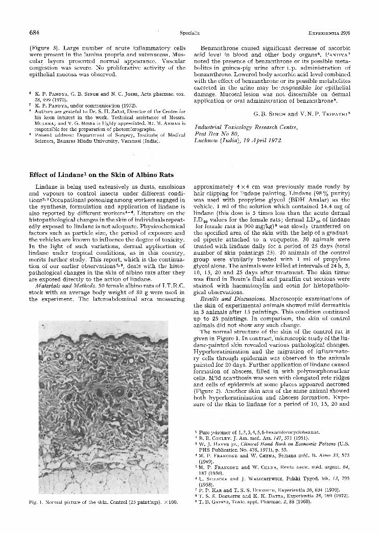
Tile normal s t ruc tu re of t he skill of ti le control ra t is given in Figure 1. In contras t , microscopic s t u d y of the 1in- dane -pa in ted skin revealed various pathological changes. Hyperke ra t in i za t ion and the migra t ion of in f l ammato- ry cells t h ro u g h epidermis was observed in the animals pa in t ed for 20 days. F u r t h e r appl ica t ion of l indane caused fo rma t ion of abscess, filled in wi th po lymorphonuc lea r cells. Mild acanthos is was seen wi th e longated re te r idges and cells of ep idermis a t some places appeared necrosed (Figure 2). Ano the r skin area of the same animal showed b o t h hype rke ra t in i za t ion and abscess format ion. Expo- sure of the skin to l indane for a per iod of 10, 15, 20 and
Fig. 1. Normal picture of the skin. Control (25 paintings). • 100.
1 Pure y-isomer of 1,2, 3, 4, 5, 6-hexachlorocyclohexane. B. E. CONLEY. J. Am. med. Ass. 147, 571 (1951). W. J. HAYES JR., Clinical Hand Book on Economic Poisons (U.S. PHS Publication No. 476, 1971), p. 53.
4 M. P. FRANCONE and W. CHELA, Semana m6d. B. Aires 55, 573 (1949).
'~ M. P. FRANCONE and W. CHENA, Revta assoc, m6d. argent. 64, 187 (1950).
G C. SIELICKA and J. WALICHIEWICZ, Polski Tygod. lek. 73, 795 (1958).
7 p. p. IKAR and T. S. S. DIKSHITH, Experientia 26, 634 (1970). 8 T. S. S. DIKStUTH and K. K. DATTA, Experientia 28, 169 (1972). 9 T. t3. G4INES , Toxic. appl. Pharmac. 2, 88 (1960).