edited by ian norton and michael bourke endoscopy -...
TRANSCRIPT

HANDBOOKEndoscopy
Second EditionUpdated: June 2017
Edited by Ian Norton and Michael Bourke

Gastroenterological Society of Australia (GESA) GESA is the professional membership society for clinicians, trainees, allied health workers and researchers in gastroenterology and hepatology in Australia. For more information, see www.gesa.org.au.
Name:
GESA Membership Number:
Login Name:
Conjoint Committee for Recognition of Training in Gastrointestinal Endoscopy (CCRTGE) The CCRTGE is the joint body of GESA, the Royal Australasian College of Physicians (RACP) and the Royal Australasian College of Surgeons (RACS). The role of the CCRTGE is to set the minimum standards for training and to recognise training in gastrointestinal endoscopy. For more information, see www.conjoint.org.au.
CCRTGE username:
CCRTGE password:
GESA Colonoscopy Recertification Program This is a voluntary periodic recertification and education program for experienced colonoscopists. The purpose is to assist experienced colonoscopists to maintain their expertise, further develop their skills and demonstrate this to peers and patients through triennial recertification. For more information, see recert.gesa.org.au.
Recertification username:
Recertification password:
Any other details
For more information, please contact [email protected]
Your details: please complete the fields below
i

This revised second edition of the Endoscopy Handbook was produced by the Gastroenterological Society of Australia (GESA). GESA would like to acknowledge the editors, Assoc Prof Ian Norton and Prof Michael Bourke, and all the authors who contributed their time and expertise to this edition:
Production of this handbook was enabled by the GESA National Colonoscopy Recertification Program with funding from the National Bowel Cancer Screening Program, Australian Department of Health.
Published by: Gastroenterological Society of Australia PO Box 5359 Pinewood Victoria 3149 Australia
www.gesa.org.au
ABN 44 001 171 115
© Gastroenterological Society of Australia 2016. This handbook is copyright and all rights reserved. It may not be reproduced in whole or in part without permission from GESA.
Second edition amended in June 2017. The most up-to-date version of this handbook will always be available as an electronic document at the GESA website (www.gesa.org.au).
iii
Mark Appleyard
John P Bate
Michael Bourke
Gregor Brown
Robyn Brown
Robert Y Chen
Sarah Cho
Lennart Choo
Crispin Corte
Philip Craig
Paul Edwards
Geoff Francis
Karl Herba
Luke Hourigan
Jason Y Huang
D Brian Jones
Di Jones
Brad Kendall
David Koorey
Vu Kwan
Nam Nguyen
Ian Norton
Darren Pavey
Jillian Rosenstengel
Mark N Schoeman
Tony Speer
Alina Stoita
Michael K L Suen
William Tam
Patrick R Walsh
David Williams
Christopher J Young
Simon Zanati
Acknowledgements

Section 1 — BACKGROUND AND PREPARATION FOR ENDOSCOPY1 How endoscopes work – Ian Norton ......................................................................................................... 5
2 Endoscope reprocessing – Robyn Brown and Di Jones ............................................................................. 11
3 Reporting, documentation and risk management – Ian Norton .............................................................. 23
4 Management of antiplatelet and anticoagulant agents for endoscopic procedures – John P Bate and Mark N Schoeman ................................................................... 31
5 Sedation and patient monitoring for gastrointestinal endoscopy – D Brian Jones .................................. 41
6 Antibiotic use in endoscopy – Sarah Cho ............................................................................................... 51
7 Endoscopic imaging – William Tam ......................................................................................................... 59
Section 2 — UPPER ENDOSCOPY8 Barrett’s oesophagus: diagnosis and management – Darren Pavey ...................................................... 75
9 Endoscopic treatment of Barrett’s high-grade dysplasia and early oesophageal adenocarcinoma – Luke Hourigan ........................................................................................................... 93
10 Management of non-variceal upper gastrointestinal bleeding – Paul Edwards and Brad Kendall ................................................................................................................ 103
11 Endoscopic management of varices and variceal haemorrhage – David Koorey and Crispin Corte ........ 113
Section 3 — COLONOSCOPY12 Colonoscopy: insertion and withdrawal technique – Michael Bourke .................................................... 123
13 Polypectomy – Michael Bourke .............................................................................................................. 139
14 Colorectal cancer screening and surveillance – Lennart Choo and Ian Norton ........................................ 155
15 Colonoscopic stenting – Michael K L Suen and Christopher J Young .......................................................... 163
16 Acute colonic bleeding – Geoff Francis and Simon Zanati ........................................................................ 173
Section 4 — ENDOSCOPIC RETROGRADE CHOLANGIOPANCREATOGRAPHY17 Biliary obstruction due to malignancy – Tony Speer ............................................................................. 185
18 Benign bilary strictures and biliary leaks – Karl Herba and Philip Craig ................................................... 195
19 Chronic pancreatitis – Nam Nguyen ...................................................................................................... 207
20 Endoscopic management of peripancreatic collections – Vu Kwan ..................................................... 217
Section 5 — ENDOSCOPIC ULTRASOUND21 Introduction to endoscopic ultrasound – Ian Norton ............................................................................. 229
22 Endoscopic ultrasound for oesophageal and mediastinal indications – Alina Stoita and David Williams.. 237
23 Endoscopic ultrasound for submucosal lesions in the upper gastrointestinal tract – Robert Y Chen .... 247
24 Endoscopic ultrasound for biliary disease and choledocholithiasis – Jason Y Huang, Jillian Rosenstengel and Patrick R Walsh ........................................... 257
Section 6 — IMAGING OF THE SMALL INTESTINE25 Approach to obscure bleeding of the gastrointestinal tract – Gregor Brown ........................................... 267
26 Endoscopic imaging of the small intestine – Mark Appleyard ................................................................. 275
List of acronyms ....................................................................................................................................... 282
Contents

Section 1
BACKGROUND AND PREPARATION FOR ENDOSCOPY

5
How endoscopes workIan Norton
It is important for endoscopists to have a general idea of how an endoscope works, primarily so they can attempt to troubleshoot equipment malfunction. From a practical point of view, when using accessory instruments with an endoscope, it is important to know that an accessory is compatible with a particular endoscope (i.e. length and channel diameter), as well as having an idea of where the accessory will appear on the visual field.
This chapter outlines the fundamental workings of an endoscope, with some reference to specialist scopes such as endoscopic ultrasound (EUS) instruments. Reference to specific proprietary aspects of any company’s instrument has been avoided as much as possible.
A bit of history
Philipp Bozini, a German physician, is credited with the earliest known attempt to visualise the interior of a body cavity, in 1805. He devised a tin tube illuminated by a candle, which was used, with limited success, to investigate the genitourinary tract. Adolf Kussmaul is credited as being the first to perform gastroscopy in 1868, using a rigid tube and a cooperative sword-swallower! Illumination and negotiating curves were insoluble problems, however, and he abandoned further development. In 1886, Viennese instrument maker Josef Leiter was the first to use the electric light bulb in a cystoscope. Subsequent rigid instruments with distal bulbs were used by ear, nose and throat surgeons until the 1960s to examine the oesophagus.
In 1932, Wolf and Schindler launched a semiflexible instrument, with a rigid proximal portion and glass prisms contained in a semiflexible portion to provide illumination. In 1950, Olympus introduced the gastrocamera, which took photographs of the stomach using microfilm and a synchronised flash.
Basil Hirschowitz introduced a flexible instrument that used fibre optics in 1958, and the “panendoscope” was introduced by ACMI in 1971. Techniques rapidly advanced. Endoscopic
SECTION 1: BACKGROUND AND PREPARATION FOR ENDOSCOPY
CHAPTER 1
back to contents
SECTION 1: BACKGROUND AND PREPARATION FOR ENDOSCOPY
6 back to contents
retrograde cholangiopancreatography (ERCP) was demonstrated with a side-viewing instrument in 1970, and endoscopic sphincterotomy was reported by Kawai and colleagues in Japan and Classen and Demling in Germany. Colonoscopy was performed in 1970 and polypectomy in 1973. Video endoscopes were introduced in 1984, and subsequent improvements have dra-matically increased the quality of imaging, as well as the comfort and ease of performing the procedure.
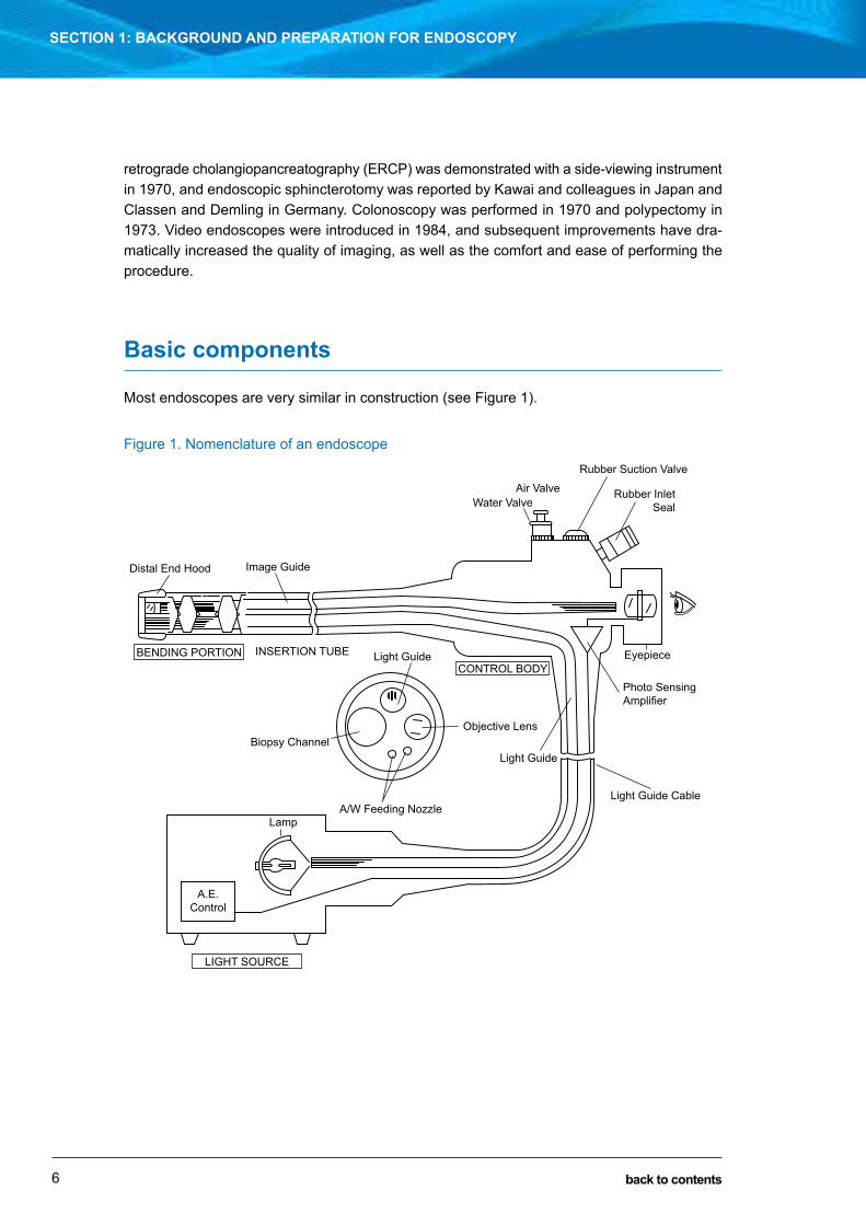
Basic components
Most endoscopes are very similar in construction (see Figure 1).
Figure 1. Nomenclature of an endoscope
Distal End Hood Image Guide
INSERTION TUBE Light GuideCONTROL BODY
Objective LensBiopsy Channel
Lamp
LIGHT SOURCE
A.E.Control
A/W Feeding Nozzle
Light Guide
Eyepiece
Rubber Suction Valve
Air ValveWater Valve
Rubber InletSeal
Photo SensingAmplifier
Light Guide Cable
BENDING PORTION

SECTION 1: BACKGROUND AND PREPARATION FOR ENDOSCOPY
7
HOW ENDOSCOPES WORKIan Norton
back to contents
COLONOSCOPE AND SIGMOIDOSCOPE
GASTROSCOPE
Water Feeding Channel
Air/Water Nozzle
Air Nozzle
Water Nozzle
Water Bottle
Video-processor
Suction Source
Suction Tube
Air Feeding Tube
Water Feeding Tube
Forward Water*Jet Connector*
Channel Inlet A/W Feeding Valve
* ALL COLONOSCOPESAND SIGMOIDOSCOPES
Suction Control Valve
Instrument Channel
Forward Water Jet Channel *
AirPump
Air Feeding Channel
These illustrations show the actual routes taken by air, water, suction and the forward water jet through a Pentax video gastroscope, colonoscope and sigmoidoscope. Note that all delivery systems have separate independent channels, all of which must first be cleaned manually, then with an enzymatic detergent, and then exposed to a high-level disinfectant or sterilant. Images courtesy of Pentax.
Insertion tube The characteristics of the insertion tube (e.g. stiffness) are the main determinant of a proce-duralist’s preference for one scope over another. The distal part of the insertion tube is made of articulated metal rings. The shaft is made of a series of metal bands spiralling in different directions, which give the scope its stiffness and torque characteristics. Variable-stiffness instruments have a series of wires running most of the length, which, when tightened, increase the rigidity of the instrument. As these wires do not extend to the tip of the instrument, the final 30 cm or so does not stiffen. Most instruments have four-way tip deflection, with left, right and down deflection to about 90° and up deflection to about 210° (depending on the instrument).
Air, water and suction channels Standard instruments have air, water and suction channels. In addition, some colonoscopes have an added water jet channel, EUS instruments have an extra channel for insufflating a balloon with water, and some therapeutic scopes have two channels for suction and appliances. When planning a procedure, you must know the location of the accessory channel relative to the image (e.g. 5 o’clock versus 7 o’clock). It is also essential to know the diameter of the

SECTION 1: BACKGROUND AND PREPARATION FOR ENDOSCOPY
8 back to contents
accessory channel relative to the therapeutic devices planned to be used (e.g. a colonic stent will not pass through a gastroscope or paediatric colonoscope).
Water to clean the lens is provided by a water bottle. This system is pressurised by a small pump. Air is always circulating across the top of the water bottle and up the umbilical cord, and effluxing from the air/water valve (which is why you can always feel air at this valve). Covering this efflux vent with your finger instead forces the air down the air/water channel and into the lumen. Depressing your finger on this valve cuts off this air flow and instead opens a channel from the water in the bottle to the air/water channel. Due to the increased pressure in the water bottle, water is forced through this system, washing the lens.
The suction channel is connected by a valve (shut in the neutral position) to another channel in the umbilical cord to the wall suction connector. Depressing this valve opens this channel to the suction system. For most of its path, this channel uses the same channel as the biopsy/accessory channel. A rubber cap prevents air escaping from the biopsy channel.
Light sourceLight is supplied by a high-intensity light source in the endoscope tower. The light is conveyed by bundles of glass fibres via the umbilical cord and instrument shaft to the instrument tip. Thus, this system removes the light bulb from the instrument tip, preventing heat build-up at the tip. The light source has an automated iris, which adjusts light output to the lumen being examined. In some systems, the light output can be manipulated to select specific wavelengths (e.g. narrow band imaging [Olympus]). In other systems, post-capture processing of the image is performed to display specific wavelengths (e.g. flexible spectral imaging colour enhancement [FICE; Fujinon], and i-scan imaging [Pentax]).
Video imaging
Video imaging is a complex field that is beyond the scope of this chapter, and only a short sum-mary is presented here. At the tip of the instrument is a charge-coupled device (CCD) silicon chip. A photon of light hitting a particular point on the surface generates an electrical charge that can then be reconstructed into a point of light on an image. Two CCD systems are in common usage: RGB sequential scopes and colour-chip scopes.
RGB sequential imagingAll colours seen by the human eye can be generated by a combination of red (R), green (G) and blue (B). These instruments have a black-and-white chip at the tip. The light used to illumi-nate the image is not continuous, but pulsed or strobed. Before entering the patient, the light is passed through a rotating wheel with red, green and blue filters. Because it is rotating too fast for the eye to see (20–30 rev/s), these red, blue and green images coalesce to replicate the original image. A disadvantage of this system is that during movement there can appear to be a strobing effect, which can be annoying for the viewer. An advantage of this system is that all CCD images are used for each capture, leading to a high-resolution image.

SECTION 1: BACKGROUND AND PREPARATION FOR ENDOSCOPY
9
HOW ENDOSCOPES WORKIan Norton
back to contents
Colour-chip imagingThis uses a multicoloured microfilter at the chip surface to instantly generate a colour rep-resentation of the image. Thus, there is no mechanical colour wheel and no strobing effect. Furthermore, as image capture is faster, there is less blurring during movement. A disadvantage is that because each pixel in the chip is colour-specific (yellow, magenta, cyan and green), the resolution of the image is less than the resolution possible with RGB sequential imaging. Complementary colours, rather than primary colours, are used to increase image brightness.
Endoscopic ultrasound
EUS is made possible by the presence of an ultrasound transducer at the tip of the instrument. An extra channel and more complicated valve system is required, to insufflate and deflate a balloon with water. This balloon helps to facilitate acoustic coupling with the mucosal surface.
There are two basic designs of EUS instrument: linear and radial. The linear instrument has a curvilinear array distal to, and in alignment with, the instrument channel. This ensures that a needle projecting from the channel will pass through the length of the ultrasound beam, permitting real-time imaging during biopsy. The radial instrument scans at 90° to the long axis of the instrument shaft (the Pentax unit has the transducer proximal to an end-viewing scope, whereas the Olympus unit has the transducer distal to an oblique-viewing instrument).
Previously, the radial instrument was the scope of choice for diagnostic work because it was easier to orientate and assess the image. Sonographers have recently become more comfort-able using the linear configuration for diagnostic work. Virtually all dedicated EUS instruments now use solid-state technology rather than a mechanical rotating transducer.
Further reading1 Modlin IR. A brief history of endoscopy. Milan: Multimed, 2000.
2 Barlow DE. How endoscopes work. In: Ginsberg GG, Kochman ML, Norton ID, Gostout CJ, editors. Clinical gastrointestinal endoscopy, 2nd ed. Philadelphia: Elsevier Press, 2011: 25-45.
Page 10 (used for notes in the hard copy) has been removed from the PDF edition of this handbook.

11
Endoscope reprocessingRobyn Brown and Di Jones
Since the first clinical report of flexible fibre-optic endoscopy in 1961, endoscopy procedures have become a commonly performed investigation. In the early years, endoscopy was often performed by physicians working in ward side rooms or other available spaces. Scant regard was given to the processes that were applied to ready the instrument before being used on the next patient. Indeed, the overarching concern was the delicacy of the endoscope and the potential for damage should anything other than a careful swabbing of the exterior of the instrument be undertaken.
As technological developments produced more robust equipment and allowed more invasive procedures to be performed, the need for attention to infection control principles became para-mount. Modern endoscopy demands high quality in all aspects of the procedure, including safety from transmission of infection during the procedure. Compliance with endoscope reprocessing guidelines is the key factor underpinning that safety.
Requirement for disinfection and sterilisation
In 1968, Earle Spaulding devised a rational approach to disinfection and sterilisation of reusa-ble medical devices. Spaulding proposed that instruments and equipment should be cleaned and reprocessed according to the level of risk associated with their intended use. The three categories he described were critical, semicritical and non-critical, based on whether a device contacted intact skin or mucous membranes or was introduced into a sterile cavity of the body (Table 1). In this schema, endoscopes are classed as semicritical.
SECTION 1: BACKGROUND AND PREPARATION FOR ENDOSCOPY
CHAPTER 2
back to contents

12
SECTION 1: BACKGROUND AND PREPARATION FOR ENDOSCOPY EnDoSCopE REpRoCESSIngRobyn Brown and Di Jones
back to contents
Table 1. Spaulding classification*
Level of risk Application ProcessCritical Entry or penetration into sterile tissue,
cavity or bloodstreamSterility required
Semicritical Contact with intact non-sterile mucosa or non-intact skin
Sterilisation preferred where possible; if sterilisation not possible, high-level chemical disinfection required
non-critical Contact with intact skin Clean as necessary with detergent and water
* http://www.health.qld.gov.au/EndoscopeReprocessing/module_2/2_1.asp
Equipment reprocessing guidelines have subsequently been framed within this categorisation and take into account the scientific knowledge of a microorganism’s resistance to disinfection. There is a hierarchy of susceptibility to the biocidal effects of disinfectants (Figure 1), and a disinfectant’s strength must match the decontamination requirements of a medical device. The biocide also needs to be in contact with all external and internal surfaces of the equipment. Thus, given 100% of surfaces in contact, the critical process parameters of biocides used in endoscope reprocessing are time, temperature and concentration of the chemical.
Figure 1. Hierarchy of microbial susceptibility to biocides
Resistance to biocides
Bacterial spores(e.g. Clostridium difficile) SterilisationMOST RESISTANT
LEAST RESISTANT
High-level disinfection
Intermediate-level disinfection
Low-level disinfection
Mycobacterial(e.g. Mycobacterium tuberculosis)
Non-lipid or small viruses(e.g. Poliovirus)
Fungi(e.g. Candida)
Lipid or medium-sized viruses (e.g. HIV)
Vegetative bacteria (e.g. Staphylococcus, Pseudomonas)
http://www.health.qld.gov.au/EndoscopeReprocessing/module_2/2_2.asp
The level of bioburden on endoscopes is also a crucial determinant of the effectiveness of disin-fection or sterilisation processes. Alfa and colleagues identified the composition of the residual soil on endoscopes both before and after cleaning. These values are used to determine the efficacy of cleaning and reprocessing practices.
Infections associated with endoscopic procedures arise from endogenous and exogenous sources. The use of antibiotics to prevent transmigration of organisms during procedures (e.g. oral flora to skin during percutaneous endoscopic gastrostomy tube insertion) is directed at

13
SECTION 1: BACKGROUND AND PREPARATION FOR ENDOSCOPY EnDoSCopE REpRoCESSIngRobyn Brown and Di Jones
back to contents
preventing infection from endogenous sources, while compliance with accepted reprocessing guidelines is thought to provide virtually no risk of transmission of patient-borne or environmental organisms via the endoscope.
The accumulation of a layer of cells and extracellular materials or biofilms can protect micro-organisms from the biocidal action of biocides. A biocide must saturate or penetrate the biofilm matrix before it can kill the microorganisms within it. Biofilms can form on surfaces of endoscopy equipment and in the tubing of automated washers and disinfectors, as well as on water filters, housings and pipes, thus protecting the embedded organisms from exposure to biocides and serving as a reservoir for continuous contamination. Scrupulous cleaning can help to reduce biofilms on endoscopes. The use of an antibiofilm agent reduces the biofilm build-up inside the reprocessing machines.
The quality of the rinse water used is a key determinant of the success of an endoscope repro-cessing procedure. Delivery of bacteria-free water for endoscope rinsing, either in manual systems or via reprocessing machines, is a complex and expensive undertaking. given the difficulty in maintaining rinse water quality, emphasis is placed on drying the endoscope after the final stage of disinfection to remove any water-borne organisms and prevent a milieu where bacteria may survive and proliferate during instrument storage.
Potential complications and adverse events
Infection transmission arising from endoscopy has been estimated to occur at a rate of one in 10 million procedures. During the period 1974–2004, gastrointestinal endoscopy (including endoscopic retrograde cholangiopancreatography [ERCp]) procedures accounted for 47.5% of endoscopy-related infections in the United States and 75% in other countries. Endemic transmission may go unrecognised because of asymptomatic infection, low frequency and the lack of disease surveillance, and infections are often only recognised if clusters occur. These limitations make it likely that the number of infections reported in the literature represent only a small fraction of actual events.
A review by Spach and colleagues identified that the most common causative agents of infection in endoscopy were Salmonella and Pseudomonas. The clinical spectrum of infection ranged from colonisation to death. More recent reviews by nelson and Seoane-Vazquez and colleagues identified some changes in the organisms involved, with no case of Salmonella transmission reported since 1987. However, the root causes of infection transmission remained unchanged, being almost always related to a failure of cleaning processes or equipment, including:
• inadequate cleaning — failing to clean all channels
• inappropriate or ineffective disinfection — incorrect exposure time, failure to perfuse some channels, failure to test concentration of the biocide, use of an ineffective or inappropriate disinfectant
• failure to follow recommended disinfection practices — using tapwater for rinsing
• flaws in the design of endoscopes or reprocessing machines.

14
SECTION 1: BACKGROUND AND PREPARATION FOR ENDOSCOPY EnDoSCopE REpRoCESSIngRobyn Brown and Di Jones
back to contents
With the exception of design problems, the other causes arise from non-compliance with the guidelines. Infection transmission of multiresistant organisms reported over the past 4 years emphasises the contribution of design issues to the complex task of endoscope reprocessing.
Principles of endoscope reprocessing
The most important step in the process of endoscope decontamination is scrupulous cleaning before disinfection.
Even minor deviations from cleaning protocols result in persistent microbiological contamination after disinfection.
Endoscopy should not be performed in centres where adequate facilities for cleaning and dis-infection are not available. These facilities include:
• personal protective equipment
♦ gloves♦ protective impervious gown♦ Face/eye protection
• Cleaning equipment
♦ Cleaning adaptors♦ Cloths♦ Syringes
• Chemicals
♦ Enzymatic/mild alkaline/biofilm removal detergents♦ Biocide♦ Alcohol 70%
• Brushes
♦ Toothbrush♦ Short stubby brush♦ Brush for each channel (select for correct size)
For cleaning to be effective, it must:
• be performed by a person conversant with the structure of the endoscope and trained in cleaning techniques
• be undertaken immediately after the endoscope is used, so that secretions do not dry and harden
• follow a protocol which, using appropriate detergents and cleaning equipment, allows all surfaces of the endoscope, internal and external, to be cleaned
• be followed by thorough rinsing to ensure all debris and detergents are removed before disinfection.
practitioners undertaking endoscope decontamination should be familiar with the particular features of the endoscope being decontaminated. It is important to ensure the manufacturer’s

15
SECTION 1: BACKGROUND AND PREPARATION FOR ENDOSCOPY EnDoSCopE REpRoCESSIngRobyn Brown and Di Jones
back to contents
endoscope cleaning instructions for each individual endoscope are available and all members of staff responsible for decontamination have been fully trained.
Mechanised cleaning of endoscopes, replacing manual cleaning, is now incorporated into some automatic flexible endoscope reprocessors (AFERs). These machines must be approved by the Therapeutic goods Administration for this extended functioning.
DocumentationClear and detailed quality management systems should be in place to ensure full compliance with all aspects of cleaning, disinfection and sterilisation protocols. Microbiological testing of the endoscopes should be undertaken at the recommended interval.
Records should be maintained to document the endoscope reprocessing steps and allow patient tracking if required. They should include:
• Date
• Instrument serial number and/or other identification
• patient details
• Identification of the person who:
♦ cleaned the endoscope and connected it to the AFER or placed it in the biocide♦ removed the endoscope from the AFER or biocide, rinsed it (if using a manual
process) and released it as safe to be used or completed the pre-storage procedures
• Details of the biocide used:
♦ batch number♦ date decanted♦ date changed or topped up♦ minimum effective concentration (MEC) of the biocide should be recorded as per
product instructions
• Critical parameters for biocidal activity (can be by exception)
♦ temperature of the biocide♦ immersion time in biocide.
Water qualityWater quality available for endoscope reprocessing should be validated by quality control measures.
The final rinse water for duodenoscopes should be free of bacteria. The final rinse water for other endoscopes should be of high quality and free of bacteria known to cause invasive clinical disease, including Pseudomonas species.

16
SECTION 1: BACKGROUND AND PREPARATION FOR ENDOSCOPY EnDoSCopE REpRoCESSIngRobyn Brown and Di Jones
back to contents
Technique
A standardised technique is important to ensure all steps are completed. Instructions will differ slightly depending on the brand of endoscope.
Before cleaning• Immediately after the procedure, wipe the insertion tube from the control head to the
distal tip with a disposable cloth dampened in a detergent solution.
• Aspirate detergent solution through suction and biopsy channels. Continue until the expelled solution is visibly clean. Alternate suctioning of fluid and air to enhance cleaning effectiveness of the aspirated solution.
• Depress and release air/water button several times.
• Follow manufacturer’s instructions to complete flushing of air/water channel with brand-specific equipment if required.
• Flush auxiliary water channel by depressing foot pedal of water jet pump or manually syringing.
• Disconnect from the processor, taking care not to contaminate the water bottle connector.
• Attach protective video cap, if required.
• Transport endoscope to the cleaning area in a manner that does not cause contamination of the environment.
Leak testingLeak testing is the process by which the external surface and internal channels of the endo-scope are placed under pressure to identify structural defects, identified by bubbles appearing from the external surface or from any of the channel openings. perforated channels of endo-scopes pose an infection control risk, and damage may also occur to parts of the endoscope not designed for fluid exposure.
• All valves and buttons should be removed before leak testing. Leak testing should be performed according to the manufacturer’s instructions.
• The leak tester should be attached and the endoscope pressurised before immersing in water.
• Careful inspection should be conducted, including bending the distal portion of the endoscope in all directions while observing for a continuous stream of bubbles (Figure 2).

17
SECTION 1: BACKGROUND AND PREPARATION FOR ENDOSCOPY EnDoSCopE REpRoCESSIngRobyn Brown and Di Jones
back to contents
Figure 2. Leak testing
http://www.health.qld.gov.au/EndoscopeReprocessing/module_5/5_3.asp
Manual cleaning1 Fill sink to cover endoscope and add detergent (accurately measure quantity as per
manufacturer’s instructions).
2 Brush buttons (ensuring all shelves and orifices are accessed), soak, rinse and place in ultrasonic cleaner for required time.
3 Remove accessories from ultrasonic cleaner, rinse in water and prepare for further processing by steam sterilisation.
4 Brush all channels, using a long brush. Clean end of brush when it has exited from the endoscope (channel access may differ with different brands of endoscope). note the three channels:
♦ Control head to suction connector 90°♦ Control head to distal tip 45°♦ Biopsy port to distal tip (Figure 3)
5 Brush valve and button seats (using a stubby brush):
♦ Air/water♦ Suction♦ Biopsy port♦ Suction connector
6 Brush control head using a toothbrush (or similar brush). Clean all grooves and recesses as grossly contaminated.
7 Brush distal tip (using a soft brush):
♦ Caution — clean the lens gently♦ For the duodenoscope distal cap — brush, flush 30 mL
8 Brush light guide plug (using a toothbrush). pay particular attention to the area under the auxiliary wash channel connector.
9 Wipe all surfaces using a disposable cloth to remove contaminants.

18
SECTION 1: BACKGROUND AND PREPARATION FOR ENDOSCOPY EnDoSCopE REpRoCESSIngRobyn Brown and Di Jones
back to contents
Figure 3. Channel brushing
Diagram courtesy of Olympus Australia.
10 Secure cleaning attachments to endoscope channels. Flush detergent through channels using a syringe or automatic pump (Figure 4, A):
♦ Syringe until bubbles cease to exit endoscope to ensure channels are flushed♦ Ensure detergent remains in contact for product-specified time.
11 All accessory channels (auxiliary water/forceps elevator) must be flushed (Figure 4, B and C).
12 Empty sink, purge detergent solution from the channels, rinse channels, rinse exterior of endoscope under running water, and dry using a lint-free cloth.
13 place endoscope in AFER or container of biocide for further processing. When cycle/immersion time is completed, remove instrument, paying particular attention to observing that all channel connections have remained attached during the cycle/immersion. If using AFER, check cycle print-out for compliance with critical parameters.

19
SECTION 1: BACKGROUND AND PREPARATION FOR ENDOSCOPY EnDoSCopE REpRoCESSIngRobyn Brown and Di Jones
back to contents
Figure 4. Flushing of channels
Diagrams courtesy of Olympus Australia.

20
SECTION 1: BACKGROUND AND PREPARATION FOR ENDOSCOPY EnDoSCopE REpRoCESSIngRobyn Brown and Di Jones
back to contents
Endoscope storageThe following steps are recommended to store the endoscope safely and enhance the drying process:
• Flush all channels with 70% alcohol (this may be completed in the AFER)
• Dry instrument channels with pressurised air (it is unlikely the drying process in the AFER will sufficiently dry the channels)
• Remove the cleaning adaptors
• Dry exterior surfaces with a soft, lint-free cloth
• Check for sheath or lens damage
• place endoscope into an endoscope drying cabinet or store in a well ventilated storage cupboard, hanging full length on safe support structures.
Endoscope disinfection in the era of multiresistant organisms
There have been multiple recent reports of outbreaks of infection with multiresistant organ-isms following endoscopic procedures. The transmission of carbapenemase-producing Enterobacteriaceae (CpE), which has resulted in death in some instances, is particularly con-cerning. Transmission of CpE has mostly been reported as occurring via ERCp in the presence of normal cleaning practices and functioning instruments. It has been concluded that changes to the design of these instruments (including the elevator mechanism and an enclosed channel that controls the elevator) have resulted in a space that is not adequately cleaned with previ-ously accepted techniques. Furthermore, the invasion of a sterile space (the biliary system) may predispose to infection with these organisms.
These outbreaks have led to new cleaning protocols with respect to duodenoscopes. In an effort to reduce biofilm build-up, it has recently been recommended in Australia that instru-ments with an elevator (duodenoscopes, linear endoscopic ultrasound scopes and therapeutic gastroscopes that have a forceps elevator) should be stored with continuous forced air drying. Further amendments to cleaning processes may come to light as more cases of scope-related infection transmission are published.
Conclusion
Reprocessing practices have evolved and current guidelines appear to be adequate for the protection of patients. It remains that where appropriate guidelines are followed, endoscopes pose minimal risk of transmission of infection. Such reassurance to patients can only be made if there is total compliance with the guidelines.

21
SECTION 1: BACKGROUND AND PREPARATION FOR ENDOSCOPY EnDoSCopE REpRoCESSIngRobyn Brown and Di Jones
back to contents
Further reading1 Spaulding EH. Chemical disinfection of medical and surgical materials. In: Lawrence CA, Block SS, editors.
Disinfection, sterilization and preservation. philadelphia: Lea & Febiger, 1968: 517-531.
2 Alfa MJ, Degagne p, olson n. Worst-case soiling levels for patient used flexible endoscopes before and after cleaning. Am J Infect Control 1999; 27: 392-401.
3 nelson DB, Mucarella LF. Current Issues in endoscope reprocessing and infection control during gastrointestinal endoscopy. World J Gastroenterol 2006; 12: 3953-3964.
4 Bisset L, Cossart YE, Selby W, et al. A prospective study of the efficacy of routine decontamination for gastrointestinal endoscopes and the risk factors for failure. Am J Infect Control 2006; 34: 274-280.
5 gastroenterological nurses College of Australia and Queensland Health Centre for Healthcare Related Infection Surveillance and prevention. Endoscope reprocessing. Module 2.2 Factors impacting on sterilisation & disinfection. http://www.health.qld.gov.au/EndoscopeReprocessing/module_2/2_2.asp (accessed oct 2016).
6 Marion K, Freney J, James g, et al. Using an efficient biofilm detaching agent is an essential step for the improvement of endoscope reprocessing protocols. J Hosp Infect 2006; 64: 136-142.
7 Joint Working group of the Hospital Infection Society and the public Health Laboratory Service. Rinse water for heat labile endoscopy equipment. J Hosp Infect 2002; 51: 7-16.
8 Seoane-Vazquez E, Rodriguez-Monguio R. Endoscopy-related infection: relic of the past? Curr Opin Infect Dis 2008; 21: 362-366.
9 Rutala WA, Weber DJ. How to assess risk of disease transmission to patients when there is a failure to follow recommended disinfection and sterilization guidelines. Infect Control Hosp Epidemiol 2007; 28: 146-155.
10 Spach DH, Silverstein FE, Stamm WE. Transmission of infection by gastrointestinal endoscopy and bronchoscopy. Ann Intern Med 1993; 118: 117-128.
11 nelson DB. Infectious disease complications of gI endoscopy: part II, exogenous infections. Gastrointest Endosc 2003; 57: 695-711.
12 Seoane-Vazquez E, Rodriguez-Monguio R, Visaria J, Carslon A. Endoscopy-related infections and toxic reactions: an international comparison. Endoscopy 2007; 39: 742-746.
13 Taylor A, Jones D, Everts R, et al, editors. Infection control in endoscopy. 3rd ed. Melbourne: gastroenterological Society of Australia and gastroenterological nurses College of Australia, 2010.
14 gastroenterological nurses College of Australia and Queensland Health Centre for Healthcare Related Infection Surveillance and prevention. Endoscope reprocessing. Module 6.2 proof of process. http://www.health.qld.gov.au/EndoscopeReprocessing/module_6/6_2.asp (accessed oct 2016).
15 gastroenterological nurses College of Australia and Queensland Health Centre for Healthcare Related Infection Surveillance and prevention. Endoscope reprocessing. Module 5.1 Reprocessing. http://www.health.qld.gov.au/EndoscopeReprocessing/module_5/5_1.asp (accessed oct 2016).
16 gastroenterological nurses College of Australia and Queensland Health Centre for Healthcare Related Infection Surveillance and prevention. Endoscope reprocessing. Module 5.3 Leak testing. http://www.health.qld.gov.au/EndoscopeReprocessing/module_5/5_3.asp (accessed oct 2016).
Page 22 (used for notes in the hard copy) has been removed from the PDF edition of this handbook.

23
Reporting, documentation and risk managementIan Norton
Procedural activities carry specific risks to the patient and expose the gastroenterologist to more potential for litigation than many other physicians. This is particularly the case because most procedures are performed on “the walking well”: patients without a defined major illness who have little expectation of a poor outcome (compared, for example, with cardiologists perform-ing infarct angioplasty). In spite of this, a review of medical claims in the United States ranked gastroenterologists 23rd of 28 specialties in number of claims.
It is a reality of practice for endoscopists that malpractice litigation is a real possibility (or even probability) during their career. Nothing can eliminate this risk, but sound medical practice, good documentation and appropriate informed consent processes will reduce the chance of both poor outcomes and litigation when adverse events occur. It is important to bear in mind that an adverse outcome is not the same as malpractice. Pancreatitis following an endoscopic retrograde cholangiopancreatography (ERCP) is not malpractice; it is a statistical certainty. The issue is whether the patient gave proper informed consent for the procedure and whether current standard practices were performed to reduce the risk.
Relationship of practitioner behaviour to litigation
In the Harvard Medical Practice Study, less than 2% of patients with an iatrogenic injury filed a claim. Clearly, factors other than the presence of injury determine whether a claim is filed. Several studies have investigated this matter and found that patient dissatisfaction and the phy-sician’s communication and interpersonal skills are major determinants of a patient’s decision regarding whether to sue. The clear message here is that communication with the patient and family is of utmost importance, particularly when mishaps occur. Patients suffering a significant complication will often have their care transferred to another appropriate specialist
SECTION 1: BACKGROUND AND PREPARATION FOR ENDOSCOPY
CHAPTER 3
back to contents

24
SECTION 1: BACKGROUND AND PREPARATION FOR ENDOSCOPY REPoRTINg, doCUMENTaTIoN aNd RISk MaNagEMENTIan Norton
back to contents
(e.g. intensivist or surgeon) for correction of the problem. Communication is an important risk management strategy to have available to the patient and family, even if you are no longer participating in the direct care of the patient. It will demonstrate empathy and help to prevent anger arising from the perception of being “abandoned” by the physician.
Open disclosure: an important aspect of patient care following a complication is that of open disclosure. That is, at an appropriate time when the patient’s medical problem has been man-aged, a formal communication between the staff involved and the patient and/or family should take place. Senior administrative staff should also be present. This provides a minuted meeting where the family can ask any questions after receiving a full and open explanation of the events. This process will help reduce the frequent criticism that problems are “covered up”.
Claims against gastroenterologists
The Physician Insurers association of america pools information from 20 member insurers and periodically publishes their data. The claims fall into the following groups:
1 Iatrogenic injury. Nearly 30% of claims relate to improper endoscopic practice causing injury. Ninety-five per cent of these cases involve perforation or laceration of the gut and its sequelae. other injuries, such as pancreatitis, haemorrhage, dental injury and falling from the bed while sedated, also result in claims.
2 Errors in diagnosis. about 25% of claims relate to errors in diagnosis, two-thirds of which are missed malignancies, particularly of the right colon and stomach. other frequent scenarios are delayed diagnosis of malignancy through failure to perform endoscopic examinations, and delayed diagnosis of non-gastrointestinal tract neoplasia, especially gynaecological and pulmonary neoplasia. The message here is that gastroenterologists must be clear where their duty of care to the patient ends, such as considering whether extragastrointestinal conditions may account for the symptoms and investigating or referring the patient appropriately. For example, a 50-year-old woman with pelvic pain and a normal colonoscopy should not just be reassured, but should be referred on for further gynaecological or other appropriate investigation.
3 Medication error. This is relatively uncommon, accounting for less than 10% of gastroenterology claims. However, two notable areas are endoscopist-supervised sedation and prescription of corticosteroids and immunosuppressive agents.
overall, about two-thirds of claims against gastroenterologists could be considered “cognitive” and one-third as “procedural mishap”. Problems with informed consent are present in about half of claim cases.

25
SECTION 1: BACKGROUND AND PREPARATION FOR ENDOSCOPY REPoRTINg, doCUMENTaTIoN aNd RISk MaNagEMENTIan Norton
back to contents
Legal principles in medical practice
Principles of tort lawClaims for medical negligence fall under the principles of tort law. Torts are “civil wrongs”, where one private citizen has brought legal proceedings against another (in this case, the physician). It does not involve criminal behaviour and is usually settled with financial compensation to the injured party (the award of “damages”).
With respect to medical negligence, tort law involves four steps:
1 A duty: the physician’s responsibility to the patient to comply with professional standards of practice
2 A breach of duty: the physician did not fulfil that responsibility
3 Causation: the physician’s failure was a cause of the patient’s suffering
4 Injury: the patient suffered a definable injury (physical or psychological).
Standard of careStandard of care is a legal concept that attempts to determine the duty which physicians must fulfil in their care of the patient. Failure to practise to this standard constitutes a breach of duty. The court usually determines this standard by hearing expert testimony, as well as relying on published data such as peer-reviewed journal articles and practice guidelines. Thus, the standard is tailored to the specific case under review and should reflect current practice at the time of the injury.
The standard of care is best described as good patient care. It is not defined as best medical practice (e.g. that provided by a world-leader in a field), but rather as what would be expected from a peer under the same circumstances.
Defining responsibilityJoint liability and comparative fault. This concept recognises that many health care workers may be involved in the circumstances leading to an adverse outcome. Therefore, the blame may be appropriately shared by many doctors, nurses and institutions. For example, a colonic perforation is not necessarily negligent. However, if it occurs in a patient who has given inade-quate consent, or it is poorly recognised by the nurse in recovery or the doctors caring for the patient on the ward, or it is subsequently mismanaged by the surgeon, there may be shared blame among many individuals.
Respondeat superior and vicarious liability. Respondeat superior is a legal term referring to the concept that a master is responsible for the mistakes of the servant. Vicarious liability means that a corporation is responsible for the acts of its employees and agents. In this sense, a consultant supervising a Fellow doing an ERCP may be liable for a proportion of the dam-ages arising from a duodenal perforation. The degree of liability will vary depending on factors

26
SECTION 1: BACKGROUND AND PREPARATION FOR ENDOSCOPY REPoRTINg, doCUMENTaTIoN aNd RISk MaNagEMENTIan Norton
back to contents
such as whether the patient had consented for the procedure to be performed by a trainee, the degree of seniority and supervision of the trainee, and whether the trainee was performing appropriately. Similarly, a physician could be responsible for a secretarial mishap leading to a poor patient outcome.
Informed consent
It is a basic legal principle that a competent individual has the right to determine what shall happen to their body. Thus, the physician must obtain the consent of the patient (or his or her legal guardian) before performing any procedure, with certain exceptions (see below).
It is crucial to understand that informed consent is a process, not a signed piece of paper. although most institutions use a signed consent form, it is usually a generic document and therefore may not reflect that the patient was aware of all the elements necessary for informed consent for that particular procedure in that particular patient. Nonetheless, a signed consent form is very useful in court as tangible evidence that a physician did go through some process of consent and gave the patient the opportunity to ask questions.
Several elements constitute informed consent:
1 Risks. all procedures have some risk and patients must be made aware of any risk which, in the view of a reasonable person, might have played a role in that specific patient’s decision to proceed. This typically includes the most severe complications (e.g. death, haemorrhage, disability), as well as common side effects. Physicians must make some attempt to frame discussion of risk in the context of the patient — losing a finger means different things to a 90-year-old in a nursing home compared with a concert pianist!
2 Benefits. The patient must understand why they are undergoing the procedure.
3 Alternatives. The patient must understand the relative risks and benefits of alternative investigations. The patient should also understand the alternative of not performing any procedure. This aspect of informed consent is often the most poorly performed.
4 The opportunity to ask questions.
You should avoid coercion of any sort and should avoid being emotionally invested in getting the patient to consent to what you believe to be the best course of management. The physician should not be judgemental or emotive.
It is often stated that obtaining informed consent in the endoscopy room immediately before the procedure could be perceived as being coercive, in that the patient, having been prepared and gowned, having taken time off work and possibly having an intravenous line in situ, is unlikely to back out of the procedure. also, the endoscopy suite environment is unlikely to provide the patient with an adequate opportunity to ask questions. These issues are especially important in the open access endoscopy setting.

27
SECTION 1: BACKGROUND AND PREPARATION FOR ENDOSCOPY REPoRTINg, doCUMENTaTIoN aNd RISk MaNagEMENTIan Norton
back to contents
obviously, informed consent must be obtained in a language suitable to the patient’s com-prehension. If the patient has difficulty understanding English, consent should be obtained through a health professional who speaks the patient’s language or an interpreter service. The patient’s friends or relatives should not be used for interpreting — this may constitute a breach of confidentiality, and the patient may be misled by the friend or relative’s own biases about what they wish the patient to hear.
Exceptions to informed consent1 Emergency.
2 Waiver. a patient may occasionally assign his or her right to determination to the physician for the management of a specific condition. This must be well documented.
3 Therapeutic privilege. This is an unusual situation where the physician believes that fully informing the patient would be a detriment to the patient. This usually refers to emotional issues. Clearly, there is a danger here that mental health patients could be denied a basic right of self-determination.
4 Legal mandate. In some circumstances, the court may order that a patient undergo a medical procedure without requiring the patient’s consent. Procedures for obtaining concealed contraband and forensic pathology specimens are examples of this.
5 Incompetency. If the patient is incompetent to make decisions, the responsibility of providing informed consent defers to the patient’s legal guardian.
Informed refusalThe inverse of informed consent is informed refusal. If a patient refuses specific medical treat-ment, there is a duty of care for the physician to ensure that the refusal is informed. For exam-ple, it is negligent to allow a patient to leave hospital against medical advice without informing them of the risks of doing so.
Documentation
Sound documentation is an extremely important risk management tool, as well as a component of good medical practice. Nothing in a patient’s management plan should be left to the memory of the physician. a case may come to trial years after the event, and in a case of conflicting memories of a conversation between a patient and a physician, the patient will often be the more convincing witness.
Medical record retention laws vary, and the physician should be acquainted with how long records of adults and minors need to be retained. The physician or institution owns the record, but the patient has the right to control access to the information. The patient has a right to see and copy the medical record.
28
SECTION 1: BACKGROUND AND PREPARATION FOR ENDOSCOPY REPoRTINg, doCUMENTaTIoN aNd RISk MaNagEMENTIan Norton
back to contents
documents should be concise, logical and legible. all entries must be dated. Never make demeaning or insulting comments about the patient (it is bad practice and it will look bad in court!). If an error occurs, it should be struck through once (so as to still be legible) and a correction made, signed and dated. Notations must never be altered. Forensic techniques are available to determine whether numbers or other text have been subsequently changed. Notations can be corrected or supplemented if the changes are clearly identified and dated.
Electronic mediaThe substance of telephone conversations should be recorded in the notes. Email is increas-ingly used to communicate with other health professionals and patients, but it has issues of confidentiality. The use of email is generally not encouraged, but if you do use it, print out a copy and keep it in the medical record.
Procedure documentationProcedures may be documented by being dictated, hand-written or generated by databases. Irrespective of the method of documentation, all endoscopic reports should contain the infor-mation shown in Table 1.
Table 1. Information to include in all endoscopic reports
● Patient name ● Unique patient identification number ● date of procedure ● Instrument used ● Names of proceduralist and assistants ● drugs used (and their doses) ● a comment regarding obtained consent ● Indication for the procedure ● Procedures undertaken ● Findings ● Whether pathological specimens were obtained ● Post-procedure instructions ● Follow-up
It is wise to include post-procedure documentation, as many patients will have post-procedural amnesia attributable to the effects of sedation at the time of discussion before leaving the endos-copy unit (in spite of appearing alert). This documentation should include advice about driving, important decision-making or dangerous activities following sedation, follow-up arrangements and a plan in case of emergency after the procedure. depending on the findings of the proce-dure and the level of relationship between the endoscopist and the patient, it may or may not be appropriate to include a summary of the findings of the procedure. There are several software reporting systems that incorporate many of the necessary elements of the report. This may also include embedding of photographs, which may be used to document both abnormalities and the adequacy of the examination (e.g. documentation of visualisation of the ileocaecal valve).

29
SECTION 1: BACKGROUND AND PREPARATION FOR ENDOSCOPY REPoRTINg, doCUMENTaTIoN aNd RISk MaNagEMENTIan Norton
back to contents
Risk management
Many of the issues already discussed comprise important aspects of risk management.
1 Sound medical practice. The best defence against poor outcomes and possible litigation is good medical practice. an important aspect of this is that the individual and the institution should make efforts to remain current with the medical literature and practise in line with government statutes and societal guidelines. It is important to note that courts have been reluctant to accept financial constraint as a mitigating factor when assessing a poor outcome (although, of course, this may shift some blame from the individual to the institution).
2 Good documentation. This is obvious, in terms of both being able to appropriately manage multiple episodes of care and communication and as a defensive tool in court.
3 Informed consent. as outlined above.
4 Peer review. This is a vital mechanism to identify endemic problems and to recognise and discuss problems to prevent their recurrence. This must always be done in a non-threatening manner so as to maintain a true reflection of the unit’s complication profile. It should be a formal process, usually involving a meeting of all senior staff on a regular basis, with recording of minutes. Individual physicians should be aware of their own complication profiles and where they stand relative to their peers. Some very experienced proceduralists may have high complication rates due to the complexity of work they perform and, in this circumstance, should have some way of illustrating their work-mix. Patients have a right to know, in general terms, a physician’s complication and outcome profile.
5 Adequate indemnity insurance. Some large institutions may self-insure their employees, but it is the responsibility of every physician to ensure that he or she has adequate indemnity cover, both for claims occurring now and for claims that may occur years into the future (although many states have a statute of limitation on medical malpractice claims).
Page 30 (used for notes in the hard copy) has been removed from the PDF edition of this handbook.

31
Management of antiplatelet and anticoagulant agents for endoscopic proceduresJohn P Bate and Mark N Schoeman
Endoscopic procedures are being performed more frequently in patients who are receiving antiplatelet and/or anticoagulant therapy. Management of these patients and their medications in the periprocedural period requires clinical judgement and an understanding of the risks involved. The latter includes both the risks of haemorrhage when performing endoscopic procedures on patients taking antiplatelet or anticoagulant therapy, and the risks of thromboembolism and other adverse events when patients cease these medications. Patients need individual assessment, and it is not possible to give guidance to cover all situations.
All endoscopic procedures have an inherent risk of bleeding. Minor bleeding is common, but clinically relevant bleeding, defined as bleeding requiring specific intervention, unplanned admission to hospital or blood transfusion, should be rare. Traditionally, procedures have been divided into those at low risk and high risk for haemorrhage (Table 1). Similarly, there are low-risk and high-risk situations with regards to ceasing antiplatelet or anticoagulant medications (Tables 2 and 3). Both these aspects need to be considered before a decision is made to cease antiplatelet or anticoagulant medication. In some high-risk situations, these medications cannot be ceased without very significant consequences.
The recommendations made in this chapter are consistent with the 2016 guidelines from the American Society for Gastrointestinal Endoscopy (ASGE), the British Society of Gastroenterology and the European Society of Gastrointestinal Endoscopy.
SECTION 1: BACKGROUND AND PREPARATION FOR ENDOSCOPY
CHAPTER 4
back to contents

32
SECTION 1: BACKGROUND AND PREPARATION FOR ENDOSCOPY MANAGEMENT of ANTIPlATElET ANd ANTIcoAGulANT AGENTS for ENdoScoPIc ProcEdurESJohn P Bate and Mark N Schoeman
back to contents
Risk stratification
All endoscopic procedures have a risk of bleeding during or after the procedure. Many patients who present for elective endoscopic procedures are taking antithrombotic therapy to reduce the risk of thromboembolic events associated with conditions such as atrial fibrillation (Af), acute coronary syndromes, deep vein thrombosis, hypercoagulable states and endoprostheses. recent guidelines summarised in this document consider the risk of bleeding from an endo-scopic intervention versus the risks of antithrombotic drug cessation. The recommendations are mostly supported by clinical evidence, but in some cases only expert opinion is available to guide the risk–benefit analysis of bleeding versus thrombosis. Many experts also recognise that bleeding is rarely fatal, whereas the consequences of a major thromboembolic event can have very serious and lifelong effects on the patient. When considering discontinuing antithrombotic
Table 2. low-risk and high-risk conditions for ceasing P2Y12 receptor antagonists
Low risk High risk ● Ischaemic heart disease without stent ● cerebrovascular disease ● Peripheral vascular disease
● Bare metal stent (within 1 month) ● drug-eluting stent (within 12 months)
Table 3. low-risk and high-risk conditions for ceasing warfarin
Low risk High risk ● Prosthetic (metal) aortic valve ● Prosthetic (xenograft) valve ● Af without valvular heart disease ● over 3 months since VTE ● Thrombophilia syndromes
● Prosthetic (metal) mitral valve ● Prosthetic (metal) valve and Af or prior
thromboembolic event ● Af and valvular heart disease (especially mitral
stenosis) ● under 3 months since VTE
Af = atrial fibrillation. VTE = venous thromboembolic event.
Table 1. Endoscopic procedures considered to involve low and high risk of bleeding
Low risk High risk ● diagnostic procedures ± biopsy ● ErcP without sphincterotomy ● Biliary or pancreatic stenting ● diagnostic EuS ● Enteroscopy ● Argon plasma coagulation ● Barrett’s ablation therapy
● Polypectomy ● ErcP with sphincterotomy or ampullectomy ● dilatation of strictures ● Therapy of varices ● PEG or PEJ insertion ● EMr or ESd ● EuS with fNA ● oesophageal, enteral or colonic stent (controversial) ● cystogastrostomy
ErcP = endoscopic retrograde cholangiopancreatography. EuS = endoscopic ultrasound. PEG = percutaneous endoscopic gastrostomy. PEJ = percutaneous endoscopic jejunostomy (direct). EMr = endoscopic mucosal resection. ESd = endoscopic submucosal dissection. fNA = fine needle aspiration.

33
SECTION 1: BACKGROUND AND PREPARATION FOR ENDOSCOPY MANAGEMENT of ANTIPlATElET ANd ANTIcoAGulANT AGENTS for ENdoScoPIc ProcEdurESJohn P Bate and Mark N Schoeman
back to contents
therapy, it is crucial to consider patient preference as well as clinical opinion. from the patient’s perspective, a post-polypectomy haemorrhage might be considered a better outcome than a major cerebrovascular accident with permanent disability.
Bleeding – low-risk proceduresProcedures considered to have a low risk of procedure-associated bleeding are listed in Table 1. These are generally diagnostic procedures with or without biopsy, but include others such as endoscopic retrograde cholangiopancreatography (ErcP) without sphincterotomy but with stent insertion, argon plasma coagulation and Barrett’s ablation therapy.
Aspirin does not increase the risk of clinically significant bleeding following low-risk endoscopic procedures.
There are limited data regarding the bleeding risk during low-risk endoscopic procedures when patients are taking P2Y12 receptor antagonists (clopidogrel, prasugrel, ticagrelor, ticlopidine). Previous recommendations were to stop P2Y12 receptor antagonists for low-risk endoscopic procedures, based indirectly on other surgical procedures. for patients taking dual antiplatelet agents, it remains common practice to consider continuing aspirin and ceasing the P2Y12 receptor antagonist in the periprocedural period. The ASGE guidelines report a systematic review of 161 cases of late drug-eluting stent thrombosis (> 30 days but < 1 year after stent placement) and very late drug-eluting stent thrombosis (> 1 year after stent placement). Patients who discontinued both aspirin and a P2Y12 receptor antagonist had a median time to throm-botic event of 7 days. In those who discontinued the P2Y12 receptor antagonist but continued taking aspirin, the median time to an event was 122 days. There were six cases (6%) of stent thrombosis within 10 days of P2Y12 receptor antagonist cessation, suggesting short-term discontinuation between 30 days and 1 year from drug-eluting coronary stent placement might be relatively safe, but still carries some risk. The British and European guidelines state more definitely that P2Y12 receptor antagonists can be continued for low-risk procedures.
There are no data regarding bleeding risk in patients undergoing low-risk procedures and taking low molecular weight heparin (lMWH). decision-making should therefore be individualised. for low-risk procedures, lMWH can probably be continued.
Patients taking warfarin can probably continue their medication for low-risk procedures. The data to support this recommendation are weak. If the drug is to be continued, the international normalised ratio (INr) should be in the therapeutic range before the procedure.
for the direct oral anticoagulants (doAcs), advice remains cautious. In general, the risks of bleeding are considered in the context of the intensity of anticoagulation provided by the medication that is used. When a patient takes warfarin, the degree of anticoagulation can be measured by the INr and the procedure may be deferred if the INr is supratherapeutic. This principle is no less important for the doAcs. The effect of doAcs is maximal 2–6 hours after oral ingestion and is not accurately measurable, making assessment of bleeding risk impos-sible. Particular caution also needs to be exercised in any patient with less than normal renal function who is taking doAcs that are predominantly cleared by the kidneys. This is important for dabigatran. dabigatran also has limited bioavailability, and high concentrations of the drug may be found in the stool. This may cause a local bowel wall effect and could account for reports

34
SECTION 1: BACKGROUND AND PREPARATION FOR ENDOSCOPY MANAGEMENT of ANTIPlATElET ANd ANTIcoAGulANT AGENTS for ENdoScoPIc ProcEdurESJohn P Bate and Mark N Schoeman
back to contents
of increased risk of lower gastrointestinal bleeding in some studies. In general, doAcs should be omitted on the morning of a low-risk procedure.
Bleeding – high-risk proceduresAspirin does not increase the risk of clinically significant bleeding following high-risk endoscopic procedures. This includes procedures such as colonic polypectomy and biliary sphincterotomy. cold snare polypectomy is associated with less delayed bleeding than diathermy-assisted polypectomy and, where possible, may be a preferred resection technique in patients taking aspirin. Advice regarding aspirin and wide-field endoscopic mucosal resection or endoscopic submucosal dissection is still not clear, and data are not conclusive. Similarly, patients under-going ampullectomy should be considered carefully, as bleeding is a significant risk. Informed consent is vital.
P2Y12 receptor antagonists should be stopped for 5 days in patients undergoing a high-risk procedure who are considered at low thrombotic risk. for patients taking dual antiplatelet agents, it is recommended to consider continuing aspirin and stopping the P2Y12 receptor antagonist 5 days before the procedure. recent data in a small number of patients showed an excessive bleeding risk when transbronchial lung biopsy specimens were taken while patients were taking clopidogrel (3.4% v 89%). The risk of bleeding after transbronchial biopsy is not increased in patients taking aspirin. The latter finding is similar to the endoscopic data and, by extrapolation, clopidogrel should be used with great caution in patients undergoing high-risk procedures. If the thrombotic risk is high, the procedure should be deferred or bridging therapy may be required.
A significant risk of haemorrhage exists when high-risk endoscopic procedures are performed on patients taking warfarin. few studies have been published, as anticoagulation is gener-ally avoided when high-risk procedures are performed, but one study did show a high rate of bleeding when colonic polypectomy was performed on patients taking warfarin (0.8% v 10.8%; odds ratio, 13.37). There are also data showing a high risk (10%–15%) of significant bleeding when warfarin is restarted within 72 hours of performing an ErcP with sphincterotomy. In patients with a low thrombotic risk, guidelines suggest ceasing warfarin 5 days before a high-risk procedure and allowing the INr to drift back to normal during this time. The INr before the procedure should be less than 1.5. If the procedure is uneventful and the risk of post-procedure bleeding is considered low, the warfarin can be restarted immediately after the procedure. In patients with a high thrombotic risk, warfarin should be ceased 5 days before the procedure and bridging therapy using lMWH should be given. The lMWH should be stopped 12 hours before the procedure. All patients should be advised that there is an increased risk of post-procedure haemorrhage compared with patients not taking anticoagulants.
for high-risk procedures, patients taking one of the DOACs should be advised to stop their medication 48 hours before the procedure. for patients with impaired renal function, the drug should be stopped 72 hours before the procedure. In patients whose condition is unstable or for urgent procedures, review by a haematologist is suggested.
35
SECTION 1: BACKGROUND AND PREPARATION FOR ENDOSCOPY MANAGEMENT of ANTIPlATElET ANd ANTIcoAGulANT AGENTS for ENdoScoPIc ProcEdurESJohn P Bate and Mark N Schoeman
back to contents
Post-procedure therapy resumptionIf antithrombotic therapy is stopped before a procedure, it is recommended it be resumed within 24–48 hours after the procedure, depending on the perceived bleeding and thrombotic risks.
ThromboembolismA significant risk of coronary stent thrombosis exists when antiplatelet therapy is prematurely discontinued. This includes both bare metal and drug-eluting stents, but the period for which most risk is present differs. clopidogrel should preferably not be stopped for at least 1 month after insertion of a bare metal stent. When clopidogrel is prescribed after placement of a drug-eluting stent, cessation of therapy should be discussed with the patient’s cardiologist. In general, antiplatelet therapy (usually dual therapy) should not be stopped within 12 months of drug-eluting stent placement. Premature discontinuation of the drug results in rates of stent thrombosis of up to 29%. Stent thrombosis causes acute closure of a major coronary vessel, resulting in myocardial infarction with a significant risk of death. If therapy does need to be ceased, the duration that the therapy is stopped should be less than 5–7 days.
cessation of warfarin results in varying risks of thromboembolism, depending on the underlying condition (Table 3). The highest risk exists for conditions such as prosthetic heart valves and Af with high-risk features, and there is a risk of thromboembolism (3.6%) in these situations despite bridging therapy with lMWH. The risk of thromboembolism in patients undergoing endoscopy who have their anticoagulation adjusted for the procedure ranges from 0.31% to 2.93%. The risk of thromboembolism in patients with Af without anticoagulation ranges from 1.9% to 18.2%, depending on concomitant risk factors. The risk of discontinuing warfarin in the setting of treatment for venous thromboembolism is probably low, particularly if more than 3 months have passed since the event.
The doAcs, including dabigatran, rivaroxaban and apixaban, are indicated in Australia for preventing venous thromboembolic events after major lower limb orthopaedic surgery (hip and knee replacement), preventing stroke and systemic embolus in patients with non-valvular Af and one or more additional stroke risk factors, and treating or preventing recurrence of deep vein thrombosis or pulmonary embolism. These are considered low-risk thrombotic conditions, and the doAcs can therefore be safely stopped before high-risk endoscopic procedures without bridging therapy. If these drugs are prescribed for conditions outside these indications, individual consideration is needed in consultation with the prescriber of the relevant agent.
Bridging therapyThe approach to bridging therapy after ceasing warfarin is shown in Table 4. In patients with non-valvular Af, bridging with lMWH is not required. A recent randomised controlled trial com-pared 1884 patients with Af who did or did not receive bridging therapy with lMWH. About half of the patients underwent endoscopic procedures. In the placebo group, there was no increase in thrombotic events (bridged group, 0.3% v placebo, 0.4%), while in the heparin group there was an increase in major bleeding events (bridged group, 3.2% v placebo, 1.3%). High-risk patients, such as those with Af and mitral stenosis or those with a high cHAdS2 score, were included in this study, but as numbers were small, conclusions for these very high-risk patients could not be drawn.

36
SECTION 1: BACKGROUND AND PREPARATION FOR ENDOSCOPY MANAGEMENT of ANTIPlATElET ANd ANTIcoAGulANT AGENTS for ENdoScoPIc ProcEdurESJohn P Bate and Mark N Schoeman
back to contents
Bridging therapy is usually required for patients with metal heart valves and appears safe and effective in this group. consultation with the patient’s cardiologist is recommended. Some patients with newer metal aortic valves may not require bridging therapy.
In patients with a thrombophilia, the risk from temporary cessation of anticoagulation therapy is minimal. Thrombophilia syndromes are no longer regarded as high-risk conditions, and bridging is therefore not required. factor V leiden and the common prothrombin G20210A mutations are low-risk thrombophilias, and bridging is not required. Patients with deficiencies of antithrombin and protein c or S are at higher risk of thrombosis, but in most cases bridging is not required.
Endoscopy procedures in a bleeding patient taking antithrombotic therapyIn general, endoscopic procedures in acutely bleeding patients taking antithrombotic therapy are reasonable and safe.
Patients who bleed while taking antiplatelet agents can be given a platelet transfusion. once bleeding is controlled, the antiplatelet therapy can resume. data suggest that for patients who develop bleeding from aspirin-related peptic ulcer disease, resumption of aspirin with concur-rent proton pump inhibitor therapy is better than switching to clopidogrel alone for prevention of recurrent bleeding. Prompt resumption of antiplatelet therapy after cessation of bleeding is crucial, as the risk of rebleeding is not significantly increased but there is a clear increase in 30-day mortality in patients who do not restart antiplatelet therapy.
In patients taking warfarin, reversal of the INr to a range of 2.5 or less allows successful inter-vention and outcomes comparable to those in non-anticoagulated patients. Various studies have found that pre-procedure INr does not appear to correlate with procedure outcome. The rockall and Glasgow-Blatchford scores assessing rebleeding risk or outcome do not include INr in their calculations. The predominant clinical concern is how best to correct the throm-botic defect and how to balance this reversal against the pre-existing thrombotic risks. Plasma products, platelet transfusions, clotting factors and vitamin K have all been used to reverse
Table 4. Approach to bridging therapy for warfarin
Condition Associated diagnosis ManagementAtrial fibrillation ● None
● cHA2dS2-VASc score < 2No bridging therapy recommended
● Mechanical valves/mitral stenosis ● History of cVA ● cHA2dS2-VASc score ≥ 2
consider bridging therapy
Valvular heart disease ● Bileaflet mechanical AVr No bridging therapy recommended
● Mechanical AVr and any thromboembolic risk factor
● older-generation mechanical AVr ● Mechanical mitral valve
Bridging therapy recommended
cHA2dS2-VASc = congestive heart failure, hypertension, age ≥ 75 years (2 points), diabetes mellitus, stroke (2 points), vascular disease, age 65–74 years, female sex. cVA = cerebrovascular accident. AVr = aortic valve replacement.

37
SECTION 1: BACKGROUND AND PREPARATION FOR ENDOSCOPY MANAGEMENT of ANTIPlATElET ANd ANTIcoAGulANT AGENTS for ENdoScoPIc ProcEdurESJohn P Bate and Mark N Schoeman
back to contents
antithrombotic agent effects. Which product to use and how aggressively therapy is given will depend on the severity of bleeding and the thrombotic risk, as well as consideration of how best to resume antithrombotic therapy once bleeding has been controlled.
In the event of massive bleeding in patients taking doAcs, haemodialysis can be used. In clinical practice, however, a patient in such an unstable condition is unlikely to be able to toler-ate haemodialysis. urgent haematological review is recommended; often clotting factors are suggested, but their effect is uncertain. Specific monoclonal antibodies have been developed as antidotes for the doAc agents and promise to reverse the anticoagulant effects within minutes. Idarucizumab, a monoclonal antibody against dabigatran, is available for use overseas but not currently in Australia. Antidotes to other doAc agents are under development, and the results of clinical trials are eagerly awaited.
Conclusion
Management of antiplatelet and anticoagulant medications for patients undergoing endoscopic procedures requires stratification of the risk of bleeding from the procedure (Table 1) balanced against the risk to the patient of ceasing anticoagulation (Tables 2 and 3). These permutations are summarised in figures 1 and 2.
figure 1. risk stratification for low-risk endoscopic procedures
Low-risk procedure (see Table 1)
Aspirin P2Y12 receptorantagonists Warfarin Direct oral
anticoagulants
No change No changeNo change
Ensure INR is not above therapeutic range
Omit on morningof procedure
INr = international normalised ratio.

38
SECTION 1: BACKGROUND AND PREPARATION FOR ENDOSCOPY MANAGEMENT of ANTIPlATElET ANd ANTIcoAGulANT AGENTS for ENdoScoPIc ProcEdurESJohn P Bate and Mark N Schoeman
back to contents
figure 2. risk stratification for high-risk endoscopic procedures in patients with low or high thrombotic risk
High-risk procedure(see Table 1)
AspirinP2Y12 receptor
antagonists(Table 2)
Warfarin(Table 3)
Direct oral anticoagulants
No change(exercise caution
with wide-field EMR, ESD and ampullectomy)
Lowthrombotic risk
Cease for 5 days
Consider aspirin alone
Highthrombotic risk
Defer procedure if possible
Consult cardiologist
Lowthrombotic risk
Cease for 5 days
Ensure INR < 1.5
Highthrombotic risk
Cease for 5 days
Use or consider LMWH as
bridging therapy(Table 4)
Cease 48 hours before procedure(72 hours if renal
impairment)
EMr = endoscopic mucosal resection. ESd = endoscopic submucosal dissection. INr = international normalised ratio. lMWH = low molecular weight heparin.
Further reading1 Acosta rd, Abraham NS, chandrasekhara V, et al; ASGE Standards of Practice committee. The management
of antithrombotic agents for patients undergoing GI endoscopy. Gastrointest Endosc 2016; 83: 3-16. doi: 10.1016/j.gie.2015.09.035.
2 Zuckerman MJ, Hirota WK, Adler dG, et al; Standards of Practice committee of the American Society for Gastrointestinal Endoscopy. ASGE guideline: the management of low-molecular-weight heparin and nonaspirin antiplatelet agents for endoscopic procedures. Gastrointest Endosc 2005; 61: 189-194.
3 Veitch AM, Vanbiervliet G, Gershlick AH, et al. Endoscopy in patients on antiplatelet or anticoagulant therapy including direct oral anticoagulants: British Society of Gastroenterology (BSG) and European Society of Gastrointestinal Endoscopy (ESGE) guidelines. Gut 2016; 65: 374-389.
4 Hui AJ, Wong rMY, ching JYl, et al. risk of colonoscopic polypectomy bleeding with anticoagulants and antiplatelet agents: analysis of 1657 cases. Gastrointest Endosc 2004; 59: 44-48.
5 Nelson dB, freeman Ml. Major hemorrhage from endoscopic sphincterotomy: risk factor analysis. J Clin Gastroenterol 1994; 19: 283-287.
6 Spandorfer JM, lynch S, Weitz HH, et al. use of enoxaparin for the chronically anticoagulated patient before and after procedures. Am J Cardiol 1999; 84: 478-480.
7 Eisenstein El, Anstrom KJ, Kong df, et al. clopidogrel use and long-term clinical outcomes after drug-eluting stent implantation. JAMA 2007; 297: 159-168.
8 cannegieter Sc, rosendaal fr, Briet E. Thromboembolic and bleeding complications in patients with mechanical heart valve prostheses. Circulation 1994; 89: 635-641.
9 Kovacs MJ, Kearon c, rodger M, et al. Single-arm study of bridging therapy with low-molecular-weight heparin for patients at risk of arterial embolism who require temporary interruption of warfarin. Circulation 2004; 110: 1658-1663.
10 Blacker dJ, Wijdicks Ef, Mcclelland rl. Stroke risk in anticoagulated patients with atrial fibrillation undergoing endoscopy. Neurology 2003; 61: 964-968.
Pages 39-40 (used for notes in the hard copy) have been removed from the PDF edition of this handbook.

41
Sedation and patient monitoring for gastrointestinal endoscopyD Brian Jones
Gastrointestinal endoscopy (GIE) has become one of the most common interventional medical procedures. In Australia, about 700 000 endoscopic procedures are billed annually to Medicare — a figure that does not include non-billable inpatients in Australian public hospitals. With the exception of flexible sigmoidoscopy, more than 95% of GIE is performed under some sort of sedation, ranging from intravenous midazolam with or without an opioid analgesic through to full general anaesthesia administered by an anaesthetist. GIE is remarkably safe. Death directly attributable to sedation for GIE or its sequelae is quoted at a rate of three per 33 854 proce-dures. Sedation is intended primarily to reduce patient anxiety and discomfort, consequently improving tolerability and satisfaction for both patient and endoscopist. Despite its benefits, the use of sedation remains problematic because it delays patient discharge, adds to the costs and is potentially associated with cardiopulmonary complications.
Aims of sedation
Sedation may be defined as a drug-induced depression in the level of consciousness that occurs along a continuum from minimal sedation to general anaesthesia (Table 1).
Patients may move from one level of sedation to another quite precipitously, depending on age, comorbidities, concurrent medications such as sedatives and opioid analgesics, and dose and rapidity of administration of intravenous sedatives.
SECTION 1: BACKGROUND AND PREPARATION FOR ENDOSCOPY
CHAPTER 5
back to contents

42
SECTION 1: BACKGROUND AND PREPARATION FOR ENDOSCOPY SEDATIon AnD PATIEnT MonITorInG for GASTroInTESTInAl EnDoScoPyD Brian Jones
back to contents
Facilities and equipment
Safe sedation for GIE is only possible in a location that is adequate in size, suitably staffed by experienced medical and nursing staff and equipped to deal with cardiopulmonary emergency. Professional guidelines produced by the Australian and new Zealand college of Anaesthetists (AnZcA) are available on its website: http://www.anzca.edu.au/documents/ps09-2014-guidelines-on-sedation-and-or-analgesia
Pre-procedure assessment
Pre-procedure assessment and patient selection are essential and can go a long way toward preventing sedation-related complications. recording of the patient’s history should include significant cardiopulmonary disease, neurological or seizure disorder, snoring or sleep apnoea, previous adverse reactions to anaesthetics or sedation, current medications and allergies, alcohol and smoking history and duration of fasting. Although not perfect, using classifications such as those of the American Society of Anesthesiologists (ASA) (Table 2) may assist with patient selection, particularly as a guide to which patients require anaesthetist support.
Table 1. continuum of sedation*
Minimal sedation Moderate sedation Deep sedation General anaesthesiaResponsiveness normal
responsiveness to verbal stimulation
Purposeful responsiveness to verbal or tactile stimulation
Purposeful responsiveness to repeated or painful tactile stimulation
Unarousable, even to painful stimulation
Airway Unaffected no intervention required
Intervention may be required
Intervention often required
Spontaneous ventilation
Unaffected Adequate May be inadequate
frequently inadequate
Cardiovascular function
Unaffected Usually maintained Usually maintained
May be impaired
* Modified from: American Society of Anesthesiologists. Task force on Sedation and Analgesia by non-Anesthesiologists. Practice guidelines for sedation and analgesia by non-anesthesiologists. Anesthesiology 2002, 96: 1004-1017.
Table 2. ASA Physical Status classification
Grade DescriptionI normal and healthy patient
II Patient with mild systemic disease not limiting activities (e.g. controlled hypertension)
III Patient with moderate systemic disease (e.g. stable angina)
IV Patient with severe systemic disease that is potentially life-threatening (e.g. congestive cardiac failure)
V Patient is morbid and at risk of death within 24 hours
E Emergency status (i.e. underlying emergency procedure in addition to class I–V)
ASA = American Society of Anesthesiologists.
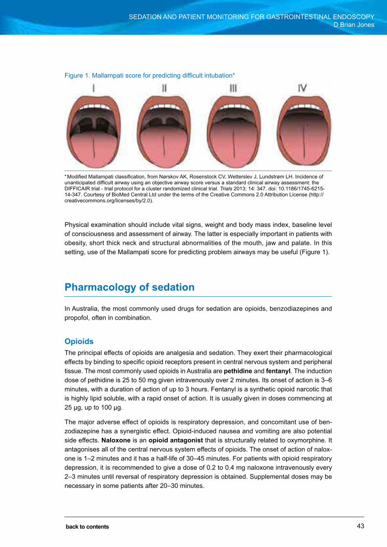
43
SECTION 1: BACKGROUND AND PREPARATION FOR ENDOSCOPY SEDATIon AnD PATIEnT MonITorInG for GASTroInTESTInAl EnDoScoPyD Brian Jones
back to contents
Physical examination should include vital signs, weight and body mass index, baseline level of consciousness and assessment of airway. The latter is especially important in patients with obesity, short thick neck and structural abnormalities of the mouth, jaw and palate. In this setting, use of the Mallampati score for predicting problem airways may be useful (figure 1).
Pharmacology of sedation
In Australia, the most commonly used drugs for sedation are opioids, benzodiazepines and propofol, often in combination.
OpioidsThe principal effects of opioids are analgesia and sedation. They exert their pharmacological effects by binding to specific opioid receptors present in central nervous system and peripheral tissue. The most commonly used opioids in Australia are pethidine and fentanyl. The induction dose of pethidine is 25 to 50 mg given intravenously over 2 minutes. Its onset of action is 3–6 minutes, with a duration of action of up to 3 hours. fentanyl is a synthetic opioid narcotic that is highly lipid soluble, with a rapid onset of action. It is usually given in doses commencing at 25 μg, up to 100 μg.
The major adverse effect of opioids is respiratory depression, and concomitant use of ben-zodiazepine has a synergistic effect. opioid-induced nausea and vomiting are also potential side effects. Naloxone is an opioid antagonist that is structurally related to oxymorphine. It antagonises all of the central nervous system effects of opioids. The onset of action of nalox-one is 1–2 minutes and it has a half-life of 30–45 minutes. for patients with opioid respiratory depression, it is recommended to give a dose of 0.2 to 0.4 mg naloxone intravenously every 2–3 minutes until reversal of respiratory depression is obtained. Supplemental doses may be necessary in some patients after 20–30 minutes.
figure 1. Mallampati score for predicting difficult intubation*
* Modified Mallampati classification, from nørskov AK, rosenstock cV, Wetterslev J, lundstrøm lH. Incidence of unanticipated difficult airway using an objective airway score versus a standard clinical airway assessment: the DIffIcAIr trial - trial protocol for a cluster randomized clinical trial. Trials 2013; 14: 347. doi: 10.1186/1745-6215-14-347. courtesy of BioMed central ltd under the terms of the creative commons 2.0 Attribution license (http://creativecommons.org/licenses/by/2.0).

44
SECTION 1: BACKGROUND AND PREPARATION FOR ENDOSCOPY SEDATIon AnD PATIEnT MonITorInG for GASTroInTESTInAl EnDoScoPyD Brian Jones
back to contents
BenzodiazepinesThe pharmacological effects of benzodiazepines include anxiolysis, amnesia, sedation, muscle relaxation and, in larger doses, anaesthesia. Benzodiazepines enhance activity of the inhibitory neurotransmitter gamma-aminobutyric acid (GABA) by binding to the GABAA receptor on the postsynaptic nerve membrane in the cerebral cortex. Although diazepam is still widely used worldwide, the most common benzodiazepine used in Australia for GIE sedation is midazolam. It has a more rapid onset and shorter duration of action than diazepam. After intravenous administration, midazolam has an onset of action within 1–2 minutes, with a peak effect in 3–4 minutes. Its duration of action is up to 80 minutes. The initial intravenous dose should be in the order of 1–2 mg, with increments of 1 mg. Most patients can be sedated with doses of up to 5–6 mg, particularly if used in conjunction with fentanyl in small doses. As for opioids, the major potential concern is oversedation (or, in effect, inadvertent general anaesthesia) with respiratory depression.
Flumazenil is structurally related to midazolam and is a specific benzodiazepine antagonist. It reverses midazolam-induced sedation and psychomotor impairment. flumazenil has a half-life of up to 1.3 hours, and the average duration of antagonism is 1 hour. Because the effects of midazolam may persist for more than 80 minutes, re-sedation may occur. Incremental intrave-nous doses of flumazenil of 0.1–0.3 mg are effective in reversing benzodiazepine oversedation. It can also be given as an infusion (0.3–0.5 mg/h).
PropofolPropofol (2,6-di-isopropofol) is a hypnotic with minimal analgesic effect. At subhypnotic doses, it produces sedation and amnesia. Propofol acts via the GABA system. It is highly lipid soluble and has a rapid onset of action of between 30 and 45 seconds (one arm–brain circulation). It is metabolised rapidly in the liver and its metabolites are excreted in the kidney. Despite this, its pharmacokinetic profile is not significantly altered in the setting of cirrhosis or renal failure. Its duration of action is short, in the order of only 4–8 minutes, and it is delivered either in mul-tiple small boluses or by infusion. The current formulation of propofol contains 1% propofol in soybean oil, glycerol and purified egg phosphatide, and should be used with caution in patients with soy or egg allergies. Pain on injection occurs in 30% of patients, and other adverse effects include a decrease in cardiac output and hypotension. There is no reversal agent for propofol, but its relatively rapid metabolism allows rapid recovery if oversedation occurs. Nonetheless, anyone using this agent must be prepared to provide mask ventilation if required.
Other agentsMost GIE in Australia is performed with midazolam and an opioid, plus or minus propofol where available. other agents used include alfentanil, ketamine, remifentanil and sevoflurane. In the United States, droperidol is occasionally used.

45
SECTION 1: BACKGROUND AND PREPARATION FOR ENDOSCOPY SEDATIon AnD PATIEnT MonITorInG for GASTroInTESTInAl EnDoScoPyD Brian Jones
back to contents
Monitoring during sedation
Patient monitoring during sedation for GIE is essential, and at least one staff member should be exclusively responsible for monitoring the sedated patient, using both visual assessment and electronic devices (figure 2), from the time of induction of sedation through to safe delivery to the recovery area. This individual should have an understanding of the pharmacology of the drugs used and the stages of sedation that can be achieved, the skills to interpret changes in physiological parameters and “rescue” the patient if complications arise, and current certification in basic and advanced life support. The level of monitoring will vary depending on many factors, including patient comorbidities, level of sedation and the type of procedure.
Many professional bodies have produced guidelines on safe sedation, and a tripartite work-ing group of AnZcA, the Gastroenterological Society of Australia and the royal Australasian college of Surgeons has published guidelines (PS09) that are available on the AnZcA website: http://www.anzca.edu.au/documents/ps09-2014-guidelines-on-sedation-and-or-analgesia
figure 2. Example of monitor data measured during sedation and anaesthesia
The minimum equipment requirements for safe administration of endoscopy with sedation are summarised in Table 3.

46
SECTION 1: BACKGROUND AND PREPARATION FOR ENDOSCOPY SEDATIon AnD PATIEnT MonITorInG for GASTroInTESTInAl EnDoScoPyD Brian Jones
back to contents
Table 3. Minimum required monitoring and emergency resuscitative equipment
Basic airway management equipment ● Supplemental oxygen ● Suction ● nasal cannulas and face masks ● Bag mask ventilation device ● oral and nasal airways (all sizes)
Advanced airway management equipment ● laryngoscope handles and blades ● Endotracheal tubes and stylets ● laryngeal masks
Cardiorespiratory equipment and monitoring devices ● Pulse oximetry ● capnography (desirable, if not essential) ● close access to defibrillator and crash trolley
Emergency drugs ● Atropine ● Antihistamines such as diphenhydramine ● Adrenaline ● flumazenil ● naloxone ● Glucose 50% ● Hydrocortisone ● lignocaine
Who should administer sedation for gastrointestinal endoscopy?
The types of sedation used in Australia vary according to the institution and whether GIE is being performed in a public or private hospital or day surgery unit (see reference 3). In private institutions, sedation is mostly provided by a medical practitioner trained in the use of seda-tive agents, including propofol. These sedationists may be either specialist anaesthetists or non-anaesthetist medical practitioners. A survey of 200 Australian anaesthetists involved in administration of sedation for GIE found that almost 100% used propofol as the main drug, usu-ally coadministered with either midazolam alone (14%), fentanyl alone (6%) or, more commonly, midazolam plus fentanyl (61%). The depth of sedation aimed for by anaesthetists equates to full general anaesthesia, according to ASA criteria (Table 1).
In public hospitals, there are at least three models of sedation delivery:
1 full propofol-based anaesthetic, with or without midazolam and/or fentanyl, administered by a specialist anaesthetist (as in the private system).

47
SECTION 1: BACKGROUND AND PREPARATION FOR ENDOSCOPY SEDATIon AnD PATIEnT MonITorInG for GASTroInTESTInAl EnDoScoPyD Brian Jones
back to contents
2 Sedation with midazolam plus or minus an opioid such as fentanyl or pethidine, administered by one of two endoscopy nurses in the procedure room, under the direction and guidance of the endoscopist. Typical doses would be between 3.5 mg and 7.5 mg of midazolam and between 50 μg and 100 μg of fentanyl.
3 Sedation with midazolam plus or minus an opioid in smaller doses than outlined above, with small increments of propofol given as frequent “top ups” according to patient discomfort. The multiple bolus technique should follow the 20:20 rule (no more than 20 mg of propofol at a time, with at least 20 seconds between each bolus). In other countries, propofol may be administered by a nurse trained in its use (so called nAPS, or nurse-administered propofol system). This approach has gained widespread use in the US and Switzerland. The alternative model is to have the propofol administered by a non-anaesthetist medical practitioner trained in its use. This system has been used very successfully in a number of Australian institutions (see reference 11). What has been striking about each of these non-anaesthetist-administered propofol systems is the incredible safety profile, with no deaths in over 220 000 cases and only one case of endotracheal intubation. Most episodes of hypoxaemia are transient and can be managed with bag mask ventilation for a few minutes until the propofol is metabolised.
The future of gastrointestinal endoscopy sedation in Australia
The questions of what constitutes optimal sedation, which drugs to use and who should admin-ister them are still somewhat controversial and evolving. Great strides have been made with the tripartite working group (representing gastroenterologists, surgeons and anaesthetists) and the PS09 guidelines will continue to evolve. The professional bodies are still developing suitable training programs for non-anaesthetist-administered sedation, using simulators and hands-on delivery of propofol and other agents at individual hospital level under the auspices of the department of anaesthetics. The current recommendations of PS09 in terms of who should administer sedation are summarised in the Appendix.
Further reading1 American Society of Anesthesiologists. Task force on Sedation and Analgesia by non-Anesthesiologists.
Practice guidelines for sedation and analgesia by non-anesthesiologists. Anesthesiology 2002, 96: 1004-1017. http://www.asahq.org/~/media/sites/asahq/files/public/resources/standards-guidelines/practice-guidelines-for-sedation-and-analgesia-by-non-anesthesiologist.pdf (accessed Sep 2016).
2 Thomson A, Andrew G, Jones DB. optimal sedation for gastrointestinal endoscopy: review and recommendations. J Gastroenterol Hepatol 2010; 25: 469-478.
3 Australian and new Zealand college of Anaesthetists. PS55: recommendations on minimum facilities for safe administration of anaesthesia in operating suites and other anaesthetising locations. Melbourne: AnZcA, 2012.

48
SECTION 1: BACKGROUND AND PREPARATION FOR ENDOSCOPY SEDATIon AnD PATIEnT MonITorInG for GASTroInTESTInAl EnDoScoPyD Brian Jones
back to contents
http://www.anzca.edu.au/documents/ps55-2012-recommendations-on-minimum-facilities-fo (accessed Sep 2016).
4 Australian and new Zealand college of Anaesthetists. PS09: Guidelines on sedation and/or analgesia for diagnostic and interventional medical, dental or surgical procedures. Melbourne: AnZcA, 2014. http://www.anzca.edu.au/documents/ps09-2014-guidelines-on-sedation-and-or-analgesia (accessed Sep 2016).
5 Padmanabhan U, leslie K. Australian anaesthetists’ practice of sedation for gastrointestinal endoscopy in adult patients. Anaesth Intensive Care 2008; 36: 436-441.
6 cohen lB, Delegge MH, Aisenberg J, et al. AGA Institute review of endoscopic sedation. Gastroenterology 2007, 133: 675-701. http://dx.doi.org/10.1053/j.gastro.2007.06.002 (accessed Sep 2016).
7 Standards of Practice committee of the American Society for Gastrointestinal Endoscopy. Sedation and anesthesia in GI endoscopy. Gastrointest Endosc 2008; 68: 815-826.
8 Symington I, McGugam E, Graham c, et al. Training in conscious sedation techniques: meeting the recommendations of the UK Academy of Medical royal colleges. Emerg Med J 2007; 24: 576-578.
9 rex DK, Deenadayalu V, Eid E. Gastroenterologist-directed propofol: an update. Gastrointest Endosc Clin N Am 2008; 18: 717-725.
10 Heuss lT, Peter S. Propofol use by gastroenterologists – the European experience. Gastrointest Endosc Clin N Am 2008; 18: 727-738.
11 clarke Ac, chiragakis l, Hillman lc, et al. Sedation for endoscopy: the safe use of propofol by general practitioner sedationists. Med J Aust 2002; 176: 159-162.
49
SECTION 1: BACKGROUND AND PREPARATION FOR ENDOSCOPY SEDATIon AnD PATIEnT MonITorInG for GASTroInTESTInAl EnDoScoPyD Brian Jones
back to contents
Appendix
Adapted from PS09: Guidelines on sedation and/or analgesia for diagnostic and interventional medical, dental or surgical procedures4
Personnel for procedural sedation and analgesia
Scenario 1: Three practitioners — sedation by proceduralist• Medical practitioner proceduralist with airway and resuscitation skills and training
in sedation• Practitioner with training in monitoring sedation• Assistant to assist both• conscious sedation in ASA grade I–II patients• Propofol, thiopentone and other intravenous anaesthetic agents must not be used
Scenario 2: Three practitioners — sedation by medical practitioner• Proceduralist• Medical practitioner with airway and resuscitation skills and training in sedation• Assistant to assist both• conscious sedation in ASA grade I–III patients• Propofol, thiopentone and other intravenous anaesthetic agents may only be used
by a medical practitioner trained in their use
Scenario 3: Four practitioners — sedation by medical practitioner• Proceduralist• Medical practitioner with airway and resuscitation skills and training in sedation• Assistant to assist each*• conscious sedation in ASA grade I–III patients (please refer to PS09)• Propofol, thiopentone and other intravenous anaesthetic agents may only be used
by a medical practitioner trained in their use
Scenario 4: Three practitioners — sedation by anaesthetist• Proceduralist• Anaesthetist• Assistant to assist both• conscious, deep sedation or general anaesthesia in all patients• All approved anaesthetic drugs may be used
Scenario 5: Four practitioners — sedation by anaesthetist• Proceduralist• Anaesthetist• Assistant to assist each*• conscious sedation, deep sedation or general anaesthesia in all patients• All approved anaesthetic drugs may be used
* recommended if assistance is likely to be required for the majority of the case (e.g. complex or emergency patients).
Page 50 (used for notes in the hard copy) has been removed from the PDF edition of this handbook.

51
Antibiotic use in endoscopySarah Cho
Concerns regarding infection associated with gastrointestinal endoscopy (GIE) are:
• bacteraemia resulting in localisation of infection in remote tissues (i.e. infective endocarditis); and
• local infection in which a typical sterile space or tissue is breached and contaminated by an endoscopic accessory or by contrast injection.
The incidence of bacteraemia during GIE procedures has been well established in numerous case series (Table 1). Procedures such as oesophageal dilatation and instrumentation of the obstructed bile ducts have been reported to have higher rates of bacteraemia, with diagnos-tic endoscopy and colonoscopy associated with lower rates of bacteraemia. Bacteraemia is uncommon even with therapeutic colonoscopic procedures, such as colon stent insertion. There are no data on the frequency of bacteraemia associated with device-assisted enteros-copy (e.g. single-balloon and double-balloon enteroscopy), but it is likely to be comparable to that of standard upper and lower endoscopic procedures. The frequency of bacteraemia after endoscopic ultrasound (EUS), with or without fine needle aspiration (FNA), is reported to be low and comparable to that for diagnostic endoscopy. There are no data available on the frequency of bacteraemia associated with newer endoscopic procedures such as endoscopic submucosal dissection, EUS-guided cystogastrostomy or biliary drainage, and peroral endoscopic myotomy.
Table 1. Approximate incidence of bacteraemia in immunocompetent individuals undergoing gastrointestinal endoscopy*
Procedure Bacteraemia incidenceDiagnostic endoscopy ± biopsy 4%
Colonoscopy 2%–4%
ERCP (no duct occlusion) 6%
ERCP (duct occlusion) 11%
EUS ± FNA 0–6%
Variceal band ligation 6%
Oesophageal dilatation or prosthesis 34%–54%
ERCP = endoscopic retrograde cholangiopancreatography. EUS = endoscopic ultrasound. FNA = fine needle aspiration. * From a British Society of Gastroenterology review.
SECTION 1: BACKGROUND AND PREPARATION FOR ENDOSCOPY
CHAPTER 6
back to contents
52
SECTION 1: BACKGROUND AND PREPARATION FOR ENDOSCOPY ANTIBIOTIC USE IN ENDOSCOPySarah Cho
back to contents
Transient bacteraemia has been shown to occur at higher rates during routine daily activity than in association with GIE (e.g. 20%–68% with brushing and flossing of teeth, and 7%–51% with simply chewing food). In most cases, endoscopy-related bacteraemia does not appear to be associated with any infection-related complications, and infective endocarditis is a very rare clinical consequence of GIE. In the United States, an estimated 14.2 million colonoscopies and 2.8 million flexible sigmoidoscopies, and perhaps as many upper endoscopies, are per-formed each year, and only 25 cases of infective endocarditis with a temporal association to an endoscopic procedure have been reported. Furthermore, there are no data that demonstrate a conclusive causal link between GIE and infective endocarditis in these cases, or that antibiotic prophylaxis before GIE prevents infective endocarditis.
Based on these considerations, the American Heart Association (AHA) revised its guidelines for prophylaxis of infective endocarditis in 2007, stating that the administration of antibiotic prophylaxis for patients with cardiac risk factors undergoing GIE was no longer recommended.
Antibiotic prophylaxis for gastrointestinal endoscopy
Prevention of infective endocarditisAs noted above, antibiotic prophylaxis solely to prevent infective endocarditis is no longer rec-ommended for patients with cardiac risk factors who undergo diagnostic or therapeutic endos-copy. Nonetheless, the possibility of infective endocarditis should be considered in patients who develop symptoms and signs of infection during the weeks after an endoscopic procedure, and they should be investigated and treated appropriately.
For patients with established gastrointestinal infections, antibiotics should have been com-menced as part of routine care before GIE, not specifically in preparation for GIE. Amoxycillin or ampicillin should be included in the antibiotic regimen for enterococcal coverage in patients:
• with cardiac conditions associated with a high risk of adverse outcomes from endocarditis (Table 2); and
• who have established gastrointestinal infections in which Enteroccoci are suspected to be part of the infecting bacterial flora (such as cholangitis); and
• particularly those who are about to undergo an endoscopic procedure that may increase the risk of bacteraemia, such as endoscopic retrograde cholangiopancreatography (ERCP).
Vancomycin may be substituted for patients who are allergic to or unable to tolerate amoxycillin or ampicillin.

53
SECTION 1: BACKGROUND AND PREPARATION FOR ENDOSCOPY ANTIBIOTIC USE IN ENDOSCOPySarah Cho
back to contents
Table 2. Cardiac conditions associated with the highest risk of an adverse outcome from infective endocarditis*
● Prosthetic cardiac valve (mechanical or bioprosthetic) ● History of infective endocarditis
● Cardiac transplant recipient with cardiac valvulopathy
● Congenital heart disease (CHD): ♦ Unrepaired CHD, including palliative conduits and shunts ♦ Completely repaired CHD with prosthetic material or device for
the first 6 months after the procedure ♦ Repaired CHD with residual defects at the site or adjacent to
the site of a prosthetic patch or device
* From the American Heart Association guidelines.
Endoscopic retrograde cholangiopancreatographyCholangitis occurs after 0.5%–3% of ERCP procedures. Antibiotic prophylaxis has been shown to reduce the incidence of bacteraemia associated with an ERCP, but clear benefit in preventing cholangitis has not been demonstrated. Therefore, routine antibiotic prophylaxis for ERCP is not recommended.
Antibiotic prophylaxis for ERCP should be given in the following settings:
• Patients with known or suspected biliary obstruction, in which there is a possibility that complete biliary drainage may not be achieved (e.g. hilar cholangiocarcinoma, primary sclerosing cholangitis)
• Patients with severe neutropenia (< 0.5 × 10⁹/L) and/or advanced haematological malignancy
• Patients with a history of liver transplantation.
Incomplete biliary drainage is a chief predictor of post-ERCP cholangitis. Therefore, antibiotic therapy is indicated if biliary drainage achieved at an ERCP is incomplete or is achieved with difficulty, such as in cases of hilar cholangiocarcinoma and primary sclerosing cholangitis. One dose of periprocedural antibiotic is unlikely to prevent cholangitis in patients with incomplete biliary drainage. Antibiotics should be continued until complete drainage is achieved. Antibiotics that cover gram-negative organisms and Enterococci should be used.
Acute cholecystitis may occur following placement of biliary self-expanding metal stents (SEMSs) due to obstruction of the cystic duct. Tumour involvement of the cystic duct orifice is a risk factor, with the incidence similar between covered and uncovered SEMSs in two meta-analyses, ranging from 1.9% to 12%. As cholecystitis is thought to occur due to impaired drainage of non-sterile bile or contrast agent caused by gall bladder outflow obstruction from SEMSs or cystic duct involvement of the tumour, prophylactic antibiotics may help to prevent this adverse event after SEMS placement, but its role is yet to be studied in this clinical setting.

54
SECTION 1: BACKGROUND AND PREPARATION FOR ENDOSCOPY ANTIBIOTIC USE IN ENDOSCOPySarah Cho
back to contents
Percutaneous endoscopic gastrostomyAntibiotic prophylaxis is effective in reducing the incidence of peristomal infection. A Cochrane systemic review of 12 randomised controlled trials involving a total of 1271 patients showed, on its pooled analysis, a statically significant reduction in the incidence of peristomal infection with administration of prophylactic antibiotics (odds ratio, 0.36; 95% CI, 0.26–0.50). A single dose of an intraveneous antibiotic that provides optimal coverage of cutaneous organisms, such as a second- or third-generation cephalosporin, should be administered 30 minutes before the procedure.
Although the role of prophylactic antibiotics before placement of percutaneous endoscopic jeju-nostomy tubes has not been studied, they should offer protection against peristomal infections similar to that observed in patients undergoing percutaneous endoscopic gastrostomy tube placement. Therefore, their use in this setting is recommended.
Methicillin-resistant Staphylococcus aureus (MRSA) decolonisation appears to be effective in reducing the risk of MRSA-related peristomal infection in patients with positive culture swabs from the nose, throat, perineum and broken skin areas.
Cirrhosis with gastrointestinal bleedingPatients with suspected variceal bleeding (or patients with decompensated liver disease who develop acute gastrointestinal bleeding) should have been already established on an intrave-nous antibiotic regimen before undergoing GIE. A meta-analysis of 12 randomised controlled trials involving 1241 patients indicated that antibiotic therapy reduced the incidence of bacterial infections and mortality in patients with cirrhosis who developed acute gastrointestinal bleed-ing. One randomised controlled trial showed that intravenous ceftriaxone was superior to oral norfloxacin in this setting.
Endoscopic ultrasoundAntibiotic prophylaxis for diagnostic EUS or EUS–FNA of solid lesions is not recommended. Clinical infection following EUS-guided FNA is uncommon but appears to occur more often in patients with cysts than solid lesions. In a retrospective series, a subgroup analysis of patients with cysts who were undergoing an EUS-guided FNA indicated a 14% risk of infectious com-plications; however, the number of cystic lesions in this series was small. In a retrospective analysis of 253 patients assessing the impact of prophylactic antibiotics during EUS–FNA of pancreatic cysts, the incidence of infectious adverse events was very low (< 1%), and antibiotics did not confer a protective effect against infections. Although benefit of prophylactic antibiotics has not been validated by prospective randomised studies, most expert opinion, as well as American Society for Gastrointestinal Endoscopy and British Society of Gastroenterology guide-lines, currently favours administration of antibiotic prophylaxis for EUS–FNA of pancreatic cystic lesions. Infections of mediastinal cysts after EUS–FNA have also been reported and appear to occur more commonly, sometimes despite use of antibiotic prophylaxis.
The role of antibiotic prophylaxis in patients undergoing various interventional EUS procedures (e.g. transgastric or transenteric drainage of pseudocysts, biliary drainage, fine needle injection

55
SECTION 1: BACKGROUND AND PREPARATION FOR ENDOSCOPY ANTIBIOTIC USE IN ENDOSCOPySarah Cho
back to contents
of cysts or tumours, fiducial placement) has not been studied. Most series have reported use of periprocedural antibiotic prophylaxis in these procedures.
Orthopaedic prostheses, vascular grafts and non-vascular cardiovascular implanted materialThe same rationale for not administering prophylactic antibiotics for infective endocarditis applies in these settings. There are only two case reports that describe pyogenic arthritis in patients with orthopaedic prostheses, and no reported cases of a vascular graft infection following GIE. Antibiotic prophylaxis before GIE is not recommended for patients with orthopaedic prostheses, synthetic vascular grafts, pacemakers, defibrillators, coronary artery stents or vena cava filters.
Peritoneal dialysisThe International Society for Peritoneal Dialysis has issued a position statement indicating that giving antibiotics such as intravenous ampicillin plus a single dose of aminoglycoside, with or without metronidazole, before GIE may lower the risk of peritonitis. An alternative is intraperitoneal administration of prophylactic antibiotics the night before the procedure. The society also recommends that the abdomen be emptied of fluid before any procedure involving the abdomen or pelvis, including colonoscopy. However, it should be noted that no randomised controlled trial data are yet available to support the recommendation. A retrospective study showed that the risk of peritonitis in patients with peritoneal dialysis after colonoscopy without antibiotic prophylaxis was 6.3%. Colon biopsy or polypectomy did not appear to increase the risk further. No peritonitis occurred in patients who received prophylactic antibiotics, although the difference was not clinically significant.
Further reading1 Allison MC, Sandoe JA, Tighe R, et al. Antibiotic prophylaxis in gastrointestinal endoscopy. Gut 2009; 58: 869-
880.
2 ASGE Standards of Practice Committee. Antibiotic prophylaxis for GI endoscopy. Gastrointest Endosc 2015; 81: 81-89.
3 Chun yJ, yoon NR, Park JM, et al. Prospective assessment of risk of bacteremia following colorectal stent placement. Dig Dis Sci 2012; 57: 1045-1049.
4 Horstkotte D, Follath F, Gutschik E, et al. Guidelines on prevention, diagnosis and treatment of infective endocarditis executive summary: the task force on infective endocarditis of the European Society of Cardiology. Eur Heart J 2004; 25: 267-276.
5 Wilson W, Taubert KA, Gewitz M, et al. Prevention of infective endocarditis: guidelines from the American Heart Association: a guideline from the American Heart Association Rheumatic Fever, Endocarditis, and Kawasaki Disease Committee, Council on Cardiovascular Disease in the young, and the Council on Clinical Cardiology, Council on Cardiovascular Surgery and Anesthesia, and the Quality of Care and Outcomes Research Interdisciplinary Working Group. Circulation 2007; 116: 1736-1754.
6 Seeff LC, Richards TB, Shapiro JA, et al. How many endoscopies are performed for colorectal cancer screening? Results from CDC’s survey of endoscopic capacity. Gastroenterology 2004; 127: 1670-1677.
7 National Institute for Health and Care Excellence. NICE Guidance [website]. http://www.nice.org.uk/guidance (accessed Sep 2016).

56
SECTION 1: BACKGROUND AND PREPARATION FOR ENDOSCOPY ANTIBIOTIC USE IN ENDOSCOPySarah Cho
back to contents
8 Freeman ML, Nelson DB, Sherman S, et al. Complications of endoscopic biliary sphincterotomy. N Engl J Med 1996; 335: 909-918.
9 Loperfido S, Angelini G, Benedetti G, et al. Major early complications from diagnostic and therapeutic ERCP: a prospective multicenter study. Gastrointest Endosc 1998; 48: 1-10.
10 Masci E, Toti G, Mariani A, et al. Complications of diagnostic and therapeutic ERCP: a prospective multicenter study. Am J Gastroenterol 2001; 1996: 417-423.
11 Sauter G, Grabein B, Huber G, et al. Antibiotic prophylaxis of infectious complications with endoscopic retrograde cholangiopancreatography. A randomized controlled study. Endoscopy 1990; 22: 164-167.
12 Niederau C, Pohlmann U, Lubke H, Thomas L. Prophylactic antibiotic treatment in therapeutic or complicated diagnostic ERCP: results of a randomized controlled clinical study. Gastrointest Endosc 1994; 40: 533-537.
13 Harris A, Chan AC, Torres-Viera C, et al. Meta-analysis of antibiotic prophylaxis in endoscopic retrograde cholangiopancreatography (ERCP). Endoscopy 1999; 31: 718-724.
14 Motte S, Deviere J, Dumonceau JM, et al. Risk factors for septicemia following endoscopic biliary stenting. Gastroenterology 1991; 101: 1374-1381.
15 Cotton PB, Connor P, Rawis E, Romagnuolo J. Infection after ERCP, and antibiotic prophylaxis: a sequential quality-improvement approach over 11 years. Gastrointest Endosc 2008; 67: 471-475.
16 De Palma GD, Galloro G, Siciliano S, et al. Unilateral versus bilateral endoscopic hepatic duct drainage in patients with malignant hilar biliary obstruction: results of a prospective, randomized, and controlled study. Gastrointest Endosc 2001; 53: 547-553.
17 Van den Hazel SJ, Speelman P, Dankert J, et al. Piperacillin to prevent cholangitis after endoscopic retrograde cholangiopancreatography. A randomized, controlled trial. Ann Intern Med 1996; 125: 442-447.
18 Byl B, Deviere J, Struelens MJ, et al. Antibiotic prophylaxis for infectious complications after therapeutic endoscopic retrograde cholangiopancreatography: a randomized, double-blind, placebo-controlled study. Clin Infect Dis 1995; 20: 1236-1240.
19 Saleem A, Leggett CL, Murad MH, Baron TH. Meta-analysis of randomized trials comparing the patency of covered and uncovered self-expandable metal stents for palliation of distal malignant bile duct obstruction. Gastrointest Endosc 2011; 74: 321-327.
20 Almadi MA, Barkun AN, Martel M. No benefit of covered vs uncovered self-expandable metal stents in patients with malignant bile duct obstruction: a meta-analysis. Clin Gastroenetrol Hepatol 2013; 11: 27-37.
21 Lipp A, Lusardi G. Systemic antimicrobial prophylaxis for percutaneous endoscopic gastrostomy. Cochrane Database Syst Rev 2013; (11): CD005571.
22 Thomas S, Cantrill S, Waghorn DJ, McIntyre A. The role of screening and antibiotic prophylaxis in the prevention of percutaneous gastrostomy site infection caused by methicillin-resistant Staphylococcus aureus. Aliment Pharmacol Ther 2007; 25: 593-597.
23 Chavez-Tapia NC, Barientos-Gutierrez T, Tellez-Avila F, et al. Meta-analysis: antibiotic prophylaxis for cirrhotic patients with upper gastrointestinal bleeding – an updated Cochrane review. Aliment Pharmacol Ther 2011; 34: 509-518.
24 Fernandez J, Ruiz del Arbol L, Gomez C, et al. Norfloxacin vs ceftriaxone in the prophylaxis of infections in patients with advanced cirrhosis and hemorrhage. Gastroenterology 2006; 131: 1049-1056; quiz 1285.
25 Williams DB, Sahai AV, Aabakken L, et al. Endoscopic ultrasound guided fine needle aspiration biopsy: a large single centre experience. Gut 1999; 44: 720-726.
26 Eloubeidi MA, Tamhane A, Varadarajulu S, Wilcox CM. Frequency of major complications after EUS-guided FNA of solid pancreatic masses: a prospective evaluation. Gastrointest Endosc 2006; 63: 622-629.
27 Wiersema MJ, Vimann P, Giovanini M, et al. Endosonography-guided fine-needle aspiration biopsy: diagnostic accuracy and complication assessment. Gastroenterology 1997; 112: 1087-1095.
28 Wildi SM, Honda RS, Fickling W, et al. Diagnosis of benign cysts of the mediastinum: the role and risks of EUS and FNA. Gastrointest Endosc 2003; 58: 362-368.
29 Ryan AG, Zamvar V, Roberts SA. Iatrogenic candidal infection of a mediatinal foregut cyst following endoscopic ultrasound-guided fine-needle aspiration. Endoscopy 2002; 34: 838-839.

57
SECTION 1: BACKGROUND AND PREPARATION FOR ENDOSCOPY ANTIBIOTIC USE IN ENDOSCOPySarah Cho
back to contents
30 Levy MJ, Norton ID, Clain JE, et al. Prospective study of bacteremia and complications with EUS FNA of rectal perirectal lesions. Clin Gastroenterol Hepatol 2007; 5: 684-689.
31 Khashab MA, Dewitt J. EUS-guided biliary drainage: is it ready for prime time? yes! Gastrointest Endosc 2013; 78: 102-105.
32 Khashab MA, Varadarajulu S. Endoscopic ultrasonography as a therapeutic modality. Curr Opin Gastroenterol 2012; 28: 467-476.
33 Zimmerli W, Trampuz A, Ochsner PE. Prosthetic-joint infections. N Engl J Med 2004; 351: 1645-1654.
34 Baddour LM, Bettmann MA, Bolger AF, et al. Nonvalvular cardiovascular-device related infections. Circulation 2003; 108: 2015-2031.
35 Piraino B, Bernardini J, Brown E, et al. ISPD position statement on reducing the risks of peritoneal dialysis-related infections. Perit Dial Int 2011; 31: 614-630.
Page 58 (used for notes in the hard copy) has been removed from the PDF edition of this handbook.

59
Endoscopic imagingWilliam Tam
Endoscopic imaging has rightfully emerged as a fundamental and core component of endoscopy that is critical in the clinical decision-making process. The ability to make detailed and accurate assessment of mucosal detail, including at the cellular level, can assist in managing lesions instantly. This chapter provides a framework for discussion, recognising that a comprehensive review of each imaging technique and its clinical utility is beyond the scope of this work.
Endoscopic imaging can be subdivided as shown in Table 1.
Table 1. Classification of endoscopic imaging
1. Conventional endoscopy (white light endoscopy)
2. Image-enhanced endoscopy ● Acetic acid ● Conventional chromoendoscopy ● Digital chromoendoscopy
♦ Narrow band imaging ♦ i-scan imaging ♦ Flexible spectral imaging colour enhancement (FICE) ♦ Autofluorescence imaging (AFI)
3. Magnification endoscopy ● Optical and electronic
4. Microscopic endoscopy ● Confocal laser endomicroscopy ● Endocytoscopy
5. Tomographic endoscopy ● Optical coherence tomography
White light endoscopy
The importance of careful examination under white light endoscopy (WLE) cannot be over-emphasised. A meticulous endoscopic assessment, noting subtle mucosal changes such as mild mucosal elevation or excavation, mucosal irregularities and nodules, is an important first step in evaluating any lesion. However, the inability to detect dysplasia and early neoplasia in
SECTION 1: BACKGROUND AND PREPARATION FOR ENDOSCOPY
CHAPTER 7
back to contents

60
SECTION 1: BACKGROUND AND PREPARATION FOR ENDOSCOPY ENDOsCOpIC IMAgINgWilliam Tam
back to contents
lesions of the gastrointestinal tract on WLE has been well documented. The shortcomings of WLE have led to the development of real-time image enhancement during endoscopy.
Image-enhanced endoscopy
Image-enhanced endoscopy is a general term loosely used to describe the myriad techniques employed to improve, in real time, the quality of images obtained during endoscopy.
Acetic acidThe use of acetic acid in gastrointestinal endoscopy has largely been limited to application in Barrett’s oesophagus. When sprayed onto Barrett’s mucosa, acetic acid 2.5% causes a revers-ible acetylation of proteins and leads to an aceto-whitening reaction, with increased opacity of the mucosal surface (Figure 1). It also causes vascular congestion and improves surface pattern evaluation.
Figure 1. Barrett’s oesophagus on white light endoscopy (A) and after acetic acid application (B)
Reprinted from Tholoor s, Bhattacharyya R, Tsagkournis O, et al. Acetic acid chromoendoscopy in Barrett’s esophagus surveillance is superior to the standardized random biopsy protocol: results from a large cohort study (with video). Gastrointest Endosc 2014; 80: 417-424, with permission from Elsevier.
Acetic acid is inexpensive and has been reported to be effective in detecting dysplasia in sur-veillance populations in uncontrolled studies. In a landmark study by Longcroft-Wheaton and colleagues, acetic acid chromoendoscopy had a sensitivity of 95.5% and specificity of 80% for detecting neoplasia in Barrett’s oesophagus.
Conventional chromoendoscopyChromoendoscopy, or dye staining, involves the topical application of various stains during endoscopy to improve visualisation of mucosal surfaces in the gastrointestinal tract (Table 2, Figure 2). The stains can be divided into three classes: absorptive, contrast and reactive. Absorptive stains (e.g. Lugol’s iodine, methylene blue, crystal violet) are absorbed into com-ponents of cellular structures in the mucosa. Differences in mucosal uptake of the absorptive

61
SECTION 1: BACKGROUND AND PREPARATION FOR ENDOSCOPY ENDOsCOpIC IMAgINgWilliam Tam
back to contents
stain can therefore be used to define different types of mucosa. Contrast stains, on the other hand, pool in the mucosal crevices, thereby accentuating the surface topography. Indigo car-mine, for instance, is used to define mucosal irregularities in colonic polyps. Reactive stains, such as Congo red and phenol red, are pH-dependent stains that change colour in response to changes in pH. Congo red turns dark blue or black in acidic conditions, while phenol red turns from yellow in an acidic environment to red in the presence of alkali.
Table 2. stains commonly used in chromoendoscopy
StainType of
stain Clinical useLugol’s iodine Absorptive squamous cell oesophageal cancer, Barrett’s oesophagus
(delineates squamous from columnar epithelium)
Toluidine blue Absorptive squamous cell oesophageal cancer, Barrett’s oesophagus (stains both gastric and intestinal metaplasia)
Methylene blue Absorptive gastric intestinal metaplasia, Barrett’s oesophagus (stains specialised columnar epithelium, not gastric type)
Crystal violet Absorptive Colonic polyps (taken up by crypt of Lieberkühn gland openings)
Indigo carmine Contrast Highlights mucosal irregularities in the oesophagus (Barrett’s) or colon (flat colon tumours)
Congo red Reactive Early gastric carcinoma, heterotopic gastric mucosa (reacts with acid-secreting gastric cells)
phenol red Reactive Helicobacter pylori infection in the stomach
Figure 2. Technique of chromoendoscopy
Optical and digital methods of image-enhanced endoscopyOptical and digital methods of image enhancement constitute the most significant advance in endoscopic imaging and have become increasingly prevalent in endoscopy suites because of their ease of use and clinical utility. Often called “special light observation” or “electronic chromoendoscopy”, these techniques involve conversion of the optical characteristics of the light used for illumination or imaging with a light source differing in optical characteristics from ordinary white light. signal processing within the video processor occurs in a specially designed way to yield enhanced images.
Application of mucolytic agent
Application of stain

62
SECTION 1: BACKGROUND AND PREPARATION FOR ENDOSCOPY ENDOsCOpIC IMAgINgWilliam Tam
back to contents
The currently available methods are:
• Narrow band imaging (NBI), generation 1 and 2 (Olympus)• i-scan imaging (pentax)• Flexible spectral imaging colour enhancement (FICE) and blue laser imaging (BLI)
(Fujinon)• Autofluorescence imaging (AFI).
Narrow band imagingNBI is an optical method of image enhancement, whereby light penetration to mucosal layers is restricted using optic filters to isolate two specific bands of light: 415 nm blue and 540 nm green. By isolating these two bands of light and taking into account their absorptive and reflec-tive properties on the mucosal surface, an image that enhances visualisation of superficial mucosal and vascular structures is created. The quality of the surface pit pattern morphology is also clearly enhanced by this technology.
Many studies have reported the superiority of NBI in detecting dysplasia and early neoplastic lesions of the digestive tract, compared with WLE.
i-scan imagingi-scan is a novel endoscopic post-processing light filter technology using sophisticated software algorithms with real-time image mapping technology embedded within its video processor. The computer-controlled digital processing provides resolution of about 1.25 megapixels per image. This module allows for three modes of image enhancement:
• surface enhancement (sE) — i-scan 1 • contrast enhancement (CE) — i-scan 2 • surface and tone enhancement (TE) — i-scan 3.
Flexible spectral imaging colour enhancementFICE is a digital method of image enhancement, whereby the displayed images are enhanced with the target’s spectral information collected by processing signals obtained during irradiation of white light. The FICE system is unique in that it uses lasers for its illumination. By manipulating the intensity of its two lasers, this system allows for detailed assessment of blood vessels and superficial mucosal structures in its BLI and BLI-bright modes.
Autofluorescence imagingWhen a xenon light source is directed at gastrointestinal mucosa, endogenous fluorophores emit a certain amount of fluorescence, while the remaining incident light is reflected, absorbed and scattered (Figure 3). The autofluorescence light can be captured and made to pass through a barrier filter and a black and white filter, and is then digitally altered to produce a composite image. The fluorescence characteristics of normal mucosa are different to those of inflamed or dysplastic mucosa, and it is this feature which is used in AFI.

63
SECTION 1: BACKGROUND AND PREPARATION FOR ENDOSCOPY ENDOsCOpIC IMAgINgWilliam Tam
back to contents
Figure 3. principle of autofluorescence imaging
Tajiri conducted a comparative study with the WLE and AFI systems to differentiate neoplastic from non-neoplastic lesions in 190 cases, and found the sensitivity, specificity and accuracy of AFI to be 98%, 92% and 99%, respectively. These results suggest that AFI might enable easy differentiation of neoplastic from non-neoplastic lesions in the colon (Figure 4).
Figure 4. Endoscopic images of a laterally spreading tumour with granular changes in the rectum on white light endoscopy (A) and autofluorescence endoscopy (B)
Reprinted from: Tajiri H, Niwa H. Recent advances in electronic endoscopes: image-enhanced endoscopy. JMAJ 2008; 51: 199-203.
High-definition and magnification endoscopy
Advances in charge-coupled device (CCD) and, more recently, complementary metal–oxide–semiconductor (CMOs) technology have resulted in smaller chips with an increased number of pixels and improved resolution. The chips used in current high-resolution or high-definition (HD)

64
SECTION 1: BACKGROUND AND PREPARATION FOR ENDOSCOPY ENDOsCOpIC IMAgINgWilliam Tam
back to contents
endoscopes produce signal images with resolutions that range from 850 000 pixels to more than one million pixels. It is important to note that to generate a true HD image, each component of the system (e.g. the endoscope CCD chip, the processor, the monitor and transmission cables) must be HD-compatible.
High-magnification endoscopes are defined by their capacity to perform optical zoom by using a movable lens at the tip of the endoscope. A translucent cap on the tip of the endoscope may be used to stabilise the focal length between the lens and the target tissue to improve image quality. Optical zoom obtains a magnified image of the target while maintaining image display quality. This is distinguished from electronic or digital magnification, which simply enlarges the image on the display, with consequent decreases in pixel density and image quality. With the proper processor, conventional endoscopes permit digital magnification of 1.5× to 2×. Although standard endoscopes magnify images 30 to 35 times, zoom endoscopes can optically magnify images up to 150 times, depending on the size of the monitor. All three endoscope manufac-turers have zoom endoscopes available in Australia, with combined optical and digital zoom. Newer Olympus endoscopes have a feature called near focus imaging (using a variable focus lens system) that allows the endoscope to be moved closer (within 2–6 mm) to the area of interest while maintaining the image in focus (Figure 5).
Figure 5. short-segment Barrett’s oesophagus on white light endoscopy (left) and on narrow band imaging (near focus mode) (right)
HD magnification endoscopy is commonly used in conjunction with the image-enhanced endos-copy techniques described above for the assessment of a wide variety of lesions in the upper and lower digestive tract.
Microscopic endoscopy
Confocal laser endomicroscopy Confocal laser endomicroscopy (CLE) enables the endoscopist to perform a real-time histolog ical assessment of the gastrointestinal tract. The most widely used CLE system is

65
SECTION 1: BACKGROUND AND PREPARATION FOR ENDOSCOPY ENDOsCOpIC IMAgINgWilliam Tam
back to contents
the “endoscope with embedded CLE technology” (eCLE) made by pentax (Tokyo, Japan) and Optiscan (Melbourne, Australia). The eCLE enables visualisation of both the epithelium and the subepithelial vascular structures with imaging at variable depths up to 250 mm and a magnification power of up to 1000 μm. Mauna Kea Technologies has created a probe-based endomicroscopy system in which the laser-scanning unit remains outside the patient, and the endomicroscopy probe is passed through the working channel of a standard endoscope. This probe-based CLE (pCLE) provides video sequence imaging at a rate of 12 images per second and allows for the compilation of images from a video sequence to create a composite video mosaic. The depth ranges from 50 to 150 mm and is fixed based on the type of probe.
These CLE systems use a wavelength of 48 nm for excitation. CLE requires the use of a con-trast agent, most commonly intravenous fluorescein sodium.
EndocytoscopyEndocytoscopy is an ultra-high-magnification technique that enables surface morphology to be assessed with magnifications in excess of 450× in real time. Endocytoscopy uses a high-power fixed-focus objective lens. The device can either be incorporated into the endoscope or used as a probe-based system. similar to the endoscope, the endocytoscope is connected to a light source and a video processor. Thus, for visualisation of images in real time, it is necessary to use two processors simultaneously.
Before the target area can be examined under the magnification of the endocytoscope, it must first be stained to enhance the cellular structures, as in conventional histopathology. This is achieved by application of a double-stain technique, which approximates haematoxylin and eosin staining in conventional histopathology. The target area is first treated with a mucolytic agent, 10% N-acetylcysteine, followed by application of 1% methylene blue, which stains the nucleus, and 0.1% crystal violet, which stains not only the nucleus but also the cytoplasm.
Tomographic endoscopy
Optical coherence tomographyOptical coherence tomography (OCT) can be thought of as analogous to ultrasound, but instead of producing an image from the scattering of sound waves, it uses optical scattering based on differences in tissue composition to form a two-dimensional image. The benefit of OCT over ultrasound is that it can generate cross-sectional images of tissues with an axial resolution of up to 10 μm, which is comparable to low-power microscopy.
Original OCT systems, or time-domain OCT, were limited to discrete locations or “point” sam-pling, due to slow acquisition rates. However, with the development of second-generation OCT, termed optical frequency-domain imaging, it is now possible to perform high-speed acquisition of large luminal surfaces in three dimensions.

66
SECTION 1: BACKGROUND AND PREPARATION FOR ENDOSCOPY ENDOsCOpIC IMAgINgWilliam Tam
back to contents
Clinical utility of endoscopic imaging
Detection of oesophageal squamous cell carcinomaNumerous studies have led to the general opinion that chromoendoscopy is useful in the detection of oesophageal squamous cell carcinoma (sCC). Lugol’s iodine stains the glycogen component of normal squamous mucosa brown; the absence of staining (i.e. “negative” staining) is therefore indicative of mucosal abnormality. This technique is also useful to delineate the margins of the tumour and therefore assists in collection of biopsy specimens and for making clinical decisions relating to treatment (e.g. endoscopic mucosal resection versus surgery).
The use of Lugol’s iodine chromoendoscopy is used in clinical practice to screen for oesopha-geal sCC in high-risk patients, such as those with head and neck cancer. several studies have now used NBI, in isolation and in conjunction with magnification endoscopy and Lugol’s iodine chromoendoscopy, to detect oesophageal sCC, with encouraging results. The blood vessel morphology (intrapapillary capillary loops; Figure 6) seen under ultra-high magnification has been used in combination with Lugol’s iodine staining of squamous epithelium in the oesoph-agus to facilitate the detection of sCC.
Figure 6. Classification of intrapapillary capillary loops (IpCLs)
IPCL type I
IPCL type II
IPCL type III
IPCL type IV
IPCL type V1
IPCL type V2
IPCL type V3
IPCL type Vn
Background
Inflammation
Inflammation/LGIN
LGIN/HGIN
m1
m2
m3/sm1
Sm2
Normal IPCL with area formation
Slight change of IPCL
Irregularly dilated IPCL
Type V1+elongation
Highly destructed IPCL
New tumor vessels
Local treatment(EMR/ESD)
ER/surgery/CRT
Surgery/CRT
Reproduced under the Creative Commons Attribution License from Minami H, Inoue H, Ikeda H, et al. Usefulness of background coloration in detection of esophago-pharyngeal lesions using NBI magnification. Gastroenterol Res Pract 2012; 2012: 529782. LgIN = low-grade neoplasia. HgIN = high-grade neoplasia. EMR = endoscopic mucosal resection. EsD = endoscopic submucosal dissection. ER = endoscopic resection. CRT = chemoradiotherapy.

67
SECTION 1: BACKGROUND AND PREPARATION FOR ENDOSCOPY ENDOsCOpIC IMAgINgWilliam Tam
back to contents
Figure 7. Kudo classification of polyps
Reproduced under the Creative Commons Attribution License from Häfner M, Liedlgruber M, Uhl A, et al. Color treatment in endoscopic image classification using multi-scale local color vector patterns. Med Image Anal 2012; 16: 75-86. doi: 10.1016/j.media.2011.05.006. Epub 2011 May 17.

68
SECTION 1: BACKGROUND AND PREPARATION FOR ENDOSCOPY ENDOsCOpIC IMAgINgWilliam Tam
back to contents
Barrett’s oesophagusseveral mucosal classification systems have now been described in the literature. They have formed the basis for further extensive work on attempting to validate the numerous optical enhancement modalities for detecting neoplasia in Barrett’s oesophagus. One of the most commonly cited is the classification system proposed by Kara and colleagues in 2006, which used NBI with magnification endoscopy. A simplified system was proposed by singh and col-leagues in 2008. A potential drawback with all the classification systems is lack of intra-observer agreement between endoscopists when using them.
Colonic polypsA large body of work in the endoscopic assessment of colonic polyps has enriched our under-standing in this field. Because of the widespread availability of image enhancement techniques, the relationship between the pit pattern seen with magnification endoscopy and the histopatho-logical characteristics of the polyp has been increasingly recognised. The Kudo classification of polyps and examples of each type are shown in Figure 7.
Figure 8. Narrow band imaging international colorectal endoscopic (NICE) classification*
Reprinted from Hayashi N, Tanaka s, Hewett Dg, et al. Endoscopic prediction of deep submucosal invasive carcinoma: validation of the Narrow-Band Imaging International Colorectal Endoscopic (NICE) classification. Gastrointest Endosc 2013; 78: 625-632, with permission from Elsevier.

69
SECTION 1: BACKGROUND AND PREPARATION FOR ENDOSCOPY ENDOsCOpIC IMAgINgWilliam Tam
back to contents
A simplified classification of polyps is the NBI international colorectal endoscopic (NICE) classification (Figure 8).
It is important to note that neither the Kudo nor NICE classification specifically identifies sessile serrated polyps. A recent study by Yamashina and colleagues has described the presence of expanded crypt openings and thick branched vessels in sessile serrated polyps, seen with magnification endoscopy and NBI.
The ability to visualise capillaries on the surface of colonic polyps using magnification endos-copy and NBI — the sano classification — has been used to facilitate the diagnosis of early colorectal neoplasia and guide its management (Figure 9).
Figure 9. sano classification
Reproduced under the Creative Commons Attribution License from Iwatate M, Ikumoto T, Hattori s, et al. NBI and NBI combined with magnifying colonoscopy. Diagn Ther Endosc 2012; 2012: 173269. doi: 10.1155/2012/173269.
Detection of dysplasia within inflammatory bowel diseaseThe role of chromoendoscopy to detect dysplasia in patients with long-standing inflamma-tory bowel disease undergoing endoscopic surveillance has not been defined. Using mag-nification endoscopy and chromoendoscopy, the extent of mucosal abnormality, including dysplasia-associated lesions or masses, could be assessed, but biopsy is still essential to obtain histological confirmation. Limited studies using the first-generation NBI system appear to show discrepant results; more studies are needed to make an authoritative comment.
70
SECTION 1: BACKGROUND AND PREPARATION FOR ENDOSCOPY ENDOsCOpIC IMAgINgWilliam Tam
back to contents
Conclusion
There is no doubt that digital chromoendoscopy has had a significant impact on endoscopic assessment, resulting in rapid uptake into routine general gastroenterological practice. Advances in optics and optical technology will continue to fuel clinical research. It is expected that a large amount of clinical data will become available in the coming years, which will, in turn, help define the critical role of endoscopic imaging.
Further reading1 Tholoor s, Bhattacharyya R, Tsagkournis O, et al. Acetic acid chromoendoscopy in Barrett’s esophagus
surveillance is superior to the standardized random biopsy protocol: results from a large cohort study (with video). Gastrointest Endosc 2014; 80: 417-424. doi: 10.1016/j.gie.2014.01.041.
2 Longcroft-Wheaton g, Duku M, Mead R, et al. Acetic acid spray is an effective tool for the endoscopic detection of neoplasia in patients with Barrett’s esophagus. Clin Gastroenterol Hepatol 2010; 8: 843-847. doi: 10.1016/j.cgh.2010.06.016.
3 Tajiri H. Autofluorescence endoscopy for the gastrointestinal tract. Proc Jpn Acad Ser B Phys Biol Sci 2007; 83: 248-255.
4 Kara MA, Ennahachi M, Fockens p, et al. Detection and classification of the mucosal and vascular patterns (mucosal morphology) in Barrett’s esophagus by using narrow band imaging. Gastrointest Endosc 2006; 64: 155-166. doi: 10.1016/j.gie.2005.11.049.
5 singh R, Anagnostopoulos gK, Yao K, et al. Narrow-band imaging with magnification in Barrett’s esophagus: validation of a simplified grading system of mucosal morphology patterns against histology. Endoscopy 2008; 40: 457-463. doi: 10.1055/s-2007-995741.
6 Kudo s, Tamura s, Nakajima T, et al. Diagnosis of colorectal tumorous lesions by magnifying endoscopy. Gastrointest Endosc 1996; 44: 8-14.
7 Hayashi N, Tanaka s, Hewett Dg, et al. Endoscopic prediction of deep submucosal invasive carcinoma: validation of the narrow-band imaging international colorectal endoscopic (NICE) classification. Gastrointest Endosc 2013; 78: 625-632. doi: 10.1016/j.gie.2013.04.185.
8 Yamashina T, Takeuchi Y, Uedo N, et al. Diagnostic features of sessile serrated adenoma/polyps on magnifying narrow band imaging: a prospective study of diagnostic accuracy. J Gastroenterol Hepatol 2015; 30: 117-123. doi: 10.1111/jgh.12688.
9 sano Y, Tanaka s, Kudo sE, et al. Narrow-band imaging (NBI) magnifying endoscopic classification of colorectal tumors proposed by the Japan NBI Expert Team. Dig Endosc 2016; 28: 526-533. doi: 10.1111/den.12644.
Pages 71-73 (used for notes in the hard copy) have been removed from the PDF edition of this handbook.

Section 2
UPPER ENDOSCOPY
7575
Barrett’s oesophagus: diagnosis and managementDarren Pavey
Barrett’s oesophagus is a metaplastic change in the oesophageal epithelium thought to result from injury due to chronic exposure to reflux gastric content. Normal oesophageal squamous epithelium is replaced by specialised intestinal epithelium, also known as intestinal metaplasia (IM). Progression of non-dysplastic IM to low-grade dysplasia (LGD), high-grade dysplasia (HGD) and oesophageal adenocarcinoma (OAC) is well described. The incidence of OAC continues to rise, particularly among the older population.
Guidelines for the diagnosis and management of Barrett’s oesophagus have recently been pub-lished by the Cancer Council of Australia. Readers are referred to these guidelines for further background on the epidemiology and risk factors for Barrett’s oesophagus.
Diagnosis of Barrett’s oesophagus
Barrett’s oesophagus has traditionally been defined as the presence of at least 1 cm of meta-plastic columnar epithelium that replaces the stratified squamous epithelium normally lining the distal oesophagus. Segments < 1 cm have instead been classified as “specialised IM of the oesophagogastric junction”. This distinction is made due to the high interobserver variability, as well as a low risk of OAC in very short-segment disease.
The definition of Barrett’s oesophagus has varied based on the requirement for the presence of IM on endoscopic biopsy samples. Debate regarding the requirement for IM on biopsy from columnar-lined epithelium segments has derived from the apparently differential risk of devel-oping OAC in such epithelium containing or not containing IM. Large population-based cohort studies have shown a substantially lower OAC risk in patients with columnar metaplasia without IM, compared with those with IM. However, not all studies have corroborated this finding. The yield for IM correlates directly with the number of endoscopic biopsy samples obtained. In a
SECTION 2: UPPER ENDOSCOPY
CHAPTER 8
back to contents

BARReTT’S OeSOPHAGuS: DIAGNOSIS AND MANAGeMeNTDarren Pavey
SECTION 2: UPPER ENDOSCOPY
76 back to contents
large retrospective study, the yield for IM was 35% with four biopsy samples and up to 68% after eight biopsies. The American College of Gastroenterology guidelines continue to suggest that only columnar-lined epithelium containing IM be defined as Barrett’s oesophagus, given the apparent differential cancer risk between epithelium with and without IM.
endoscopic assessment of Barrett’s oesophagus should use the Prague classification, whereby the circumferential (C) and maximal (M) extent of tongue-like protrusions are reported (e.g. C3M4). Any segment of Barrett’s oesophagus measuring > 3 cm has been classified as long-segment Barrett’s, with segments < 3 cm classified as short-segment Barrett’s oesophagus.
Prevalence of Barrett’s oesophagus
The prevalence of Barrett’s oesophagus in the general population remains uncertain because of the need for endoscopy to define the condition. Studies have reported prevalences between 1.3% and 1.6% in unselected adult populations undergoing endoscopic screening. About 10% of the population with chronic reflux have Barrett’s oesophagus.
The significance of Barrett’s oesophagus is its association with an increased risk of OAC. The incidence of OAC is progressively increasing, and it remains highly lethal, with a 5-year survival rate of less than 20%.
Barrett’s oesophagus with no evidence of cellular atypia is classified as non-dysplastic. IM may develop with progressively more abnormal features, including LGD, then HGD and eventually adenocarcinoma. The natural history of Barrett’s oesophagus is still being determined. In a large study that identified 1376 patients with a first-ever diagnosis of non-dysplastic Barrett’s oesophagus, 618 patients had long-term follow-up data at a mean of 4.12 years. OAC developed in 12 patients (2%), HGD in 22 (3.6%), and LGD in 100 (16.1%). There was no progression to dysplasia in 484 patients. Overall, 21.7% of patients with non-dysplastic Barrett’s oesophagus progressed to dysplasia or adenocarcinoma.
Screening for Barrett’s oesophagus
The concept of the metaplasia–dysplasia–carcinoma sequence has led to the hypothesis that screening for Barrett’s oesophagus and implementing endoscopic surveillance to detect dyspla-sia, followed by endoscopic intervention, will lead to a decreased incidence of OAC. Screening also detects prevalent dysplasia or carcinoma that may be treated with endoscopic therapy. However, the evidence to support this hypothesis consists of retrospective studies that may be subject to bias. Indeed, > 90% of OACs are diagnosed in patients without a prior Barrett’s oesophagus diagnosis, despite the increasing use of endoscopy.
Screening for Barrett’s oesophagus may be considered in men with chronic (> 5 years) and/or frequent (weekly or more) symptoms of gastro-oesophageal reflux disease (GORD; heartburn or acid regurgitation) and two or more risk factors for Barrett’s oesophagus or OAC: age > 50

77
BARReTT’S OeSOPHAGuS: DIAGNOSIS AND MANAGeMeNTDarren Pavey
SECTION 2: UPPER ENDOSCOPY
back to contents
years, white ethnicity, central obesity (waist circumference > 102 cm or waist–hip ratio > 0.9), current or past history of smoking, and a confirmed family history (in a first-degree relative) of Barrett’s oesophagus or OAC. Screening is not currently advocated in Australia.
Given the substantially lower risk of OAC in women with chronic GORD symptoms, screening for Barrett’s oesophagus in women is not recommended. However, screening could be considered in individual women based on the presence of multiple risk factors for Barrett’s oesophagus or OAC (risk factors as for men, with central obesity defined as waist circumference > 88 cm, waist–hip ratio > 0.8). Screening of the general population is not recommended. Before screen-ing is performed, the overall life expectancy of the patient should be considered, and implications if Barrett’s oesophagus with dysplasia is diagnosed, such as the need for periodic endoscopic surveillance and therapy, should be discussed with the patient.
If initial endoscopic evaluation is negative for Barrett’s oesophagus, repeating it is not recom-mended. If endoscopy finds oesophagitis (Los Angeles classification B, C or D), repeat endo-scopic assessment after 8–12 weeks of proton pump inhibitor (PPI) therapy is recommended to ensure healing of the oesophagitis and to exclude the presence of underlying Barrett’s oesophagus.
Surveillance of Barrett’s oesophagus
Survival in OAC is stage-dependent, and early spread before the onset of symptoms is char-acteristic. Lymph node metastases are a clear prognostic factor for decreased survival. Thus, the best hope for improved survival of patients with OAC remains detection of cancer at an early and potentially curable stage.
Observational studies have suggested that patients with Barrett’s oesophagus in whom OAC was detected in a surveillance program had their cancers detected at an earlier stage, with markedly improved survival, compared with similar patients not undergoing routine endoscopic surveillance (Table 1). Furthermore, nodal involvement is far less likely in surveilled patients. These studies suggest that survival may be enhanced by endoscopic surveillance. Recent population-based studies have also found a survival advantage for patients with OAC who received adequate surveillance, whereas a case–control study from the Northern California Kaiser Permanente population found no evidence that endoscopic surveillance improved sur-vival from OAC. Although there are no prospective clinical trial data demonstrating a benefit of endoscopic surveillance, the heterogeneity of available evidence makes it prudent to continue to perform endoscopic surveillance of patients with Barrett’s oesophagus.
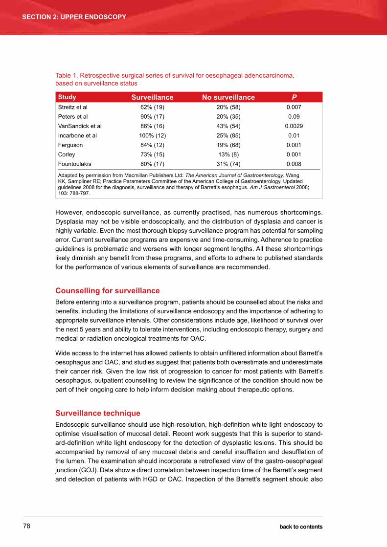
BARReTT’S OeSOPHAGuS: DIAGNOSIS AND MANAGeMeNTDarren Pavey
SECTION 2: UPPER ENDOSCOPY
78 back to contents
Table 1. Retrospective surgical series of survival for oesophageal adenocarcinoma, based on surveillance status
Study Surveillance No surveillance PStreitz et al 62% (19) 20% (58) 0.007Peters et al 90% (17) 20% (35) 0.09VanSandick et al 86% (16) 43% (54) 0.0029Incarbone et al 100% (12) 25% (85) 0.01Ferguson 84% (12) 19% (68) 0.001Corley 73% (15) 13% (8) 0.001Fountoulakis 80% (17) 31% (74) 0.008
Adapted by permission from Macmillan Publishers Ltd: The American Journal of Gastroenterology. Wang KK, Sampliner Re; Practice Parameters Committee of the American College of Gastroenterology. updated guidelines 2008 for the diagnosis, surveillance and therapy of Barrett’s esophagus. Am J Gastroenterol 2008; 103: 788-797.
However, endoscopic surveillance, as currently practised, has numerous shortcomings. Dysplasia may not be visible endoscopically, and the distribution of dysplasia and cancer is highly variable. even the most thorough biopsy surveillance program has potential for sampling error. Current surveillance programs are expensive and time-consuming. Adherence to practice guidelines is problematic and worsens with longer segment lengths. All these shortcomings likely diminish any benefit from these programs, and efforts to adhere to published standards for the performance of various elements of surveillance are recommended.
Counselling for surveillanceBefore entering into a surveillance program, patients should be counselled about the risks and benefits, including the limitations of surveillance endoscopy and the importance of adhering to appropriate surveillance intervals. Other considerations include age, likelihood of survival over the next 5 years and ability to tolerate interventions, including endoscopic therapy, surgery and medical or radiation oncological treatments for OAC.
Wide access to the internet has allowed patients to obtain unfiltered information about Barrett’s oesophagus and OAC, and studies suggest that patients both overestimate and underestimate their cancer risk. Given the low risk of progression to cancer for most patients with Barrett’s oesophagus, outpatient counselling to review the significance of the condition should now be part of their ongoing care to help inform decision making about therapeutic options.
Surveillance techniqueendoscopic surveillance should use high-resolution, high-definition white light endoscopy to optimise visualisation of mucosal detail. Recent work suggests that this is superior to stand-ard-definition white light endoscopy for the detection of dysplastic lesions. This should be accompanied by removal of any mucosal debris and careful insufflation and desufflation of the lumen. The examination should incorporate a retroflexed view of the gastro-oesophageal junction (GOJ). Data show a direct correlation between inspection time of the Barrett’s segment and detection of patients with HGD or OAC. Inspection of the Barrett’s segment should also

79
BARReTT’S OeSOPHAGuS: DIAGNOSIS AND MANAGeMeNTDarren Pavey
SECTION 2: UPPER ENDOSCOPY
back to contents
involve careful attention to the right hemisphere of the segment, extending from the 12 o’clock to 6 o’clock location, where early cancer appears to have a predilection for developing.
The aim of surveillance is detection of dysplasia. The description of dysplasia should use a standard five-tier system: (i) negative for dysplasia; (ii) indefinite for dysplasia; (iii) LGD; (iv) HGD; and (v) carcinoma. As active inflammation makes it more difficult to distinguish dyspla-sia from reparative changes, surveillance biopsies should only be performed after any active inflammation related to GORD is controlled with antisecretory therapy. The presence of ongoing erosive oesophagitis is a relative contraindication to performing surveillance biopsies. Once any inflammation related to GORD is controlled, obtaining systematic four-quadrant biopsy samples at 2 cm intervals along the entire length of the Barrett’s segment remains the standard for endoscopic surveillance of non-dysplastic Barrett’s oesophagus.
Subtle mucosal abnormalities, such as ulceration, erosion, plaque, nodule, stricture or other luminal irregularity in the Barrett’s segment, should also be sampled, as such lesions have an association with underlying cancer. Mucosal abnormalities encountered during surveillance of patients with known dysplasia should undergo endoscopic mucosal resection (eMR). eMR will change the diagnosis in about half of patients, when compared with endoscopic biopsies. Interobserver agreement among pathologists is also improved. The safety of systematic endo-scopic biopsy protocols has been demonstrated. The addition of routine cytological sampling appears to add little to surveillance endoscopic biopsies. Currently, the finding of subsquamous Barrett’s oesophagus on surveillance biopsy samples of an untreated patient does not change patient management, based on the most advanced histological findings from the combination of targeted and random biopsies.
Advanced endoscopic imaging techniquesVarious enhancements to endoscopic imaging with white light endoscopy have been studied to allow for detailed inspection of the Barrett’s segment. This “electronic chromoendoscopy” allows for detailed imaging of the mucosal and vascular surface patterns in Barrett’s oesopha-gus without the need for chromoendoscopy dye sprays. This may be accomplished with either narrow band imaging (NBI), which uses optical filters to narrow the bandwidth of white light to blue light, or by post-processing software systems to accomplish similar visualisation. Most of the literature has examined NBI in conjunction with magnification endoscopy. A randomised clinical trial of NBI versus high-definition white light endoscopy found no difference in the number of patients detected with dysplasia or neoplasia, but fewer biopsies were required for NBI. A recent meta-analysis also suggests that the use of enhanced imaging may increase detection of dysplasia. Other image enhancement techniques have been studied, including methylene blue staining, acetic acid staining, indigo carmine staining, autofluorescence endoscopy, confocal laser endomicroscopy, volumetric laser endomicroscopy, spectroscopy and molecular imaging, but none of these methods appear ready for widespread clinical use at present.
Importance of confirmation of dysplasiaDysplasia remains the best clinically available marker of cancer risk in patients with Barrett’s oesophagus. Although there is considerable interobserver variability in the interpretation of dysplasia, there is reasonable interobserver agreement among gastrointestinal pathologists

BARReTT’S OeSOPHAGuS: DIAGNOSIS AND MANAGeMeNTDarren Pavey
SECTION 2: UPPER ENDOSCOPY
80 back to contents
for the extremes of dysplasia, namely IM without dysplasia and HGD or OAC. There is more difficulty in the interpretation of “indefinite for dysplasia” and LGD. Recent studies show that 73% to 85% of patients diagnosed with LGD in the community had their diagnoses downgraded to no dysplasia or indefinite for dysplasia after review by two experienced gastrointestinal pathol-ogists. Other studies suggest that community-based pathologists have difficulty in interpreting both non-dysplastic Barrett’s oesophagus and dysplasia. All readings of dysplasia should be confirmed by a second pathologist with extensive experience in interpreting Barrett’s oesophagus-associated neoplasia.
Surveillance intervalsSurveillance intervals are determined by the presence and grade of dysplasia and are currently governed by expert opinion. Given the low risk of progression of Barrett’s oesophagus to OAC, surveillance at 3- to 5-year intervals remains reasonable in patients without dysplasia (Table 2).
Table 2. Dysplasia grade and surveillance interval
Dysplasia Documentation Follow-upNone ● Two OGDs with biopsy within 1 year ● endoscopy every 3–5 yearsLow-grade ● Highest grade on repeat OGD with
biopsies within 6 months ● expert pathologist confirmation
● 1-year interval until no dysplasia on two examinations
High-grade ● Mucosal irregularity ● Repeat OGD with biopsies to rule
out OAC within 3 months ● expert pathologist confirmation
● endoscopic resection ● Continued 3-month surveillance or
intervention based on results and patient
OGD = oesophagogastroduodenoscopy. OAC = oesophageal adenocarcinoma. Adapted by permission from Macmillan Publishers Ltd: The American Journal of Gastroenterology. Wang KK, Sampliner Re; Practice Parameters Committee of the American College of Gastroenterology. updated guidelines 2008 for the diagnosis, surveillance and therapy of Barrett’s esophagus. Am J Gastroenterol 2008; 103: 788-797.
There is a paucity of data to guide the management of Barrett’s oesophagus patients with biopsy samples indefinite for dysplasia. It is reasonable to use double-dose PPI therapy to decrease any ongoing inflammation. A retrospective study found that being indefinite for dysplasia was associated with a similar risk of progression to cancer as LGD. More recent data suggest an especially high risk of progression to higher grades of dysplasia within the first year of diag-nosis, but a risk comparable to non-dysplastic Barrett’s oesophagus after the first year. The progression risk may be more pronounced in multifocal (i.e. indefinite for dysplasia in biopsy samples from more than one level of the oesophagus) than focal indefinite for dysplasia. Thus, surveillance in these patients should follow the recommendations for LGD described below.
If LGD is found, it should be confirmed by a second pathologist with expertise in Barrett’s oesophagus. Patients should receive aggressive antisecretory PPI therapy for reflux disease to decrease the changes associated with regeneration or inflammation. A repeat endoscopy after optimisation of acid suppression therapy may result in downgrading of the LGD diagnosis. If LGD is confirmed and endoscopic therapy not performed, annual surveillance is recommended until two examinations in a row are negative for dysplasia, after which surveillance intervals for non-dysplastic Barrett’s oesophagus can be followed. A protocol of four-quadrant biopsy

81
BARReTT’S OeSOPHAGuS: DIAGNOSIS AND MANAGeMeNTDarren Pavey
SECTION 2: UPPER ENDOSCOPY
back to contents
samples at 1 cm intervals is advisable, given that anatomical studies suggest dysplasia can occur in a mosaic pattern and involve small portions of the overall surface area of the oesoph-agus. eMR should be performed if any mucosal abnormality is present.
If HGD is found, the diagnosis should be confirmed by a second pathologist with experience in gastrointestinal pathology. The presence of any mucosal abnormality warrants eMR to maximise staging accuracy. If HGD is confirmed, endoscopic intervention is warranted, as described below.
Biomarkers of increased riskGiven the limitations of endoscopic surveillance and histological findings of dysplasia as a risk stratification tool, molecular markers to identify patients at increased risk of progression have been studied. DNA content abnormalities, chromosomal abnormalities, gene mutations, methylation changes and clonal diversity measurements define patients at increased risk of progression to cancer. These genetic abnormalities appear to occur early in disease develop-ment. However, given the complexity and diversity of alterations observed in the progression sequence, a panel of biomarkers may be required for risk stratification. No biomarkers are yet ready for clinical practice.
Treatment of Barrett’s oesophagus
Endoscopic therapyAdvances in endoscopic therapy in the past decade have broadened the pool of patients with Barrett’s oesophagus who may be considered for intervention, and also diminished the need for oesophagectomy in this patient population. Consideration of any endoscopic therapy begins with a close inspection of the Barrett’s oesophagus mucosa. Identification of mucosal irregularities, including nodularity, ulceration or flat but irregular mucosal contour, is essential to detecting the areas of highest yield for neoplasia. The adjunct use of a narrow light spectrum imaging technology such as NBI may aid in detecting mucosal irregularity. If irregularity is detected, the next management step should be eMR or endoscopic submucosal dissection, both for thera-peutic benefit and to allow staging of the lesion.
Figure 1 demonstrates the recommended actions for surveillance endoscopy of non-nodular Barrett’s oesophagus.
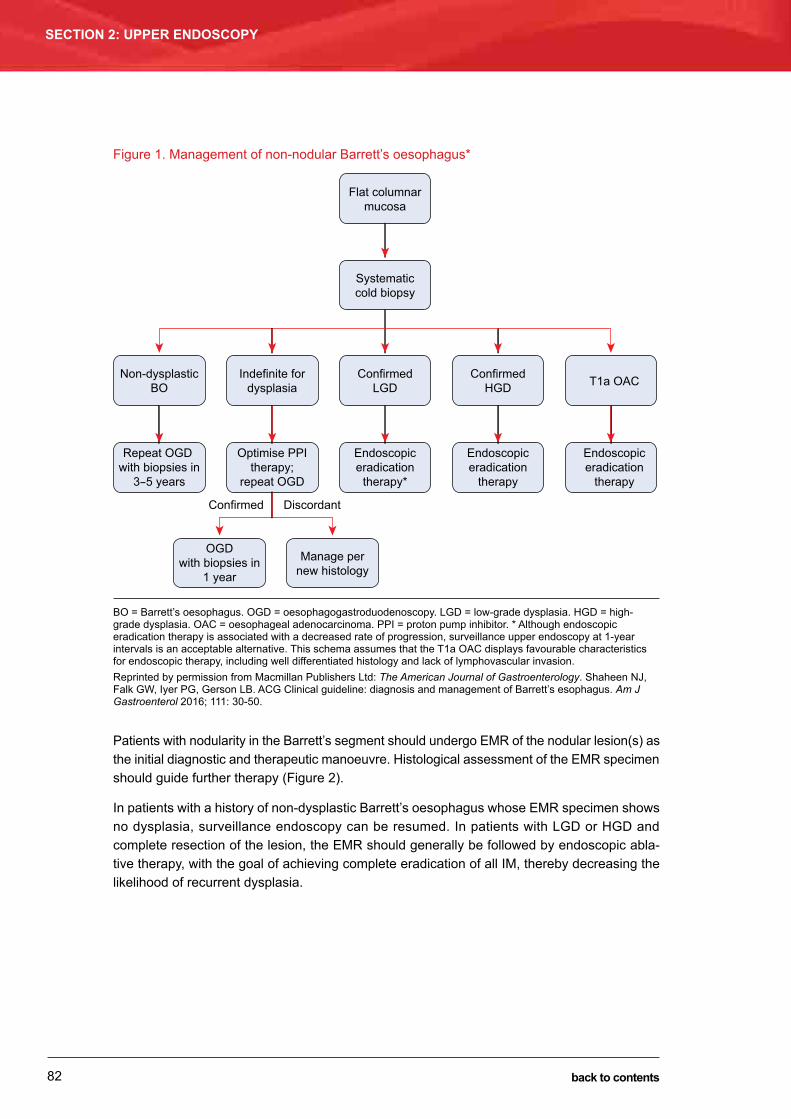
BARReTT’S OeSOPHAGuS: DIAGNOSIS AND MANAGeMeNTDarren Pavey
SECTION 2: UPPER ENDOSCOPY
82 back to contents
Figure 1. Management of non-nodular Barrett’s oesophagus*
Flat columnarmucosa
Systematiccold biopsy
Non-dysplasticBO
Indefinite fordysplasia
ConfirmedLGD
ConfirmedHGD T1a OAC
Repeat OGD with biopsies in
3-5 years
Optimise PPItherapy;
repeat OGD
Confirmed Discordant
Endoscopiceradication
therapy*
OGDwith biopsies in
1 year
Manage pernew histology
Endoscopiceradication
therapy
Endoscopiceradication
therapy
BO = Barrett’s oesophagus. OGD = oesophagogastroduodenoscopy. LGD = low-grade dysplasia. HGD = high-grade dysplasia. OAC = oesophageal adenocarcinoma. PPI = proton pump inhibitor. * Although endoscopic eradication therapy is associated with a decreased rate of progression, surveillance upper endoscopy at 1-year intervals is an acceptable alternative. This schema assumes that the T1a OAC displays favourable characteristics for endoscopic therapy, including well differentiated histology and lack of lymphovascular invasion.Reprinted by permission from Macmillan Publishers Ltd: The American Journal of Gastroenterology. Shaheen NJ, Falk GW, Iyer PG, Gerson LB. ACG Clinical guideline: diagnosis and management of Barrett’s esophagus. Am J Gastroenterol 2016; 111: 30-50.
Patients with nodularity in the Barrett’s segment should undergo eMR of the nodular lesion(s) as the initial diagnostic and therapeutic manoeuvre. Histological assessment of the eMR specimen should guide further therapy (Figure 2).
In patients with a history of non-dysplastic Barrett’s oesophagus whose eMR specimen shows no dysplasia, surveillance endoscopy can be resumed. In patients with LGD or HGD and complete resection of the lesion, the eMR should generally be followed by endoscopic abla-tive therapy, with the goal of achieving complete eradication of all IM, thereby decreasing the likelihood of recurrent dysplasia.

83
BARReTT’S OeSOPHAGuS: DIAGNOSIS AND MANAGeMeNTDarren Pavey
SECTION 2: UPPER ENDOSCOPY
back to contents
Figure 2. Management of nodular Barrett’s oesophagus
Endoscopically visible
nodularity in BO
Endoscopic mucosalresection
Low-gradedysplasia
High-gradedysplasia
T1a OAC
Yes
No
T1b OAC
Endoscopicablativetherapy*
Endoscopicablativetherapy
Favourablehistology?
Endoscopicablativetherapy
Discussion atmultidisciplinaryoncology group
BO = Barrett’s oesophagus. OAC = oesophageal adenocarcinoma. * Few data exist on the clinical course of patients with low-grade dysplasia managed by endoscopic surveillance after endoscopic mucosal resection (eMR), although this is an alternative treatment strategy. endoscopic submucosal dissection is an alternative to eMR. Favourable histology consists of no lymphatic or vascular invasion and moderate to well differentiated disease. Reprinted by permission from Macmillan Publishers Ltd: The American Journal of Gastroenterology. Shaheen NJ, Falk GW, Iyer PG, Gerson LB. ACG Clinical guideline: diagnosis and management of Barrett’s esophagus. Am J Gastroenterol 2016; 111: 30-50.
In patients with non-nodular Barrett’s oesophagus, the utility of ablative therapy is becoming clearer. In patients with Barrett’s oesophagus and HGD, ablative therapy should be preferred over either oesophagectomy or intensive endoscopic surveillance because of its proven efficacy and a side-effect profile superior to surgery. Recent data show that in patients with Barrett’s oesophagus and LGD confirmed by a second pathologist, ablative therapy results in a statis-tically and clinically significant reduction in progression to the combined end point of HGD or OAC, or to OAC alone.
In contrast, in patients with non-dysplastic Barrett’s oesophagus, recent data suggest lower rates of progression than previously believed. Given the low rate of progression, the low but real rate of complications of endoscopic therapy and the costs associated with its delivery, ablative therapy cannot be recommended in these patients. Whether these therapies are warranted in patients judged to have a higher lifetime risk of cancer, such as those with familial Barrett’s oesophagus or OAC and young patients with long segments of Barrett’s oesophagus, is unclear.
In patients with OAC, depth of invasion decides the curative potential of endoscopic therapy (Figure 3). Lesions confined to the mucosa (T1a) have a very low rate of lymphatic involvement

BARReTT’S OeSOPHAGuS: DIAGNOSIS AND MANAGeMeNTDarren Pavey
SECTION 2: UPPER ENDOSCOPY
84 back to contents
(< 1%), making these lesions optimally treated by eMR, followed by a mucosal ablative therapy to eradicate the remaining Barrett’s oesophagus. Lesions with superficial submucosal invasion (T1bSM1) have conflicting data regarding the likelihood of lymph node invasion (approximate risk of lymph node invasion: SM1, 20%; SM2, 35%; SM3, 50%), making consideration of surgery and/or multimodality therapy appropriate. However, in patients at high risk of complications with oesophagectomy, endoscopic therapy can be considered as an alternative. If endoscopic therapy is being considered for definitive therapy for such patients, well differentiated tumours, as well as those with no lymphovascular invasion, have the best prognosis.
Lesions with invasion into the mid or deep submucosa (T1bSM2 or T1bSM3) are associated with high rates of lymphatic involvement. endoscopic therapy performed on these lesions should not be assumed to be curative. Currently, the added value of endoscopic therapy as part of a scheme of multimodality therapy (e.g. endoscopic therapy plus chemotherapy and/or radiotherapy) is not well described in the literature.
Figure 3. TNM staging of oesophageal cancer
Reprinted by permission of the American Joint Committee on Cancer (AJCC), Chicago, Illinois, and Springer, New York, uSA, from the AJCC Cancer Staging Manual, 7th edition, 2010.
The role of imaging modalities such as endoscopic ultrasound (euS), positron emission tomo-graphy (PeT) and computed tomography (CT) scanning is becoming clearer. As substantial minorities of patients with superficial OAC will be incorrectly T-staged by euS, the routine use of euS before eMR is unwarranted, as clinical decision making will rest with the eMR findings. euS may have a limited role in endoscopic therapy of early oesophageal neoplasia in the setting of T1b disease. For patients with T1b disease being considered for endoscopic therapy, evidence of locoregional lymph node involvement, especially if substantiated by fine needle aspiration, means any attempt at endoscopic therapy would be palliative and that other

85
BARReTT’S OeSOPHAGuS: DIAGNOSIS AND MANAGeMeNTDarren Pavey
SECTION 2: UPPER ENDOSCOPY
back to contents
modalities need to be invoked for curative intent. Given the low likelihood of distant involvement in intramucosal (T1a) cancer or patients with dysplastic Barrett’s oesophagus, PeT–CT has no demonstrated benefit in these clinical settings. PeT–CT may have value in detecting distant involvement after a diagnosis of T1b disease.
eMR is not adequate as sole therapy for T1a or T1b OAC. up to a third of patients treated with eMR who achieve complete resection of the primary lesion will subsequently develop recurrent HGD or OAC. Whether these subsequent lesions represent undetected metachronous lesions or a field effect in the susceptible patient is unclear. However, endoscopic ablative treatment of the remainder of the Barrett’s oesophagus markedly decreases this risk. Therefore, all patients with successful resection of a T1a OAC, as well as any T1b lesions selected for endoscopic therapy, should undergo subsequent ablation of the remainder of the Barrett’s segment.
Successful endoscopic ablative therapy is defined as complete eradication of all dysplasia, as well as all IM, in the tubular oesophagus. To demonstrate this outcome, biopsy specimens in four quadrants at the GOJ, as well as every 1 cm through the extent of previous Barrett’s oesophagus, are taken. In addition, as several case series report occurrence of neoplasia in the cardia or at the GOJ after successful ablative therapy, surveillance biopsies of the cardia should be routinely performed.
The decision of when to say that endoscopic ablative therapy has failed in a patient depends on the patient’s clinical situation, the amount of progress made with initial attempts at ablation and the likely mechanism of failure. Cohort studies show that even among patients who have undergone four sessions of radiofrequency ablation (RFA) without complete eradication of IM (CeIM), more than half eventually attain this goal with subsequent therapy, suggesting that a firm cut-off for defining failure is not advisable.
A wide variety of ablative modalities have been reported to be effective in eradicating IM. Currently, level 1 evidence for prevention of incident cancer exists in three clinical scenarios: photodynamic therapy in the setting of Barrett’s oesophagus with HGD, RFA in the setting of HGD (Figures 4 and 5), and RFA in the setting of LGD. Given the large body of data supporting the safety and efficacy of RFA, and the costs and side-effect profile of photodynamic therapy, RFA appears to currently be the preferred therapy for most patients.

BARReTT’S OeSOPHAGuS: DIAGNOSIS AND MANAGeMeNTDarren Pavey
SECTION 2: UPPER ENDOSCOPY
86 back to contents
Figure 4. endoscopic appearance of a circumferential ablation procedure using the HALO360 system
C4M4 Barrett’s oesophagus with high-grade dysplasia (A). HALO360 ablation balloon (Medtronic) positioned 1 cm above the maximum extent of the Barrett’s oesophagus (B) and after being inflated (C). Ablation effect immediately after deflation of the balloon (D), after ablation of the whole Barrett’s oesophagus (e) and after cleaning off the coagulum (F). Reprinted courtesy of Georg Thieme Verlag KG, Stuttgart, New York: Gondrie JJ, Pouw Re, Sondermeijer CM, et al. Stepwise circumferential and focal ablation of Barrett’s esophagus with high grade dysplasia: results of the first prospective series of 11 patients. Endoscopy 2008; 40: 359-369.
Figure 5. endoscopic treatment of a C4M5 Barrett’s oesophagus with a combination of endoscopic resection and ablation therapy
endoscopic treatment of a C4M5 Barrett’s oesophagus with high-grade dysplasia (HGD) and a visible lesion at the lower end of the Barrett’s segment (A, B), with a combination of endoscopic resection and ablation therapy. The visible abnormality was first focally removed with endoscopic resection (C). The resection specimen showed a radically resected well differentiated mucosal cancer (D); biopsy samples from the remaining Barrett’s oesophagus showed HGD. The Barrett’s oesophagus was subsequently ablated using the HALO360 system (Medtronic) supplemented with focal ablation with the HALO90 (e) system (not shown) for a complete endoscopic and histological removal of all dysplasia and all Barrett’s oesophagus without any stenosis (F–H). Reprinted courtesy of Georg Thieme Verlag KG, Stuttgart, New York: Gondrie JJ, Pouw Re, Sondermeijer CM, et al. Stepwise circumferential and focal ablation of Barrett’s esophagus with high grade dysplasia: results of the first prospective series of 11 patients. Endoscopy 2008; 40: 359-369.

87
BARReTT’S OeSOPHAGuS: DIAGNOSIS AND MANAGeMeNTDarren Pavey
SECTION 2: UPPER ENDOSCOPY
back to contents
Surgical therapySeveral studies have investigated the relative value of surgical antireflux procedures in prevent-ing OAC in the setting of Barrett’s oesophagus, with conflicting results. Given the weak nature of the data, along with the overall very low incidence of cancer in patients with non-dysplastic Barrett’s oesophagus, antireflux surgery should not be considered as an antineoplastic measure in these patients. The indications for this procedure in patients with Barrett’s oesophagus are the same as those in general GORD patients — principally, GORD symptoms or oesophagitis not well controlled by medical therapy. With respect to optimising medical therapy, dosages of PPI beyond twice daily have not shown a beneficial effect in patients with Barrett’s oesophagus. Once-daily PPI therapy is recommended for patients with Barrett’s oesophagus unless GORD symptoms require twice-daily PPIs for adequate symptom control.
Oesophagectomy has a well established role in the care of patients with Barrett’s oesophagus and OAC. It is the treatment of choice for fit candidates with T1bSM2–3 disease, either alone or in combination with radiation therapy and/or chemotherapy. In patients with T1aSM1 or T1bSM1 OAC and unfavourable prognostic factors, such as poor differentiation or lymphovas-cular invasion, surgical consultation should be obtained.
Management of Barrett’s oesophagus after endoscopic therapy
Following CeIM, the recurrence rate for IM is not inconsiderable, with some cohorts having rates of ≥ 20% at 2–3 years after CeIM. Although most recurrences are non-dysplastic, up to a quarter may be dysplastic, including OAC. Cohorts treated with either combination therapy (eMR followed by ablation) or single modality therapy (eMR alone) have reported comparable recurrence rates. Recurrence also appears to be similar across different ablation modalities, with similar rates being described after cryotherapy and photodynamic therapy for the treatment of dysplastic Barrett’s oesophagus.
Careful endoscopic surveillance with biopsies is recommended after CeIM to detect recurrent IM. Careful inspection of both tubular oesophagus (in the region of the prior Barrett’s segment) and the GOJ (on antegrade and retroflexed views) is important. Both the interval of these examinations and the biopsy protocol are based on expert opinion and on intervals reported in published cohort studies. endoscopic surveillance for patients with baseline HGD every 3 months in the first year after CeIM, every 6 months in the second year and annually thereafter is currently recommended. For patients with baseline LGD, endoscopic surveillance is rec-ommended every 6 months in the first year after CeIM, and annually thereafter. Most studies use four-quadrant biopsy samples every 1 cm throughout the previous Barrett’s segment, with additional targeted biopsy sampling of any endoscopic abnormality, although this approach has not been compared with other biopsy regimens. There is currently no evidence to support discontinuing surveillance after multiple negative surveillance endoscopies, given reports of recurrent neoplasia several years after CeIM.

BARReTT’S OeSOPHAGuS: DIAGNOSIS AND MANAGeMeNTDarren Pavey
SECTION 2: UPPER ENDOSCOPY
88 back to contents
Biopsy samples from the tubular oesophagus and GOJ should be obtained in separate bottles to allow localisation and treatment of recurrent Barrett’s oesophagus. The optimal number of biopsy samples needed for adequate surveillance is unknown. Despite concerns regarding depth of biopsy sampling after ablation, the prevalence of subsquamous Barrett’s oesophagus after ablation is variable, with rates ranging from 0.9% after RFA to 14.2% after photodynamic therapy. Although neosquamous epithelium may be more permeable than normal squamous epithelium, it does not appear to harbour genetic abnormalities.
When detected by surveillance, most recurrent metaplasia and dysplasia is amenable to endoscopic therapy, including eMR and additional ablation. However, a few cases requiring oesophagectomy for invasive carcinoma have been reported. Predictors of recurrence are not well defined, with some studies suggesting that older age, a longer pre-ablation Barrett’s oesophagus segment, presence of a larger hiatal hernia and higher grade of dysplasia before ablation are associated with higher rates of recurrence.
There is some evidence from uncontrolled observational studies that incomplete control of reflux may be associated with increased recurrence rates after successful endotherapy. Treatment of reflux after successful ablation should follow the same principles as outlined above. The goal of medical treatment should be the control of symptoms of reflux and the prevention or healing of oesophagitis.
Conclusion
Care of the patient with Barrett’s oesophagus has evolved rapidly in the past decade. The recommendations given here should not be construed as practice standards or quality meas-ures — clinical circumstances should dictate the best care for each patient. Recent changes in these recommendations include the expanded use of endoscopic ablative therapy, especially its extension to patients with LGD based on evidence showing a diminished risk of progression or adenocarcinoma after treatment. There has been further refinement of screening recom-mendations, based on data showing both a lower risk of OAC in patients with non-dysplastic Barrett’s oesophagus and a better understanding of the impact of sex and anthropomorphics on risk. The most important change is the recommendation that women with GORD symptoms should no longer undergo routine screening. Finally, surveillance recommendations have been attenuated to recognise the relatively rare occurrence of progression in non-dysplastic Barrett’s oesophagus, as well as the unclear nature of benefit inherent in endoscopic surveillance.
It is likely that the development of several technologies will cause further evolution in the care of patients with Barrett’s oesophagus. Several areas appear poised for paradigm-shifting advances, including the evolution of biomarkers to predict risk, the use of advanced imaging and biomolecular technologies to allow recognition of areas of neoplasia within Barrett’s oesoph-agus, and the advent of less invasive and less expensive modalities for screening patients.

89
BARReTT’S OeSOPHAGuS: DIAGNOSIS AND MANAGeMeNTDarren Pavey
SECTION 2: UPPER ENDOSCOPY
back to contents
Further reading1 Shaheen NJ, Falk GW, Iyer PG, Gerson LB. ACG clinical guideline: diagnosis and management of Barrett’s
esophagus. Am J Gastroenterol 2016; 111: 30-50.
2 Whiteman DC, Appleyard M, Bahin FF, et al. Australian clinical practice guidelines for the diagnosis and management of Barrett’s esophagus and early esophageal adenocarcinoma. J Gastroenterol Hepatol 2015; 30: 804-820.
3 Fitzgerald RC, di Pietro M, Ragunath K, et al. British Society of Gastroenterology guidelines on the diagnosis and management of Barrett’s oesophagus. Gut 2014; 63: 7-42.
4 Jung KW, Talley NJ, Romero Y. epidemiology and natural history of intestinal metaplasia of the gastroesophageal junction and Barrett’s esophagus: a population-based study. Am J Gastroenterol 2011; 106: 1447-1455.
5 Sharma P, Dent J, Armstrong D. The development and validation of an endoscopic grading system for Barrett’s esophagus: the Prague C & M criteria. Gastroenterology 2006; 131: 1392-1399.
6 Sharma P, Falk GW, Weston AP, et al. Dysplasia and cancer in a large multicenter cohort of patients with Barrett’s esophagus. Clin Gastroenterol Hepatol 2006; 4: 566-572.
7 Spechler SJ. Barrett esophagus and risk of esophageal cancer: a clinical review. JAMA 2013; 310: 627-636.
8 Hvid-Jensen F, Pedersen L, Drewes AM. Incidence of adenocarcinoma among patients with Barrett’s esophagus. N Engl J Med 2011; 365: 1375-1383.
9 Incarbone R, Bonavina L, Saino G. Outcome of esophageal adenocarcinoma detected during endoscopic biopsy surveillance for Barrett’s esophagus. Surg Endosc 2002; 16: 263-266.
10 Ferguson MK, Durkin A. Long-term survival after esophagectomy for Barrett’s adenocarcinoma in endoscopically surveyed and nonsurveyed patients. J Gastrointest Surg 2002; 6: 29-35.
11 Verbeek Re, Leenders M, Ten Kate FJ. Surveillance of Barrett’s esophagus and mortality from esophageal adenocarcinoma: a population-based cohort study. Am J Gastroenterol 2014; 109: 1215-1222.
12 Bhat SK, McManus DT, Coleman HG. Oesophageal adenocarcinoma and prior diagnosis of Barrett’s oesophagus: a population-based study. Gut 2015; 64: 20-25.
13 Corley DA, Mehtani K, Quesenberry C. Impact of endoscopic surveillance on mortality from Barrett’s esophagus-associated esophageal adenocarcinomas. Gastroenterology 2013; 145: 312-319.
14 Sikkema M, de Jonge PJ, Steyerberg eW. Risk of esophageal adenocarcinoma and mortality in patients with Barrett’s esophagus: a systematic review and meta-analysis. Clin Gastroenterol Hepatol 2010; 8: 235-244.
15 Sami SS, Subramanian V, Butt WM, et al. High definition versus standard definition white light endoscopy for detecting dysplasia in patients with Barrett’s esophagus. Dis Esophagus 2015; 28: 742-749.
16 enestvedt BK, Lugo R, Guarner-Argente C. Location, location, location: does early cancer in Barrett’s esophagus have a preference? Gastrointest Endosc 2013; 78: 462-467.
17 Kumaravel A, Lopez R, Brainard J. Brush cytology vs. endoscopic biopsy for the surveillance of Barrett’s esophagus. Endoscopy 2010; 42: 800-805.
18 Sharma P, Hawes RH, Bansal A. Standard endoscopy with random biopsies vs. narrow band imaging targeted biopsies in Barrett’s oesophagus: a prospective, international, randomised controlled trial. Gut 2013; 62: 15-21.
19 Qumseya BJ, Wang H, Badie N. Advanced imaging technologies increase detection of dysplasia and neoplasia in patients with Barrett’s esophagus: a meta-analysis and systematic review. Clin Gastroenterol Hepatol 2013; 11: 1562-1570.
20 Curvers WL, ten Kate FJ, Krishnadath KK. Low-grade dysplasia in Barrett’s esophagus: overdiagnosed and underestimated. Am J Gastroenterol 2010; 105: 1523-1530.
21 Horvath B, Singh P, Xie H. Risk for esophageal neoplasia in Barrett’s esophagus patients with mucosal changes indefinite for dysplasia. J Gastroenterol Hepatol 2014; 30: 262-267.
22 Fels elliott DR, Fitzgerald RC. Molecular markers for Barrett’s esophagus and its progression to cancer. Curr Opin Gastroenterol 2013; 29: 437-445.

BARReTT’S OeSOPHAGuS: DIAGNOSIS AND MANAGeMeNTDarren Pavey
SECTION 2: UPPER ENDOSCOPY
90 back to contents
23 Merlo LM, Shah NA, Li X. A comprehensive survey of clonal diversity measures in Barrett’s esophagus as biomarkers of progression to esophageal adenocarcinoma. Cancer Prev Res (Phila) 2010; 3: 1388-1397.
24 Weaver JM, Ross-Innes CS, Shannon N. Ordering of mutations in preinvasive disease stages of esophageal carcinogenesis. Nat Genet 2014; 46: 837-843.
25 Manner H, Pech O, Heldmann Y. efficacy, safety, and long-term results of endoscopic treatment for early stage adenocarcinoma of the esophagus with low-risk sm1 invasion. Clin Gastroenterol Hepatol 2013; 11: 630-635.
26 Shaheen NJ, Sharma P, Overholt BF. Radiofrequency ablation in Barrett’s esophagus with dysplasia. N Engl J Med 2009; 360: 2277-2288.
27 Phoa KN, van Vilsteren FG, Weusten BL, et al. Radiofrequency ablation vs endoscopic surveillance for patients with Barrett esophagus and low-grade dysplasia: a randomized clinical trial. JAMA 2014; 311: 1209-1217.
28 Bulsiewicz WJ, Kim HP, Dellon eS. Safety and efficacy of endoscopic mucosal therapy with radiofrequency ablation for patients with neoplastic Barrett’s esophagus. Clin Gastroenterol Hepatol 2013; 11: 636-642.
29 Pohl H, Wrobel K, Bojarski C. Risk factors in the development of esophageal adenocarcinoma. Am J Gastroenterol 2013; 108: 200-207.
30 Pech O, Bollschweiler e, Manner H. Comparison between endoscopic and surgical resection of mucosal esophageal adenocarcinoma in Barrett’s esophagus at two high-volume centers. Ann Surg 2011; 254: 67-72.
31 Nentwich MF, von Loga K, Reeh M. Depth of submucosal tumor infiltration and its relevance in lymphatic metastasis formation for T1b squamous cell and adenocarcinomas of the esophagus. J Gastrointest Surg 2014; 18: 242-249.
32 Bulsiewicz WJ, Dellon eS, Rogers AJ. The impact of endoscopic ultrasound findings on clinical decision making in Barrett’s esophagus with high-grade dysplasia or early esophageal adenocarcinoma. Dis Esophagus 2014; 27: 409-417.
33 Thosani N, Singh H, Kapadia A, et al. Diagnostic accuracy of euS in differentiating mucosal versus submucosal invasion of superficial esophageal cancers: a systematic review and meta-analysis. Gastrointestinal Endoscopy 2012; 75: 242-253.
34 Orman eS, Kim HP, Bulsiewicz WJ. Intestinal metaplasia recurs infrequently in patients successfully treated for Barrett’s esophagus with radiofrequency ablation. Am J Gastroenterol 2013; 108: 187-195.
35 Shaheen NJ, Overholt BF, Sampliner Re. Durability of radiofrequency ablation in Barrett’s esophagus with dysplasia. Gastroenterology 2011; 141: 460-468.
36 Pasricha S, Bulsiewicz WJ, Hathorn Ke. Durability and predictors of successful radiofrequency ablation for Barrett’s esophagus. Clin Gastroenterol Hepatol 2014; 12: 1840-1847.
37 Titi M, Overhiser A, ulusarac O. Development of subsquamous high-grade dysplasia and adenocarcinoma after successful radiofrequency ablation of Barrett’s esophagus. Gastroenterology 2012; 143: 564-566.
38 Guarner-Argente C, Buoncristiano T, Furth ee. Long-term outcomes of patients with Barrett’s esophagus and high-grade dysplasia or early cancer treated with endoluminal therapies with intention to complete eradication. Gastrointest Endosc 2013; 77: 190-199.
39 Haidry RJ, Dunn JM, Butt MA. Radiofrequency ablation and endoscopic mucosal resection for dysplastic Barrett’s esophagus and early esophageal adenocarcinoma: outcomes of the uK national halo RFA registry. Gastroenterology 2013; 145: 87-95.
40 Gray NA, Odze RD, Spechler SJ. Buried metaplasia after endoscopic ablation of Barrett’s esophagus: a systematic review. Am J Gastroenterol 2011; 106: 1899-1908.
41 Pouw Re, Gondrie JJ, Rygiel AM. Properties of the neosquamous epithelium after radiofrequency ablation of Barrett’s esophagus containing neoplasia. Am J Gastroenterol 2009; 104: 1366-1373.
42 Yasuda K, Choi Se, Nishioka NS. Incidence and predictors of adenocarcinoma following endoscopic ablation of Barrett’s esophagus. Dig Dis Sci 2014; 59: 1560-1566.
43 Ferraris R, Fracchia M, Foti M. Barrett’s oesophagus: long-term follow-up after complete ablation with argon plasma coagulation and the factors that determine its recurrence. Aliment Pharmacol Ther 2007; 25: 835-840.
Pages 91-92 (used for notes in the hard copy) have been removed from the PDF edition of this handbook.

9375
Barrett’s oesophagus: diagnosis and managementDarren Pavey
Barrett’s oesophagus is a metaplastic change in the oesophageal epithelium thought to result from injury due to chronic exposure to reflux gastric content. Normal oesophageal squamous epithelium is replaced by specialised intestinal epithelium, also known as intestinal metaplasia (IM). Progression of non-dysplastic IM to low-grade dysplasia (LGD), high-grade dysplasia (HGD) and oesophageal adenocarcinoma (OAC) is well described. The incidence of OAC continues to rise, particularly among the older population.
Guidelines for the diagnosis and management of Barrett’s oesophagus have recently been pub-lished by the Cancer Council of Australia. Readers are referred to these guidelines for further background on the epidemiology and risk factors for Barrett’s oesophagus.
Diagnosis of Barrett’s oesophagus
Barrett’s oesophagus has traditionally been defined as the presence of at least 1 cm of meta-plastic columnar epithelium that replaces the stratified squamous epithelium normally lining the distal oesophagus. Segments < 1 cm have instead been classified as “specialised IM of the oesophagogastric junction”. This distinction is made due to the high interobserver variability, as well as a low risk of OAC in very short-segment disease.
The definition of Barrett’s oesophagus has varied based on the requirement for the presence of IM on endoscopic biopsy samples. Debate regarding the requirement for IM on biopsy from columnar-lined epithelium segments has derived from the apparently differential risk of devel-oping OAC in such epithelium containing or not containing IM. Large population-based cohort studies have shown a substantially lower OAC risk in patients with columnar metaplasia without IM, compared with those with IM. However, not all studies have corroborated this finding. The yield for IM correlates directly with the number of endoscopic biopsy samples obtained. In a
SECTION 2: UPPER ENDOSCOPY
CHAPTER 8
Endoscopic treatment of Barrett’s high-grade dysplasia and early oesophageal adenocarcinomaLuke Hourigan
The endoscopic approach to Barrett’s high-grade dysplasia (HGD) and early oesophageal adenocarcinoma (OAC) has undergone significant change over the past 10 years. Endoscopic management of high-grade dysplastic Barrett’s oesophagus and related early OAC is now accepted as first-line therapy. The most widely accepted endoscopic methods are transparent cap endoscopic mucosal resection (EMR) and radiofrequency ablation (RFA).
These endoscopic techniques have radically changed the approach to high-grade dysplastic Barrett’s oesophagus and early OAC. Before their advent, the management approach was based on endoscopic surveillance of Barrett’s mucosa, with eventual surgery for those with HGD or malignancy. The contemporary approach is surveillance with subsequent endoscopic management of Barrett’s dysplasia and early OAC. Oesophagectomy is now primarily required only for patients with adenocarcinoma invading the submucosa.
General principles
Endoscopic detection and the proper staging of patients with Barrett’s dysplasia or early OAC are essential before therapy.
Endoscopic resection is the cornerstone of endoscopic management of early OAC, as it enables histological correlation and optimal patient selection. Patients with submucosal invasion should
SECTION 2: UPPER ENDOSCOPY
CHAPTER 9
back to contents

SECTION 2: UPPER ENDOSCOPY
94 back to contents
be referred for surgery, as they have a 15%–30% risk of positive local lymph node involvement. This risk is minimal in patients with intramucosal adenocarcinoma or HGD.
The compromise for patients undergoing endoscopic management is the need to accept an intense regimen of initial endoscopic staging and treatment, followed by continued surveillance.
EMR is complemented by RFA, which is currently the most widely accepted ablation technique. RFA has shown an impressive efficacy and safety profile in multiple studies, and the neosqua-mous mucosa that develops after ablation has been shown to be free of genetic abnormalities. There is also a very low occurrence of residual areas of columnar mucosa underneath the neosquamous mucosa (buried glands). Treatment of low-grade dysplastic Barrett’s oesopha-gus is becoming more widely accepted. Potential RFA management of non-dysplastic Barrett’s oesophagus is a controversial issue, and it is too early to embrace RFA for this use.
Successful endoscopic management of high-grade dysplastic Barrett’s oesophagus and early oesophageal malignancy is dependent on good patient selection, accurate grading and staging of the Barrett’s segment, and access to expertise in pathological assessment and endoscopic skills.
Techniques
Endoscopic work-up of early Barrett’s neoplasiaThe goal of endoscopic surveillance of patients with Barrett’s oesophagus is the detection of early neoplastic lesions. There are three useful basic rules:
1 Use the best endoscope you have available.
2 You do not detect what you see, but you detect what you recognise.
3 Perform a systematic endoscopic inspection.
Essential endoscopic imaging modalities for detection of early neoplasia and Barrett’s oesoph-agus include high-definition endoscopy and narrow band imaging. Other useful technologies include chromoendoscopy, autofluorescence imaging and confocal microscopy.
The importance of endoscopic work-up and staging of patients with suspected early Barrett’s oesophageal neoplasia is in identifying patients who are eligible for endoscopic therapy and selecting those patients who require surgical management for curative treatment. The endo-scopic work-up should focus on identifying the most suspicious areas in the Barrett’s segment that subsequently need to be removed by endoscopic resection. EMR allows for optimal diag-nosis and staging, is potentially curative and guides the selection of patients for endoscopic therapy. Patients who are eligible for endoscopic therapy are those with HGD or well or mod-erately differentiated cancers limited to the mucosa without lymphovascular invasion.
Endoscopic ultrasound (EUS) has a limited role in the T- and N-staging of early neoplasia in Barrett’s oesophagus. Differentiation of intramucosal and submucosal lesions is much more

95
ENDOSCOPiC TREATMENT OF BARRETT’S HiGH-GRADE DYSPLASiA AND EARLY OESOPHAGEAL ADENOCARCiNOMALuke Hourigan
SECTION 2: UPPER ENDOSCOPY
back to contents
difficult with Barrett’s oesophagus than with squamous cell lesions. Ultimately, the best method for assessing the risk of lymph node involvement may not be EUS, but a diagnostic endoscopic resection with assessment of the depth of involvement.
Computed tomography and positron emission tomography scanning have minimal value in locoregional staging of early neoplasia in Barrett’s oesophagus.
Macroscopic appearances of early neoplasia in Barrett’s oesophagusVisible lesions in Barrett’s oesophagus may be classified according to the Paris classification (adopted from the Japanese gastric classification of gastric carcinoma) (Figure 1).
Depressed lesions carry a higher risk of submucosal invasion than flat or elevated lesions. Type 0-iii lesions always have deep submucosal invasion and are usually accompanied by a dense desmoplastic reaction. These are therefore not suitable for endoscopic treatment.
Figure 1. Paris classification of lesions in Barrett’s oesophagus, and examples
A: Is lesion. B: IIa lesion. C: Same IIa lesion (outlined).

SECTION 2: UPPER ENDOSCOPY
96 back to contents
Staging and depth of infiltration
The most important risk factor predicting lymph node metastasis in early neoplasia of Barrett’s oesophagus is the depth of infiltration of the lesion. Mucosal lesions (T1a) are subdivided into three categories: T1aM1, which is limited to the epithelial layer; T1aM2, with infiltration into the lamina propria; and T1aM3, with infiltration into the muscularis mucosa. Submucosal lesions (T1b) are divided into T1bSM1, with infiltration into the superficial third; T1bSM2, with infiltration into the intermediate third; and T1bSM3, with infiltration into the deepest third.
Patients with mucosal adenocarcinoma are considered to have a negligible risk of lymph node metastases and are therefore eligible for curative endoscopic treatment. Patients with deep sub-mucosal invasion are generally considered to be at risk of lymphatic dissemination and should be considered for surgical treatment. Recent studies suggest that T1bSM1 tumours show a much lower risk (0–8%) of lymph node involvement than T1bSM2–3 tumours (26%–67%). Given the risks of oesophagectomy and relatively poor outcome when there are positive lymph nodes, this suggests that endoscopic treatment may also be a valid treatment option for patients with superficial submucosal invasion evident in their endoscopic resection specimens. The manage-ment of all lesions with submucosal invasion should be discussed in a multidisciplinary setting.
Barrett’s histology
The presence of a double muscularis in Barrett’s oesophagus makes interpretation of dysplasia and adenocarcinoma difficult, particularly with biopsy specimens.
Determination of the precise location of neoplastic involvement in Barrett’s oesophagus is important, as several studies have shown that tumours that infiltrate into, and even through, the new muscularis mucosa have a very low risk of metastasis compared with tumours that penetrate the deeper (original) muscularis mucosa into the true submucosal layer.
Endoscopic mucosal resection
The two most commonly used devices for EMR are the original inoue device (Olympus) and the Duette Multi-Band Mucosectomy device (Cook Medical). There is also a new device, the Captivator EMR Device (Boston Scientific). it is possible to achieve good overlapping of margins during serial EMR with all devices.
Submucosal pre-injection is not essential with the Multi-Band Mucosectomy device, but it can be helpful, especially at the cardio-oesophageal junction.
Total endoscopic resection of Barrett’s oesophagus can be performed in a single session or as a staged procedure, with a hemi-circumferential resection in the first session, depending on the length of the Barrett’s oesophagus (Figure 2). Although circumferential EMR in a single ses-sion is associated with stricture formation, the circumstances for obtaining complete resection

97
ENDOSCOPiC TREATMENT OF BARRETT’S HiGH-GRADE DYSPLASiA AND EARLY OESOPHAGEAL ADENOCARCiNOMALuke Hourigan
SECTION 2: UPPER ENDOSCOPY
back to contents
are optimal in the first EMR session. Fibrosis with scar formation makes EMR more difficult in subsequent sessions. For a staged procedure, repeat endoscopy and EMR are performed at 4–6-week intervals until complete resection is achieved. Patients are treated with high-dose proton pump inhibitors during this period.
Figure 2. Single Duette resection of type iia early oesophageal adenocarcinoma (top), and two Duette resections required to remove a larger type iia oesophageal adenocarcinoma (bottom)
Results of total endoscopic resectioninitially, with localised EMR only, reported rates of recurrence or metachronous carcinomas at 34-month follow-up have been as high as 23%–30%. The higher recurrence rate was due to the presence of multifocal synchronous lesions overlooked by biopsy before EMR, as well as metachronous development of new foci of dysplasia. This confirms the need to resect or ablate the whole Barrett’s segment.
Fortunately, the frequency of complications associated with EMR is modest. Significant bleed-ing is observed in up to 14% of patients. However, most patients with bleeding are managed endoscopically in an outpatient setting. Perforation is observed in up to 1.8% of procedures, and may even be treated effectively with medical therapy (i.e. clips) in some circumstances.
Stricture formation is a common complication of extensive mucosal resection, with reported rates ranging from zero to 70%, and can be a major problem for both total endoscopic resec-tion and endoscopic submucosal dissection (ESD). Strictures can be treated endoscopically with balloon dilatation. To reduce the risk of stricture formation, total endoscopic resection may be carried out in multiple sessions. The use of total endoscopic resection should be limited to Barrett’s segments less than 5 cm in length, as application to longer segments can result in long strictures that are often difficult to dilate after total endoscopic resection.

SECTION 2: UPPER ENDOSCOPY
98 back to contents
En bloc resection with endoscopic submucosal dissection
ESD potentially permits en bloc resection of lesions larger than 15 mm that would otherwise require serial resection with cap-based techniques. The benefits of pursuing en bloc resection remain questionable, as serial EMR of larger lesions has been proven to be highly clinically efficacious while also being technically easier to perform. ESD is technically more demanding and time-consuming.
Ablation techniques
Photodynamic therapyPhotodynamic therapy was the first treatment shown to significantly decrease HGD and cancer in patients with Barrett’s oesophagus. However, its use has been limited, primarily because of its side effects, which include oesophageal strictures, cutaneous photosensitivity, chest pain, nausea and vomiting.
Argon plasma coagulationMost experts do not recommend argon plasma coagulation as a primary ablation method for dysplastic Barrett’s oesophagus. it is, however, widely used for very focal residual Barrett’s oesophagus after EMR and/or RFA.
Radiofrequency ablationCurrent data suggest that RFA is a very encouraging modality for eradication of Barrett’s oesophagus, with many appealing features. it has proven to be highly effective in eradicating intestinal metaplasia and its associated dysplasia. RFA has a low complication rate, preserves the functional integrity of the oesophagus and is relatively easy to apply. The regenerating neosquamous epithelium is free of the pre-existing oncogenetic alterations. For patients with intramucosal adenocarcinoma and HGD, RFA appears to be a valid and less invasive alternative to photodynamic therapy, argon plasma coagulation or oesophagectomy, albeit after a thorough endoscopic work-up and endoscopic resection of intramucosal adenocarcinoma. The role of RFA treatment in patients with low-grade dysplasia is becoming more widely accepted, but its role in non-dysplastic Barrett’s oesophagus requires more study.
99
ENDOSCOPiC TREATMENT OF BARRETT’S HiGH-GRADE DYSPLASiA AND EARLY OESOPHAGEAL ADENOCARCiNOMALuke Hourigan
SECTION 2: UPPER ENDOSCOPY
back to contents
Radiofrequency ablation procedures
Barrx 360 balloon catheters and Barrx focal ablation catheters Long-segment Barrett’s dysplasia usually requires initial step-wise circumferential balloon catheter ablation, with subsequent focal ablation of residual Barrett’s. initially, the oesophageal landmarks are recorded. After cleaning the oesophageal wall with acetylcysteine (1%) or vig-orous irrigation to remove excess mucus, the top of the gastric folds and the furthest proximal extent of the Barrett’s oesophagus are recorded for reference during the sizing and ablation procedures. A stiff guidewire or Savary metal wire is then introduced, and the endoscope is removed.
The next step is sizing the inner oesophageal diameter. This has traditionally been performed with a separate measuring catheter; however, new technology has seen the introduction of an “all in one” measuring and treatment balloon catheter — the Barrx 360 Express RFA Balloon Catheter (Covidien).
First circumferential ablation passThe RFA balloon catheter is introduced, followed by the endoscope. Under endoscopic vis-ualisation, the proximal margin of the electrode is placed 10 mm above the furthest proximal extent of the Barrett’s oesophagus. The balloon is inflated, and the electrode is activated via a foot switch. Moving from proximal to distal, the balloon is repositioned, allowing a small overlap of about 5–10 mm with the previous ablation zone, until the entire Barrett’s oesophagus has been ablated.
Cleaning procedure between ablation cyclesAfter the first ablation pass, the ablation catheter is removed and the electrode surface cleaned. Some operators prefer to fit a specialised transparent cap on the tip of the endoscope to remove the coagulum from the ablation zone before the second ablation pass. Although the extensive cleaning procedure requires extra procedure time, there are data that support this approach for increased efficacy. This is followed by a second ablation pass.
At a minimum of 8 weeks after the first circumferential ablation treatment, patients are sched-uled for a review gastroscopy to assess the need for a second ablation. Most patients with any residual circumferential Barrett’s oesophagus can have their ablation completed with a focal RFA device (i.e. Barrx 90, Barrx 60, Barrx Ultra Long or Barrx Channel device [Covidien]). Barrett’s segments less than 3 cm in length can usually be treated entirely with a Barrx focal ablation catheter.
Barrx focal ablation catheters allow direct application of RFA. The electrode is brought into close contact with the mucosa and kept in place. it is immediately activated, resulting in a double application of energy. Ablation of the entire Z-line is recommended to ensure eradication of intestinal metaplasia at the gastro-oesophageal junction. After all residual Barrett’s oesophagus has been ablated, the coagulum is carefully scraped off the oesophageal wall with the leading edge of the electrode or with a transparent cap, followed by cleaning of the electrode outside

SECTION 2: UPPER ENDOSCOPY
100 back to contents
the patient and cleaning of the ablation zone with forceful spraying of water. A second ablation pass is then performed (Figure 3). Ablation can be repeated every 2–3 months until all Barrett’s oesophagus has been eradicated visually and confirmed histologically.
Figure 3. Appearance after initial balloon radiofrequency ablation (left) and subsequent preparation for second treatment after removal of coagulum (right)
Neosquamous epithelium after radiofrequency ablationAfter RFA, Barrett’s oesophagus is re-epithelialised by newly developed squamous epithelium, referred to as neosquamous epithelium.
Conclusion
There have been significant advances in the endoscopic approach to Barrett’s HGD and early OAC. Whereas endoscopy was previously used only for diagnosis and surveillance, it now offers intervention with staging and/or cure in well selected patients. This paradigm shift has resulted from the development of effective EMR, complemented by RFA. At the very least, EMR of early oesophageal malignant lesions provides histological staging to assist with the decision between ongoing endoscopic management or surgery. The future will see the development of further consensus on the application of the endoscopic approach and the requirement for ongoing surveillance.
Further reading1 Sharma VK, Kim HJ, Das A, et al. A prospective pilot trial of ablation of Barrett’s esophagus with low-grade
dysplasia using stepwise circumferential and focal ablation (HALO system). Endoscopy 2008; 40: 380-387.
2 Sharma VK, Jae Kim H, Das A, et al. Circumferential and focal ablation of Barrett’s esophagus containing dysplasia. Am J Gastroenterol 2009; 104: 310-317.
3 Sharma P, Wani S, Rastogi A. Endoscopic therapy for high-grade dysplasia in Barrett’s esophagus: ablate, resect, or both? Gastrointest Endosc 2007; 66: 469-474.
101
ENDOSCOPiC TREATMENT OF BARRETT’S HiGH-GRADE DYSPLASiA AND EARLY OESOPHAGEAL ADENOCARCiNOMALuke Hourigan
SECTION 2: UPPER ENDOSCOPY
back to contents
4 Bergman JJ, Sharma P. Total Barrett eradication: is it ready for prime time? Endoscopy 2008; 40: 991-993.
5 Curvers WL, Bansal A, Sharma P, Bergman JJ. Endoscopic work-up of early Barrett’s neoplasia. Endoscopy 2008; 40: 1000-1007.
6 Curvers WL, Singh R, Song LM, et al. Endoscopic tri-modal imaging for detection of early neoplasia in Barrett’s oesophagus: a multi-centre feasibility study using high-resolution endoscopy, autofluorescence imaging and narrow band imaging incorporated in one endoscopy system. Gut 2008; 57: 167-172.
7 Westerterp M, Koppert LB, Buskens CJ, et al. Outcome of surgical treatment for early adenocarcinoma of the esophagus or gastro-esophageal junction. Virchows Arch 2005; 446: 497-504.
8 Wang VS, Hornick JL, Sepulveda JA, et al. Low prevalence of submucosal invasive carcinoma at esophagectomy for high-grade dysplasia or intramucosal adenocarcinoma in Barrett’s esophagus: a 20-year experience. Gastrointest Endosc 2009; 69: 777-783.
9 Odze RD, Lauwers GY. Histopathology of Barrett’s esophagus after ablation and endoscopic mucosal resection therapy. Endoscopy 2008; 40: 1008-1015.
10 Seewald S, Ang TL, Gotoda T, Soehendra N. Total endoscopic resection of Barrett esophagus. Endoscopy 2008; 40: 1016-1020.
11 Wang KK, Lutzke L, Borkenhagen L, et al. Photodynamic therapy for Barrett’s esophagus: does light still have a role? Endoscopy 2008; 40: 1021-1025.
12 Pouw RE, Sharma VK, Bergman JJ, Fleischer DE. Radiofrequency ablation for total Barrett’s eradication: a description of the endoscopic technique, its clinical results and future prospects. Endoscopy 2008; 40: 1033-1040.
13 Pouw RE, Gondrie JJ, Rygiel AM, et al. Properties of the neosquamous epithelium after radiofrequency ablation of Barrett’s esophagus containing neoplasia. Am J Gastroenterol 2009; 104: 1366-1373.
14 Pouw RE, Peters FP, Sempoux C, et al. Stepwise radical endoscopic resection for Barrett’s esophagus with early neoplasia: report on a Brussels’ cohort. Endoscopy 2008; 40: 892-898.
15 Shaheen NJ, Sharma P, Overholt BF, et al. Radiofrequency ablation in Barrett’s esophagus with dysplasia. N Engl J Med 2009; 360: 2277-2288.
16 Moss A, Bourke MJ, Hourigan LF, et al. Endoscopic resection for Barrett’s high-grade dysplasia and early esophageal adenocarcinoma: an essential staging procedure with long-term therapeutic benefit. Am J Gastroenterol 2010; 105: 1276-1283.
Page 102 (used for notes in the hard copy) has been removed from the PDF edition of this handbook.

10375
Barrett’s oesophagus: diagnosis and managementDarren Pavey
Barrett’s oesophagus is a metaplastic change in the oesophageal epithelium thought to result from injury due to chronic exposure to reflux gastric content. Normal oesophageal squamous epithelium is replaced by specialised intestinal epithelium, also known as intestinal metaplasia (IM). Progression of non-dysplastic IM to low-grade dysplasia (LGD), high-grade dysplasia (HGD) and oesophageal adenocarcinoma (OAC) is well described. The incidence of OAC continues to rise, particularly among the older population.
Guidelines for the diagnosis and management of Barrett’s oesophagus have recently been pub-lished by the Cancer Council of Australia. Readers are referred to these guidelines for further background on the epidemiology and risk factors for Barrett’s oesophagus.
Diagnosis of Barrett’s oesophagus
Barrett’s oesophagus has traditionally been defined as the presence of at least 1 cm of meta-plastic columnar epithelium that replaces the stratified squamous epithelium normally lining the distal oesophagus. Segments < 1 cm have instead been classified as “specialised IM of the oesophagogastric junction”. This distinction is made due to the high interobserver variability, as well as a low risk of OAC in very short-segment disease.
The definition of Barrett’s oesophagus has varied based on the requirement for the presence of IM on endoscopic biopsy samples. Debate regarding the requirement for IM on biopsy from columnar-lined epithelium segments has derived from the apparently differential risk of devel-oping OAC in such epithelium containing or not containing IM. Large population-based cohort studies have shown a substantially lower OAC risk in patients with columnar metaplasia without IM, compared with those with IM. However, not all studies have corroborated this finding. The yield for IM correlates directly with the number of endoscopic biopsy samples obtained. In a
SECTION 2: UPPER ENDOSCOPY
CHAPTER 8
Management of non-variceal upper gastrointestinal bleedingPaul Edwards and Brad Kendall
The management of non-variceal upper gastrointestinal bleeding (NVUGIB) — bleeding prox-imal to the ligament of Treitz — has changed profoundly over the past 30 years. The endos-copist’s role has morphed from localising the source of bleeding for the surgeon to being the leader of a multidisciplinary team, where the expected clinical norm is to:
• effectively resuscitate the patient• accurately predict the patient’s prognosis using well validated risk stratification tools• endoscopically identify the source of bleeding• arrest bleeding by using multiple therapeutic modalities, all delivered via the channel of
the endoscope, in the endoscopy suite or the operating theatre.
Epidemiology and pathology
Data from the United States for NVUGIB are roughly applicable to the Australian setting. There are 160–180 hospital admissions per 100 000 population. More than two-thirds of patients are aged over 60 years, and 25% are older than 80 years. A dramatic trend is that an increasing proportion of admissions for bleeding (especially moderate to severe bleeding) are associated with the use of aspirin, non-steroidal anti-inflammatory drugs (NSAIDs), platelet inhibitors and other anticoagulants. Gastroduodenal peptic ulceration accounts for the majority of lesions causing NVUGIB (see Table 1 for a comprehensive list of aetiologies). The mortality rate ranges between 5% and 10%, although experts in the field believe that we may now be witnessing better outcomes in terms of mortality and morbidity as management guidelines are both stand-ardised and improved.
SECTION 2: UPPER ENDOSCOPY
CHAPTER 10
back to contents

104
SECTION 2: UPPER ENDOSCOPY
back to contents
Table 1. Causes of upper gastrointestinal bleeding
Aetiology FrequencyPeptic ulcer disease 35%–50% (25% duodenal ulcer, 20% gastric ulcer)Gastroduodenal erosions 8%–15%Oesophagitis 5%–15%Oesophageal varices 5%–10%Mallory–Weiss tears 15%Upper gastrointestinal malignancy 1%Vascular malformations 5%Rare causes* < 5%
* Including Dieulafoy lesion, angiodysplasia, haemobilia, pancreatic pseudocyst and pseudoaneurysm, aortoenteric fistula, bleeding diathesis, Ehlers–Danlos syndrome, pseudoxanthoma syndrome, gastric antral vascular ectasia, Osler–Weber–Rendu syndrome.
Clinical assessment and risk stratification
The most frequent presenting signs of acute upper gastrointestinal tract haemorrhage are haematemesis and melaena (Table 2). Older or frail patients and those with tachycardia (heart rate > 100 beats/min), hypotension (systolic blood pressure < 90 mmHg) or a haemoglobin level less than 100 g/L require urgent and vigorous resuscitation using supplemental oxygen, airway management, transfusion of plasma expanders with or without the use of packed red blood cells, correction of coagulopathy and intravascular monitoring. In patients who are less seriously compromised, volume replacement with crystalloid intravenous fluids will usually restore haemodynamic stability. The recent European Society of Gastrointestinal Endoscopy guidelines have recommended a restrictive red blood cell transfusion strategy in patients with NVUGIB, aiming for a target haemoglobin level between 70 g/L and 90 g/L, with consideration of a higher target haemoglobin level in patients with significant comorbidity (e.g. ischaemic cardiovascular disease).
Table 2. Frequency of presenting symptoms in non-variceal upper gastrointestinal bleeding
Symptom FrequencyHaematemesis, including coffee-ground emesis 40%–50%Melaena 70%–80%Haematochezia (red or maroon stool) 15%–20%Syncope 14%Presyncope 43%Dyspepsia 18%Epigastric pain 41%

105
MANAGEMENT OF NON-VARICEAL UPPER GASTROINTESTINAL BLEEDINGPaul Edwards and Brad Kendall
SECTION 2: UPPER ENDOSCOPY
back to contents
There is overwhelming evidence that clinical and endoscopic criteria are powerful predictors of subsequent mortality and rebleeding; hence, early risk stratification is both useful and effective.
The Rockall score (Table 3) is the most widely accepted risk stratification tool for all upper gastrointestinal haemorrhage, and has been well validated. It accurately predicts risk of both rebleeding and death. The Rockall score uses both clinical and endoscopic data, with scores ranging from 0 to 11 (higher scores indicate higher risk). Numerous other scoring algorithms are “variations on a theme”; however, they all effectively stratify the risk for patients with NVUGIB.
Table 3. Rockall score for risk stratification of patients with upper gastrointestinal bleeding
ScoreVariable 0 1 2 3
Age (years) < 60 60–79 > 80
Shock No shock Pulse > 100 beats/min
SBP < 100 mmHg
Comorbidity Nil major CCF, IHD, major morbidity
Renal failure, liver failure, metastatic
cancerDiagnosis Mallory–
Weiss tearAll other
diagnosesGastrointestinal
malignancyEvidence of bleeding
None Blood, adherent clot, spurting vessel
SBP = systolic blood pressure. CCF = congestive cardiac failure. IHD = ischaemic heart disease.
A study from a large Asian centre reported 7.1% mortality in over 3000 high-risk patients and found that significant predictors of death were: age (> 70 years), the presence of more than one listed comorbidity, haematemesis at presentation, systolic blood pressure lower than 100 mmHg, in-hospital bleeding, rebleeding and a need for surgery. Interestingly, having a Helicobacter pylori-related ulcer was significantly associated with a lower risk of mortality.
In terms of non-patient-related prognostic factors, data from two North American studies sug-gested that weekend admission of patients with NVUGIB is associated with excess mortality (odds ratios of 1.21 [95% CI, 1.09–1.35] and 1.08 [95% CI, 1.02–1.15]). These publications illustrate that logistical issues (i.e. the difficulty in providing after-hours endoscopy services compared with “classical” after-hours urgent surgery) may also be significant determinants of patient outcome.
Pharmacotherapy
The goal of pharmacotherapy for NVUGIB is to achieve profound acid suppression using proton pump inhibitors (PPIs), as increasing and maintaining the intragastric pH to 6 or more proba-bly promotes clot stability. The use of histamine H2-receptor antagonists has not significantly improved outcomes in patients with NVUGIB.

106
SECTION 2: UPPER ENDOSCOPY
back to contents
Studies have shown that PPIs significantly reduce the risk of ulcer rebleeding, the need for urgent surgery and the risk of death. However, in high-risk patients with severe bleeding, the mortality rate is lower only in patients who have first undergone endoscopic therapy. Hence, it is now an accepted cornerstone of therapy to use an intravenous PPI bolus followed by a continuous infusion for 72 hours after endoscopic haemostasis. There is emerging evidence to suggest that use of high-dose intravenous PPIs, while the patient is awaiting endoscopy, may be associated with a beneficial “down staging” of endoscopic lesions.
The use of prokinetic agents such as erythromycin before endoscopy has been shown to improve visualisation of the upper gastrointestinal tract.
Role of endoscopy
Staff and trainingThere are no standardised guidelines dictating who actually performs therapeutic endoscopy for NVUGIB in Australian hospitals.
Most cases of NVUGIB in city and large regional teaching hospitals are managed by endos-copists with Conjoint Committee for Recognition of Training in Gastrointestinal Endoscopy recognition in gastroscopy (mostly physicians). At smaller city hospitals and country hospitals, general or gastrointestinal surgeons may do the bulk of this urgent endoscopic work.
Performing endoscopy for patients with NVUGIB requires the endoscopist to discern between the different pathological causes and decide which therapeutic modalities (e.g. injection, heater, gold probes, clips, rubber bands, glue) are most appropriate. Specialised training is required to gain the necessary skills and competence levels. The requirements for recognition of training in upper gastrointestinal endoscopy in Australia include successful completion of 20 supervised therapeutic upper gastrointestinal endoscopies (not necessarily for gastrointestinal bleeding).
Skilled endoscopy nurse assistants play a crucial role in the successful delivery of therapeutic modalities for NVUGIB. In many instances, after-hours urgent endoscopy is performed by the endoscopist, usually in the operating theatre, without the help of specialist nursing assistants who are available during normal working hours. Some units have programs whereby general nurses gain proficiency in endoscopic procedures, while other hospitals have specialist endos-copy nurses on 24-hour call.
Timing of endoscopyPatients who are actively bleeding and/or at high risk with poor predicted outcomes should be hospitalised, commence treatment with appropriate pharmacotherapy and receive urgent endoscopic therapy, and then be triaged to a monitored setting or intensive care unit within the first 24 hours. Given that the risk of rebleeding in this group is greatest in the first 72 hours, the minimum predicted hospital stay would be at least 3 days.

107
MANAGEMENT OF NON-VARICEAL UPPER GASTROINTESTINAL BLEEDINGPaul Edwards and Brad Kendall
SECTION 2: UPPER ENDOSCOPY
back to contents
Those patients who need urgent or immediate endoscopy and therapy are usually obvious. The exact meaning of “urgent” is loosely defined, but most urgent cases of NVUGIB can be triaged using surgical systems: (1) life-threatening problem that needs immediate surgery and must be in theatre within an hour; or (2) organ-threatening disease, but relatively stable and can wait up to 4 hours. There is a subgroup of patients who become stable with resuscitation and can wait (in a monitored setting, such as the intensive care unit, and receiving a high-dose PPI infusion) for 6–8 hours, then have a “daylight/working hours” endoscopy, often when trained staff are readily available. Most units would perform endoscopy on stable or low-risk patients with NVUGIB on the same-day list or the next working day.
Endoscopy performed within 24 hours after presentation has been shown to improve outcomes, such as the number of units of blood transfused and the length of hospital stay, for selected high-risk patients. Endoscopic haemostasis has been shown to decrease rebleeding rates, the need for urgent surgery and mortality rates. Endoscopic treatments may include injection therapy with saline, vasoconstrictors, sclerosing agents or tissue adhesives; thermal therapy with contact or non-contact methods; and mechanical therapy, usually with endoscopic clips or rubber band ligation.
Peptic ulcer bleeding
Risk stratification using endoscopic criteriaThe Forrest classification uses the endoscopic appearance of a bleeding ulcer to predict the likelihood of recurrent bleeding (Table 4). High-risk lesions are those with active blood spurting (grade IA) (Figure 1) or oozing (grade IB), a non-bleeding “visible vessel” (actually a fibrin plug) appearing as a pigmented protuberance (grade IIA) (Figures 2 and 3), and an adherent clot that cannot be dislodged by suction or forceful irrigation (grade IIB). Low-risk lesions are flat pigmented spots (grade IIC) and clean-base ulcers (grade III).
Table 4. Forrest classification and risk of rebleeding
Forrest grade Type of lesion Risk of rebleeding if untreated (range)IA Arterial spurting bleeding 100%IB Arterial oozing bleeding 55% (17%–100%)IIA Visible vessel 43% (8%–81%)IIB Adherent clot 22% (14%–36%)IIC Haematin-covered flat spot 10% (0–13%)III No stigmata of haemorrhage 5% (0–10%)
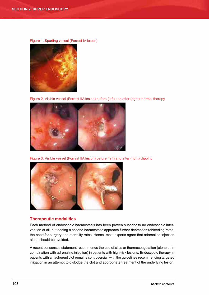
108
SECTION 2: UPPER ENDOSCOPY
back to contents
Figure 1. Spurting vessel (Forrest IA lesion)
Figure 2. Visible vessel (Forrest IIA lesion) before (left) and after (right) thermal therapy
Figure 3. Visible vessel (Forrest IIA lesion) before (left) and after (right) clipping
Therapeutic modalitiesEach method of endoscopic haemostasis has been proven superior to no endoscopic inter-vention at all, but adding a second haemostatic approach further decreases rebleeding rates, the need for surgery and mortality rates. Hence, most experts agree that adrenaline injection alone should be avoided.
A recent consensus statement recommends the use of clips or thermocoagulation (alone or in combination with adrenaline injection) in patients with high-risk lesions. Endoscopic therapy in patients with an adherent clot remains controversial, with the guidelines recommending targeted irrigation in an attempt to dislodge the clot and appropriate treatment of the underlying lesion.

109
MANAGEMENT OF NON-VARICEAL UPPER GASTROINTESTINAL BLEEDINGPaul Edwards and Brad Kendall
SECTION 2: UPPER ENDOSCOPY
back to contents
If the clot cannot be dislodged with irrigation, endoscopic therapy may be considered, although intensive PPI therapy alone may be sufficient. Planned second-look endoscopy within 24 hours after initial endoscopic therapy is not routinely recommended, but a second endoscopy may be considered on a case-by-case basis for clinical signs of recurrent bleeding or uncertainty regarding the effectiveness of initial haemostasis.
Patients with minor acute NVUGIB and endoscopic low-risk lesions (i.e. flat pigmented spots and clean-base ulcers) can safely undergo early discharge from hospital with medical therapy, provided they have support at home and are not geographically isolated.
Approach to rebleeding and endoscopic failureDespite the huge advances in endoscopic management, surgical rates for management of a bleeding peptic ulcer are approximately 5% to 7% over most series. This reflects the dictum that “if a bleeding vessel is big enough to have its own name, then endoscopic therapy is unlikely to be effective”. The surgical approach has also changed significantly in the past three decades, given our new understanding of the causation of peptic ulcer disease. The aim of emergency surgery is no longer to cure the disease (by oversew, resection and/or vagotomy), but rather to stop the haemorrhage when endoscopic therapy has failed or is unavailable.
As a consequence, the use of angiography with transcatheter embolisation has become a much more viable option to treat rebleeding and endoscopic failures. Such radiological services are usually only available at large tertiary care hospitals. The provision of these services to this subgroup of patients with endoscopic failure may also pose significant logistical difficulties, as radiology suites may be much more “anaesthetic/resuscitation unfriendly” than endoscopy suites.
At one of our institutions, 70% of patients with rebleeding and endoscopic treatment failure are successfully treated by percutaneous transcatheter embolisation (PTE) and 30% have surgery. Often, the consultant surgeon makes the request for PTE. In an interesting parallel to the endo-scopic literature, a study by Loffroy and colleagues concluded that angiographic embolisation should be performed early in the course of rebleeding and that multiple modalities of embolisa-tion (i.e. coils, gelatin sponge, particles or glue) should be applied to obtain the best outcomes.
Endoscopic treatment of non-peptic ulcer bleeding
The most common cause of non-peptic ulcer NVUGIB is a Mallory–Weiss tear. In addition to the techniques used in treating peptic ulcer bleeding, band ligation has also been used successfully to treat this condition. Upper gastrointestinal neoplasia can occasionally present with upper gastrointestinal bleeding. The techniques used in peptic ulcer bleeding can also be used in the treatment of neoplastic upper gastrointestinal bleeding, but adequate haemostasis may be more difficult to achieve because of the underlying neoplastic process. In these circumstances, radiological or surgical treatment may be required to stop the bleeding.

110
SECTION 2: UPPER ENDOSCOPY
back to contents
Post-discharge management
In all patients with peptic ulcer bleeding, H. pylori infection should be thoroughly excluded. H. pylori-positive patients should undergo a course of H. pylori treatment once they are medically stable. H. pylori eradication should be confirmed by follow-up testing. In those patients with peptic ulcer bleeding and a negative H. pylori test result, a repeat test using a second, different, method should be performed to confirm the negative status.
Decisions regarding ongoing use of medications that may have contributed to the bleeding, such as antiplatelet and anticoagulation medications, should be made in conjunction with the physician who is managing the patient’s condition for which the medications have been pre-scribed. In patients for whom the underlying risk factor for the peptic ulcer has been treated (H. pylori) or permanently removed (aspirin or NSAID), a 2-month course of standard-dose PPI should be given. Whether PPI treatment is needed beyond that period is uncertain. In patients with no identifiable risk factors for peptic ulcer bleeding, long-term PPI should be considered.
In patients with a gastric ulcer, a repeat endoscopy should be performed in about 2 months to ensure ulcer healing and exclude a malignant ulcer. If there is a high endoscopic suspicion of an underlying malignancy, earlier repeat endoscopy with biopsy should be performed when the patient has recovered from the acute bleeding episode.
Conclusion
In patients with NVUGIB, various clinical and endoscopic determinants are powerful predictors of subsequent mortality, underpinning the importance of early risk stratification. Major outcomes including mortality, rebleeding and the need for surgery have improved dramatically through enhanced resuscitative and supportive measures, multiple modality endoscopic therapy and profound acid suppression. The optimal timing of the diagnostic or therapeutic endoscopy requires further study but, at the very least, early endoscopy (i.e. within 24 hours of admission) results in shorter and cheaper hospital stays. Logistical and staffing issues are also important. The gastroenterologist now effectively leads a multidisciplinary team (emergency physicians, intensivists, surgeons and radiologists) in the management of NVUGIB. Practical tips for man-aging patients with NVUGIB are shown in Table 5.

111
MANAGEMENT OF NON-VARICEAL UPPER GASTROINTESTINAL BLEEDINGPaul Edwards and Brad Kendall
SECTION 2: UPPER ENDOSCOPY
back to contents
Table 5. Practical tips for management of non-variceal upper gastrointestinal bleeding (NVUGIB)
● Use clinical and endoscopic data to determine early risk stratification, and base clinical decisions on this.
● Give high-dose intravenous proton pump inhibitor (PPI) bolus and then start PPI infusion.
● Consider giving a prokinetic agent such as erythromycin 30 minutes before the procedure.
● Develop adequate logistical plans to manage NVUGIB at your hospital. Endoscopy should be performed in a safe environment for the patient and a familiar working environment for the endoscopist and anaesthetic/resuscitation staff.
● At endoscopy, use further risk stratification based on the Forrest classification. Arrest bleeding using multiple therapies; this usually starts with volume tamponade using adrenaline, followed by thermal coagulation (e.g. gold probe), then consider endoscopic clips as further therapy if required.
● In the presence of rebleeding, consider further endoscopic therapy, percutaneous transcatheter embolisation or surgical options.
● In all patients with peptic ulcer bleeding, Helicobacter pylori infection should be thoroughly excluded. If identified, it should be treated and post-treatment testing performed to confirm eradication.
Further reading1 Chiu PW, Ng EK, Cheung FK, et al. Predicting mortality in patients with bleeding peptic ulcers after therapeutic
endoscopy. Clin Gastroenterol Hepatol 2009; 7: 311-316.
2 Rockall TA, Logan RF, Devlin HB, Northfield TC. Variation in outcome after acute upper gastrointestinal haemorrhage. The National Audit of Acute Upper Gastrointestinal Haemorrhage. Lancet 1995; 346: 346-350.
3 Ananthakrishnan AN, McGinley EL, Saeian K. Outcomes of weekend admissions for upper gastrointestinal hemorrhage: a nationwide analysis. Clin Gastroenterol Hepatol 2009; 7: 296-302e1.
4 Shaheen AA, Kaplan GG, Myers RP. Weekend versus weekday admission and mortality from gastrointestinal hemorrhage caused by peptic ulcer disease. Clin Gastroenterol Hepatol 2009; 7: 303-310.
5 Barkun A, Sabbah S, Enns R, et al. The Canadian Registry on Nonvariceal Upper Gastrointestinal Bleeding and Endoscopy (RUGBE): endoscopic hemostasis and proton pump inhibition are associated with improved outcomes in a real-life setting. Am J Gastroenterol 2004; 99: 1238-1246.
6 Gralnek IM, Barkun AN, Bardou M. Management of acute bleeding from a peptic ulcer. N Engl J Med 2008; 359: 928-937.
7 Barkun AN, Bardou M, Kuipers EJ; International Consensus Upper Gastrointestinal Bleeding Conference Group. International consensus recommendations on the management of patients with nonvariceal upper gastrointestinal bleeding. Ann Intern Med 2010; 152: 101-113.
8 Loffroy R, Guiu B, D’Athis P, et al. Arterial embolotherapy for endoscopically unmanageable acute gastroduodenal hemorrhage predictors of early rebleeding. Clin Gastroenterol Hepatol 2009; 7: 515-523.
9 Gralnek IM, Dumonceau JM, Kuipers EJ, et al. Diagnosis and management of nonvariceal upper gastrointestinal hemorrhage: European Society of Gastrointestinal Endoscopy (ESGE) Guideline. Endoscopy 2015; 47: a1-46.
Page 112 (used for notes in the hard copy) has been removed from the PDF edition of this handbook.

11375
Barrett’s oesophagus: diagnosis and managementDarren Pavey
Barrett’s oesophagus is a metaplastic change in the oesophageal epithelium thought to result from injury due to chronic exposure to reflux gastric content. Normal oesophageal squamous epithelium is replaced by specialised intestinal epithelium, also known as intestinal metaplasia (IM). Progression of non-dysplastic IM to low-grade dysplasia (LGD), high-grade dysplasia (HGD) and oesophageal adenocarcinoma (OAC) is well described. The incidence of OAC continues to rise, particularly among the older population.
Guidelines for the diagnosis and management of Barrett’s oesophagus have recently been pub-lished by the Cancer Council of Australia. Readers are referred to these guidelines for further background on the epidemiology and risk factors for Barrett’s oesophagus.
Diagnosis of Barrett’s oesophagus
Barrett’s oesophagus has traditionally been defined as the presence of at least 1 cm of meta-plastic columnar epithelium that replaces the stratified squamous epithelium normally lining the distal oesophagus. Segments < 1 cm have instead been classified as “specialised IM of the oesophagogastric junction”. This distinction is made due to the high interobserver variability, as well as a low risk of OAC in very short-segment disease.
The definition of Barrett’s oesophagus has varied based on the requirement for the presence of IM on endoscopic biopsy samples. Debate regarding the requirement for IM on biopsy from columnar-lined epithelium segments has derived from the apparently differential risk of devel-oping OAC in such epithelium containing or not containing IM. Large population-based cohort studies have shown a substantially lower OAC risk in patients with columnar metaplasia without IM, compared with those with IM. However, not all studies have corroborated this finding. The yield for IM correlates directly with the number of endoscopic biopsy samples obtained. In a
SECTION 2: UPPER ENDOSCOPY
CHAPTER 8
Endoscopic management of varices and variceal haemorrhageDavid Koorey and Crispin Corte
Up to 30% of patients with well compensated cirrhosis and 60% of those with decompen-sated disease will have varices. The mortality rate from a first episode of variceal bleeding is about 15%–30%, and 1-year survival figures ranging from 30% to 80% have been reported. Variceal bleeding is therefore a common and important complication of chronic liver disease. Gastroenterologists need to be able to identify patients at risk of bleeding, reduce that risk (primary prophylaxis), manage acute variceal bleeding and prevent rebleeding (secondary prophylaxis). This chapter provides an overview of variceal bleeding management, focusing on endoscopic intervention.
General principles of management
Primary prophylaxisRecent guidelines recommend that all patients with cirrhosis undergo upper endoscopy. Clues to the presence of portal hypertension, such as splenomegaly, thrombocytopenia or portosys-temic collaterals on imaging, should prompt early assessment. If the initial examination finds no varices, follow-up endoscopy after 2 years has been recommended. If varices are found, the risk of bleeding should be assessed. Key factors predicting an increased risk include severity of the liver disease, size of the varices and presence of red markings — red wale markings, cherry red spots and haematocystic spots (Figure 1).
SECTION 2: UPPER ENDOSCOPY
CHAPTER 11
back to contents

114
SECTION 2: UPPER ENDOSCOPY
back to contents
Figure 1. Varices with red markings
Red wale markings: longitudinal dilated venules that resemble whip marks (left); cherry red spots: small red spots up to about 2 mm in diameter (middle); and haematocystic spot: a large (> 3 mm), round, crimson projection resembling a blood blister (right).
Oesophageal varices are graded endoscopically as:
• Grade I: small straight varices• Grade II: enlarged tortuous varices occupying less than one-third of the lumen• Grade III: large coil-shaped varices occupying more than one-third of the lumen.
Patients with small (Grade I) varices, no red markings and well compensated disease can be fol-lowed with annual endoscopy. In patients with higher-risk varices (Grade I with red markings or decompensated disease, or Grade II–III), options for primary prophylaxis include non-selective β-blockade and variceal band ligation. Both reduce first bleeding episodes and bleeding-related mortality. If a non-selective β-blocker (e.g. propranolol) is used, aim for a 25% reduction in resting heart rate or a heart rate of 50–55 beats/min. Some groups measure wedged hepatic venous pressure gradients to assess response to β-blockers. In most patients, non-selective β-blockers should be used first, in view of the inconvenience, discomfort and occasionally serious complications associated with banding. Variceal band ligation is appropriate in patients with high-risk varices if there are contraindications to β-blockers or if they are poorly tolerated.
Acute variceal haemorrhageAcute variceal haemorrhage is a medical emergency and should be suspected in any patient with upper gastrointestinal bleeding in the context of chronic liver disease, established portal hypertension or clinical, radiological or pathological indicators of portal hypertension.
Initial management should focus on adequate resuscitation, pharmacological intervention and early endoscopic treatment. Venous access sufficient to allow rapid transfusion is essential. Excessive volume replacement should be avoided, as this may raise the portal venous pres-sure and aggravate bleeding. Aiming to maintain a systolic blood pressure of 100 mmHg has been suggested. A target haemoglobin level of 80 g/L appears appropriate unless there are significant cardiorespiratory comorbidities. Coagulopathy and thrombocytopenia should be corrected if there is active bleeding. The safety of the airway should be evaluated, given the risk of aspiration in patients with reduced consciousness due to encephalopathy and ongoing haematemesis. Intravenous terlipressin or octreotide should be commenced before endoscopy if variceal bleeding is suspected; these agents reduce portal pressures and improve early haemostasis. Prophylactic broad-spectrum antibiotics (e.g. intravenous ceftriaxone) should be

115
EnDOSCOPIC mAnAGEmEnT OF VARICES AnD VARICEAL HAEmORRHAGEDavid Koorey and Crispin Corte
SECTION 2: UPPER ENDOSCOPY
back to contents
given routinely, as they reduce infections and rebleeding and improve survival in patients with acute variceal haemorrhage.
Only 40%–50% of variceal bleeding will cease spontaneously (compared with about 90% of non-variceal upper gastrointestinal bleeding), and early endoscopy is therefore a priority. Rarely, massive blood loss will be immediately life-threatening, and balloon tamponade for suspected varices will be necessary without preceding endoscopy. In most cases, however, resuscitation and pharmacological intervention will allow early endoscopy. Whenever possible, this should be performed with an anaesthetist present and the patient intubated to protect the airway. It is important to ensure that all equipment likely to be required is available: a banding device and an endoscope of suitable size for the device (larger, dual-channel scopes may not readily accept a standard banding device); cyanoacrylate glue, lipiodol and an appropriate injector; a Linton tube, Sengstaken tube or similar for uncontrollable variceal bleeding; and items such as injectors, a heater probe and clips should a non-variceal source be found. Where available, covered stents specifically designed to be used for control of variceal haemorrhage (Danis stent; Endotherapeutics) should also be considered.
Band ligation will control bleeding from oesophageal varices in 80%–90% of cases, with fewer complications and lower mortality than sclerotherapy. When bleeding from gastric varices is identified, injection of cyanoacrylate glue is generally the treatment of choice. Band ligation can be used for type I gastro-oesophageal varices (a continuation of oesophageal varices a short distance along the lesser curvature of the stomach). When bleeding cannot be controlled at initial endoscopy, balloon tamponade should be considered, with a view to a second attempt at endoscopic management the following day (endoscopists should familiarise themselves with the protocols for balloon tamponade before the need arises). If endoscopic therapy again fails, salvage transjugular intrahepatic portosystemic shunts (TIPS) may be an option, but outcomes are likely to be poor in the setting of advanced liver disease, multiorgan failure and ongoing bleeding.
Recently, use of the Danis stent (a specifically designed removable covered metal stent) has been reported in the control of refractory acute bleeding from oesophageal varices. This may prove to be an alternative strategy to balloon tamponade as a bridge to TIPS in selected patients. This stent is deployed in a similar fashion to a Sengstaken or Linton tube (whereby inflation of a gastric balloon secures appropriate placement with respect to the cardio-oesoph-ageal junction) and does not require radiological guidance. Where the stent is available, endos-copists who manage variceal haemorrhage should familiarise themselves with the mechanism of stent deployment and removal. Splenectomy or splenic artery embolisation can be considered in patients with sinistral (left-sided) portal hypertension due to splenic vein thrombosis.
Secondary prophylaxis In patients who survive a first episode of variceal haemorrhage, 40%–60% will rebleed by 6 weeks and 60%–80% by 1 year. Rebleeding is predicted by the severity of the initial bleed, the severity of the liver disease and portal hypertension, active bleeding at the initial endoscopy and large varices with high-risk stigmata. Variceal band ligation should be repeated at intervals of 2–4 weeks until varices have been obliterated. non-selective β-blockade reduces rebleed-ing and bleeding-related mortality when used as secondary prophylaxis. The combination of

116
SECTION 2: UPPER ENDOSCOPY
back to contents
variceal band ligation with β-blockade appears to provide the best reduction in rebleeding rates. nevertheless, 5%–20% of patients will rebleed despite secondary prophylaxis, and TIPS should be considered in these patients.
Endoscopic oesophageal band ligation
TechniqueIn the elective setting of primary or secondary prophylaxis, the goal is obliteration of the varices while minimising the risk of complications. more than one treatment session is usually required. Correction of mild to moderate thrombocytopenia or coagulopathy is generally not needed. In patients with ascites, prophylactic antibiotics may reduce the risk of spontaneous bacterial peritonitis after banding.
Elective band ligation can generally be performed under conscious sedation in day patients. The endoscope is first passed without the banding device to assess the varices. note should be made of the lowest level at which the varices appear, as this is where banding should commence; generally, it is at or just below the cardio-oesophageal junction. Retroflexion in the stomach is important to check for emerging gastric varices (Figure 2).
The endoscope is then reintroduced with the banding device attached. Various multiband ligators are available, and the endoscopist should be familiar with those used in their institution. The first band is generally deployed at the lowest point of the largest varix. The varix should be sucked into the ligator until the view is obliterated (“red-out”), and the band then deployed. Slight, gentle alternating clockwise and anticlockwise twisting of the endoscope can facilitate suction of the varix. Occasionally a varix will rupture during suction, often at a point of wall thinness, such as a haematocystic spot. In this situation, it is important to maintain
suction and deploy the band without delay. The second and subsequent bands are deployed on the remaining varices, initially targeting the largest varices and those with high-risk stigmata. The endoscope should be withdrawn slightly as banding proceeds.
Although banding will generally be concentrated in the distal oesophagus, multiple bands should not be deployed at exactly the same level, and some intervening mucosa should be left between adjacent bandings (Figure 3). This helps to reduce the risk of confluent post-banding ulceration.
Figure 2. Gastric varices seen on retroflexion
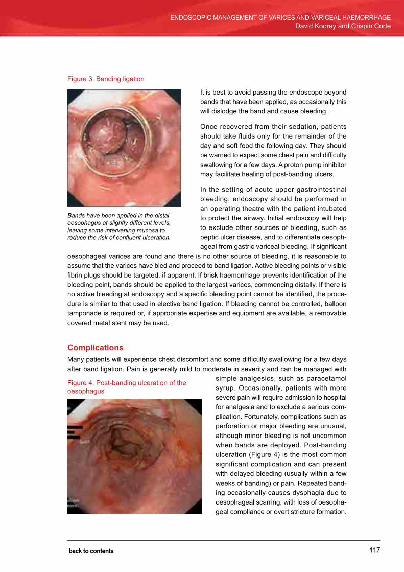
117
EnDOSCOPIC mAnAGEmEnT OF VARICES AnD VARICEAL HAEmORRHAGEDavid Koorey and Crispin Corte
SECTION 2: UPPER ENDOSCOPY
back to contents
Figure 3. Banding ligation
It is best to avoid passing the endoscope beyond bands that have been applied, as occasionally this will dislodge the band and cause bleeding.
Once recovered from their sedation, patients should take fluids only for the remainder of the day and soft food the following day. They should be warned to expect some chest pain and difficulty swallowing for a few days. A proton pump inhibitor may facilitate healing of post-banding ulcers.
In the setting of acute upper gastrointestinal bleeding, endoscopy should be performed in an operating theatre with the patient intubated to protect the airway. Initial endoscopy will help to exclude other sources of bleeding, such as peptic ulcer disease, and to differentiate oesoph-ageal from gastric variceal bleeding. If significant
oesophageal varices are found and there is no other source of bleeding, it is reasonable to assume that the varices have bled and proceed to band ligation. Active bleeding points or visible fibrin plugs should be targeted, if apparent. If brisk haemorrhage prevents identification of the bleeding point, bands should be applied to the largest varices, commencing distally. If there is no active bleeding at endoscopy and a specific bleeding point cannot be identified, the proce-dure is similar to that used in elective band ligation. If bleeding cannot be controlled, balloon tamponade is required or, if appropriate expertise and equipment are available, a removable covered metal stent may be used.
Complicationsmany patients will experience chest discomfort and some difficulty swallowing for a few days after band ligation. Pain is generally mild to moderate in severity and can be managed with
simple analgesics, such as paracetamol syrup. Occasionally, patients with more severe pain will require admission to hospital for analgesia and to exclude a serious com-plication. Fortunately, complications such as perforation or major bleeding are unusual, although minor bleeding is not uncommon when bands are deployed. Post-banding ulceration (Figure 4) is the most common significant complication and can present with delayed bleeding (usually within a few weeks of banding) or pain. Repeated band-ing occasionally causes dysphagia due to oesophageal scarring, with loss of oesopha-geal compliance or overt stricture formation.
Bands have been applied in the distal oesophagus at slightly different levels, leaving some intervening mucosa to reduce the risk of confluent ulceration.
Figure 4. Post-banding ulceration of the oesophagus

118
SECTION 2: UPPER ENDOSCOPY
back to contents
Glue injection of gastric varices
TechniqueWhen gastric varices bleed, they often bleed massively. The upper stomach may be obscured by blood and clot. Rolling the patient to the right lateral position should provide better visualis-ation. Prolonged washing and aspiration may still be necessary. In some cases, if the bleeding point cannot be visualised, balloon tamponade will be needed, with continued pharmacological therapy and repeat endoscopy after 12 to 24 hours.
Injection of cyanoacrylate glue remains the prime endoscopic therapy for gastric variceal haemorrhage. The endoscopist and assistant need to be familiar with the procedure and fol-low protocols carefully to optimise results and minimise the risk of equipment damage. Only needles suitable for use with glue should be used. The injecting needle is first primed with lipiodol and then partly primed with a histoacryl–lipiodol mix to about 10 cm from the needle tip (the histoacryl is purple and should be visible in the injector). It is important not to fully prime with histoacryl–lipiodol, to avoid glue contaminating the channel as the injector is introduced through the endoscope.
After obtaining a satisfactory view and good position, the injector is passed down the channel. The bleeding varix, the varix with a fibrin plug, ulceration or red marks, or the largest varix, if a bleeding point cannot be identified, is injected with histoacryl–lipiodol, followed by lipiodol alone to push the remainder of the glue into the varix. The needle should be withdrawn from the varix (but not into the endoscope) immediately after injection is completed, to avoid the needle becoming stuck in the varix.
The procedure can be repeated for additional injections, but at no stage should the injector be withdrawn into the endoscope. After the last injection, the injector is flushed with water. The needle tip is retracted into the injector, but the injector is not retracted into the endoscope. The endoscope is removed from the patient with the injector still protruding. The injector is then again flushed repeatedly with water and wiped several times with a water-moistened gauze before it is removed from the endoscope. The biopsy channel is then immediately flushed with water.
ComplicationsBleeding may occur at the time of the procedure (related to injection) or weeks after injection (related to sloughing of the glue cast and resulting ulceration). Glue embolism, both pulmonary and paradoxical, can occur.
Conclusion
Identification and management of patients at risk of variceal haemorrhage; resuscitation, medical management and endoscopic control of active variceal bleeding; and prevention of rebleeding are all essential skills for practising gastroenterologists. Primary and secondary
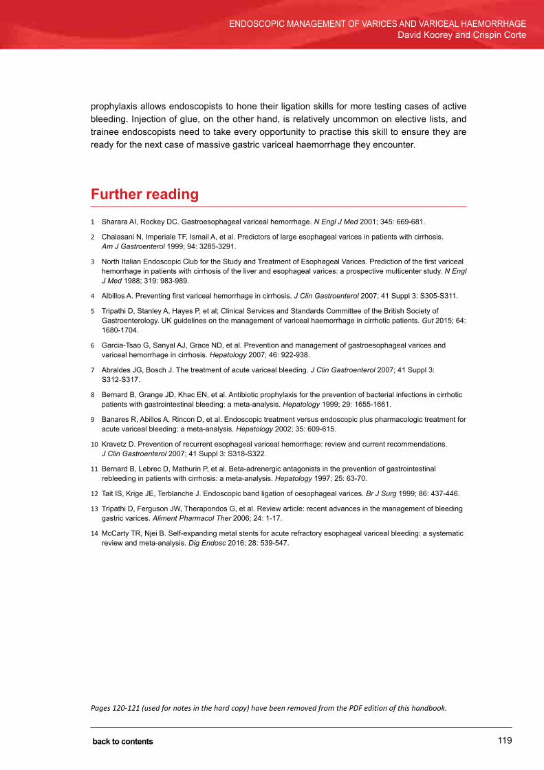
119
EnDOSCOPIC mAnAGEmEnT OF VARICES AnD VARICEAL HAEmORRHAGEDavid Koorey and Crispin Corte
SECTION 2: UPPER ENDOSCOPY
back to contents
prophylaxis allows endoscopists to hone their ligation skills for more testing cases of active bleeding. Injection of glue, on the other hand, is relatively uncommon on elective lists, and trainee endoscopists need to take every opportunity to practise this skill to ensure they are ready for the next case of massive gastric variceal haemorrhage they encounter.
Further reading1 Sharara AI, Rockey DC. Gastroesophageal variceal hemorrhage. N Engl J Med 2001; 345: 669-681.
2 Chalasani n, Imperiale TF, Ismail A, et al. Predictors of large esophageal varices in patients with cirrhosis. Am J Gastroenterol 1999; 94: 3285-3291.
3 north Italian Endoscopic Club for the Study and Treatment of Esophageal Varices. Prediction of the first variceal hemorrhage in patients with cirrhosis of the liver and esophageal varices: a prospective multicenter study. N Engl J Med 1988; 319: 983-989.
4 Albillos A. Preventing first variceal hemorrhage in cirrhosis. J Clin Gastroenterol 2007; 41 Suppl 3: S305-S311.
5 Tripathi D, Stanley A, Hayes P, et al; Clinical Services and Standards Committee of the British Society of Gastroenterology. UK guidelines on the management of variceal haemorrhage in cirrhotic patients. Gut 2015; 64: 1680-1704.
6 Garcia-Tsao G, Sanyal AJ, Grace nD, et al. Prevention and management of gastroesophageal varices and variceal hemorrhage in cirrhosis. Hepatology 2007; 46: 922-938.
7 Abraldes JG, Bosch J. The treatment of acute variceal bleeding. J Clin Gastroenterol 2007; 41 Suppl 3: S312-S317.
8 Bernard B, Grange JD, Khac En, et al. Antibiotic prophylaxis for the prevention of bacterial infections in cirrhotic patients with gastrointestinal bleeding: a meta-analysis. Hepatology 1999; 29: 1655-1661.
9 Banares R, Abillos A, Rincon D, et al. Endoscopic treatment versus endoscopic plus pharmacologic treatment for acute variceal bleeding: a meta-analysis. Hepatology 2002; 35: 609-615.
10 Kravetz D. Prevention of recurrent esophageal variceal hemorrhage: review and current recommendations. J Clin Gastroenterol 2007; 41 Suppl 3: S318-S322.
11 Bernard B, Lebrec D, mathurin P, et al. Beta-adrenergic antagonists in the prevention of gastrointestinal rebleeding in patients with cirrhosis: a meta-analysis. Hepatology 1997; 25: 63-70.
12 Tait IS, Krige JE, Terblanche J. Endoscopic band ligation of oesophageal varices. Br J Surg 1999; 86: 437-446.
13 Tripathi D, Ferguson JW, Therapondos G, et al. Review article: recent advances in the management of bleeding gastric varices. Aliment Pharmacol Ther 2006; 24: 1-17.
14 mcCarty TR, njei B. Self-expanding metal stents for acute refractory esophageal variceal bleeding: a systematic review and meta-analysis. Dig Endosc 2016; 28: 539-547.
Pages 120-121 (used for notes in the hard copy) have been removed from the PDF edition of this handbook.

Section 3
COLONOSCOPY
123
Colonoscopy: insertion and withdrawal techniqueMichael Bourke
Successful colonoscopy, like all high-quality endoscopy, starts with careful preparation. This may seem obvious, but it is often overlooked and cannot be overemphasised. Nothing should be taken for granted or assumed. The endoscopist must be familiar and comfortable with the equipment, the colonoscope of choice, the nursing staff, sedationists and other assistants, and the environment in which the procedure will be completed. A useful analogy is to envisage every colonoscopic procedure unfolding like a Mozart clarinet concerto, with the endoscopist as the conductor. Each professional has a role to fulfil, and the endoscopist and his or her coworkers should be continuously cognisant of the music of the procedure room as the procedure evolves. When endoscopy units and procedural teams are able to generate this level of awareness, rhythm and harmony, it becomes immediately obvious to all involved when something or some-one is out of tune. In such situations, a major complication or problem may be imminent; this can be avoided through early recognition by any member of the group, with remedial action taken by the appropriate member of the team.
The endoscopist must know the patient well. Important features include a history of previous abdominopelvic surgery, disease or treatment, such as previous hysterectomy or pelvic radio-therapy. A thorough understanding of the cardiorespiratory status and physiological reserve of all patients scheduled for endoscopy is essential. Some patients will tolerate a prolonged or difficult procedure very poorly due to poor general medical health.
It is possible to learn from almost every colonoscopy performed, whether you perform five or 50 per week. Challenging cases create the opportunity to reflect on technique and how this might be generally improved or specifically adapted to particular challenges.
Eight key points for safe and effective colonoscopy are shown in Table 1.
SECTION 3: COLONOSCOPY
CHAPTER 12
back to contents

SECTION 3: COLONOSCOPY
124 back to contents
Table 1. Key points for safe and effective colonoscopy
● Know and check your instruments and processor before your colonoscopy list commences. For most colonoscopy procedures, the air setting should be on low. Carbon dioxide is proven to be superior to air for insufflation during colonoscopy, with advantages of reduced post-procedural pain and swifter patient recovery.
● Check the compliance or degree of tension of the patient’s abdominal wall (while the patient is relaxed in the left lateral position) just before starting, and monitor this periodically during the procedure.
● Be a gentle perfectionist. Every instrument manipulation should be completed precisely and as well as it possibly can be.
● Keep the scope as short and straight as possible, and withdraw and straighten after completing the insertion for each segment of colon and all major corners.
● Try to maintain continuous awareness of your anatomical location and the three-dimensional position of the scope.
● Never try to push through fixed resistance; force does not work in endoscopy.
● If you are not making progress, change your strategy. Doing the same thing over and over is unlikely to be successful.
● Master the left colon; this holds the key to maximising caecal intubation success and minimising insertion time.
Basic insertion technique
Caecal intubation should be as efficient as reasonably possible, as this is the most uncomfort-able phase of colonoscopy. Much of the colon is a mobile elastic tube, particularly the sigmoid, and thus a hurried, forceful intubation results in looping, patient discomfort and a potentially failed complete examination. The great paradox of colonoscopy is “slower is faster”. Purposeful and controlled movements will result in a more complete examination than will a flurry of exu-berant actions.
Endoscope handlingThe predominant technique is that of torque steering. This implies that the right hand remains on the insertion tube (IT) of the instrument, with the fingers and wrist of that hand in coordination with the left thumb on the large wheel (or up–down control) of the endoscope. Most changes of direction in colonoscopy are coordinated movements between the right wrist and the left thumb. Torque steering involves first gently angulating up (most frequent) or down with the thumb on the large wheel, while rotating the shaft (most frequently clockwise on insertion through the sigmoid and descending colon) (Figure 1). Angulation, even slight, confers lateral deviation of the tip of the instrument as the twist on the shaft is applied. Without tip angulation, no lateral deviation will occur, hence giving no ability to steer through a corner.

125
ColoNoSCoPy: INSErTIoN AND WIThDrAWAl TEChNIquEMichael Bourke
SECTION 3: COLONOSCOPY
back to contents
hold the IT with your fingers as if it were a pencil (not like a tennis racquet) (Figure 2). This will maximise sensory feed-back, informing the proceduralist on the amount of tension within the IT. When the scope is straight, the IT at the level of the anus will feel relatively “floppy” and seem-ingly fall onto the bed as it exits the anus. In contrast, when the scope is severely angulated or looped, the IT will feel stiff and does not fall onto the bed.
For major corners, rotate the IT so that the axis of the corner is in the 6–12 o’clock ori-entation, which is the most powerful bend-ing direction of the instrument (especially upwards). resist the temptation to move the right hand off the IT up to the left–right control (small wheel). This interrupts the fluidity of the insertion technique and adds very little to the overall angulation that you can obtain at the tip if using a torque steer-ing technique.
With optimal technique, the right hand is almost exclusively responsible for rotation of the IT, while the left hand may adjust the position of the scope head and, with the thumb, move the up–down control and occasionally the smaller left–right wheel. Disciplined coordination between the two hands is the foundation of good technique.
If at any point you lose your view (“red out”), simply pull the instrument back gently and insufflate.
Most movements are small, as they are being effected on a long lever — the endoscope — and thus the net result at the tip can be significant.
Minimise air insufflation on insertion and suction air frequently. The colon is a readily distensi-ble, mobile elastic tube which, when overinflated, becomes long and tortuous, enhancing the difficulty of the insertion procedure.
When advancing through straight segments, use suction liberally as you progress forward. This makes such segments easier to traverse and concertinas the colon onto the scope, ultimately making the insertion less difficult.
When aspirating fluid, use a two-finger technique, with the index finger over the air button and the middle finger on suction. This avoids unnecessary suction of the bowel wall, which, if it
Coordinated movements between the right wrist and the left thumb comfortably negotiate most corners.
Hold the insertion tube like a pencil; the fingertips sense tension in the insertion tube and inform on the degree of resistance at the scope tip.
Figure 2. Correct handling of the insertion tube
Figure 1. Torque steering technique

SECTION 3: COLONOSCOPY
126 back to contents
occurs, impedes the view and mandates gentle withdrawal of the instrument and a time delay with reorientation of the scope tip before insertion can recommence.
When suctioning fluid, perform a submarine periscope technique to maintain a luminal view. The working channel (and suction channel) of the colonoscope is in the 5 to 6 o’clock position. As the relationship between the lens, light source and working channel is fixed, this orientation is constant. Position your view just above the fluid, thus the suction port will be beneath the surface.
Looping, pressure and non-progressionIn ideal insertion conditions, the length of instrument inserted at the anus results in an equal (or greater, if the colon is being shortened onto the IT) distance of endoscope tip progression in the colon. This is termed one-to-one progress. During insertion, loss of one-to-one progress indicates that the scope is bowing (consider the situation in gastroscopy when advancing to the pylorus; the flexible endoscope will bow for a variable distance on the greater curve of the stomach and then, as it is splinted by the fixed gastric wall, progress towards the pylorus will resume) or a loop may be developing. This is a frequent occurrence in the left colon and is acceptable if, after 5–8 cm of insertion, progress resumes with marginally increased resistance. This should be relatively painless and should not require a substantial increase in force. After the next corner, the scope can be “straightened” by gentle withdrawal, suction and torque.
If, however, there is a seemingly long, straight segment of resistance-free insertion, a loop is probably being formed. This should be relatively painless and tension in the IT should be only marginally increased. At the end of the loop where all the “slack” of the redundant loop (and loose mesentery) has been taken up, progress will steadily halt, tension in the IT will gradually increase and the patient may experience pain. The loop can then be resolved by simultaneous (usually clockwise) torque, suction and gentle withdrawal of the IT. The IT may rotate more than 180° in the endoscopist’s hand. If the loop is being resolved correctly, the scope tip will not fall back more than a few centimetres and will often advance proximally in the colon, and tension in the IT will rapidly decrease.
Abdominal pressure is a very effective technique when used strategically. Knowledge of the endoscope’s location allows logical application. It is most useful when the scope is straight and the tip is beyond the sigmoid colon. After 5–6 cm of non-progression, apply pressure to where bowing of the IT is most likely occurring (see later sections).
Segment-specific insertion technique
There are aspects of core technique that are particular to each segment of the colon, and it is not uncommon that subtle manoeuvres adopted early when difficulty is encountered will easily overcome what may at first appear to be a major obstacle to complete examination. Key among these is the concept of mastering the left colon. About two-thirds of your total insertion time should be spent in the left colon. All loops should be reduced and the scope straight before you
127
ColoNoSCoPy: INSErTIoN AND WIThDrAWAl TEChNIquEMichael Bourke
SECTION 3: COLONOSCOPY
back to contents
move beyond the splenic flexure. All people are created approximately equal in length between the anus and the splenic flexure; at this point, insertion length should be between 45 and 50 cm.
use suction liberally during insertion to encourage the colon to concertina onto the colonoscope, especially when advancing easily through straight segments. use pressure for short periods of time and try to be specific and algorithmic in its application. In best-practice colonoscopy, caecal intubation is achieved expeditiously with a short, straight scope, limited use of the insertion length of the instrument and minimal patient discomfort. Insertion length at the caecal pole is ideally between 65 and 90 cm. When difficulty on insertion arises, there are generally three potential areas for remediation or problem avoidance:
1 endoscope handling and insertion technique
2 abdominal pressure
3 change in position.
The order from 1 to 3 reflects the relative ease with which each of these interventions can be completed, with change of position being the most difficult, particularly in large, heavily sedated patients. Nevertheless, at times this is vitally important. The underlying principle is to do the simple things first and to change strategy promptly and sequentially if successive techniques fail to resolve the problem. In addition, a change of instrument is necessary in specific situations (e.g. stenosing diverticular disease).
Rectum and rectosigmoidAfter digital rectal examination, the instrument, with its distal 5 cm well lubricated, is inserted beyond the anus into the rectum. hold the endoscope between one and two hands’ breadths from the buttocks. It is useful to pause at the anorectal junction and carefully inspect the rectum in front of you. The anorectal junction is best examined in retroflexion. retroflexion in the rectum is relatively contraindicated, particularly with an adult colonoscope in individuals with poorly distensible rectums, such as those with chronic colitis or previous radiotherapy. If necessary, it may be safely performed in these patients with a gastroscope. Passing from the rectum into the sigmoid colon is accomplished by torque steering. This angle may be very acute, particularly in:
• older patients with stenosing diverticular disease• patients with a history of previous low abdominal or pelvic surgery, such as previous
abdominal hysterectomy
• young women.
Even when this angle is easily overcome, it is best not to push forcefully through, elongating the sigmoid colon on the instrument, but rather keep a short scope and use torque steering and rotation of the IT (particularly clockwise) to reach the descending colon. The scope may tend to form a loop on the bed exterior to the patient. This can be resolved upon reaching the descending colon or splenic flexure. In a sharply angulated rectosigmoid junction (usually at about 20 cm of insertion), right hypogastric pressure directed medially and downwards above the pubis will often resolve the difficulty (Figure 3).

SECTION 3: COLONOSCOPY
128 back to contents
Sigmoid colonIn general, this is the most difficult segment during insertion. The sigmoid colon is more like an accordion than any other portion of colon and can be stretched to as much as 70–80 cm, but will shorten to about 25–30 cm with a straight scope located in the caecum (at 65–70 cm of insertion). Thus, precise anatom-ical location within the sigmoid colon by virtue of distance on insertion is very unreliable. It may be useful on withdrawal. If a polyp is not removed on insertion, it is very important to precisely note its position with a straight scope so that it can be readily identified on withdrawal — its clockface position within the lumen may have completely changed. you may even con-sider leaving a suction mark on the adjacent mucosa or taking a biopsy sample to aid local-isation on withdrawal. The sigmoid mesentery is highly variable in length and mobility and thus, in some individuals, the sigmoid colon can pass well into the upper abdomen before making a loop back down to the descending colon. In contrast, after hysterectomy, the sig-
moid tends to be rather angulated and may be fixed into the area previously occupied by the uterus. This can create difficulties during insertion.
Due to the shape of the pelvis, with the curved sacrum, in general the colonoscope will naturally pass from posterior to anterior, and in more than 80% of cases will take a clockwise anteroposte-rior spiral into the descending colon. Some degree of upward looping with a convex arc forming towards the diaphragm always occurs as the colonoscope is advanced through the sigmoid colon. If you advance beyond the rectum into a long, straight (often relatively featureless) seg-ment, this often reflects a lengthy or mobile sigmoid mesentery, and a large loop may be forming. In this situation, the colonoscope will often pass relatively easily into the descending colon, forming a relatively open N loop (Figure 4). Eventually such a loop will cause the patient pain, and the endoscopist will be aware of its formation due to non-progression up the descending colon, the excessive length of instrument inserted and the long, straight insertion phase beyond the rectum. Such a loop is easily withdrawn by applying clockwise torque of between 90° and 180° and withdrawing the instrument slowly. From this point, it is often easy to advance up to the splenic flexure. Applying loop resolution techniques when halfway through a loop will only cause the colonoscope to drop back below the loop without genuine progress being achieved.
A sharply angulated rectosigmoid junction may be overcome with right hypogastric pressure directed medially and downwards above the pubis.
Figure 3. Applying pressure to a sharply angulated rectosigmoid junction

129
ColoNoSCoPy: INSErTIoN AND WIThDrAWAl TEChNIquEMichael Bourke
SECTION 3: COLONOSCOPY
back to contents
Almost all colonoscopy procedures require some degree of sigmoid loop resolution. The scope must be straight at the splenic flexure so that the remainder of the procedure can proceed smoothly, but a sigmoid loop may still tend to reform. Thus, while moving through the proximal colon, it is important to repeatedly straighten and pull back to keep the sigmoid colon straight. on occasion, non-specific left iliac fossa pressure may assist in maintaining a straight sigmoid colon.
Aim to completely avoid pushing blindly around corners, but rather rotate through these by torque steering. Enter the corner and rotate the IT slowly within the right hand up to 180°, while gently insufflating and pulling back very slightly. If a straight segment of lumen comes into view beyond this corner, try not to push directly into it; rather, aspirate air and with small forward and backward movements, inch your way forward into the segment. This technique will concertina the colon onto the endoscope rather than stretching the colon over a forcefully inserted instrument. on occasion, a “slide by” technique is acceptable for a few centimetres,
where you may see the normal mucosal vascular pattern slide by. If mucosal blanching occurs, this is indicative of excessive force by the colonoscope tip on the mucosa, and perforation may be imminent. The scope must be withdrawn.
In severe stenosing or tightly angulated diverticular disease, a change of instrument may be preferable. In general, paediatric colonoscopes allow easier passage of the sigmoid colon, and many colonoscopists use them as the preferred instrument. Sometimes a gastroscope may even be necessary. When compared with an adult colonoscope, the paediatric colonoscope and gastroscope have smaller tip diameters (12.3 mm, 11.3 mm and 8.8 mm, respectively) and take a more compact arc with complete “up” movement to achieve 180° angulation (6.6 × 5.6 cm for an adult colonoscope versus 3.8 × 3.4 cm for a gastroscope) (Figure 5). hence, narrow, severely angulated corners can be traversed more easily. Even when using a gastroscope, the caecum can often be easily reached as the relatively fixed sigmoid colon acts as a splint, tending not to loop, and thus instruments with short insertion lengths will succeed.
An N sigmoid loop is forming — after the loop is passed, this can be resolved by simultaneous clockwise torque and slow scope withdrawal.
Figure 4. Formation of an N sigmoid loop

SECTION 3: COLONOSCOPY
130 back to contents
Figure 5. Comparison of 180° turning arcs of adult (left) and paediatric (middle) colonoscopes and a gastroscope (right)
Narrow-calibre instruments that are able to adopt a tighter arc are easier to manoeuvre through tightly angulated segments of colon, such as in severely stenosing diverticular disease.
Descending colon, splenic flexure and distal transverse colonoften, sigmoid loop resolution will paradoxically advance the colonoscope forward in the descending colon. Progress in the descending colon may also be direct. right or left hypogas-tric pressure or left iliac fossa pressure may be necessary. Pressure should only be applied for 30 seconds initially, to determine its effect. The colonoscope must be straight at the splenic flexure, with only 45–50 cm of instrument inserted. This flexure is usually relatively fixed by the phrenicocolic ligament (a peritoneal fold of variable length), but it may be mobile. The transition to the transverse colon is usually relatively obvious when the patient is in the left lateral position, as there is generally little fluid and the lumen has a triangular configuration.
Passing beyond the splenic flexure may be difficult. A convenient technique is to take a wider corner, aiming for the 12 o’clock position. As you insert the scope, there will be 2–3 cm of non-advancement, but then the “slack” will be taken up and the scope will advance into the distal transverse colon. After 4–5 cm, you can pull back a little and straighten, aspirate air and move on to the mid transverse colon. If available, applying the variable stiffener may help. This will stiffen the proximal shaft of the IT, which at this point is within the sigmoid colon, and thus prevent or minimise looping at this level. occasionally, change of patient position to supine will be necessary. Gentle forward and backward movements while aspirating will also assist.
on occasion, much of the left colon is relatively mobile, and a sense of the true anatomy may completely disappear. Again, a clue to this situation is the finding of a long, straight segment of insertion at 20–50 cm without obvious angulations. At the appropriate point where non-pro-gression occurs, it may be best to apply the less conventional counterclockwise torque on withdrawal to resolve what is actually a reverse alpha loop (Figure 6).

131
ColoNoSCoPy: INSErTIoN AND WIThDrAWAl TEChNIquEMichael Bourke
SECTION 3: COLONOSCOPY
back to contents
Figure 6. A sharply angulated or mobile splenic flexure that “loops” upward may be overcome by left upper quadrant pressure
Proximal transverse colon, hepatic flexure and ascending colonAs the scope has often “buckled up” the splenic flexure, most often you can shorten into the proximal transverse colon by with-drawal and clockwise torque (beyond the mid transverse angulation). Try to enter the ascending colon with only 70–90 cm of scope inserted. often you can rotate clock-wise into the ascending colon with gentle advancement and then, by aspirating and gentle backward and forward movements, proceed to the caecum. remember that brisk or forceful movements when the scope is in the right colon will result in looping of the relatively unfixed left colon. If there is difficulty passing the hepatic flexure, rotate the patient halfway back to supine (moving only the shoulders) and then, if needed, to full supine position (see below).
Caecum and ileocaecal valve intubationAll landmarks should be confidently identified in the caecum. The scope should be able to comfortably touch the appendiceal orifice. This indicates that deep caecal intubation has been achieved. The ileocaecal valve can be considered as being located on the medial or poster-omedial wall and, in consideration of this landmark, generally the anterior and lateral walls of the caecum are easily seen. A frequent blind spot is the region immediately inferior to the ileo-caecal valve, and between here and the appendiceal orifice. Difficult-to-detect sessile lesions that may later lead to interval caecal cancers may lurk in this area. If difficulty is encountered, it is crucial that a deliberate effort is made to view this area. This can be done by aspirating air and applying counterclockwise torque, hugging the medial wall of the ascending colon and working gently backwards and forwards with 2 cm movements to insert the tip of the colono-scope beyond the ileocaecal valve.
Several different techniques may be used to advance a straight colonoscope from the hepatic flexure down to the caecal pole. If difficulty is encountered in the ascending colon, as a starting point, it often helps to roll the patient’s shoulders (but not necessarily the hips) backwards toward the supine position; full supine position may be necessary but requires much more effort. This opens up the hepatic flexure and will often allow the scope to pass unobstructed and without difficulty to the base of the caecum. Gentle forward and backward movements while aspirating air to concertina the colon onto the IT may also assist, as may applying counterclockwise torque while advancing along the medial wall of the ascending colon. In larger, more voluminous right colons (e.g. in middle aged and older men with elevated body mass index), it is helpful on occasion to advance the colonoscope in the 12 o’clock direction (this being the anterolateral

SECTION 3: COLONOSCOPY
132 back to contents
wall of the ascending colon and caecum). Sometimes pressure in the left iliac fossa and left upper quadrant is necessary. There may be 2–3 cm of non-progression, then the scope will advance and you will reach the caecum and be able to shorten to an 80–90 cm position at the anus, indicating there has been a small amount of looping.
There are several techniques to achieve ileal intubation. If you draw an imaginary arrow on the curved “bow” opening of the appendiceal orifice, the arrow generally points directly to the orifice of the ileocaecal valve. If the orifice of the valve cannot be seen en face (more than 50% of occasions), aspirating air for periods of 2 or 3 seconds at a time may encourage the flow of gas and luminal contents out of the valve and, viewed from above, this can also help to identify the precise location. My preferred technique for intubating the ileum is to identify the appendiceal orifice, rotate the IT gently counterclockwise a few degrees and slowly withdraw the instrument on the medial wall of the caecum until I come across the inferior lip of the valve. It is then easy to pause for a moment, gently insufflate and drop into the orifice. Some prefer to deflex the tip of the colonoscope and drag back into the valve. on occasion, it is necessary to retroflex to identify an ileocaecal valve, but such manoeuvres to identify the valve are only possible in individuals who have a fairly deep (long) and capacious caecum.
Problem solving
The most common problems that result in failed caecal intubation and their potential solutions are listed in Table 2. Knowledge (or a reasonable estimate) of the location of the scope tip and the IT’s status is necessary to apply these strategies successfully. If these are not obvious, they can be approximated by assessing the amount of scope inserted, the tension in the IT and knowledge of the colonic anatomy to that point. A resistance-free insertion through featureless colon to 80 cm with few angulations suggests the formation of a large sigmoid loop. This will need to be resolved before progression to the right colon. In contrast, a straight 50 cm scope at the splenic flexure with non-progression on insertion suggests a mobile sigmoid colon or “high” splenic flexure. use of the stiffener or specific pressure will control the problem.

133
ColoNoSCoPy: INSErTIoN AND WIThDrAWAl TEChNIquEMichael Bourke
SECTION 3: COLONOSCOPY
back to contents
Table 2. Colonoscopy insertion problem and sequential response strategy
Sequential responseProblem 1 2 3Stenotic or sharply angulated–fixed rectosigmoid junction or sigmoid colon
right hypogastric pressure directed medially and downwards above the pubis (Figure 3)
Change position to supine ± apply pressure (as in 1)
right lateral position or change of scope
long resistance-free left colon forming an N, alpha or complex loop
recognise, advance to the end of the loop and resolve with clockwise torque
resolve with slow counterclockwise torque, often > 180°
Withdraw the scope to the rectum or until it is straight, apply specific pressure and reinsert slowly
Trouble passing the splenic flexure, high and mobile splenic flexure
Apply variable stiffener and aim for 12 o’clock ± left upper quadrant pressure just beneath the left costal margin pushing postero-inferiorly (Figure 6)
Supine position ± pressure (as in 1) or left iliac fossa pressure
right lateral position ± pressure (as in 1 and/or 2)
Non-progression in mid or proximal transverse colon
Apply variable stiffener if available ± left upper quadrant pressure
right hypogastric or left iliac fossa pressure
Supine or right lateral position
Scope in the ascending colon but caecum in the distance
use gentle forward and backward movements while aspirating air and applying counterclockwise torque to pass the scope down the medial wall of the ascending colon, aiming for the ileocaecal valve
Adopt a halfway back position (shoulders but not hips rotated back toward the supine position) ± left iliac fossa pressure and advance, aiming for 12 o’clock
Supine or right lateral position
AssumptionsIt is assumed the patient is initially in the left lateral position, the scope is straight and the patient is sedated. on occasion, a change in position may be a preferred strategy before applying pressure, especially when the patient can be repositioned with relative ease. Attempt each response only once or twice. If it does not work after two attempts, the strategy is unlikely to succeed, and the next logical step in the hierarchy should be chosen. Expert colonoscopists who quickly and painlessly intubate the caecum are generally not more dexterous than their less experienced colleagues, but make better decisions more quickly and without repetition of manoeuvres that have failed previously. These are the preferred initial strategies, but this is not an exhaustive list and other techniques may assist.
When to reconsiderAs a general rule, if the caecum has not been reached within 20 minutes or progress during insertion comes to a halt for more than 10 minutes, then, particularly for trainees, the situa-tion should be reconsidered. Always remember that safety is the primary objective. Complete colonic examination may not be necessary (non-endoscopic alternatives exist) and as technical difficulty increases, the balance of procedure-related risk against clinical benefit may reach a tipping point where the benefit does not justify the risk. When difficulty is encountered, train-ees should involve their teacher or supervisor early, as at this point he or she may be able to talk them through the resolution of the problem as an educational exercise. The trainees who ultimately complete their training with the most skill and technical insight are those who involve

SECTION 3: COLONOSCOPY
134 back to contents
their teachers early. If assistance is delayed, the consultant may need to take over or, worse, the procedure may need to be aborted.
Withdrawal technique
Colorectal cancer is the second leading cause of cancer death in Australia, and many colonos-copies are now performed for screening and adenoma detection. recent evidence confirms that, even when performed under optimal conditions, colonoscopy is an imperfect test. Thus, institutional and individual adherence to quality measures that maximise mucosal visualisation and lesion detection are critical in optimising the efficacy of colonoscopy in cancer prevention and diagnosis.
Although not a universal finding in all studies, an expanding body of evidence indicates that withdrawal times in excess of 6–7 minutes are associated with enhanced adenoma detection rates. Furthermore, evidence from large prospective studies indicates that, within screening programs, endoscopists with an adenoma detection rate in excess of 20% provide effective cancer prevention, with a significantly lower or absent incidence of interval cancers when compared with endoscopists with lower adenoma detection rates. Thus, adenoma detection rate is a valuable surrogate marker for the efficacy of colonoscopy in cancer prevention. Many experts and quality programs now report adenoma detection rates in excess of 50% for screen-ing populations of average risk. The consensus is that adenoma detection rates in screening populations should exceed 20% as a baseline, with figures above 30% reflecting best practice.
It is intuitively obvious that bowel preparation is a critical factor in achieving an accurate colo-noscopic assessment. responsibility for the quality of the bowel preparation primarily rests with the endoscopist and his or her team. Patients who are likely to have poor bowel preparation (e.g. non-English-speaking people, inpatients, patients with diabetes or chronic constipation) should be identified before the procedure. All procedure reports should describe the quality of the bowel preparation. In some clinical situations, considerations for repeating the procedure may be necessary. Photo documentation of poorly prepared areas helps to justify to patients and other third parties the potential necessity for a repeat procedure, possibly with an enhanced bowel preparation.
Beyond time considerations, it is my view that the withdrawal procedure should incorporate a protocol that mandates examination of known and potential “blind” spots. Some components of this “colonic mucosal visualisation” protocol will be completed on insertion. ultimately, such a protocol may be a standard feature of colonoscopy reports, just as bowel preparation already is. Areas where the mucosa may be incompletely visualised include the medial aspects of the caecum and the major flexures, particularly the inferomedial aspects. Two-pass examination means that the endoscope is slowly withdrawn, slowly reinserted and withdrawn again, with the field of view centred on areas where visualisation was suboptimal in the first pass. More than two passes may be required. A similar strategy can be applied to the sigmoid colon in selected cases. Skilful endoscopists achieve this easily, underscoring the importance of good technique.
At the technical level, the key aspect is torque of the IT with the right hand and use of the up–down control with the left thumb. This should allow visualisation of the entire mucosal surface

135
ColoNoSCoPy: INSErTIoN AND WIThDrAWAl TEChNIquEMichael Bourke
SECTION 3: COLONOSCOPY
back to contents
and deflection or flattening of mucosal folds to view the proximal side with an economy of movement. A slow, staccato, in-and-out movement may be used across sharp angulations or excessively telescoped portions of colon where mucosa may “fly past” — withdraw 3–5 cm, then insert 2–3 cm and slowly work your way past the corner. This technique can also be used in the transverse colon or other poorly visualised areas to provide an insertion component to the examination phase.
Although the endoscopist does not wish to leave a patient uncomfortably distended, excessive suctioning of air during withdrawal may compromise mucosal visualisation. Successive folds that have collapsed onto each other may obscure lesions lurking in the space between them. Minimising unnecessary air insufflation on insertion will assist. Good insertion technique facil-itates swift caecal intubation, reducing gas use. Periodic palpitation of the patient’s abdomen to assess distension is easily done and also assists. A useful technique is to deflate each segment when examination of that portion of the colon is complete. Thus, having completed the assessment of the caecum, ascending colon and hepatic flexure, air is aspirated and the ascending colon completely deflated before moving back to the transverse colon. If, at the end of the procedure, a patient is clearly distended, a good endoscopist can easily intubate the colon beyond the sigmoid colon or splenic flexure to suction gas. Be aware that in very long procedures or patients with patulous ileocaecal valves, much of the gas trapping may be in the small bowel, and removing colonic gas will not greatly help.
In contrast, adequately suctioning all pools of fluid and residue with irrigation, as required, is very important. A significant lesion may be hidden beneath such collections. unless the pool is large, use a two-finger technique to avoid suctioning mucosa. Poorly distensible areas on withdrawal are often dependent or contain redundant colon. Even small changes in position significantly improve distension and visualisation.
A potential algorithm for colonoscopy withdrawal is summarised in Table 3. Certain areas of the colon are best seen on insertion, particularly the transverse colon when the patient is in the left lateral position. optimal views on withdrawal may require placing the patient in the supine position. Similarly, the sigmoid colon is inflated in front of the endoscope on insertion, but is often concertinaed over the instrument on withdrawal.
Table 3. Suggested features of a standardised colonoscopy withdrawal protocol
● Withdrawal time of ≥ 6 minutes
● Ileal intubation with careful inspection of the region immediately beneath the ileocaecal valve, and between the valve and the appendiceal orifice
● Consider retroflexion in the ascending colon and examination of the proximal aspect of folds up to the hepatic flexure; if not safely possible, perform careful inspection of the medial and anterior walls of the ascending colon
● Two-pass examination of the hepatic flexure: reinsertion of the scope beyond the flexure and an additional withdrawal examination of this area once the first withdrawal examination has been completed
● Two-pass examination of the splenic flexure and distal transverse colon
● Consider two-pass examination of the sigmoid colon where it “flies past” quickly due to telescoping on the insertion tube
● Slow, staccato withdrawal and insertion for areas that are difficult to visualise completely
● retroflexion in the rectum and inspection of the anorectal junction and distal rectum

SECTION 3: COLONOSCOPY
136 back to contents
Conclusion
With its recognition as a pivotal component of colorectal cancer screening, diagnosis and therapy, high-quality colonoscopy has become a critical part of endoscopic practice. All those training in colonoscopy should diligently apply themselves to becoming the best colonoscopists they can possibly be. Satisfying the training requirements of the relevant statutory bodies and qualifying for independent practice should not be seen as the end of the training experience. Attaining this sort of formal approval also carries with it a substantial responsibility.
Sadly, interval cancers and their attendant devastating impact on patients and families remain not infrequent. quality assurance activities, either individually or as part of a group, are an impor-tant ongoing component of high-quality endoscopic practice. Prospectively recording parameters such as adenoma detection and caecal intubation rates, as well as bowel preparation adequacy, provides important information on the quality of an individual’s colonoscopy practice.
This chapter is not an exhaustive summary of colonoscopy technique but is intended to serve as a backbone upon which a trainee can develop his or her skills and as a valuable resource for those involved in teaching colonoscopy.
Further reading1 Williams CB. Colonoscopy and flexible sigmoidoscopy. In: Cotton PB, Williams CB, editors. Practical
gastrointestinal endoscopy – the fundamentals, 6th ed. oxford: Blackwell Publishing, 2008.
2 Barclay rl, Vicari JJ, Doughtey AS, et al. Colonoscopic withdrawal times in adenoma detection during screening colonoscopy. N Engl J Med 2006; 355: 2533-2541.
3 Kaminski MF, regula J, Kraszewska E, et al. quality indicators for colonoscopy and the risk of interval cancer. N Engl J Med 2010; 362: 1795-1803.
4 Bourke MJ. Making every colonoscopy count: ensuring quality in endoscopy. J Gastroenterol Hepatol 2009; 24 Suppl 3: S43-S50.
Pages 137-138 (used for notes in the hard copy) have been removed from the PDF edition of this handbook.
139
PolypectomyMichael Bourke
Polypectomy is a remarkably effective therapeutic intervention. It reduces the expected inci-dence of colorectal cancer among patients with colonic adenomas by the order of 75%–90%. About 70%–80% of colorectal neoplasia arises from conventional adenomatous polyps, and it is by this means that polypectomy exerts its influential effect. Colonic adenomas are common, with a prevalence of more than 30% among average-risk 50-year-olds. Most colonic polyps are relatively small, at less than 10 mm in diameter, which has facilitated the ease and success of their treatment by colonoscopy. Only 10%–20% of polyps are over 10 mm in size.
Lesion assessment
Lesion assessment should routinely include a morphological description, a size estimate and an assessment of the polyp’s relationship to the surrounding mucosa if relevant. For example, polyps may have a saddle distribution over a fold, or an invasive lesion may be relatively fixed with effacement of the surrounding mucosal folds. Colorectal polyps, particularly lesions > 7 mm, should be described morphologically according to the Paris system of endoscopic classification (Figure 1). For significant polyps (> 10 mm) in the left colon, their distance from the anus with a straight scope (on withdrawal) should be precisely recorded. Below the splenic flexure, this distance can be accurately reproduced.
SECTION 3: COLONOSCOPY
CHAPTER 13
back to contents

140
SECTION 3: COLONOSCOPY
back to contents
Figure 1. Paris classification of superficial neoplastic lesions
Polypoid
Type 0
Slightlyelevated
Flat
0-IIb0-IIa0-I(Ip,Is)
0-Ip 0-Is
0-IIa 0-IIc0-IIb
0-III0-IIC 0-III
Slightlydepressed
Excavated(ulcer)
Protruded, pedunculated Protruded, sessile
Superficial shallow,depressed
FlatSuperficial, elevated
Excavated
Non-Polypoid
Schematic representation of the major variants of type 0 neoplastic lesions of the digestive tract: polypoid (lp and ls), non-polypoid (IIa, IIb and IIc), non-polypoid and excavated (III). Terminology as proposed in a consensus macroscopic description of superficial neoplastic lesions.
Reprinted from: Participants in the Paris Workshop. The Paris endoscopic classification of superficial neoplastic lesions: esophagus, stomach, and colon: November 30 to December 1, 2002. Gastrointest Endosc 2003; 58: S3-S43, with permission from Elsevier.
General principles of polypectomy
Safe polypectomy implies the ability to resect and completely remove a polyp while achieving haemostasis and maintaining the integrity of the colonic wall. Two complementary forces operate during polypectomy: monopolar current delivered by the wire snare leads to cauterisation and haemostasis, while the tightening of the wire loop against the plastic sheath of the snare exerts a shearing force that ultimately will transect the polyp at the desired point. These two forces must operate simultaneously to result in a clean, bloodless polypectomy without excessive thermal injury to the colonic wall. Either force alone will not safely sever a polyp or ensnared tissue ≥ 10 mm in size. Small polyps of ≤ 6 mm can be safely removed by cold snaring.
Application of currentClosure of the snare around the pedicle of a polyp with the application of a current seals the vessel by the principle of coaptive coagulation. This is the means by which thermal probes are used to arrest peptic ulcer haemorrhage. As a result of firm pressure, the walls of the bleeding vessel are coapted together, with a small amount of heat being delivered to seal the union. Coagulating current alone is used by most experienced endoscopists, on an approximate set-ting of 20–30 W, although with older generators this is not standardised. Most endoscopists find a power setting they are comfortable with and use this for almost all their polypectomies, whether for large sessile or small polyps. The delivery of energy is continuous once polypec-tomy is commenced, and the snare is closed slowly over 2–3 seconds. Cutting current alone is not appropriate, as this explodes cells and will result in immediate haemorrhage in a signif-icant proportion of cases. The use of blended current may be appropriate for situations where snare entrapment has occurred as a result of stalling during polypectomy or when removing polyps with very thick pedicles, where stalling with the use of coagulating current alone is a significant risk (on the proviso that the endoscopist is prepared for immediate haemorrhage, should it occur).

141
POLyPECTOMyMichael Bourke
SECTION 3: COLONOSCOPY
back to contents
More recently, devices that continuously sense tissue resistance and adjust power output within a predetermined range have become more widely used. Using a combination of short pulses of cutting current in combination with longer segments of coagulating current may provide a more controlled and safer polypectomy, with less uncontrolled dispersion of thermal energy beyond the target area, particularly for large sessile lesions. Such microprocessor-controlled electrosur-gical generators capable of alternating cycles of pulse cutting and coagulation current include:
• Erbe VIO 300 (Erbe, Tübingen, Germany)
• Olympus ESG-100 (Olympus, Tokyo, Japan)
• ConMed electrosurgical generators (ConMed, Englewood, Colo, USA)
In theory, these generators confer a significant safety and technical advantage for major endo-scopic resection.
SnaresCurrent snare nomenclature is misleading. The standard snare (truly a large snare) is 5.5–6 cm in length and 2.5–3 cm in width. It will not open to its full width to allow tissue capture until the snare has completely exited the sheath; this usually implies that there must be 5–6 cm of lumen in front of the end of the sheath into which the snare can comfortably open. This is not always the case, particularly in stenotic diverticular disease (where the lumen is unable to completely accommodate the fully deployed snare). It can also be cumbersome to use, especially for the removal of small polyps or saddle-type lesions that wrap over a fold. It was the first snare manufactured, but has been superseded by the preferred snare for most polypectomies: the “mini-snare” (3 × 1–1.5 cm, depending on the manufacturer). This smaller snare would be better termed the standard snare. It is a better “all purpose” snare and will easily remove more than 90% of polyps encountered by the average colonoscopist. It is far easier to manipulate and deploy than the larger snare. Smaller 1–1.5 cm oval to circular snares are now also widely available. Endoscopists should familiarise themselves with the various snares available.
A monofilament or thin wire cuts faster and has an advantage in certain situations, either for cold guillotining of small polyps (not mandatory) or to avoid stalling and snare entrapment when transecting giant pedicles. However, most endoscopists use a braided wire (0.4 to 0.5 mm diameter), which manages most common situations very well. Many snares now come with a stopper or markers on the handle, which allow the endoscopist to estimate the degree of clo-sure of the snare (i.e. how much tissue is entrapped). This can be rehearsed before inserting the snare down the working channel by closing the handle until the tip of the wire just abuts on the end of the plastic sheath. This is the desired position for most polypectomies. An ink mark on the snare handle could serve as an alternative, should the stopper not be available. I prefer to handle the snare myself while transecting the polyp. Although this is not common practice amongst endoscopists, I feel the information gained is invaluable, particularly when removing large lesions.
Positioning of the target lesionThe working channel of most colonoscopes exits between 5 and 6 o’clock; hence, devices passed down this channel, including the polypectomy snare, will undergo an obligatory trajectory

142
SECTION 3: COLONOSCOPY
back to contents
from 5 o’clock (on exiting the scope) towards 11 o’clock as the device is advanced. For large or difficult polyps, the target lesion should generally be positioned between 5 and 6 o’clock. Smaller, less challenging lesions can be comfortably ensnared along this 5 to 11 o’clock arc. Positioning of the target lesion is crucial to successful and safe polypectomy.
Small polyps, if in the appropriate field, can be removed quickly on the way through to the caecum. This has the advantage of eliminating the not uncommon difficulty of identifying a small lesion on withdrawal. However, it is important to bear in mind that if a significant amount of time is spent trying to remove a diminutive polyp on insertion, there is a risk of excessive air insufflation, which could increase the difficulty of caecal intubation and lengthen the time for total colonoscopy. For this reason, among others, large lesions are probably best left to be dealt with on withdrawal. Total colonoscopy is important, as 50% of patients who have one adenoma in their colon will have at least one further adenoma elsewhere in the colon. Removing lesions on withdrawal has the additional advantage of a straight instrument. This means that movement of the wheels or torque of the shaft is transmitted directly to the tip of the instrument, rather than being absorbed by any loops that may have been created during insertion. If a target lesion is not within the preferred 5 to 11 o’clock arc, a straight scope can easily be rotated to deliver the lesion into the target zone.
Basic polypectomy
More than 80% of polyps encountered at colonoscopy are ≤ 10 mm in size, and most of these are sessile and minimally elevated. A small snare, no more than three times larger than the lesion, is preferred.
The key steps are as follows:
• Position the lesion 2–3 cm beyond the distal tip of the scope and approximately at 6 o’clock by rotating the insertion tube. your assistant may need to hold the insertion tube for you.
• Advance the snare sheath above and slightly beyond the lesion.
• Ask your assistant to open the snare and gently withdraw it slightly, and lower the open snare around the lesion.
• In general, there are two options for snare closure:
♦ Standard approach (Figure 2)
○ Gently advance the snare so that the polyp is at the base of the open snare and tip of the sheath and slowly close.
○ The snare sheath remains largely stationary in the fingers of the endoscopist and the wire snare captures the polyp under visual control. Minor adjustments of the sheath by the endoscopist may be necessary during the final stages of closure. This is the core technique for most polypectomies. It works well when the axis of the snare is parallel to the wall of the colon. It is safe, requires limited technical

143
POLyPECTOMyMichael Bourke
SECTION 3: COLONOSCOPY
back to contents
skill and carries little risk of unintentional entrapment of tissue proximal to the polyp.
Figure 2. Standard approach to polypectomy
The base of the open snare is positioned at the interface between the polyp and the surrounding normal mucosa. The snare is then closed slowly, with gentle suction.
♦ Traction technique (Figure 3)
This approach may be preferable when access to the lesion is difficult, especially if the projected axis of the snare is not parallel to the colonic wall (> 30°) and in situations where access to the lesion is often difficult: saddling a fold; proximal to a fold that obscures the view; or narrow lumens where even a small snare cannot open completely. This technique avoids unintentional entrapment of tissue proximal to the polyp.
○ Gently pull the open snare backwards until it abuts the target lesion and slightly distorts.
○ Working carefully with your assistant, maintain the anchor you have on the lesion and, while your assistant closes the snare, advance the sheath to closure.
Figure 3. Traction technique for polypectomy (left) and post-polypectomy site (right)

144
SECTION 3: COLONOSCOPY
back to contents
Challenging polypectomy
In general, there are three types of polyps that can create therapeutic difficulties for even the most experienced and proficient endoscopist. These can be considered as follows:
• Small, flat sessile polyps
• Pedunculated polyps with very large pedicles
• Large flat sessile lesions or laterally spreading tumours
Small flat polyps (< 10 mm) Technically challenging, minimally elevated or flat lesions between 2 and 10 mm in size can be found throughout the colon. There is always the temptation to use a hot biopsy forceps tech-nique, but I strongly advocate resisting this urge. The hot biopsy forceps technique is associated with a small but unacceptable risk of transmural injury leading to either perforation or post-pol-ypectomy serositis, along with the significant risk of delayed post-polypectomy haemorrhage resulting from the ensuing, occasionally deep, unpredictable cautery ulcer. Numerous case reports attest to this problem. A better alternative is cold guillotining of these lesions. However, it can be difficult to discretely and precisely capture the target lesion within the snare. There is an easier way to do this: the suction pseudopolyp technique.
Ensure that the bowel is not overdistended. Distension of the bowel stretches these lesions out, placing them under tension and making them difficult to ensnare. Ensure that the mini-snare is ready to be used, the diathermy plate is attached (if you wish to use this) and the foot pedal is in the correct position.
Aspirate the lesion into the suction channel of the colonoscope. This requires precise targeting but is easily learnt. Once the polyp has entered the working channel (bearing in mind it is still attached to the mucosa and has not been resected), continuous suction is applied while pulling the colonoscope backwards for a distance of 3–5 cm. The suction is then released and the colonic mucosa springs back to its original position; however, you will observe that instead of a flat lesion, there is now a small pea-like polyp. This can be easily ensnared and cold-guillotined (safe for polyps up to 6 mm) or removed by standard polypectomy with diathermy.
This technique works particularly well in the colon because the mucosa is very loosely attached to the underlying muscularis propria, a feature that is not shared by the oesophagus or stomach.
Pedunculated polyps with very large pediclesThe concerns here are threefold:
• The risk of post-polypectomy haemorrhage — it may be worth considering a haemostatic intervention to the stalk, either before or after polypectomy, particularly where the head of the lesion is in excess of 3 cm.
• Stalling during transection — this occurs when, despite the application of continuous current on appropriate settings, the snare becomes embedded in the polyp stalk and will not make any further progress in transecting the stalk.

145
POLyPECTOMyMichael Bourke
SECTION 3: COLONOSCOPY
back to contents
• Contralateral burn — this results from current leakage from the tip of the polyp into the contralateral wall, where the head of the lesion abuts during polypectomy. This is an infrequent occurrence that can be avoided by moving the polyp back and forth during polypectomy. It will not be discussed further.
Stalk pre-treatmentIn current practice, stalk pre-treatment is rarely performed, but if employed it entails the use of either a clip or an endoloop. It may take more than one clip to secure the pedicle. Haemostasis is confirmed by blue discolouration of the head of the lesion. The polyp stalk is then transected above the level of the clip.
The endoloop is a detachable nylon snare with a noose, which can be locked at the base of a large pedicle, rendering the tissue above ischaemic and occluding the vessels within the stalk. It is a useful tool but has limited expansile force; hence, complete deployment of the loop in a stenotic segment may be difficult, and a truly giant polyp may not be readily captured. Ironically, endoloops are easier to use in areas where they are less likely to be required, such as the ascending colon, where the lumen is more capacious but large lesions are not frequently pedunculated. A clip (or several) is a useful alternative but will not confer reliable pre-resec-tion haemostasis for lesions with thick pedicles (> 10–12 mm) due to its inability to completely capture the stalk.
Stalling during polypectomyThis may happen, despite the appropriate diathermy settings, with extremely thick stalks or where there is malignant invasion of the stalk. In lesions with thick pedicles, the delivery of energy should generally be continuous once polypectomy is commenced, as this decreases the chances of stalling. Smart generators that sense tissue resistance offer greater flexibility in these situations, and stalling is virtually impossible.
Stalling occurs when the encircled tissue at the core of the snare has been completely desic-cated, but the cell walls have not been disrupted. A thick core of desiccated tissue of woody consistency forms. Further application of coagulating current is unlikely to transect the stalk. In a pedunculated lesion, it is usually very safe to simply change to a blended or pure/primar-ily cutting current and (by using the cut foot pedal) continue to transect the stalk with tension applied to the snare handle. Haemostatic devices should be on hand in case of bleeding. Pre-treatment of the polyp stalk with either a clip or endoloop provides additional confidence with this manoeuvre. The stalk shortens significantly during the application of diathermy, so the clip or loop acts as a reference point to prevent the endoscopist becoming concerned about the application of current close to the true colonic wall. Before removing a polyp with a large stalk, it is important to ensure that the snare retracts for at least 1.5–2 cm into the plastic sheath of the snare. Mechanical force is also an important component of tissue transection.
146
SECTION 3: COLONOSCOPY
back to contents
Large sessile lesions or laterally spreading tumoursIn general, submucosal injection of saline or another solution during polypectomy should be considered:
• in the right colon when the base of the lesion exceeds 10 mm
• in the left colon when the base of the lesion exceeds 15 mm
• if a lesion is hidden behind a fold, pre-injection on the more proximal side beyond the lesion will often elevate it forwards towards the colonoscope and facilitate an easy resection.
Submucosal injection is uniquely successful in the colon, more so than with any other mucosal surface throughout the gastrointestinal tract, due to the relatively loose attachment of the overlying mucosal layer to the deeper muscularis propria. The loose, spongy areolar tissue of the submucosal layer allows the deposition of large amounts of fluid. The mucosa, muscularis propria or serosa will not accept any form of injected substance. An injection in the correct mucosal plane immediately results in a visible bleb. The use of a fluid cushion lifts the mucosa away from the underlying muscularis propria and creates a large vertical plane through which safe thermal transection can occur. The fluid also serves as a heat sink, minimising transmission of thermal energy to the colonic serosa.
The colonic wall is normally between 2 and 2.5 mm in total thickness. With the use of a submu-cosal injection, this can be increased substantially. This technique allows the safe piecemeal resection of extremely large sessile lesions. In the hands of experts, it is possible to safely resect lesions in excess of 120 cm maximum dimension extending over more than two haus-tral folds and occupying more than two-thirds of the circumference of the colon. However, an endoscopist who does not perform this technique frequently should consider the options for removal of a polyp if it:
• occupies more than one-third of the circumference of the colonic wall
• crosses over two haustral folds (as invariably the tissue caught in the valley between the folds will be difficult to remove).
During piecemeal endoscopic mucosal resection, as the plane of excision is cutting through the loose areolar tissue of the submucosa, which has been extensively infiltrated by fluid, the snare should transect this tissue easily. If there is stalling during polypectomy, this is due to either:
• malignant invasion (which is unlikely if the lesion has been properly assessed and has elevated well on injection); or
• the snare is operating in a plane deeper than the submucosa (i.e. the muscularis propria). The endoscopist should stop this part of the piecemeal polypectomy and reassess.
It is also important to remember the “one chance rule” — that is, the best opportunity of obtain-ing a discrete and clear resection without the need for subsequent excavating polypectomies and argon plasma coagulation exists only with the first intervention. Once thermal energy has been applied to the polyp, there is invariably submucosal fibrosis, which limits the ability of the submucosa to expand and can make further attempts at appropriate endoscopic mucosal

147
POLyPECTOMyMichael Bourke
SECTION 3: COLONOSCOPY
back to contents
resection extremely difficult. In this situation, after multiple attempts have previously been made at another institution, a compromise position at the tertiary institution may be necessary, wherein the adenomatous tissue is shaved closely away from the bowel wall and the defect is then widely treated with argon plasma coagulation. This can be successful in eliminating all adenomatous tissue during long-term follow-up, but does not represent optimal management for these types of lesions. In essence, resection of lesions in excess of 30–35 mm should only be attempted by experienced endoscopists who are confident they can safely and completely excise the lesion. If this is not the case, the polyp should be referred on to an appropriate expert.
Injection technique
The first few submucosal injections set the stage for a successful procedure, and great care should be taken at this point. Poorly placed or excessive injections, particularly within relatively narrow lumens (e.g. stenosing sigmoid diverticular disease), may create major difficulties and potentially render the procedure impossible. A carefully placed submucosal injection should make the procedure easier by lifting the lesion out into the lumen and toward the colonoscope. This is particularly important for poorly accessible lesions located on the proximal sides of folds or within tight angulations. A transparent short cap can be used to deflect folds and facilitate access to the proximal aspect of lesions saddling folds. For extensive piecemeal endoscopic resection, I prefer a sequential inject and resect technique, thus avoiding elevating the entire lesion at the outset. I perform one or two resections for each one to two sequential injections. Elevating the entirety of a large lesion (> 40 mm) may create difficulty with access but also excessive tension within the cushion, limiting purchase of the snare and decreasing the size of sequential piecemeal resections.
Where access is unrestricted and en bloc excision is being considered (< 20 mm, particularly in distal colon and rectum), use the injection to elevate the lesion toward the colonoscope.
• Divide the lesion into thirds and make the initial injection at the junction of the middle and furthest thirds (from the scope tip).
• Position the needle tip tangentially to the mucosal surface and gently touch the surface.
• Ask your assistant to commence the injection while simultaneously “stabbing” the mucosa with the needle tip by a rapid 1–2 cm movement, with the right hand holding the injection catheter. This technique accesses the submucosal plane swiftly and accurately.
• The correct plane is confirmed by an immediate elevation of the mucosa. Ongoing injection without tissue elevation or intraluminal fluid escape indicates transmural placement of the needle tip with extramural injection. Slowly withdraw the needle, and the tissue should elevate.
• Pull back slightly on the injection catheter or colonoscope, while maintaining the position of the needle tip in the submucosal plane. This will reduce the deformity on the mucosal surface and maximise fluid deposition immediately beneath the lesion, limiting dispersion of the fluid cushion beyond the perimeter of the lesion. you may even gently rotate the mucosa (which is impaled on the needle tip) out into the lumen by torque on the endoscope shaft.
148
SECTION 3: COLONOSCOPY
back to contents
• After satisfactory tissue elevation (usually a 5–8 mL submucosal injection), resect this area first.
• In cases of submucosal fibrosis, where the needle tip is placed correctly but the submucosal plane is obliterated by fibrosis, a “jet sign” may be seen: a jet of fluid exits the lesion at high pressure.
• Alternatively, a canyoning effect may occur, where the lesion remains anchored in its original position but the tissue of the perimeter elevates. The injection should be terminated immediately. The peripheral elevation will make the lesion very difficult to access, ensnare and resect.
• For more extensive lesions, beyond or straddling haustral folds or angulations, plan to resect the least accessible area first and use the first injection to facilitate access to this area.
Resection technique
After careful lesion assessment, an endoscopic resection plan is loosely formulated, taking into account the orientation, size and position of the lesion in relation to the endoscope and its location in the colon. Generally, a more aggressive approach can be adopted in the rectum, whereas great care needs to be taken in the caecum. Plan to remove the lesion in as few pieces as safely possible. En bloc and oligo-piecemeal resections create fewer opportunities for error, more accurate histological assessment and, theoretically, a reduced risk of recurrence compared with removing lesions in numerous pieces (poly-piecemeal excision). Figures 4–6 show some basic examples.
• Orientate the target so that it is in the 5–6 o’clock position.
• Resect the most inaccessible and difficult aspect first.
• Work sequentially from the point of first entry into the submucosal plane, using the edge of the defect as the base for subsequent piecemeal resections.
• Excise a 2–3 mm margin of normal tissue at the edge of the lesion; this eliminates the risk of small amounts of residual tissue at the edge of the defect. These can be difficult to treat.
• Align the snare at the edge of the advancing mucosal defect to minimise occurrence of tissue islands within the defect. These are difficult to remove subsequently.
• Open the snare completely above your target and push down firmly on the fluid cushion with the up–down control, while aspirating air. Deflating the lumen reduces colonic wall tension and decreases the footprint of the neoplasm on that wall, maximising tissue capture.
• Close the snare tightly. If using a spiral or serrated snare of more than 10 mm diameter, it is not possible to resect the tissue without the use of diathermy. Individual snares have different handling characteristics. Endoscopists who perform advanced endoscopic resection should become familiar with the performance characteristics of their preferred

149
POLyPECTOMyMichael Bourke
SECTION 3: COLONOSCOPY
back to contents
snares. I use the 20 mm spiral snare as the general workhorse of extended piecemeal and en bloc endoscopic mucosal resection. Closing the snare maximally excludes muscularis propria from the captured tissue, analogous to the use of rubber band ligation during multiband mucosectomy in the oesophagus.
• Safe tissue capture is confirmed by three manoeuvres:
♦ assessing mobility of the ensnared tissue relative to the adjacent colonic wall — the captured tissue should be able to move back and forth quickly and seemingly slide a short distance over the surface of the colon;
♦ the degree of closure of the snare handle — for a spiral snare, the snare handle should be such that the distance between the thumb and fingers is less than 1 cm; and
♦ the speed of transection — this phase should be short-lived. The snare is kept tightly closed while the foot pedal is depressed. With a microprocessor-controlled generator, between one and three pulses transect the tissue. A more prolonged transection phase indicates either potential entrapment of the muscularis propria or deeper neoplastic invasion. In the right colon, the endoscopist generally taps the pedal, essentially cutting the tissue from the colonic wall in a fashion similar to endoscopic submucosal dissection.
Figure 4. Resection of a Paris 0-IIa 15 mm non-granular lesion
The site before (A) and after (B) indigo-carmine saline injection to lift the lesion ready for resection; after en bloc resection (C), showing exposed submucosa; and the end result (D), showing no residual adenoma and clean margins; two lesions have been resected.

150
SECTION 3: COLONOSCOPY
back to contents
Figure 5. Resection of a Paris 0-IIa 15 mm sessile serrated adenoma
The site before (A) and after (B) indigo-carmine saline injection to lift the lesion ready for resection; after en bloc resection (C), showing exposed submucosa; and the end result (D), showing no residual adenoma.

151
POLyPECTOMyMichael Bourke
SECTION 3: COLONOSCOPY
back to contents
Figure 6. Resection of a Paris 0-IIa 30 mm granular lesion overlying a fold in saddle disposition
The site before (A) and after (B) indigo-carmine saline injection to lift the lesion ready for resection; after the first resection (C), showing exposed submucosa; and the end result after four-piece resection (D), showing no residual adenoma.
Conclusion
This chapter is not an exhaustive summary of polypectomy technique, but lays out the general principles and is intended to serve as a backbone upon which a trainee can develop his or her skills and as a valuable resource for those involved in teaching colonoscopy and polypectomy. Some of the discussion in relation to the more advanced techniques will not be relevant to all, but it is helpful to have an understanding of the principles involved.

152
SECTION 3: COLONOSCOPY
back to contents
Further reading1 Polypectomy. In: Waye JD, Rex DK, Williams CB, editors. Colonoscopy principles and practice. Oxford:
Blackwell Scientific Publication, 2003.
2 Pattullo V, Bourke MJ, Tran KL, et al. The suction pseudopolyp technique: a novel method for the removal of small flat nonpolypoid lesions of the colon and rectum. Endoscopy 2009; 41: 1032-1037. doi: 10.1055/s-0029-1215294.
3 Swan MP, Bourke MJ, Alexander S, et al. Large refractory colonic polyps: is it time to change our practice? A prospective study of the clinical and economic impact of a tertiary referral colonic mucosal resection and polypectomy service (with videos). Gastrointest Endosc 2009; 70: 1128-1136. doi: 10.1016/j.gie.2009.05.039.
4 Endoscopic Classification Review Group. Update on the Paris classification of superficial neoplastic lesions in the digestive tract. Endoscopy 2005; 37: 570-578.
5 Bourke M. Endoscopic resection of advanced mucosal neoplasia and early cancer in the colon. In: Sharma P, Reddy DN, editors. Gastrointestinal cancers: endoscopic imaging and treatment. New Delhi: Jaypee Brothers Medical Publishers, 2010.
Pages 153-154 (used for notes in the hard copy) have been removed from the PDF edition of this handbook.

155
Colorectal cancer screening and surveillanceLennart Choo and Ian Norton
Colorectal cancer (CRC) is the second leading cause of cancer-related death in Australia. In 2015, there were more than 17 500 new diagnoses of colorectal cancer and more than 4100 deaths associated with the disease.
Survival is directly related to stage at diagnosis. Stage I CRC (limited to the mucosa and sub-mucosa) has a 5-year survival rate of over 95%. The 5-year survival rates for stage II (T2–T3), stage III (lymph node involvement) and stage IV (distant metastases) disease are 80%, 50% and 10%, respectively. Early diagnosis therefore improves survival, but CRC is usually asymp-tomatic until the disease is in a late stage.
It is well established that screening of asymptomatic people with an average level of risk can detect cancers at an earlier, and therefore more curable, stage, resulting in a reduction in mor-tality. Since its launch in Australia in May 2006, the National Bowel Cancer Screening Program (NBCSP) has had a measurable impact on CRC stage at diagnosis. In the program’s first 2 years, 40% of NBCSP-detected cancers were stage I, compared with only 14% of symptomatic cancers. Based on this, improvement in survival is anticipated with longitudinal follow-up. Furthermore, as CRC is preceded by a long premalignant phase (polyp) for which there is an effective intervention (polypectomy), this is an ideal disease for screening strategies.
The NBCSP modality is immunochemical faecal ocult blood testing. When fully implemented, the program will invite those aged between 50 and 74 years to participate on a biennial basis.
Individual risk of colorectal cancer
Three risk categories are used to stratify people for appropriate screening: high risk, increased risk and average risk. Only 25% of new cases of CRC occur in those with easily identifiable risk factors. The remaining 75% occur in people considered to be at average risk of the disease.
SECTION 3: COLONOSCOPY
CHAPTER 14
back to contents

156
SECTION 3: COLONOSCOPY
back to contents
High-risk group
Familial adenomatous polyposisAlthough most people with familial adenomatous polyposis (FAP) have a family history of the disease, 20% of those affected have secondary to spontaneous mutations and thus could be the first affected member in the family. FAP accounts for only 1% of all CRC; however, its pen-etrance is nearly always 100%, as is the risk of developing CRC. Although people with classical FAP start to express their phenotype in the early teenage years, developing hundreds of colonic polyps, attenuated FAP is being increasingly recognised. In attenuated FAP, fewer than 100 adenomas may be present, often only in the proximal colon, and they tend to develop at a later age and progress to CRC at a slower rate than in classical FAP.
Where an APC gene mutation has been identified in a family, individuals in the family may be evaluated by genetic testing for the APC mutation, or be enrolled in a colonic screening program from their second decade of life until such time as colectomy is deemed by both physician and patient to be the best treatment. This screening involves annual flexible sigmoidoscopy until colonic adenomas are detected, and annual colonoscopy thereafter.
It is important to note that these patients are also at increased risk of duodenal (and ampullary) cancer and adenomas, and gastric adenomas. Therefore, upper endoscopic surveillance is also recommended for patients with FAP, including with a side-viewing scope to evaluate the ampulla, and this surveillance should continue after colectomy. Additionally, Helicobacter pylori infection should be sought in patients with FAP and eradicated if present, due to the increased risk of chronic active gastritis and subsequent gastric adenomas.
Hereditary non-polyposis colorectal cancerHereditary non-polyposis colorectal cancer (HNPCC) comprises 3%–5% of all CRCs and tends to cause more right-sided cancers than occur in the sporadic population. HNPCC is associated with many other cancers, including endometrial, ovarian, pelvi-ureteric, gastric, small bowel, pancreatic and hepatobiliary cancer. Although disease penetrance is less than for FAP, nearly 70% of individuals with HNPCC will eventually develop a malignancy.
Individuals may be clinically screened for the possibility of HNPCC by using the revised Bethesda criteria (Table 1). Those who fulfil the criteria should have any tumour stained immu-nohistochemically for the mismatch repair gene products (proteins hMLH1, hMSH2, hMSH6 and hPMS2), and genetic testing should be offered to those in whom a negative stain suggests a deficient protein. Individuals with positive genetic test results should undergo colonoscopy every 1–2 years, beginning either at the age of 25 years or 5 years younger than the youngest person in the family to have been diagnosed with CRC, until the age of 40 years, then annually thereafter. Separate screening guidelines exist for the other cancers affecting HNPCC patients.
Increased-risk groupIndividuals may be at increased risk of CRC based on family history, personal history of previous adenomatous polyps or CRC, or a history of inflammatory bowel disease (IBD). These people have a two- to sixfold increased risk of CRC, and should ideally be screened by colonoscopy.

157
COLORECTAL CANCER SCREENINg AND SuRVEILLANCELennart Choo and Ian Norton
SECTION 3: COLONOSCOPY
back to contents
Table 1. Revised Bethesda criteria for clinical evaluation of risk for hereditary non-polyposis colorectal cancer (HNPCC)
Individuals with any of the following:
● CRC before age 50 years ● Synchronous or metachronous CRC or other HNPCC-related tumours,* regardless of age ● CRC with MSI-high morphology before age 60 years ● CRC with one or more first-degree relatives with CRC or other HNPCC-related tumours,*
one cancer diagnosed before age 50 years, or an adenoma before age 40 years ● CRC with two or more relatives with CRC or other HNPCC-related tumours,* regardless of age
CRC = colorectal cancer. MSI = microsatellite instability. * Endometrial, ovarian, pelvi-ureteric, gastric, small bowel, pancreatic or hepatobiliary cancer.
Family historyPeople with one first-degree relative with CRC before the age of 55 years, or two first-degree relatives with CRC at any age, or a first-degree relative and a second-degree relative with CRC at any age on the same side of the family should be screened beginning at the age of 50 years, or 10 years before the earliest age of occurrence of CRC in the family. These individuals should be screened every 5 years with a colonoscopy, assuming a normal preceding colonoscopy.
Individuals with one first-degree relative with CRC at the age of 55 years or older or two sec-ond-degree relatives with CRC at any age should be screened as average-risk individuals, as their lifetime risk is increased only 1.5-fold compared with the general population.
Personal historyIndividuals with a personal history of CRC who have undergone a surgical resection for curative intent should ideally have had a full colonoscopy before resection, as 5% will harbour a syn-chronous cancer and 20%–40% a synchronous adenomatous polyp. If complete colonoscopy was not performed before resection, it may be performed intraoperatively or 3–6 months after resection. Thereafter, patients should undergo colonoscopy 1 year after resection of the CRC. If adenomatous polyps are identified, these patients are surveilled for future disease based on the size, histopathology and number of polyps (Table 2). If no adenomatous polyps are identified, these patients are surveilled with repeat colonoscopy every 5 years.
Patients with adenomatous polyps that were incompletely excised or were excised by piecemeal nature have a high rate of recurrence and, as such, should be re-evaluated with another colonoscopy in 2–6 months. Patients with polyps that cannot be completely excised or who have multiple recur-rences should be considered for surgical resection or advanced endoscopic resection techniques, such as a full thickness resection with a novel device that is deployed over the colonoscope.
Individuals with 10 or more adenomas on a single examination should have a repeat colon-oscopy within a year, and the possibility of a hereditary syndrome should be considered. Patients with five to nine adenomas should have a repeat colonoscopy in 1 year. Patients with three to four adenomas, or one adenoma ≥ 10 mm, or any adenoma with villous features or high-grade dysplasia should have a repeat colonoscopy in 3 years. If the follow-up colonoscopy is normal or shows only one or two small tubular adenomas with low-grade dysplasia, then the interval for the subsequent examination should be 5 years. Patients with serrated adenomas with any dysplasia should have a repeat colonoscopy in 3 years. Patients with one or two small tubular adenomas with low-grade dysplasia should have a repeat colonoscopy in 5 years.

158
SECTION 3: COLONOSCOPY
back to contents
Table 2. Surveillance for patients in whom adenomatous polyps are identified after resection of colorectal cancer
Adenoma number and characteristics Repeat colonoscopy interval ● Incompletely or piecemeal excised adenomas 2–6 months ● 10 or more adenomas < 1 year* ● 5–9 adenomas 1 year ● 3–4 adenomas, or 1 adenoma ≥ 10 mm, or any adenoma
with villous features or high-grade dysplasia3 years†
● Serrated polyps with any dysplasia 3 years ● 1–2 small tubular adenomas with low-grade dysplasia 5 years ● No adenomas 5 years
* Consider possibility of a hereditary syndrome. † If follow-up colonoscopy is normal or shows only 1–2 small tubular adenomas with low-grade dysplasia, the interval for subsequent examination is 5 years.
Individuals with hyperplastic polyps in the rectosigmoid should be subsequently screened as average-risk individuals, except in the case of a hyperplastic polyposis syndrome. Patients with hyperplastic polyposis syndrome should have a colonoscopy every 1–2 years, and colectomy should be considered when it is not possible to achieve control of the polyps endoscopically. Hyperplastic polyposis syndrome is fulfilled if patients satisfy any of the following criteria:
• ≥ 5 serrated class polyps proximal to the sigmoid colon, of which ≥ 2 are ≥ 10 mm• any number of serrated class polyps proximal to the sigmoid colon in an individual
who has a first-degree relative with hyperplastic polyposis syndrome• ≥ 20 serrated class polyps of any size, distributed throughout the colon.
Inflammatory bowel diseasePatients with idiopathic IBD — either ulcerative colitis (uC) or Crohn’s disease (CD) — are at an increased risk of developing CRC after 8–10 years of chronic colitis. This risk is estimated at 0.25% per year of disease, and is four to five times higher in the presence of primary scle-rosing cholangitis. Surveillance colonoscopy should commence 8 years after the onset of uC extending proximal to the sigmoid colon or 8 years after CD affects more than one-third of the colon; or 12 to 15 years after the onset of left-sided colitis. If primary sclerosing cholangitis is present, it is recommended to begin surveillance from the time of diagnosis.
Surveillance interval varies with the extent of colitis, the activity of the disease and the pres-ence of primary sclerosing cholangitis. Annual colonoscopic surveillance is recommended for patients with uC extending proximal to the sigmoid colon or in patients with CD affecting more than one-third of the colon if any of the following risk factors are present:
• active disease• primary sclerosing cholangitis• family history of CRC in a first-degree relative aged < 50 years• colonic stricture, multiple inflammatory polyps or shortened colon.
Three-yearly colonoscopy is recommended for patients with IBD and a family history of CRC in a first-degree relative aged > 50 years. Five-yearly colonoscopy is recommended for patients in whom two previous colonoscopies were macroscopically and histologically normal.

159
COLORECTAL CANCER SCREENINg AND SuRVEILLANCELennart Choo and Ian Norton
SECTION 3: COLONOSCOPY
back to contents
Average-risk groupIndividuals without a personal or family history of CRC or polyps and no IBD have an increasing risk of CRC with increasing age (Table 3). Population screening is recommended from the age of 50 years in completely asymptomatic individuals. The available options for CRC screening, with their advantages and disadvantages, are summarised in Table 4.
Table 3. Absolute risk of colorectal cancer in people without risk factors
Risk of colorectal cancer within time periodAge (years) 5 years 10 years 15 years 20 years30 1 in 7000 1 in 2000 1 in 700 1 in 35040 1 in 1200 1 in 400 1 in 200 1 in 9050 1 in 300 1 in 100 1 in 50 1 in 3060 1 in 100 1 in 50 1 in 30 1 in 2070 1 in 65 1 in 30 1 in 20 1 in 1580 1 in 50 1 in 25 — —
Faecal immunohistochemical testingThe immunochemical faecal occult blood test (iFOBT) specifically detects non-degraded human globin and thus identifies bleeding in the colon and rectum only (blood from the upper gastro-intestinal tract is degraded to heme products before its transit to the colon). It does not require dietary and medicinal exclusion before testing. These advantages have translated to increased participation rates in population screening studies. Two large randomised controlled trials have recently shown the superiority of the iFOBT over guaiac faecal occult blood testing, detecting advanced neoplasms and cancer 2–2.5 times more often when used as a screening tool. For these reasons, the iFOBT was adopted by the federal government for the NBCSP. An analysis by the Australian Institute of Health and Welfare in 2014 found the test to be highly accurate, with the following characteristics:
• 3.6% positive predictive value and 99.9% negative predictive value (i.e. 3.6% of those with a positive result and fewer than 0.1% of those with a negative result were diagnosed with bowel cancer within 2 years)
• 83% sensitivity and 93% specificity (i.e. 83% of those diagnosed with bowel cancer within 2 years of their screening test had received a positive result, and 93% of those who did not go on to be diagnosed with bowel cancer had received a negative result).
By 2019, all Australian residents aged between 50 and 74 years will be invited to participate in the NBCSP every second year. An initial explanatory letter from the program is followed by an iFOBT kit with instructions. In the event of a positive test result, the participant and his or her general practitioner are notified of the result, and colonoscopy is recommended. The program is in alignment with current NHMRC guidelines (currently under review).
Guaiac faecal occult blood testingguaiac faecal occult blood tests (gFOBTs) detect blood in the stool based on a reaction with the pseudoperoxidase activity of heme. The test is not specific for human haemoglobin and can cross-react with peroxidases in fruits, vegetables and non-human blood, thus requiring a strict 3-day elimination diet of all meats and some raw vegetables and fruits (including melons) before

160
SECTION 3: COLONOSCOPY
back to contents
testing. Non-steroidal anti-inflammatory drugs and vitamin C should also be avoided before testing to minimise false-positive and false-negative results, respectively. The test also requires collection of stool samples over 3 consecutive days, and does not distinguish upper from lower gastrointestinal bleeding. Despite these limitations, in a systematic review of four randomised controlled trials involving more than 320 000 individuals, a 16% reduction in the relative risk of death from CRC was noted overall, and a 25% reduction was seen when adjusted for screening attendance. The Hemoccult SENSA gFOBT kit (Beckman Coulter) has sensitivity for cancer and advanced adenomas of 86.7% and 87.5%, respectively, with a specificity of 87.5% for both. However, as discussed above, this test is not the preferred method of faecal occult blood testing.
Flexible sigmoidoscopyFlexible sigmoidoscopy is an endoscopic procedure that examines the distal part of the colon lumen; however, this is where most cancers are found. It is typically performed without sedation (thus no prior fasting is required) and with more limited bowel preparation than colonoscopy (usually just an enema before investigation). As sedation is not required, it can be performed in office-based settings and patients do not require the whole day off work, nor are they restricted by requiring an escort home. Any adenoma identified on flexible sigmoidoscopy requires a sub-sequent colonoscopy for further evaluation. Sigmoidoscopy is associated with a 60% to 80% reduction in CRC mortality. Emerging evidence has also shown that, despite lower participation rates, as a once-off screening tool, flexible sigmoidoscopy detects three and six times more advanced neoplasia or CRC than the iFOBT and gFOBT, respectively. However, because of resource limitations and patient inconvenience, this strategy has not been developed in Australia.
ColonoscopyColonoscopy is the common endpoint for all screening studies and is considered the gold stand-ard for the diagnosis of both colon and rectal polyps and malignancy. Although effective at both diagnosis and treatment, colonoscopy requires a large amount of patient participation. A liquid
Table 4. Summary of screening tests for colorectal cancer
Test Advantages DisadvantagesFIT ● Non-invasive
● No time off work ● Inexpensive
● Non-bleeding polyps and cancers will not be detected
● Requires continued yearly testing
gFOBT ● Non-invasive ● No time off work ● Inexpensive
● Requires multiple samples ● Requires dietary restrictions ● Lower sensitivity and specificity than FIT
Flexible sigmoidoscopy
● Minimal bowel preparation ● No fasting required
● No sedation, thus uncomfortable
Colonoscopy ● Complete bowel examination ● Diagnostic and therapeutic
● Complete bowel preparation required ● Conscious sedation used; day off work
and chaperone required ● Risks include perforation and bleeding ● Expensive
FIT = faecal immunohistochemical testing. gFOBT = guaiac faecal occult blood test (Hemoccult SENSA; Beckman Coulter).
161
COLORECTAL CANCER SCREENINg AND SuRVEILLANCELennart Choo and Ian Norton
SECTION 3: COLONOSCOPY
back to contents
diet is generally recommended the day before, with the ingestion of a large volume of lavage or laxative solutions. During the procedure, patients typically receive sedation to decrease the discomfort, which disallows them to work the same day and requires them to be escorted home by a family member or friend. Many large population studies have demonstrated the decreased incidence of CRC after clearance colonoscopy, although there have been no randomised con-trolled trials of colonoscopy screening to assess benefits over risks. The reduction in incidence has been estimated at 70% to 90%. However, despite it being the best investigation for diag-nosis and treatment, colonoscopy is not infallible. Controlled studies have found a miss rate of about 6%–12% for 10 mm polyps, illustrating that optimal bowel preparation and adequate training of the proceduralist are paramount. Additionally, complications are more frequent and severe than with the other investigations, with a significant bleeding rate of one in 500, bowel perforation rate of one in 1000, and death rate of one in 10 000 colonoscopies. Due to its lim-itations, colonoscopy is not recommended for screening of people at average risk in Australia.
Further reading1 Lin JS, Piper MA, Perdue LA, et al. Screening for colorectal cancer: updated evidence report and systemic
review of the uS Preventive Services Task Force. JAMA 2016; 315: 2576-2594. doi: 10.1001/jama.2016.3332.
2 Hewitson P, glasziou P, Irwig L, et al. Screening for colorectal cancer using the faecal occult blood test, Hemoccult. Cochrane Database Syst Rev 2007; (1): CD001216.
3 Ananda SS, McLaughlin SJ, Chen F, et al. Initial impact of Australia’s National Bowel Cancer Screening Program. Med J Aust 2009; 191: 378-381.
4 Vasen HF, Moslein g, Alonso A, et al. guidelines for the clinical management of familial adenomatous polyposis (FAP). Gut 2008; 57: 704-713. doi: 10.1136/gut.2007.136127.
5 Nakamura S, Matsumoto T, Kobori Y, Iida M. Impact of Helicobacter pylori infection and mucosal atrophy on gastric lesions in patients with familial adenomatous polyposis. Gut 2002; 51: 485-489.
6 umar A, Boland CR, Terdiman JP, et al. Revised Bethesda guidelines for hereditary nonpolyposis colorectal cancer (Lynch syndrome) and microsatellite instability. J Natl Cancer Inst 2004; 96: 261-268.
7 Jenkins MA, Baglietto L, Dowty Jg, et al. Cancer risks for mismatch repair gene mutation carriers: a population-based early onset case-family study. Clin Gastroenterol Hepatol 2006; 4: 489-498.
8 Australian Cancer Network Colorectal Cancer guidelines Revision Committee. Clinical practice guidelines for the prevention, early detection and management of colorectal cancer. Sydney: Cancer Council Australia and Australian Cancer Network, 2005. (NHMRC approved; currently under review.)
9 Winawer SJ, Zauber Ag, Fletcher RH, et al. guidelines for colonoscopy surveillance after polypectomy: a consensus update by the uS Multi-Society Task Force on Colorectal Cancer and the American Cancer Society. CA Cancer J Clin 2006; 56: 143-159.
10 Hewitson P, glasziou P, Watson E, et al. Cochrane systematic review of colorectal cancer screening using the fecal occult blood test (Hemoccult): an update. Am J Gastroenterol 2008; 103: 1541-1549.
11 Hol L, van Leerdam ME, van Ballegooijen M, et al. Screening for colorectal cancer: randomised trial comparing guaiac-based and immunochemical faecal occult blood testing and flexible sigmoidoscopy. Gut 2010; 59: 62-68.
12 Cancer Council Australia Colonoscopy Surveillance Working Party. Clinical practice guidelines for surveillance colonoscopy — in adenoma follow-up; following curative resection of colorectal cancer; and for cancer surveillance in inflammatory bowel disease. Sydney: Cancer Council Australia, 2011.
13 Rex DK, Ahnen DJ, Baron JA, et al. Serrated lesions of the colorectum: review and recommendations from an expert panel. Am J Gastroenterol 2012; 107: 1315-1329.
14 Australian Institute of Health and Welfare. Analysis of bowel cancer outcomes for the National Bowel Cancer Screening Program. (AIHW Cat. No. CAN 87.) Canberra: AIHW, 2014.
Page 162 (used for notes in the hard copy) has been removed from the PDF edition of this handbook.

163
Colonoscopic stentingMichael K L Suen and Christopher J Young
Colorectal cancer is one of the most common cancers in Western society, and malignant obstruction of the colon accounts for 8%–29% of all large bowel obstructions. Conventional treatment of patients with malignant obstruction requiring urgent surgery is associated with a greater physiological insult on already nutritionally deficient patients. Conventional surgical treat-ment is associated with mortality in 15%–34% of patients and morbidity in 32%–64%. Patients with large bowel obstruction will have fluid and electrolyte depletion and possibly renal failure. Faecal loading and distension of the colon can make the operation technically more difficult, with impending colonic ischaemia and perforation if the obstruction is not relieved, while nutri-tional deficiency will affect wound healing. The traditional surgical management involves one to three operations, and a significant proportion of these patients end up with a permanent stoma.
Colonic stenting has the potential to immediately relieve large bowel obstruction and to avoid emergency operations in these sick patients, which may in turn improve their clinical outcomes.
General principles
A self-expanding metal stent (SEMS) is a tubular metal mesh stent that expands radially on deployment. Similar stents were first used in the management of malignant biliary obstruction, with great success. Their use in the management of large bowel lesions was first described by Dohmoto in 1991 for an obstructing rectal cancer. The stents are usually inserted under fluoro-scopic or endoscopic guidance, or a combination of both, to relieve the obstruction, which can provide permanent relief in the palliative setting and temporary relief in the curative setting. In 1994, Tejero and colleagues described the use of stents as a “bridge to surgery”. This use has been the subject of many reviews, which have highlighted its efficacy, particulary in reducing ostomy rates, allowing quicker return to oral diet, minimising extended post-operative stays and producing quality-of-life benefits.
SECTION 3: COLONOSCOPY
CHAPTER 15
back to contents

164
SECTION 3: COLONOSCOPY
back to contents
In the palliative setting, patients can benefit from avoiding an open procedure and the high likelihood of stoma formation usually seen in this setting. Their quality of life should improve as a result.
Techniques
Types of stentsThere are a few different types of stents available. The ones that we have used include the Wallstent, WallFlex and Ultraflex stents (all from Boston Scientific) and Evolution stents (Cook Medical) (Figure 1). We no longer use the Wallstent.
Figure 1. Four different types of self-expanding metal stents
Top three stents supplied by Boston Scientific and lower stent supplied by Cook Medical.
Wallstent9cm
Wallflex 12cm
Ultraflex12cm
Evolution10cm

165
CoLonoSCoPIC STEnTIngMichael K L Suen and Christopher J Young
SECTION 3: COLONOSCOPY
back to contents
The key difference between these stents is that the WallFlex, Wallstent and Evolution stents can be placed through the biopsy channel of a colonoscope, which is 3 mm (or 10 Fr) in diameter, while the Ultraflex stent cannot be placed through the colonoscope. This allows the WallFlex, Wallstent and Evolution stents to be placed anywhere in the colon or rectum that the colono-scope can reach.
The WallFlex and Evolution stents are made of nitinol (nickel–titanium alloy) and have rounded metal ends and a proximal flare that may make them less likely to dislodge if placed through a stricture. The Wallstent is made of a cobalt–chromium alloy, and its ends are potentially sharp barbs that can penetrate probing fingers or soft bowel. The Ultraflex stent is made of nitinol and comes on a larger delivery system. It cannot be placed through the colonoscope, so its use is limited more to the rectum and sigmoid colon.
It is the weave of the nitinol wires that creates the differences in parameters between the WallFlex and Ultraflex nitinol stents. Unlike the WallFlex, Wallstent and Evolution systems, the Ultraflex stent does not lengthen when radial pressure is applied externally, so that while it cannot be placed in a narrow delivery system to go down a biopsy channel, it has a greater radial force to withstand the external pressure placed on it by lower bowel strictures and pro-pulsive forces.
The stenting procedureWe prefer to perform the procedure in the operating theatre under combined endoscopic and fluoroscopic guidance. All patients are placed on a trans-x operating table in a supine position, with their legs in a “frog-leg” position for ease of access of equipment. The patient is usually brought down towards the foot of the table so that the abdomen can be viewed with the fluor-oscopic machine.
The fluoroscopic screen is placed to the top left of the table, with the video monitor of the colo-noscope and the endoscopist positioned to the right of the table. The C-arm of the fluoroscopic image intensification machine comes in from the left-hand side of the patient, along with the radiographer.
nursing staff are usually on the right-hand side of the patient, with one assistant at the foot of the table to help hold the scope, guidewire or stent in a particular position if required.
Under the colonoscopic view, a guidewire is inserted. Air or a 50:50 solution of iopromide and saline can be used as a contrast medium to confirm the length of the stricture. A triple lumen endoscopic retrograde cholangiopancreatography cannula is used if contrast needs to be injected. Access to angulated strictures may be facilitated by the use of a bowed sphincterotome. given the disproportions that arise from fluoroscopy and the screen, the 5 cm guidewire tip is the best judge of distance to adequately measure stricture length and allow appropriate stent selection.
The endoscopist needs to commit to deploying the stent using either the colonoscopic view or the fluoroscopic view. As the WallFlex or Evolution stent is deployed, the stent pushes away from the end of the colonoscope, and the colonoscope is pushed away from the stricture, therefore pushing the video image further away. Simultaneous viewing of the video image while deploying

166
SECTION 3: COLONOSCOPY
back to contents
the stent requires the colonoscope to be reinserted 2–5 cm and the stent apparatus to be pulled back into the colonoscope by the same amount. The WallFlex, Wallstent and Evolution stents are approximately twice their final length while in the delivery device. This all needs to be taken into account while deploying the stent (Figure 2). The Ultraflex stent is already at its full length in the delivery device, so the proximal and distal markers at either end of the stent show exactly where the stent will end up once deployed (Figure 3).
The guidewire should not be removed from the stricture or the deployed stent until the endos-copist is satisfied with the position of the stent.
Figure 2. WallFlex stent used in palliative treatment of older man (ASA grade IV) with obstructing sigmoid colon carcinoma
Top left: Abdominal x-ray on presentation. Top middle: Computed tomography scan, coronal view of tumour. Top right: Tumour viewed through colonoscope. Bottom left: Guidewire placed. Bottom middle: Image intensification of partly deployed stent. Bottom right: Abdominal x-ray of fully deployed stent.
ASA = American Society of Anesthesiologists.

167
CoLonoSCoPIC STEnTIngMichael K L Suen and Christopher J Young
SECTION 3: COLONOSCOPY
back to contents
Figure 3. Ultraflex stent used in palliative treatment of older woman (ASA grade IV) with obstructing mid-rectal carcinoma
Top left: Gastrografin enema image of stricture. Top middle: Colonoscopic view of tumour. Top right: Guidewire through stricture. Bottom left: Image intensification of stent apparatus before deployment. Bottom middle: Fully deployed stent. Bottom right: Colonoscopic view through deployed stent.
ASA = American Society of Anesthesiologists.
Relevant literature
OutcomesSebastian and colleagues reported the largest pooled analysis of SEMSs in 2004, combining results from 1198 patients in 54 studies (66% with palliative and 34% with temporary intent). They reported an overall technical success rate — defined as a successful stent placement at the first attempt, with correct deployment confirmed radiologically — of 93%, with a techni-cal success rate of 93% in the palliative group and 92% in the bridge-to-surgery group. This technical success was also noted in the review by Khot and colleagues. A higher success rate was seen in primary colorectal cancer cases (94%) than in extrinsic compression cases (78%). Reasons for technical failure included inability to place a guidewire, and long and tortuous strictures making adequate stenting difficult.

168
SECTION 3: COLONOSCOPY
back to contents
A 2011 Cochrane review reported an average time of clinical relief of obstruction of 0.66 days in the colonic stenting group and 3.55 days in the emergency surgery group, with an overall success rate of 86.02%. In a 2012 study by Ho and colleagues, the placement of SEMSs took a median time of 35 min (range, 20–80 min). Fourteen of 20 patients (70%) had successful stenting and bowel decompression. They resumed a soft diet after a median of 2 days (range, 1–4 days) and were discharged a median of 4 days (range, 2–6 days) later. They returned for elective surgery at a median of 10 days (range, 9–38 days) after stenting. Stenting failed in six of the 20 patients, primarily due to technical failure (5 patients; 25%). The main cause was the inability to pass the guidewire across the stenotic cancer (in four patients), which may be overcome with the use of a pediatric nasogastroscope.
The use of colonic stents in obstructed patients has also been associated with significant reductions in mortality rate and medical complications. In a systematic review specifically com-paring stenting with open surgery for malignant large bowel obstruction, Tilney and colleagues reported a mortality rate of 5.7% in the stent group compared with 12.1% in the group treated by emergency surgery. Fewer medical complications were found in the stent group, with a reported odds ratio of 0.18.
Tilney et al also reported that stoma formation at any point during management in the stent group was 8.2%. Although a direct comparison was not made in the literature, this is much lower than the rate reported by Seah and colleagues, who found a 68% permanent stoma rate in patients who had undergone the Hartmann procedure. In a meta-analysis, Cennamo and colleagues reported a permanent stoma creation rate of 25.0% (38/152) in the stent group and 48.1% (78/162) in the surgical group; the pooled analysis showed a significantly higher rate in the surgical group. In a randomised controlled trial conducted by Young and colleagues, none of the 19 of 26 patients in the stent group who were successfully stented required a stoma, while 24 of 26 in the surgery group required a stoma to be fashioned (P < 0.001).
When colonic stents were used as a bridge to surgery, single-stage surgery with primary anas-tomosis was achieved in 72% of patients. The most common reasons for failure of single-stage surgery included locally advanced tumour, inadequate bowel preparation, stent perforation and stent migration. Long-term survival data are still lacking in the literature; however, among a few such studies, Saida and colleagues showed no significant difference in overall survival between emergency surgery and the stent procedure at 3 years (50% v 48%) and 5 years (44% v 40%).
Cost-effectiveness of up to a 50% reduction in costs in the palliative group and 12% in the bridge-to-surgery group has been reported in the literature. Most cost reduction was attributable to shorter hospital stays, fewer days in the intensive care unit and fewer surgical procedures. In a trial conducted by Fiori and colleagues, the median hospital stay was 2.6 days for the stent group and 8.1 days for the stoma group.
Potential complications and adverse eventsReported complications associated with SEMS insertion include death, perforation, migration, obstruction, bleeding, pain, incontinence and faecal impaction. Sebastian and colleagues reported a 4% perforation rate, 12% migration rate, 7% obstruction rate and a 0.6% stent-related mortality rate.

169
CoLonoSCoPIC STEnTIngMichael K L Suen and Christopher J Young
SECTION 3: COLONOSCOPY
back to contents
The most important factor associated with stent-related perforation is balloon dilatation. Migration rates can be reduced by using stents with proximal flare, such as the WallFlex, Ultraflex and Evolution stents, which may enhance stent retention. Most stent obstruction is associated with tumour ingrowth, which can be treated by laser therapy or repeat stenting, without the need for open surgery. Most stent-related bleeding resolves spontaneously. Pain and incontinence are problems specifically related to lower rectal stents — in 2004, Baron stated that, in general, stent insertion greater than 2 cm proximal to the anal canal does not interfere with anal function.
An important consideration with stent insertion used as a bridge to surgery is the potential for the dilatation — or, even worse, a perforation — resulting from SEMSs to cause dissemination of otherwise localised curable tumours. Although no difference in long-term survival was reported in a few studies, a lack of matching in the studies analysed for this outcome could have caused this finding to be affected by selection bias.
Quality of lifeIncreasing evidence has been published regarding the quality of life of patients undergoing stenting and surgical intervention for the management of malignant bowel obstruction. In a 2011 study by Van Hooft and colleagues, no significant difference was noted between the two groups in the primary outcome of global health status. More recently, in their 2015 trial, Young and colleagues observed that 15 of 26 patients (58%) in the stent group were recorded as having an increase in quality of life from baseline to 1 week, compared with 7 of 26 (27%) in the surgery group (P = 0.02). While both groups reported reduced quality of life after the 1-week follow-up, the surgery group had significantly lower quality of life than the stent group from baseline to 1 and 2 weeks (P < 0.001 and P < 0.012) and from baseline to 12 months (P = 0.01). There were no significant differences in whether the patient had an increased or decreased quality of life at any other time point.
Conclusion
Colonic stents are safe and effective and are associated with lower morbidity and mortality rates compared with open surgery in the management of large bowel obstruction. They also have the potential to reduce stoma formation rates and lower the overall management cost in this group of patients. They are a useful adjunct to colorectal surgery. Most of the reported literature concerns their usage for palliative indications, and their use as a bridge to surgery still needs to be further assessed.
Further reading1 Deans gT, Krukowski ZH, Irwin ST. Malignant obstruction of the left colon. Br J Surg 1994; 81: 1270-1276.
2 Leitman IM, Sullivan JD, Brams D, DeCosse JJ. Multivariate analysis of morbidity and mortality from the initial surgical management of obstructing carcinoma of the colon. Surg Gynecol Obstet 1992; 174: 513-518.

170
SECTION 3: COLONOSCOPY
back to contents
3 Mulcahy HE, Skelly MM, Hussain A, o’Donoghue DP. Long-term outcome following curative surgery for malignant large bowel obstruction. Br J Surg 1996; 83: 46-50.
4 Seah DW, Ibrahim S, Tay KH. Hartmann procedure: is it still relevant today? ANZ J Surg 2005; 75: 436-440.
5 Dohmoto M. new method – endoscopic implantation of rectal stent in palliative treatment of malignant stenosis. Endosc Dig 1991; 3: 1507-1512.
6 Fiori E, Lamazza A, Burza A, et al. Malignant intestinal obstruction: useful technical advice in self-expanding metallic stent placement. Anticancer Res 2004; 24 (5B): 3153-3155.
7 Pothuri B, guirguis A, gerdes H, et al. The use of colorectal stents for palliation of large-bowel obstruction due to recurrent gynecologic cancer. Gynecol Oncol 2004; 95: 513-517.
8 Lopera JE, Ferral H, Wholey M, et al. Treatment of colonic obstructions with metallic stents: indications, technique, and complications. AJR Am J Roentgenol 1997; 169: 1285-1290.
9 Mainar A, De gregorio Ariza MA, Tejero E, et al. Acute colorectal obstruction: treatment with self-expandable metallic stents before scheduled surgery — results of a multicenter study. Radiology 1999; 210: 65-69.
10 Sebastian S, Johnston S, geoghegan T, et al. Pooled analysis of the efficacy and safety of self-expanding metal stenting in malignant colorectal obstruction. Am J Gastroenterol 2004; 99: 2051-2057.
11 Khot UP, Lang AW, Murali K, Parker MC. Systematic review of the efficacy and safety of colorectal stents. Br J Surg 2002; 89: 1096-1102.
12 Tilney HS, Lovegrove RE, Purkayastha S, et al. Comparison of colonic stenting and open surgery for malignant large bowel obstruction. Surg Endosc 2007; 21: 225-233.
13 Saida Y, Sumiyama Y, nagao J, Uramatsu M. Long-term prognosis of preoperative ‘‘bridge to surgery’’ expandable metallic stent insertion for obstructive colorectal cancer: comparison with emergency operation. Dis Colon Rectum 2003; 46 (10 Suppl): S44-S49.
14 osman HS, Rashid HI, Sathananthan n, Parker MC. The cost effectiveness of self-expanding metal stents in the management of malignant left-sided large bowel obstruction. Colorectal Dis 2000; 2: 233-237.
15 Baron TH. Indications and results of endoscopic rectal stenting. J Gastrointest Surg 2004; 8: 266-269.
16 Young CJ, De-Loyde KJ, Young JM, et al. Improving quality of life for people with incurable large-bowel obstruction: randomized control trial of colonic stent insertion. Dis Colon Rectum 2015; 58: 838-849.
17 Tejero E, Mainar A, Fernandez L, et al. new procedure for the treatment of colorectal neoplastic obstructions. Dis Colon Rectum 1994; 37: 1158-1159.
18 Fiori E, Lamazza A, De Cesare A, et al. Palliative management of malignant rectosigmoidal obstruction. Colostomy vs. endoscopic stenting. A randomized prospective trial. Anticancer Res 2004; 24: 265-268.
19 Sagar J. Colorectal stents for the management of malignant colonic obstructions. Cochrane Database Syst Rev 2011; (11): CD007378. doi: 10.1002/14651858.cd007378.pub2.
20 Ho KS, Quah HM, Lim JF, et al. Endoscopic stenting and elective surgery versus emergency surgery for left-sided malignant colonic obstruction: a prospective randomized trial. Int J Colorectal Dis 2012; 27: 355-362. doi: 10.1007/s00384-011-1331-4.
21 Cennamo V, Luigiano C, Coccolini F, et al. Meta-analysis of randomized trials comparing endoscopic stenting and surgical decompression for colorectal cancer obstruction. Int J Colorectal Dis 2013; 28: 855-863.
22 Lamazza A, Fiori E, Schillaci A, Sterpetti AV. A new technique for placement of a self-expanding metallic stent (SEMS) in patients with colon rectal obstruction: a prospective study of 43 patients. Surg Endosc 2013; 27: 1045-1048.
23 Van Hooft JE, Bemelman WA, oldenburg B, et al. Colonic stenting versus emergency surgery for acute left-sided malignant colonic obstruction: a multicentre randomised trial. Lancet Oncol 2011; 12: 344-352.
Pages 171-172 (used for notes in the hard copy) have been removed from the PDF edition of this handbook.
173
Acute colonic bleedingGeoff Francis and Simon Zanati
Acute lower gastrointestinal bleeding — defined as bleeding distal to the ligament of Treitz — accounts for approximately 20%–25% of major gastrointestinal bleeding. This chapter focuses primarily on those patients with large-volume acute lower gastrointestinal bleeding who require hospitalisation and emergency care.
The aetiology of lower gastrointestinal bleeding can be generally classified as anatomical, vascu-lar, inflammatory or autoimmune, neoplastic and iatrogenic (Table 1), with the incidence of each cause varying in different age groups. Most lower gastrointestinal bleeding will spontaneously resolve, requiring acute non-specific resuscitation and circulatory support, followed by early colonoscopy to establish the diagnosis. Mortality from lower gastrointestinal bleeding is about 4%, with advanced age, comorbid illness and ischaemic aetiology increasing the risk of death.
Table 1. Major causes of large-volume lower gastrointestinal bleeding
● Diverticular disease ● Vascular malformations (angiodysplasia) ● Ischaemic colitis ● Haemorrhoids ● Inflammatory bowel disease (ulcerative colitis, Crohn’s disease) ● Neoplasia (polyps or carcinoma) ● Radiation enteropathy
All patients presenting with rectal bleeding require a full history and respiratory, cardiac and abdominal examination, including digital rectal examination and anal inspection. Up to 40% of rectal carcinomas are palpable. Generally, left colonic bleeding produces bright red blood, while right colonic bleeding is coloured maroon and can be mixed with stool. Upper gastrointestinal bleeding typically presents with melaena. However, about 15% of patients presenting with severe acute haematochezia will have an upper gastrointestinal cause diagnosed at upper endoscopy. Small bowel causes of severe haematochezia account for up to 9% of cases. The location for gastrointestinal bleeding cannot be solely determined by the colour of blood in the stool.
SECTION 3: COLONOSCOPY
CHAPTER 16
back to contents

174
SECTION 3: COLONOSCOPY
back to contents
General principles
Initial management of patients with lower gastrointestinal bleeding is the same as for patients with acute upper gastrointestinal haemorrhage. Early assessment of the severity of bleeding and haemodynamic stabilisation occur concurrently. Subsequent consideration of the site, likely pathological cause and specific therapy for bleeding follows and should be guided by the locally available expertise in gastroenterology, radiology and surgery.
Overt rectal bleeding requires investigation in all cases. For severe acute bleeding (transfusion requirement of > 2 units), persistent active bleeding or where a patient has evidence of an acute abdomen or serious comorbidities, hospitalisation, resuscitation and early involvement of gastroenterological and surgical units are necessary. Consideration needs to be given to intensive care admission for haemodynamically unstable patients.
Resuscitation involves insertion of two large-bore (> 18G) peripheral intravenous catheters. Blood investigations, including an urgent full blood count, cross-match, coagulation profile, elec-trolytes and blood urea nitrogen level, should be undertaken. Patients with thrombocytopenia with a platelet count < 50 000/µL should be given platelet transfusion. Coagulopathy (interna-tional normalised ratio > 1.5) should be corrected with fresh frozen plasma. If a patient is taking warfarin, reversal with intravenous vitamin K and Prothrombinex-VF is indicated. Anaemia may require packed red cell transfusion. Target haemoglobin level varies depending on a patient’s background cardiorespiratory status and the likelihood that there is active bleeding. Significant comorbid illness or active bleeding warrants transfusion up to 100 g/L, whereas the absence of these features makes a target haemoglobin level of at least 70 g/L desirable. If there is concern about the possibility of colonic perforation or acute bowel obstruction, plain abdominal and erect chest x-rays should be obtained.
Where the history suggests anorectal abnormality, proctoscopy or sigmoidoscopy may provide the diagnosis and offer therapeutic options. Investigation of large-volume haematochezia should commence with upper endoscopy to exclude an upper gastrointestinal source, with the intention of proceeding to colonoscopy if the examination is negative.
Colonoscopy technique
Emergency colonoscopy allows for accurate localisation of the bleeding site, collection of any required pathological specimens and the application of haemostatic therapy, which is required in 10%–15% of cases. Such interventions have been demonstrated to control colonic bleeding and reduce the risk of rebleeding from diverticular disease, when compared with conservative therapy. Early colonoscopy (within 24 hours) reduces morbidity, the need for blood transfusion and the length of hospital stay. Colonoscopic localisation and diagnosis direct further manage-ment and, where surgery is required, directed segmental resection is associated with superior clinical outcomes.
Complete colonoscopy should be performed to the level of the caecum and is achievable for acute lower gastrointestinal bleeding in more than 95% of cases. Blood is a powerful cathartic,

175
ACUTE COLONIC BLEEDINGGeoff Francis and Simon Zanati
SECTION 3: COLONOSCOPY
back to contents
and some experts advocate proceeding to colonoscopy without any bowel preparation. In our experience, rapid colon cleansing achieves superior results and can be achieved with 2–3 litres of polyethylene glycol-based purge taken orally or via nasogastric tube over 60 minutes. Administration of a promotility agent, such as erythromycin or metoclopramide, may reduce the risk of vomiting. A period of 2 hours is recommended between ingestion of a clear fluid and administration of anaesthetic agents.
The therapeutic modalities to control bleeding in the lower gastrointestinal tract are the same as those in the upper gastrointestinal tract. These include local injection of 1:10 000 adrenaline, thermal coagulation using heater or gold probes, coagulation graspers, haemostatic mechanical clipping and argon plasma coagulation.
Unlike the thick muscular wall of the stomach, the right colon is relatively thin. Prolonged thermal effect at higher power carries the increased risk of necrosis and bowel perforation. This risk can be significantly decreased by submucosal injection of saline to act as a buffer, as well as a reduction in power settings. Visible vessels can be treated by bipolar coagulation at 10–15 W of power, applying moderate appositional pressure directly on the vessel, and 1-second pulses until good coagulation and flattening of the vessel is achieved.
Argon plasma coagulation allows for a non-contact method of thermoablation, which achieves superficial thermal injury with reduced risk of colonic perforation. For this reason, it has become very popular in the management of vascular lesions such as angiodysplasia and radiation proctopathyproctitis, where it can be used to quickly and safely ablate diffuse lesions and has been shown to decrease transfusion requirements.
Overall, therapeutic manoeuvres in lower gastrointestinal bleeding have a technical success rate of 90%–100% and a clinical success rate of 70%–100%, with rare complications. The rates of successful treatment are increased in the setting of post-polypectomy haemorrhage where bleeding origin is already known.
Imaging
Various imaging modalities may be used when massive haemorrhage precludes colonoscopy or when a bleeding source cannot be identified at colonoscopy.
Radionuclide imagingRadionuclide scanning is more sensitive than angiography and is able to detect bleeding rates of 0.1–0.5 mL/min. However, it can only localise bleeding to an area of the abdomen, rather than a definitive anatomical site. Localisation is further hampered by blood moving in either peristaltic or antiperistaltic directions in the colon lumen. Overall, accuracy has been reported to range between 24% and 91%. Two types of radio-labelling substrate can be used, with different benefits. Technetium sulphur colloid has a short half-life, so the patient needs to be actively bleeding at the time of injection. However, it is extremely sensitive and can detect bleeding rates as low as 0.1 mL/min. Technetium-99m pertechnate-labelled red cells, although
176
SECTION 3: COLONOSCOPY
back to contents
less sensitive, can allow for repeated scanning up to 24 hours after infusion, so may be more useful in detecting intermittent bleeding.
Radionuclide imaging is acknowledged to be less specific than a positive finding at colonoscopy or angiography. Its primary use may be to determine if there is sufficient active bleeding likely to result in a positive angiographic study.
Multidetector computed tomographyComputed tomography (CT) has been evaluated in cohort studies, which suggest it has an evolving role in localising acute lower gastrointestinal bleeding, as well as predicting the treat-ment potential of arteriography and embolisation. Sensitivity and specificity of 85% and 99%, respectively, have been reported in severe or massive bleeding. It has the benefit of anatomical correlation to determine bleeding site. Like angiography, a positive scan requires active bleed-ing. Image quality may be limited by artefacts resulting from contrast extravasation. Depending on local expertise, multidetector CT may be preferable to radionuclide imaging as the first-line diagnostic imaging modality to localise bleeding for subsequent directed endoscopic, angio-graphic or surgical therapy.
AngiographyAngiography requires a rate of bleeding of 1–1.5 mL/min for accurate bleeding localisation. The overall yield of angiography for the detection of a bleeding source ranges from 40% to 78%. A positive result confers a high likelihood of the need for transcatheter embolisation or surgery.
As 50%–80% of diverticular bleeds and 100% of angiodysplastic bleeds are fed by the superior mesenteric artery, this is the first vessel interrogated during angiography. This is followed by the inferior mesenteric and coeliac systems, giving an overall success rate in localisation of 14%–72%.
The major advantages of angiography are that it does not require bowel preparation and that it may permit therapy at the same procedure.
Transcatheter embolisation is a definitive means of controlling haemorrhage and is clinically effective in 80%–91% of cases. It comes with the risk of bowel infarction, which complicates 10%–20% of procedures and 2%–9% of “superselective” procedures. Other complications include arterial thrombosis, embolisation of vessels distal to the intended target and renal failure. The low sensitivity of the test and significant complication rates, when compared with colonoscopy, make angiography a second-line investigation in lower gastrointestinal bleeding.
Surgery
Most patients will not require surgery. Occasionally, severe life-threatening lower gastrointes-tinal bleeding does not spontaneously abate or respond to the aforementioned interventions, and surgical management is unavoidable. In the acute setting, such surgery is associated with significant morbidity and mortality, particularly in older patients with multiple comorbidities.

177
ACUTE COLONIC BLEEDINGGeoff Francis and Simon Zanati
SECTION 3: COLONOSCOPY
back to contents
Wherever possible, all efforts to localise the lesion should be undertaken, so as to direct the appropriate surgical intervention. Directed segmental resection is preferable to a blind subtotal colectomy (which will not manage a small bowel source of bleeding), as it is associated with a lower rate of rebleeding, preserved bowel function and less overall morbidity and mortality.
Conclusion
An ageing population and increasing reliance on antiplatelet agents and novel oral anticoag-ulants is likely to make management of lower gastrointestinal bleeding an important skill for the gastroenterologist. Advances in interventional techniques have expanded the role for early colonoscopy, which is central to management but dependent on locally available expertise. Where a bleeding source is not detected or unabated brisk bleeding precludes colonoscopy, nuclear or CT imaging may guide angiographic intervention or definitive surgery.
A management algorithm is proposed in Figure 1, and illustrations of colonic bleeding and its treatment are shown in Figure 2.
Figure 1. Management algorithm for lower gastrointestinal bleeding*
Large-volume haematochezia
History, physical examination, resuscitation
GastroscopyUpper GI Bleed = Yes
Treat
Treat
No Yes
Upper GI Bleed = No
Active bleeding
Colonoscopy
Sourceidentified
Source not identified,adequate examination
Source not identified,inadequate examination
CT/angiography+/- embolisation
+/- surgery
Enteroscopy +/- capsuleendoscopy
GI = gastrointestinal. CT = computed tomography. * Adapted with permission from: Farrell JJ, Friedman LS. Review article: the management of lower gastrointestinal bleeding. Aliment Pharmacol Ther 2005; 21: 1281-1298.
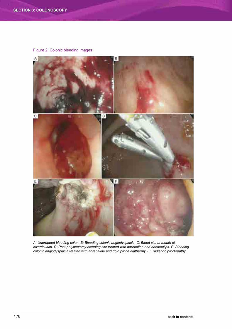
178
SECTION 3: COLONOSCOPY
back to contents
Figure 2. Colonic bleeding images
A: Unprepped bleeding colon. B: Bleeding colonic angiodysplasia. C: Blood clot at mouth of diverticulum. D: Post-polypectomy bleeding site treated with adrenaline and haemoclips. E: Bleeding colonic angiodysplasia treated with adrenaline and gold probe diathermy. F: Radiation proctopathy.

179
ACUTE COLONIC BLEEDINGGeoff Francis and Simon Zanati
SECTION 3: COLONOSCOPY
back to contents
Further reading1 Gostout CJ. Acute lower GI bleeding. In: Brandt LJ, editor. Clinical practice of gastroenterology. Philadelphia:
Churchill Livingstone, 1998: 651-662.
2 Gostout CJ, Wang KK, Ahlquist DA, et al. Acute gastrointestinal bleeding. Experience of a specialized management team. J Clin Gastroenterol 1992; 14: 260-267.
3 Farrell JJ, Friedman LS. Gastrointestinal bleeding in the elderly. Gastrenterol Clin North Am 2001; 30: 377-407.
4 Longstreth GF. Epidemiology and outcome of patients hospitalized with acute lower intestinal hemorrhage: a population-based study. Am J Gastroenterol 1997; 92: 419-424.
5 Strate LL, Ayanian JZ, Kotler G, Syngal S. Risk factors for mortality in lower intestinal bleeding. Clin Gastroenterol Hepatol 2008; 6: 1004-1010.
6 Jensen DM, Machicado DA. Diagnosis and treatment of severe hematochezia. The role of acute colonoscopy after purge. Gastroenterology 1988; 95: 1569-1574.
7 Steer ML, Silen W. Diagnostic procedures in gastrointestinal haemorrhage. N Engl J Med 1983; 309: 646-650.
8 Farrell JJ, Friedman LS. Review article: the management of lower gastrointestinal bleeding. Aliment Pharmacol Ther 2005; 21: 1281-1298.
9 Zuckerman GR, Trellis TR, Sherman TM, Clouse RE. An objective measure of stool color for differentiating upper from lower gastrointestinal bleeding. Dig Dis Sci 1995; 40: 1614-1621.
10 ASGE Standards of Practice Committee. The role of endoscopy in the patient with lower GI bleeding. Gastrointest Endosc 2014; 79: 875-885.
11 Strate LL, Syngal S. Timing of colonoscopy: impact on length of hospital stay in patients with acute lower intestinal bleeding. Am J Gastroenterol 2003; 98: 317-322.
12 Schmulewitz N, Fisher DA, Rockey DC. Early colonoscopy for acute lower GI bleeding predicts shorter hospital stay: a retrospective study of experience in a single center. Gastrointest Endosc 2003; 58: 841-846.
13 Jensen DM, Machicado GA, Jutabha R, Kovacs TO. Urgent colonoscopy for the diagnosis and treatment of severe diverticular hemorrhage. N Engl J Med 2000; 342: 78-82.
14 Navaneethan U, Njei B, Venkatesh PG, Sanaka MR. Timing of colonoscopy and outcomes in patients with lower GI bleeding: a nationwide population-based study. Gastrointest Endosc 2014; 79: 297-306.
15 Management of acute upper and lower gastrointestinal bleeding a national guideline. Edinburgh: Scottish Intercollegiate Guidelines Network, 2008.
16 Gianfrancisco JA, Abcarian H. Pitfalls in the treatment of massive lower gastrointestinal bleeding with “blind” subtotal colectomy. Dis Colon Rectum 1982; 25: 441-445.
17 Waye JD, Bashkoff E. Total colonoscopy: is it always possible? Gastrointest Endosc 1991; 37: 152-154.
18 Brady M, Kinn S, Stuart P. Preoperative fasting for adults to prevent perioperative complications. Cochrane Database Syst Rev 2003; (4): CD004423. doi: 10.1002/14651858.CD004423.
19 Norton ID, Wang L, Levine SA, et al. Efficacy of colonic submucosal saline solution injection for the reduction of iatrogenic thermal injury. Gastrointest Endosc 2002; 56: 95-99.
20 Silva RA, Correia AJ, Dias LM, et al. Argon plasma coagulation therapy for hemorrhagic radiation proctosigmoiditis. Gastrointest Endosc 1999; 50: 221-224.
21 Rex DK, Lewis BS, Waye JD. Colonoscopy and endoscopic therapy for delayed post- polypectomy hemorrhage. Gastrointest Endosc 1992; 38: 127-129.
22 Parra-Blanco A, Kaminaga N, Kojima T, et al. Hemoclipping for postpolypectomy and postbiopsy colonic bleeding. Gastrointest Endosc 2000; 51: 37-41.
23 Nicholson ML, Neoptolemos JP, Sharp JF, et al. Localization of lower gastrointestinal bleeding using in vivo technetium-99m-labeled red blood cell scintigraphy. Br J Surg 1989; 76: 358-361.
24 Olds GD, Cooper GS, Chak A, et al. The yield of bleeding scans in acute lower gastrointestinal hemorrhage. J Clin Gastroenterol 2005; 39: 273-277.
180
SECTION 3: COLONOSCOPY
back to contents
25 Feingold DL, Caliendo FJ, Chinn BT, et al. Does hemodynamic instability predict positive technetium-labeled red blood cell scintigraphy in patients with acute lower gastrointestinal bleeding? A review of 50 patients. Dis Colon Rectum 2005; 48: 1001-1004.
26 García-Blázquez V, Vicente-Bártulos A, Olavarria-Delgado A, et al. Accuracy of CT angiography in the diagnosis of acute gastrointestinal bleeding: systematic review and meta-analysis. Eur Radiol 2013; 23: 1181-1190.
27 Yoon W, Jeong YY, Shin SS, et al. Acute massive gastrointestinal bleeding: detection and localization with arterial phase multi-detector row helical CT. Radiology 2006; 239: 160-167.
28 Martí M, Artigas JM, Garzón G, et al. Acute lower intestinal bleeding: feasibility and diagnostic performance of CT angiography. Radiology 2012; 262: 109-116.
29 Fiorito JJ, Brandt LJ, Kozicky O, et al. The diagnostic yield of superior mesenteric angiography: correlation with the pattern of gastrointestinal bleeding. Am J Gastroenterol 1989; 84: 878-881.
30 Reinus JF, Brandt LJ. Vascular ectasias and diverticulosis. Common causes of lower gastrointestinal bleeding. Gastroenterol Clin North Am 1994; 23: 1-20.
31 Cohn SM, Moller BA, Zieg PM, et al. Angiography for preoperative evaluation in patients with lower gastrointestinal bleeding: are the benefits worth the risks? Arch Surg 1998; 133: 50-55.
32 Browder W, Cerise EJ, Litwin MS. Impact of emergency angiography in massive lower gastrointestinal bleeding. Ann Surg 1986; 204: 530-536.
33 Kuo WT, Lee DE, Saad WE, et al. Superselective microcoil embolization for the treatment of lower gastrointestinal hemorrhage. J Vasc Interv Radiol 2003; 14: 1503-1509.
34 Lipof T, Sardella WV, Bartus CM, et al. The efficacy and durability of super-selective embolization in the treatment of lower gastrointestinal bleeding. Dis Colon Rectum 2008; 51: 301-305.
35 Walker TG, Salazar GM, Waltman AC. Angiographic evaluation and management of acute gastrointestinal hemorrhage. World J Gastroenterol 2012; 18: 1191-1201.
36 Renzuli P, Maurer CA, Netzer P, et al. Subtotal colectomy with primary ileorectostomy is effective for unlocalized diverticular hemorrhage. Langenbecks Arch Surg 2002; 387: 67-71.
37 Baker R, Senagore A. Abdominal colectomy offers safe management for massive lower GI bleed. Am Surg 1994; 60: 578-582.
Pages 181-183 (used for notes in the hard copy) have been removed from the PDF edition of this handbook.

Section 4
ENDOSCOPIC RETROGRADE CHOLANGIOPANCREATOGRAPHY

185
Biliary obstruction due to malignancyTony Speer
The most common malignancy causing biliary obstruction is carcinoma of the pancreas. Other malignancies include cholangiocarcinoma, carcinoma of the gall bladder and tumour metasta-ses to lymph nodes or the liver. Biliary obstruction due to malignancy is conveniently divided into low bile duct obstruction and hilar obstruction; the investigation and treatment of these two groups differ.
Identifying obstruction: stones and strictures
The initial results of liver function tests will roughly triage patients into those with chronic liver disease, hepatitis or “cholestasis”. Cholestatic liver function test results typically show moderate elevations of alkaline phosphatase and gamma-glutamyl transferase levels and minor eleva-tions of transaminase levels (2–5 ×). Cholestatic liver function test results may be due to either biliary obstruction or hepatocellular disease. A careful history often identifies a likely cause of hepatocellular disease. Abdominal ultrasound is the initial investigation to triage patients into those with obstruction and those with hepatocellular disease.
If the bile duct is obstructed, ultrasound imaging will usually show a dilated duct and identify the level of obstruction. Good-quality ultrasound may also identify the lesion itself, particularly with carcinoma of the pancreas or gall bladder, metastases or, less frequently, cholangiocarcinoma. In low bile duct obstruction due to malignancy, the gall bladder is usually dilated. Ultrasound readily identifies stones in the gall bladder, but their presence does not necessarily signify bile duct obstruction due to stones. Ultrasound has good specificity (90%–95%) for identifying stones in the duct, but lower sensitivity (30%–70%); that is, ultrasound misses stones in the common bile duct in about 50% of cases.
SECTION 4: ENDOSCOPIC RETROGRADE CHOLANGIOPANCREATOGRAPHY
CHAPTER 17
back to contents
BIlIAry OBSTrUCTIOn dUe TO mAlIGnAnCyTony Speer
SECTION 4: ENDOSCOPIC RETROGRADE CHOLANGIOPANCREATOGRAPHY
186 back to contents
A common scenario is jaundice, with ultrasound showing a dilated bile duct but no causative lesion. The clinical presentation usually separates patients into two broad groups: those with stones and those with malignancy.
Low bile duct obstruction
If ultrasound shows the bile duct dilated down to the pancreas and a dilated gall bladder, the most likely cause is carcinoma of the pancreas. Important differential diagnoses are carci-noma of the ampulla, autoimmune pancreatitis, other pancreatic tumours and, occasionally, an impacted gallstone. A pancreatic protocol computed tomography (CT) scan of the abdomen will help differentiate between these diagnoses and stage a carcinoma.
After intravenous contrast injection, fine-cut CT scans are taken through the pancreas during arterial and portal venous phases. A pancreatic carcinoma is best seen on the arterial phase. Involvement of arterial and venous structures can also be identified. A routine CT scan of the abdomen and pelvis does not usually include arterial-phase imaging, and the slices through the pancreas are much broader, showing less detail. A standard abdominal and pelvic CT scan may miss small pancreatic tumours.
If the pancreatic protocol CT scan does not identify a mass lesion or is equivocal, endoscopic ultrasound (eUS) may identify a small lesion. eUS shows detailed images of the pancreas, and fine needle aspiration biopsy can be performed on suspicious mass lesions and enlarged lymph nodes under ultrasound control. An impacted gallstone and carcinoma of the ampulla are readily identified by eUS.
Carcinoma of the ampulla is a less common cause of low bile duct obstruction, but it is an important differential diagnosis. The tumour is less aggressive than carcinoma of the pancreas, and surgical resection provides a cure in most patients. The tumour is slower-growing, and imaging often shows marked dilation of the common bile duct and pancreatic duct. It is difficult to identify a mass lesion within the lumen of the duodenum on CT or ultrasound scans with any confidence. Patients with carcinoma of the ampulla are often less jaundiced than those with carcinoma of the pancreas, and cholangitis and pancreatitis are common symptoms. diagnosis can be confirmed by direct biopsy at either eUS or endoscopic retrograde cholangiopancrea-tography (erCP).
Atypical imagingnot all pancreatic masses obstructing the bile duct are carcinomas. Pancreatic adenocarci-noma arises in the duct, causing obstruction with dilation and atrophy upstream. A mass with a non-dilated main pancreatic duct should raise suspicion of an alternative diagnosis. The most common cause — a carcinoma in the uncinate process, not invoving the main duct — is easily diagnosed with eUS and biopsy. lymphoma may present with an unusally large mass in an otherwise well patient. Surrounding lymph nodes are not always enlarged.
187
BIlIAry OBSTrUCTIOn dUe TO mAlIGnAnCyTony Speer
SECTION 4: ENDOSCOPIC RETROGRADE CHOLANGIOPANCREATOGRAPHY
back to contents
Autoimmune pancreatitis presents with a mass obstructing the bile duct; however, the pancre-atic duct is not dilated, although it may have irregular strictures. The pancreas looks “plump”, not atrophied, on CT and magnetic resonance imaging (mrI). Often the patient is mildly jaun-diced, and the level of jaundice remains stable rather than progressively increasing, as it does in carcinoma.
Autoimmune pancreatitisTwo subtypes of autoimmune pancreatitis have been described.
Patients with type 1 autoimmune pancreatitis are usually older men (mean age, 67 years); their serum IgG4 levels may be elevated, and acute pancreatitis and pain are rare. Type 1 is the pancreatic manifestation of IgG4-related disease, a multiorgan immune-mediated condition that mimics many malignant, infectious and inflammatory disorders. Other manifestations of IgG4-related disease — sialadenitis, dacryoadenitis, retroperitoneal fibrosis and autoimmune sclerosing cholangitis — may be present concurrently or develop metachronously over months to years. An elevated serum IgG4 level is one of several diagnostic criteria, but it is neither sensitive nor specific. About 20% of patients with type 1 autoimmune pancreatitis have a normal serum IgG4 level. The IgG4 level is elevated in 10% of patients with carcinoma of the pancreas and 4%–10% of healthy controls.
Type 2 autoimmune pancreatitis occurs in younger patients (median age, 43 years), affecting men and women equally. It presents with obstructive jaundice or acute pancreatitis and is asso-ciated with inflammatory bowel disease (in 30% of patients). Serum IgG4 levels are normal.
If imaging is atypical, an eUS and biopsy should always be performed to confirm a carcinoma before undertaking resection, and the possibility of lymphoma or autoimmune pancreatitis should be considered before inserting a metal stent. An uncovered metal stent in a benign lesion is impossible to remove and will cause many years of discomfort and complications for the patient and the endoscopist.
Staging carcinoma of the pancreasA confident diagnosis of carcinoma of the pancreas can usually be made based on history plus imaging. Typically, an older patient presents with painless progressive jaundice and associated weight loss (Figure 1); ultrasound shows a dilated bile duct and gall bladder, and CT confirms a mass in the head of the pancreas and a dilated pancreatic duct.
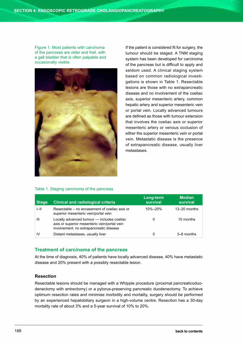
BIlIAry OBSTrUCTIOn dUe TO mAlIGnAnCyTony Speer
SECTION 4: ENDOSCOPIC RETROGRADE CHOLANGIOPANCREATOGRAPHY
188 back to contents
If the patient is considered fit for surgery, the tumour should be staged. A Tnm staging system has been developed for carcinoma of the pancreas but is difficult to apply and seldom used. A clinical staging system based on common radiological investi-gations is shown in Table 1. resectable lesions are those with no extrapancreatic disease and no involvement of the coeliac axis, superior mesenteric artery, common hepatic artery and superior mesenteric vein or portal vein. locally advanced tumours are defined as those with tumour extension that involves the coeliac axis or superior mesenteric artery or venous occlusion of either the superior mesenteric vein or portal vein. metastatic disease is the presence of extrapancreatic disease, usually liver metastases.
Table 1. Staging carcinoma of the pancreas
Stage Clinical and radiological criteriaLong-term
survivalMedian survival
I–II resectable – no encasement of coeliac axis or superior mesenteric vein/portal vein
10%–20% 13–20 months
III locally advanced tumour — includes coeliac axis or superior mesenteric vein/portal vein involvement; no extrapancreatic disease
0 10 months
IV distant metastases, usually liver 0 3–6 months
Treatment of carcinoma of the pancreasAt the time of diagnosis, 40% of patients have locally advanced disease, 40% have metastatic disease and 20% present with a possibly resectable lesion.
Resectionresectable lesions should be managed with a Whipple procedure (proximal pancreaticoduo-denectomy with antrectomy) or a pylorus-preserving pancreatic duodenectomy. To achieve optimum resection rates and minimise morbidity and mortality, surgery should be performed by an experienced hepatobiliary surgeon in a high-volume centre. resection has a 30-day mortality rate of about 3% and a 5-year survival of 10% to 20%.
Figure 1. most patients with carcinoma of the pancreas are older and frail, with a gall bladder that is often palpable and occasionally visible

189
BIlIAry OBSTrUCTIOn dUe TO mAlIGnAnCyTony Speer
SECTION 4: ENDOSCOPIC RETROGRADE CHOLANGIOPANCREATOGRAPHY
back to contents
Imaging often underestimates the amount of disease, and patients should be advised that laparotomy is the final staging procedure. Unresectable local disease or distant metastases are found in up to 50% of patients undergoing a Whipple resection; surgical bypass, hepatico-jejunostomy with or without a gastroenterostomy, provides palliation in these patients.
Patients considered for Whipple resections have in the past been managed with pre-operative biliary stents. A randomised trial of pre-operative endoscopic drainage found an increased rate of overall complications in the pre-operative drainage group. The patients selected for this study had mild to moderate jaundice, with a bilirubin level less than 200 μmol/l; pre-operative drainage is not beneficial in this group. Pre-operative drainage could be considered in selected patients with deep jaundice, renal impairment or cholangitis.
Palliationmost patients present with unresectable disease and are managed with palliation.
Biliary obstruction causes jaundice, pruritis, malabsorption and poor appetite, and prolonged obstruction may lead to renal impairment. These symptoms resolve with biliary decompression. endoscopic biliary stenting is the treatment of choice. This can be achieved in 90%–95% of patients, with relief of jaundice in 90%, a 30-day mortality rate of 5%–7% and a median survival of 5.5 months. expanding metal stents are more expensive than plastic stents but have a lower rate of early complications, better drainage and a lower late blockage rate, and are cost-effective when these advantages are taken into account. late stent blockages occur in 25%–30% of patients with metal stents, causing recurrent jaundice or cholangitis, and are easily managed with repeat erCP, dilation of the stricture and insertion of another stent.
Chemotherapy for advanced pancreatic cancerGemcitabine is a nucleoside analogue with activity across a broad range of solid tumours. Gemcitabine reduces symptoms in patients with carcinoma of the pancreas and provides a modest survival benefit with low toxicity. Patients with a good performance status could be considered for FOlFIrInOX therapy (folinic acid, fluorouracil, irinotecan and oxaliplatin).
Chemoradiotherapy for locally advanced pancreatic cancerThe optimal therapy for patients with locally advanced unresectable pancreatic cancer remains controversial. Chemoradiotherapy could be considered for those with a good performance status.
Other palliative measuresduodenal obstruction is often best palliated with expanding metal stents or a laparoscopic bypass in patients with a good performance status.
Persistent severe pain is a common problem, and treatment should start with regular analgesia. Other management that could be added includes chemotherapy or radiotherapy and coeliac plexus block.
malabsorption secondary to pancreatic duct obstruction responds well to enzyme replacement.

BIlIAry OBSTrUCTIOn dUe TO mAlIGnAnCyTony Speer
SECTION 4: ENDOSCOPIC RETROGRADE CHOLANGIOPANCREATOGRAPHY
190 back to contents
Histologynot all tumours in the pancreas are adenocarcinomas. Histological proof of malignancy should be obtained for all unresectable lesions. eUS-guided fine needle aspiration biopsy has a sen-sitivity of between 80% and 95% and a specificity of 95% to 100%. Ultrasound or CT-guided percutaneous biopsy, of either the primary tumour or a metastasis, can also be performed. If a lesion is resectable and the imaging is typical of a carcinoma of the pancreas, it is not neces-sary to obtain pre-operative histological confirmation. A negative biopsy does not necessarily exclude a carcinoma.
Tumour markersTumour markers are of limited diagnostic value. Cancer antigen 19-9 (CA 19-9) is often elevated in carcinoma of the pancreas, but it is also elevated in biliary obstruction, cholangitis and liver failure. CA 19-9 testing is often performed as a baseline measure to guide treatment follow-up. If a neuroendocrine tumour is suspected, chromogranin A testing should be requested.
Hilar strictures
Hilar strictures are defined as those involving the proximal 2 cm of the common hepatic duct and/or the left or right hepatic ducts and their branches. The most common malignancies that obstruct the hilum are cholangiocarcinoma (50%), carcinoma of the gall bladder (20%) and metastases to the lymph nodes or liver (20%). Important differential diagnoses are benign strictures, including sclerosing cholangitis, mirizzi’s syndrome and benign idiopathic stricture.
most patients present with jaundice, pruritis, pale stools and dark urine. right upper quadrant pain occurs in patients with carcinoma of the gall bladder but not those with cholangiocarcinoma. Fatigue, malaise and weight loss are common in those with advanced disease.
Ultrasound is the first imaging procedure. Carcinoma of the gall bladder and cholangiocarcinoma can be detected as mass lesions. Small tumours may be missed on ultrasound; ductal dilation with an abrupt change in duct diameter may indicate the presence of a tumour.
mrI with gadolinium contrast gives excellent visualisation of the hepatic parenchyma, as well as the biliary tree and vascular structures. magnetic resonance cholangiopancreatography has replaced erCP and percutaneous transhepatic cholangiography as the best imaging modality to show biliary anatomy, and it should be performed on all suspected hilar strictures (Figure 2).

191
BIlIAry OBSTrUCTIOn dUe TO mAlIGnAnCyTony Speer
SECTION 4: ENDOSCOPIC RETROGRADE CHOLANGIOPANCREATOGRAPHY
back to contents
Figure 2. Hilar stricture on magnetic resonance cholangiopancreatography
Hilar stricture with a stricture in the left hepatic duct and multiple strictures in the right hepatic duct. The left duct is the best for stenting.
Abdominal CT scans may provide additional information on mass lesions, vascular involvement and lobar atrophy.
Staging of cholangiocarcinoma depends on the extent of the stricture, vascular involvement and lobar atrophy and hypertrophy and is best performed in consultation with an experienced hepa-tobiliary surgeon and hepatobiliary radiologist. Criteria for unresectability are shown in Table 2.
Table 2. Criteria for unresectability of cholangiocarcinoma
● Bilateral extensions into or beyond second-order ducts ● Significant lobar atrophy with insufficient remaining liver ● Involvement of main or both right and left portal veins or hepatic arteries ● Biliary stricture involving more than three hepatic segments ● metastatic spread beyond the liver or bile ducts
HistologyBiliary brushings performed during erCP have a specificity of 95%–100% but a low sensitivity of only 50%. Histological confirmation is not necessary before resection, but biliary brushings should be performed on unresectable lesions when a stent is being inserted. Ultrasound or CT-guided biopsy can be performed on mass lesions in unresectable disease.
Surgical resection has a 5-year survival of 9%–18%, with a 30-day mortality rate of 5%–12%. routine use of pre-operative biliary drainage is not recommended. In three randomised trials, two showed no difference in outcomes and one showed that the pre-operatively drained patients had worse outcomes.
Patients with unresectable lesions should be assessed to see whether they would benefit from biliary drainage with an endoscopic stent. The risks of attempted stenting may outweigh the benefits in those with multiple intrahepatic strictures or multiple liver metastases causing

BIlIAry OBSTrUCTIOn dUe TO mAlIGnAnCyTony Speer
SECTION 4: ENDOSCOPIC RETROGRADE CHOLANGIOPANCREATOGRAPHY
192 back to contents
impaired hepatocellular function, as judged by ascites, hypoalbuminaemia or prolonged inter-national normalised ratio, and those who are only mildly jaundiced. discussions with the patient and the patient’s family should include realistic expectations of the benefits of stenting and an appreciation of the risks. If the patient decides to proceed, the imaging should be reviewed to identify the best segment to drain. Atrophied segments and those replaced by metastatic disease should be avoided. A randomised trial has confirmed that a single stent placed in the best segment provides effective palliation and has a lower risk of cholangitis than attempting to place two stents. A single stent can be placed successfully in 75%–90% of patients, with a 30-day mortality rate of 10%–20% and an overall mean survival of about 6 months.
Tumour markersThere are no tumour markers specific for cholangiocarcinoma. CA 19-9 is elevated in up to 85% of patients with cholangiocarcinoma, but it is also elevated in those with benign biliary obstruction and hepatic injury.
Further reading1 Strobel O, Buchler mW. Pancreatic cancer: clinical practice guidelines – what is the evidence? Nat Rev Clin
Oncol 2016; 13: 593-594. doi: 10.1038/nrclinonc.2016.127.
2 Kamisawa T, Chari ST, lerch mm, et al. recent advances in autoimmune pancreatitis: type 1 and type 2. Gut 2013; 62: 1373-1380. doi: 10.1136/gutjnl-2012-304224.
3 Kamisawa T, Zen y, Pillai S, Stone JH. IgG4-related disease. Lancet 2015; 385: 1460-1471. doi: 10.1016/S0140-6736(14)60720-0.
4 lim eJ, Bhathal PS, Tagkalidis PP, Speer AG. Catching a chameleon: IgG4-related systemic disease. Med J Aust 2010; 193: 418-420.
5 Hii mWJ, Gibson rn. role of radiology in the treatment of malignant hilar biliary strictures 1: review of the literature. Australas Radiol 2004; 48: 3-13. doi: 10.1111/j.1440-1673.2004.01233.x.
6 de Palma Gd, Galloro G, Sicilliano S, et al. Unilateral venous bilateral endoscopic hepatic duct drainage in patients with malignant hilar biliary obstruction: results of a prospective, randomized, and controlled study. Gastrointest Endosc 2001; 53: 547-553. doi: http://dx.doi.org/10.1067/mge.2001.113381.
7 eckel F, Brunner T, Jelic S; eSmO Guidelines Working Group. Biliary cancer: eSmO clinical practice guidelines for diagnosis, treatment and followup. Ann Oncol 2011; 22 Suppl 6: vi40-vi44. doi: 10.1093/annonc/mdr375.
8 dumonceau Jm, Tringali A, Blero d, et al. Biliary stenting: indications, choice of stents and results: european Society of Gastrointestinal endoscopy (eSGe) clinical guideline. Endoscopy 2012; 44: 277-298. doi: 10.1055/s-0031-1291633.
Pages 193-194 (used for notes in the hard copy) have been removed from the PDF edition of this handbook.

195
Benign Biliary strictures and Biliary leakskarl Herba and Philip craig
SECTION 4: ENDOSCOPIC RETROGRADE CHOLANGIOPANCREATOGRAPHY
back to contents
Benign biliary strictures and biliary leakskarl Herba and Philip craig
the aetiology of benign biliary strictures (BBs) and leaks is diverse (table 1). Patients most commonly present with symptomatic cholestasis or cholangitis, and it is important to exclude underlying malignancy at presentation. Many strictures are suitable for endoscopic therapy, which is usually indicated to prevent the development of cirrhosis. the focus of this chapter is the management of the most common causes of biliary strictures and leaks.
table 1. causes of benign biliary strictures
● Post-operative ● Post-endoscopic sphincterotomy ● chronic pancreatitis ● Primary sclerosing cholangitis ● secondary sclerosing cholangitis
♦ recurrent pyogenic cholangitis (oriental cholangitis) ♦ acquired immunodeficiency syndrome (aids) cholangiopathy ♦ Hepatic arterial chemotherapy (floxuridine) ♦ congenital biliary disorders
● igg4-related cholangiopathy ● Portal biliopathy ● radiation-induced ● ischaemia (including vasculitis)
Post-operative biliary leaks and strictures
although bile leaks and strictures may occur after any operation on the biliary tract, they most often occur following laparoscopic cholecystectomy. the risk of bile duct injury following this procedure is 0.5%, which appears to be two to five times higher than that for open cholecys-tectomy. strictures may also develop at the site of biliary anastomoses after hepatic resection
SECTION 4: ENDOSCOPIC RETROGRADE CHOLANGIOPANCREATOGRAPHY
CHAPTER 18

196
Benign Biliary strictures and Biliary leakskarl Herba and Philip craig
SECTION 4: ENDOSCOPIC RETROGRADE CHOLANGIOPANCREATOGRAPHY
back to contents
or liver transplantation. Bergman and colleagues have classified biliary injuries into four types, which are assessed as either major or minor:
Type A: Biliary leaks involving either the cystic duct or peripheral hepatic radicles (minor)
Type B: Bile duct leaks with or without biliary strictures (major)
Type C: Bile duct strictures without leaks (major)
Type D: complete transection with or without excision of part of the biliary tree (major).
the timing and presentation of post-operative biliary injuries vary depending on the nature of the injury. Patients may present acutely with either jaundice or cholangitis or with an external fistula or intraperitoneal collection. However, most strictures (70%–80%) have a delayed pres-entation, often 6 to 12 months after surgery, with symptoms of cholestasis or elevation in liver function test (lFt) results. On cholangiography, biliary strictures are usually short with sharp edges, located near the cystic duct stump. strictures resulting from ischaemic injury are usually longer and extend to the hilum.
in the early post-operative period, endoscopic retrograde cholangiopancreatography (ercP) has a role in both defining and treating biliary injuries. if ercP is unsuccessful or does not fully define the biliary tree, magnetic resonance cholangiopancreatography (MrcP) provides useful information to classify types of injury. When the presentation of biliary strictures is delayed, abdominal ultrasound, MrcP or computed tomography (ct) cholangiography before ercP can help plan management. Percutaneous transhepatic cholangiography — an alternative therapeutic approach to ercP — is limited by lower success rates and higher complication rates and is therefore reserved for failed endoscopic procedures. causes of biliary injury in the setting of cholecystectomy include anatomical variations in the origin of the cystic duct and right-sided biliary segments, previous surgery and active cholecystitis or cholangitis.
apart from type d injuries, where it is impossible to gain access to the proximal biliary tree with a guidewire, all other biliary injuries are worth attempted endoscopic therapy. to treat early post-operative biliary leaks, appropriate ercP technique includes slow contrast injection to define the level of the injury and to detect associated calculi (Figure 1). the aim of therapy is to abolish the transpapillary pressure gradient, thereby promoting the flow of bile into the duodenum. leaks from the cystic duct stump are the most common and are often associated with a retained bile duct calculus. severing of the ducts of luschka, which are peripheral ducts connecting the intrahepatic system within the gall bladder bed, also produces minor leaks. Both situations can be managed by a combination of endoscopic sphincterotomy (es) and/or short-term biliary stenting, with resultant leak closure within a week. Both es and stenting have specific advantages and disadvantages. type B biliary injuries produce leaks from either the common bile duct or one of the intrahepatic ducts. these injuries usually require large-bore plastic stents, with or without es, to cover the hole and help prevent late stricture formation. these stents should be left in place for at least 2 months, with expected resolution in 70%–80% of cases.

197
Benign Biliary strictures and Biliary leakskarl Herba and Philip craig
SECTION 4: ENDOSCOPIC RETROGRADE CHOLANGIOPANCREATOGRAPHY
back to contents
Figure 1. Post-cholecystectomy bile leak managed with endoscopic sphincterotomy
late biliary strictures are usually managed by a combination of balloon dilatation and the place-ment of multiple stents (Figure 2), as dilatation alone is associated with high recurrence rates. these strictures are often tight and need to be traversed with narrow hydrophilic guidewires. an es is required to allow placement of multiple stents and to facilitate repeat procedures. the biliary stents are exchanged every 3 months to avoid cholangitis and usually left in place for 1 year until stricture resolution is seen on cholangiography. For difficult cases, a combined percutaneous–endoscopic approach may be required for initial stent placement. Bergman and colleagues reported that using two 10 French (Fr) plastic stents for 12 months enabled 80% of post-operative stenoses to resolve endoscopically. However, costamagna’s group advocated even more aggressive management, with balloon dilatation and the placement of as many 10 Fr stents as possible (mean, 3.2) over a mean 12-month period, until complete stricture resolution. this regimen was confirmed by kuzela and colleagues, with long-term stricture resolution seen in 90%–100% of patients over 16–48 months of follow-up. early complications in 9% of patients were all managed conservatively.
Overall, surgical and endoscopic approaches appear at least equivalent in treating BBs, although there have been no comparative trials. However, as initial endoscopic therapy does not preclude later surgical intervention, it is usually the initial treatment of choice. although multiple endoscopic procedures are usually required, the morbidity and mortality from surgery is higher.

198
Benign Biliary strictures and Biliary leakskarl Herba and Philip craig
SECTION 4: ENDOSCOPIC RETROGRADE CHOLANGIOPANCREATOGRAPHY
back to contents
Figure 2. Balloon dilatation (left), followed by placement of multiple plastic stents (right), for a benign post-operative biliary stricture
Post-transplant anastomotic biliary strictures are managed with similar endoscopic approaches, combining biliary dilatation with the placement of multiple plastic stents. Post-sphincterotomy distal biliary strictures may occur in up to 2% of patients. again, stricture dilatation using multiple plastic stents produces stricture resolution in 90% of patients. uncovered self-expanding metal stents (seMss) are not indicated for most BBs, as they cannot be removed and may develop a late granulation reaction, which can preclude surgery. However, in poor operative candidates requiring long-term stenting, uncovered seMss may be useful.
Chronic pancreatitis
symptomatic biliary stenoses develop in 10%–30% of patients with chronic pancreatitis. acute biliary obstruction from either pancreatic inflammation or pseudocysts usually resolves with short-term biliary stenting. the long-term results of endoscopic therapy for fibrotic stricture in patients with chronic pancreatitis are, however, variable. cahen’s overall success rate for aggressively stenting 58 patients with distal strictures for 12 months was just 38%. results with seMss have been mixed, and these have usually been recommended for poor operative candidates. in a multicentre study of 60 patients with existing plastic biliary stents for chronic pancreatitis-related BBs, patients were randomly assigned to receive either covered seMss or three plastic stents. two-year stricture-free success rates were comparable between the plastic stent group (90%) and the covered seMs group (92%). the use of covered seMss is also associated with fewer ercPs.
Promising preliminary data using short-term (3-month) stenting with large-bore, covered seMss for BBs (Figure 3) suggest an effective alternative to plastic stenting. unlike uncovered seMss, these stents can be removed. a prospective non-randomised study of covered seMss

199
Benign Biliary strictures and Biliary leakskarl Herba and Philip craig
SECTION 4: ENDOSCOPIC RETROGRADE CHOLANGIOPANCREATOGRAPHY
back to contents
in patients with BBs assessed rates of eventual successful stent removal and stricture reso-lution at either 6 months (for liver transplant strictures) or 12 months (for chronic pancreatitis or post-cholecystectomy strictures). stent removal was successful in 75% and subsequent long-term stricture resolution occurred in 75% of patients. a similar study of covered seMss confirmed that longer (> 90 days) stenting was associated with higher stricture resolution rates. Finally, a recent multicentre study in patients with BBs compared outcomes between patients receiving multiple plastic stents or covered seMss. stricture resolution rates at 12 months after placement were 93% with covered seMss and 85% in the multiple plastic stent group (P < 0.001). in addition, patients with covered seMss required fewer ercPs (2.1 v 3.2).
Figure 3. Mid post-operative bile duct stricture (left) successfully managed with a fully covered metal stent (middle) and at 6-month follow-up after stent removal (right)
Primary sclerosing cholangitis
Primary sclerosing cholangitis (Psc) is a chronic inflammatory disease characterised by stric-turing and dilatation of the intrahepatic and/or extrahepatic biliary tree. it is associated with inflammatory bowel disease in two thirds of patients. Psc leads to chronic cholestasis, but patients may present at any time with asymptomatic elevation of serum alkaline phosphatase level, pruritus, jaundice or upper abdominal pain. the disease may progress to cirrhosis, and 10%–30% of patients with Psc will develop cholangiocarcinoma. traditionally, ercP has been used to radiologically diagnose Psc. However, in expert hands, MrcP and ct cholangiography are almost as sensitive, giving MrcP a diagnostic accuracy of 83%–90% in comparison to ercP. However, MrcP has difficulties in diagnosing early Psc, assessing extrahepatic dis-ease and differentiating strictures due to cholangiocarcinoma, and it does not allow therapeutic interventions in cholestatic patients. ct cholangiography appears more sensitive than MrcP at diagnosing Psc (sensitivity, 94% v 63%), and it is superior in diagnosing extrahepatic involve-ment (69% v 25%). secondary causes of sclerosing cholangitis need to be excluded (table 1). in addition, other conditions, such as multicentric cholangiocarcinoma or igg4-associated cholangiopathy, may mimic the cholangiographic appearances of Psc.
the role of ercP in Psc is to aid diagnosis in difficult cases, to help differentiate between benign and malignant strictures and to treat symptomatic dominant biliary strictures, which develop in 15%–20% of patients. at presentation, 25% of dominant strictures are dysplastic

200
Benign Biliary strictures and Biliary leakskarl Herba and Philip craig
SECTION 4: ENDOSCOPIC RETROGRADE CHOLANGIOPANCREATOGRAPHY
back to contents
or malignant. in a cohort of swedish patients with Psc followed for 5.7 years after diagnosis, the frequency of cholangiocarcinoma was 13%. confirmation of cholangiocarcinoma in Psc is often difficult, but it should be suspected with the development of progressive cholestasis or a sudden rise in tumour markers. such individuals should undergo cross-sectional imaging and cholangiography, with brushings and biopsies of any suspicious strictures. a carcinoembryonic antigen (CEA) level over 5.2 μg/L has been shown to have a sensitivity of 68% and specificity of 82% for cholangiocarcinoma, while a cancer antigen 19-9 level over 180 ku/l has sensitivity of 67% and specificity of 98%. the sensitivity of brush cytology for diagnosing cholangiocarcinoma is about 50%. Psc patients with cytological high-grade dysplasia, and possibly those with low-grade dysplasia, should be strongly considered for liver transplantation. Other modalities used to diagnose cholangiocarcinoma in patients with Psc that appear to be superior to ercP for detecting malignancy are intraduct ultrasound (sensitivity, 88% v 63%; specificity, 91% v 53%) and cholangioscopy (sensitivity, 92% v 66%; specificity, 93% v 51%).
the prognosis of patients with Psc may be assessed using a scoring system combining the initial cholangiographic features with age at first ercP. Patients undergoing ercP should be treated with broad-spectrum antibiotics for several days to prevent cholangitis, while care should be taken to avoid overfilling obstructed intrahepatic biliary segments unsuitable for drainage. some patients have either coexistent biliary pigment stones or a papillary stenosis, which respond to es. the optimal endoscopic management of dominant biliary strictures is, however, controversial. the available literature consists of case series, and a variety of techniques have been used. the term “dominant biliary stricture” usually refers to a stenosis of < 1.5 mm diam-eter involving the extraheptic duct or < 1 mm of the right or left hepatic ducts. Biliary dilatation upstream of a stricture may not be present in patients with Psc.
in patients without significant cholestasis, whether dominant strictures should be endoscopically treated is controversial. However, in jaundiced patients with symptoms of biliary obstruction or those likely to have cholangiocarcinoma, endoscopic tissue sampling and therapy of dominant strictures should be undertaken. Most experts recommend initial es, then use of narrow hydro-philic guidewires to traverse the stricture before dilatation. some series suggest that dilatation alone is superior to prolonged endoscopic stenting, with lower rates of cholangitis. Moreover, since the bile ducts in patients with Psc are usually narrow, these are often not amenable to the placement of multiple stents. However, more recent series employing short-term plastic stenting suggest improved benefits extending several years. Ponsioen and colleagues described 32 patients with dominant strictures receiving plastic stents for a mean of just 11 days; 60% of patients were intervention-free at 3-year follow-up. the addition of ursodeoxycholic acid to endoscopic therapy for dominant biliary strictures may also improve survival, as assessed by the Mayo clinic model. Overall complications for therapeutic procedures in patients with Psc are no more common than for other groups having ercP, although episodes of post-procedure cholangitis appear more common. liver transplantation should be considered in cirrhotic patients with dominant biliary strictures. unfortunately, most Psc patients who develop symptomatic cholangiocarcinomas are unsuitable for either liver transplantation or curative resection. in this situation, most patients are best managed with long-term biliary stenting, most often using covered seMss.
cholangioscopy has an important role in differentiating benign from malignant strictures. a multicentre cohort study of 226 patients undergoing single-operator cholangioscopy (spyglass;

201
Benign Biliary strictures and Biliary leakskarl Herba and Philip craig
SECTION 4: ENDOSCOPIC RETROGRADE CHOLANGIOPANCREATOGRAPHY
back to contents
Boston scientific) for indeterminate biliary strictures or stone lithotripsy demonstrated high technical success rates (93%), while histologically adequate specimens were obtained in 88% of 140 biopsy cases. a similar technical success rate (93%) was found in a cohort of 59 patients in an australian centre undergoing balloon-assisted cholangioscopy (Bac) using slim gastro-scopes (Figure 4). among 34 cases of indeterminate biliary strictures, Bac-directed biopsies had a sensitivity of 60% and a specificity of 100% for detecting malignancy. Bac image quality was excellent and, even without biopsies, had a sensitivity of 100% for the confirmation of benign strictures. a systematic review of 456 patients undergoing direct cholangioscopy with the spyglass system confirmed a similar sensitivity (60%) and specificity (98%) for detecting malignant strictures.
Figure 4. dominant hilar stricture in primary sclerosing cholangitis
Cholangiogram and brush cytology sampling (left) were indeterminate for malignancy. Subsequent balloon-assisted cholangioscopy using a slim gastroscope introduced over a guidewire and balloon catheter (right) found that the stricture appearance was benign, and results of biopsies were negative for malignancy.
Secondary sclerosing cholangitis
Acquired immunodeficiency syndrome cholangiopathyacquired immunodeficiency syndrome (aids) cholangiopathy produces right upper quadrant pain in 90% of patients, and cholestasis. the cholangiopathy often develops late in aids, with cd4+ cell counts below 200/mm3. Biliary opportunistic infections such as Cryptosporidium par-vum (two-thirds of cases) or Cytomegalovirus are often implicated. the diagnostic appearances of aids cholangiopathy on ercP include papillary stenosis with extrahepatic biliary dilatation and stricturing of the intrahepatic and extrahepatic biliary tree. Other features include abnor-mal pancreatograms in 50% of patients and an increased incidence of cholangiocarcinoma. Organisms are found in relevant duodenal, papillary and biliary samples from over 90% of patients. therapies include appropriate antimicrobial agents, antiretroviral therapy and urso-deoxycholic acid. es often palliates pain without improving lFt results.

202
Benign Biliary strictures and Biliary leakskarl Herba and Philip craig
SECTION 4: ENDOSCOPIC RETROGRADE CHOLANGIOPANCREATOGRAPHY
back to contents
IgG4-related cholangiopathy
igg4-related cholangiopathy is characterised by segmental biliary strictures, usually in the set-ting of autoimmune pancreatitis. the usual presentation is obstructive jaundice, with over 80% of affected patients being men aged over 60 years. thus, the common differential diagnosis is biliary malignancy. there is histological evidence of bile duct fibrosis with a lymphoplasmacytic infiltrate, with positive igg4 staining on plasma cells. these histological findings may be noted in biliary, liver or ampullary biopsy specimens. in cases of igg4-related cholangiopathy without autoimmune pancreatitis, strictures of the bile duct most commonly involve the hilar region and can mimic cholangiocarcinoma. additional features include multiorgan involvement and elevated serum igg4 levels. a Japanese study group for this condition has proposed clinical diagnostic criteria based on a combination of biliary imaging, increased serum igg4 levels, coexistence of other organ involvement, and characteristic histopathological features with or without a response to steroids (table 2). Jaundiced patients respond to biliary stenting, while the strictures usually disappear with corticosteroid treatment. long-term immunosuppression is eventually required in 20% of patients.
table 2. Features of igg4-related cholangiopathy, from Japanese clinical diagnostic criteria
Characteristic feature FrequencyBiliary tract imaging: diffuse or segmental narrowing of intrahepatic and/or extrahepatic bile duct with wall thickening
distal bile duct 70%
Proximal bile duct 34%
intrahepatic 36%
Haematological examination
elevated serum igg4 concentrations (typically > 1.35 g/l)
Histological features
Marked lymphocyte and plasmacyte infiltration and fibrosis
infiltration of igg4-positive plasma cells: > 10 per high-power field
storiform fibrosis
Obliterative phlebitis
coexistence of other diseases
autoimmune pancreatitis
igg4-related dacryoadenitis or sialadenitis
igg4-related retroperitoneal fibrosis
clinical response to steroids
203
Benign Biliary strictures and Biliary leakskarl Herba and Philip craig
SECTION 4: ENDOSCOPIC RETROGRADE CHOLANGIOPANCREATOGRAPHY
back to contents
Conclusion
the management of BBs is generally dependent on the underlying stricture aetiology. endoscopic approaches are usually indicated for diagnosis, and therapy consists of a combi-nation of stricture dilatation and/or biliary stenting.
Further reading1 MacFayden BV Jr, Vecchio r, ricardo ae, Mathis cr. Bile duct injury after laparoscopic cholecystectomy. the
united states experience. Surg Endosc 1998; 12: 315-321.
2 Bergman JJ, van den Brink gr, rauws ea, et al. treatment of bile duct lesions after laparoscopic cholecystectomy. Gut 1996; 38: 141-147.
3 Pitt Ha, Miyamoto t, Parapatis sk, et al. Factors influencing outcome in patients with postoperative biliary strictures. Am J Surg 1982; 144: 14-21.
4 craig Pi, Williams, sJ, Hatfield arW, et al. endoscopic therapy in the management of benign biliary strictures. Gut 1990; 31: a1216.
5 Vitellas kM, el-dieb a, Vaswani k, et al. detection of bile duct leaks using Mr cholangiography with mangfodipir trisodium (teslascan). J Comput Assist Tomogr 2001; 25: 102-105.
6 Mueller Pr, vansonnenberg e, Ferrucci Jt, Jr, et al. Biliary stricture dilatation: multicenter review of clinical management in 73 patients. Radiology 1986; 160: 17-22.
7 Bjorkman dJ, carr-locke dl, lichtenstein dr, et al. Postsurgical bile leaks: endoscopic obliteration of the transpapillary pressure gradient is enough. Am J Gastroenterol 1995; 90: 2128-2133.
8 Woods Ms, traverso lW, kozarek ra, et al. characteristics of biliary tract complications during laparoscopic cholecystectomy: a multi-institutional study. Am J Surg 1994; 167: 27-33; discussion 33-4.
9 Bergman JJ, Burgemeister l, Bruno MJ, et al. long-term follow-up after biliary stent placement for postoperative bile duct stenosis. Gastrointest Endosc 2001; 54: 154-161.
10 costamagna g, Pandolfi M, Mutignani M, et al. long-term results of endoscopic management of postoperative bile duct strictures with increasing numbers of stents. Gastrointest Endosc 2001; 54: 162-168.
11 kuzela l, Oltman M, sutka J, et al. Prospective follow-up of patients with bile duct strictures secondary to laparoscopic cholecystectomy, treated endoscopically with multiple stents. Hepatogastroenterology 2005; 52: 1357-1361.
12 Morelli g, Fazel a, Judah J, et al. rapid-sequence endoscopic management of posttransplant anastomotic biliary strictures. Gastrointest Endosc 2008; 67: 879-885.
13 Pozsar J, sahin P, laszlo F, topa l. endoscopic treatment of sphincterotomy-associated distal common bile duct strictures by using sequential insertion of multiple plastic stents. Gastrointest Endosc 2005; 62: 85-91.
14 O’Brien sM, Hatfield ar, craig Pi, Williams sP. a 5-year follow-up of self-expanding metal stents in the endoscopic management of patients with benign bile duct strictures. Eur J Gastroenterol Hepatol 1998; 10: 141-145.
15 Jakobs r, riemann JF. the role of endoscopy in acute recurrent and chronic pancreatitis and pancreatic cancer. Gastroenterol Clin North Am 1999; 28: 783-800, xii.
16 craig Pi. role of endoscopic stenting for biliary strictures in chronic pancreatitis. Dig Endosc 2012; 24 suppl 1: 38-42.
17 van Berkel a, cahen d, van Westerloo d, et al. self-expanding metal stents in benign biliary strictures due to chronic pancreatitis. Endoscopy 2004; 36: 381-384.

204
Benign Biliary strictures and Biliary leakskarl Herba and Philip craig
SECTION 4: ENDOSCOPIC RETROGRADE CHOLANGIOPANCREATOGRAPHY
back to contents
18 Haapamäki c, kylänpää l, udd M, et al. randomized multicenter study of multiple plastic stents vs. covered self-expandable metallic stent in the treatment of biliary stricture in chronic pancreatitis. Endoscopy 2015; 47: 605-610.
19 siiki a, Helminen M, sand J, laukkarinen J. covered self-expanding metal stents may be preferable to plastic stents in the treatment of chronic pancreatitis-related biliary strictures: a systematic review comparing 2 methods of stent therapy in benign biliary strictures. J Clin Gastroenterol 2014; 48: 635-643.
20 Mahajan a, Ho H, sauer B, et al. temporary placement of fully covered self-expandable metal stents in benign biliary strictures: midterm evaluation (with video). Gastrointest Endosc 2009; 70: 303-309.
21 devière J, nageshwar reddy d, Püspök a, et al. successful management of benign biliary strictures with fully covered self-expanding metal stents. Gastroenterology 2014; 147: 385-395.
22 kahaleh M, Brijbassie a, sethi a, et al. Multicenter trial evaluating the use of covered self-expanding metal stents in benign biliary strictures: time to revisit our therapeutic options? J Clin Gastroenterol 2013; 47: 695-699.
23 coté ga, slivka a, tarnasky P, et al. effect of covered metallic stents compared with plastic stents on benign biliary stricture resolution: a randomized clinical trial. JAMA 2016; 315: 1250-1257.
24 cullen sn, chapman rW. review article: current management of primary sclerosing cholangitis. Aliment Pharmacol Ther 2005; 21: 933-948.
25 Boberg kM, Bergquist a, Mitchell s, et al. cholangiocarcinoma in primary sclerosing cholangitis: risk factors and clinical presentation. Scand J Gastroenterol 2002; 37: 1205-1211.
26 Weber c, kuhlencordt r, grotelueschen r, et al. Magnetic resonance cholangiopancreatography in the diagnosis of primary sclerosing cholangitis. Endoscopy 2008; 40: 739-745.
27 Macchi V, Floreani a, Marchesi P, et al. imaging of primary sclerosing cholangitis: preliminary results by two new non-invasive techniques. Dig Liver Dis 2004; 36: 614-621.
28 Bjornsson e, Olsson r. dominant strictures in patients with primary sclerosing cholangitis-revisited. Am J Gastroenterol 2004; 99: 2281.
29 tischendorf JJ, kruger M, trautwein c, et al. cholangioscopic characterization of dominant bile duct stenoses in patients with primary sclerosing cholangitis. Endoscopy 2006; 38: 665-669.
30 Bergquist a, ekbom a, Olsson r, et al. Hepatic and extrahepatic malignancies in primary sclerosing cholangitis. J Hepatol 2002; 36: 321-327.
31 Ponsioen cy, lam k, van Milligen de Wit aW, et al. Four years experience with short term stenting in primary sclerosing cholangitis. Am J Gastroenterol 1999; 94: 2403-2407.
32 Boberg kM, Jebsen P, clausen OP, et al. diagnostic benefit of biliary brush cytology in cholangiocarcinoma in primary sclerosing cholangitis. J Hepatol 2006; 45: 568-574.
33 tischendorf JJ, Meier Pn, schneider a, et al. transpapillary intraductal ultrasound in the evaluation of dominant bile duct stenoses in patients with primary sclerosing cholangitis. Scand J Gastroenterol 2007; 42: 1011-1017.
34 Ponsioen cy, Vrouenraets sM, Prawirodirdjo W, et al. natural history of primary sclerosing cholangitis and prognostic value of cholangiography in a dutch population. Gut 2002; 51: 562-566.
35 craig P, Hatfield a. endoscopic therapy in primary sclerosing cholangitis: primary sclerosing cholangitis: recent developments. Eur J Gastroenterol Hepatol 1992; 4: 284-287.
36 kaya M, Petersen Bt, angulo P, et al. Balloon dilation compared to stenting of dominant strictures in primary sclerosing cholangitis. Am J Gastroenterol 2001; 96: 1059-1066.
37 Ponsioen c, lam k, van Milligen de Wit aW, et al. Four years experience with short term stenting in primary sclerosing cholangitis. Am J Gastroenterol 1999; 94: 2403-2407.
38 gluck M, cantone nr, Brandabur JJ, et al. a twenty-year experience with endoscopic therapy for symptomatic primary sclerosing cholangitis. J Clin Gastroenterol 2008; 42: 1032-1039.
39 chen yk, Parsi Ma, Binmoeller kF, et al. single-operator cholangioscopy in patients requiring evaluation of bile duct disease or therapy of biliary stones (with videos). Gastrointest Endosc 2011; 74: 805-814.
40 lim P, aggarwal V, craig P. role of balloon-assisted cholangioscopy in a multiethnic cohort to assess complex biliary disease (with videos). Gastrointest Endosc 2015; 81: 932-942.

205
Benign Biliary strictures and Biliary leakskarl Herba and Philip craig
SECTION 4: ENDOSCOPIC RETROGRADE CHOLANGIOPANCREATOGRAPHY
back to contents
41 navaneethan u, Hasan Mk, lourdusamy V, et al. single-operator cholangioscopy and targeted biopsies in the diagnosis of indeterminate biliary strictures: a systematic review. Gastrointest Endosc 2015; 82: 608-614.e2.
42 cello J. aids-related biliary tract disease. Gastrointest Endosc Clin N Am 1998; 8: 963.
43 Bjornsson e, chari st, smyrk tc, lindor k. immunoglobulin g4 associated cholangitis: description of an emerging clinical entity based on review of the literature. Hepatology 2007; 45: 1547-1554.
44 ghazale a, chari st, Zhang l, et al. immunoglobulin g4-associated cholangitis: clinical profile and response to therapy. Gastroenterology 2008; 134: 706-715.
45 tanaka a, tazuma s, Okazaki k, et al. nationwide survey for primary sclerosing cholangitis and igg4-related sclerosing cholangitis in Japan. J Hepatobiliary Pancreat Sci 2014; 21: 43-50.
46 Ohara H, Okazaki k, tsubouchi H, et al. clinical diagnostic criteria of igg4-related sclerosing cholangitis 2012. J Hepatobiliary Pancreat Sci 2012; 19: 536-542.
Page 206 (used for notes in the hard copy) has been removed from the PDF edition of this handbook.

207
Chronic pancreatitisNam Nguyen
Chronic pancreatitis (CP) is characterised by progressive inflammation of the pancreas, which leads to irreversible structural damage and development of exocrine and/or endocrine impair-ment. Alcohol use and recurrent bouts of gallstone pancreatitis are the most common causes. In less than 20% of cases, the underlying causes are autoimmune and hereditary in origin (including cystic fibrosis gene mutations and SPINK1 and cationic trypsinogen mutations). Even in so-called idiopathic CP, up to 65% of patients are found to have a genetic defect that is responsible for the disease. The most common presentation in patients with CP is abdominal pain. Symptoms of pancreatic insufficiency, such as diabetes, weight loss, malabsorption or steatorrhoea, are late manifestations.
In most cases, the presence and severity of CP can be assessed by non-invasive imaging techniques, using computed tomography (CT), magnetic resonance imaging (MRI) or magnetic resonance cholangiopancreatography (MRCP). Changes of early CP, however, can be subtle and may not be well visualised by conventional imaging, thus requiring further evaluation with endoscopic ultrasound (EUS) or endoscopic retrograde cholangiopancreatography (ERCP). This chapter outlines the diagnostic and therapeutic roles of these endoscopic modalities in the management of CP.
Diagnostic endoscopic procedures for chronic pancreatitis
ERCP is no longer considered the gold standard for the diagnosis of CP because of the risk of ERCP-induced complications and the fact that the parenchymal abnormality cannot be assessed. With the ability to detect pancreatic parenchymal and ductal changes with high sensitivity, EUS has emerged as the technique of choice for the morphological diagnosis of CP. When used in conjunction with secretin stimulation and fine needle biopsy, both functional and histological assessments can also be achieved by EUS.
SECTION 4: ENDOSCOPIC RETROGRADE CHOLANGIOPANCREATOGRAPHY
CHAPTER 19
back to contents

208
ChRoNIC PANCREATITISNam Nguyen
SECTION 4: ENDOSCOPIC RETROGRADE CHOLANGIOPANCREATOGRAPHY
back to contents
Endoscopic ultrasoundThe consensus from an international working group is that EUS can diagnose CP by the pres-ence of the major and minor Rosemont criteria, which comprise five parenchymal and five ductal criteria. Based on the number and character of positive criteria, EUS evaluation is classified as “consistent with CP”, “suggestive of CP”, “indeterminate for CP” or “normal” (Table 1). By using 1–2 EUS criteria for diagnosing mild pancreatitis, 3–5 for moderate pancreatitis and > 5 for severe pancreatitis, agreement between the results of a classic secretin functional test and EUS was 100% for normal parenchyma and severe disease, 50% for moderate CP and 13% for mild disease. The cut-off of ≥ 4 EUS criteria has the best accuracy (86%) and specificity (90%) for predicting the presence of pancreatic insufficiency and histological findings of CP.
Table 1. (A) Consensus-based criteria of parenchymal and ductal changes for diagnosing CP by EUS, and (B) the likelihood of a diagnosis of CP*
A. Rosemont criteriaFeatures Definition
Major criteria hyperechoic foci with shadowing (A)
Echogenic structures ≥ 2 mm in length and width that shadow
MPD calculi (A) Echogenic structure(s) within MPD with acoustic shadowing
Lobularity with honeycombing (B)
Well circumscribed, ≥ 5 mm structures with enhancing rim and relatively echo-poor centre; three or more contiguous lobules visualised
Minor criteria hyperechoic foci without shadowing
Echogenic structures ≥ 2 mm in both length and width with no shadowing
Cysts Anechoic, rounded or elliptical structures with or without septations
Stranding Hyperechoic lines of ≥ 3 mm in length in at least two different directions with respect to the imaged plane
Irregular MPD contour Uneven or irregular outline and ectatic course
Dilated side branches ≥ 3 tubular anechoic structures, each measuring ≥ 1 mm in width, budding from the MPD
MPD dilation ≥ 3.5 mm body or ≥ 1.5 mm tail
hyperechoic MPD margin Echogenic, distinct structure greater than 50% of entire MPD in the body and tail
B. Likelihood of a diagnosis of CP based on the above criteriaNormal •≤ 2 minor features, no major featuresIndeterminate for CP •3–4 minor features, no major features
•major B feature alone or with < 3 minor featuresSuggestive of CP •1 major A feature plus < 3 minor features
•1 major B feature plus ≥ 3 minor features•≥ 5 minor features (any)
Consistent with CP •1 major A feature plus ≥ 3 minor features•1 major A feature plus major B feature•2 major A features
CP = chronic pancreatitis. EUS = endoscopic ultrasound. MPD = main pancreatic duct. * Adapted from: Catalano MF, Sahai A, Levy M, et al. EUS-based criteria for the diagnosis of chronic pancreatitis: the Rosemont classification. Gastrointest Endosc 2009; 69: 1251-1261.

209
ChRoNIC PANCREATITISNam Nguyen
SECTION 4: ENDOSCOPIC RETROGRADE CHOLANGIOPANCREATOGRAPHY
back to contents
The major weakness of EUS in the diagnosis of CP, however, is the relatively poor interobserver agreement. Calcification, duct dilation, lobularity with honeycombing, hyperechoic strands and cysts (Figure 1) have the highest kappa scores, but the interobserver agreement for the other criteria is lower. Another shortcoming is that some of the minor EUS criteria for CP, such as duct dilation and stranding, can be seen with normal ageing; thus, it is important to be aware of the patient’s age when interpreting the findings. Together, these weaknesses mean that EUS tends to overdiagnose early CP, which may be minimised by the use of an endoscopic function test. Such a combination of tests has been shown to prevent false-positive results from EUS for the evaluation of CP.
Figure 1. Examples of sonographic appearance of chronic pancreatitis
These images show a range of features, including stranding (a), lobularity (b), calcification in parenchyma and pancreatic ducts (c, e), cystic lesion (d), dilated pancreatic duct and marked parenchymal atrophy (f).
More recently, quantitative elastography and contrast-enhanced techniques have been inte-grated into standard EUS to improve the assessment of CP. Strain elastography is an imaging modality that can measure tissue stiffness by assessing the tissue’s response to an externally generated force. It is based on the fact that stiffer tissues have lower strains, meaning that they deform less under compression, compared with softer tissues that deform more. In a large prospective study (n = 191) that used quantitative EUS elastography to assess the presence of CP, the strain ratio correlated strongly with the number of EUS criteria for CP (r = 0.813), and the overall accuracy of EUS elastography for diagnosing CP was 91% (cut-off strain ratio, 2.25). A recent study has also indicated that EUS elastography is highly accurate in grading the degree of pancreatic fibrosis. Both elastography and contrast-enhanced EUS have been found to be extremely useful in differentiating between pancreatic mass lesions related to CP and carcinoma.

210
ChRoNIC PANCREATITISNam Nguyen
SECTION 4: ENDOSCOPIC RETROGRADE CHOLANGIOPANCREATOGRAPHY
back to contents
Endoscopic retrograde cholangiopancreatographyWith the advances in EUS, MRI and MRCP, the diagnostic role of ERCP in the evaluation of CP is limited and is only relevant to centres that do not have access to EUS or MRI. historically, ERCP was used diagnostically to outline pancreatic ductal abnormalities of CP, including irregular dilatations and strictures of the main duct, clubbing or ectasia of side-branches or ductal stones. The Cambridge classification is the most commonly used scoring system to categorise the ductal changes, as “equivocal changes”, “mild to moderate changes” or “consid-erable changes”, which strongly correlates with the severity of CP and pancreatic insufficiency. however, ERCP is not useful in patients with early CP, where ductal changes can be absent or too subtle to be appreciated. In these patients, the risk of ERCP-induced pancreatitis or related complications is higher than its diagnostic benefit, and ERCP should therefore not be used. one exception is autoimmune pancreatitis: ERCP is found to be more sensitive than MRCP in differentiating focal forms of autoimmune pancreatitis and pancreatic neoplasm, and it is routinely performed in Asia, especially Japan.
Therapeutic endoscopic procedures for chronic pancreatitis
In contrast to its limited diagnostic potential, ERCP has a key therapeutic role in the manage-ment of CP, including relief of pancreatic and biliary ductal obstruction, stone extraction and treatment of pancreatic fistulae and pseudocysts. Similarly, with the development of appropriate accessories, EUS has also evolved from diagnostics to therapeutics, allowing safe and effective approaches for drainage of peripancreatic fluid collections, biliary and pancreatic duct (PD) obstruction that is not accessible by ERCP, and coeliac plexus neurolysis.
Endoscopic retrograde cholangiopancreatography
Biliary endotherapyUp to 30% of patients with advanced CP develop symptomatic biliary strictures, which are most often the result of fibrosis or oedema from acute on chronic pancreatitis affecting the head of pancreas, pseudocyst compression and, rarely, malignant transformation. Patients with IgG4-related autoimmune pancreatitis can develop biliary obstruction from either pancreatic disease or associated IgG4 cholangiopathy. The most common presenting complaints are abdominal pain, jaundice and cholestatic elevation of liver enzyme levels. Except for IgG4 disease, the location of most biliary obstruction related to CP is in the distal third of the common bile duct (CBD).
In addition to obtaining a cholangiogram to characterise the nature of the stricture, ERCP plays a key role in decompressing the biliary tree by a combination of sphincterotomy, balloon dilation of the stricture and/or stent insertion. In cases where the stricture is irregular or suspicious of malignancy, brushing can also be done for cytology, although the diagnostic yield is expected to be low (about 30%). Given the fibrotic nature of most of these strictures, long-term success with a single plastic stent is low, especially in patients who have calcific CP in the head region.
211
ChRoNIC PANCREATITISNam Nguyen
SECTION 4: ENDOSCOPIC RETROGRADE CHOLANGIOPANCREATOGRAPHY
back to contents
To reduce the need for repeated ERCPs for stent replacement, “aggressive” insertion of mul-tiple (three to five) plastic stents after balloon stricturoplasty over a 12-month period has been proposed to overcome the restenosis and “stretch” the stricture. Technical success with this approach can be achieved in 90% of cases, and the rate of stricture resolution after 12 months varies from 44% to 92%.
Recently, the development of fully covered, removable, self-expanding metal stents (SEMSs) has provided an alternative to the multiple plastic stents approach. This involves much less technical demand, and success can be achieved in 100% of cases. The shortcomings of SEMSs are the higher cost and the risk of stent migration. In a recent large, non-randomised, multicen-tre, international study (n = 198) that used SEMSs for CP stricture over 12 months, stricture resolution was found in 80% of patients after a median follow-up period of 20 months. The rate of stent migration, however, was significantly high (29%) and was associated with a lower rate of stricture resolution. In addition, the rate of serious stent-related or stent removal-related adverse events was high (27%), with cholangitis the most common complication. In a recent small but randomised trial that compared SEMSs with multiple plastic stents for CP strictures, both long-term success and rate of stent migration were similar between the groups, but the SEMS group had a higher technical success rate and fewer endoscopic sessions required, which may account for the higher reported cost-effectiveness. Based on these findings, SEMSs have been proposed as the first-line option for fibrotic strictures of CP. Further validation with larger randomised controlled trials is required before this recommendation can be implemented.
Pancreatic endotherapyIn most centres, pancreatic endotherapy is reserved for patients with CP who have CP pain, ductal stones and obstruction (i.e. stricture with pre-stenotic dilatation) demonstrated on MRCP or EUS. In these patients, pancreatic sphincterotomy, pancreatic stricture dilatation and stenting with multiple plastic stents or SEMSs during ERCP proved to be useful to achieve long-term symptomatic improvement. The rationale is based on the hypothesis that ductal hypertension leads to pain, which can be resolved by either endoscopic or surgical decompression. Given the endoscopic approach is much less invasive, the European Society of Gastrointestinal Endoscopy (ESGE) has recommended endoscopic therapy as the first-line therapy for painful uncomplicated CP.
It is important to understand, however, that the link between ductal hypertension and pain is not straightforward, as many patients with a dilated PD from stricture or stones are pain-free. Vice versa, a significant proportion of CP patients with pain have no PD obstruction or stones, indi-cating factors such as parenchymal ischaemia, inflammation or neuropathic hypersensitivity are also important in the pathogenesis of pain in CP. Although ductal decompression has also been proposed to minimise the deterioration in pancreatic function, the data are conflicting, as further losses of endocrine and exocrine function are observed even after pancreaticojejunostomy.
Endotherapy for PD strictures: The outcomes of endoscopic ductal decompression by stent-ing in patients with CP are only modest, with the greatest benefit seen in those with dominant strictures and dilated ducts. In a large multicentre study of 1018 patients with CP followed up for a mean of 4.9 years, although endoscopic PD stenting relieved pain in two-thirds of patients, one-quarter of patients subsequently had surgery, and pancreatic function failed to improve. Compared with a single stent, placing multiple plastic pancreatic stents is associated

212
ChRoNIC PANCREATITISNam Nguyen
SECTION 4: ENDOSCOPIC RETROGRADE CHOLANGIOPANCREATOGRAPHY
back to contents
with a higher rate of stricture resolution and fewer repeated ERCPs. While recent case reports suggest promising results from using fully covered metal pancreatic stents for PD strictures, further study with a larger sample size and longer follow-up is warranted.
Compared with surgical drainage, endoscopic decompression appears to be less effective. In a small randomised trial, 39 patients with surgical decompression had lower pain scores, better physical health summary scores and fewer procedures than did endoscopically treated patients over the 2-year follow-up. There were no differences in the rate of complications, length of hos-pital stay or changes in pancreatic function between the groups. Given patients undergoing PD stenting who subsequently required surgical drainage were found to have more perioperative complications than those without prior stenting, it is important to carefully assess the need for endotherapy in these patients in a multidisciplinary setting.
Endotherapy for PD stones: Pancreatic stones can be removed successfully in 50%–75% of cases after pancreatic (major or minor) sphincterotomy using balloon or basket extraction, which leads to symptomatic improvement in two-thirds of patients. however, if there is a stricture downstream of the stone, stricture dilatation with a balloon is required before attempting stone extraction. Factors that favour successful complete extraction of PD stones are stone size less than 1 cm, fewer than three stones, stones confined to the head and body of the pancreas, and absence of impacted stones or stones upstream (towards the tail) of a stricture. Stones that are larger, impacted or upstream of a stricture frequently require fragmentation via mechanical lithotripsy, intraductal lithotripsy with a pulsed dye laser or electrohydraulic lithotripsy, or extra-corporeal shockwave lithotripsy before attempted extraction.
Endotherapy for pancreatic fistulae and pseudocysts: Disruption of the main PD can occur in about 10% of patients with CP from an upstream blowout of obstructing strictures or stones, and can manifest as pancreatic ascites, internal fistulae (e.g. pseudocysts, pleural effusion to other organs) or external cutaneous fistulae. Diverting the pancreatic juice flow away from the fistulae, and if possible bridging the pancreatic leak with transpapillary stents, can successfully treat the fistulae in 56% to 74% of cases. Endoscopic injection of fibrin glue into the fistulous tract in conjunction with stenting has been shown to increase the resolution rate. While the majority of pseudocysts are now managed with an EUS approach (see below), transpapillary stenting via ERCP still has a role in treating pseudocysts communicating with the main PD in the head or body of the pancreas. Stents should be left in place for a longer duration, as their removal within 2 months is associated with a higher incidence of pseudocyst recurrence.
Endoscopic ultrasound
Therapy for pseudocystsCurrently, EUS has the key therapeutic role in the management of most pseudocysts. In a recent randomised trial, EUS-guided drainage was found to be as effective as a surgical approach, but with fewer complications, shorter length of hospital stay and greater cost-effectiveness. Compared with an isolated endoscopic approach based on the cystic bulge, the EUS-guided approach was significantly safer, given its ability to characterise the cystic lesion and iden-tify vascular structures. Thus, it has been recommended by the the American Society for Gastrointestinal Endoscopy as first-line therapy for managing symptomatic lesions (abdominal

213
ChRoNIC PANCREATITISNam Nguyen
SECTION 4: ENDOSCOPIC RETROGRADE CHOLANGIOPANCREATOGRAPHY
back to contents
pain, gastric outlet obstruction, early satiety, weight loss or jaundice), infected cysts or enlarging cysts.
The principle of direct cystoenterostomy is to create a communication between the cyst lumen and the gastric or duodenal lumen. This can be achieved by puncturing the cyst under EUS guidance with either a 19G needle or a needle knife, with the track subsequently dilated with a balloon. Two or more double pigtail stents are then placed transmurally into the cyst cavity (Figure 2, a-c). For pseudocysts with significant debris or necrosis, a nasocystic drain can be used to lavage the cyst content (Figure 3, a-b). Recently, the development of large-diameter fully covered metal stents (Figure 2, d) has allowed direct endoscopic necrosectomy immediately after cystoenterostomy or on subsequent examination (Figure 3, c-d). The choice of plastic versus metal stents for cystoenterostomy is often dictated by the presence of debris and the need for necrosectomy.
Regardless of the type of stent, the overall procedural technical success rate is over 90%, and in those cases the rate of cyst resolution is approximately 90% and the long-term recurrence rate is 10%–15%. The risk of complications, primarily bleeding and perforation, is less than 10%. The stents can be removed on resolution of the pseudocyst, the size of which is monitored with CT imaging at 4–6-weekly intervals.
Figure 2. Principal steps in endoscopic ultrasound-guided pseudocyst drainage
These steps involve puncturing the cyst with a 19G needle (a), placing a guidewire for balloon dilatation of a cystogastrostomy tract (b) and subsequent insertion of plastic pigtail stents (c). In pseudocysts with significant necrosis, metal stents can be used instead of multiple plastic stents (d).
Figure 3. Management of pseudocysts with significant debris and necrosis
To manage pseudocysts with significant debris and necrosis (a), insertion of a nasocystic drain (b) for irrigation is recommended in the first 48 hours. Direct endoscopic necrosectomy can also be performed to remove the infective material (c, d).
Coeliac plexus blockEUS-guided coeliac plexus block may have a role in CP patients with intractable pain, and it has been proposed by the ESGE as the second-line treatment for pain in patients with CP.

214
ChRoNIC PANCREATITISNam Nguyen
SECTION 4: ENDOSCOPIC RETROGRADE CHOLANGIOPANCREATOGRAPHY
back to contents
Given its better safety profile, the EUS approach should be preferred over percutaneous coe-liac plexus block. The procedure involves identifying the coeliac plexus or individual ganglionic body for injection of local anaesthesia (e.g. 0.5% bupivacaine and absolute alcohol. Injection of corticosteroids (triamcinolone) is significantly less effective than alcohol (38% v 80%). Although EUS-guided coeliac plexus block alleviates pain in 55% to 70% of patients with short-term follow-up, long-term pain relief is disappointing. At best, only 30% of patients have long-term pain relief at 24 weeks after injection.
The side effects of this method are diarrhoea and hypotension due to parasympathetic activity. Pain exacerbation occurs in 9% of patients for about 48 hours after the injection. Paraparesis, peripancreatic abscess and retroperitoneal haemorrhage are rare side effects (about 0.6%). Given these shortcomings, careful patient selection must be undertaken.
Endoscopic ultrasound drainage of the main pancreatic duct or common bile ductThis procedure is required on rare occasions when ERCP is unsuccessful, which is caused by the inability to cannulate the main PD or CBD (due to severe inflammation, previous surgery or postsurgical stricture) or difficult endotherapy (tight stenosis, large stone or pancreas divisum), and surgery cannot be performed safely. The technique involves puncturing the main PD or CBD through the gastric or duodenal wall, creating a fistula that allows drainage from the main PD or CBD to the gastric or duodenal lumen via the stent. Alternatively, the technique can be used to advance the guidewire into the main PD or CBD, through the papillae and out into the second part of the duodenum, allowing a rendezvous procedure to be performed. Available data indicate the success of this technique is high for biliary drainage (> 90%) but only modest for main PD drainage (about 70%). In patients who have a successful procedure, the reported rate of significant pain relief varies from 69% to 75%. The rate of complications can be as high as 43%. Thus, EUS drainage of the main PD should continue to be confined to tertiary care centres and very experienced endoscopists.
Further reading1 Catalano MF, Sahai A, Levy M, et al. EUS-based criteria for the diagnosis of chronic pancreatitis: the Rosemont
classification. Gastrointest Endosc 2009; 69: 1251-1261.
2 Sahai AV, Zimmerman M, Aabakken L, et al. Prospective assessment of the ability of endoscopic ultrasound to diagnose, exclude, or establish the severity of chronic pancreatitis found by endoscopic retrograde cholangiopancreatography. Gastrointest Endosc 1998; 48: 18-25.
3 Rosch T, Daniel S, Scholz M, et al; European Society of Gastrointestinal Endoscopy Research Group. Endoscopic treatment of chronic pancreatitis: a multicenter study of 1000 patients with long-term follow-up. Endoscopy 2002; 34: 765-771.
4 Neuhaus h. Therapeutic pancreatic endoscopy. Endoscopy 2004; 36: 8-16.
5 Cahen DL, Gouma DJ, Nio Y, et al. Endoscopic versus surgical drainage of the pancreatic duct in chronic pancreatitis. N Engl J Med 2007; 356: 676-684.
6 Kozarek RA, Brandabur JJ, Ball TJ, et al. Clinical outcomes in patients who undergo extracorporeal shock wave lithotripsy for chronic calcific pancreatitis. Gastrointest Endosc 2002; 56: 496-500.

215
ChRoNIC PANCREATITISNam Nguyen
SECTION 4: ENDOSCOPIC RETROGRADE CHOLANGIOPANCREATOGRAPHY
back to contents
7 Mahajan A, ho h, Sauer B, et al. Temporary placement of fully covered self-expandable metal stents in benign biliary strictures: midterm evaluation (with video). Gastrointest Endosc 2009; 70: 303-309.
8 Baron Th, harewood GC, Morgan DE, Yates MR. outcome differences after endoscopic drainage of pancreatic necrosis, acute pancreatic pseudocysts, and chronic pancreatic pseudocysts. Gastrointest Endosc 2002; 56: 7-17.
9 Varadarajulu S, Christein JD, Tamhane A, et al. Prospective randomized trial comparing EUS and EGD for transmural drainage of pancreatic pseudocysts (with videos). Gastrointest Endosc 2008; 68: 1102-1111.
10 Varadarajulu S, Bang JY, Sutton BS, et al. Equal efficacy of endoscopic and surgical cystogastrostomy for pancreatic pseudocyst drainage in a randomized trial. Gastroenterology 2013; 145: 583-590.
11 Santosh D, Lakhtakia S, Gupta R, et al. Clinical trial: a randomized trial comparing fluoroscopy guided percutaneous technique vs. endoscopic ultrasound guided technique of coeliac plexus block for treatment of pain in chronic pancreatitis. Aliment Pharmacol Ther 2009; 29: 979-984.
Page 216 (used for notes in the hard copy) has been removed from the PDF edition of this handbook.

217
Endoscopic management of peripancreatic collectionsVu Kwan
Although numerous innovative techniques for the endoscopic management of peripancreatic collections have emerged in recent years, the principles of management remain unchanged: a clear clinical indication for treatment should exist; a distinction between pseudocyst and pancreatic necrosis should be made; and drainage procedures should only be performed in experienced centres with multidisciplinary support.
General principles
Four important questions should be posed when assessing a peripancreatic collection:
• Is there a clinical indication for drainage of the collection?• What is the nature of the collection?• Is it suitable for endoscopic drainage?• Is there communication with the main pancreatic duct (PD)?
Clinical indicationIt is imperative to keep in mind that 50% of acute peripancreatic collections will resolve spon-taneously. Therefore, a clear clinical indication should exist for draining persistent collections. Important clinical indications for drainage include gastric outlet obstruction, infection and abdom-inal pain. Additionally, asymptomatic collections that have not resolved after 13 weeks warrant consideration for drainage, as early surgical studies suggest that spontaneous resolution after this period is rare and that the complication rate rises sharply.
SECTION 4: ENDOSCOPIC RETROGRADE CHOLANGIOPANCREATOGRAPHY
CHAPTER 20
back to contents

218
EnDoscoPIc mAnAgEmEnT oF PErIPAncrEATIc collEcTIonsVu KWAn
SECTION 4: ENDOSCOPIC RETROGRADE CHOLANGIOPANCREATOGRAPHY
back to contents
Nature of the collectionThe distinction between pseudocyst and walled-off pancreatic necrosis is of utmost importance. A pseudocyst is a collection of pancreatic juice encased in reactive granulation tissue, for which simple drainage using stents will usually suffice. Pancreatic necrosis is non-viable pancreatic parenchyma that becomes walled off. It may be sterile or infected. sterile pancreatic necrosis may be managed conservatively in the first instance; in fact, this is an ideal approach as liqu-efaction may occur, converting it from a solid debris collection to a fluid collection that can be drained. However, infected pancreatic necrosis is a distinctly different situation that is associated with significant morbidity and mortality and generally requires both drainage and debridement.
Suitability for endoscopic drainage
MaturityIt is important that the collection is mature, with a defined circumscribing wall. A poorly defined collection is not an enclosed system of fixed pressure and will not decompress into the gut lumen when a tract is created.
LocationProximity to the gut wall is of great importance, but, with the advent of endoscopic ultrasound (Eus), the requirement for the collection to be causing a “bulge” of compression into the gut lumen (Figure 1) is no longer imperative. Eus allows clear visualisation of structures lying adjacent to the gut wall and, with its Doppler ultrasound capability, can detect the presence of blood vessels lying in between the gut wall and the collection.
Figure 1. A perigastric collection creating a bulge of external compression on the antrum of the stomach
Collection compositionAs noted above, the nature of the collection’s internal contents needs to be considered. If the collection is composed purely of fluid, then a simple endoscopic drainage procedure will suffice.
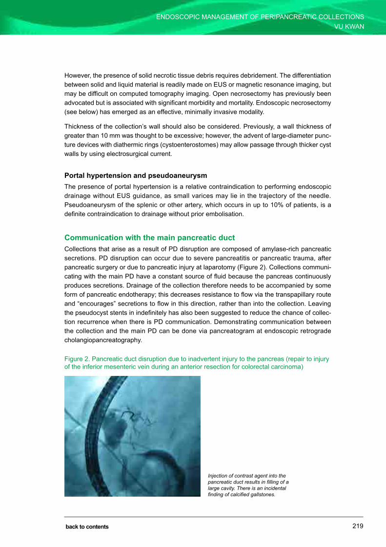
219
EnDoscoPIc mAnAgEmEnT oF PErIPAncrEATIc collEcTIonsVu KWAn
SECTION 4: ENDOSCOPIC RETROGRADE CHOLANGIOPANCREATOGRAPHY
back to contents
However, the presence of solid necrotic tissue debris requires debridement. The differentiation between solid and liquid material is readily made on Eus or magnetic resonance imaging, but may be difficult on computed tomography imaging. open necrosectomy has previously been advocated but is associated with significant morbidity and mortality. Endoscopic necrosectomy (see below) has emerged as an effective, minimally invasive modality.
Thickness of the collection’s wall should also be considered. Previously, a wall thickness of greater than 10 mm was thought to be excessive; however, the advent of large-diameter punc-ture devices with diathermic rings (cystoenterostomes) may allow passage through thicker cyst walls by using electrosurgical current.
Portal hypertension and pseudoaneurysmThe presence of portal hypertension is a relative contraindication to performing endoscopic drainage without Eus guidance, as small varices may lie in the trajectory of the needle. Pseudoaneurysm of the splenic or other artery, which occurs in up to 10% of patients, is a definite contraindication to drainage without prior embolisation.
Communication with the main pancreatic ductcollections that arise as a result of PD disruption are composed of amylase-rich pancreatic secretions. PD disruption can occur due to severe pancreatitis or pancreatic trauma, after pancreatic surgery or due to pancreatic injury at laparotomy (Figure 2). collections communi-cating with the main PD have a constant source of fluid because the pancreas continuously produces secretions. Drainage of the collection therefore needs to be accompanied by some form of pancreatic endotherapy; this decreases resistance to flow via the transpapillary route and “encourages” secretions to flow in this direction, rather than into the collection. leaving the pseudocyst stents in indefinitely has also been suggested to reduce the chance of collec-tion recurrence when there is PD communication. Demonstrating communication between the collection and the main PD can be done via pancreatogram at endoscopic retrograde cholangiopancreatography.
Figure 2. Pancreatic duct disruption due to inadvertent injury to the pancreas (repair to injury of the inferior mesenteric vein during an anterior resection for colorectal carcinoma)
Injection of contrast agent into the pancreatic duct results in filling of a large cavity. There is an incidental finding of calcified gallstones.

220
EnDoscoPIc mAnAgEmEnT oF PErIPAncrEATIc collEcTIonsVu KWAn
SECTION 4: ENDOSCOPIC RETROGRADE CHOLANGIOPANCREATOGRAPHY
back to contents
Technique
EndoscopesPreviously, the therapeutic duodenoscope was the major workhorse for pseudocyst drainage, but this relatively blind technique requires the presence of visible bulge into the gut lumen (Figure 1). The advent of Eus has significantly altered the practice of transluminal drainage, with the following advantages:
• clear visualisation of the collection and its relationship to the gut wall• assessment of internal composition: presence of solid debris that requires debridement,
presence of features that suggest an alternative diagnosis (e.g. septation of a cystic tumour)
• detection of intervening vessels or varices using Doppler ultrasound• real-time visualisation of the needle as it is passed into the collection.
StentsTraditionally, at least two double pigtail plastic stents have been placed in all patients to keep the cystogastrostomy tract open and allow ongoing drainage until the collection resolves (Figure 3). These plastic stents remain the conventional method of drainage. However, newer lumen-apposing fully covered metal stents have been developed, which have the theoretical advantages of ease of insertion, maintenance of a larger-diameter tract (e.g. allowing future endoscopic necrosectomy) and reduced risk of intra-abdominal leak. These metal stents must be removed after 3–5 months (as the gastric acid degrades the plastic covering), so are gen-erally used for collections that are not likely to recur.
Figure 3. Insertion of first stent
The first stent is deployed over the first guidewire, leaving in situ the second guidewire, over which the second stent can be placed immediately thereafter.

221
EnDoscoPIc mAnAgEmEnT oF PErIPAncrEATIc collEcTIonsVu KWAn
SECTION 4: ENDOSCOPIC RETROGRADE CHOLANGIOPANCREATOGRAPHY
back to contents
Post-procedural careuncomplicated pseudocyst drainage in an otherwise well patient can be performed as a day-stay procedure if it is performed early in the morning and the patient is observed for the remainder of the day after the procedure. However, sick or septic patients must be observed as inpatients. If the collection is infected, ongoing intravenous antibiotics appropriate to the organisms cultured from the fluid should be administered. The patient should be restricted from eating and drinking (nil by mouth) for 24 hours after the procedure.
Follow-up imaging should be performed in 1–2 months to determine whether or not the collec-tion has resolved. If the collection does not recur after about 3 months, the stents are generally removed. A small randomised controlled trial suggested that collection recurrence could be reduced by leaving stents in indefinitely, particularly in patients with communication with the PD. If the collection has not resolved in 4–6 weeks, repeat dilatation and stent replacement, perhaps with larger-calibre stents, can be performed.
Necrosectomynecrosectomy can be performed as part of the initial drainage procedure or as a two-stage procedure, allowing time for the tract to be well established and for decompression of any infected fluid component.
A forward-viewing gastroscope is used. The existing tract is dilated to 15–20 mm using a bal-loon dilator. The gastroscope is then entered into the cavity. A variety of equipment can then be used to remove the necrotic tissue:
• Basket devices (roth net [us Endoscopy], biliary stone extraction devices) (Figure 4)• Polypectomy snare• Tripod foreign body grasping forceps
Figure 4. A roth net basket device being opened within the collection of necrotic material
It is important that only the necrotic pancreatic tissue (black in colour) is removed, not the healthy granulation tissue (pink in colour) (Figure 5). If there is adherent tissue that cannot be removed, a repeat procedure after further nasocystic catheter irrigation is advisable. The aim is to have no residual necrotic tissue, leaving behind only a cavity lined by healthy granulation tissue.

222
EnDoscoPIc mAnAgEmEnT oF PErIPAncrEATIc collEcTIonsVu KWAn
SECTION 4: ENDOSCOPIC RETROGRADE CHOLANGIOPANCREATOGRAPHY
back to contents
Figure 5. The interior of a collection of infected pancreatic necrosis
Note the pink granulation tissue lining the cavity — this need not be debrided. The black solid debris in the body of the cavity is the necrotic pancreatic tissue — this is the target of debridement.
Relevant literature
no randomised controlled trials regarding the best way to manage peripancreatic collections have been conducted. However, endoscopic drainage has become an accepted alternative to surgery when conducted in experienced centres.
The outcomes of endoscopic drainage of pseudocysts have been documented by several expert centres. The initial landmark report in 1989 described its use in patients with pseudo-cysts in the setting of chronic pancreatitis. While it has previously been considered that higher success rates are achieved in chronic pancreatitis (> 90%) compared with acute pancreatitis (70%–80%), a more recent series examining larger numbers of patients does not suggest this is the case, with equivalent success rates for both groups (about 90% overall). There is a lower long-term success rate in patients with pancreatic necrosis, also shown in data from other cen-tres. However, with the development of endoscopic debridement techniques, success rates of over 90% have been demonstrated.
complications of transmural drainage include:
• Acute and delayed bleeding• Perforation• systemic infection• stent migration.
The frequency of these complications is variable in the published literature, ranging from 11% to 37%. no controlled data exist to compare Eus-guided and conventional drainage techniques. studies to date were not designed to detect such a difference, as in both series, the patients selected to undergo Eus-guided drainage were those in whom non-Eus-guided drainage had failed or who had features such as non-bulging collections and portal hyperten-sion. nonetheless, both series showed that the two groups had comparable complication rates.

223
EnDoscoPIc mAnAgEmEnT oF PErIPAncrEATIc collEcTIonsVu KWAn
SECTION 4: ENDOSCOPIC RETROGRADE CHOLANGIOPANCREATOGRAPHY
back to contents
Conclusion
The endoscopic management of peripancreatic collections offers patients a safe and effective alternative to major surgery. refinements in technical aspects and equipment over recent years have rendered the practice easier to execute, with excellent long-term results. nonetheless, it remains a technique that should only be performed in expert centres with adequate multidis-ciplinary support.
Further reading1 Yeo cJ, Bastidas JA, lynch-nyhan A, et al. The natural history of pancreatic pseudocysts documented by
computed tomography. Surg Gynecol Obstet 1990; 170: 411-417.
2 Vitas gJ, sarr mg. selected management of pancreatic pseudocysts: operative versus expectant management. Surgery 1992; 111: 123-130.
3 Bradley El, clements Jl Jr, gonzalez Ac. The natural history of pancreatic pseudocysts: a unified concept of management. Am J Surg 1979; 137: 135-141.
4 Warshaw Al, rattner DW. Timing of surgical drainage for pancreatic pseudocyst. clinical and chemical criteria. Ann Surg 1985; 202: 720-724.
5 Baillie J. Pancreatic pseudocysts (Part II). Gastrointest Endosc 2004; 60: 105-113.
6 El Hamel A, Parc r, Adda g, et al. Bleeding pseudocysts and pseudoaneurysms in chronic pancreatitis. Br J Surg 1991; 78: 1059-1063.
7 Kiviluoto T, schroder T, Kivilaakso E, lempinen m. Acute haemorrhage associated with pancreatic pseudocyst and chronic pancreatitis. Ann Chir Gynaecol 1984; 73: 214-218.
8 Pitkaranta P, Haapiainen r, Kivisaari l, schroder T. Diagnostic evaluation and aggressive surgical approach in bleeding pseudoaneurysms associated with pancreatic pseudocysts. Scand J Gastroenterol 1991; 26: 58-64.
9 Arvanitakis m, Delhaye m, Bali mA, et al. Pancreatic-fluid collections: a randomized controlled trial regarding stent removal after endoscopic transmural drainage. Gastrointest Endosc 2007; 65: 609-619.
10 gillams Ar, Kurzawinski T, lees Wr. Diagnosis of duct disruption and assessment of pancreatic leak with dynamic secretin-stimulated mr cholangiopancreatography. AJR Am J Roentgenol 2006; 186: 499-506.
11 Voermans rP, Eisendrath P, Bruno mJ, et al. Initial evaluation of a novel prototype forward-viewing us endoscope in transmural drainage of pancreatic pseudocysts (with videos). Gastrointest Endosc 2007; 66: 1013-1017.
12 Kozarek rA. Endoscopic treatment of pancreatic pseudocysts. Gastrointest Endosc Clin N Am 1997; 7: 271-283.
13 cremer m, Deviere J, Engelholm l. Endoscopic management of cysts and pseudocysts in chronic pancreatitis: long-term follow-up after 7 years of experience. Gastrointest Endosc 1989; 35: 1-9.
14 Jacobson Bc, Baron TH, Adler Dg, et al. AsgE guideline: The role of endoscopy in the diagnosis and the management of cystic lesions and inflammatory fluid collections of the pancreas. Gastrointest Endosc 2005; 61: 363-370.
15 Hookey lc, Debroux s, Delhaye m, et al. Endoscopic drainage of pancreatic-fluid collections in 116 patients: a comparison of etiologies, drainage techniques, and outcomes. Gastrointest Endosc 2006; 63: 635-643.
16 Baron TH, Harewood gc, morgan DE, Yates mr. outcome differences after endoscopic drainage of pancreatic necrosis, acute pancreatic pseudocysts, and chronic pancreatic pseudocysts. Gastrointest Endosc 2002; 56: 7-17.
17 Voermans rP, Veldkamp mc, rauws EA, et al. Endoscopic transmural debridement of symptomatic organized pancreatic necrosis (with videos). Gastrointest Endosc 2007; 66: 909-916.

224
EnDoscoPIc mAnAgEmEnT oF PErIPAncrEATIc collEcTIonsVu KWAn
SECTION 4: ENDOSCOPIC RETROGRADE CHOLANGIOPANCREATOGRAPHY
back to contents
18 charnley rm, lochan r, gray H, et al. Endoscopic necrosectomy as primary therapy in the management of infected pancreatic necrosis. Endoscopy 2006; 38: 925-928.
19 seewald s, groth s, omar s, et al. Aggressive endoscopic therapy for pancreatic necrosis and pancreatic abscess: a new safe and effective treatment algorithm (videos). Gastrointest Endosc 2005; 62: 92-100.
20 Antillon mr, shah rJ, stiegmann g, chen YK. single-step Eus-guided transmural drainage of simple and complicated pancreatic pseudocysts. Gastrointest Endosc 2006; 63: 797-803.
21 Kahaleh m, shami Vm, conaway mr, et al. Endoscopic ultrasound drainage of pancreatic pseudocyst: a prospective comparison with conventional endoscopic drainage. Endoscopy 2006; 38: 355-359.
22 cahen D, rauws E, Fockens P, et al. Endoscopic drainage of pancreatic pseudocysts: long-term outcome and procedural factors associated with safe and successful treatment. Endoscopy 2005; 37: 977-983.
23 Varadarajulu s, Wilcox cm, Tamhane A, et al. role of Eus in drainage of peripancreatic fluid collections not amenable for endoscopic transmural drainage. Gastrointest Endosc 2007; 66: 1107-1119.
Pages 225-227 (used for notes in the hard copy) have been removed from the PDF edition of this handbook.

Section 5
ENDOSCOPIC ULTRASOUND

229
Introduction to endoscopic ultrasoundIan Norton
Endoscopic ultrasound (EUS) developed rapidly in the 1990s and is now a core part of most large endoscopy units, particularly those dealing with hepatobiliary malignancy. However, the technology has remained farily static over the past 10 years, while other imaging modalities have continued to develop. Thus, some areas of EUS have become more limited (e.g. staging of oesophageal and pancreatic malignancy), while others have grown (e.g. suspected chole-docholithiasis and non-small cell carcinoma of the lung). This chapter outlines the common diagnostic and therapeutic scenarios for which EUS is currently used.
Equipment
EUS uses a flexible endoscope to pass an ultrasound probe to a point in the gastrointestinal tract adjacent to an area of interest. The procedures are performed on an outpatient basis, usually with titrated conscious sedation. The frequencies used range from 5.5 MHz to 30 MHz. Higher frequencies produce high-resolution images with limited depth of view, whereas lower frequencies provide imaging of lower resolution but greater penetration (up to about 5 cm). Acoustic coupling with the mucosal surface is achieved by a water-filled balloon attached to the instrument tip and/or infusion of water into the lumen. Three types of instruments are currently in routine practice.
1 Radial scanning endoscope. This is the diagnostic “workhorse” of EUS. It com-prises a modified endoscope with a circular ultrasound array around the instrument, near the tip. This produces a circular ultrasound image (with the instrument at the centre of the circle) perpendicular to the long axis of the endoscope. Previously, these instruments involved a mechanical rotating transducer, but most instruments
SECTION 5: ENDOSCOPIC ULTRASOUND
CHAPTER 21
back to contents

INTrodUcTIoN To ENdoScoPIc UlTrASoUNdIan Norton
SECTION 5: ENDOSCOPIC ULTRASOUND
230 back to contents
in use now have a solid-state transducer with doppler capability. These instruments do not have fine needle aspiration (FNA) capability.
2 Linear scanning endoscope. This has a solid-state, curved linear array transducer in the endoscope tip, providing an image parallel to the axis of the endoscope. The ultrasound beam is aligned with the endoscope’s biopsy channel, such that a needle (or other accessory) is in the ultrasound beam as it passes through tissue. This permits sampling of tissues deep in the lumen (such as lymph nodes) under real-time ultrasound control. Similarly, periluminal cystic lesions can be aspirated for diagnostic and therapeutic purposes. The solid-state nature of these transducers also permits doppler interrogation of the area of interest.
3 Catheter probes. These are small-calibre catheters (about 7 French) that are passed through a routine endoscope. They have a mechanical rotating transducer at the tip. In particular, high-frequency probes (12–30 MHz) are used to obtain fine detail of mucosal lesions. Wire-guided versions of these probes have been used to perform intraductal ultrasound within the pancreaticobiliary system. In general, catheter probes are not widely used as they require a separate drive motor, and their main application (high-frequency definition of mucosal lesions) has largely been superseded by endoscopic mucosal resection (EMr) techniques.
Luminal applications
Mucosal lesionsStandard EUS scanning resolves the gastrointestinal wall into five “layers”, representing a reflective interface, the mucosa, submucosa, muscularis propria and serosa. Higher-frequency probes provide more definition, with the muscularis mucosa visible, as well as separation of the muscularis propria into circular and longitudinal layers. Therefore, EUS is a very sensitive modality for determining the layer in which a lesion arises, as well as the depth of penetration of a mucosal-based lesion (T-staging).
T-staging of luminal malignancyIn general, T-staging of mucosal-based tumours is dependent on the relationship of the lesion to the muscularis propria. T1 lesions are superficial to the muscle layer, T2 lesions invade the muscle and T3 lesions extend beyond the muscle. EUS has been shown to be more accurate than computed tomography (cT) in the detection and T-staging of oesophageal, gastric and rectal malignancies. The T-stage accuracy of EUS is 80%–89% for early gastric cancer and 76%–96% for rectal lesions. However, EUS is poor at differentiating mucosal-based disease from submucosal involvement (both T1). This distinction is important because oesophageal lesions with submucosal invasion have a much higher risk of lymphatic involvement (up to 30%) than mucosal lesions. Furthermore, the extent of submucosal depth of invasion has prognos-tic significance and is not stratified by EUS. Therefore, as EMr techniques have advanced

231
INTrodUcTIoN To ENdoScoPIc UlTrASoUNdIan Norton
SECTION 5: ENDOSCOPIC ULTRASOUND
back to contents
(definitively substaging T1 tumours pathologically), EUS is not commonly used for assessment of early oesophageal neoplasia.
Nonetheless, because T-staging has direct prognostic significance, accurate T-staging of malignancy by EUS may assist in the preoperative stratification of patients enrolled in oncolog-ical trials. Furthermore, preoperative diagnosis of locally advanced (T4) lesions may prevent explorative surgery of incurable lesions.
Mucosal thickeningThickened gastric folds are easily characterised by EUS. loss of normal acoustic layering may suggest malignant infiltration. Unfortunately, Zollinger–Ellison syndrome, Ménétrier’s disease, diffuse gastric malignancy and mucosa-associated lymphoid tissue (MAlT) lymphoma may all cause gastric wall thickening with preservation of wall layers, making EUS differentiation of these conditions difficult. However, EUS can be useful in targeting appropriate areas for large particle endoscopic biopsy (or EMr), as well as confirming whether the process is in the mucosa or submucosa and thus amenable to endoscopic biopsy. In situations where endoscopic mucosal biopsies are negative, needle biopsy under EUS guidance may be performed, yielding a diagnostic accuracy of about 65%.
Submucosal lesions with normal overlying mucosaThese are a frequent indication for EUS, which can differentiate lipoma (hyperdense) from leio-myoma (hypodense). EUS imaging cannot differentiate leiomyoma from gastrointestinal stromal tumours (GISTs), but FNA may be used for immunostaining to differentiate these lesions. EUS criteria for determining high-risk GISTs (size > 3 cm, irregular border, heterogeneous structure and cystic spaces) have been published.
less common submucosal lesions that are also well characterised by EUS include carcinoid tumours, duplication cysts and pancreatic rests. EUS is also occasionally used to assess gastro-oesophageal varices or other vascular lesions (usually in the setting of excluding a varix before biopsy).
Assessment of periluminal tissuesEUS may be helpful in assessing sphincter integrity in patients with faecal incontinence. EUS may be as sensitive as magnetic resonance imaging (MrI) or examination under anaesthaesia for characterisation of perirectal fistulae in patients with crohn’s disease. However, it is more operator-dependent than MrI and is therefore not commonly used for this purpose.
Assessment of peri-intestinal lymph nodes and masses
EUS with or without EUS-guided FNA biopsy provides the most sensitive and specific means of nodal staging of periluminal malignancy. N-stage accuracy rates of 70%–85% for rectal tumours

INTrodUcTIoN To ENdoScoPIc UlTrASoUNdIan Norton
SECTION 5: ENDOSCOPIC ULTRASOUND
232 back to contents
and 79%–93% for oesophageal tumours have been reported. of particular note, in one study, EUS–FNA increased diagnostic accuracy for N-staging of oesophageal malignancy from 70% to 93%, compared with radial EUS alone. EUS-guided FNA proving malignant involvement of selected node groups is particularly important in cases where the nodal staging precludes resection with curative intent (e.g. coeliac or cervical lymphadenopathy in oesophageal cancer and contralateral mediastinal adenopathy in non-small cell carcinoma of the lung).
Multiple studies have documented the safety and efficacy of transoesophageal EUS-guided biopsy of mediastinal masses. EUS–FNA has been shown to be at least as accurate as trans-bronchial biopsy in the diagnosis of mediastinal masses. Furthermore, cost-effectiveness analysis of EUS–FNA of mediastinal masses has demonstrated significant savings compared with mediastinoscopy. Therefore, EUS is a safe and relatively inexpensive method of sampling mediastinal masses and lymph nodes, particularly those relatively inaccessible by other modal-ities (e.g. subcarinal and posterior mediastinum).
Extraluminal applications
Diagnosis and staging of pancreatic malignancyEUS is the most sensitive test for detecting and characterising pancreatic masses, especially those smaller than 3 cm in diameter. The accuracy of detection of vascular involvement has been reported to be as high as 80%–85%. However, locoregional staging by multislice cT has replaced EUS in most units. linear-scanning EUS permits biopsy of suspicious pancreatic lesions.
Pancreatic neuroendocrine tumoursEUS is a sensitive test for detecting neuroendocrine tumours. one comparative study reported EUS to be superior to cT or transabdominal ultrasound and equivalent to somatostatin receptor scintigraphy (SrS) for gastrinomas, and superior to SrS for insulinomas. Biopsy of suspicious lesions via EUS is also straightforward, although usually not necessary.
Pancreatic cystic lesionsEUS is the most accurate imaging technique in the characterisation of pancreatic cysts. This is important because some lesions are benign (post-inflammatory and serous cystadenomas), while others have malignant potential (mucinous tumours). cyst carcinoembryonic antigen level has been shown to be the most accurate test for differentiating benign from malignant lesions.
CholedocholithiasisEUS is at least as sensitive as endoscopic retrograde cholangiography or magnetic resonance cholangiography in diagnosing common bile duct stones. EUS does not carry the risk of pan-creatitis associated with endoscopic retrograde cholangiopancreatography (ErcP). As such,

233
INTrodUcTIoN To ENdoScoPIc UlTrASoUNdIan Norton
SECTION 5: ENDOSCOPIC ULTRASOUND
back to contents
ErcP should rarely be performed as a diagnostic test for the investigation of “biliary-type” abdominal pain.
Chronic pancreatitisSonographic changes in the pancreatic parenchyma and duct system in chronic pancreatitis have been reported and characterised. Although several studies have advocated the use of EUS as a means of early diagnosis of chronic pancreatitis, these studies lacked a “gold stand-ard” for early disease and ignored age-related changes that may occur in the pancreas. More data are needed before advocating EUS as a standard diagnostic test for chronic pancreatitis. It should also be noted that differentiating focal chronic or acute pancreatitis from malignancy can be difficult, even with EUS.
Therapeutic interventions using endoscopic ultrasound
The ability to place a needle under real-time guidance using the linear-scanning instrument has opened up the possibility of therapeutic intervention using EUS; specifically, drainage of fluid collections and injection of therapeutic substances into specific areas.
Drainage of peripancreatic fluid collectionsEndoscopic drainage of symptomatic pancreatic pseudocysts has become widely established. EUS may be of use in confirming the diagnosis of pseudocysts, as well as in determining the most advantageous site of cyst and gut apposition. EUS also has the advantage of delineating adjacent vascular structures to be avoided during cyst puncture and aspiration. Many series have reported successful drainage and stenting of cyst gastrostomy entirely under EUS real-time guidance using linear EUS scanning. This is now the standard approach for transgastric drainage of peripancreatic collections.
Coeliac ganglion neurolysisThe coeliac ganglion drapes across the coeliac artery, an area easily visualised during EUS and within several millimetres of the posterior gastric wall. coeliac plexus neurolysis under direct EUS guidance has been advocated for patients with unresectable pancreatic cancer. EUS may stage the lesion, obtain biopsy confirmation and perform neurolysis all in one procedure. However, in rare cases this technique has resulted in spinal cord infarction, which has limited the enthusiasm for this approach.
INTrodUcTIoN To ENdoScoPIc UlTrASoUNdIan Norton
SECTION 5: ENDOSCOPIC ULTRASOUND
234 back to contents
Complications of endoscopic ultrasound
complications of EUS relate to either the endoscopic portion of the procedure or the FNA biopsy. dilatation of oesophageal malignancies to a diameter sufficient to pass the EUS instrument (12 mm) is associated with risk of perforation. large pancreatic neoplasms may invade and fix the second part of the duodenum, leading to increased risk of perforation as the relatively stiff instrument traverses this fixed segment of bowel.
Three large published series (totalling over 1100 patients) have investigated complications of EUS–FNA. comprising over 900 EUS–FNA biopsies of solid tissue structures, there were no complications relating to the biopsy. In a large multicentre trial involving 554 consecutive mass or lymph node biopsies, only five complications were observed, all of which were non-fatal. Two patients had endoscope-induced perforation, two had febrile episodes following aspiration of cystic lesions, and one had haemorrhage from the wall of a pseudocyst. Pneumoperitoneum has been reported when endoscopy closely followed EUS–FNA, suggesting that intestinal insufflation should be minimised soon after EUS–FNA. A risk (< 2%) of pancreatitis (usually mild) has been observed following biopsy of pancreatic lesions.
Needle aspiration is not a sterile procedure. While this is not a concern with sampling solid lesions, cystic fluid aspiration should be done with antibiotic prophylaxis, and lesions should, if possible, be aspirated to dryness. Furthermore, infecting proteinaceous cystic lesions, such as mediastinal cysts, is potentially very dangerous and should be avoided.
Further reading1 dietrich cF. Endoscopic ultrasound: an introductory manual and atlas. 2nd ed. Stuttgart: Thieme, 2011.
2 Faigel do, Kochman Ml, editors. Endoscopic oncology: gastrointestinal endoscopy and cancer management. Totowa, NJ: Humana Press, 2006.
3 American Society for Gastrointestinal Endoscopy. Technology status evaluation: endoscopic ultrasonography. Gastrointest Endosc 1998; 48: 705-707.
4 American Society for Gastrointestinal Endoscopy. Technology status evaluation: endoscopic ultrasound probes. Gastrointest Endosc 2006; 63: 751-754.
5 chak A, canto MI, rosch T, et al. Endosonographic differentiation of benign and malignant stromal cell tumors. Gastrointest Endosc 1997; 45: 468-473.
6 Buscail l, Pagès P, Berthélemy P, et al. role of EUS in the management of pancreatic and ampullary carcinoma: a prospective study assessing resectability and prognosis. Gastrointest Endosc 1999; 50: 34-40.
7 Ahmad NA, lewis Jd, Ginsberg GG, et al. EUS in preoperative staging of pancreatic cancer. Gastrointest Endosc 2000; 52: 463-468.
8 Zimmer T, Stolzel U, Bader M, et al. Endoscopic ultrasonography and somatostatin receptor scintigraphy in the preoperative localisation of insulinomas and gastrinomas. Gut 1996; 39: 562-568.
9 Gunaratnam NT, Wong GY, Wiersema MJ. EUS-guided celiac plexus block for the management of pancreatic pain. Gastrointest Endosc 2000; 52 (6 Suppl): S28-S34.
10 Wiersema MJ, Vilmann P, Giovannini M, et al. Endosonography-guided fine-needle aspiration biopsy: diagnostic accuracy and complication assessment. Gastroenterology 1997; 112: 1087-1095.
11 Williams dB, Sahai AV, Aabakken l, et al. Endoscopic ultrasound guided fine needle aspiration biopsy: a large single centre experience. Gut 1999; 44: 720-726.
Pages 235-236 (used for notes in the hard copy) have been removed from the PDF edition of this handbook.

237
Endoscopic ultrasound for oesophageal and mediastinal indicationsAlina Stoita and David Williams
Endoscopic ultrasound (EUS) combines endoscopic visualisation with high-frequency ultra-sound. This allows precise differentiation of the individual layers of the oesophageal wall and direct imaging of the surrounding tissues. As such, EUS is an ideal imaging modality for cancer staging (TNM classification), it allows extraluminal tissue diagnoses using EUS-guided fine needle aspiration (FNA) biopsy, and therapeutic procedures (coeliac plexus neurolysis, medi-astinal abscess drainage) can be performed with EUS guidance.
Technique
The oesophagus can be evaluated with either a radial or a linear echoendoscope, but if tissue acquisition is not needed, the imaging from a radial scope is easier to interpret. The mediasti-num is usually evaluated with a linear scope, as in most cases a biopsy sample is required to diagnose mediastinal lesions and to stage non-small cell lung cancer (NSCLC). EUS-guided FNA biopsy is usually performed with 22G or 25G needles. Newer EchoTip ProCore 22G, 25G and 19G needles (Cook Medical) and the SharkCore EUS–FNA needle (Covidien) are used to aquire histological specimens when tissue architecture is important (e.g. diagnosis of lym-phoma). These needles allow collection of larger and multiple samples that can be used for molecular studies and genomic analysis, and they will start playing an important role in the new area of personalised medicine. On-site cytopathological interpretation of the sample improves the diagnostic accuracy of EUS-guided FNA and minimises the number of passes.
An oesophageal cancer and mediastinal study starts with inserting the endoscope in the stomach and imaging the liver, left adrenal gland and coeliac axis for metastases (M stage). The scope is then slowly withdrawn into the oesophagus looking for mediastinal lymph nodes
SECTION 5: ENDOSCOPIC ULTRASOUND
CHAPTER 22
back to contents

238
ENDOSCOPiC ULTrASOUND FOr OESOPhAGEAL AND MEDiASTiNAL iNDiCATiONSAlina Stoita and David Williams
SECTION 5: ENDOSCOPIC ULTRASOUND
back to contents
(N stage). Once the oesophageal tumour is reached, the frequency is adjusted to obtain detailed images of the wall layers (T stage). Positive and relevant negative findings are carefully doc-umented in the report.
ComplicationsEUS is a safe procedure, and complications are rare. The reported risk of perforation is 0.4%, mostly related to inexperienced operators inserting an oblique-viewing echoendoscope with a long rigid tip. As the risk of infection after aspirating mediastinal cysts has been reported to be as high as 9%, cyst aspiration is avoided or is performed under antibiotic prophylaxis.
Indications
OesophagusOesophageal cancer stagingin Australia, oesophageal cancer has a low incidence and prevalence (about 1300 new cases per year). Males are three times more likely than females to develop oesophageal cancer. Cigarette smoking, alcohol consumption, high body mass index and obesity, low intake of fresh fruit and vegetables, and gastro-oesophageal reflux disease are risk factors responsible for an estimated 79% of oesophageal adenocarcinoma cases. Cigarette smoking, excess alcohol consumption and low fruit and vegetable intake are responsible for an estimated 89% of squa-mous cell carcinoma cases.
Oesophageal cancer remains a devastating disease, with overall 5-year survival of only 17%. Accurate tumour staging (Figure 1, Table 1) is essential to help define treatment options and determine patient outcomes.
Figure 1. Staging of oesophageal cancer
reprinted by permission of the American Joint Committee on Cancer (AJCC), Chicago, illinois, and Springer, New York, USA, from the AJCC Cancer Staging Manual, 7th edition, 2010.
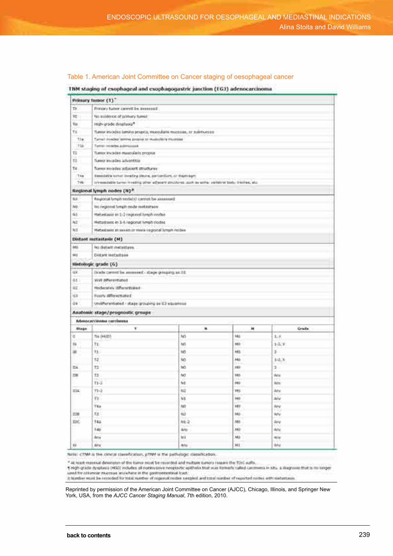
239
ENDOSCOPiC ULTrASOUND FOr OESOPhAGEAL AND MEDiASTiNAL iNDiCATiONSAlina Stoita and David Williams
SECTION 5: ENDOSCOPIC ULTRASOUND
back to contents
Table 1. American Joint Committee on Cancer staging of oesophageal cancer
reprinted by permission of the American Joint Committee on Cancer (AJCC), Chicago, illinois, and Springer New York, USA, from the AJCC Cancer Staging Manual, 7th edition, 2010.

240
ENDOSCOPiC ULTrASOUND FOr OESOPhAGEAL AND MEDiASTiNAL iNDiCATiONSAlina Stoita and David Williams
SECTION 5: ENDOSCOPIC ULTRASOUND
back to contents
Therapeutic impact studies show that the benefits of EUS staging include:
• avoiding unnecessary surgery in patients with advanced disease (e.g. previously unidentified metastases, T4 disease)
• selecting patients with nodal involvement for neoadjuvant therapy (meta-analyses have shown that for stage iiA, iiB and iii disease, neoadjuvant chemoradiotherapy followed by surgery, rather than surgery alone, improves 2-year and 3-year survival)
• identifying patients with early cancers for definitive endoscopic mucosal resection (e.g. T1mN0 disease).
For oesophageal cancer, EUS has a T-stage accuracy of 85% and N-stage accuracy of 80%. When EUS–FNA of lymph nodes is performed, the N-stage accuracy increases to 87%. Standard endosonographic criteria for a malignancy include nodes that are round, well defined, hypoechoic and larger than 5–10 mm. When all four criteria are present, the chance of malig-nancy is 80%; albeit, this occurs in less than 25% of cases. Suggested modified criteria (the four standard criteria plus presence of coeliac lymph nodes, > 5 lymph nodes and EUS T3–T4 tumour) can better differentiate malignant lymph nodes. The presence of > 6 or < 1 modified criteria has 100% accuracy for predicting if a lymph node is malignant or benign, respectively.
Elastography is an emerging EUS technology relying on variations in tissue stiffness to identify malignant lymph nodes. however, the reported sensitivity and specificity of this technique are only 80%–87%, and EUS–FNA remains the gold standard in confirming malignant adenopathy.
Numerous studies have shown that EUS is consistently superior to computed tomography (CT) in detecting tumour stage and locoregional adenopathy. in addition, EUS has been reported to be superior to positron emission tomography (PET) in detecting nodal metastases, and can provide sampling of the lymph nodes.
Malignant oesophageal strictures can sometimes be problematic, as they can prevent insertion of the large-bore echoendoscope (outer diameter about 13 mm) and hence prevent a complete staging study. however, if it is felt that EUS findings would significantly alter patient management (e.g. coeliac nodal metastases, T4 disease), dilatation to allow passage of the echoendoscope can be performed before EUS, although risk of perforation needs to be weighed against the potential benefits.
restaging of oesophageal cancer with EUS imaging alone after neoadjuvant therapy is less accurate than primary TN staging, as it is difficult to differentiate tumour from necrosis or inflammation. however, EUS–FNA can be used to exclude patients with persistent extensive disease, T4 tumours or new metastases from undergoing surgery.
EUS is the best locoregional staging modality for oesophageal cancer and should be incorpo-rated into the staging plan of patients who are considered suitable candidates for treatment. The Cancer institute New South Wales supports this central role of EUS in oesophageal cancer by acknowledging that most quality-adjusted life-years for patients are achieved when EUS–FNA is used in staging strategies.

241
ENDOSCOPiC ULTrASOUND FOr OESOPhAGEAL AND MEDiASTiNAL iNDiCATiONSAlina Stoita and David Williams
SECTION 5: ENDOSCOPIC ULTRASOUND
back to contents
Barrett’s oesophagusThe aims of staging Barrett’s oesophagus-related early cancers or high-grade dysplasia are to detect occult cancers, identify submucosal invasion (T1sm) and define nodal disease (N1). it is particularly important to identify submucosal invasion, as tumours limited to the mucosa have < 5% chance of metastasising to lymph nodes, whereas tumours invading to submucosa have up to a 27% chance of nodal disease. Multiple studies suggest that EUS cannot readily detect dysplasia, nor occult cancers, and cannot reliably distinguish T1m from T1sm invasion, particularly in flat lesions, as it has a tendency to overstage. As such, EUS best serves as a staging adjunct to careful high-quality endoscopy and endoscopic mucosal resection of defined lesions, whereby histologically proven T1sm (or beyond) disease can be assessed for nodal disease. At present, there is no defined role for EUS in the assessment of non-dysplastic Barrett’s mucosa or low-grade dysplasia.
Submucosal lesionsEUS is important in the assessment of submucosal lesions in the oesophagus, with more common lesions including mesenchymal tumours, granular cell tumours and cysts (bronchio-genic, oesophageal simple and duplication cysts). EUS can readily differentiate mural lesions and extraluminal compression from mediastinal mass lesions. With respect to mural lesions, EUS can provide information about layer of origin, size, borders, homogenicity and vascularity, which can help define differential diagnoses. While not often required, EUS–FNA can confirm diagnosis, although cytological samples obtained are often only small. in addition, EUS can indicate whether endoscopic resection is appropriate. The most common oesophageal lesion is leiomyoma, which appears as a well defined, hypoechoic, homogeneous lesion that arises from the muscularis propria (fourth echo-layer). Gastrointestinal stromal tumours (GiSTs) are not common in the oesophagus.
EUS can help differentiate pseudoachalasia from achalasia. Patients with achalasia have a thickened muscularis propria in the distal oesophagus but otherwise normal wall architecture. in pseudoachalasia, tumour infiltration into the oesophageal wall is evidenced by an irregular hypoechogenic infiltration, with loss of the normal wall architecture and possible adjacent mass lesion.
Mediastinum
Diagnosing and staging non-small cell lung cancerLung cancer is the fourth most common cancer in Australia and has the highest cancer-related mortality, with a 5-year survival rate of 14%. As prognosis of lung cancer is directly related to disease TNM stage, accurate staging is essential for choosing the most beneficial therapies. Up to 25% of patients undergoing thoracotomy after staging CT and bronchoscopy alone are found to have mediastinal disease that would have altered their management, either by precluding them from curative resection or requiring them to have neoadjuvant treatment before surgery.

242
ENDOSCOPiC ULTrASOUND FOr OESOPhAGEAL AND MEDiASTiNAL iNDiCATiONSAlina Stoita and David Williams
SECTION 5: ENDOSCOPIC ULTRASOUND
back to contents
The role of EUS–FNA in NSCLC is:
• diagnosing lung cancer with tumour near the oesophagus• diagnosing suspected lung cancer with enlarged mediastinal lymph nodes• assessing tumour invasion (T4) in centrally located tumours• mediastinal nodal staging of lung cancer (N2–N3 disease)• mediastinal restaging after induction chemotherapy• assessing PET-positive lymph nodes.
According to the American Joint Committee on Cancer’s lung cancer classification, patients with stage i or ii disease are offered surgery alone, and those with stage iiiA disease (N1 and minimal N2) can be considered as candidates for neoadjuvant therapy. Patients with stage iiiB disease (T4 or N3) are in general not considered suitable candidates for resection.
Staging modalities for NSCLC include CT, PET, EUS–FNA, endobronchial ultrasound (EBUS) and mediastinoscopy. Compared with CT, EUS–FNA is more sensitive (88% v 57%) and specific (91% v 82%) for mediastinal staging, and percutaneous biopsy carries a risk of pneumothorax. EUS–FNA and PET have similar sensitivities, but EUS is more specific (100% v 72%) and can detect malignant lymph nodes < 1 cm in the longest axis. Any lymph nodes > 1 cm on cross-sec-tional imaging or any positive nodes on PET should be sampled to exclude malignancy. EUS and mediastinoscopy have similar accuracy (90%), but they are complementary as they provide access to different mediastinal nodal stations. EUS provides access to posterior stations 5, 7, 8 and 9, and mediastinoscopy provides access to stations 2 and 4 and the ventral part of station 7. however, as mediastinoscopy is an invasive procedure requiring general anaesthesia, with a 2% complication rate, it should be considered only when EUS–FNA has not provided a definitive diagnosis. Studies confirm that EUS–FNA can thereby reduce unnecessary thoracotomies and mediastinoscopies in a substantial number of patients.
EBUS is an emerging endoscopic modality that can evaluate subcarinal and anterior mediastinal lymph nodes (stations 7, 2, 3 and 4). The combination of EUS–FNA and EBUS–FNA carries the promise of complete endoscopic mediastinal nodal staging, with reported 93% sensitivity and 100% specificity.
EUS–FNA and EBUS–FNA have been demonstrated to be a feasible and effective method for collecting high-quality multiple core tissue samples for EGFR and KRAS mutation analysis and other molecular profiling. This can guide correct subtyping of lung cancer and help guide clinical and therapeutic decisions. American Society for Gastrointestinal Endoscopy guidelines recom-mend EUS–FNA as the procedure of choice for sampling subcarinal, aortopulmonary window and perioesophageal lymph nodes (stations 5, 7 and 8) in lung cancer patients with enlarged lymph nodes on cross-sectional imaging. EUS–FNA should also be considered in preoperative staging of patients with negative mediastinal adenopathy on imaging, as a significant number of mediastinal nodal metastases can be detected by EUS.
Unexplained mediastinal lypmphadenopathy and mediastinal mass lesionsAs with mediastinal staging of known lung cancer, EUS–FNA is an important modality for assessment of unexplained mediastinal adenopathy and mass lesions.
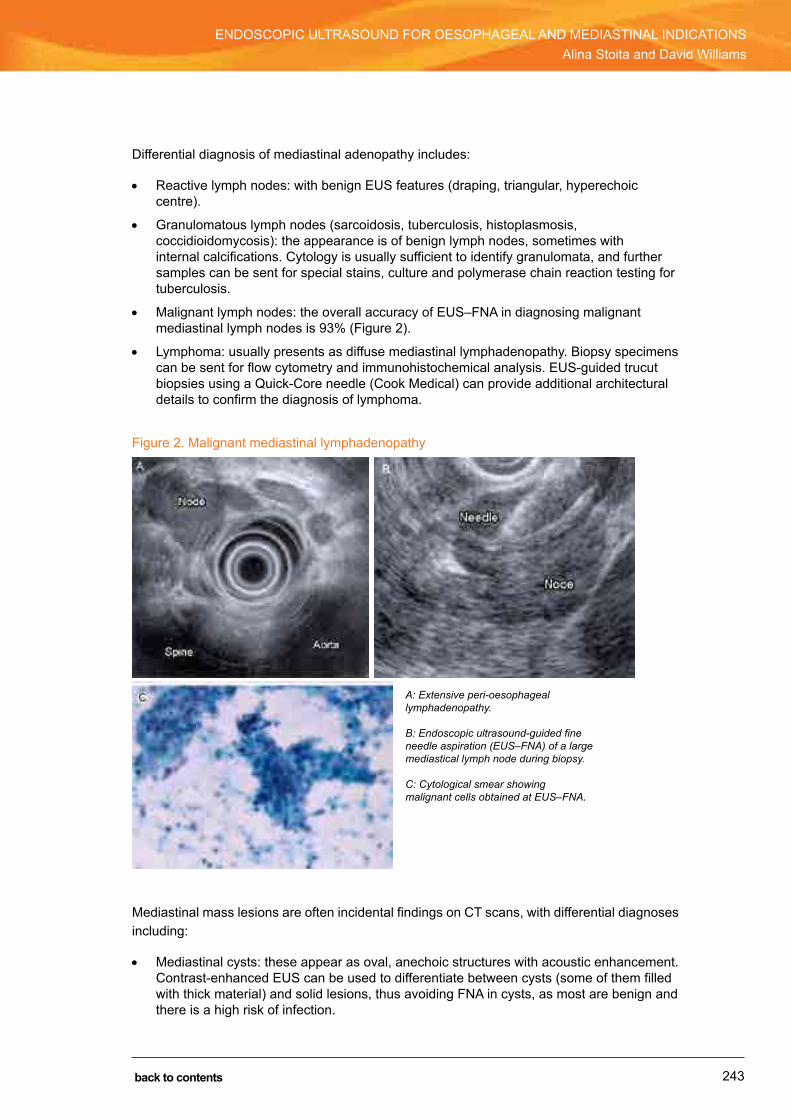
243
ENDOSCOPiC ULTrASOUND FOr OESOPhAGEAL AND MEDiASTiNAL iNDiCATiONSAlina Stoita and David Williams
SECTION 5: ENDOSCOPIC ULTRASOUND
back to contents
Differential diagnosis of mediastinal adenopathy includes:
• reactive lymph nodes: with benign EUS features (draping, triangular, hyperechoic centre).
• Granulomatous lymph nodes (sarcoidosis, tuberculosis, histoplasmosis, coccidioidomycosis): the appearance is of benign lymph nodes, sometimes with internal calcifications. Cytology is usually sufficient to identify granulomata, and further samples can be sent for special stains, culture and polymerase chain reaction testing for tuberculosis.
• Malignant lymph nodes: the overall accuracy of EUS–FNA in diagnosing malignant mediastinal lymph nodes is 93% (Figure 2).
• Lymphoma: usually presents as diffuse mediastinal lymphadenopathy. Biopsy specimens can be sent for flow cytometry and immunohistochemical analysis. EUS-guided trucut biopsies using a Quick-Core needle (Cook Medical) can provide additional architectural details to confirm the diagnosis of lymphoma.
Figure 2. Malignant mediastinal lymphadenopathy
Mediastinal mass lesions are often incidental findings on CT scans, with differential diagnoses including:
• Mediastinal cysts: these appear as oval, anechoic structures with acoustic enhancement. Contrast-enhanced EUS can be used to differentiate between cysts (some of them filled with thick material) and solid lesions, thus avoiding FNA in cysts, as most are benign and there is a high risk of infection.
A: Extensive peri-oesophageal lymphadenopathy. B: Endoscopic ultrasound-guided fine needle aspiration (EUS–FNA) of a large mediastical lymph node during biopsy. C: Cytological smear showing malignant cells obtained at EUS–FNA.

244
ENDOSCOPiC ULTrASOUND FOr OESOPhAGEAL AND MEDiASTiNAL iNDiCATiONSAlina Stoita and David Williams
SECTION 5: ENDOSCOPIC ULTRASOUND
back to contents
• Mediastinitis and abscess: an abscess is suggested by an inhomogeneous, well demarcated, hypoechoic area in a patient with fever and appropriate history, usually after surgical intervention. There are case reports of successful EUS-guided drainage of a mediastinal abscess, using much the same technique as for drainage of pancreatic and pelvic fluid collections.
• Primary neoplasms of the posterior mediastinum: these are rare. Most are neurogenic and arise from peripheral nerves (schwannoma, neurofibroma), sympathetic ganglia (ganglioneuroma, ganglioneuroblastoma, neuroblastoma), parasympathetic ganglia (paraganglioma) or sarcoma.
• Metastases: these could be from lung, breast, oesophageal, colon, renal or testicular cancers.
Conclusion
EUS with or without FNA is an established, highly effective and safe method for diagnosing and staging lesions of the oesophagus and mediastinum that has a significant impact on patient management.
Further reading and videos1 EUS FNA Standardization Committee. Standard imaging techniques of endoscopic ultrasound-guided fine-
needle aspiration using a curved linear array echoendoscope. Dig Endosc 2007; 19 Suppl 1: S180-S205.
2 inui K, Kida M, Fujita N, et al. Standard imaging techniques in the pancreatobiliary recognition using radial scanning endoscopic ultrasonography. Dig Endosc 2004; 16 Suppl 1: S118-S133.
3 herth FJ, Krasnik M, Kazuhiro Y, et al. Endobronchial ultrasound-guided transbronchial needle aspiration. J Bronchology Interv Pulmonol 2006; 13: 84-91.
4 hawes rh, Fockens P, Varadarajulu S. Endosonography, 3rd ed. Saunders Elsevier, 2015.
5 rosch T, Will U, Chang KJ, editors. Longitudinal endosonography: atlas and manual for use in the upper gastrointestinal tract. heidelberg: Springer Verlag, 2001.
6 Bhutani MS. interventional endoscopic ultrasonography. harwood Academic Publishers, 1999.
7 hawes rh, Van Dam J, Varadarajulu S, editors. Diagnostic and interventional Endoscopic Ultrasound: 16th international Symposium on Endoscopic Ultrasonography (includes working group recommendations, program, & abstracts). Gastrointest Endosc 2009; 69 (2 Suppl): S1-S266.
8 Meining A, editor. Endoscopic ultrasonography: the basics [video]. European Society of Gastrointestinal Endoscopy, 2013. http://www.esge.com/esge-elibrary-endoscopic-ultrasonography-the-basics.html (accessed Oct 2016).
9 Cook Medical. Endoscopic ultrasound video library. https://www.cookmedical.com/esc/feature/echovideos/home/index.html (accessed Oct 2016).
10 American Society for Gastrointestinal Endoscopy. Education. http://www.asge.org/Education (accessed Oct 2016).
11 Kelsey PB, Bounds BC, raju GS, Collier DF, editors. DAVE Project: Digital Atlas of Video Education — Gastroenterology. https://itunes.apple.com/us/podcast/dave-project-gastroenterology/id134278089?mt=2 (accessed Oct 2016).

245
ENDOSCOPiC ULTrASOUND FOr OESOPhAGEAL AND MEDiASTiNAL iNDiCATiONSAlina Stoita and David Williams
SECTION 5: ENDOSCOPIC ULTRASOUND
back to contents
12 Acosta rD, Abraham NS, Chandrasekhara V, et al; ASGE Standards of Practice Committee. The management of antithrombotic agents for patients undergoing Gi endoscopy. Gastrointest Endosc 2016; 83: 3-16. doi: 10.1016/j.gie.2015.09.035.
13 Evans JA, Early DS, Chandraskhara V, et al; ASGE Standards of Practice Committee. The role of endoscopy in the assessment and treatment of esophageal cancer. Gastrointest Endosc. 2013; 77: 328-334. doi: 10.1016/j.gie.2012.10.001.
14 Edge S, Byrd Dr, Compton CC, et al; editors. AJCC Cancer staging manual, 7th ed. New York: Springer-Verlag, 2010.
Page 246 (used for notes in the hard copy) has been removed from the PDF edition of this handbook.
247
Endoscopic ultrasound for submucosal lesions in the upper gastrointestinal tractRobert Y Chen
It is not uncommon to see a submucosal lesion on routine endoscopy. This is characterised by protrusion of a lesion into the lumen of the oesophagus or stomach (i.e. “lump” or “nodule”) with a normal-appearing overlying mucosa. Endoscopic appearance and biopsy are generally non-diagnostic. Computed tomography imaging can often only visualise large lesions; it cannot locate small lesions, nor can it give detailed information regarding the nature of a submucosal lesion.
Endoscopic ultrasound (EUS) is a combination of endoscopy and ultrasonography that uses an echoendoscope with an ultrasound transducer mounted at the tip of the endoscope. Submucosal lesions can be scanned in close range (by endoscopically placing the tip of the echoendoscope close to the lesion) with high frequency. As a result, detailed images of the layers of the wall (Figure 1) and the lesion can be obtained.
SECTION 5: ENDOSCOPIC ULTRASOUND
CHAPTER 23
back to contents

248
EndoSCopIC UlTRASoUnd FoR SUbmUCoSAl lESIonS In ThE UppER gASTRoInTESTInAl TRACTRobert Y Chen
SECTION 5: ENDOSCOPIC ULTRASOUND
back to contents
Figure 1. normal gastric wall on endoscopic ultrasound
First layer = superficial mucosa (hyperechoic). Second layer = deep mucosa (hypoechoic). Third layer = submucosa (hyperechoic). Fourth layer = muscularis propria (hypoechoic). Fifth layer = serosa (hyperechoic).
Differential diagnoses of submucosal lesions
The differential diagnosis of a submucosal lesion can be divided into lesions arising from outside the wall (extramural) and those arising from within the wall (intramural). The sensitivity and specificity of EUS in differentiating intramural lesions and extramural compression are 92% and 100%, respectively.
Extramural lesions can be caused by:
• indentation by normal structures, such as surrounding organs (e.g. gall bladder, liver) or vessels
• indentation by a tumour, mass or aneurysm.
Intramural lesions have multiple differential diagnoses that include: • gastrointestinal stromal tumour (gIST)• leiomyoma• leiomyosarcoma• lipoma• cyst• pancreatic rest• carcinoid lesions• granular cell tumour and blood vessel (varices) • other rare lesions (metastases, fibromas or haematomas).
249
EndoSCopIC UlTRASoUnd FoR SUbmUCoSAl lESIonS In ThE UppER gASTRoInTESTInAl TRACTRobert Y Chen
SECTION 5: ENDOSCOPIC ULTRASOUND
back to contents
General principles
The specific technique for using EUS for submucosal tumours is to instil fluid (either water or saline) into the gastrointestinal lumen (for gastric and duodenum lumen, but not the oesoph-agus due to the aspiration risk) to “submerge” the lesion, and aspirate intraluminal air. This allows the lesion to be clearly visualised by optimising acoustic coupling, with no interference by intraluminal air. In addition, a miniprobe can be used for small submucosal lesions.
Endoscopic ultrasound features of submucosal lesions
Extramural lesionson EUS, extramural lesions are seen to be arising from outside the wall of the stomach or oesophagus and compressing into the lumen. The normal wall layers are preserved between the gastrointestinal lumen and the extramural lesion.
The most common organs or vessels that inadvertently give rise to an impression of a submu-cosal lesion are the splenic artery (at the posterior wall of the upper stomach), liver (anterior wall), spleen (fundus) and gall bladder (antrum). The important feature to note is that submu-cosal lesions arising from compression from external organs or vessels tend to be present only at significant inflation of the stomach with air, and disappear on deflation of the stomach.
pathological extramural lesions could arise from liver tumours, splenic aneurysms, pancreatic pseudocysts or tumours, or intra-abdominal lymphadenopathy.
Intramural lesions
Gastrointestinal stromal tumourEven though gISTs account for less than 1% of all primary gut tumours, they are the most common submucosal lesion found. gISTs are mesenchymal tumours originating from neoplastic transformation of the interstitial pacemaker cells (interstitial cells of Cajal). microscopically, the appearance of a gIST can be either spindle cell tumour (70%) or epitheloid (20%), or both. The most characteristic feature is a positive test result for the immunohistochemical marker cell surface antigen Cd117 (KIT), which is a growth factor transmembrane receptor and a product of proto-oncogene c-Kit (chromosome 4).
The estimated incidence of gISTs is 10–20 per one million people. gISTs can occur throughout the gastrointestinal tract, with approximately 60% occurring in the stomach, 35% in the small intestine and 5% in the oesophagus and rectum.

250
EndoSCopIC UlTRASoUnd FoR SUbmUCoSAl lESIonS In ThE UppER gASTRoInTESTInAl TRACTRobert Y Chen
SECTION 5: ENDOSCOPIC ULTRASOUND
back to contents
on EUS, a gIST is usually hypoechoic and arising from the muscularis propria layer (fourth echo-layer), is homogeneous and has a smooth and well defined margin (Figure 2). however, it can involve or arise from other layers (such as the muscularis mucosa).
most gISTs are homogeneous in echotexture. Features suggestive of a high-risk lesion include inhomogeneity of the echotexture or the presence of pockets of anechoic areas, and indistinct or irregular margins.
Figure 2. gastrointestinal stromal tumour on endoscopic ultrasound
Leiomyoma and leiomyosarcomaEndoscopically, there is no clear-cut discriminating feature between gIST and leiomyoma. histologically, gIST tends to have both spindle and epitheloid cell types, whereas leiomyoma tends to have spindle cell types only. on immunohistochemical staining, gISTs are positive to Cd117, whereas leiomyoma submucosal lesions are positive to smooth muscle actin and desmin.
Lipomalipomas are benign tumours of mature lipocytes, with no malignant potential. on EUS, they arise from the submucosal layer (third echo-layer) and are hyperechoic, with a smooth margin (Figure 3).
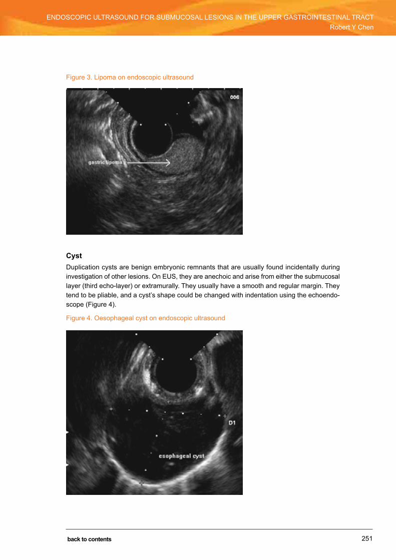
251
EndoSCopIC UlTRASoUnd FoR SUbmUCoSAl lESIonS In ThE UppER gASTRoInTESTInAl TRACTRobert Y Chen
SECTION 5: ENDOSCOPIC ULTRASOUND
back to contents
Figure 3. lipoma on endoscopic ultrasound
Cystduplication cysts are benign embryonic remnants that are usually found incidentally during investigation of other lesions. on EUS, they are anechoic and arise from either the submucosal layer (third echo-layer) or extramurally. They usually have a smooth and regular margin. They tend to be pliable, and a cyst’s shape could be changed with indentation using the echoendo-scope (Figure 4).
Figure 4. oesophageal cyst on endoscopic ultrasound

252
EndoSCopIC UlTRASoUnd FoR SUbmUCoSAl lESIonS In ThE UppER gASTRoInTESTInAl TRACTRobert Y Chen
SECTION 5: ENDOSCOPIC ULTRASOUND
back to contents
Pancreatic restpancreatic rest is ectopic pancreatic tissue arising as an embryonic remnant. It is composed of exocrine cells with cystic dilatation and is usually located on the inferior aspect of the antrum, or occasionally in the duodenum. Endoscopically, pancreatic rest usually has a central “umbilicus”. on EUS, pancreatic rest arises from the submucosal layer (third echo-layer) in most cases. It is of mixed echogenicity, with isoechoic areas mixed with anechoic areas corresponding to small ducts.
Carcinoid lesionsCarcinoid lesions are intramucosal tumours with malignant potential. These lesions tend to be small and found incidentally. on EUS, they are usually hypoechoic or isoechoic and arise from the deep mucosa (second echo-layer). They may also involve the submucosal layer (third echo-layer).
Granular cell tumourThis is an uncommon oesophageal tumour that in general runs a benign course. It tends to be located in the oesophagus and rarely in the stomach. on EUS, a granular cell tumour is usually hypoechoic, arising mainly from the deep mucosa (second echo-layer) and sometimes the submucosal layer (third echo-layer).
VaricesVarices usually appear as bluish, tortuous submucosal lesions on endoscopy. Clinical infor-mation generally aids with the diagnosis. on EUS, varices appear as anechoic areas, which are positive on doppler ultrasound, arising from the deep mucosa (second echo-layer) or submucosa (third echo-layer). during scanning, it can be seen that these form a tubular rather than spherical structure.
Role of EUS-guided fine needle aspiration
Even though EUS-guided fine needle aspiration (EUS–FnA) is very accurate in obtaining tissue diagnosis of pancreatic or mediastinal lesions, its yield for solid intramural gastric or oesoph-ageal lesions is not high and varies significantly, from as low as 19% up to 100%. The overall average yield is about 60%.
This variability in yield is caused by a combination of factors: the lesions are often small and more mobile during FnA (especially in the stomach), and there are scanty cells within the lesions. In addition, the aggressiveness of gIST lesions is difficult to predict with cytology obtained on EUS–FnA alone. Factors that may play a role in determining aggressiveness of gISTs include tumour size, mitotic activity, tumour necrosis, and histological and immunochemi-cal characteristics. Thus, even if FnA shows evidence of cells supporting the diagnosis of gIST, it is hard to determine the malignant nature or aggressiveness of the lesion.

253
EndoSCopIC UlTRASoUnd FoR SUbmUCoSAl lESIonS In ThE UppER gASTRoInTESTInAl TRACTRobert Y Chen
SECTION 5: ENDOSCOPIC ULTRASOUND
back to contents
Although trucut biopsy sampling can be done via EUS, its yield is not ideal either, and is reported to be about 63%. Recent advances in needle design have seen increases in histological yield of up to 80%–85%; however, larger studies are required to confirm these findings.
Thus, EUS–FnA or trucut biopsy is not done routinely for submucosal lesions, and whether tissue sampling is performed depends on the preference of the echoendoscopist and the indi-vidual clinical scenario.
Approach to managing submucosal lesions
Gastrointestinal stromal tumourmanagement of gIST is controversial. Although there are many factors affecting the aggres-siveness or malignant tendency of gISTs, the most easily applicable factors are size and mitotic activity.
Surgery should be considered if a gIST is larger than 5 cm, as lesions of this size are of at least intermittent risk of having metastasis or malignant tendency. If a lesion is sized between 2 and 5 cm, some endoscopists will elect to monitor the lesion, while others will consider surgery, depending on the individual clinical situation. As gISTs smaller than 2 cm may not grow rapidly or change significantly in size, many endoscopists will elect to observe them without surgery. however, it should be borne in mind that even small gISTs can be associated with malignant change or metastasis if the mitotic activity is high.
LeiomyomaAs it is hard to distinguish leiomyoma from gIST endoscopically or on EUS, management of leiomyoma will be similar to that described above for gIST. If leiomyosarcoma is suspected on EUS, surgery clearly needs to be considered.
LipomaIf benign lipoma is found and is confidently diagnosed on EUS, no further follow-up is needed.
CystsCysts are, in general, benign and do not need regular follow-up with EUS.
Pancreatic restIn general, pancreatic rests are mostly benign. It is not routinely recommended to follow up a patient with pancreatic rest with regular surveillance.

254
EndoSCopIC UlTRASoUnd FoR SUbmUCoSAl lESIonS In ThE UppER gASTRoInTESTInAl TRACTRobert Y Chen
SECTION 5: ENDOSCOPIC ULTRASOUND
back to contents
Carcinoid lesionsCarcinoid lesions can be either sporadic or associated with atrophic gastritis or multiple endo-crine neoplasia syndromes. management will depend on the type of carcinoid lesion. In general, the lesion should be removed, either endoscopically or via surgery.
Treatment
generally, submucosal lesions are followed up endoscopically or removed by surgery. There is a small amount of literature on endoscopic submucosal dissection of submucosal lesions, but this technique is very time-consuming and technically challenging. It may be considered if surgery cannot be carried out, and should be performed in centres with personnel well trained in endoscopic submucosal dissection. other treatment methods, such as band ligation, have been described, but should not be performed on lesions arising from the muscularis propria layer. large gISTs or gISTs with metastasis can be treated with imatinib.
Acknowledgementlennart Choo contributed to this chapter by updating it for this edition of the handbook.
Further reading1 Rosch T, Kapfer b, Will U, et al. Accuracy of endoscopic ultrasonography in upper gastrointestinal submucosal
lesions: a prospective multicenter study. Scand J Gastroenterol 2002; 37: 856-862.
2 Quek R, george S. gastrointestinal stromal tumor: a clinical overview. Hematol Oncol Clin North Am 2009; 23: 69-78, viii.
3 badalamenti g, Rodolico V, Fulfaro F, et al. gastrointestinal stromal tumors (gISTs): focus on histopathological diagnosis and biomolecular features. Ann Oncol 2007; 18 Suppl 6: vi136-140.
4 Fletcher Cd, berman JJ, Corless C, et al. diagnosis of gastrointestinal stromal tumors: a consensus approach. Hum Pathol 2002; 33: 459-465.
5 Zhu X, Zhang XQ, li bm, et al. Esophageal mesenchymal tumors: endoscopy, pathology and immunohistochemistry. World J Gastroenterol 2007; 13: 768-773.
6 mucciarini C, Rossi g, bertolini F, et al. Incidence and clinicopathologic features of gastrointestinal stromal tumors. A population-based study. BMC Cancer 2007; 7: 230.
7 nilsson b, bumming p, meis-Kindblom Jm, et al. gastrointestinal stromal tumors: the incidence, prevalence, clinical course, and prognostication in the preimatinib mesylate era — a population-based study in western Sweden. Cancer 2005; 103: 821-829.
8 miettinen m, lasota J. gastrointestinal stromal tumors: review on morphology, molecular pathology, prognosis, and differential diagnosis. Arch Pathol Lab Med 2006; 130: 1466-1478.
9 palazzo l, landi b, Cellier C, et al. Endosonographic features predictive of benign and malignant gastrointestinal stromal cell tumours. Gut 2000; 46: 88-92.
10 Chak A, Canto mI, Rosch T, et al. Endosonographic differentiation of benign and malignant stromal cell tumors. Gastrointest Endosc 1997; 45: 468-473.

255
EndoSCopIC UlTRASoUnd FoR SUbmUCoSAl lESIonS In ThE UppER gASTRoInTESTInAl TRACTRobert Y Chen
SECTION 5: ENDOSCOPIC ULTRASOUND
back to contents
11 patti R, Almasio pl, di Vita g. granular cell tumor of stomach: a case report and review of literature. World J Gastroenterol 2006; 12: 3442-3445.
12 Wilson Jl, Kalade A, prasad S, et al. diagnosis of solid pancreatic masses by endoscopic ultrasound-guided fine-needle aspiration. Intern Med J 2009; 39: 32-37.
13 Caddy g, Conron m, Wright g, et al. The accuracy of EUS-FnA in assessing mediastinal lymphadenopathy and staging patients with nSClC. Eur Respir J 2005; 25: 410-415.
14 okubo K, Yamao K, nakamura T, et al. Endoscopic ultrasound-guided fine-needle aspiration biopsy for the diagnosis of gastrointestinal stromal tumors in the stomach. J Gastroenterol 2004; 39: 747-753.
15 Chen VK, Eloubeidi mA. Endoscopic ultrasound-guided fine-needle aspiration of intramural and extraintestinal mass lesions: diagnostic accuracy, complication assessment, and impact on management. Endoscopy 2005; 37: 984-989.
16 polkowski m, gerke W, Jarosz d, et al. diagnostic yield and safety of endoscopic- ultrasound guided trucut biopsy in patients with gastric submucosal tumors: a prospective study. Endoscopy 2009; 41: 329-334.
17 blay JY, bonvalot S, Casali p, et al. Consensus meeting for the management of gastrointestinal stromal tumors. Report of the gIST Consensus Conference of 20-21 march 2004, under the auspices of ESmo. Ann Oncol 2005; 16: 566-578.
18 Sato T, peiper m, Fritscher-Ravens A, et al. Strategy of treatment of submucosal gastric tumors. Eur J Med Res 2005; 10: 292-295.
19 orlowska J, pachlewski J, gugulski A, butruk E. A conservative approach to granular cell tumors of the esophagus: four case reports and literature review. Am J Gastroenterol 1993; 88: 311-315.
Page 256 (used for notes in the hard copy) has been removed from the PDF edition of this handbook.

257
Endoscopic ultrasound for biliary disease and choledocholithiasisJason Y Huang, Jillian Rosenstengel and Patrick R Walsh
Endoscopic ultrasound (EUS) is an ideal modality for assessing biliary disease and choledo-cholithiasis because of its high sensitivity and specificity and low morbidity. The usual algorithm begins with transabdominal ultrasound, which is the least invasive method but has significant limitations, typically related to patient factors. Computed tomography (CT) imaging is useful for detecting ductal dilation, and associated mass lesions and stones are usually also visible. Magnetic resonance cholangiopancreatography (MRCP) is a sensitive method for identifying ductal changes or filling defects; however, small lesions, the nature of the filling defect and periampullary abnormality can be challenging to elucidate or characterise. Endoscopic retro-grade cholangiopancreatography (ERCP) is now largely a therapeutic tool, as complication rates (2%–5% risk of pancreatitis) are relatively high.
EUS is an essential complementary modality to preliminary cross-sectional imaging, especially when prior findings are ambiguous or tissue acquisition is required. Historically, EUS was largely a diagnostic modality, but with recent developments in equipment and techniques, EUS-guided biliary intervention is coming of age.
General principles
Ultrasound imaging of tissue is achieved by transmitting short pulses of ultrasound energy into the tissue and receiving reflected signals. Fluid-filled structures (e.g. gall bladder) have few reflections or refractions and are therefore dark or “anechoic”. Solid structures (e.g. stones) lead to reflection and scattering and therefore appear bright or “hyperechoic”, with posterior shadowing. Tissues containing fat are also brighter (more echogenic) than lean tissues.
SECTION 5: ENDOSCOPIC ULTRASOUND
CHAPTER 24
back to contents

258
EndoSCoPiC UlTRaSoUnd FoR biliaRY diSEaSE and CHolEdoCHoliTHiaSiSJason Y Huang, Jillian Rosenstengel and Patrick R Walsh
SECTION 5: ENDOSCOPIC ULTRASOUND
back to contents
Stones are seen as echogenic structures with or without posterior acoustic shadowing. They may be mobile, and there may be associated signs: dilated and/or thickened bile duct walls, cholelithiasis and pericholecystic fluid. “Sludge” is hyperechoic but does not have shadowing.
Strictures are seen as narrowing of the bile duct. This narrowing may be extrinsic or intrinsic and benign or malignant in nature. Malignant strictures are typically hypoechoic. There is either an associated mass (intrinsic or extrinsic) or, less commonly, ill defined thickening of the bile duct. in these circumstances, the area of pre-stenotic ductal dilation (“transition zone”) may hold the key to a definitive diagnosis.
Diagnostic performance
Procedurea diagnostic EUS is usually done as a day-only procedure under deep sedation (with propofol and/or midazolam and fentanyl). overall, it is a very similar experience to a gastroscopy for the patient.
StonesMRCP is a non-invasive imaging technique that is regarded as superior to CT for the diagnosis of biliary stones. However, MRCP has limited spatial resolution and can miss stones of small size (< 5 mm) or located in the periampullary region. Performance comparison is complicated by the absence of a true gold standard (both ERCP and intraoperative cholangiography can also miss small stones).
a recent meta-analysis involving 538 patients compared EUS and MRCP for stone detection, and found sensitivity of 93.7% and 83.5%, specificity of 88.5% and 91.5%, positive predictive value of 89% and 87.8% and, most importantly, negative predictive value of 96.9% and 87.8%, respectively. EUS is therefore the most accurate test for common bile duct (Cbd) stones.
imaging is usually the first line in the investigation pathway for patients with suspected biliary stones and, if stones are seen, ERCP is recommended. Patients with history and biochemical data suggestive of stones, but with negative findings on imaging, should proceed to EUS before ERCP. if EUS findings are negative, then ERCP is avoided.
StricturesStrictures are either benign or malignant. They can further be categorised as intraductal or extraductal in origin. EUS can readily visualise the bile duct and accurately characterise the thickness of the ductal wall and nature of the stricture. Tissue acquisition by EUS-guided fine needle aspiration (EUS–Fna) can secure a definitive diagnosis. intraductal ultrasound and cholangioscopy, although not readily available, provide detailed assessment of biliary strictures (see below). a recently released digital cholangioscope has dramatically improved intraductal visualisation.

259
EndoSCoPiC UlTRaSoUnd FoR biliaRY diSEaSE and CHolEdoCHoliTHiaSiSJason Y Huang, Jillian Rosenstengel and Patrick R Walsh
SECTION 5: ENDOSCOPIC ULTRASOUND
back to contents
ComplicationsComplications from EUS are rare. The standard risk from a diagnostic EUS is similar to that of a diagnostic gastroscopy. The risks include those associated with sedation (e.g. aspiration, drug reaction) or the procedure itself. Procedural complications include minor trauma, bleed-ing and perforation. Perforation may occur in the cervical oesophagus, gastric lesser curve or duodenal bulb. Fna-related complications include infection, bleeding and pancreatitis, which occurs in about 1%–1.5% of patients. infection risk is highest if a non-draining fluid structure is accessed, necessitating antibiotic prophylaxis.
Technique of diagnostic examination
Historically, assessment of Cbd abnormality was performed using a radial echoendoscope. This provides a “long” view of the Cbd from the common hepatic duct and cystic duct take-off to the ampulla. Currently, the linear echoendoscope is the scope of first choice for pancreaticobiliary interrogation. The linear instrument provides a piecemeal view of the Cbd.
General principles of a comprehensive EUS examination include the use of fixed common guiding “stations” and anatomical relationship with surrounding organs to facilitate a complete examination of the targeted viscera. However, there are several variations, or “roadmaps”, depending on style of examination and choice of scope.
Linear echoendoscopeThe Cbd can be visualised through a transgastric or transduodenal approach. The transgastric approach reveals the proximal Cbd and common hepatic duct, including the intrahepatic bile ducts. Pushing the scope into the antrum typically reveals the gall bladder and, in some cases (depending on the EUS system), the ampulla. The view in this approach is typically longitudinal.
The transduodenal approach typically begins in the duodenal bulb (d1), followed by scope withdrawal through the descending duodenum (d2). The d1 view is useful for scanning the entire Cbd up into the hilum, including the cystic duct and gall bladder. as the typical view is in cross-section, an oval hypoechoic structure is seen. The d2 approach provides the best view of the ampulla, as well as the terminal portion of the Cbd and pancreatic duct (Pd). a longitudinal view is obtained on withdrawal of the scope, and the entire Cbd and Pd can be seen. doppler ultrasound can help distinguish duct from adjacent vascular structures as required.
Radial echoendoscopeThe radial scope is placed into the duodenal bulb. The balloon is inflated and wedged at the apex of the d1–d2 junction with maximal downwards deflection of the large wheel on the scope. The portal vein is then identified and placed on the left side of the screen. The liver and gall bladder should be on the superior half of the image. The scope is then slowly advanced with a gentle clockwise torque to provide an image of the Cbd. once this is obtained, the Cbd is traced towards both the ampulla and the liver until a long view is obtained. as the Cbd is traced
260
EndoSCoPiC UlTRaSoUnd FoR biliaRY diSEaSE and CHolEdoCHoliTHiaSiSJason Y Huang, Jillian Rosenstengel and Patrick R Walsh
SECTION 5: ENDOSCOPIC ULTRASOUND
back to contents
to the ampulla, the main Pd will come into view. This completes the stack view (portal vein, Cbd and Pd). The Cbd is traced to the duodenal fall-off (where the Cbd enters the duodenum).
The scope is then placed in d2 and straightened (as in ERCP). The ampulla is visualised endo-scopically. The balloon is inflated and manoeuvred so it “kisses” the ampulla. air is aspirated and the scope is then slowly withdrawn. The ampulla will come into view as a relatively hypoechoic area containing two adjacent oval anechoic structures. The one closest to the probe is the Cbd; the deeper structure is the Pd. The ducts can then be traced from the ampulla.
Diagnosis of biliary disease
biliary abnormalities detected on EUS include stones and sludge (discussed above), strictures, thickened walls and related disorders in the ampulla and gall bladder.
Bile duct strictures and massesdifferentiating benign from malignant biliary strictures remains a challenge. EUS improves the diagnosis in the setting of a known or suspected bile duct stricture despite negative findings from ERCP-facilitated tissue sampling and cross-sectional imaging. in this setting, the sensitivity and specificity for malignancy for the finding of a pancreatic head mass and/or an irregular bile duct wall are 88% and 100%, respectively. a bile duct wall thickness ≥ 3 mm has a sensitivity of 79% and a specificity of 79% for malignancy; the sensitivity of EUS–Fna for malignancy is 47%, with a specificity of 100%. The sensitivity of EUS–Fna improves from 36% to 57% when a pancreatic mass is seen on EUS (as opposed to a thickened bile duct wall alone). in sus-pected hilar cholangiocarcinoma where ERCP brushings are negative, the accuracy, sensitivity and specificity of EUS–Fna are reportedly 91%, 89% and 100%, respectively. EUS–Fna has resulted in avoidance of major surgery in 20% of cases. in a similar series of proximal biliary strictures subjected to EUS–Fna, the sensitivity, specificity and accuracy of EUS–Fna were 77%, 100% and 79%, respectively. as EUS–Fna has a low negative predictive value, a negative Fna result does not permit reliable exclusion of malignancy.
The question of which endoscopic technique to use to diagnose biliary strictures is not always straightforward. in the setting of biliary obstruction requiring decompression, ERCP is the obvious first step, with subsequent EUS if a tissue diagnosis is not made. The most common approach in australia is to start with ERCP when a biliary tumour is suspected, and EUS when a pancreatic tumour is suspected.
although intraductal ultrasound is not readily available, its features of malignancy include a hypoechoic appearance of the stricture, with irregular margins, whereas benign strictures are more commonly hyperechoic or isoechoic, with smooth borders. Endoscopically placed stents render the interpretation of findings at both EUS and intraductal ultrasound more difficult. The stent tends to change the characteristics of the wall (thickened) and size of the duct (decompres-sion, loss of transition zone) and introduce artefacts (intraductal and/or intraluminal air artefacts).

261
EndoSCoPiC UlTRaSoUnd FoR biliaRY diSEaSE and CHolEdoCHoliTHiaSiSJason Y Huang, Jillian Rosenstengel and Patrick R Walsh
SECTION 5: ENDOSCOPIC ULTRASOUND
back to contents
an alternative sequence would be to have EUS precede the ERCP to help clarify intraductal or extraductal abnormality (e.g. stricture characterisation, number of stones) and facilitate cyto-logical diagnosis by EUS–Fna, if logistically possible. While the Fna material is undergoing in-room assessment, ERCP can be carried out, and intraductal biopsies and brushings can be done if the initial EUS–Fna is non-diagnostic. With the additional diagnostic information from the preceding EUS–Fna, this may influence appropriate choice of biliary stent. This will result in a preliminary diagnosis and completion of endoscopic therapy in the one session. This “one-stop shop” approach will help reduce the need for repeat procedures for tissue acquisition or change of stents from plastic to metal.
AmpullaEUS is a very sensitive modality for detecting periampullary tumours. The sensitivity of EUS for detecting ampullary tumours ranges from 95% to 100%, compared with transabdominal ultra-sound (5%–24%), CT (19%–68%) and MRi (81.3%). The sensitivity of ERCP for detection of ampullary tumours is equivalent to EUS. EUS can be useful in determining endoscopic resect-ability of lesions. diagnostic accuracy of EUS for ampullary lesions with low-grade dysplasia or adenocarcinoma is 72% and 96%, respectively. a duodenoscope is needed occasionally when the ampulla is not visible because of restricted viewing angles of the oblique-viewing linear echoendoscope.
Gall bladderThe gall bladder is best seen from the gastric antrum on EUS. it is essential to obtain a history of cholecystectomy from a patient before EUS to avoid a prolonged search for the “elusive gall bladder”, although typically clips are visible once the cystic duct is traced. The sensitivity and specificity of EUS for the diagnosis of gallstones, when previous imaging findings have been negative, are 96% and 86%, respectively.
in the healthy population, 4%–7% of people have gall bladder polyps, which are classified as either neoplastic (adenomas and adenocarcinomas) or non-neoplastic (cholesterol polyps, inflammatory polyps, adenomyomatosis). EUS can depict the two-layer structure of the gall bladder wall with high resolution, and its superiority to transabdominal ultrasound for definition of small polypoid lesions and staging of gall bladder carcinoma has been reported in several studies. Sadamoto and colleagues have proposed an endosonographic scoring system to differentiate benign from malignant polyps. More recently, Cho and colleagues described the finding of hypoechoic foci as a sensitive and specific predictor of malignancy in gall bladder polyps (90% and 89%, respectively).
Malignant gall bladder lesions tend to be sessile and broad-based rather than pedunculated. imaging appearances on EUS include hypoechoic heterogeneous masses, thickening of the wall, polypoidal masses, loss of border between the liver and gall bladder, and focal calcifica-tion of the gall bladder wall. The accuracy of EUS for the T-staging of gall bladder carcinoma in one study was 100% for pTis, 75.6% for pT1, 85.3% for pT2 and 92.7% for pT3–4 tumours. EUS–Fna of the gall bladder has been reported without complications in small series, but is not currently recommended due to the risk of bile leak with peritonitis and tumour seeding, even with a 25G Fna needle.

262
EndoSCoPiC UlTRaSoUnd FoR biliaRY diSEaSE and CHolEdoCHoliTHiaSiSJason Y Huang, Jillian Rosenstengel and Patrick R Walsh
SECTION 5: ENDOSCOPIC ULTRASOUND
back to contents
Interventions for biliary disease
The most common indication for EUS-guided biliary drainage (EUS–bd) is failed ERCP, which typically occurs in < 5% of cases and is generally related to patient factors, such as significant anatomical deformity (duodenum or ampulla) leading to an inaccessible ampulla or unidentifi-able ampullary orifice. Traditionally, these patients have undergone percutaneous transhepatic cholangiography to gain biliary access and subsequent drainage. EUS–bd is an emerging technique that is not quite yet mainstream.
EUS–bd can be achieved via the transduodenal or transgastric transhepatic route. a wire can be passed down the bile duct in an anterograde fashion through the major papilla to facilitate conventional ERCP, or a stent can be placed into the extrahepatic or intrahepatic bile duct for direct drainage into the duodenum or stomach, respectively. With the recent development of lumen-apposing stents, direct drainage of the gall bladder into the duodenum is also possible.
Conclusion
EUS is a highly accurate imaging modality for the biliary tree. it is the most sensitive method for detecting small stones in the Cbd. EUS is also useful in further defining biliary strictures identified on cross-sectional imaging. it provides additional information to that from ERCP and is less invasive. EUS–Fna allows cytological confirmation of the nature of the stricture or surrounding lymph nodes. EUS is also useful in the assessment of gall bladder and ampullary tumours. EUS–bd is a growing area and, with the recent release of lumen-apposing stents, additional indications will soon emerge.
Further reading1 de Castro Vl, Moura EG, Chaves dM, et al. Endoscopic ultrasound versus magnetic resonance
cholangiopancreatography in suspected choledocholithiasis: a systematic review. Endosc Ultrasound 2016; 5: 118-128. doi: 10.4103/2303-9027.180476.
2 Sadamoto Y, oda S, Tanaka M, et al. a useful approach to the differential diagnosis of small polypoid lesions of the gallbladder, utilizing an endoscopic ultrasound scoring system. Endoscopy 2002; 34: 959-965.
3 Cho JH, Park JY, Kim YJ, et al. Hypoechoic foci on EUS are simple and strong predictive factors for neoplastic gallbladder polyps. Gastrointest Endosc 2009; 69: 1244-1250. doi: 10.1016/j.gie.2008.10.017.
4 Walter d, Teoh aY, itoi T, et al. EUS-guided gall bladder drainage with a lumen-apposing metal stent: a prospective long-term evaluation. Gut 2016; 65: 6-8. doi: 10.1136/gutjnl-2015-309925.
Pages 263-265 (used for notes in the hard copy) have been removed from the PDF edition of this handbook.

Section 6
IMAGING OF THE SMALL INTESTINE

267
Approach to obscure bleeding of the gastrointestinal tractGregor Brown
“Obscure gastrointestinal bleeding” (OGIB) is defined as clinically significant gastrointestinal bleeding for which no cause is found at gastroscopy and colonoscopy. The implication is that the bleeding is from the small bowel, although there are a range of lesions that may be missed at routine endoscopy. OGIB may be overt (melaena or haematochezia) or non-overt (iron deficiency anaemia, without the obvious passage of blood through the rectum), which to some extent will dictate the causes of the bleeding and the urgency with which it should be investigated.
Over the past 10 years, the investigation and management of OGIB have undergone a para-digm shift due to advances in endoscope technology, namely capsule endoscopy (CE) and balloon enteroscopy (BE), as well as the increasing availability of accurate computed tomog-raphy angiography (CTA). Previously, patients with OGIB faced a choice of either repeated blood transfusions and iron infusions or (often speculative) laparotomy with or without on-table enteroscopy. The management of OGIB is now not only less morbid but also more successful, although it must be said that the outcome data remain incomplete.
Causes of obscure gastrointestinal bleeding
The causes of OGIB may be divided into those within reach of standard endoscopy and those beyond it (i.e. in the small bowel). Routine gastroscopy and colonoscopy have known miss rates, which depend on the lesion, endoscopist factors (experience, skill, care, equipment, time taken) and patient factors (ease of intubation, quality of preparation, tolerability of the procedure). Commonly missed lesions include portal hypertensive gastropathy, vascular malformations (either isolated, particularly in the fundus or caecum, or multiple, such as gastric antral vasc-ular ectasia), ulcers of the stomach (Dieulafoy lesions or hiatus hernia-associated Cameron
SECTION 6: IMAGING OF THE SMALL INTESTINE
CHAPTER 25
back to contents

APPROACh TO OBsCuRE BlEEDInG Of ThE GAsTROInTEsTInAl TRACTGregor Brown
SECTION 6: IMAGING OF THE SMALL INTESTINE
268 back to contents
ulcers) or duodenum (posterior cap or second part) and colorectal cancer. Occasionally, rou-tine endoscopy may need to be repeated before embarking on a search for true small bowel causes of OGIB.
The causes of true OGIB are listed in Table 1. This list is not exhaustive. By far the most common lesions are vascular malformations, usually found in the proximal jejunum, and inflammatory lesions caused by either non-steroidal anti-inflammatory drugs (nsAIDs) or small bowel Crohn’s disease. small bowel malignancy remains rare.
Table 1. small bowel causes of obscure gastrointestinal bleeding
● Angiodysplasia ● non-steroidal enteropathy ● Crohn’s disease ● Polyps (adenoma, hyperplastic, hamartoma) ● Benign tumours (lipoma, leiomyoma) ● Malignancy (adenocarcinoma, lymphoma, gastrointestinal
stromal tumour, carcinoid, metastases — especially melanoma) ● Jejunal diverticula ● Radiation enteritis ● Enteric varices ● Dieulafoy lesion ● Parasites ● Meckel diverticulum ● Aorto-enteric fistula ● Blue rubber bleb naevus syndrome ● Osler–Weber–Rendu syndrome (hereditary haemorrhagic
telangiectasia)
Investigating obscure gastrointestinal bleeding
CE and BE have changed the approach to investigating OGIB, as has the introduction of highly sensitive and routinely available CTA. how these tools are used depends on the clinical sce-nario, local expertise and availability. Broadly, the heavier the bleeding, the more accurate and relevant CTA becomes. however, endoscopy remains the mainstay of investigation of OGIB.
All patients with anaemia should have at least a gastroscopy, which should be done urgently if there is heavy bleeding. Even bright rectal bleeding (if heavy) can derive from an upper gas-trointestinal source, and gastroscopy can be therapeutic as well as diagnostic.
Overt obscure gastrointestinal bleedingIn overt heavy gastrointestinal bleeding, the next diagnostic step after normal findings on gastroscopy will depend on the factors alluded to previously. If the patient is cardiovascularly unstable, CTA may be the most appropriate next step. If a bleeding point can be identified, it can be followed by formal angiography and embolisation. Colonoscopy in this situation is difficult, time-consuming and potentially risky, and should only be undertaken by those with suitable experience, expertise in colonic haemostasis and available equipment (i.e. a water

269
APPROACh TO OBsCuRE BlEEDInG Of ThE GAsTROInTEsTInAl TRACTGregor Brown
SECTION 6: IMAGING OF THE SMALL INTESTINE
back to contents
pump and a colonoscope with a flushing channel). from a therapeutic perspective, the time to do a colonoscopy is when the patient is actively bleeding, although this necessarily limits the diagnostic capability. Importantly, the most common cause of haematochezia is diverticular haemorrhage, which in most cases will settle spontaneously, so it is very reasonable to simply observe a stable patient with haematochezia and normal findings on gastroscopy.
“urgent” CE may have a role for an actively bleeding patient with normal findings on gastros-copy, particularly when colonoscopy and ileoscopy show transported blood from the ileum. however, the delay implicit in CE means CTA is usually more appropriate, being not only quicker but also more likely to accurately localise the lesion for therapeutic angiography (or surgery) if the study is positive.
Non-overt obscure gastrointestinal bleedingCE comes into its own for non-overt OGIB (i.e. iron deficiency anaemia) when good-quality gastroscopy and colonoscopy (often done in combination on the same occasion) are unhelpful and there is less urgency. In this setting, the yield of CE for causative small bowel lesions is over 50%. CE is also simple, safe and well tolerated. As with any form of endoscopy, exper-tise is required to accurately review and report CE studies. In its current form, CE is purely diagnostic, having no therapeutic capability. It also struggles to accurately localise small bowel lesions, unless they are located at either extremity of the small intestine. In experienced hands, however, it can provide uniquely useful information to guide BE, which offers the full gamut of endoscopic interventional therapeutics deep in the small bowel.
There is debate around the world as to the interplay between CE and BE and how they should be used. In Australia, CE is fully funded for this indication, provided certain clinical criteria are met, so we are able to use it as a screening tool for the more risky and time-consuming BE. By using CE, we are able to select those patients with lesions that are likely to be reachable with BE, which can thereby confer its therapeutic potential more fully and consistently. As such, the two modalities are very much complementary.
Specific modalities used in obscure gastrointestinal bleeding
GastroscopyCareful and timely gastroscopy is essential in the work-up of OGIB. It should be done urgently (< 24 hours) for overt bleeding and within 30 days for iron deficiency anaemia. It is the best diagnostic modality for upper gastrointestinal bleeding and can often also be therapeutic. Blood in the upper gastrointestinal tract mandates a careful search for a source — if one cannot be found, the examination should be repeated once the blood has cleared. Important lesions to consider are varices, ulcers (particularly hiatus hernia-associated Cameron ulcers, subtle duo-denal ulcers) and vascular malformations. Duodenal biopsy samples should routinely be taken to check for histological evidence of coeliac disease in patients with iron deficiency anaemia.

APPROACh TO OBsCuRE BlEEDInG Of ThE GAsTROInTEsTInAl TRACTGregor Brown
SECTION 6: IMAGING OF THE SMALL INTESTINE
270 back to contents
ColonoscopyColonoscopy is often combined with gastroscopy in the investigation of iron deficiency anaemia because of the risk of dual abnormalities, as well as patient convenience. The yield diminishes in younger women, where its use can be restricted to those with significant anaemia or relevant symptoms at the clinician’s discretion.
In patients with persistent or severe overt bleeding with anaemia, urgent colonoscopy carries significant risk, as well as being difficult and time-consuming. Bowel preparation is useful if time allows, and appropriate equipment (water pump and suitable colonoscope with a flushing channel) and experience are essential. A strong argument can be made for observing stable patients with haematochezia, as this will most often be diverticular in origin and will settle spon-taneously. In unstable or persistently bleeding patients, CTA may be more suitable as a means of localising bleeding to direct therapy (colonoscopy, angiography or surgery), depending on the clinical circumstances and local availability and expertise.
Capsule endoscopyIn Australia, CE is fully funded by Medicare for recurrent or persistent bleeding and anaemia, as long as a gastroscopy and colonoscopy have not found a cause. The yield in this situation is over 50%.
With the “standard preparation” for CE (clear fluids after lunch the day before, fast from mid-night), about 20% of studies are incomplete (i.e. the capsule has not entered the colon by the end of the study — usually 8 hours). Drugs that enhance gastric emptying (e.g. metoclopramide) or bowel preparation may improve the completion rate, but the data are mixed.
The major complication of CE is non-excretion of the capsule, although this occurs in < 1% of studies. usually it is due to small bowel strictures (Crohn’s disease, nsAIDs or malignancy), but overt small bowel obstruction is extremely rare. The capsule can be removed if reachable endoscopically, but surgery may be required — importantly, the cause of the capsule retention (i.e. the stricture) can usually be dealt with at the same time.
Balloon enteroscopyThere are two BE systems — double (DBE) and single (sBE) — from different manufacturers. Both have a flexible overtube with a balloon on the tip; the DBE system has an additional balloon on the tip of the scope itself. The balloons are independently inflated and deflated by a pump under the control of the operator, gently fixing the instrument to the otherwise mobile small bowel wall to allow deep small bowel intubation. Performing BE is complex and time-consuming (often taking an hour or more) and generally requires two operators. The risk of aspiration is significant, and many practitioners choose to routinely intubate patients having antegrade BE. Post-procedure discomfort is common but is reduced by the use of carbon dioxide for insuffla-tion — this also improves insertion depth.
In Australia, BE is usually performed to further investigate or treat a small bowel abnormality identified by CE (or another imaging modality). The “direction” of BE is determined by the CE findings — if the lesions are in the proximal 60% of the small bowel by transit time, antegrade BE is used; if in the distal 40%, retrograde BE is used. If the lesion is not reached, a tattoo can

271
APPROACh TO OBsCuRE BlEEDInG Of ThE GAsTROInTEsTInAl TRACTGregor Brown
SECTION 6: IMAGING OF THE SMALL INTESTINE
back to contents
be placed at the limit of insertion and the alternative direction can be used (usually as a separate procedure), but total enteroscopy is rarely achieved in the Australian setting.
As BE usually follows CE to define suspected lesions, the rate of therapeutic intervention is higher than in “routine” endoscopy, with a concomitant increase in the risk of the procedure, particularly for bleeding and perforation. Pancreatitis is a rare risk of BE, possibly due to traction on the ampulla during repeated insertion and withdrawal cycles. While virtually the full gamut of endotherapy is available with BE, the length of the scope (210 cm), current narrowness of the working channel (2.8–3.2 mm) and routine formation of loops when deep in the small bowel mean that some interventions (e.g. balloon dilatation, stent insertion) may be difficult or impossible. hopefully, technical developments in both the enteroscopes and relevant therapeu-tic consumables will improve these aspects of BE, but currently it is a procedure that should only be performed in referral centres with the necessary interest, expertise and case volume.
Computed tomography angiographyAs multislice computed tomography scanners have become routine, CTA has become increas-ingly available and can usually be performed quickly and without preparation or sedation. It is not limited by “access” to the gastrointestinal tract, which can be problematic for endoscopy. The major risk is of contrast-related renal impairment, and the major limitation is the need for active bleeding at the time of the scan to accurately define a bleeding source. Once defined, however, therapeutic angiography is an immediate option, where available and clinically appro-priate. The major risk of embolisation is infarction of the target organ, but this is diminishing with increasingly selective embolisation.
Conclusion
The approach to OGIB has changed dramatically in recent years, making it less morbid and more successful. Gastroscopy remains the index investigation, with subsequent investigations determined by the clinical scenario, particularly whether the bleeding is overt or occult and whether the patient’s condition is stable or not. When the presentation is one of iron deficiency anaemia, colonoscopy is the next investigation; if not informative, it is followed by CE as a screening test for BE or surgery. When the bleeding is overt, watchful waiting may be appropri-ate, with elective colonoscopy to clarify the cause of bleeding once it has settled. If the bleeding persists or the patient’s condition is unstable, CTA can be useful to localise the bleeding for therapeutic angiography. In the right hands, urgent colonoscopy can be both diagnostic and therapeutic, but it comes with an increased risk of harm. CE is of limited utility in this setting, due to the inherent delays and difficulty identifying and localising causative lesions in the presence of large amounts of blood.

APPROACh TO OBsCuRE BlEEDInG Of ThE GAsTROInTEsTInAl TRACTGregor Brown
SECTION 6: IMAGING OF THE SMALL INTESTINE
272 back to contents
Further reading1 Alexander JA, leighton JA. Capsule endoscopy and balloon-assisted endoscopy: competing or complementary
technologies in the evaluation of small bowel disease? Curr Opin Gastroenterol 2009; 25: 433-437.
2 Postgate A, Tekkis P, Patterson n, et al. Are bowel purgatives and prokinetics useful for small-bowel capsule endoscopy? A prospective randomized controlled study. Gastrointest Endosc 2009; 69: 1120-1128.
3 selby W. Complete small-bowel transit in patients undergoing capsule endoscopy: determining factors and improvement with metoclopramide. Gastrointest Endosc 2005; 61: 80-85.
4 Domagk D, Bretthauer M, lenz P, et al. Carbon dioxide insufflation improves intubation depth in double-balloon enteroscopy: a randomized, controlled, double-blind trial. Endoscopy 2007; 39: 1064-1067.
5 li X, Chen h, Dai J, et al. Predictive role of capsule endoscopy on the insertion route of double-balloon enteroscopy. Endoscopy 2009; 41: 762-766.
Pages 273-274 (used for notes in the hard copy) have been removed from the PDF edition of this handbook.
275
Endoscopic imaging of the small intestineMark Appleyard
Endoscopic imaging of the entire small bowel has now been available for over a decade. The entire small bowel was first reliably seen with the development of capsule endoscopy (CE), quickly followed by balloon enteroscopy (BE). Extensive research has aimed to clarify the role of these new tools in the management and therapeutics of the small bowel. These tools have allowed a paradigm shift in the diagnosis of patients with obscure gastrointestinal bleeding (OGIB), with BE providing an effective therapeutic endoscopy alternative to more traditional therapeutic modalities, such as embolisation during angiography and surgery. With the improved diagnostic and therapeutic capabilities of these newer techniques, intraoperative enteroscopy is now rarely required and will not be discussed further in this chapter.
There are also some newer devices, in addition to balloons, that can allow therapeutic interven-tion into the small bowel (e.g. spiral enteroscopy overtube [Spirus Medical]), but the evidence for these is limited. Hence, comments on therapeutic small bowel intervention will be largely confined to BE.
The management algorithms now available for small bowel disease include both these endo-scopic developments (BE and CE), with new indications and innovations on the horizon.
General principles
Two key issues help decide which endoscopic tool is best to use in the small bowel: the indi-cation and the need for a diagnosis or therapy are crucial elements. CE is essentially a diag-nostic test, with BE being both diagnostic and therapeutic. The more traditional and available push endoscope (commonly a colonoscope used orally) might still play a similar role to BE for proximal small bowel lesions.
SECTION 6: IMAGING OF THE SMALL INTESTINE
CHAPTER 26
back to contents

276
EndOSCOpIC IMAGInG Of THE SMAll InTESTInEMark Appleyard
SECTION 6: IMAGING OF THE SMALL INTESTINE
back to contents
The American College of Gastroenterology (ACG) has recently suggested dropping the term OGIB for suspected small bowel bleeding, but most other authorities continue to use this term.
IndicationMost small bowel endoscopy is performed for OGIB, which is defined as recurrent or persistent gastrointestinal bleeding with normal findings on endoscopy and colonoscopy. In Australia, OGIB is the main Medicare-rebateable indication for CE, with peutz–Jeghers syndrome more recently added to the Medicare Benefits Schedule. In 2007, the American Gastroenterological Association position paper on management of OGIB, the ACG guidelines and the European clinical guidelines all placed CE as the first diagnostic test and the most pivotal step in the investigation of OGIB. The traditional techniques of small bowel x-ray, cross-sectional radiol-ogy and angiography play a very limited role in the early investigation of OGIB and should not precede CE.
Angioectasias account for half of the abnormal findings in patients with OGIB in Western cohorts, so investigation with mucosal viewing is optimally required. Bidirectional BE has a similar diagnostic rate to CE for OGIB, but with inferior total enteroscopy rates; hence, CE is the first step for diagnosis in Western guidelines. The next appropriate step can be determined based on the result of CE. As most patients will not need endoscopic intervention, the need for more invasive diagnostic and therapeutic modalities can be minimised.
A less common indication is suspected small bowel Crohn’s disease. Although CE can play an important role in the diagnosis of this disease, the risk of a retained capsule rises significantly if a patient is known to have Crohn’s disease, so disease assessment is not usually an indication. BE, however, can have an important role — not only diagnostic, with the addition of endoscopic biopsies and mucosal assessment, but also an important therapeutic role for stricturing disease.
CE can be useful in small bowel polyposis syndromes, while most studies show other indications to have a very low yield for both procedures.
Technique
Capsule endoscopyCE does not require a traditional endoscopic environment or skill set and can be easily per-formed in a clinician’s rooms without sedation. The placement of recording equipment and swallowing of the capsule are simple and easy. The key technical issues are related more to the preparation for this technique, with cleansing and prokinetic use. The general consensus is that bowel preparation improves the quality of the capsule imaging, but data regarding improved diagnostic yield or total enteroscopy rates are conflicting. There is ongoing controversy about prokinetic use, with many authors supporting the regular use of prokinetics to maximise total enteroscopy rates, while others reject this notion. Software advances have shown promise regarding electronic diagnostics, but manual reading is still required for now.

277
EndOSCOpIC IMAGInG Of THE SMAll InTESTInEMark Appleyard
SECTION 6: IMAGING OF THE SMALL INTESTINE
back to contents
Reading the capsule images requires time and experience; there is no agreed minimum num-ber of procedures reviewed to enable determination of competency. The Australian Conjoint Committee for the Recognition of Training in Gastrointestinal Endoscopy has set a requirement for a minimum of 50 capsule examinations for certification.
Push enteroscopypush enteroscopy (pE) is a relatively simple and readily available technique that is a very good option for proximal small bowel lesions (figure 1). It is performed in the left lateral position, typical of endoscopy, mainly under conscious sedation. There are no good guidelines to deter-mine which proximal lesions are best targeted by pE, but it is unlikely to reach lesions more than 25% down the small bowel. The lack of tip control, compared with BE, is likely to make BE the choice for all therapeutic enteroscopy, where available.
figure 1. Multiple classic angioectasias in the proximal small bowel have been successfully ablated using argon plasma coagulation during standard push enteroscopy
Balloon enteroscopy
BE aims to pleat the small bowel over an overtube with a push–pull technique, using balloons. The first technique described was double balloon enteroscopy (dBE; fujinon), which uses bal-loons on the overtube and endoscope. The more recent addition of single balloon enteroscopy (SBE; Olympus) uses a balloon only on the overtube. dBE and SBE require a more traditional endoscopic setup and skill set than CE, even though they have technical nuances. In most cases, these procedures will be used to target known lesions that have been diagnosed by CE or cross-sectional imaging (figure 2).

278
EndOSCOpIC IMAGInG Of THE SMAll InTESTInEMark Appleyard
SECTION 6: IMAGING OF THE SMALL INTESTINE
back to contents
figure 2. Capsule endoscopy followed by double balloon enteroscopy
A: Capsule study shows a stricture before non-passage of the capsule. B: Double balloon enteroscopy identifies the stricture and shows similar endoscopic features. C: After successful endoscopic balloon dilatation, the capsule is retrieved with a standard endoscopic snare.
As a general principle, lesions in the proximal two-thirds of the small bowel are approached orally (antegrade procedure), whereas those in the distal third are approached anally (retrograde procedure). Antegrade procedures require fasting for at least 4 hours, unless a small bowel stric-ture is suspected, in which case a fluid diet for 24 hours before the procedure is recommended so that food debris is not encountered. Retrograde procedures require a comprehensive bowel preparation as any residual stool can create friction between the enteroscope and overtube, thus making the procedure difficult and impairing visualisation.
Most procedures are performed under deep sedation with anaesthetic support; general anaes-thetic with endotracheal intubation is rarely required. patients having antegrade procedures are more likely than those having retrograde procedures to need anaesthetic support. patient positioning is similar to routine endoscopy; however, the supine position can be helpful in accessing stable ileal intubation.
Relevant literature
OutcomesOGIB is the main indication for small bowel endoscopy, and most of the literature is based on these patients. The initial studies of CE in patients with OGIB showed its clear diagnostic supe-riority over traditional tools such as small bowel series, computed tomography (CT) and pE. The overall diagnostic yield of CE in OGIB is about 60%; this figure has remained consistent among most series, except in cases of acute ongoing OGIB, where the diagnostic yield can approach 100%. Generally, repeat endoscopy or colonoscopy is not required before CE. Very rapid bleeding might be better evaluated with BE or CT angiography than CE, as the small bowel transit time and evaluation always take several hours.
depending on the CE finding, a decision can then be made as to whether a therapeutic endos-copy procedure is required. Most patients will not require enteroscopy; however, in those in whom a lesion is identified or who continue to bleed, BE is generally the procedure used for

279
EndOSCOpIC IMAGInG Of THE SMAll InTESTInEMark Appleyard
SECTION 6: IMAGING OF THE SMALL INTESTINE
back to contents
therapy. CE has been found to miss lesions, and it is likely that BE is more accurate in the proximal small bowel. The average number of BE procedures required for those who need endoscopic intervention is about 1.3 per person. The CE findings can also help determine the most appropriate endoscopic tool, with proximal lesions possibly being suitable for pE or ante-grade BE, while deeper or very distal lesions are best suited for antegrade or retrograde BE. There is ongoing debate about whether dBE achieves greater depth than SBE. Thankfully, a negative CE has a high negative predictive value, so recurrent bleeding is uncommon in this scenario. If there is further bleeding, the approach should be individualised according to the clinical presentation.
patients with Crohn’s disease often have small bowel involvement, and determining diagnosis, disease extent and severity is very important for managing their disease. Although CE is the most sensitive tool to diagnose mucosal lesions in suspected small bowel Crohn’s disease, this is generally only indicated after ileocolonoscopy and faecal or blood biomarkers suggest inflammation. faecal calprotectin seems particularly promising in this regard. In comparison to OGIB, these patients have a higher rate of retained capsules, with about 2% retention in patients suspected of having Crohn’s disease, and up to 15% retention if they are known to have it. Hence, CE can be problematic in patients with known small bowel Crohn’s disease, and cross-sectional radiological imaging is preferred. If CE is required, using a patency capsule to “test run” the small bowel before using the real capsule can be useful in patients with known small bowel Crohn’s disease.
patients with polyposis syndromes are emerging as a group who can benefit from these small bowel imaging techniques. Currently in Australia, peutz–Jeghers syndrome is approved for sur-veillance CE. CE is probably the most accurate test for polyp detection, but magnetic resonance elastography is also useful and might be more accurate for size estimation and positioning. pE or BE can then be used to remove lesions (figure 3), thus avoiding the need for small bowel surgery in many patients. prior surgery can make BE difficult, and surgery is still required for lesions that cannot be reached.
figure 3. Successful polypectomy during antegrade double balloon enteroscopy after capsule endoscopy performed for peutz–Jeghers syndrome identified the lesion in the proximal small bowel
Emerging indications for BE include endoscopic retrograde cholangiopancreatography in patients with altered anatomy, with new modifications and accessories improving success rates.

280
EndOSCOpIC IMAGInG Of THE SMAll InTESTInEMark Appleyard
SECTION 6: IMAGING OF THE SMALL INTESTINE
back to contents
Complications and adverse eventsComplications of CE are rare, with retained capsules being the best recognised. depending on the indication, the risk of a retained capsule can be as high as 15% in patients with known Crohn’s disease. Many argue that surgery for retained capsules is not a complication, as surgery would have been the definitive intervention in these cases anyway.
Enteroscopy literature is now extensive, with good data regarding complications. It is expected that the rate of any complication for a diagnostic BE should be less than 1%, but therapeutic BE procedures can have complication rates up to 4%, including perforation, bleeding, pancreatitis and anaesthetic complications.
Conclusion
Endoscopic imaging of the small bowel has developed rapidly over the past decade, with new diagnostic and therapeutic options. new algorithms have been developed to guide patient man-agement, and these vary according to indication. future developments in this field will probably be in refining the use of these tools; however, exciting new modifications and accessories will expand their indications for both small bowel and non-small bowel use.
Further reading1 Raju GS, Gerson l, das A, lewis B; American Gastroenterological Association. American Gastroenterological
Association (AGA) Institute medical position statement on obscure gastrointestinal bleeding. Gastroenterology 2007; 133: 1694-1696.
2 Gerson lB, fidler Jl, Cave dR, leighton JA. ACG clinical guideline: diagnosis and management of small bowel bleeding. Am J Gastroenterol 2015; 110: 1265-1287.
3 pennazio M, Spada C, Eliakim R, et al. Small-bowel capsule endoscopy and device-assisted enteroscopy for diagnosis and treatment of small-bowel disorders: European Society of Gastrointestinal Endoscopy Clinical Guideline. Endoscopy 2015; 47: 352-376.
Page 281 (used for notes in the hard copy) has been removed from the PDF edition of this handbook.

EndOSCOpIC IMAGInG Of THE SMAll InTESTInEMark Appleyard
SECTION 6: IMAGING OF THE SMALL INTESTINE
ACG American College of Gastroenterology Af atrial fibrillation AfER automated flexible endoscope reprocessor AfI autofluorescence imaging AHA American Heart Association AnZCA Australian and new Zealand College of Anaesthetists ASA American Society of Anesthesiologists ASGE American Society for Gastrointestinal Endoscopy BAC balloon-assisted cholangioscopy BBS benign biliary strictures BE balloon enteroscopy BlI blue laser imaging CBd common bile duct CCd charge-coupled device Cd Crohn’s disease CE capsule endoscopy CEA carcinoembryonic antigen CEIM complete eradication of intestinal metaplasia ClE confocal laser endomicroscopy Cp chronic pancreatitis CpE carbapenemase-producing Enterobacteriaceae CRC colorectal cancer CT computed tomography CTA computed tomography angiography d1 duodenal bulb d2 descending duodenum dBE double balloon enteroscopy dOAC direct oral anticoagulant EBUS endobronchial ultrasound EMR endoscopic mucosal resection ERCp endoscopic retrograde cholangiopancreatography ES endoscopic sphincterotomy ESd endoscopic submucosal dissection ESGE European Society of Gastrointestinal Endoscopy EUS endoscopic ultrasound fAp familial adenomatous polyposis fICE flexible spectral imaging colour enhancement fIT faecal immunohistochemical testing fnA fine needle aspiration fr french
List of acronyms
282 back to contents

GABA gamma-aminobutyric acid gfOBT guaiac faecal occult blood test GIE gastrointestinal endoscopy GIST gastrointestinal stromal tumour GOJ gastro-oesophageal junction GORd gastro-oesophageal reflux disease Hd high-definition HGd high-grade dysplasia HnpCC hereditary non-polyposis colorectal cancer IBd inflammatory bowel disease IM intestinal metaplasia InR international normalised ratio IT insertion tube lfT liver function test lGd low-grade dysplasia lMWH low molecular weight heparin Mpd main pancreatic duct MRCp magnetic resonance cholangiopancreatography MRI magnetic resonance imaging nBI narrow band imaging nSAId non-steroidal anti-inflammatory drug nSClC non-small cell lung cancer nVUGIB non-variceal upper gastrointestinal bleeding OAC oesophageal adenocarcinoma OGd oesophagogastroduodenoscopy OGIB obscure gastrointestinal bleeding pd pancreatic duct pET positron emission tomography ppI proton pump inhibitor pSC primary sclerosing cholangitis RfA radiofrequency ablation SBE single balloon enteroscopy SCC squamous cell carcinoma SEMS self-expanding metal stent TIpS transjugular intrahepatic portosystemic shunts UC ulcerative colitis WlE white light endoscopy
283 back to contents

Conjoint Committee for Recognition of Training in Gastrointestinal Endoscopy: www.conjoint.org.au
GESA Colonoscopy Recertification program: recert.gesa.org.au