edge-affected context for adaptive contrast enhancement · edge-affected context for adaptive...
TRANSCRIPT

EDGE-AFFECTED CONTEXT FOR ADAPTIVE CONTRAST ENHANCEMENT
Abstract
R Cromartie! and S M Pizerl,2
1 Department of Computer Science 2Departments of Radiology and Radiation Oncology
The University of North Carolina at Chapel Hill Chapel Hill, N.C. 27599
Contrast enhancement is a fundamental step in the display of digital images. The end result of display is the perceived brightness occurring in the human observer; design of effective contrast enhancement mappings therefore requires understanding of human brightness perception. Recent advances in this area have emphasized the importance of image structure in determining our perception of brightnesses, and consequently contrast enhancement methods which attempt to use structural information are being widely investigated. In this paper we present two promising methods we feel are strong competitors to presently-used techniques. We begin with a survey of contrast enhancement techniques for use with medical images. Classical adaptive algorithms use one or more statistics of the intensity distribution of local image areas to compute the displayed pixel values. More recently, techniques which attempt to take direct account of local structural information have been developed. The use of this structural information, in particular edge strengths, in defining contextual regions seems especially important. Two new methods based on this idea are presented and discussed, namely edge-affected unsharp masking followed by contrastlimited adaptive histogram equalization (AHE), and diffusive histogram equalization, a variant of AHE in which weighted contextual regions are calculated by edge-affected diffusion. Results on typical medical images are given.
Keywords
Edge-limited diffusion, human vision.
1. INTRODUCTION
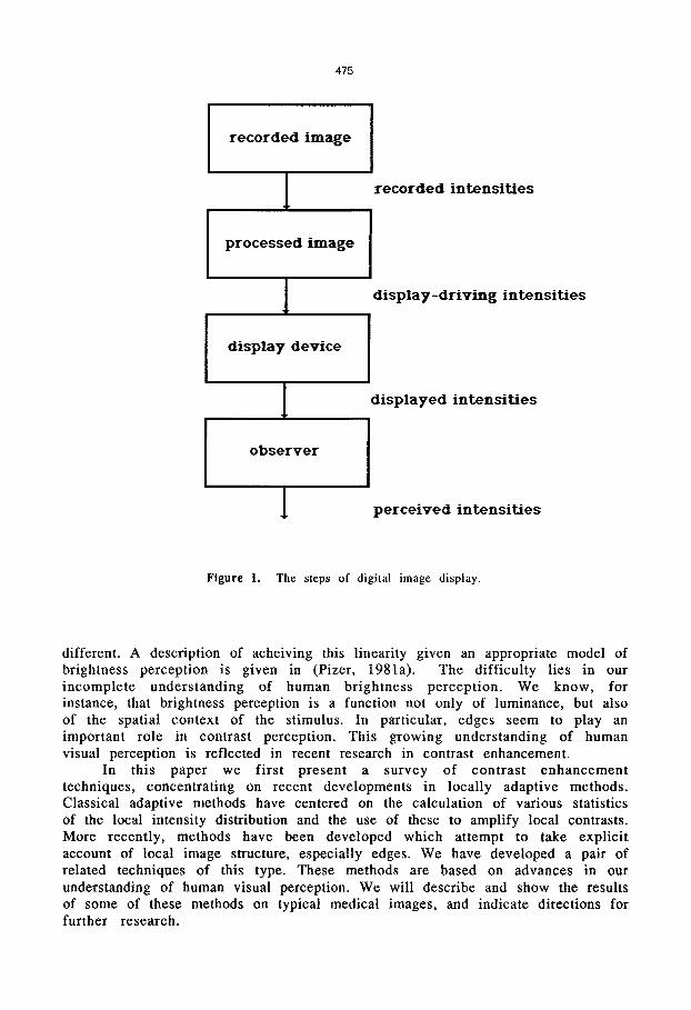
Display of a digital image is the process by which the array of recorded intensities is presented to the human observer as a light image. For medical images the overall goal of display is the detection, localization and qualitative characterization of anatomical objects represented by the intensity variations in the recorded data. A necessary step in this process is the rule by which the recorded intensities of the original image are mapped to the display-driving intensities of the display device (Figure 1). When performed explicitly, this step is called contrast enhancement. After this mapping is performed, the image undergoes further transformations, first within the display device and then in the human visual system. The effective design of contrast enhancement mappings requires a thorough understanding of these transformations. It would be ideal if the display device/observer combination could be made linear, so that equal differences in display-driving intensity would be perceived as equally
475
recorded image
1 recorded intensities
processed image
1 display-driving intensities
display device
1 displayed intensities
observer
1 perceived intensities
Figure 1. The steps of digital image display.
different. A description of acheiving this linearity given an appropriate model of brightness perception is given in (Pizer, 1981a). The difficulty lies in our incomplete understanding of human brightness perception. We know, for instance, that brightness perception is a function not only of luminance, but also of the spatial context of the stimulus. In particular, edges seem to play an important role in contrast perception. This growing understanding of human visual perception is reflected in recent research in contrast enhancement.
In this paper we first present a survey of contrast enhancement techniques, concentrating on recent developments in locally adaptive methods. Classical adaptive methods have centered on the calculation of various statistics of the local intensity distribution and the use of these to amplify local contrasts. More recently, methods have been developed which attempt to take explicit account of local image structure, especially edges. We have developed a pair of related techniques of this type. These methods are based on advances in our understanding of human visual perception. We will describe and show the results of some of these methods on typical medical images, and indicate directions for further research.

476
2. CONTRAST ENHANCEMENT APPROACHES IN THE LITERATURE
2.1. Global (Stationary) Contrast Enhancement
A global or stationary enhancement mapping is a grey-level transformation based solely on the intensity of each pixel:
I'(x,y) = f(I(x,y)).
The goal is to find a function which best utilizes the full range of display grey levels. Among these methods are intensity windowing, histogram equalization and histogram hyperbolization.
If we identify a subrange of image grey levels corresponding to features of interest this subrange can be expanded linearly to fill the full range of intensities. This technique is called intesity windowing. Pixels whose values fall outside the selected range are mapped to the minimum or maximum level. This technique is commonly used in the presentation of CT images. For example, in chest CT images, a "lung window" and a "mediastinum window" are chosen and applied, producing two images. These two images are then viewed side-by-side by the radiologist. This method has the advantage of being easily computed and in the case of CRT displays can be made interactive by an implementation which directly manipulates the lookup table of the display device. One difficulty is that objects occupying widely separated areas of the intensity range cannot be well presented in a single image. A perhaps more serious difficulty is that the perception of object boundary locations can depend critically on window selection.
Global histogram equalization is justified by the argument that for noisefree images it maximally transmits information as to scene intensity values (Cormack, 1981). In this method, a pixel's grey level is mapped to its rank in thegrey-level histogram of the entire image, scaled so that the output image fills the full range of intensities. The enhancement mapping is thus proportional to the cumulative distribution function of the image intensities. The result is that intensity values having greater numbers of pixels will be allocated a greater number of display levels, and the resulting histogram will be as flat as possible. There is however the compression of intensities that occur less frequently in the global histogram, which results in a loss of contrast in some areas of the image (Figure 2).
In histogram hyperbolization (Frei, 1977), a transformation of intensities is sought that results in a flat histogram of perceived intensities. Since the luminance response of the first stage of the human visual system is approximately logarithmic, it is argued that the shape of the histogram of displayed intensities should be approximately hyperbolic. Essentially, what is sought is histogram equalization after the effect of retinal processing. Thus a histogram-equalized image presented on a perceptually-linearized display should result in perceived brightnesses very close to those of a histogram-hyperbolized image displayed without linearization. This approach assumes a display device which is linear in its presentation of absolute luminances. Its main weakness is the strong dependence of our visual system on local context; brightness perception is not a simple function of absolute luminance.

477
Figure 2. Chest CT scan -- original (left) and processed using global histogram equalization (right).
2.2. Adaptive Contrast Enhancement
An adaptive contrast enhancement mapping is one in which the new intensity value for a pixel is calculated from its original value and some further information derived from local image properties:
I'(x,y) = f(I(x,y),DN(x,y)),
where N (x,y), the contextual region, is some spatial neighborhood of (x,y) in the image which includes the pixel of interest. For computational efficiency, it is most usual for N to be a square region centered on (x,y), but as we shall see, this need not be the case. Furthermore, the size and shape of the contextual region may itself vary throughout the image, based on either local statistics or local structural information.
A large number of adaptive contrast enhancement methods can be viewed as some variation of high-pass filtering. The oldest and most widely-used of these is unsharp masking. Known in its photographic form for at least sixty years, unsharp masking has also been applied to digital images. It can be defined as:
I'(x,y) = y(I(x,y) - I*N(x,y)) + I*N(x,y),
= y(I(x,y)) + (y-1 )(I*N(x,y)),
where I*N(x,y) is a weighted average of intensities over the contextual region
and y is a constant gain factor. The term (l(x,y) - I*N(x,y)) is a high-frequency
component sometimes referred to as the detail image. A y between 0 and 1 results in a smoothing of the image, a y greater than 1 results in emphasis of the high-frequency detail image. Unsharp masking has been applied and tested with varying success on a wide range of medical images (Loo et al., 1985, Sorenson, 1987). It has a noticeable sharpening effect on edges, but when the gain factor is

478
high enough to present very small details well, ringing artifacts are introduced across strong edges, and breakup of image objects can occur (Figure 3).
Unsharp masking can be generalized in a number of ways. One way is to replace the constant gain with separate weights for the high and low-frequency terms:
l'(x,y) = A(I(x,y) - I*N(x,y)) + B(I*N(x,y)).
An example of a method using this formulation is the statistical difference filter (Wallis, 1976, Harris, 1977). In this method, A is chosen so that the variance within the contextual region is made as nearly constant as possible, subject to a preset maximum gain to avoid over-enhancement of areas of very small standard deviation. B is a constant which serves to restore part of the lowfrequency component. The method has been shown to produce objectionable artifacts and finding suitable values for the weighting factors, the maximum gain and the window size proves difficult. A related method (Gordon et al., 1984, Dhawan et al., 1986) is based on the definition of a measure of the contrast at a pixel:
C = II(x,y) - I*(x,y)l I (I(x,y) + I*(x,y)),
which yields a value in the range 0-1. Enhancement consists of computing a new contrast C' and modifying the intensity of the pixel according to this new contrast as follows:
l'(x,y) = I*(x,y) (1 +C') I (1-C'), if l(x,y) > I*(x,y)
= I*(x,y) (1-C') I (l+C'), if I(x,y) < l*(x,y).
An advantage of this method is that arbitrary enhancement functions can be easily applied. Results depend critically on window size, however, and
Figure 3. Unsharp masking applied to the same image as in Figure 2 with two different gain factors -- y = 2 (left) and y = 5 (right).

479
blurring of edges is a problem. Moreover, the need to rescale the range of I' to the original range defeats the attempt to actually acheive the contrasts C' that are desired.
In adaptive histogram equalization (AHE) (Pizer, 198lb, Zimmerman, 1985), the histogram is calculated for the contextual region of a pixel, and the transformation is that which equalizes this local histogram. It development is logical both from the point of view of the information theory basis of global histogram equalization and from our knowledge of the human visual system. We are very sensitive to local relative contrasts, but insensitive to both absolute luminance and widely-separated relative luminances. ARE provides a single displayed image in which contrasts in widely-varying recorded intensities can be easily perceived. ARE has demonstrated its effectiveness in the display of images from a wide range of imaging modalities, including CT, MRI and Radiotherapy portal fims.
While providing excellent enhancement of the signal component of the image, ARE also enhances noise. In addition, shadowing of strong edges can occur in certain types of images. This latter problem has been analyzed and a suggested remedy given in the context of high resolution digital chest radiographs in (Rehm et al., 1990). In contrast-limited adaptive histogram equalization (CLARE) (Pizer et al., 1987), the enhancement calculation is modified by imposing a userspecified maximum on the the height of the local histogram, and thus on the slope of the cumulative histogram which defines the mapping. The enhancement is thereby reduced in very uniform areas of the image, which prevents overenhancement of noise and reduces the edge-shadowing effect of unlimited ARE (Figure 4). Several investigators have examined the possibility of using unsharp masking as a pre-processing step for CLARE (Blume, 1987, Rehm, 1990). More about this will be said later.
2.3. Methods Incorporating Structure
It has been recognized for some time that local image structure plays a crucial role in our perception of contrast, and enhancement techniques which incorporate local structural information are a logical result. There are two ways in which the above methods may be extended to include structural information.
'Figure 4. Images processed with AHE (left) and CLAHE (right).
480
One is to change the enhancement calculation itself; the other is to change the contextual region over which the calculations are done. Examples of each of these approaches are presented below.
An interesting extension of Gordon's technique (Beghdadi and Le Negrate, 1989) uses a modified contrast definition based on the detection of edges within the contextual region. In essence, the edge-grey-value of a pixel is its intensity weighted by the local edge strength at that pixel as computed by the Sobel, Laplacian or other edge operator. These edge-weighted values are then used in the calculation of the contrast measure as before. This method has an edgeenhancing effect when compared to the original formulation, but the exact effect depends strongly on the shape of the enhancement function and the choice of window size.
Several ways have been proposed of adjusting the contextual region over which the contrast enhancement is calculated. The idea is to adaptively restrict the local context to that which is relevant to perception of the pixel under consideration. Exactly what constitutes relevance in this sense is not entirely clear and depends to a large extent on the visual model we employ, but it is certain that perceived object boundaries are important in defining relevant context.
Gordon's method has also been extended by introducing -a limited set of different window sizes, and choosing the appropriate size on a pixel-by-pixel basis throughout the image. This is done by analysing how the contrast function changes across these different window sizes. As the window size increases, the contrast of a central object will increase until the inner window just covers the object. This window is then used to calculate the enhancement as before. Even by restricting the available windows to a few possible sizes, the computational burden is large. Moreover, the use of square windows limits the ability to adapt to actual image structure, and as is the case with all the variants of this method, the rescaling problem remains.
Kim and Yaroslavskii ( 1986) use analysis of the local histogram to define subsets of the contextual region, and the enhancement mappings are applied over these subsets rather than the entire region. One method uses only a portion of the histogram centered on the pixel of interest, the includes only those pixels of the contextual region which fall within a certain intensity range surrounding the value of that pixel. To the extent that nearness in the histogram or nearness in absolute intensity corresponds to closeness within the image, this has the effect of restricting the calculations to within object boundaries. These measures are, however of doubtful validity -- either method may result in a contextual region of disconnected pixels. Moreover, while the contextual region does indeed change across the image, the overall window size remains fixed. To be entirely satisfactory, an adaptive neighborhood must both have some mechanism for responding to object boundaries and also not be limited by an imposed overall shape. Two methods which meet both these criteria are now examined.
3. NEW METHODS INCORPORATING EDGE STRUCTURE
While many of the techniques discussed thus far are quite useful, they have as their weakness that context is determined in a way that is at best indirectly related to the grouping schemes used by visual systems. Thus we seek some way of determining the relevant context, and we can use recent advances

481
in understanding human VISIOn to help us. We will apply this idea in extending two of the most attractive methods from those discussed earlier: unsharp masking followed by AHE, and CLARE.
We know that an important early stage of human vision involves the calculation of an edge map. Best evidence seems to suggest that our perception of brightness is controlled by a sort of diffusion process in which the perceived contrast of these edges acts as an insulation strength that partially blocks the diffusion (Cohen and Grossberg, 1984).
Originally developed in the context of edge detection and the theory of scale-space, anisotropic diffusion (Perona and Malik, 1988) offers a way of producing truly variable contextual regions in a manner very like the above description of the calculations of the human visual system. Stationary blurring, which corresponds to the pixel averaging discussed above performed over unvarying contextual regions, can be modeled as a solution to the heatconduction or diffusion equation:
where c is a constant controlling the rate of diffusion. If we let c vary according to local image features, we obtain
It = div(c(x,y,t)L\1) = c(x,y,t)VI + L\ci\1,
the anisotropic diffusion equation. Here V is the gradient and L\ the Laplacian operator. If we knew for a given time t the location of object boundaries, we could set c to be 1 within those boundaries and 0 outside. In this way, we could entirely eliminate interaction across region boundaries. Since we do not know precisely the object boundaries, this is not possible; moreover this is not the result we want. We can chose c to be a monotonically decreasing function of the edge strength such that diffusion across edges is limited, but not eliminated. As an example, we use the function
c(IVII) = exp(-IVII2 /2K2),
where the parameter K can be viewed as selecting an edge strength up to which diffusion is allowed and beyond which the diffusion is strongly limited. Thus, diffusion using a large value of K blurs all but the very strongest edges, while selecting a very small K blocks diffusion except across very weak edges.
3.1 Unsharp Masking Using Edge-Limited Blurring
The context for unsharp masking is the blurring kerneL We have used an implementation of anisotropic diffusion (Gerig and de Moliner, 1989) in an variant of unsharp masking which can be viewed as the standard unsharp masking formula using a variable rather than fixed contextual region. The result is a relative increase in the blurring of low-contrast edges. This means, on amplification of the detail image, a relative increase in the enhancement of small details. A comparison of unsharp masking using isotropic Gaussian blurring and using edge-limited blurring is given in Figure 5. With edge-limited blurring,
482
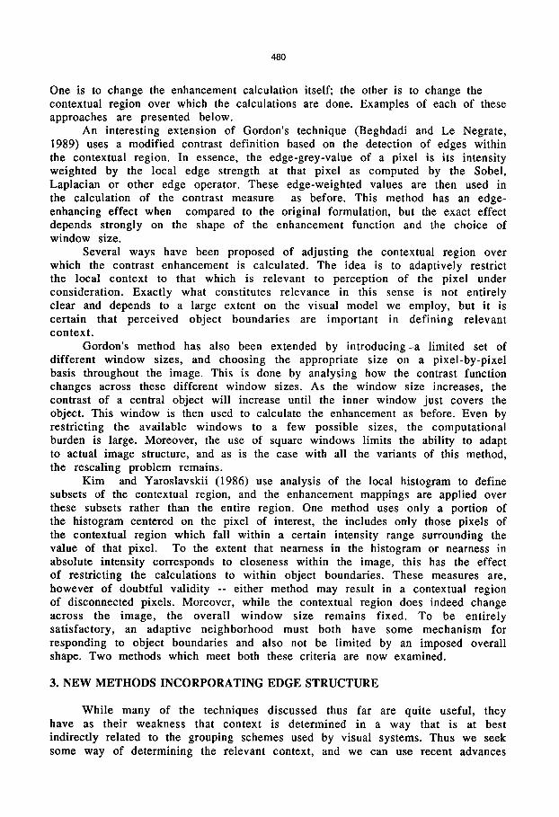
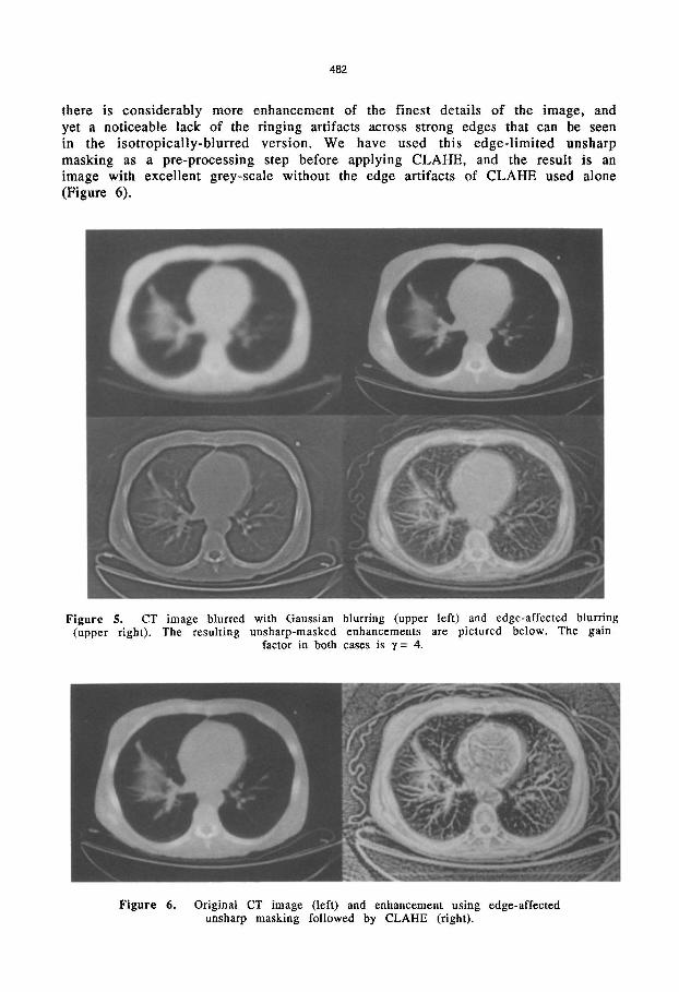
there is considerably more enhancement of the finest details of the image, and yet a noticeable lack of the ringing artifacts across strong edges that can be seen in the isotropically-blurred version. We have used this edge-limited unsharp masking as a pre-processing step before applying CLAHE, and the result is an image with excellent grey-scale without the edge artifacts of CLAHE used alone (Figure 6).
Figure S. CT image blurred with Gaussian blurring (upper left) and edge-affected blurring (upper right). The resulting unsharp-masked enhancements are pictured below. The gain
factor in both cases is y = 4.
Figure 6. Original CT image (left) and enhancement using edge-affected unsharp masking followed by CLARE (right).

483
3.2. Diffusive Histogram Equalization
For AHE, the context is all the pixels contributing to the histogram from which we calculate the output grey level. These contributions can have variable weights, so the weight values are also part of the definition of the contextual region. Here, too, we have used edge-affected blurring to achieve a variableneighborhood adaptive histogram equalization algorithm we call diffusive histogram equalization (DHE). In this method, we calculate the effect of all pixels of a given grey-level on the histograms of all pixels in the image in a single step. This is done by applying edge-limited blurring to an image made by placing a positive starting value in each pixel of the current grey level and zeros eveywhere else. The edges used to limit the diffusion are the edges of the original image. After diffusing, the histogram value for each pixel is contained in this image. Thus the influence of one pixel on another is limited by intervening edges. The contextual region is truly variable, not only in overall shape (potentially the entire image could be the contextual region) but also in the variable weighting of each pixel within the region. Figure 7 shows the contextual region for one pixel in the chest CT image, and the result of applying DHE to that image.
4. DISCUSSION
We have applied the two methods discussed above to a number of images from different modalities with encouraging results. We expect to conduct formal analysis of these methods in the near future. It is important in designing tests for evaluation of contrast enhancement methods to keep in mind the particular task being performed. Enhancement methods are often compared on the basis of their ability to increase detectablity of either standard test patterns or very subtle artificially-produced lesions imposed on real medical images. This detection task is certainly important for many imaging modalities, but may not be the most important in every case. Boundary localization, shape characterization and comparison of absolute luminances are some other viewing tasks which may be
Figure 7. Diffusive histogram equalization -- edge-affected contextual region for one pixel superimposed on original image (left) and final enhanced image (right).

484
of importance. Diffusive histogram equalization is a particularly aggressive enhancement method, yielding images exceptionally rich in structural detail. We expect it might not be particularly well-suited for lesion detection. By comparison, the images produced by edge-affected unsharp masking followed by CLAHE are perhaps more suited to making the qualitative judgements required by lesion detection or appreciation of gross shape features. The ultimate test of any contrast enhancement method designed for use with medical images is whether or not it provides increased diagnostic accuracy or efficiency in a clinical setting. In choosing among contrast enhancement methods, we must generally be content with some approximation to this test. Moreover, it may take a considerable amount of training for the clinician to effectively use images processed by means of these enhancements.
Another task-related matter is how noise is treated. Noise is unwanted image detail, so its definition depends on what detail is wanted, as well as what is known about the properties of the image-formation process. With such a decision, the contrast enhancement must be chosen not simply to convey signal differences, but to convey them relative to noise. This problem is acute for DHE, which tends to bring out contrast at all image levels. This can be controlled to some extent by allowing the diffusion to continue relatively long, effectively increasing the size of the contextual region. It would certainly be possible to apply a contrast-limitation factor, as in CLARE, which would result in images with less apparent noise.
We have discussed these techniques without paying particular attention to the cost of computing them. The anisotropic diffusion calculations are very expensive, both in processor time and memory requirements. Certainly, for a method to be usable in the real world, the implementation must be relatively fast, even real-time. Many of the methods discussed above, particularly the adaptive ones, are too computationally expensive to be clinically valuable. Many can be speeded up by using recently developed image processing computers or other specialized hardware.
5. CONCLUSION
Digital image processing is becoming more and more important in medical imaging as we move from film-based to computer-based imaging systems. An important part of this is the effective display of these digital images, and contrast enhancement is an essential step of the display process. We have tried to give an indication of the importance of an accurate visual model in the development of these techniques, and have outlined the development of two contrast enhancement methods, edge-affected unsharp masking followed by AHE and diffusive histogram equalization. Both of these methods are based on our knowledge of how the visual system determines context. As better models of human visual perception are formulated, we will be able to design contrast enhancement methods which more effectively complement our perceptual capabilities.

485
Acknowledgements
We wish to thank Dr. Julian Rosenman, leader of the project entitled "Adaptive Histogram Equalization for Radiotherapy" within the research program in Medical Image Presentation at The University of North Carolina at Chapel Hill. This work is supported in part by NIH Grant #POl CA47982.
References
Beghdadi A and Le Negrate A (1989). Contrast enhancement technique based on local detection of edges. Computer Vision, Graphics, and Image Processing ~162-17 4.
Blume H and Kamiya K (1987). Auto-ranging and normalization versus histogram modifications for automatic image processing of digital radiographs. Proc. S.P.LE. 1..fU Medical Imaging: 371-386.
Cormack J and Hutton BF (1981). Quantitation and optimization of digitized scintigraphic display characteristics using information theory. Medical Image Processing: Proceedings of the Vllth International Meeting on Information Processing in Medical Imaging, Stanford University: 240-263.
Dhawan AT, Buelloni G and Gordon R (1986). Enhancement of mammographic features by optimal neighborhood image processing. IEEE Transactions on Medical Imaging M I- 5 No. 1: 8-15.
Frei W (1977). Image enhancement by histogram hyperbolization. Computer Graphics and Image Processing .6.: 286-294.
Gerig, G and de Moliner R (1989). Personal communication. Gordon R (1986). Enhancement of mammographic features by optimal neighborhood image
processing. IEEE Transactions on Medical Imaging MI-5 No 1: 8-15. Grossberg S (1984). Neural dynamics of brightness perception: features, boundaries,
diffusion, and resonance. Perception and Psychophysics .16..._(5}: 428-456. Harris, Jr. JL (1977). Constant variance enhancement: a digital processing technique.
Applied Optics .1..6.: 1268-1271. Kim V and Yaroslavskii L (1986). Rank algorithms for picture processing. Computer Vision,
Graphics, and Image Processing ll: 234-258. Loo LD, Doi K and Metz C (1985). Investigation of basic imaging properties in digital
radiography 4. Effect of unsharp masking on the delectability of simple patterns. Medical Physics ll: 209-214.
Perona P and Malik J (1988). Scale-space and edge detection using anisotropic diffusion. Report UCB/CSD 88/483, Computer Science Division University of California, Berkeley, CA.
Pizer SM (1981a). Intensity mappings to linearize display devices. Computer Graphics and Image Processing 11: 262-268.
Pizer SM (1981b). An automatic intensity mapping for the display of CT scans and other images. Medical Image Processing: Proceedings of the VIIth International Meeting on Information Processing in Medical Imaging, Stanford University: 276-309.
Pizer SM, Amburn EP, Austin JD, Cromartie R, Geselowitz A, ter Haar Romeny B, Zimmerman JB and Zuiderveld K (1987). Adaptive histogram equalization and its variations. Computer Vision, Graphics, and Image Processing .3.2.: 355-368.
Rehm K, Seely GW, Dallas WJ, Ovitt TW and Seeger JF (1990). Design and testing of artifactsuppressed adaptive histogram equalization: A contrast-enhancement technique for the display of digital chest radiographs. Journal of Thoracic Imaging 5. No 1: 85-91.
Sorenson J (1987). Effects of improved contrast on lung-nodule detection A clinical ROC study. Investigative Radiology ll: 772-780.
Wallis R (1976). An approach to the space variant restoration and enhancement of images. Proceedings of the Symposium on Current Mathematical Problems in Image Science, Monterey, California, Naval Postgraduate School.
Zimmerman JB (1985). Effectiveness of Adaptive Contrast Enhancement. Ph.D. dissertation, Department of Computer Science, The University of North Carolina at Chapel.