ectopic pregnancy michael t. fitch, m.d., ph.d. … · · 2017-08-072009-01-15 · 2. understand...
TRANSCRIPT

Ectopic Pregnancy Simulation Case Fitch MT and Fisher CJ
Page 1
ECTOPIC PREGNANCY A case for high-fidelity simulation in emergency medicine
Michael T. Fitch, M.D., Ph.D. Assistant Professor
Director, Emergency Department Simulation Program
Christen J. Fisher, M.D. Clinical Instructor
Department of Emergency Medicine Wake Forest University
Medical Center Boulevard, Winston-Salem, North Carolina 27157 Phone: 336-716-4626 Fax: 336-716-5438
http://www.EmergencySimulation.com/
This Resource Successfully Peer Reviewed and Accepted with Acclamation by MedEdPORTAL on 1/15/2009. MedEdPORTAL Publication Number: 1673 Alterations to this Resource Created After This Date Have Not Been Reviewed By MedEdPORTAL. Subsequent Revisions: None.
ABSTRACT
This educational resource provides the information and materials for a high-fidelity simulation case suitable for resident physicians in emergency medicine. This case is currently in use at our institution for upper-level emergency medicine residents completing required educational time in our Emergency Department Simulation Program. This case has been used for the past three years in our program and has recently been edited and expanded to its existing form.
This high-fidelity patient simulation case involves a young woman who presents with syncope and abdominal pain. A pregnancy test in the emergency department is positive, which is a concerning finding for this patient who has had a tubal ligation. She is hypotensive and tachycardic with a bedside ultrasound significant for intraabdominal free fluid and a possible left sided ectopic pregnancy. Appropriate treatment includes fluid resuscitation, obstetrical consultation, blood transfusion if necessary, and disposition to the operating room for a presumed ruptured ectopic pregnancy. Debriefing materials are provided to illustrate and stimulate discussion of the important concepts for diagnosing and treating patients with ectopic pregnancy.

Ectopic Pregnancy Simulation Case Fitch MT and Fisher CJ
Page 2
I. TITLE OF CASE: Ectopic Pregnancy II. TARGET AUDIENCE: Resident physicians or medical students III. LEARNING OBJECTIVES: A. Primary Learning Objectives
1. Demonstrate an appropriate initial approach to a patient with hypotension and tachycardia
2. Identify features concerning for a possible ectopic pregnancy 3. Demonstrate appropriate management of ruptured ectopic pregnancy B. Secondary Goals 1. List the risk factors for ectopic pregnancy
2. Understand benefits and limitations of ultrasound in the diagnosis of an ectopic pregnancy 3. Describe non-surgical management options for ectopic pregnancy
C. Critical Actions 1. Obtain appropriate history of present illness from patient. 2. Identify unstable vital signs. 3. Initiate IV fluid resuscitation. 4. Order pregnancy testing. 5. Order type and cross for blood products. 6. Obstetrical consultation (before or after bedside ultrasound) 7. Recommend operative therapy for presumed ruptured ectopic pregnancy. IV. ENVIRONMENT:
A. Lab set up – Emergency Department bed #18 in simulation laboratory. B. Mannequin set up – The mannequin will be positioned in the bed in a head-raised
position, with street clothes on. At the beginning of the simulation, there are no monitor leads on the patient, no IV has been placed, and patient is not on oxygen.
C. Props – Available for use will be a heart monitor with leads, blood pressure cuff, and
pulse oximeter. Supplemental oxygen by nasal cannula and non-rebreather will be available. There will be a medicine cart or tray with a full complement of vasoactive agents, ACLS medications, medicines necessary for sedation, rapid sequence intubation, and analgesia. In addition, a fully stocked code cart with defibrillator will be available for use, along with a selection of direct laryngoscopes and intubation supplies. Bedside ultrasound is available if requested by the physician.

Ectopic Pregnancy Simulation Case Fitch MT and Fisher CJ
Page 3
D. Audiovisual – (See Appendix A) Available for review when asked for by participants will be an electrocardiogram showing sinus tachycardia, and images from a bedside ultrasound that demonstrate intraabdominal free fluid. Transvaginal ultrasound images (if obtained) demonstrate no intrauterine pregnancy and a suspicious left adnexal anechoic mass concerning for etopic pregnancy. A positive urine pregnancy test, a quantitative serum beta-hCG, maternal blood type, and CBC results are available if requested.
E. Distractors – Distractors can be added at the director’s discretion. However, as
written, this is the only patient that needs to be cared for and there will be no extraneous inputs to distract the participants’ attention from the case at hand.
V. ACTORS:
A. Roles can vary depending on the number of participants in the simulation session and on potential actor availability. This case can be implemented using the minimum of one participant and one facilitator/operator who can provide oral feedback via overhead audio and play additional roles via voice only. Realism can be enhanced by using physical actors, such as nursing staff members, but these are not required to successfully implement this case.
B. Roles may be played by resident or faculty physicians, nurses, or medical students. C. Actions for the roles will be as follows:
a. Primary physician - The main scenario participant will act as the primary physician and do the primary evaluation of the patient to include obtaining a history, conducting a physical exam, and ordering any desired ancillary testing. The primary physician can perform any needed procedures, or can delegate these to other physicians.
b. Secondary physicians - Other participants in the scenario will serve as collaborators, assistants for any necessary procedures, and consultants.
c. Nursing staff - The role of the nurse will be to administer medications, verify orders, and perform other tasks as directed by the physicians. The nurse can also make observations as needed to stimulate case progression.
VI. CASE NARRATIVE:
A. Scenario Background
a. Chief Complaint: “Passed out” / Abdominal pain b. Triage Nursing Note: Patient is a 32 year old previously healthy female, who
came to the ED today because she had a syncopal episode while shopping for groceries at a local store. Felt “dizzy” prior to passing out. She has had intermittent abdominal pain for past one week.

Ectopic Pregnancy Simulation Case Fitch MT and Fisher CJ
Page 4
c. Vital Signs: Heart rate 119; Blood pressure 91/56; Respiratory rate 22; Pulse oximetry 99% on room air; Temperature 99.1 degrees Fahrenheit
d. Past Medical History: (Only give if requested by physician) Nephrolithiasis, right knee meniscus repair 2 years ago, seasonal allergies. G3P3 with bilateral tubal ligation 2 years ago for desired sterility. If specifically asked, will report no history of infertility treatments, PID, STDs, or ectopic pregnancy.
e. Medications and Allergies: Claritin and ibuprofen as needed. No allergies. f. Family and Social History: Lives with husband and three young girls ages 7,
5, and 3. Works part time. No tobacco or illicit drug use. Occasional wine with dinner.
B. Initial Scenario Conditions
a. History given by patient: She was shopping at a local grocery store in the dairy aisle when she suddenly felt hot and dizzy. She does not remember any further events. She awoke to EMS providers caring for her on the floor of the store, and was transported to the ED via ambulance.
b. Circumstances at symptom onset: She denies vertigo, palpitations, chest pain, shortness of breath, nausea, or vomiting.
c. Secondary complaint: Patient reports low abdominal pain intermittently over the past one week. Was seen several days ago at an outside facility for this complaint, where a workup was negative for a kidney stone per her report. Today she feels the pain is diffusely throughout lower abdomen, which is somewhat different than the right sided sharp and stabbing pain that took her to the outside ED for evaluation. Pain is dull and radiates to her right shoulder. Pain is worse with movement and there are no alleviating factors. Pain is 9/10, and patient reports no nausea, vomiting, or diarrhea. No fever, No dysuria, hematuria, or urinary urgency. Last menstrual period was 6 weeks ago, and she reports a history of “irregular” periods.
d. Initial Exam: i. General: Patient is an obese female. She is awake, alert, and
appropriately oriented but appears uncomfortable, pale, and diaphoretic.
ii. Head, Ears, Eyes, Nose, Throat: There is no evidence of head trauma. Pupils are equal, round, and reactive from 6mm to 4mm. Extraocular movements are fully intact. Ears are normal, there is no discharge, the tympanic membranes are clear, with good light reflex, no evidence of perforation. No mucus membrane rashes, dryness, or swelling.
iii. Skin: Pale and clammy. Mild diaphoresis. No rashes, petechia, or purpura.
iv. Cardiovascular: Tachycardia with regular rhythm. Palpable but weak radial pulses. No murmurs, rubs, or gallops
v. Lungs: Clear to auscultation without wheezes, crackles, or rales. Equal breath sounds bilaterally.

Ectopic Pregnancy Simulation Case Fitch MT and Fisher CJ
Page 5
vi. Abdomen: Significant diffuse tenderness, greatest in bilateral lower quadrants right > left. Involuntary guarding is present. Does have rebound tenderness when palpating lower abdomen only.
vii. Genitourinary: Normal external female genitalia. Scant blood at cervical os, which is closed. No cervical motion tenderness. Bilateral adnexal and suprapubic tenderness. No masses palpated.
viii. Back: No tenderness to palpation, no costovertebral angle tenderness. ix. Extremities: No muscle tenderness with full range of motion in all
extremities. No swelling or edema. Symmetric extremities. x. Neurological: Alert and oriented with normal mental status. Pupils
equal, round, and reactive to light and accommodation. Cranial nerves II-XII intact. 2+ deep tendon reflexes in all extremities. No sensory or motor deficits. Normal finger to nose pointing, negative Romberg, and normal gait.
e. Physiology: (appears when placed patient is placed on monitor)
i. Heart rate begins at 120, gradually increases to 140 until 2 liters of normal saline are administered IV
ii. Blood pressure is 90/50, gradually decreases to 80/35 until 2 liters of normal saline are administered IV. BP will remain in low 90’s/50’s after IV fluids are given.
iii. Pulse oximetry is 98% on room air, 100% if patient is on oxygen iv. Respiratory rate is 18-20 breaths/minute, will gradually increase to
respiratory rate of 30 as case progresses.
C. Scenario Branch Points
a. Changes in patient condition: The patient’s condition will gradually worsen (increased tachycardia and hypotension) until IV fluids are administered via large bore peripheral IV’s. Blood products will take a significant amount of time to type and cross, and when available administration of these alone will temporarily improve the patient but she will complain of worsening abdominal pain throughout transfusion.
b. Request for old records: Records from outside ED visit several days ago are
unavailable.
c. Differential diagnosis: The physician managing this case may consider acute appendicitis, nephrolithiasis, ovaria torsion, PID, tuboovarian abscess, or threatened abortion in addition to ectopic pregnancy in this case.
d. Intravenous fluid administration: Blood pressure will remain in 90’s/50’s and
not decline if IV fluids are given as boluses, tachycardia will improve to 110’s but will not resolve during case.

Ectopic Pregnancy Simulation Case Fitch MT and Fisher CJ
Page 6
e. Blood transfusion: Blood transfusion will improve tachycardia and hypotension somewhat, but patient will continue to complain of worsening abdominal pain.
f. Laboratory studies: Patient has a positive urine pregnancy test, which will
hopefully prompt the physician to consider ectopic pregnancy as a possible diagnosis. If a type and cross for blood products was not initially ordered, this should be added on when the diagnosis of ectopic pregnancy is entertained.
g. Bedside ultrasound: The positive pregnancy test will hopefully prompt the
physician to consider an ultrasound or to move directly to OB consultation. The ultrasound will demonstrate intraabdominal free fluid. Note that if this test is ordered to be done in the radiology suite, the patient will abruptly worsen while gone to radiology.
h. Consultation of obstetrician: Initial conversation with consultant will reveal
that OB does not appreciate the potential severity of this patient’s illness. She will downplay the blood pressure (that can be a normal blood pressure for a young healthy woman) and attribute the tachycardia to pain (why don’t you give her some IV pain medicine and see how she does?). She will recommend a transvaginal ultrasound to be done in the radiology suite and asks that she be called after it is done. The hope is that the physician managing the case will insist that the OB come to see the patient immediately for a suspected ruptured ectopic pregnancy.
i. Radiology suite ultrasound: The patient should not be sent to radiology
ultrasound with her unstable vital signs. However, if the decision is made to allow this to happen, patient will significantly worsen and become abruptly more tachycardic and hypotensive.
D. Scenario Outcome
a. Ultimately the obstetrical consultant must be involved in this patient with a suspected ruptured ectopic pregnancy. The patient remains tachycardic and hypotensive despite aggressive therapy, and operative management should be recommended.
VII. INSTRUCTOR NOTES:
A. Scenario flow – Instructors can directly influence the flow of the scenario by
providing the initial patient history via both nursing report and patient verbal responses, since the patient is awake and talking and a source of information.

Ectopic Pregnancy Simulation Case Fitch MT and Fisher CJ
Page 7
a. Urine pregnancy testing - One key aspect of the case flow is requesting a pregnancy test, either urine or serum. If this is not done, it is very unlikely the physician managing the case will successfully navigate this patient encounter.
b. Initial treatment – The physician managing this case should recognize the vital sign abnormalities and take appropriate steps to address them. Ideally, this will include 2 large bore IV lines, normal saline fluid boluses, and type and cross of blood products.
B. Information for actors – This case can easily be presented without formal “actors”
and by using other participants to serve as the nursing staff or other collaborating physicians. Any specific actors used outside of participants should be briefed about the critical actions and anticipated flow of the case ahead of time.
C. Scenario programming – The settings for a high-fidelity patient simulator are fairly
straightforward for this scenario and do not require specific programming. Our program typically presents this scenario with the initial vital signs as presented above, with the two major branch points to be acute clinical worsening (with increased tachycardia and hypotension) or temporary stabilization of the abnormalities without completely correcting them.
VII. DEBRIEFING PLAN:
A. Method of debriefing – A post-case debriefing conference can be completed
immediately following the end of the scenario. Consider including some of the following elements:
a. Open-ended questions by facilitator – Consider beginning the session with a question to the primary participant about how they felt the scenario went. This often leads to extensive participant-led discussion that will touch on many of the major issues in the case. Invite any secondary participants and/or observers to comment about how the case unfolded.
b. Brief didactic review – Potential materials for review after the scenario have been provided in Appendix B.
c. Formal participant evaluation – A potential form for evaluation is provided in Appendix C.
B. Actual debriefing materials – See Appendix B for debriefing materials. C. Rules for the debriefing – You may find that an informal discussion format for the
initial portion of the debriefing leads to an open discussion of aspects of the case management that were good, and those areas where improvement can be made. Encourage your participants to discuss the case management decisions in a non-judgemental way. When this case is used for experiential learning (without formal participant evaluation on the specific case) such discussions may be more productive as compared to situations where formal feedback is anticipated. We have found that placing an emphasis on the learning that occurs from experiencing a case like this is

Ectopic Pregnancy Simulation Case Fitch MT and Fisher CJ
Page 8
very effective, rather than focusing on the “correct answer” or whether the specific management decisions turned out to be the most appropriate.
D. Questions to facilitate the debriefing –
a. How do you decide when a patient’s vital signs are stable or unstable? b. What is the best way to deal with a consultant on the telephone who is
reluctant to come to see a patient as rapidly as you would like? c. What is the differential for abdominal pain, vaginal bleeding or both? d. What are risk factors for ectopic pregnancy? e. What is the role of transvaginal ultrasound in the stable and unstable patient? f. How would you manage this case differently if you practiced at an institution
without the immediate availability of bedside ultrasound? g. In the stable patient, what are treatment options for an ectopic pregnancy?
IX. PILOT TESTING AND LESSONS LEARNED:
A. Number of participants – This scenario has been presented for small groups of 3-4
participants, and has been field tested approximately 6 times over the past 3 years. All participants have been PGY1, PGY2, and PGY3 emergency medicine residents.
B. Performance expectations – Our experience to date has been that experienced
emergency medicine residents, typically PGY3 and late-PGY2 levels of training, are able to successfully negotiate this case without significant difficulty. As our program has significantly increased our training in bedside ultrasound over the past several years, many of our residents go immediately to ultrasound in this case to look for intraabdominal free fluid. This may not be the case with learners who are not as familiar with the uses of bedside ultrasound.
C. Anticipated management mistakes –
a. Failure to send a pregnancy test – As discussed above, it is very unlikely that this case will be successfully managed if a pregnancy test is not ordered. It is possible that it could be correctly managed under the assumption of ectopic pregnancy with the patient’s history of tubal ligation and late menstrual period.
b. Inadequate fluid resuscitation – Immediate treatment with IV fluid bolus is an important goal of the case after the physician recognizes the hypotension and tachycardia. Delays in initiating treatment will lead to worsening patient status and increased difficulty in stabilizing vital signs even temporarily.
c. Patient is sent out of the department for testing – Despite the suggestion from the OB consultant to obtain a transvaginal ultrasound in the radiology suite, this patient should not leave the department for testing – and if she does her clinical condition will precipitously decline.
D. Evaluation form for participants – See Appendix C

Ectopic Pregnancy Simulation Case Fitch MT and Fisher CJ
Page 9
X. AUTHORS AND THEIR AFFILIATIONS:
Michael T. Fitch, M.D., Ph.D. Assistant Professor Director, Emergency Department Simulation Program
Christen J. Fisher, M.D. Clinical Instructor Department of Emergency Medicine Wake Forest University School of Medicine Medical Center Boulevard, Winston-Salem, North Carolina 27157
XI. REFERENCES:
• Murray H, Baakdah H, Bardell, T, Tulandi T. Diagnosis and treatment of ectopic pregnancy. CMAJ 2005 October 173 (8).
• Valley VT, Mateer JR, Aiman EJ, Thoma ME, Phelan MB. Serum progesterone and endovaginal sonography by emergency physicians in the evaluation of ectopic pregnancy. Acad Emerg Med 1998;5: 309-13.
• Pisarska MD, Carson SA, Buster JE. Ectopic pregnancy. Lancet 1998;351:1115-20.
• Tenore JL, et al. Ectopic Pregnancy. Am Fam Physician. 2000 Feb 15;61(4):1080-8.
• Tay JI, et al. Ectopic Pregnancy. BMJ. 2000 April; 320: 916-919.
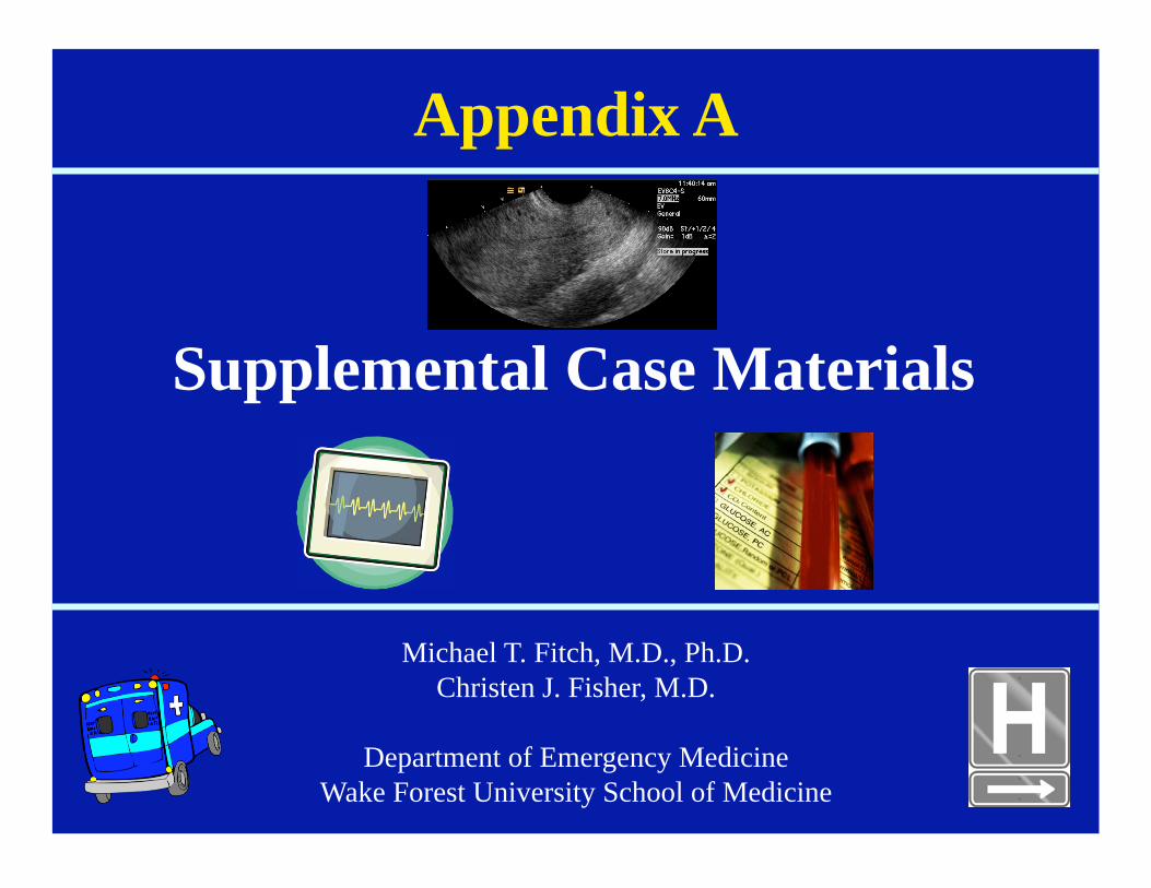
Appendix A
Michael T. Fitch, M.D., Ph.D.
Christen J. Fisher, M.D.
Department of Emergency Medicine
Wake Forest University School of Medicine
Supplemental Case Materials

Patient ECG

Abdominal Ultrasound – Right Upper Quadrant

Abdominal Ultrasound – Left Upper Quadrant

Transvaginal Ultrasound – Left Ovary

Transvaginal Ultrasound – Right Ovary

Transvaginal Ultrasound – Uterus Longitudinal View

Transvaginal Ultrasound – Uterus Transverse View
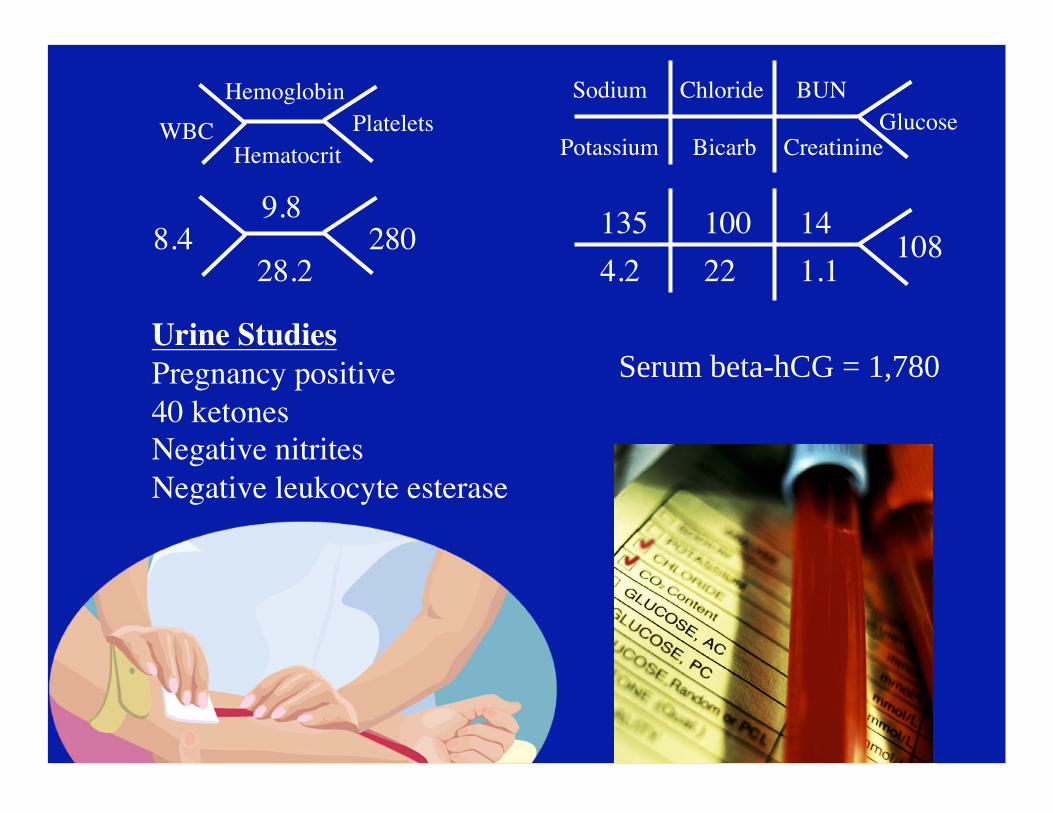
8.49.8
28028.2
135
4.2
100 14
22 1.1108
Urine StudiesPregnancy positive40 ketonesNegative nitritesNegative leukocyte esterase
WBC
Hemoglobin
HematocritPlatelets
Sodium
PotassiumGlucose
Chloride
Bicarb
BUN
Creatinine
Serum beta-hCG = 1,780

Appendix B
Michael T. Fitch, M.D., Ph.D.
Christen J. Fisher, M.D.
Department of Emergency Medicine
Wake Forest University School of Medicine
Case Debriefing Materials

Ectopic Pregnancy
Michael T. Fitch, M.D., Ph.D.
Christen J. Fisher, M.D.
Department of Emergency Medicine
Wake Forest University School of Medicine

• Incidence in general population is about 2% - has steadily increased over past 30 years
• Incidence in women presenting to the ED with first trimester bleeding or pain is 6-16%
• Usually diagnosed at 6-10 weeks
• Between 40 and 50 percent of ectopic pregnancies are misdiagnosed at the initial visit to an emergency department
• Ruptured ectopic pregnancy is the leading cause of maternal mortality in the 1st trimester and accounts for 10-15 percent of all maternal deaths
Ectopic Pregnancy

Possible Ectopic Pregnancy
• Ectopic pregnancy
• Ruptured ectopic pregnancy
• Appendicitis
• Cervicitis
• Pelvic inflammatory disease
• Tuboovarian abscess
• Urinary tract infection
• Pyelonephritis
Differential Diagnosis of Pelvic Pain

Risk Factors for Ectopic Pregnancy
• History of pelvic inflammatory disease, chlamydial or gonococcal cervicitis (7-fold increase in risk)
• Fallopian tube surgery, including tubal ligation
• Previous ectopic pregnancy
• Intrauterine device use
• Assisted reproductive techniques (3-fold increase in risk)
• History of infertility
• Increased age

Pathophysiology
• Ectopic pregnancy can be caused by:
– Mechanical alterations in tubal transport mechanism
– Functional and hormonal factors that alter the fertilized ovum
• Tubal rupture is most often spontaneous – May also be precipitated by trauma (i.e., coitus, bimanual exam)
• Typically occurs in early pregnancy – Usually presents at 6-10 weeks
– Can present as late as 16 weeks

Clinical Features
• “Classic” findings ectopic pregnancy: – Abdominal pain (90%)
– Vaginal bleeding (80%)
– Amenorrhea (70%)
– Adnexal tenderness (54%)
– Previous ectopic pregnancy (11%)
• Abdominal pain is variable – May be sudden, lateralized, and extreme
– May be minor and diffuse
– Referred pain to shoulder / upper abdomen if hemoperitoneum
• Vaginal bleeding is usually light

• Presenting vital signs may be normal – Poor correlation of hemoperitoneum with vital signs
– Relative bradycardia may occur with rupture and hemorrhage
• Abdominal exam highly variable – Local or diffuse tenderness
– Peritoneal signs may or may not be present
• Pelvic exam can be normal or can find: – Cervical motion tenderness
– Adnexal tenderness
– Palpable mass
Physical Findings

Diagnostic Studies
• Urine pregnancy
• Serum -hCG
• Hemoglobin
• Blood for type and crossmatch – If unstable and/or anticipate transfusion
– Rhogam as indicated for Rh negative mothers
• Ultrasound – Bedside abdominal ultrasound for hemoperitoneum
– Bedside transvaginal ultrasound for ectopic
– Formal ultrasound an option if patient is stable

Unstable vital signs or concerning physical exam
• Resuscitation in the Emergency Department
– Monitor
– Two large bore IV lines or other intravascular access
– IV fluid boluses
– Type and cross for possible blood transfusion
• Immediate consultation to Ob-Gyn specialist
– Hypotension
– Tachycardia
– Peritoneal signs on abdominal exam
– Free fluid on ultrasound examination
ED Evaluation and Management

ED Evaluation and Management
• Transvaginal Ultrasound plus serum hCG – 95-100% sensitivity and specificity
• ‘Discriminatory Zone’ - level of serum ß-hCG above
which an IUP can be consistently visualized
– Transabdominal sonography 6,500 mIU per mL
– Transvaginal sonography 1,000 to 1,500 mIU per mL
– Ectopic should be suspected if the transvaginal ultrasound does not
detect an IUP when ß-hCG level is > 1,000 to 1,500 mIU per mL
– Ectopic pregnancy can accompany a “psuedo-gestational sac” in the
uterus; therefore an intrauterine sac alone cannot confirm IUP
Stable vital signs and no peritonitis or free fluid

Transvaginal Ultrasound
• Intrauterine pregnancy confirmed on ultrasound – Unlikely ectopic pregnancy
– Heterotopic pregnancy occurs in 0.003% of general population and up to 3% in those with assisted fertility
– Abnormal pelvic free fluid may suggest ruptured heterotopic
• No intrauterine pregnancy on ultrasound + adnexal mass – Concerning for ectopic pregnancy
– Pelvic free fluid concerning for ruptured ectopic pregnancy
– OB-Gyn consultation
• No intrauterine pregnancy without an adnexal mass – Termed “Indeterminant result”
– Management decisions may be based on quantitative -hCG results
Categorizing transvaginal ultrasound findings

Transvaginal Ultrasound
• Diagnostic algorithm for indeterminant ultrasound– Stable vital signs
– No abnormal pelvic or intraabdominal free fluid on ultrasound
• Quantitative -hCG < 1,000 - 1,500– Early intrauterine pregnancy vs. early ectopic pregnancy
– Arrange repeat -hCG and OB-Gyn followup in two daysIntrauterine pregnancy should show 66% increase in -hCGEctopic pregnancy would show a slower increase or plateau in -hCG
• Quantitative -hCG > 1,000 - 1,500– Highly suspicious for ectopic pregnancy– Consultation with OB-Gyn specialist– May need D&C (dilation and curettage), laparotomy, or methotrexate
Management of Indeterminant Ultrasound Result

Ectopic Pregnancy
• Consultation with OB-Gyn specialist in the ED
• Operative treatment – Laparoscopy versus laparotomy
– Dilation and Currettage
• Medical treatment with methotrexate therapy – ß-hCG levels higher than 1,500 have a higher risk of treatment failure
– ß-hCG levels higher than 5,000 usually do not respond to MTX therapy
– Criteria for MTX include: • hemodynamic stability
• confirmation of ectopic pregnancy by ultrasound examination
• significant risk associated with general anesthesia
• patient compliance
• small size of ectopic mass and lack of fetal cardiac activity
Treatment Options for Diagnosed Ectopic

References
• Murray H, Baakdah H, Bardell, T, Tulandi T. Diagnosis and
treatment of ectopic pregnancy. CMAJ 2005 October 173 (8).
• Valley VT, Mateer JR, Aiman EJ, Thoma ME, Phelan MB. Serum
progesterone and endovaginal sonography by emergency
physicians in the evaluation of ectopic pregnancy. Acad Emerg
Med 1998; 5: 309-13.
• Pisarska MD, Carson SA, Buster JE. Ectopic pregnancy. Lancet
1998; 351:1115-20.
• Tenore JL, et al. Ectopic Pregnancy. Am Fam Physician. 2000 Feb
15; 61(4):1080-8.
• Tay JI, et al. Ectopic Pregnancy. BMJ. 2000 April; 320: 916-919.

Ectopic Pregnancy
Michael T. Fitch, M.D., Ph.D.
Christen J. Fisher, M.D.
Department of Emergency Medicine
Wake Forest University School of Medicine

Appendix C
Ectopic Pregnancy Simulation Evaluation
Participant Name ___________________________________ Date _____________________ Action/Objective Un Sat Ex N/A Core
Competency Obtain appropriate history of present illness from patient.
PR, MK, PC
Identify unstable vital signs. PC
Initiate IV fluid resuscitation. MK, PC
Order pregnancy testing. PC
Order type and cross for blood products. PC
Obstetrical consultation (before or after bedside ultrasound)
SBP, PC
Recommend operative therapy to consultant for presumed ruptured ectopic pregnancy
MK, SBP
Overall
Demonstrates an appropriate initial approach to a patient with hypotension and tachycardia
Identifies features concerning for a possible ectopic pregnancy
Demonstrates appropriate management of ruptured ectopic pregnancy
PR: professionalism Un: unsatisfactory IPS: interpersonal skills Sat: satisfactory PC: patient care Ex: excellent MK: medical knowledge N/A: not applicable SBP: systems‐based practice