ecological study of aquatic midges and reference to · mosquitoes, which they resemble only in...
TRANSCRIPT
SOMEREFERENCE TO
ECOLOGICAL STUDY OF AQUATIC MIDGES ANDRELATED INSECTS WITH SPECIALFEEDING HABITS.
,;J.
By ADELBERT I,.. LEATHERS.
,;J.
CONTENTS'.
• 2
36999II
21,
13IS161717ISISIS191920.
20
2929
3°3°3°3°31
3 1
31
32
Page.2Chironomidre. . . . . . . . . . . . . . . . . . . . .
Introduction " , , .Technique .Structure and function of head of Chironomus braseniai with reference to feeding habits .Subfamily Chironominre .
Group I.-Chironomus lobiferus Say ' .Habitat .Uses made of silk .Method of spinning silk .Silk structures '-Related forms .
Group II.-TanytarS1ts pusio Meigen , : .Construction of tube .Variations in tubes .The net .Net making , .Silk spinning ' .Adaptability , .
Group III.-ehironomus caJlugm Johannsen .Habitat :.-:-The burrow .Feeding habits - 22
Group IV.-ehironomus braseniGl!, n. sp., a leaf-eating chironomid .Introduction .General habits .Life history " ' ' .Penetrating the epidermis .The burrow .Respiration .Feeding habits .Economic importance .ControL : .Description of Chironomus brasenia, n. sp .
Larva .Pupa ~ .Male " " .Female.............•.......................................................
Group V.-Trichocladius nitidellus Malloch .Feeding habits..........•.......................................................The burrow ~ '" .
I

2 BULLETIN OF THE BUREAU OF FISHERIES.
Chironomidre-Continued.
Group VI. .Group VI: Subgroup A.-Metriocnemus knabi Coquillett .
Head structures .Feeding habits " .
Group VI: Subgroup B.-0rthocladius sp.(?) .Larval characters .Feeding habits .
Group VI: Subgroup C.-Prodiamesa sp , ,Body structures .Mouth parts .Feeding habits " .
Subfamily Tanypinre .Mouth parts .Feeding habits. .. .. .. .. . . . . . . . . . . . . . . . . .. . .
Subfamily Ceratopogoninre. " .Body structures .Head structures .Feeding habits. . . . . . . . . . . .. .. . . . . . . . . . .
Summary......................... . .Orphnephilidre.. .. .. .. . .. . . .. .. .. . . . . . . .. . . . .
Habitat.......................... .. . .Feeding habits. . .. .. .. . . .. .. . . . . . . . . . . . . . . . . . . . . . . . . . . .
Explanation of figures............................ . _.. ., __ ..Bibliography. .. .. .. .. . . . . . . . . . . . . . . . . . . . . . . . .
Page.
3333333435353536
3636373739
4°42
4344454648
485051
60
CHIRONOMIDlE.
INTRODUCTION.
The insect.s belonging to the family Chironomidse, commonly known as midges,constitute an obscure group of Diptera which, on account of their small size and inoffensive habits, have very largely escaped notice except as they may have been mistaken formosquitoes, which they resemble only in general appearence. They are, however, verycommon in every community from the polar region to the Tropics. The adults are oftenseen on moist evenings flying in dense swarms near the ground, over sidewalks, orunder trees by the roadside, and it is in this brief period of their existence, consistingof from 5 to IO days, that they are most familiar to the general public.
Closely related to the Chironomidee are the Orphnephilidse, a family of semiaquaticinsects as scarce as the Chironomidee are common, the only known habitat in this countrybeing in the environment of Ithaca, N. Y. The larval stages of the Chironomidse,which extend over a period varying from all winter to 2S or 30 days, according to foodand weather conditions, are only infrequently observed, chiefly because of the smallsize and secluded habits of the larvee. They are aquatic, mainly fresh-water, .insectsliving in burrows which they construct by fastening together the debris found at thebottom of ponds with silk secreted by their salivary glands. The great abundance ofthese larvse and their relation to other aquatic organisms were the fundamental considerations that gave impetus to this study. It was hoped that an investigation oftheir feeding habits would give a clue to the chief adaptations which have given rise totheir numerical dominance and widespread distribution.

AQUATIC MIDGES AND SOME RELATED INSECTS. 3
That they do subsist in great numbers has been called to the author's attention.not alone by his own observations, but by various published and unpublished works ofstudents who have recorded them as forming an important part of the food of trout,suckers, and various other fish, and of salamanders, dragonflies, mayflies, and a varietyof other predacious aquatic organisms. Herein lies the chief interest of these observations from the fish-cultural point of view, that a careful study is made of a particulargroup of animals which are engaged in converting vegetable detritus and other organicmaterials existing in fishponds into a form suitable for consumption by fish. Howuseful they are as a direct medium in transforming and conserving the food supplyfurnished by the microorganisms found in small quantities in all habitats will be shownin greater detail in the subsequent discussion.
In beginning this work the larvee of many species were examined in order to determine their stomach contents. The organisms found were so similar, both in numberand variety, to those available in a given locality that there seemed to be little or nosorting in their method of feeding. Consequently, attention was directed more to theirmethod of capturing food than to the substances eaten, and it was here that the fundamental adaptations were found which enable the different genera and species to live ina similar environment with a minimum amount of competition. The small size of theIarvte and their great power of tiding over periods of food shortage, together with theircapacity to live in habitats containing a scanty supply of oxygen, readily enable themto subsist where a larger animal would find the food supply insufficient or the environment unsuited to its manner of life.
In this study of the feeding habits the author has endeavored to associate intogroups those Iarvsewhich obtain their food in an essentially similar manner. An attempthas been made to cover the entire family: The subfamily Chironominse has been dividedinto six groups, while the subfamilies Tanypinee and Ceratopogoninee each constitutebut a single group. The number of these divisions shows in a somewhat graphic waythe relative size and amount of specialization of the three subfamilies. It is to be hopedthat these groups will be found sufficient to accommodate all the various species of thefamily, although the two consecutive seasons devoted to this work, in the absence ofany considerable literature on the feeding habits of the larvse, are all too short a time toexhaust a study involving such small and relatively obscure organisms.
This work was done in the entomological laboratory of Cornell University, underthe direction of Prof. J. G. Needham, to whom the author is greatly indebted for muchcounsel, assistance, and encouragement in the prosecution of the work. The authorwishes to acknowledge his appreciation of the assistance rendered by Prof. O. A. Johannsen in the identification of specimens, general suggestions, and sympathetic interest andencouragement in every phase of the work. .He is also greatly indebted for many favorsfrom the various members of the Department, to whom he wishes to express his appreciation for the thoughtfulness that prompted such generous cooperation.
TECHNIQU.E.
In order to carryon the laboratory experiments with various chironomids it wasfound desirable to keep a number of living larvee always on hand. For most larvse verysimple containers proved most satisfactory. Those that live in the manner describedunder Group III were brought home, together with a small mass fl)f the debris in which
4 BULLETIN OF THE BUREAU OF FISHERIES.
they were living, and placed in shallow agateware trays. The debris containing thelarvre was usually spread out, so as not to be more than one-fourth of an inch deep, andwas then covered to a depth of half an inch with tap' water. After a day or so, when thelarvre were especially numerous, as a rule they used all the loose debris in constructingrather long U-shaped tubes, where they usually succeeded in maintaining themselves forweeks at a time. The shallow agateware trays were rather generally used for the variousforms of larvre that could be collected in numbers. They are especially to be recommended on account of the large amount of water surface exposed to the air, thus facilitating aeration.
In breeding the various species collected a considerable number of individualreceptacles were required. In the early experiments square watch glasses with coverswere used successfully. They were later discarded in favor of medium-sized test tubes.Such a tube with a cotton wool plug has numerous points of advantage over a watchglass. First, the cotton plug permits a free exchange of gases. This circulation prevents the accumulation of moisture on the inside of the test tube, so that the newlyimmerged fly is not so liable to be caught in a water film and drowned. Second, thecotton plug makes a very satisfactory surface to which a freshly immerged fly maycling. Third, a number of test tubes may be placed together in a slanting position, sothat the water which they contain will exposea proportionally large surface to the air,thus insuring perfect aeration. Fourth, a considerable number of tubes may be placedin a tray and a uniform temperature maintained either by flowing water or by evaporation from the surface of standing water. Fifth, the data concerning the larva may bewritten on a small piece of paper and inserted with the cotton in the mouth of the testtube.
Mectriocnemus knabi larvre were kept for several weeks by bringing in the leavesof the pitcher plant and placing them so that they would remain in an upright position.They were kept full by the occasional addition of small amounts of water. The Iarvrewere also kept for weeks at a time in petri dishes containing the water and insect remainsobtained by emptying the leaves of the pitcher plant. They do not appear to be soexacting in their environmental requirements as most chironomid Iarvre and can doubtless be reared in most any sort of a container.
Chironomus lobijerus were brought into the laboratory in water-soaked Sparganium .stems, which were allowed to float freely in trays filled with water. In this conditionthe Iarvee maintained themselves for considerable periods at a time. Upon removingthem from their burrow they were found to adjust themselves to various artificialreceptacles. The most satisfactory glass preparations for the observation of the habitsof Chironomus lobijerus larvse were constructed so as to give flat horizontal surfaces.This was accomplished by cementing two rectangular strips of glass cut from cover slipsto either side of parallel capillary glass tubes. The size of the capillary tubes used wasslightly larger than the full-grown larva.
It was found that King's "Microscopical Cement" is more satisfactory than acement made by dissolving asphaltum in turpentine or xylol, especially when it isdesired to make permanent mounts of the silken tubes. It is necessary to dehydraterapidly in order not to dissolve this cement, but even with this defect it is more satis
.factory than other cements soluble in xylol. Because of the uniform thickness and the

AQUATIC MIDGES AND SOME RELATED INSECTS. 5
flatness of the surfaces (fig. 26) this type of glass preparation is more satisfactory thanany flattened tube the author was able to manufacture.
Ehrlich's acid hematoxlin was found to be the most satisfactory of any stain usedfor bringing out the silk structures. . "Licht green" and "eosin" were also used but.were not found satisfactory. The licht green, while staining the silk glands and thesilk within the silk duct, did not stain the silk outside of the body. The eosin, wflilestaining the silk slightly, was found unsatisfactory because of the ease with which it wasremoved in dehydration. .
There are few animals that lend themselves more readily to a laboratory or lecturedemonstration than do these stem-dwelling larvee. They seem to differ decidedly fromother chironomids, especially those that characteristically live in a mud burrow, intheir reactions to strong light. Their greater tolerance of light enables one to demonstrate the silk-spinning movements and the general behavior of the larvre by means ofa stereopticon. The only requirements are that the larvse shall have recently built afresh, clean silk tube in a glass preparation, and that the temperature of the water bekept down to normal room temperature. The reason for requiring that the silkentube be a fresh one is that after a week or two the silk becomes much discolored by thelodgment of fine particles as well 'as by the deliberate attachment of masses of castingsto the ends of the burrow. It is only necessary to remove the larva by a jet of waterand then the tube can be removed by a needle. A few hours will usually suffice toenable them to again replace the silken tube. The temperature is easily controlled byhaving a specially constructed lantern slide through which a current of water can bemade to pass. A type used with considerable"success was constructed as follows:
Two pieces of sheet brass the size of a lantern slide were cut so as to give a symmetrically placed rectangular opening I~ by 2 inches near their center. Thesetwobrass strips were drilled and fitted with screw bolts. Two sheets of transparent celluloidand a single sheet of rubber packing material about an eighth of an inch in thickness
.were punched so that the holes coincided with those in the brass plates. The rubberpacking was cut so as to give a rectangular opening which coincided with that in thebrass plates, and the parts were assembled in the following order: Sheet brass, celluloid,rubber packing, celluloid, and brass. Two one-eighth-inch rubber tubes were connectedwith the inclosed chamber by openings at diagonally opposite comers. It was foundeasier to make this connection through one of the sheets of celluloid than through therubber packing material. These tubes were fused in with beeswax and their endsweighted and put into separate jars. The tube opening at the bottom of the lantern slidewas used as the intake tube and the jar to which it was connected was filled with coldwater. Then by gravity the water was made to flow through the chamber. Two adjustable pinchcocks were provided and the flow of water stopped while the glass preparations containing the larvte were being placed in the chamber. Then by regulating theflow of water by means of the pinchcocks the preparation was used as long as desired.When the water had all been. passed through, the jars were changed and it was sentthrough a second time. It was found desirable to have the water removed from thetop of such a chamber on account of air bubbles which tend to accumulate when wellaerated water is heated slightly.

6 BULLETIN OF THE BUREAU OF FISHERIES.
STRUCTURE AND FUNCTION OF HEAD OF Chironomus braseniae WITH. REFERENCE TO FEEDING HABITS.
The head of the chironomid larva, while so constructed as to be wonderfully adaptedfor feeding upon a large variety of foods in diverse environmental conditions, neverthelessshows a wide range of variations. These variations, while especially well marked in thesubfamilies, are also to be found among the different genera and to a lesser extent withinthe genus. They have been taken advantage of by the systematists, who have figuredthe structures that best lend themselves to their purposes. Miall and Hammond (1900)and more recently Goetghebuer (191 1) have made more careful studies of these structures,with special consideration of their morphology. The object in this discussion is thereforeto consider the special adaptation of the mouth parts, with particular reference to theirfunction in the feeding habits. As certain of these structures have already been treatedmore fully than others, only those parts whose function appears to the author to be eitherpoorly or inadequately discussed elsewhere will be considered in great detail here.
In this study the head parts of Chironomus brasenie n. sp, are figured, and an attempt is made to point out the more conspicuous differences between this species and thelarger and better known species upon which Miall and Hammond (1900) worked. Themost noticeable feature about the head of the larva of C. braseniai is its great width relative to the length. The labrum is also unusually narrow and the head has a roughlytriangular outline (fig. 10).
The labrum undergoes a remarkable amount of variation in minor ,details, such asthe presence or absence of a thin triangular labral comb, variously arranged pectinatehairs, and paired lobular bodies. The' structures located on the ventral surface of thelabrum are commonly assigned to the epipharynx and consist of a three to many toothedepipharyngeal comb located on the anterior border, a thickened chitinized horseshoeshaped area just posterior to it, within which are attached a variable number of claws orspines, and just outside of these spines a pair of peculiarly rnandiblelike structures, knownas premandibles (Goetghebuer) or lateral anus (Johannsen). The corresponding structures of C. braseniai are peculiar in being reduced in number, larger in size, and morestrongly chitinized as an adaptation to its leaf-eating habits. The function of the labrumis that of a very complexed scraping organ, and the degree of specialization of its variousparts is usually found to be correlated with the nature of the food and its method ofcollection.
The labrum is rather less specialized in this species than in Chironomus cayugm andthe others included by Goetghebuer (1911) in his Group 1. The pectinate hairs are fewerin number and simpler (figs. 1 and 2, s). The epipharyngeal comb (figs. 1 and 2, co) consists of three large rounded teeth with smooth inner surfaces. The horseshoe-shapedchitinous area (figs. 1 and 2, h) on the ventral surface of the epipharynx is in this speciesmuch less horseshoe shaped than usual. It is here represented by two chitinous barswhich articulate in front with the thickened anterior border of the labrum and posteriorly with a median caudad projecting process (fig. 1) within this horseshoe area. Thereare four pairs of chitinous hooks (figs. 1 and 2, e), rather blunt in outline in Chironomusbrasenie; but often very much specialized and developed as minutely serrate plates. Justlateral to the posterior end of the horseshoe-shaped chitinous area are the "lateral arms"of Johannsen or "premandibles" of Goetghebuer (figs. 1 and 2, a). These are provided

AQUATIC MIDGES AND SOME RELATED INSECTS. 7
with a mesad projecting process, which loosely articulates which the central chitinousstructure (figs, 1 and 2). These arms are provided with muscles and are capable of awide variety of movements.
The lateral arms, while figured for a considerable number of species, do not seem tohave been treated at all from a functional standpoint. From the author's experience itseems possible that the small size of the head and the constant activity of the larva haveserved to vitiate many attempts in this direction. It is easy to see from the study of alarge number of dead larvte that the arms are to be found in a variety of positions, themost frequently observed position being that found when the labrum is drawn in betweenthe maxillse, When the labrum is in this position, the arms project posteriorly down intothe pharynx, just above the surface of the hypopharynx. When the labrum is raisedsomewhat, they are seen to lie just above the labium. When the labium is elevated, as inthe normal feeding, the ends of the arms are farther forward.
Several times while examining the labium of living larvse "the author has observedwhat he considers the normal movements of these appendages. They are moved forwardand toward each other when the labrum is elevated, so that their setigerous anterior margins (fig. 2, a) scrape the chitinous claws (figs. 1 and 2, e) attached within the horseshoearea, removing any food material that they may have collected. They are then swungbackward in close proximity to each other as the labrum is pressed down. From theseoccasional observations, together with the structure of associated parts of the pharynx,it seems reasonable to conclude that the lateral arms have an important function, as theyconvey the food down the alimentary tract to such a level that the circular muscles of theesophagus can act upon it in the swallowing process. They would, therefore, appear tosupplement the mandibles and maxillas, which may have lost something of their primitivefunctions as an adaptation to their present manner of life.
The mandibles have been figured by a large number of authors; especially from thesystematic standpoint. The author has tried to show in detail the method of articulation of the mandibles with the head because of the restricted movements of these appendages resulting from their method of attachment. The anterior median margins of theepicranial plate (fig. 5) carry on their inner surfaces special internal chitinous processes(figs. 4 and 5, i) upon which the mandibles articulate. These processes alone would givethe mandibles a considerable.freedom of movement. This movement, however, is somewhat restricted by the process q (figs. 3 and 6) and the plate st (figs. 4, 1 I, and 12). Theirchief movements are consequently confined to plains approximately at right angles toeach other. In this motion they oppose the labium rather than each other. Thecomplexity of the adductor muscles, however, enables the mandibles to oppose eachother when elevated. The external process of the mandible (fig. 3, q), which projectsbeyond the point of articulation out over the thickened margin of the epicranial plate(fig. 12, pr), adds considerable firmness and rigidity. Thefunction of the mandibles isof especial interest; because they, next to the labium, limit the range of adaptability ofthe chironomids. This is especially emphasized in the discussion of the adaptability ofChironomus brasenie and C. lobi/crus.
The maxilla (fig. II) has been the object of considerable speculation especially asregards its homologies. Mundy (1909) figures vibrissse, which he considers as replacingthe striated structures shown in figures II and 12, c. . This- structure Goetghebuer(191 I) considers a part of the labium. The attachment of the movable parts of the
~02850_22-2

8 BULLETIN OF THE BUREAU OF FISHERIES.
maxilla is especially interesting in this connection. The structures below p (fig. II)are attached to the chitinous plate st (fig. II) along its outer margin. The partsmarked land g (fig. II) are attached to the part beneath the letter p (fig. II) and arecapable of being folded over it. They articulate at w(fig. II) and swing inward. Inattempting to homologize these structures the plate st (figs. II and 12) is considered asrepresenting the stipes, which is fused to the anterior margin of the epicranial plate p.Evidently p (fig. II) represents the palpus and the structures below it (P) the palpifer;land g represent the combined lacinia and galea and c the two cardos, one beneath theother. .
The movements of the rnaxillee are restricted by their attachment to a fixed plateoutside the plane of movement of the mandibles. It seems probable that their functionhas been largely taken over by the labrum and especially the lateral arms. The anteriorpart (fig. 4, g and l) is capable of a considerable movement in a lateromedial directionand although rather thin is doubtless an important factor in the concentration of the foodparticles. This part of the maxilla, as well as the palpifer, carries a number of sensepapillse which doubtless have more or less well-developed taste cells, as it is easy to seethat the Iarvse have very acute taste organs in this part.of the head. It therefore seemsprobable that as the function of the maxilla has decreased the maxilla itself has becomevery much modified.
The labium in the family Chironomidse is very important in the determination ofthe larva and consequently is a familiar structure in the systematic literature. Thisstructure is developed as a thickened plate with an anterior toothed margin. It is soclosely fused with the lower surface of the epicranial plate that in many species it seems .to be only a modification of the anterior border of this part of the head. This is especiallynoticeable in those species which show a suture between these plates iri the labial region.This structure is, however, capable of being removed as a separate plate, and morecomplete study will doubtless show a similar arrangement throughout the subfamily.Its function is that of a scraping and cutting edge, and it is next to the mandibles in itsimportance in governing the range of adaptability of the species.
The hypopharynx (figs. 7 and 8) is furnished with chitinous plates th and a varietyof spines and setse, This anterior portion is separated from the posterior by a cavity z(fig. 7), which is continuous with the salivary ducts d (fig. 8). It is supported by achitinous ring f (fig. 8). The posterior part is furnished with a large number of backwardpointing setse on its dorsal surface and is supported by a chitinous skeleton shown infigure 8, k and /. The arms (fig. 7, k)'of the hypopharynx form a point of attachmentfor the upper end of the pharynx and hold this part extended. .
The function of the hypopharynx is doubtless sensory to a large extent, as its roleof guarding the entrance to the alimentary tract and the exit of the salivary ductswould naturally demand. It seems probable that the backward projecting setee (fig.7, e) at the entrance of the pharynx may also serve to disentangle the food materialbrought in by the lateral arms. The structure of the anterior and posterior borders ofthe cavity in the hypopharynx through which the silk escapes is of special interest inconnection with the study of the silk structures spun both by this and other species ofChironomidse, although the part which it plays is !i,till uncertain.

AQUATIC MIDGES AND SOME RELATED INSECTS.
SUBFAMILY CHIRONOMINlE.
Group 1.-ChironomUB lobiferuB Say.
9
In this first group the author wishes to consider as a type one of the chironomid larvathat seems to have departed most widely from the more familiar examples. This species,however, is capable of living in a loose mud burrow and of collecting and eating itsfood directly from the surface of the accumulated debris about it, but this is not its mostcharacteristic method of feeding when living 'in competition with other species.
HABITAT.
The burrows of Chironomus lobiierus may be found on floating logs, at th~ bottomsof ponds, or attached to stems, stumps, and other perpendicular surfaces. In thesehabitats the larvre live by straining the fine particles from the water which passesthrough their burrows. A still more unique mode of life is shown by C. lObiJerus in thereadiness and frequency with which it penetrates the stems of aquatic plants. A list of .the plants attacked includes so nearly all the submerged aquatics that it is concludedthat the structure of the epidermis is the important limiting factor.
The presence of larvre within a stem is easily recognized by two small round openmgs through the epidermis which they make at either end of that portion of the tissueoccupied by their burrows. These openings enable the larvas to set up a current throughtheir burrows by throwing their bodies into an undulatory motion. In this way the larveeare able to obtain food and carryon their respiratory processes at the same time. Thegeneral behavior of Chironomus sparganii Kieffer [lobiferus(?)] larvse.has been observedand well described by Willem (1908).
. The general facts are as follows: The larvre are found in both dead and living stemsof Sparganium, in the softer tissue where the chlorophyll is lacking. They are commonlylocated some 8 or 10 inches below the surface of the water. In the dead and especiallythe well water-soaked stems of Sparganium they are to be found in abundance: Theirburrows communicate with the exterior by two small openings from one-quarter to onehalf millimeter in diameter. The openings are at varying distances from each other, butusually measure in a rough way the relative lengths of the larvre, the average distancesbeing about 15 millimeters.
The method by which the larvee penetrate these stems seems not to have been observed nor questioned so far as the literature is concerned. In Group IV is discussed theadaptation of the head of Chironomus brasenie for burrowing, and evidence is given thatthe penetration of the uninjured epidermis is a matter of very considerable difficulty.The larvee of C. brasenice, however, show a unique adaptation to this procedure byspinning a special silken arch by which they are able to apply pressure more advantageously to their mouth parts. This phenomenon was not seen in a considerable series ofC. lobiferus larvas that were kept under observation for this purpose, and it is concludedthat this species has not yet developed such anadaptation. .
The experiments set up for the purpose of testing out the ability of a larva to enter anuninjured stem were of two kinds: First, outdoor experiments with uninjured stemsfastened together as rafts and placed among the infested stems; and, second, smallsections of infested stems taken into the laboratory and placed in watch glasses, in which
10 BULLETIN OF THE BUREAU OF FISHERIES.
severallarvre removed from similar stems were placed. In the outdoor experiments therafts were made of freshly cut stems about 2 feet long and were left to float freely inan infested portion of a pool where similar larvee could be taken at any time during theyear. These stems were observed at intervals for two months, and none showed any signsof the presence of the larvse. In this relatively short period they showed but slightsigns of decay and practically no accumulation of diatoms.
In the laboratory experiments with sections of similar stems in a more advancedstage of decay only such stems as had already been infested were used. It was soon foundthat the larvee would readily accept these stems, which they usually entered by creepinginto the openings at the ends. The 'sections cut to fit into a Syracuse watch glass couldordinarily be entered from the ends and were usually short enough to enable the larvee tomaintain their water current without penetrating the epidermis. In order to make itnecessary for the larvse to penetrate the epidermis, the cut ends were coated with meltedparaffin. The result in many cases was that they simply crawled under the stems andspun their tJilken tubes, fastening them to the stem above and the glass below. In onlyone instance did a larva penetrate the stem from the side. In this case the opening wasrough and jagged in outline and was located near one of the lower comers of the stem,where it seems fair to assume that the larva might have gained some advantage (bycatching its posterior end under the edge of the stem) from the sharpness of the anglethat would in a way compensate for its light weight in bringing pressure to bear on themouth parts. This seems especially possible when it is observed that the claws of the posterior prolegs point forward and are capable of holding the posterior end of the body inplace while the muscles of the body are used in flexing the body and holding the mouthparts of the larvee in contact with the epidermis.
Observation on a series of stems selected at random from among a considerablenumber dipped up from the bottom of a pool where the larvre were abundant showeda greater number of larvre near the ends of the stems. In some cases a larva was solocated that one end of its tube opened at the end of the stem and the other by anopening bored through the epidermis. Several stems were found to have openingsalong their entire length, but all were confined to what had been the inner or uppersurface of the leaf where the epidermis was thinnest. In other cases the larvre hadan opening on one side of the leaf with a long vertical tube leading to its gallery whichwas on the opposite side, where it opened to the surface through a thickened epidermis.Old Typha stems were occasionally found with larvee located near the broken ends,but in no case was there noticed a larval-made opening penetrating the epidermis.When the Typha stems were tested with a sharp point, the epiderinis was found tobe very much tougher than that of Sparganium which is most frequently inhabitedby the larvre.
The thickness and texture of the epidermis of a .stem is an especially importantfactor, as the above observations indicate. This, however, is not the only source ofevidence, but when considered in connection with the fact that two larval molts outof six examined had one of the lateral teeth- of the labial plate broken (fig. 28) itbecomes evident that the larvee exert themselves to the limit in penetrating the variousplants in which they construct their galleries. That the larvre more frequently penetratethe epidermis from the inside than from the outside seems to be shown from the greater

AQUATIC MIDGES AND SOME RELATED INSECTS. II
abundance of openings near the broken end of stems, and that the penetration is moreeasily accomplished in a small gallery, where the larva is able to brace its body againsta somewhat resistant parenchymous tissue, is obvious when the nature of the larval mouthparts is understood.
These structures have been fully discussed in connection with Chironomus braseniai,and it is only necessary to consider them very briefly here. The labium is used as acutting edge and is applied at an angle of about 45° to the surface. Pressure is broughtto bear upon it by the mandibles, which on a flat surface have to be widely extendedin order to bring their pointed tips into use. This pressure is therefore applied verylargely as a sidewise pull and has the effect of 'using the labium more or less like a scraper.Hence, the strength of the larva and the toughness of its labial plate are importantfactors limiting its attack on plant tiss~e.
USES MADE OF SILK.
The fact that the larva of Chironomus lobijerus lives as it does in a burrow whichcommunicates with the exterior by two small openings, too small to allow the larvawithin to extend its body, naturally makes one curious to know how it is able t9 obtainfood. The natural conclusion, of course, would be that it ate the plant tissue, butthis is not found to be the case when the stomach content of the larva is examined.Willem (1908) observed this and stated that the stomach content was composed oforganic debris analogous to that which floats in the water-" desmids, diatoms, Pediastrum, Clathrocystis, spicules of Spongilla, carapace of hydrachnids, rotifers, togetherwith grains of sand and sometimes the fragments of plant diaphragms." The author'sstudy of stomach contents fully corroborated the above observations, although at thetime the author was not acquainted with Willem's work. .
In the author's study-of the behavior of the larvte a number of burrows were cutfrom the stems with just enough tissue to prevent disturbing the silk lining. Thesepreparations were placed in Syracuse watch glasses and observed under a binocularmicroscope. Considerable difficulty was encountered in seeing through the epidermis,'so it was cut away and replaced with a cover glass. The larvee readily readjustedthemselves by making their burrows open at the ends of the section of tissue insteadof up through the epidermis. In this way the behavior could be watched much moreexactly, but it was not until one of the most characteristic performances, over an areawhere the underlying tissue had been entirely removed, was observed that a clew tothe method by which the larvse obtain their food was discovered. Willem (1908) dismisses this subject by stating that the food is removed by adhering to the walls of theburrow near: the end at which it enters. The following statement, translated from thesame source, seems to refer to the movement that gave this clue:
Sometimes the larva is fixed posteriorly retracting and elongating in the act of going and comingrhythmically, its body playing the role of a piston for renewing the water in the tube.
This movement, so well described by Willem, the author has been able to demonstrate is. concerned in the spinning of a thin conical net across the end of the burrow:This net is used to strain the floating organisms out ~f the water which the larva forcesthrough it by the rhythmic undulatory motion of its body. In this process the I~va

12 BULLETIN OF THE BUREAU OF FISHERIES.
clings to the silk with which its burrow is lined by means of the hooked claws on theanterior and posterior prolegs.
The current of water which is driven through the burrow by the undulating motionof the body of the larva serves the double function of bathing the branchial gills, thusrenewing the oxygen supply, and of bringing in whatever particles may be floating inthe adjacent water that are of use to the larva as food. The normal undulations movefrom the head backward, and the larva always turns about after spinning its net, sothat the current is driven into the open end of the conical net. The position assumedby the larva places the caudal filaments in such proximity to the net that they are ableto serve a more or less important tactile function. When the larva has maintainedthis current for about 10 minutes (the time element appearing more uniform than theamount of food actually present in the net at anyone time), it turns about in itsburrow quickly and gathers in and swallows the catch, net and all.
The net is "hauled" in a very characteristic way. The larva seizes that portionof the rim with which it first comes in contact. The mandibles, the labrum, and probably the lateral arm of the epipharynx are brought into use, and the flimsy net is tomaway from the silk of the burrow and crowded down the throat of the larva by thelabrum. Then the larva rotates its body and seizes the other side, which is swallowedat once. Then the remainder of the net is swallowed while the larva rotates its bodyfirst to one side and then to the other as if to wring out or twist up the net, so that itcan be more easily swallowed. The conical tip of the net usually contains a considerable variety of plankton organisms ranging from bacteria, which are either stuck tothe net or caught in its meshes, to crustaceans and various rotifers, which sometimessucceed in escaping but are nevertheless often captured. The entire process of "hauling" the net and eating it takes only about six seconds.
The most striking and fundamental use made of silk by Chironomus lobiferus is inthe construction of a net by means of which the larva obtains its entire food supply.Silk has, however, other uses of very great adaptive importance even in this unusualhabitat. Many burrows are found where old openings have been entirely sealed upby its use. The regular openings through the epidermis are usually made round andsmaller in size by the addition of a silk margin, and the burrow itself is lined with silkwhich is uniformly made of such a diameter that the movements of the larva are especially effective. This ability to spin a thin, flexible, and at the same time practicallywater-tight lining enables the larva to adapt itself to cavities of varying sizes.
The small size of the openings at the ends of the burrow seems to be a specialadaptation, for when the larvte live under the very different conditions afforded byglass tubes they retain this same habit. It seems probable that the narrow openingsincrease the speed of the current and so prevent Protozoa, Crustacea, and other smallorganisms from swimming against it. Large particles are also prevented from enteringthe burrow. In Case these small openings are plugged by an accumulation of particlesthe larva stops itsrhythmic undulatory movements and suddenly throws its body intoseveral much shorter waves which move in the reverse direction. This sets up a strong
, countercurrent which usually dislodges the obstruction, although the contents of thenet are usually lost. In case an obstruction is not readily dislodged the larva creepsforward and brings its mandibles and labrum into play.

AQUATIC MIDGES AND SOME RELATED INSECTS.
METHOD OF SPINNING SILK.
13
The method by which CMr01WmUS lobijerus larvse spin or spread out the silk used inthe construction of their burrows and in the formation of the little conical nets mentionedabove is very simple. The anterior pair of prolegs is the' chief implement employedand so far as can be observed the only part of the body used for this purpose. Thestructure of these appendages takes on a new significance when function is suggested,and we at once notice the difference in structure between the anterior and posteriorprolegs.
The chitinous claws of the posterior pair are widened at their base (fig. 33), arefew in number, and are arranged around the front and lateral margins of the prolegs(fig. IS). The muscles of the prolegs are so arranged as to set these hooks into thesilken lining of the burrow, and thus hold the larva firmly in place. The hooks pointoutward and are so attached that by the contraction of the muscles of the proleg theyare all brought close together in the center. When extended, the hooks all moveoutward in different directions, with the result that the prolegs are hooked fast to thesilk lining of the burrow. 'their function is preeminently that of an attachment, andit is to this specialization of the posterior appendages that the anterior prolegs owe theirgreater freedom of movement.
The anterior prolegs are often mistaken for a part of the head because of theirposition just posterior to the chitinized portion of the head proper. They commonlyappear as a mass of bristles radiating in all directions. From the side they appear asone, because they are always moved together and are so completely covered by relatively long spines that it is hard to see how they are attached. A sagittal view showsthem to be made up of two rounded lobes separated by a narrow depression. Thespines are graded in length from mere tubercles in front to long narrow hooked andbarbed spines in the centre and again decreasing in size on the posterior surface. Herethe short spines have rather wide bases and the tips are deeply serrate and somewhathooked (fig. 34). The spines are obviously arranged in rows which diverge somewhatfrom the mid line laterally (fig. 35). The spines located near the centre of the prolegsare the best developed and are probably the most used in silk spinning. They arecurved backward and hooked at their tips. Near the end there are a number of barbson both the anterior and posterior edges. They are flattened laterally and are capableof being condensed into a very compact mass by the contraction of the muscles of theproleg, The hooks at the end of the spines point backward, and all the long spinesare hooked except a few of the very outer spines, which seem to be slimmer and morehairlike.
The actual process of silk spinning is much more easily studied by observing theconstruction of the conical net mentioned above than in any other way. It is constructedout free from other substances and is consumed and replaced. every 10 minutes nightand day until the activities of the larva are slowed down by the approach of the pupalstage. The larva begins the spinning by extending its body well forward and makingseveral fairly rapid passes with its anterior prolegs in various radial directions. Thesemovements place 'the silk strands that form the attachment for the apex of the net.Then, withdrawing its body somewhat and attaching the silk to the place where theseradiating strands fuse with each other, the larva retracts its body, drawing out a ribbon

BULLETIN OF THE BUREAU OF FISHERIES.
of silk spread by the prolegs. During this retraction the prolegs are held pointingforward at an angle of about 45 degrees with the body, and their exact use can only besurmised, but from their position and the speed of the movements it seems possiblethat the semifluid silk is spread either by the short spines in front or what seems moreprobable by the carding effect of variously hooked and serrate spines located fartherback on the prolegs. When the larva reaches the end of its backward movement,the prolegs are spread and rapidly touched to the silk lining of the burrow at twonearby points. Then the forward movement is carried out. In this movement theprolegs are extended slightly forward and are more or less spread out. When theend of this movement is reached, the thread is attached either by. the contact of thehead or the prolegs to this central point of attachment and the process repeated. Itis impossible to tell whether the head takes part in the process of attachment or not,because both the head. and the prolegs are so close together at this point. It wouldseem probable from the small size of the apex of the finished structure that at somepoint in its construction the head occupying such an advanced position would be theonly possible part of the body that could accomplish the attachment of the fibers. Itis obvious, however, that the head does nottouch the wall in the process of attachingthe silk at the rim of the net, for the head is held projecting straight out and the movements of the prolegs are unmistakable.
The forward and backward movements of the body are accomplished largely throughthe instrumentality of the posterior prolegs. These are held attached to the silk, andthe last three or four segments of the body are flexed on them as axes. On the forwardmovement the body is straightened and the prolegs extended forward; on the backwardstroke the prolegs point backward according to the degree with which the body is flexed.
The silk net (figs. 26 and 27) is too long to be spun from one place by the simpleflexing of the body. This means that it has to be spun in two sections, The overlapping of the sections gives the appearance of a continuous sheet of silk extending fromthe apex to the base of the net, and the original posterior attachments of the first sectionappear as radiating strands from the sides of the net.
The entire process of constructing the net requires less than half a minute and involves the spinning of 42 to 44 ribbons or sheets, as determined by counting the movements. When this process is completed and the larva turns about and begins forcingthe water into the net, it can readily be made visible by adding a few drops of water containing powdered carmine.
The method by which the silk lining of a burrow is spun is not so easy to observeas the process of spinning a net. It takes longer, and the number of movements is sogreat that it is almost impossible to correlate them with any definite. structure laterobserved. But even here, if proof were lacking that the prolegs are the one necessaryfactor to explain the entire process, there are structures that bear unmistakable evidence of their use. The lining, as the silk net, is spun in sections which, while not ofuniform length all the way around the tube, are nevertheless approximately so.
The exact way in which the first section is constructed is not so easily understood,but from this on the process involves a considerable repetition of the method employedin the construction of the net. The body is extended and retracted in the process ofattaching the sheets of silk to the first section, to each other, and to whatever support

AQUATIC MIDGES AND SOME RELATED INSECTS. 15
there may be available. These silken sheets are held extended by the thin branching~.~~~4~Lof silk (figs. 36 and 37). The whole aggregation of silk sheets and threadsi~ held under tension by silk layers. attached in a spiral position. The supportingthreads are then originally used as attachment fibers to hold a section of the tubeextended and are later pulled into a position nearly at right angles to the lining by thetension exerted by the addition of another section. In this way the lining or tubeappears slung in the centre of a cavity with numerous threads radiating in variousdirections (fig. 26).
SII.K STRUCTURES.
The completed silk lining shows relatively little structure as far as the tube itself isconcerned, but the supporting lines thrown out when the larva fastens this lining betweentwo parallel glass surfaces are quite interesting. In studying the structure the tube isseen to be of afairly uniform diameter and to be composed of a thick layer of silk, whichshows no definite layers or strands. At intervals the silk is pulled out into conical enlargements. At these points the tube is seen to be made up of more than one layer, forthe lining continues straight. on leaving a space. The lining is held extended in theform of a cylinder by very interesting branchedthreads. These threads are often moreor less sheetlike next to the tube, but divide and subdivide toward their point of attachment where they are much niore widely spread out than at their origin (fig. 37). Thesestructures show unquestionably the use of the prolegs, for it is inconceivable that suchfine threads often ending in more than one plane could have been attached in any otherway.
The structure of the conical net is not easily made out even under high powersof the microscope, but the addition of powdered carmine to the water passing throughthe net gives it such a uniform coat that the author is inclined to think the entirestructure porous. At' times Protozoa and other relatively large organisms are seen'to be forced into one of these nets and to escape by a circuitous route, which wouldsuggest a breach between ribbons or sheets of silk. . In other cases relatively largegaps, opening directly through One side of the net, are indicated by the escape ofparticles. When the .net is collapsed, as it always is when the larva is not forcingwater through it (the condition always existing in stained material), none but thegrosser structures. are visible (fig. 27).
The net, as explained above, is spun in sections, bat the position of the threadsattached to its sides, as well as the observed behavior of the larva, shows these sectionsto be less regular than one might infer from the previous description. Thearrangementof the net in sections in a manner similar to that of the lining of the gallery suggeststhe possibility of narrow slits in its surface of the same nature as those in the attachments of the tube (fig. 36).' It is probable, however, that the impact of the currentis necessary to open them wide enough to allow water to pass through.
The conical net is spun exceedingly thin, as one would expect from the frequencewith which it is consumed and replaced. This is doubtless correlated with the speedof the movements involved in its construction. In fact, it seems reasonable to concludethat the nature of the silk rather than the psychology of the larva dictates the speedof' its movements. The spinning of one part of the net upon another in such rapid
802850-22-3
16 BULLETIN OF THE BUREAU OF FISHERIES.·
succession indicates that the silk hardens very quickly on contact with the water.The quickness with which the silk hardens determines the speed at which the larvamust work in order to spin silk of a given thickness. Hence, the thinner the structurethe greater the speed required, because of the greater surface relative to the volumeexposed. Thus it appears that the very rapid movements of the larvae are dictatedby considerations of economy in the silk used.
Rl!;I.A'tF;D FORMS.
Goetghebuer (191I) in a special examination of the external structures of thelarvae of the genus Chironomus established three groups, as follows:
Group I.-eontaining those species possessing two pairs of branching filamentson the eleventh segment; a thickened oval area on the labrum; an epipharyngealcomb composed of a row of regular teeth; the antennas with five segments; and theabdominal segments of the pupa without spinose protuberances.
Group n.-Branchial filaments of the eleventh segment lacking; the mediananterior piece of the labrum simple without the thickened oval area; the comb of theepipharynx not composed of a regular row of teeth; the antennas with five segmentswithout Lauterborn's organs; and the pupa without spinose protuberances.
Group nL-Agrees with Grou.p II except in the presence of small granulationson the labrum of the larva and the presence of spinose protuberances on the posteriorabdominal segments of the pupa.
The specimens upon which the last two groups were founded allliv:e in the parenchyma of submerged leaves of numerous aquatic plants and are as follows: Chironomus .sparganii Kieffer, C. viridis Macquert, C. niverpennis Fabricus, C. te~ Fabricus,and C. dispar Meigen. The list of plants in which these larvae were found as givenby Goetghebuer is as follows: Stratiotes aloides, Sparganium ramosum, Butomus umbellatus, and Alisma plantago.
In addition to this list the author has bred Chironomus lobiferus Say, C. pedeUusDeger and Tanytarsus obediens Johannsen, from Sparganium stems, and Needham(1908) reports Chironomus albistria Walker from Nympheea stems. While, of course,only the bred specimens have actually been observed to build conical feeding nets,yet the similarity of their extemalstructures and the nature of their habitat give aconsiderable justification for including them in this group, especially when it is observedthat Tanytarsus obediens, a member of another genus, possesses this habit.
A bit of information regarding the similarity of the larvae of C4ironomus sparganiiKieffer is contained in a paper by Willem (1908). He finds the uniform punctationsof the abdominal tergites, the posterior teeth of the lateral plate of the eighth segment,and especially' the peculiar process carried by certain abdominal segments wouldsuggest C. lobiferus Say, the description of which was found in Johannsen's monograph.He finds his most striking difference in the fact that Johannsen says that these processesoccur on all the segments, while he finds them on segments two to six only, Uponexamining his own material the author finds this to be also true for C. lobiferus Say,as well as C. sparganii. . Dr. O. A. Johannsen has also observed the author's materialand agrees' with him in the identification of this species. While it is not known howgreat weight this observation had 'with Kieffer in establishing the species C. sparganii,

AQUA'tIC MIDGES AND SOME RELATED INSECTS. 17
yet there is no doubt whatever that the two species will be found to resemble each othervery closely, as it is difficult to find any satisfactory distinctions between them fromtheir descriptions.
Group II.-Tanytaraua puaio Meigen.
For this group Tanytarsus pusio Meigen has been selected as a type, because Mundy(1909) has already studied it so completely that there is relatively little new materialto be added. The only larva whose feeding habits he describes is the species givenabove, but he designates "Larva No. I" and apparently "Larva No. 18" as also feedingin a similar manner. "Larva No. 18," lie says,"builds a still more elaborate case,composed of long stalks to which is attached a short tube with three long arms givenoff at the free end. The case is not quite so opaque as that of T. pusio and is of a lightbrown color." The author has bred T. exiguus from similar tubes and observed itshabits, which resemble very. closely those of T. pusio, as described by Mundy.
CONSTRUCTION OF TUBE.
The following description is taken from Mundy's work (1909). The latter part iscondensed from a more complete description.
The first thing the larva does is to gather a number of particles of mud together and form them intoa short, strap or band passing across the body and fixed to the dish on each side. Using this band as astarting point the larva sets about building a simple straight tube closely applied to the dish and openat both ends. At first the band is merely broadened so as to cover more of the body, but soon it isshortened as well until length and breadth change places and a real tube is formed. * * *.
Anchored, as it were, to the strap by its anal feet it rapidly sweeps through an angle of about 60°,touching the surface here and there with its mouth as it passes. Then, firmly grasping a particle bymeans of the labial armament and the anterior appendages, it powerfully contracts its body, thusdrawing the particles toward the centre of operations; but not only do the above mentioned particlesmove, but all those touched during the sweeping movement follow in its wake, having been unitedtogether by silk threads or mucus during the first action. In this way abundance of material is collected and the building of the case proceeds rapidly.·
According to Mundy (1909) Tasiytarsu« pusioand "Larva No. I," which builds a'stalk case, begin their tubes and construct them to a large extent exactly alike. Whenthe tube of" No. I" is 3 millimeters long, it begins to build it up horizontally, removingmaterial from the opposite end of the old tube for this purpose. This is carried onuntil there is only a narrow stalk projecting up from one side of the original burrowsupporting on its top end a short tube. This tube is later strengthened by the additionof saliva' especially at the attachment. Then, three arms' are provided and the webattached. .
In strengthening an arm the larva twists its head right around it, describing thereby a completecircle, completing the forward and return movements with the greatest rapidity. [Mundy, 1909.]
Tcn.ytarsu« Pusio makes a dark-brown mud tube fastened together with salivabut not lined with a distinct silk lining.
The tube is attached for a variable length to rock or moss.stem in the bedof a river, but it gradually curves awayfrom its support, so that the anterior end projects freely in the water. This end isthe widest, from which it gradually tapers toward the base. '..'
~ \

18 BULLETIN OF ,THE BUREAU OF FISHERIES.
, VARIATIONS IN, TUBES.
The tubes, as explained above, are composed of debris fastened securely togetherwith silk. Taylor (1905) and Lauterborn (1905) have described other closely relatedlarvee, living in similar situations,that spin tubes of nearly pure silk. In texture thetubes of Tanytarsus jJusio Meigen and T. exiguus Johannsen are intermediate betweenthose of pure silk, such as are spun by Chironomus lobijerus when living within a stem,and those composed of a mass of debris only loosely fastened together with silk, such asare characteristic of the group that is represented by C. cayugce.
The tubes, figured by Mundy for Tanytarsus />Usio are rather different in structureand proportions from those of T. exiguus. The substance of the tubes gives them a gray,slatey appearance that closely resembles the general color of the bottom. The armsare proportionally stouter and are represented on the sides of the burrow by elevatedridges. The tubes are as often fastened flat down to the. surface upon which theyrest as elevated at the end, apparently depending upon the convexity of such surfaces.
The arrangement of the tubes is not to a1J.Y great extent dependent upon thedirection of the current. Small stones having from 8 to 10 tubes on their undersidesusually showed such a variety in the arrangement of those tubes that it would seemthat free space was of more importance than the direction of the current.
Johannsen (1905) says of the tubes of Tanytarsus exiguus:
During the early summer most of the cases will be found attached by the stems alone, but later intheseason most of them lie flat on the rocks and are attached on one side like Simulium pupal cases.
It seems evident that this species varies considerably in, the type of tube whichit builds. The author's observations on this species in nature are confined to smallstreams, which were not very rapid, and in these localities the food supply has beenfairly abundant, as shown by the number and variety of the population, In suchhabitats the predominance of the attached type of tube would seem to indicate that thestrength of the current and perhaps the food supply are the governing factors. Sincethe writer's observations were made both in the fall and in the spring, the effect ofseasonal changes should be eliminated unless these tubes were able to persist throughoutthe winter, which seems improbable in most cases considering the erosion to whichsuch small streams are subjected.
THE NET.
The arms, as before mentioned, .are connected by webs so as to form a net to retain all passingobjects; but even with a high-power lens I have been unable to detect single threads. The networkseems only to be made up of irregular bands of slime or mucus passing between the neighboring arms, soprobably it issues from the creature's mouth in this form. [Mundy, I909.]
NET MAKING.
To build its net the larva proceeds as follows: Running up one of the arms for some distance itswings across to the next arm, carrying with it a thread of silk, then quickly back again, at the sametime retreating somewhat into its case. This zigzag movement is repeated two or three times until thebase of the arms is reached, when the whole process may be repeated over again until a sufficient number of threads have been stretched across to make a rude network which, whatever its workmanshipcompared with that of a spider, is at any rate goodenough for its purpose and effectually stops all objects
AQUATIC MIDGES AND SOME RELATED INSECTS 19
floating by. In the case of larva No.1 this has only to be done twice, but in Tanytarsus pusio from fourto seven times, according to the number of arms present. From time to time the larva pulls down the netbetween two arms, using the labrum and thoracic feet to collect the particles together into a compactmass, which may then be used for further building operations or may be pressed into the mouth tobe consumed. [Mundy, 1909.]
SILK SPINNING.
This process has not been treated in any considerable detail by Mundy exceptthat he rightly inferred that the silk was made up of bands of slime or mucus instead ofthreads. The author has followed the activities of Tanytarsus exiguus in its silk-spinningmovements and finds it a very difficult species to observe in this particular. Its chiefsilk-spinning activities consist of the rapid movements of its head and anterior prolegsin such close proximity to the surface that in spite of the numerous repetitions of thesame movement, while applying layer after layer of silk to the rim of its burrow, theauthor was unable to determine that the head did not play an equally important partin this process. It was more nearly possible to distinguish the use of the prolegs in thework of reinforcing the arms. Here the most characteristic movements were upward,in which movements the head was held somewhat away from the arm as the bodyencircled it.
The most satisfactory movements in this process were those concerned in the construction of the web mentioned above. The specimen studied in this particular had atube fastened to the bottom of a glass vessel. This tube had two radiating arms onwhich the larva spun a single thread. This web was attached to the glass as far out asthe larva could reach, then to the nearest arm, and from this arm to the second anddown to the glass again on the opposite side. In this process the larva in swinging fromone arm to the other repeatedly struck the end with its prolegs while its head projectedwell beyond.
The silk is especially viscous, and the particles swept against it by the current readilystick fast. By this means the single thread spun by Tan.ytarsus exiguus was very effectivein catching particles. At intervals this thread was pulled down and consumed, thelabrum and maxillae playing the important part in the process. The prolegs were notseen to be employed in the pulling down or rather pulling in of this single thread, asstated by Mundy.
ADAPTABILITY.
The Tawytarsus pusio larvse were taken from flowing water and placed in dishescontaining only about a quarter of an inch of water with relatively few fatalities, considering the crude methods employed in removing their burrows. The dead larvse wereremoved and a small amount of organic debris added. This the larvee raked togetherin masses near the ends of their burrows and consumed in what seemed tremendousamounts for such small larvee, They simply placed their heads against one side of amass, and by the motion of the appendages of the head alone the food was passed downtheir throats in a steady stream. It was apparently fastened together by the silk spunduring the process of collecting !t together. In this connection Mundy's observation,

20 BULLETIN OF THE BUREAU OF FISHERIES.
quoted above, on the method of collecting particles in the construction of their tubesseemed to be related phenomena.
Perhaps the most remarkable change in the behavior of these larvse was that exhibited by a specimen which, after living a week in quiet water, suddenly found its foodswept out of reach by a current. This larva in less than IS minutes raked away a partof the rim that it had spun between the radiating arms and after reinforcing these outrakers spun a web upon them. The current was set up by a pipette operated by hand,and gave a very satisfactory means of testing the reactions of the larva, for the strengthand direction of the current could be changed at will. A complete reversal in the direction of the current seemed to alter the behavior of the larva not at all in regard to itsweb or any other observed activity.
It would seem feasible to demonstrate the behavior of this larva in such a speciallantern slide as recommended for this purpose with Chironomus lobijerus. The larvre,while sensitive to a jar, do not seem to notice the light particularly, and the adhesivenature of the silk makes it possible to use powdered carmine or India ink to make thestrands visible. The current recommended to keep the temperature down could beadjusted to answer for the natural flow of a stream.
Group I1I.-ehironomu8 cayugee Johannsen.
This group is based upon a recently described species which, so far as can be judgedfrom direct observation as well as indirect references, will prove to be one of the mostwidely distributed species of the family. It is on this account, as well as upon itsunique habit of living in watering troughs where it is easily accessible; that Chironomuscayuga: Johannsen has been selected for the purposes of this study. I t is a type of avery large group which is included in Goetghebuer's first group and characterized bythe presence of two pairs of branchial filaments on the eleventh segment, a thickenedoval area on the labrum, an epipharyngeal comb composed of a row of regular teeth,antennre with five segments, and the pupa without spinose protuberances on the abdominal segments. The division includes most of the bigger red chironomid larvee and isprobably of greater economic importance than any other group in the family. At this
, point it may be of interest to recall that most, if not all, of the species of Group I understress of circumstances adopt the habitat and behavior of this group.
HABITAT.
These larvee are fitted by their extra branchial filaments and red blood for life inthe debris at the bottom of lakes, ponds, and stagnant pools. The larvee of the speciesselected as the type, while living in various other habitats, are especially common inhorse troughs, having been taken by the author from troughs in Orrington, Me.: WoodsHole, Mass.; Ithaca, N. Y.; Dayton, Ohio; Greencastle, Ind.; Evanston, Ill.; and Milwaukee, Wis. The troughs most carefully studied are those in Woods Hoie, Mass.,Ithaca, N. Y., and Greencastle, Ind. In none of these troughs was the author able tofind any other species belonging to this group represented, and it seems that by somespecial adaptation this species has succeeded in adjusting itself to conditions differentfrom those common to the group. It is also found associated with Chironomus decorusand others in the debris at the bottoms of larger bodies of water, and it is obvious that it

AQUATIC MIDGES AND SOME RELATED INSECTS. 21
is' not only capable of living in the same conditions as they, but, as can be shown by asimple experiment, both are able to live on the debris found in a watering trough.
It seems possible, then, that the difference may consist in such a simple adaptationas in the manner of depositing eggs. Needham (1906) has referred to the habit ofChironomus annularis Degeer of extruding its eggs while in flight and depositing themfree in the water. Mundy (1909) has referred to the fact that Tanytarsus jJusw eggswere found attached to leaves several centimeters below the surface. J. T. Lloydinforms the author that he has observed masses of chironomid eggs of considerableextent blown upon the shore of Cayuga Lake. The author has observed Chironomushyperboreus Staeger depositing its eggs upon the surface. Some of these females werecaught in flight and found to have a considerable mass of eggs ready to be deposited.On the other hand, Chironomus cayuglB females were observed just at dusk to light uponsmall stones which projected slightly above the water level and to thrust the tips oftheir abdomens beneath the surface of the water and there deposit egg masses attachedto the stones. All the eggs taken from troughs have been found attached, and it seems
-possible that by this habit alone C. cayuglBmay be especially adapted to such a singularhabitat.
It is interesting to note that troughs fed from flowing streams where a considerableamount of silt is constantly present have in every case been found to contain but fewor no larvee, and it seems probable that the choking out or covering up of the foodsupply is the controlling factor.. Other members of this group are found in streams, ponds, reservoirs, and lakes,even at very great depths, as in Lac Leman. Here Mlle. Zebrowska (1914) foundspecimens designated as Chironomus "B" abundant to a depth of 20 meters and rareto the extreme depth of 100. .
THE BURROW.
The process of building a burrow has probably been observed in this group morefrequently than in any other, because the larvee when out of their burrows are veryrestless and at once begin to rake particles together. The pectinate hairs and comb ofthe labrum and the epipharyngeal comb are used in this work. The anterior prolegsusually form the limit of the backward stroke of the head, and it is difficult to say forcertain that they remove the accumulated debris j but it is clear that this debris isfastened together with silk, and it seems possible that they may be instrumental inspreading it.. When a certain amount of debris is accumulated, it is raked back by a looping of
the body, so that the posterior prolegs hook into the silk that holds the particles together.When a sufficient amount has been so accumulated, the larva seizes the mass adheringto its posterior prolegs by means of its head and anterior prolegs and fastens it overthe posterior end of the body in such a manner as to form a narrow band or strap, whichis referred to in Group II of Mundy's description. This narrow band has the dimensionsof a cross section of a burrow, and with this as a beginning the construction work consists of a direct application of building material to either side of the strap. From thisstage on the behavior is the same as that observed in the ordinary lengthening of theburrow. The larva now reaches out and grasps by means of its labrum, mandibles, andprolegs a mass of debris and draws it in and puts it in place at the edge of the burrow.

22 BULLETIN OF THE BUREAU OF FISHERIES.
Then silk is spun by the obvious use of the prolegs, as in the case of Chironomus lobijerus.Each addition of debris is fastened in place by silk which is attached to the older partsof the burrow and spun out and part-way around this material. In this way the larvreconstruct long tubes that give them protection from enemies and at the same timehelp support them on the surface of the soft debris where they are usually found. Thetubes are often U-shaped, and thus serve to bring in fresh water from which the larvreare able to carry on their respiration while living among decaying organisms at thebottom.
F:E:EDlNG HABITs.
The author has found it exceedingly difficult to satisfy himself that the membersof this group are not really similar in habit to those included in Group I, but repeatedexperiment has convinced him that their habits are distinct. -When a large numberof these larvre are scraped up together with a mass of the surrounding debris and thenspread out in a shallow dish, they literally spin every bit of the debris into loose interwoven U-shaped burrows. When one tests the current in these burrows, the water isfound to be flowing through them in a definite direction.
Methods have been repeatedly tried to get these larvre to adapt themselves to glasstubes of the sort used so successfully with Chironomus lobijerus, but in no case havethese experiments succeeded except when sufficient debris was present to make it possiblefor the larva to completely conceal itself.
Several larvre were put upon pure sand with the hope that it would furnish protection, if that was what was desired, and at the same time fail to serve as food. Thelarvee were obviously not well satisfied with their surroundings and moved about overthe surface apparently in search of more suitable conditions. In removing the larvrefrom their burrows a small piece of the organic debris of which their tubes are characteristically composed was left adhering to one of them. This the larva kept clinging toand trying to roll up into a burrow. The other larvse as SOon as they encountered thisdebris attempted to get possession of it; after a few hoursthey all made burrows outof sand. The current was tried by means of powdered carmine but without satisfactoryresults.
In another experiment several larvse which had well-constructed tubes wereremoved, tubes and all, to a flat dish. These larvre were keeping a strong current ofwater flowing through their burrows. After being removed they were placed in shallowwater. The tubes were well separated from each other, and the bottom was lightlysprinkled with loose debris similar to that from which the tubes were constructed.After a few hours the Iarvee ceased to maintain so strong a current and in most casesmaintained it only spasmodically. The debris sprinkled over the surface was not
. disturbed even after being left over night. The current was repeatedly tested andfound to be insufficient to furnish any considerable amount of food.
The tubes were then dissected under a microscope and their inner surfaces werefound to be eaten full of rounded holes and enlarged in places.' From this it was determined that the larva ate away the substance of its burrow from within. While thelarva is in such a tube it would probably not be possible to maintain a sufficient currentthrough the burrow to bring in much food on account of the number of openings throughits wall. It seems pO,ssible, nevertheless, that the current would at times bring in and
AQUATIC MIDGES AND SOME RELATED INSECTS. 23
deposit substances that could be used as food. This seems especially possible, becausethe larvse of Group I are known to eat and replace certain parts of the silk composingthe wall of their burrows at irregular intervals.
The larvee, on the 'other hand, are known to have the habit of scraping up substancesto be eaten directly as food. The author has observed this behavior in the case of wellsoaked pieces of cracked com. These the larvte seemed to have eaten exclusively, fortheir stomachs were full of the starch grains. The larvte frequently reached out forsome distance and unless the fragments were easily moved did not seem to attempt todrag them in. Once a piece of com was found it was usually eaten out until nothingbut the hull remained.
An examination of the debris at the bottom of the Greencastle (Ind.) troughsshowed the greatest number of larvre per square foot yet found, which by count of asmaller area was estimated to be 500 to the foot. Here an abundance of diatoms, interspersed with com and oats brought in the mouths of the horses from a near-by liverystable, formed a layer about an inch and a half in depth. The flowing water and theundulating motion of the-larvse kept the conditions suitable to favor the development ofdiatoms, as was indicated by the great abundance of a relatively few species. Thepresence of a considerable amount of horse champings did not seem to upset the balance,
. as a too liberal addition of com has been found to do' in laboratory cultures. Miss'Tilbury (1913) found it possible to rear the larvse of this species from egg to adult onPotamogeton crispus alone. This she grated up andfed to them in small amounts.
It will be seen from the above observations that Chironomus cayugCB is well suitedto experimental culture methods, and it seems probable that. the group as a whole isequally hardy. Their large size and overlapping broods offer. considerable encouragement to the hope that they may sometime be an important factor in fish culture.
Group IV.-Chironomua brasenlee, n, sp., a loaf-eating chironomid.
INTRODUCTION.
While on a lymnological trip to North Spencer, N. Y., the author's attention wascalled by Dr. Needham to the work of an insect larva that was cutting burrows in thefloating leaves of the water shield, Brasenia schreberi, and to a lesser extent in theleaves of the sweet-scented water lily, Castalia odoraia. The larvee were found to bethose of a midge of the genus Chironomus and apparently an undescribed species, although this or a species with similar habits seems to have been observed by workers inseveral different parts of the country. Mr. Isley, of the U. S. Bureau of Entomology,informs the writer that he has seen Iarvse of this genus with similar habits in the vicinityof Washington, D. C. Dr. R.. H. Pettit has referred to a species which he bred from theleaves of both Nupha« advena and N ympkea odoratain the Wild Gardens, Forest Hill, Mass.He also observed this same species at Pine Lake, Ingham County, Mich. The author hasseen specimens from Fair Haven and North Spencer, N. Y. Dr. Pettit's note on anundescribed species published in the first report of the Michigan Academy of Science(1900) is the only reference the writer has found in the literature, however, to a speciesof Chironomus with similar habits, although the closely related genus of Cricatopus,according to' a brief note by C. W. Johnson published in the Entomological News (vol.
80285°-22-4

BUL-L-EnN OF THE BUREAU OF FISHERIES.
12, P.30) apparently contains a leaf-eating species, as Mr. Johnson states that Cricotopus syluestris was bred by Prof. Smith from the leaves of Victoria regia. These twospecies, so far as the author knows, are the only ones that feed directly on living planttissue. Other larvse, however, are found in the large air spaces of dead and livingaquatic plants, where they maintain themselves it! the same way as while living in aburrow made of trash.
Dr. Pettit's note gives only a brief explanation of the nature of the damage doneand a general description of the larva and pupa based on the color characters. He bredthe adults and states that they belong to the genus Chironomus and are probably a newspecies. Since the above quoted studies are substantially in agreement with the writer'sobservations it would seem probable that all the above chironomid records refer to thisspecies. The unusual food habits and other unique adaptations seem to justify a rathermore comprehensive study of this species.
GENERAl, HABITS.
The larvee of Chironomus brasenue from a superficial examination would appearto be true leaf miners. The straight or winding galleries are covered with a greenridge, which closely resembles the epidermis in color, and the lower epidermis is leftintact. Closer examination, however, shows that the cover of the larval burrow is notthe upp~r epidermis but rather an ~rtificial cover made up of plant fragments fastenedtogether with silk and moistened with a film of water which floods the entire burrowand spreads out in a thin film a little way on each side of the burrow. The larvre canalso be seen when at work to actually project their body out onto the surface .of theleaf at times. They are completely immersed in the water which constantly floodstheir burrows,and they breathe by blood gills. Their burrows are lined with silk, whichis also used in constructing the coverings of the burrows. The larvee of the typical representatives of this genus do not carry their cases about with them as was erroneously~tated in his discussion of this species by Dr. Pettit. On the contrary, this larvaresembles the other members of the genus except in the method of obtaining and thenature of its food.
L,IFE HISTORY.
The life history differs in several important particulars from that of the typicalspecies of the genus Chironomus, The eggs are laid on the surface of partially submergedleaves of both the water shield and the sweet-scented pond lily (fig. 19). They arelaid in strings which tend to show a double arrangement of the eggs, due doubtless toan egg coming from each ovary simultaneously. These egg strings are wound aboutand crisscrossed in such a way that they iorm a somewhat disklike mass which tendsto be only one layer deep, the gelatinous coating fusing to unite the whole into a singlemass. In the limited area where the eggs were found most abundantly the leaves ofCastalia were selected rather more frequently than those of Brasenia, although the latteris clearly preferred by the larvee. It seems probable that the chief factor governmg theselection of these leaves is their partial submergence, as the eggs are laid on the topsurface of the leaf and are unable to endure desiccation.
The young larvee obtained from these eggs were placed on sections of Brasenia leavesand confined there in drops of water. These preparations were placed in watch glasses

AQUATIC MIDGES AND SOME RBLATED INSECTS.
and the normal conditions maintained for several days. Careful observations showedno signs of the larvee having begun burrows. They were then allowed to enter thewater beneath the leaf where they lived for several weeks, but failed to develop, eventhough a miscellaneous supply of aquatic organisms was furnished. Several of thelarvee died and the experiment was abandoned after two months' observation.
Just how the very young Iarvse maintain themselves is still undetermined. Athorough search in the early spring before the leaves of Castalia or Brasenia had reachedthe surface failed to reveal the presence of any of the Iarvre on the roots, stems, or leavesof these, their characteristic host plants, or of any of the other near-by aquatic vegetation.From the facts that none of the small Iarvee succeeded in penetrating the leaves underlaboratory conditions, that only large larvre were found in all the burrows opened,and that the burrows are of a uniform width in practically all cases, it seems probablethat the larvee do not enter the burrows before mid-Iarval1ife (figs. 20, 21, 22).
The author has been unable to make any direct observations on the length oftime spent in feeding on the leaves, as the larvse are not to be found in any of theapparently similar aquatic situations about Ithaca and when brought into the laboratoryon leaves frequently leave their old burrows and start new ones. This confusion,together with the writer's inability to rear the young under laboratory conditions,forced him to use indirect means in determining the length of time spent in. feeding onthe leaf tissue. Late in the. season a dozen leaves containing active larvse were eachlabeled by pinning a square piece of paper so that a marked comer came opposite theend of the burrow. The label carried the number of the larva, and the rate of progresswas measured daily. These results showed that not more than 10 days on the averagewould be required to construct a burrow of average length, while the larva that madethe greatest progress would not have required more than seven days. Subsequentexamination of these larvre showed 100 per cent infested with a Gordian worm, so theresults are doubtless inaccurate, although all larvre that ceased burrowing after a dayor two were omitted in making up the average.
The larva transforms to a pupa on the leaf where it has been feeding. The pupalchamber can often be seen at the end of a burrow 2 inches in length on the leaves ofBrasenia and somewhat shorter on the leaves of Castalia. A burrow of this lengthrepresents the work as a rule of a single individual. The pupal chamber can be easilypicked out because of its club-Shaped appearance (fig. 24), the big portion being at theend of the burrow. The pupal chamber is, however, often completely separated fromthe ordinary burrow, suggesting a vagrant tendency on the part of the larva just beforepupation. When separated from the larval burrow it has the same general shape andappearance as when attached. It is but little longer than the pupa and is in roughagreement with it in general outline. The pupa lies in this burrow with the head nextto the large open end. When ready to transform it wriggles through this opening andthe imago escapes. The pupal molt is usually' ft with the thoracic part projectingfrom the pupal chamber (fig. 24). The length of ime spent as a pupa is about five days,varying considerably with the temperature and the condition of the individual pupa andthe larva from which it transforms.
The adults, both males and females, were found among the bushes and underbrushalong the banks, and several were shaken from the tops of 'trees 6 to 8 feet high. The :

26 BUr..r..ETIN OF THE BUREAU OF FISHERIES.
leaves found with egg masses on them were near these trees and were shaded during apart of the day. Mating and egg laying probably takes place in the early evening, asis the habit of the family.
When infested leaves are brought into the laboratory, the adults begin to emergein a day or so and continue to transform a few at a time until they have all completedtheir development. This lack of uniformity in. the time of transforming is characteristic of the Chironomidee and is an important factor in their adaptation to the dominantposition that they hold in the life of the fresh waters. In the case of Chironomus brasenie this adaptation makes it a worse pest.
Dr. Pettit bred his first specimens about the middle of May while at ForestHill,Mass., and in his note before the Academy of Science he states that a second brood wasseen on August I at Pine Lake, Ingham County, Mich. It seems probable that theterm" brood" is here rather loosely applied.
PENETRATING THE EPIDERMIS.
The author has given evidence above to show that the leaf-mining method of feeding seems not to be adopted before mid-larval life, and hence is doubtless a less primitive habit than that of the young larvas.
That this method of feeding is probably impossible for the young larvse seemsborne out by observations made upon the behavior of the half-grown larvas in penetrating the epidermis of aquatic leaves. In attempting to induce the larvse to start newburrows the writer removed them from their old ones and placed them on leaves wherethere were no unoccupied burrows. This work was for the most part rewarded onlyby observing the larvee searching vainly for their original burrows. If by chance Oneencountered the burrow of another larva it crept boldly in, only to bemet by the owner,who usually administered a sharp nip by means of its mandibles. Since' the membersof the genus are chiefly herbivorous and therefore for the most part peaceably inclined,the intruder usually retires quickly. It often repeats its attempt to enter the sameburrow several times in succession, each time more cautiously, until it finally gives upor in some cases enters an unused part of the burrow and begins feeding. In this caseit extends its burrow as a continuation of the original burrow or as a side branch, showing that it finds a decided advantage in using a burrow already started.
In case the larva gives up the attempt to enter an already formed burrow it begins anew,one: The first requisite is the spinning of an arch formed of many thicknesses of silkabout the size of an ordinary burrow. This, from the author's laboratory observations,is preferably located near another burrow, perhaps because of the water film that alwaysaccompanies the burrow. The larva next enters this silken arch, which is about aslong as wide, turns itself on its back, and bends its head backward. This positionenables the larva to brace its anterior prolegs against the underside of the silken archand so bring pressure upon its head, which is at such an angle to the surface that themandibles and the labrum are in contact with the surface of the leaf. In this position,with the head bent backwards a little more than at right angles, it extends its mandiblesand rasps its way slowly through the epidermis, The spinning of the arch and thepenetrating of the epidermis take over an hour and are doubtless severe tests on the
. strength of so small a larva.

AQUATIC MIDGES AND SOME RELATED IKSECTS.
Chironomids labor under greater handicaps than other gnawing larvre in not havingstrong legs provided with claws for holding them in position and in not having mandiblesthat are opposable for cutting the tissue. Instead they have to depend upon indirectmethods, of applying pressure to their mouth parts, and the utilization of their labialplates in conjunction with their mandibles for cutting the tissue.
THE BURROW.
The method of penetrating the epidermis in the beginning of the burrow is thesame for the leaves of Brasenia schreberi as for Castalia odoraia, but from this point onit differs markedly, due to differences in the texture of the plant. That chironomidsshow adaptability in their feeding habits is well shown by the differences in their burrows in B. schreberi and C. odoraia, resulting from a difference in the thickness andtexture of the leaves of these two plants.
We will take up first the nature of the burrow made on the leaves of Brasenia,since they are thinner, more easily penetrated, and where equally available more seriously attacked. This shows an evident selective power on the part of the larva. Thewriter is, however, aware that the softness of texture may be the deciding factor. Whenthe larva has penetrated the epidermis of a leaf, it is able to bring pressure to bear moredirectly on the labial plate. The mandibles are hooked under the edge, and they,together with the pressure derived from the anterior prolegs, readily force the labialplate down through the epidermis. Then the larva moves a little to one side and repeatsthe operation. In this way the epidermis and parenchymatous tissue are removed fromunder the silken arch. Then the larva commences in the mid line and makes a cut asexplained above from the center of the burrow to the outside edge. The larva duringthis operation is inverted, with its head turned backward. It next assumes an uprightposition, grasps the strip near its free end between its mandibles and labial plate, andpulls the strip backward, raising it upward at the same time. Then by bending itsbody to one side in its burrow it gets tinder the loose end and scrapes it clean of thegreen parenchymatous cells that adhere to it and fastens it in place against the silkenarch (fig. 23). It next rakes this exposed area free of all the parenchymatous cellsdown to the lower epidermis. This removal of tissue usually results inthe admissionof water, probably through the mucous gland, as no openings are visible; at any ratethe burrow becomes flooded and capillarity keeps it wet both inside and out.
The larva continues to cut slabs and to extend its burrow as long as it cares forfood. These slabs are twisted backward and fastened in an upright position and theirtips bound together with silk secreted by the salivary glands. The bottom as well asthe sides and top are lined with silk. In the leaves of Brasenia the bottom and sides ofthe burrow have a very thin layer of silk which is closely applied to the surroundingtissue. When castings are to be extruded, the larva turns about in its burrow and thepartially digested material is fastened to the arched top of the burrow by silk in such away as to serve as sort of a porch. It is held extended by silk threads which are fastened out on the surface of the leaf. This porch or canopy serves a threefold purpose,being a shelter from the sun, a means of retaining a film of water over the area that isbeing excavated, and an entanglement in which the free end of the slab of epidermisbecomes lodged so as to be held up while the tissue is being removed from its underside.

28 BULLETIN OF THE BUREAU OF FISHERIES.
Later it becomes a part of the roof. Figure 23 is a diagram representing the nature ofthe burrow and an area with the top removed to show the general appearance of thelower epidermis, which forms the floor of the burrow.
In the case of the leaves of Castalia odorata the larva is obliged to bite the epidermisto pieces and remove it by sections because of its thickness. The head is applied atdifferent angles and pieces of varying sizes are removed. Those that are small enoughare swallowed and the rest are used in the construction of the sides and top of the burrow.The large spines are also removed and woven into the burrow with silk. On accountof their relatively large size they greatly tax the strength of the larva. The thicknessof these leaves is considerably greater than that of Brasenia leaves and the work .ofexcavating greater, This necessitates a much greater thickness in the layer of silkmaking up the bottom of the burrow, especially as its bottom is at a higher level thanthe lower epidermis of the leaf. The castings are utilized in the same way as explainedabove for Brasenia, and the parenchymatous cells are eaten as food. The above changesin the method of procedure show a marked contrast to the habits of many other insectlarvre. This ability to adapt themselves to a.variety of conditions has doubtless been animportant factor in the adoption of their present unusual feeding habits. The pupalburrow is essentially similar to the larval burrow in structure, but is made up morelargely of silk, is larger in diameter, and persists longer than the larval burrow (fig. 24).
RESPIRATION.
The larva breathes by means of four blood gills located on the posterior part of thelast segment. These gills are longer. and more pointed than in the species having redblood (fig. IS) ..The water in the burrow while small in amount is kept in circulationby an undulating motion of the body during the intervals while the larva is not feeding.The current flows from the head backward over the gills. and out through the chinksin the sides of the burrow, passes forward over the surface of the leaf in thin films oneither side, and again enters the open end of the burrow. The water should be wellaerated, since it is exposed in thin films both to the air and to the surface of the leafwhile flowing forward outside of the burrow. It is also exposed to favorable conditionsfor the desired exchange of gases while within the burrow, as it comes in contact withthe air in the air-containing spaces of the parenchyma which is rich in oxygen andpoor in carbon dioxide. .That the oxygen supply is rich seems to be demonstrated byrepeated accidental experiments where leaves were submerged overnight with the resultthat the larvre died in the submerged leaves but lived in those on the surface.
The pupa is active and continues to aerate its burrow by occasional undulationsof its abdomen. The repiratory filaments consist of several much-branched tuftslocated on each side of the thorax.
FEEDING HABITS.
The larvre feed intermittently. They find an abundant food supply at hand, andthe only limit set them is the rate at which digestion can be carried on. They are yellowish white in color, their blood lacking the hremoglobin which gives the characteristiccolor to the other chironomid larvre which are known as bloodworms. The green foodmaterial can be readily seen through the translucent body. It is seldom that the,

AQUATIC MIDGES AND SOME RELATED INSECTS.
stomach is allowed to become more than half empty, and often the larva resumes itsfeeding operations when the stomach is practically full.
An attempt was made to determine the length of time that the food remained inthe alimentary canal, but the small size of the larvre and the acute discrimination intheir feeding habits prevented the use of any coloring substances to mark any portionof their food. Direct observation was resorted to but proved too tedious to afford accuratedata. The larvre often withdraw from the end of their burrows and remain almostmotionless for an hour or so at a time. Then they will begin feeding again, workingfor a half or three quarters of an hour at a time with only occasional short intermissionsfor the purpose of renewing their air supply by setting the water in circulatfon. Bynoting the intervals between feeding and resting it seems doubtful jf the food remainsin the body for more than two hours.
There is no special masticating apparatus present, and the result is that a verylarge per cent of the parenchymatous cells swallowed pass through the body entirelyunaltered. The use"of the castings for roofing material in connection. with the burrowplaces these cells in a position which, while artificial, nevertheless offers. conditionsunder which the carrying on of their life processes should be partially possible. Thesecells are held suspended in a silken mesh, bathed in water rich in the mineral salts,resulting from the digestion of similar cells, and favorably placed for the obtaining 'ofcarbon dioxide. That the covering of a larval burrow remains green for a considerabletime is readily observed, and it seems possible that the larvse have in this matter hitupon a favorable adaptation. I .
E;CONOMIC IMPORTANCE;.
The aquatic conditions of life required by Chironomus brasenie larvas confine theirattacks to leaves at or beneath the surface where their burrows may be flooded withwater. This requirement limits their attacks to a restricted variety of plants. Thewriter's observations on the injury done aquatic plants by C. brasenia: are confined tothe one place where they occur within a reasonable distance of Ithaca, N. Y., which isSpencer Lake. Here the conditions seem to be excellently adapted to the 'growth ofaquatic plants. The lake is shallow and Brasenia schreberi is the dominant plant withfloating leaves, while Castalia ordoraia is present in various parts of the lake and is nextin abundance. Observations made on July 22 show a. very considerable proportionof the leaves of Brasenia infested, while only one or two doubtful cases of the infestationof Castalia were observed. On October 7 the entire pond was examined, and a leaf ofBrasenia which had not been injured by this larva was so rare as to make it difficultto explain how it escaped. The leaves of Castalia showed a greater percentage ofinfestation later in the season than at the time of the author's earlier visit, but theywere not badly injured. Dr. Pettit says of the damage to water lilies: .
The pads of both Nwphar advena and of Nymphea odorata were furrowed by some miner. Thepads had been badly eaten in some places and many contained living larvee and pupre.
CONTROL.
The injury done by these larvee in parks arid private gardens may some time becomeso great that methods of control will be necessary. At first thought it would not seemfeasible to spray for an aquatic larva, but, as shown above, the water is kept circulating .
BULLETIN OF THE BUREAU OF FISHERIES.
through the burrow and out on the surface of the leaf again. This use of the same waterover and over, except as it is removed by evaporation and replaced by a fresh supplydrawn in by capillarity through minute openings, prevents the dilution of any poisonthat may be added. Any arsenical spray should be effective. There is, however, animportant difficulty encountered by the lack of uniformity in the rate of developmentof the larvee. They are present in increasing numbers from the first of the season tolate in September and infestation is taking place constantly.
Where feasible the larvee may be destroyed in the early part of the season by drainingthe pond and allowing the bottom to become dry for a few days. The Iarvee are unableto breathe unless immersed in water and are, therefore, easily destroyed by a relatively
. short period of drying. In small pools the mechanical removal and destruction ofeggs and larvse should be effective.
DESCRIPTION OF CHIRONOMUS BRASENIJE, N. SP.
Lar'lla.-Light green in color, the chitinized areas such as the head and claws reddishbrown; antennre slender, about three-quarters as long as the mandibles, the basaljoint four-ninths of the whole length; a small spine on the apex of the basal joint andanother at the apex of the second joint probably represent Lauterbom's organ. Eacheye consists of two black spots in such close contact as to appear as one on superficialexamination. The labrum much narrowed anteriorally, with a few setse and four pectinate hairs. The epipharynx with three blunt teeth on its anterior border, the usualchitinized horseshoe area laterally compressed with the usual pectinate setse, a posteriorly projecting median process and the two lateral arms articulate with the posteriormargin of this area. The lateral arms also have dorsally projecting portions for theattachment of muscles. They are furnished with a median projecting membraneousflap. Maxilla with short palpus, several setse, and two mesad projecting lobes. Mandibles with blackened teeth, the two median and outermost teeth not much blackened.Labrum with blunt-pointed margin, the teeth with rounded outline. Posterior prolegswith bilobed claws. Anal blood gills long and somewhat pointed.' The posterior dorsal tufts of setse are each placed upon a papilla, which is about as broad as long (fig.IS). There is also a pair of setee just dorsal to the anal gills. Length, 7 millimeters.
Pupa.-Light green in color, the chitinized parts somewhat infuscated. Respiratoryorgans consist of a pair of tufts of white filaments. Dorsal surface of the second to sixthabdominal segments with a well-developed anterior band of brown setse, the second andthird segments with a posterior row of coarse spines, the entire surface covered withminute setse, which are slightly smaller on a few irregularly placed areas, thus giving thesurface a slightly mottled appearance. The lateral fin of the eighth segment with theusual set of four filaments and a brownish slightly toothed chitinized portion seen best
. in the pupal molt (fig. 16). The caudal fin has the usual fringe of filaments. Length,5 millimeters.
Male.-Head, proboscis, palpi, and basal joint of antennas yellow, tubercle slightly'developed, eyes black. Antennal shaft and verticils brown. .Antennse with 14 joints,the terminal two-thirds as long as the rest of the antennse. Pronotum projecting laterally, but not reaching 'the level of the mesonotum dorsally. Mesonotum greenishyellow, translucent, somewhat pruinose; vittre of a light buff color; scutellum and

AQUATIC MIDGES AND SOME RELATED INSECTS. 31
halteres yellow; metanotum and stemopleura buff colored. Wings white, longitudinalveins and cross veins not infuscated. Cubitus forking distinctly beyond the cross vein;third and fourth veins ending about equally distant from the apex of the wing. Abdomenlight green, densely clothed with long yellow hairs. Segments without distinct fascia.Hypopygium as in figure 17. Legs whitish, fore tarsus not bearded, second and thirdjoints densely bearded for their entire length. Tibial comb darkened on all legs; basalsegments of fore tarsus more than one-half longer than the tibia, proportions as 47 : 30.Pulvilli well developed, empodium, narrow. Length, 4 millimeters.
Female.-Antennre yellow, apical joint slightly infuscated, seven jointed; posteriormargins of the abdominal segments with a narrow whitish fascia. Otherwise like themale. Length, 3.5 to 4 millimeters. .
Group V.-Trichocladius nitidellus Malloch.
To this group belong those larvse which feed directly and apparently by preferenceon filaments of Spirogyra. Triclwcladius nitidellus Malloch, a species described in 1915, isthe only species that the author has thus far found which properly belongs here, althoughLyonet. (1832) described the habits of a species which obviously belongs to this group.Miall and Hammond (i900) state that this species has been rediscovered and studiedby T. H. Taylor.. They give in considerable detail the habits and behavior of this larvabased on Taylor's observations. But it seems to the writer that there is a considerabledifference between the two species and that the larva studied by Taylor agrees more nearlywith that of Trichocladius nitidellus, which the author has studied, than with that observed by Lyonet, This difference will be more readily understood after the feedinghabits of Trichocladius have been outlined.
FEEDING HABITS.
The description of the habits of this group has been well given by Miall and Hammond (1900, p. n-17Y based on Taylor's studies. Triclwcladius nitidellus differs onlyin a few details. Filaments of Spirogyra are eaten exclusively by the older Iarvee. Thereseems to be some selective ability exercised in the choice of filaments when more thanone species of Spirogyra is present. This selection favors the smaller filaments. Thelarva often seizes a filament near the middle and forces the loop down its throat twofibers abreast. The same thing is often done with the larger filaments, and only occasionally are they. bitten off completely. In this respect the author's observations differfrom those of Taylor, who says: "A :filament of Spirogyra is seized by the mandibles andbitten in two."
Taylor also states: "The labrum beginning at one end of the :filament draws it intothe gullet by a stroking action." The labrum is not so well adapted to meet the requirements of this method of feeding as the lateral arms which are located on the epipharynx.This will become more obvious when it is understood that the filaments are only crusheda little between the mandibles and possibly also between the labrum and the labium.This leaves the filaments in so natural a condition that when evacuated they immediatelystraighten out into their original shape. The stroking action of the labrum on a smoothfilament, it would seem, is a far less effective method of forcing such a filament downinto the stomach than the contact on either side by a well-developed pair of lateral arms

BULLETIN OF THE BUREAU OF FISHERIES.
which can be swung through an especially wide angle due to their position on the underside of the labrum. The movements of the lateral arms are so correlated with those ofthe labrum that the apparent stroking action observed by Taylor might well have beenmisinterpreted. The correctness of this line of reasoning has been confirmed by theauthor by direct observation with the low power of the compound microscope on alarva which was feeding with the ventral side up.
Digestion in Trichocladius nitidellus as in Chironomus brasenie is incomplete, manycells appearing to be unaltered in the course of their passage through the body.
THE BURROW.
The burrow, as far as the author has been able to observe, is quite variable. Inthe older species it is made up of filaments of Spirogyra which have already passedthrough the body, as Taylor has also observed. In the case of the very young larvathe burrow seems to be made up of dark-colored debris due to a different type of foodeaten by the young larva. It is found to be made up of organic debris in which diatomsfigure very largely. The writer is not sure at what stage the larvse begin to feed uponSpirogyra, but it is certain that specimens not more than one-fourth the size of themature Iarvre do feed upon Spirogyra. These Iarvse have been observed to drag theirburrow after them in a manner similar to that described by Lyonet. His observationswhen translated are approximately as follows:
Its activity in transporting itself from one region to another is very great and its behavior ispeculiar. It extends its head for this purpose, seizes in its teeth all objects which it encounters, retiringquickly without relaxing its hold. The claws of the anterior prolegs hook themselves into the objectseized by the teeth, loosening them it elongates itself again in order to seize some more distant objectand draw itself forward.
Taylor does not seem to have noticed this habit, for he states under the heading of" locomotion" :
As the case is not fixed the larva can travel without leaving it. It does not creep like a caddislarva, but jerks itself forward by a few powerful undulations, in which the flexible case participates.
The older larvee, however, according to repeated observations made on Trichocla-. dius nitidellus both in the laboratory and out of doors, show so wide a range of behaviorthat it is impossible to confirm or disprove any of the above statements. Larvee ofthis species found living on the algee near the surface in a watering trough were placedin watch glasses and fed diatomaceous debris, which they ate and from which they constructed tubes in no way different from those characteristic of Chironomus CGJlugce, asdescribed above. Others, fed on a scant amount of Spirogyra, built no tube at all.About 30 larvre were found at the bottom of a small pool clinging to an old and partlydecayed table leg. These had no tubes. It is not difficult to find all intermediatestages between the attached, the free tube, and the larvse without any tubes.
The food supply seems to be a controlling factor in the nature of the tube built.Since the larva lives on Spirogyra by preference, it eats away all the filaments in itsimmediate vicinity unless they are very abundant and closely matted. In that case itselects out the filaments which it prefers and simply extends the case. When the foodbecomes scarce, the larva is able to feed upon whatever debris it finds available, andwhen feeding in this manner it constructs the tube characteristic of larvee feeding inthis manner.

AQUATIC MIDGES AND SOME RELATED INSECTS. 33
Taylor's observation upon the use of evacuated Spirogyra filaments in the construction of the tube is quite correct for Trichocladius nitidellus, as is also his observation ofthe use of silk to fasten the fragments together. "Fibrous structure," which Taylorsaw only "faintly," was not noticed.
The larva carries on its respiration in the usual way; but since it habitually lives ina well-aerated environment it lacks hremoglobin in its blood and simply takes the colorof the food contained in its stomach. Such larvre characteristically have much betterdeveloped tracheal systems than those provided with hsemoglobin. This fact, togetherwith Taylor's drawing ,(Miall and Hammond, 1900, p. 15, fig. 8), which shows a welldeveloped tracheal system, tends to corroborate his identification to the genus Orthocladius to which all the species of Trichocladius were formerly referred.
The available literature on the feeding habits of all the Orthocladius larvre is solimited that it is impossible to tell whether any considerable number feed upon differentfilamentous algre or not. It seems probable that the specimen observed by Taylor andassigned to this genus is either identical or closely related to Trichocladius nitidellus.
Group VI.
This group or subdivision of the Chironominre is erected to include several speclesknown to live throughout the larval stage without building any semblance of a tube.Since there are several genera represented by these forms which have relatively littlemore in common than their free living manner of life, the author has divided theminto SUbgroups based upon the structure of their mouth parts and the nature of theirhabitats.
Group VI: Subgroup A.-Metriocnemua knabi Coquillett.
'these larvre are quite unique in their habitat, food, and manner of life. They wereapparently first studied in the larval condition by Knab (1905), who made a number ofobservations' on the larvreand pupre. He found them in the pitchers of the pitcherplant (Sorracema jJurjJurea) , and, so far as known, the larvre are not found in any otherhabitat. Although confined apparently to a single limited habitat, they are evidentlywidely distributed, for Knab found them at Westfield, Springfield, and Wilbraham,Mass., and at Cedar Lake, Ill. The writer has found them at McLean, N. Y., and theyare doubtless to be found wherever Sarracenia jJurjJurea occurs.
The larvre of the present species live at the bottom of the water-filled leafcups of Sarracenia pur.purea, burrowing in the closely packed debris composed of the fragments of decomposing insects. Evidently their food is from this source. [Knab, 1905.)
HEAD STRUCTURES.
The head structures for this subfamily have already been discussed, but this particular species and doubtless the entire genus (for the larval stage of very few are known)show' decided modifications in their mouth parts. These modifications are well adaptedto the present mode of life of the larvee, .
The labrum has the usual epipharyngeal comb well, though not strongly developed.It also has the labral comb well developed, very wide, and finely toothed. The usualhooks, pectinate hairs, and spines are present,' but somewhat modified. The pectinatehairs are here quite broad at their tip and fumished with a straight and uniformly

34 BULLETIN OF THE BUREAU OF FISHERIES.
toothed margin. There are two pairs of these pectinate hairs, which, together withthe other spines and processes, make quite a formidable and closely set array of scrapingimplements. The labium is, on the whole, not very different from the one figured forChironomus brasenice except in the number and relative lengths of the teeth. In M etriocnemus knabi the teeth of the labium are of nearly uniform length.
The maxillas are quite remarkably different. The inner mesad projecting portionhas a number of long close set spines which lie in the same plane as the labium anddoubtless are of great assistance in supplementing it and the hypopharynx in theirscraping action. The basal portion labeled for Chironomus bra~enice is in M etriocnemusknabi narrower and less firmly united to the epicranial plate.. The fan-shaped structuremarked c (figs. I I and 12) is entirely lacking in this species.
The hypopharynx is long and well supplied with short, blunt processes. I ts marginlacks the chitinized plates shown forChironomus brasenue and is obviously specializedas a delicately sensitive scraping structure. Its general appearance is that of a softand somewhat flexible tonguelike structure covered with processes that are doubtlesstactile in function. The epipharynx is provided with the usual pair of lateral arms whichare here similar but less strongly developed than is the case with Chironomus brasenie.
FEEDING HABITS.
Knab's remark that the larvee burrow among the fragments of decayed insects andevidently obtain their food from this source is true as far 'as it goes. It leaves one indoubt, however, as to the actual food of the Iarvee, Several times the author saw Iarveewith broad chitinous bands around their bodies, evidently segments of insects' legs.Other observations have shown the larvse with considerable parts of their bodies extending into these narrow insect appendages where the larvse were apparently feeding.This would lead one to think that the larvse feed upon the decaying tissues found there.This is doubtless true, for they are quite adaptive in their habits; but from other observations it would seem that they were, perhaps, even here feed'ing indirectly on the insectstructures by devouring the large numbers of bacteria that in tum break down the insecttissues. This conclusion has been reached after considerable. experience with theselarvse under artificial conditions.
Larvee were removed from the pitcher plants and placed in petri dishes, togetherwith the insect debris in which they were living. They were found to be perfectly wellfitted to live in this manner. I t was also found that they could live on beef broth,smoked beef, and decaying plant material. It is obvious, therefore, that insects are notthe only source of food for these larvse,
The question at once arises, Why is this species found so universally in the pitcherplant and nowhere else if it can live upon so wide a range of food? The answer is obviously given in the adaptation of this larva to a particular kind of food. In this adaptation the mouth parts are doubtless most fundamental. As explained above, they arefitted with a number of combs, spines, and fingerJike processes. 'the structure andlength of the hypopharynx also indicate thatit, t-oo, is used as a delicate scraping organ,which is of prime importance in assembling the scattered bacteria. In this connectionthe presence of well-developed silk glands in close association with the hypopharynx.suggests the possible function of their secretion in assembling the bacteria. upon whichthe larva largely subsists.

AQUATIC MIDGES AND SOME RELATED INSECTS. 35
That the larvse prefer bacteria to the more solid tissues can be observed by theirbehavior in a petri dish, where they move about in a very characteristic manner. Thehead is carried at an angle of about 45° to the bottom, and the anterior prolegs are thechief organs of locomotion, They alternate with the head in supporting the body andare provided with about three rows of strong-coarse spines. The larva moves along bya rapid alternate depression of the head and backward stroke of the anterior prolegs.The posterior prolegs are little used, and the larva curves its body up first on one sideand then on the other, thus aiding the head and anterior prolegs in their forward movement. The larvse always move forward, and the spines on the inner border of the maxillee and hypopharynx are doubtless of prime importance in collecting the fine organisms,inJcorrelauon with this progressive method of feeding.
The pitcher plant seems to be the chief natural environment where such food substances are available. While its inner surface is covered with closely placed spines whichall point inward, it nevertheless offers a favorable environment to such small Iarvse,for they are able to move about among these spines and collect the bacteria and moldspores which accumulate there. That these larvte may find conditions at least imperfectly suited to their method of feeding in other environments would seem to simplifythe explanation of their distribution. The swamps are widely separated, and thepitcher plants are not numerous, and if a few larvee could live in other environments thedistribution would be more readily accounted for.
Group VI: Su6group B.-orthocladiu8 sp. (?).
These larveehave not been bred, but are abundant and will doubtless be found, to beone of the common species. They are found in flowing streams about Ithaca and werecollected the first part or-June among the debris resulting from the disintegration ofCladophora. They were at that time very numerous, but the writer has been unableto find them on several occasions during the last of July and the first part of August.As forms found in flowing water are hard to rear, none of those taken early in the seasonwere reared.
I,ARVAI, CHARACTERS.
The larvre are bluish green in color and have several rows of coarse black claws bothon the anterior and posterior prolegs, The caudal filaments are placed on very smallshort papillse, and the filaments themselves are very short, scarcely extending beyondthe anal gills. The head parts are rather stout (figs. 29 and 30), and the fan-shape membranes are entirely absent.
The larvse apparently do not make use of their well-developed silk glands for thepurpose of building tubes. It seems probable that they would not be so well developedunless they had some important function. The author has concluded therefore thattheir development is correlated with the nature of the food eaten.
FEEDING HABITS.
The larvre creep about on the surface of submerged stones and even out of the waterwhere the rocks are only moist. A very noticeable feature in the behavior of theselarvse is the frequency with which they turn over. .This habit of rolling over every few

BULLETIN OF THE BUREAU OF FISHERIES.
minutes while creeping over moist stones seems to be an adaptive measure. The upperportion of the body would become dry and the surface film would break away from thelarva if it were not for the frequent moistening of the entire surface of the body. Thisrevolving of the body about on its long axis the larva accomplishes by bending its headand the first two or three segments of its body off to one side, then, ~ relaxing theunderneath muscles and stressing the upper muscles, this angle is made to revolve aboutthe body until the larva again reaches an upright position.
These larvre feed upon the organic debris to be found on the ledges in flowing water.Those whose stomach contents were studied had eaten Cladophora almost exclusively,and both the cell contents and the cell walls could be recognized. There were also a fewdiatoms, several filaments of Oscillatoria, and a few spherical cells, possibly of Aph&capsa sp. The well-developed silk glands are doubtless used by the larvre as digestiveglands, and their development may be due to the coarse organic nature of their food.
Group VI: Subgroup C.-Prodiamesa .p.
The genus Prodiamesa was established by Kieffer in 1906. In the "GeneraInsectorum" he refers six European species to this genus, and the writer has foundonly one South American species which has subsequently been referred to it. Thereis therefore no record of the occurrence of. the species of this genus 'in this country.The species upon which these observations are based has been bred, and, from thelarval mouth parts figured by Kieffer and Thienemann (1908), it is found to resembleProdiamesa /Wmcox very closely. The adult description, however, does not agree with
-that of P. praxox, and it seems probable that it will be found to be a new species.The larvse are yellowish white with reddish-brown heads and brownish claws on
the prolegs. They, as well as the larvre of the genus Diamesa, are characteristicallyfound in flowing streams. The larvre of Prodiamesa were found burr.owing throughthe coarse debris that had accumulated in a roadside watering trough which was fedby a rapidly flowing stream from a near-by hillside. The trough was nearly full ofsticks, grass, and leaf mold in various stages of decay, and it was through this ratherloosely aggregated material that the larvre were seeking their food.
BODY STRUcTURES.
This genus as represented by the writer's material (which is essentially in agreement with the larva of Prodiamesaprmcox) is structurally quite similar to the burrowingforms. This is especially true of the anterior prolegs which are made up of severalrows of fine spines. The branchial gills of the eleventh segment are absent in thisgenus. The caudal filaments are well developed.
MOUTH PARTS.
The most characteristic modifications in the mouth parts are to be observed inconnection with the maxilla, the hypopharynx, and another structure the homologyof which has not yet been satisfactorily established.
The maxilla is capable of a great deal of free movement, as the basal portion (as inthe other species in this group) is not fused to the adjacent epicranial plate (fig. 31).Its inner mesad projecting process (fig. 31, l) is provided with numerous fingerlike

AQUATIC MIDGES AND SOME REL.ATED INSECTS. 37
.processes resembling those observed in M eiriocnemus koobi. The hypopharynx alsosuggests that of M. knabi and doubtless shows a close generic relationship.
The structures referred to above as not having their homologies well establishedare those to be found on the ventral side of the head (fig. 32, c), and suggest at oncethe fan-shaped membrane which the writer has labeled c in figure II. Mundy (1909) ,has observed and mentioned these structures. He also gives a figure (PI. V, fig. 18)and states that it is his opinion that the long filaments which the author has shown infigure 32 doubtless fuse and form the fanlike membrane which is so characteristic ofthe genus Chironomus. The author has been unable to establish the connection between
, these structures and the articulation of the maxilla shown in figure I I and is thereforedoubtful about their identity. The other mouth parts are very similar to those givenfor Chironomus brasenim and therefore need no special.mention.
FEEDIr'm HABITS.
The feeding habits, judging from the food found in the stomach, involve a processof selection. The larvse, as indicated above, creep along through the trash at the bottom of streams and consume whatever they encounter that seems most edible. Thoseexamined had a considerable quantity of plant fragments and some soft, brown unrecognizable substance in their alimentary canal, but no diatoms were found. It wassuggested by Mundy that the vibrissre on the ventral side of the head of this larvamight serve as the vibrissre on the sides of a eat's head. It seems probable to the writerthat their function is a tactile one. They doubtless enable the larva to distinguishthe different substances which are more or less edible and in this way supplement themore delicate sense structures within the head of the larva.
The group as a whole contains larvre with mouth parts that allow greater freedomof movement, especially in connection with the maxillse, than in any of the other groups.Associated with this modification of the maxillre is the entire lack or slight developmentof the fanlike membrane on each side of the labrum. The hypopharynx seems to bedeveloped as a more efficient organ of sense and doubtless serves an important functionin the selection of the food. The other structures of the head are in general similarto those characteristic of the previously discussed groups. It seems probable from thestructure of the mouth parts that tliis is the more primitive group, as Goetghebuer hasalso suggested. The free-living habits of the larvre seem also to supplement thisconclusion,
SUBFAMILY TANYPINlE.
This subfamily contains seven genera, and at least 45 species are known to occur inNorth America; but in spite of the number of known adults the author has failed tofind any considerable literature bearing on the larval habits. Fr. Meinert (1886) madea number of observations upon a Tanypus species which he figures. These observationshave been followed quite closely by Miall and Hammond (1900) and Johannsen (1903).This literature while good as a general treatise fails to give any very adequate idea of thefeeding habits beyond the statement that the larvre are predacious. Mialland Hammond,however, state:
Bloodworms are preyed upon by many aquatic insects as well as by fishes. Caddis worms, Perlalarvre, Sialis larvre, and Tanypus larveedevour them greedily. A number of empty heads of the blood.worms may often be seen in the stomachs of a single Perla or Tanypus larva.

BULLETIN OF' THE BUREAU OF' F'ISHERIES.
The behavior of the larvse belonging to this SUbfamilyis quite similar for the membersof the group but very different from that of the larvee of the other subfamilies. TheTanypinre differ from the Chironominre in the length and structure of the head and thefunction .and arrangement of the mouth parts. The anterior prolegs are less stronglydeveloped in Tanypus and are capable of being entirely retracted, so as not to give evena protuberance on that part of the body. The posterior prolegs are more stronglydeveloped, are longer, and are furnished with claws of greater length than those of theChironominee. In other respects they differ less from the Chironominee in appearancethan in structure. They are not all nearly colorless, as Miall (1895) states, but thereare a few which are blood.red in color. These differ in their behavior as will be explainedbelow. They-also differ from all the aquatic members of the subfamily Ceratopoqonina:in the possession of both anterior and posterior prolegs.
In his study of this group the author has observed as many different species as hecould find, but his chief attention has been given to Tanypus carneus, T. hirtipennis, T.monilis, and T. dyari, which have also been bred in the course of this work.
Tanypus hirtipennis differs from the other species mentioned in having red bloodwith which is correlated a burrowing habit. The presence of hremoglobin in the bloodseems to enable this species to live in a less well-aerated environment in the same waythat it does in the case of certain members of the subfamily Chironominse. They donot build tubes as so many of the true bloodworms, however, but simply prowl aroundpushing their inquisitive heads here and there among the organic debris at the bottom.On this particular point Meinert (1886) states that the Tanypus larvee construct tubeswhere they remain concealed. Dr. Johannsen tells the writer that inno case has heobserved them to behave in this manner. Since the salivary glands are much smallerin proportion to the rest of the body, since the larvse live upon tube-dwelling larvse,and since the pupse are active like those of Culex, it seems probable that they are onlyfound in tubes where they have gone in pursuit of their normal food. This also seemsmost likely from the fact that the Tanypus larvse when disturbed flap themselves out ofthe tube or debris where they are, as readily as otherwise, which is not the case withthe tube-building Iarvee. This species, except for its adaptation to a lower level whereit is more protected from bottom-crawling enemies .as well as impeded in its locomotion,differs but little from the surface-dwelling forms.
Tanypus carneus is perhaps the best representative of a surface-dwelling memberof this subfamily. It is slim, has a head about three times as long as wide, and itsanterior proleg is long and slim and shows its double nature only toward the tip, whereit is divided into two rounded branches provided with a few rather delicate claws. This'proleg is capable of being completely withdrawn and thus adapts the larva to life amongfilamentous algre where it seems most at ~ome. The long posterior prolegs enable thelarvre to glide along snakelike through the filaments. When an enemy approaches, theyare able to withdraw by a backward flexing of these prolegs and the posterior 'end ofthe body. Their behavior when sufficiently stimulated resembles that of the crayfish.So rapid is their movement that whether their prolegs catch on any solid particles ornot they shoot backward far out, of danger. When at rest on the debris beneath thesurface, the stimulation of their caudal setee causes them to give a little flip to theirbodies which brings their heads almost exactly at the point where their posterior endshad been. This power to rapidly right about face, while especially characteristic of all

AQUATIC MIDGES AND SOME RELATED INSECTS. 39
the Tanypinee, is not confined to this subfamily, but is a common reaction in all freeliving species of the Chironominre.
The author's chief justification for considering the feeding habits of this entiresubfamily together is the similarity in the structures of the head and the mouth parts.The figures and the discussion of these parts found chiefly in systematic works havemisinterpreted the homology of these parts. On this account, as well as the fact thatthe mouth parts in this group are of prime importance in any discussion of their feedinghabits, they will be discussed in considerable detail.
•MOUTH PARTS.
The hypopharynx of the larva of the subfamily Tanypinre has commonly beencalled the labium. But it seems probable from figures 42 and 43, th, that what hashitherto been called the labial plate is really an especially well chitinized anterior border ofthe hypopharynx. Its strong development is here associated with its very much greaterfunctional importance in this species. The strong muscles attached to this part of thehead swing the plate upward and backward, the entire chitinized framework of thehypopharynx taking part in this movement. The toothed border of the upper chitinized bar also serves a similar scraping and cutting function (figs. 42 and 43. hy).The labium proper is double and has been labeled hypopharynx. From its position itseems more properly called the labium, and its double nature finds a partial counterpart in the labium of Chironomus digitatus Malloch (Malloch, 1915, pl. 30, fig. 13).
In the latter species the central part of the labium is a large rounded light-coloredprocess, while the two sides are black and toothed in a manner very similar to the twotoothed areas in the Tanypinee. The central area appears to be homologous to the"labial papillae" of Malloch, 1915 (pI. 25, figs. 4 and 9; see also fig. 41 in this work).This centrally arranged flap is soft and muscular and has a band of roughened scaleson its dorsal surface. The mandibles are opposable and very pointed. They are alsoable to be used in opposition to the labium. In Tanypus dyari they are furnishedwith a row-of seven lateral teeth (fig. 41, md.), the first of which is especially welldeveloped in practically all species, as is the case in T. dyari.· It seems possible thatthis long-pointed tip, together with the first well-developed tooth, are structureshomologous to the double tip so common in the Chironominse, which is frequentlymentioned as of specific value.
The maxilla in the Tanypinee (fig.40) is very different from the homologous structure in the Chironominse. Here instead of being attached to a flattened plate (figsI I and 12) it is capable of a considerable movement. It consists of a flattened appendage with a roughly circular chitinous supporting structure made up of several partiallyfused sc1erites (fig. 40). This freedom of movement and increased functional importance of the maxilla have an important bearing on the freedom of the movement ofthe mandible, as one can readily appreciate who is familiar with the restricted movement of the latter in the genus Chironomus due to the fusion of the maxilla to the sidesof the head. Correlated with this freedom of movement of the maxilla one is able tonote that the mandibles may be employed either above or below the maxilla. Theanterior portion of the maxilla (fig. 40) is furnished with a large number of thin platelike processes which doubtless have a tactile function.

BULLETIN OF THE BUREAU OF FISHERIES.
The labrum (fig. 39) is thin and but slightly developed compared to the labrumin the Chironominse. It is furnished on its anterior border with several processesapparently possessing sensory functions. These processes Seem to be of more or lessspecific value and may sometime be of use, as this group is lacking in really good larvalcharacters of systematic value. The chitinous processes (cP) may be homologous tothe lateral arms of the hypopharynx of the Chironominse.
The head structure of Tanypus is quite unique, being developed for-a special mannerof life, and the constant recurrence of this structure throughout the group, togetherwith the great similarity in the habits of these species, seems to abundantly justifythe placing of all the species in one group. From the ventral side of the head one isable to distinguish longitudinally arranged muscles which are attached at one end tothe chitinous framework of the hypopharynx and at the other end to the posteriorborder of the head. From the dorsal surface, however, one sees a very different muscular arrangement. Here the muscles in the anterior part of the head' radiate anteriolaterad from the mid-dorsal line, and in the posterior part of the head they radiateposterior-laterad from this same mid-dorsal line. Here in the center of the head isan area which doubtless serves as a sort of pump and to which are attached long muscleswhich radiate anteriorly and posteriorly. This pump is a structure present throughoutthe subfamily so far as the author has observed.
Another unique feature of the head is the presence of retractile antennse, whichMeinert (1882) has figured and described in detail. He does not seem to have observedtheir functional significance, however, but considers them only from the standpoint oftheir anatomy and homology. It will, therefore, be sufficient to state that theantennre are withdrawn into the head capsule itself where there are special chitinoussheaths to receive them. They are withdrawn by special well-developed muscles andare said to be extended by blood pressure. They are of great functional importancein that they enable the larvee to actually measure the distance from their prey. A feweasily made observations enable one to see how constantly they are used. The larvseprowl about with their antennse partially extended, and upon encountering an activeobject they withdraw them as they approach, thus keeping in touch until near enoughto seize the object.
FEEDING HABITS.
. The larvee are all predacious as far as the author has been able to determine,although it is very difficult to actually observe them feeding. Numerous studies ofthe stomach contents of the larvee have shown such an array of diatoms and desmidsas to entirely mislead one looking for proof of their predacious habits. Meinert (1886)states that he has observed a living Simocephalus in the intestine of a Tanypus larva,while Miall and Hammond (1900) have apparently observed indubitable evidence thatthey were predacious on bloodworms from the presence of the heads of these larvee intheir stomachs.
It was not, however, until the author had starved a Tanypus comeus larva for aweek that he was able to observe the actual feeding habits. This larva when put in adish containing a number of large Cyprididsewould apparently strike at them when theycame in contact with its head. The striking seemed to he a more or less involuntaryreaction, for when the nature and size of these crustaceans were discovered they were

AQUATIC MIDGES AND SOME RELATED INSECTS. 41
allowed to go uninjured. One of these crustaceans was killed and placed near the headof the hungry larva, but it was left undisturbed until movement was imparted to it bythe aid of a needle. From repeated observations it seems apparent that the larvee ofthis subfamily will not touch anything which is not moving. When movement wasimparted artificially to dead pyschodid Iarvee they were attacked, but before the skinwas broken the larva abandoned them. When tried a day or so later on this larva itwould not touch it, thus showing that decaying or dead material is not eaten evenwhen the larva is very hungry, a fact in decided contrast with the behavior of a numberof Chironomus larvse, especially Chironomus lobijerus. The Tanypus larva to whichwas offered a freshly killed crustacean, however, ate it readily. It nevertheless showeda preference for small recently hatched bloodworms. These were s\valIowed whole andwere apparently uninjured, as they were capable of moving for a time after beingswallowed.
The method of attack and the defensive attitude of the larvte of this subfamily. were well shown in an encounter which occurred between two larvee which the authorwas keeping on short rations preparatory to making observations on their feedinghabits. The larvee were of different species, one having a smaller and longer head thanthe other. The encounter was a head-on collision, each apparently striving to defenditself. They were taken under the compound microscope and their behavior observed.The head of the smaller larva was apparently not much within that of the larger one, butit was easy to see that the muscles within its head were being sucked toward the anteriortip of the head. The result was that the smaller larva was killed, although it was notconsumed nor were any of the muscles of the head actually sucked out. The survivor,although unprovided with food from any other source, left its victim undisturbed as aresult, doubtless, of its lack of movement. .
An observation on Tanypus carneus well illustrates the function of the head as asucking organ. A specimen that had been without food for four or five days was placedin a watch glass with a very active bloodworm (Chironomus sp.?) which was about thesame size as the Tanypus larva. The Tanypus larva attacked the bloodwoi:m just backof the head, employing its sharp mandibles to hold the larva. Very soon a reddish colorcould be observed in the head of the attacking larva, showing that it was beginning tosuck the blood of the other. Then the alimentary canal was cut off, probably by themandibles, and with its contents (diatoms, etc.) sucked into the body of the Tanypus.This left only the collapsed body wall of the larva to be consumed. This was accomplished by the use of the same powerful sucking apparatus. The body wall was drawninto the mouth while the hypopharynx rasped a hole through it, then the continuedsqueezing and sucking action of the head removed the muscles of the body wall. Thismethod of treatment was repeated on different parts of the body until finally all themuscles of the body wall were removed. In this case the head was not swallowed andthe muscle fragments of the bloodworm were in such a broken state that they wouldalmost defy identification.
Miall and Hammond have remarked on the presence of red coloring matter in thebody of Tanypus larvee which they consider due to the bloodworms they have eaten.It is easy to confuse a natural red color with the color due to the food eaten, but a littleexperience will enable one to see a difference in the intensity of the color that is unmistakable.
42 BULLETIN OF THE BUREAU OF FISHERIES.
The silk glands (fig. 44) are small and egg-shaped in general outline. Their ductsfuse some little distance posterior to their opening, which is situated just dorsal to theanterior border of the hypopharynx. The shape, relative size, and transparency ofthese salivary glands, together with the very different functions of the head and mouthparts in the Tanypinse, at least suggest that their function is more exclusively that ofa digestive gland than it is in the Chironominse,
The alimentary canal is developed rather differently in the Tanypinse 'than in theChironominse. Miall (1895) figures the alimentary tract for Tanypus maculatus butdoes not label the parts. In comparing these structures with the drawings of the alimentary canal of Chironomus sp.? given by Miall and Hammond the croplike enlargement so easily distinguished in the Tanypinse (fig. 44, cr) is represented only by a narrowesophagus. The cardial chamber (fig. 44, c) is narrow and sharply marked off, and itssurface is covered with longer coeca than in the Chironomus, but, on the whole, not sovery different from it. The stomach proper (fig. 44, st) is proportionally shorter and ofless functional importance. The remainder of the alimentary Canal is quite similar inboth subfamilies.
The food is retained in the crop (fig. 44, cr) part of the alimentary canal when firstconsumed and is constantly being stirred about by a peristaltic motion. When specimens are starved for a considerable time, the food is retained in this part of the alimentary tract often for the greater part of a week, which would seem to indicate itsrelative importance.
The peritrophic membrane, if present, is very thin and inconspicuous. The authorhas been unable to discover its presence by gross dissection and has consequently concluded that in this respect the Tanypinse are decidedly modified as a result of theircarnivorous habits. From the length of time that the diatoms are retained in the alimentary canal it would seem probable that they also are as well digested as they are inthe stomach of the Chironominee. As stated above, the digestion in this latter group isquite incomplete and any comparative statement mustbe relative in its nature.
It will be clear from the above considerations that .the fundamental structures ofthis subfamily are closely correlated with its peculiar manner of life. It seems probablethat this subfamily represents a more primitive type of insect than those included inthe Chironominse. This conclusion is based not alone on the free-living active behaviorof the Iarvse, but also upon the pupee, which resemble the pupee of Culex in their mannerof life, as well as upon the primitive venation of the wings of the adults.
SUBFAMILY CERATOPOGONIN)E..
This is a widely distributed group. Many of the adults are known as blood-suckinginsects, some attacking other insects exclusively and some turning their attention tothe higher animals including man,. while others appear not to take any food in theadult condition.
In the larval condition their habits are also variable. Guerin (1833) found thelarvse of Ceraiopogon. geniculatus Guerin and C. [lavijrons Guerin under the bark ofdead trees in a humid environment. Dufour (1845) found larvee of a species which heidentified as Ceratopogon geniculatus Guerin in decomposing onions. Perris (1847)found the Iarvee of Ceratopogon brunmipes Perris in' decomposing mushrooms at the

AQUATIC MIDGES AND SOME RELATED INSECTS. 43
base of a poplar tree. He also found Ceratopogp» lucorum Mirgen in a heap of decomposing elm leaves and succeeded in rearing them indoors in this same material Laboulbene (1869) found the larvee of an unidentified species of Ceratopogon in the ulcers orinjured places in elm trees where they were living in what Dufour (1845) calls "Iamarmelade de l'Orme." These larvee were reared and. the species named Ceratopogo«diufour·i in honor. of Leon Dufour by Laboulbene, Long (1902) found Ceratopogonbrumalis Long in great numbers on the underside of nearly dry cow dung. He alsofound several hundred larvse of all ages on the undersurface of a piece of moist rottingelm -wood. He found similar larvre and pupse in the nests of the common foraging ants(Eciton coecum). The larvre of Ceratopogon. specularis Coquillett were found by Longto live gregariously in cow dung. Larvse of Ceratopogon stenomatis Long were foundby Dr. W. M. Wheeler in an ant nest, where they were moving about in the refuseheaped up by the ants in certain portions of their nests,
The larvse of Ceraiopogpn. taxanus Long (Long, 1902) were found beneath thebark of old dead trees in moist places or on the underside of very damp rotting wood. .The only other habitat so far as known where the larvte are commonly found is astrictly aquatic one. This latter environment according to Johannsen (1905) isoccupied by the species having smooth wings. An examination of Malloch's (1915)keys, which cover only the Illinois species of this subfamily, shows 4 genera and 22species with hairy wings to 9 genera and 72 species with smooth wings. It wouldappear, therefore (granting the supposition that smooth wings and aquatic habitatfor the larvee are correlated characters), that the greater number of the species areaquatic, but so few species are known in the immature stages that it is impossible tosay whether the greater number undergoes development in water or in some moredistinctly terrestrial environment. .
BODY STRUCTURES.
The bodies of the aquatic larvre are long and tapering, and their heads are proportionally longer and slimmer than those of the semiaquatic and terrestrial forms. Theaquatic larvse are entirely devoid of. walking appendages, and the only external bodystructures that link them up with their near allies are the caudal filaments. Thesehave either been considered homologous with claws of the posterior prolegs or left without any attempt at a comparison.";' In a permanent preparation of thelarva of a Culicoidies sp.? the author has discovered that these filaments are arranged in two groups(figs. 45 and 48), which clearly suggest that they are homologous with the caudalfilaments of the Chironominse, Several authors have suggested that these structures,since they can be made to point either forward or backward, function as locomotorappendages. This observation is apparently correct. The great relative size andlength of these caudal filaments seem to be functional modifications, for they are senseorgans in other genera of the family.
The semiaquatic species (fig.• 49) found in the sap flows of injured elms here atIthaca, N. Y.,resembles the one described by Laboulbene (1869), which he namedCeratopogon dufouri. These larvee differ from the aquatic forms in having only poorlydeveloped caudal setse and in the presence of very short and contractile posterior prolegs, which are fused together and providedwith a circle of hooked claws (fig.50). This

44 BULLETIN OF THE BUREAU OF FISHERIES.
form was not bred, but from the mouth parts it seems possible that this is a mycetophilid and not a chironomid at all.
The typically terrestrial forms have well-developed anterior and posterior prolegs.Perris's (1847) observations on Ceratopogpn. brunnipes and C. lucorum, together witha study of the aquatic species already mentioned, would indicate a series of intermediatestages between those with both anterior and posterior prolegs and those without either.The author has indicated one stage of this series above. "The first stage has caudalfilaments which replace the posterior prolegs. The' other stages are represented bythe two species described by. Perris. The first, Ceratopogon. brunnipes, he found indecaying mushrooms at the foot of a poplar tree. Of this species he says the anteriorprolegs are deeply dilobed and each lobe is furnished with a few claws. These arecompletely retractile, but those of the posterior prolegs are not. The other species,Certuopogo« lucorum, he says, appears to have a proleg formed of two pieces united by asuture, each of which is feebly bilobed. The exterior lobe is bare, and the inner lobeis furnished with fine spines. The last species, found in decaying elm leaves, resemblesthe typically terrestrial forms in general i besides possessing both anterior and posteriorprolegs it has a spiny body.
In considering the terrestrial nature of these larvee Laboulbene (1869) says thatstigmates certainly exist but that he has not been able to count the openings. Hisopinion that they really do exist seems to be due to his observations on the arrangement of the tracheas. The authors quoted in regard to 'the variety of habitats occupiedby the larvee of the Ceratopogoninse all emphasize the humid condition of the habitat,and in the absence of any direct observations on the presence of spiracles it seemsprobable that in this respect at least the group is a unit.
HEAD STRUCTURES.
To understand the feeding habits of any insect, the structure of the mouth partslends an important clue. This is equally true of the mouth parts of the larvee of theCeratopogoninee, The frequency with which the early students complain of the difficulties of such a study is a sufficient justification for a somewhat incompleteconsideration of these structures here.
In the aquatic larvse Culicoides sp.?, probably C.guttipennis, the head is long andslim, about four times as long as wide. The antennse, so conspicuous and useful in theother. subfamilies, are here very slightly developed, scarcely reaching to the anteriorborder of the head. Their location (fig. 45) on the dorsa-lateral border of the head,together with their slight development, fits them to serve as a sense organ with only avery limited function. This slight development of the antennse is' characteristic of theentire subfamily and is doubtless associated with the nature of the food consumed.
The mandibles, in Culicoides sp.?, possibly guttipennis, are quite characteristicstructures (figs. 46, 47, md). They are so hinged as to be capable of being extendedbeyond the head and are opposable. They are also often observed within the headwith their tips pointing backward, showing that they have a wide range in their movements. The fact that the mandibles are capable of being swung through such a wideangle shows that they are doubtless very essential to the feeding habits of the larvee.
The larvae resembling Laboulbene's larvrewhich the author found in the sap flowson elm trees about Ithaca have mandibles with teeth resembling those on the typical

AQUATIC MIDGES AND SOME RELATED INSECTS. 45
Chironomus mandibles (fig. 51). These were used by the larvee for the purpose oflocomotion. They move alternately, and the head is tipped down slightly, so that theirmotion in a dorso-ventral direction enables them to function as feet. In this speciesthe anterior prolegs are entirely lacking, and the larva moves by a gliding eel-like motionaided by the mandibles, which also help to clear the way. A similar function of themandibles in Culicoides sp. is suggested (figs. 46 and 47).
The labium has several times been figured showing a strongly chitinized centraltooth and in some species a single pair of lateral points on the otherwise smooth, somewhat thickened lower margin of the head.. This central tooth appears to be the hypopharynx. Its probable function as a piercing organ would doubtless be suggested toeveryone by its shape.
The epipharynx in Culicoides sp. seems to be located near the middle of the headand, so far as it is possible to tell from drawings, what the author considers as the epipharynx (figs. 46 and 47, ep) is what has been called the hypopharynx by Malloch(1915). Its function seems to be that of a strainer or comb, coupled doubtless with atactile function.
The ventral half of the head seems to be fitted with long muscles which doubtlessoperate the mouth parts. The dorsal half of the head posterior to the epipharynxseems to be filled with radiating muscles, as described in the case of the Tanypinee,which doubtless serve a similar function, namely, that of a pump or sucking organ.
FEEDING HABITS.
Culicoides (guttipennis9) larvee, which were under observation for some time, wereextremely hard to observe while feeding. The only case actually seen was that of alarva feeding within the dead body of a pupa. This pupa was of the same species asthe larva. When observed, the body of the larva was thrust deep into the nearly emptyshell. The larva was revolving its head and first two segments about, and of coursecould not bring pressure upon its mouth parts because of its lack of prolegs. It seemsprobable that the mandibles came into use at this time, as they are the only mouthparts adequate to the purpose. It was impossible to observe the activity of the mouthparts on account of the thickness of the chitinous wall.'
The author's experience in trying to study the stomach contents of these species isexactly parallel to that of Miall and Hammond (1900), who were unable to identify thesmall particles occasionally found in the stomach. According to these same authors"The digestive system is straight and simple and apparently adapted to the wants of acarnivorous animal.'
From the obvious specialization of these aquatic larvee, as shown by their relativelygreat length and slight breadth, it seems' fair to assume that they are adapted to anenvironment where they are able to reach food inaccessible to thicker and more chubbylarvas. During the winter larvee of an aquatic species were found deep in variousdecaying stems, especially those of Typha and Sparganium. The following summerthe writer was unable to find them in connection with these stems, but by dipping upmasses of floating green algte in thesame pool he found them in considerable numbers.
What then is the nature of the food upon which the larvee of the Ceratopogoninrelive? It seems probable that larvee living under decayingbark, in rotting onions,among decaying elm leaves, or under cow dung would have but little choice in the food
BULLETIN OF 'tHE BUREAU OF FISHERIES.
which they obtain, especially when we learn that these larvte do not burrow but simplywiggle and creep along through the moist and semifluid portions of their environment.That they in all probability live on decaying organic matter, together with the bacteriaand mold which are always present in such substances, seems obviously a case of necessity. That an aquatic environment offers a greater opportunity for larvse adapted tosuch a life to select their food is readily seen from the study of even a limited habitat.
The fact that aquatic larvee kept in confinement will eat animal material is wellshown by the observations mentioned above of a larva found eating the tissue of a deadpupa of its own kind. Their presence among filamentous algre suggests that they mayalso eat out the protoplasmic contents of the larger filaments. It seems probable thatvarious otherorganisms might become entangled in these filaments and be used as foodby these larvae.
In conclusion, it may be said that the larvte of this group as a whole show a specialization of the mouth parts which fit them to live on soft substances. The various habitats in which the larvas are found seem to bear testimony to the organic nature of thefood consumed. The uniform failure of all attempts at microscopic analysis of thestomach contents of these larvse suggests the structureless nature of the food taken.It seems apparent, therefore, that, since all decomposing organic matter offers a verysimilar food supply, the larvee of this subfamily are capable of adapting themselves toa wide range of food substances. The humidity more than anyone other factor doubtless limits this adaptability, since moisture not only aids decay, thus making hard inedible substances available as food, but also serves as a factor of prime importance fromthe standpoint of respiration to a larva lacking spiracles.
SUMMARY.
In the following groups the author has tried to show the more striking differencesin the feeding habits of the Chironomidse. In each group the mouth parts and generalbehavior have been made use of in determining the feeding habits. The stomach contents have been depended upon only as a 'Confirmation of activities actually observed,thus avoiding several errors in connection with the predacious forms. The family as awhole shows a wide range of structural variations and a wider range, if possible, ofspecial adaptations.
In Group I the larvee, although somewhat variable in habits, can and do live to avery considerable extent upon bacteria, Protozoa, diatoms, small Crustacea and otherfree-floating aquatic organisms, which they strain from the currentdriven through theirburrows by means of delicate silken nets.
In Group II the Iarvee utilize the natural flow of the stream and subsist on theplankton organisms found there. The individuals of this group are usually very numerous, but as Group I consists of forms characteristic of quiet water, Group II does notcompete with it. .
Oroup III contains the greater number of the typical Chironomus larvre, known asbloodworms. They are found wherever any considerable accumulation of diatomsand plant debris occurs. In laboratory experiments they were found to be able to subsist for considerable periods upon a very scanty supply of food material. Thus, wefind them to be a group capable of utilizing and conserving whatever amount of diatoms,algse, and plant debris may chance to fall upon the bottoms of fresh-water ponds. Their

AQUATIC MIDGES AND SOME RELATED INSECTS. 47
large size and overlapping broods indicate their possible importance to fish life and in. fish culture.
Group IV contains an aberrant species that feeds directly upon floating aquaticleaves and is noteworthy chiefly for its direct injury to those plants.
Group V contains at least one species (Trichocladius nitidellus) that promises tobe of considerable importance, as it is able to subsist entirely upon filamentous algre,chiefly Spirogyra.
Group VI includes a number of free-living forms that occupy somewhat uniquehabitats but constitute a group of minor importance.
The entire subfamily Tanypinre .consists of predacious forms, which as a group. apparently do not contribute anything of economic importance to the Chironomidee as
a whole. They, however, do occasionally feed upon small Crustacea and the morerapidly moving diatoms and in this way help to counteract their otherwise well-meritedposition as an economically undesirable group, from the standpoint of the fish-culturist,
The subfamily Ceratopogoninre are scavengers as a group and as such fulfill auseful function.
From the consideration of these rather arbitrary divisions, as well as the naturalsubdivisions of this large family, it becomes evident that there is a wide range in theadaptations of its different members. Some of these adaptations are of generic value,while others seem to vary within the genus, as in Tanytarsus obediens, which is included in Group I, although the other members of the group belong to the genusChironomus. In a similar manner the red color of the larvse seems to occur withlittle or no relation to the genus or subfamily but is rather more closely associatedwith the nature of the environment.
It is obvious from the above that the family has become specialized for differenthabitats. While the author has tried to point out what seems to be the behavior normally characterizing each group, it is easily apparent from a few observations that thegreat adaptability of all these species when under the stress of adverse conditions reducesthem to what is probably the primitive habit of the group, namely, that of direct feedingon the debris about them.
The degree of departure from the primitive method of feeding, however, variesconsiderably. In the Tanypinre we have a form that is strictly predacious, while in theCeratopogoninre we have a form that is adapted to live on dead and even decayingorganic matter. The latter group seems to be about as abundantly represented insemiterrestrial environments as in those that are strictly aquatic, and it is this groupthat doubtless contains the most or perhaps the least primitive representatives of thefamily.. The Tanypinee doubtless come next and then the Chironominre.
In the Chironominre it would seem that from direct feeding by the use of silk toattach together the particles fed upon the use of silk in entangling particles in a streamwould be but a simple step. Then, from this beginning an artificial current, madenecessary by the poor supply of air, might readily lead up through a series of stages,from th~ entanglement of particles in the lining of their burrows to the present highlyspecialized silk net, which characterizes Group I.
The small size of these larvee and their adaptability to such a wide range of habitatsenable them to take possession of an environment where the food supply would be
BULLETIN OF THE BUREAU OF FISHERIES.
insufficient for a larger form with similar food requirements. It is this factor that seemsmost readily to explain the wide range and great numerical dominance of the family.It is this fact, too, which seems best to account for their numerous enemies among theaquatic animals.
ORPHNEPFlILIDlE.This family is included here because of its close kinship with the Chironornidse, as
shown by the structure of the adult. It is also of considerable interest on account ofthe unique and little known habits of the larva, which lives on the surface of ledgescovered by only a thin film of water (fig. 38) and breathes by means of a trachea, rendering it entirely unable to live submerged for any considerable time. As might beinferred from these two conditioning factors this family is not likely, to occur in manyparts of the country. That it is really scarce is well illustrated by the fact that therecord for the family in this country prior to 1916, so far as known, was based on threespecimens found by Dr. O. A. Johannsen at Ithaca, N. Y. Dr. V. L. Kellogg (1902)states that he has examined specimens of every family of the Nematocera exceptOrphnephila. References in the literature to this family are so infrequent as to make italmost unknown except to a specialist in Diptera. Thienemann (1909), however,found and described the larva which he obtained from mountain streams in Europe.His paper considers the nature of the habitat, the distribution, and the method oflocomotion of the larvee of Orphnephila testacea. According to Kellogg (19°5, p. 327)this species is the only one representing the family in this country and as far as theauthor is aware the only species known, if the American and European forms are actually identical.
The three adult flies found by Dr. Johannsen referred to above were taken in sweeping for insects, and none were taken in a manner to reveal the whereabouts of theirimmature stages. It was therefore a very pleasant surprise to the author to accidentallyrun across the habitat of this most unique semiaquatic insect in ·the environment ofIthaca, the only place in this countrywhere this species isknown to occur.
As the interests of the author centered about the ecology of the species, especiallyas it concerns the feeding habits of the larvse, he several times attempted to take specimens into the laboratory that the necessary conditions of their environment might bemore readily studied. Several of these attempts were failures because the larvee weredrowned while en route, but by lining test tubes with moist cheesecloth it was foundvery easy to carry any number of the larvse considerable distances under perfectlynormal conditions.
HABITAT.
While Thienemann's description is in substantial agreement with the writer's ownobservations, it seems best to summarize the conditions under which the larvse werefound.
The horizontal strata of the rock, so characteristic of all the gorges and" hangingvalleys" in the environment of Ithaca, together with the usually rather irregular vertical-cleavage, frequently gives rise to a stair-stepped bottom to the streams that enterthe deeper valleys. The only habitat in which these larvse were found was on a seriesof "giant steps" (fig. 38), where a smallstream spreads out over these broad and nearlyhorizontal stones in its precipitous descent to the. valley of Six Mile Creek. Here thelarvee were found rather more frequently on the vertical than the horizontal 'surfaces of

AQUATIC MIDGES AND SOME RELATED INSECTS. 49
the ledges., They also seemed to select those parts of the rocks which were free fromany vegetable growth, a selection probably closely correlated with their method oflocomotion.
That the larvse are unable to live on the surface of any other than perfectly quietwater was discovered by repeated attempts, as above stated, to carry living larveehome in bottles half filled with water. The result was nearly 100 per cent fatalities.This does not mean that they can not move over quiet water, for they are verymuch athome in such conditions; but as soon as the surface film rises above the ventral third ofthe body, which is distinguished by being somewhat flattened and white in color, theresult is total submergence. Total submergence is, of course, only fatal in larvsewhich breathe by means of tracheae and have no special' means of escaping from thewater. The larva; of Orphnephila do breathe by trachese, and, while they are very welladapted to move rapidly on the surface of moist ledges, their very peculiar sidewise movement of the body is not at all suited to locomotion, beneath the surface film. In fact,they depend very largely upon the surface film which holds them so closely in contactwith, the rock's surface that with the claws of their anterior and posterior prolegs theyare able to anchor one end wliile the other is being swung around in a horizontal plane.This zigzag sidewise movement is sufficiently rapid to enable them to move four or fivetimes as fast as a chironomid can crawl and gives them the appearance of being verysprightly. -
This poor adaption to an aquatic environment is one of the factors that doubtlessmakes for their infrequent occurrence, next to the nature of their habitat, which is ofitself rather unique. In several laboratory experiments in which the author attemptedto duplicate natural conditions the larvse were observed leaving the moist stones uponwhich they had been placed and voluntarily subjecting themselves to the current whichswept them over the edge of the dish; So far as it is possible to judge, the same thing isliable to occur in nature, and the results are doubtless fatal, for the bigger streams areconstantly agitated by swirls and cross currents which would submerge and drown theIarvse.
In order to try to eliminate the nonessentials in the environment of the Iarvse, theauthor began searching for suitable methods of rearing them' in captivity. At first anexperiment, referred to above, was set up in which rocks taken from the natural habitatwere placed in a tray through which a stream of gently flowing water was maintained.The result was that the larvse allowed themselves to be carried over the edge and werelost down the sink spout. At Dr. Johannsen's suggestion a cheesecloth pocket was usedand resulted in the successful completion of the transformation of some five or six adults.
This pocket was made by placing a double thickness of cheesecloth over the top of awide lamp chimney and pressing it down so that it would hang in a conical sort of apocket. This was covered with two thicknesses of cheesecloth after the larvse and afair supply of. food had been placed within. Then both the cover and the pocket weremade fast by successive coils of white thread, which were wound about so tightly that thelarvee were unable to creep out between the layers of the cheesecloth. The pocket was.moistened by water which was kept dripping slowly over its surface. To insure themore uniform distribution of the water to all parts of the receptacle, a mass of cotton
.wool was placed on the cheesecloth. cover.' That this sort of an artificial environmentseemed to meet their every need was demonstrated by the fact that the larvae lived under

5° BULLETIN OF THE BUREAU OF FISHERIES.
these conditions for several weeks with very few fatalities, even though the water ceaseddripping several times for a number of hours, thus causing a considerable drying out.The pupre, which seem never to have been found in nature, were observed to be locatedin a fold in the pocket where the water supply was more uniform and where greatersecurity of position was doubtless possible.
The author was unable to find a pupa out of doors even after he had bred the pupeein an artificial environment. The larvee, while quite abundant in the one habitat inwhich the author succeeded' in finding them, either do not live to transform to pupee inany considerable numbers or else they possess some unusual habits which entirely escapedthe writer's notice, for repeated search for pupee in the most likely places and at suchwidely separated intervals was made that it does not seem possible that they could havebeen abundant in the environment occupied by the larvae. That the pupa can live underthe same conditions in which the larvse are found is amply demonstrated by the author'slaboratory experiments, where upwards of 50 per cent of the nearly mature larva- transformed to adults. Another source of information which seems to corroborate the notionthat the pupee are not abundant was the fact that repeated sweeping over these rocksand in the adjoining region failed to give even a single adult specimen. The eggs sofar as is known have never been found, and nothing is known of the mating habits ofthe adults. .
FEEDING HABITS.
The feeding habits of Orphnephila are no less unique than its other environmentaladaptations, Let us first take up the structure and arrangement of the mouth parts,as a knowledge of their nature and position is fundamental to all ecological considerations.
Thienemann (1909), as mentioned above, has figured the more commonly observedmouth parts of Orphnephila, but the separate drawings give no adequate notion of therelative position of each part. The author has found it necessary to draw the mouthparts as they appear in position and then for the sake of comparison several of themseparately. The assembled mouth parts (figs. 53 and 54) show that the mandibles,instead of moving from the outside inward toward the mid line, as described in the caseof the chironomids, are so hinged as to move outward from the mid line when in use forthe purpose of scraping food from the rocks. This arrangement of the. mandibles inOrphnephila, so far as the author is aware, is unique among Arthropoda. Correlatedwith the mandibles are the maxillre which are furnished With a border of spoon-shapedplates which are opposable to the mandibles. This arrangement makes their functionas collecting baskets, for gathering in the particles scraped free from the stones by themandibles, quite obvious. The rods shown in figure 54, rd are supporting structures whichfuse with the c1ypeal plate and extending beneath the mandibles form a partial supportfor the articulation of the maxillas. The very marked development of the labrumsuggests at least its probable function, and while the writer has not been able to observethis particular mouth part in use it is probably brought into play in connection with themandibles in such a way as to scrape an intermediate area not touched by them.
The rather narrow labrum is provided with a considerable number and variety ofspines at its terminal end and, togetherwith the somewhat similarIyclothed hypopharynx,is doubtless instrumental in collecting the food scraped loose by the labrum, as well asin the removal of the food particles assembled by the maxillre (fig. 53, lb).
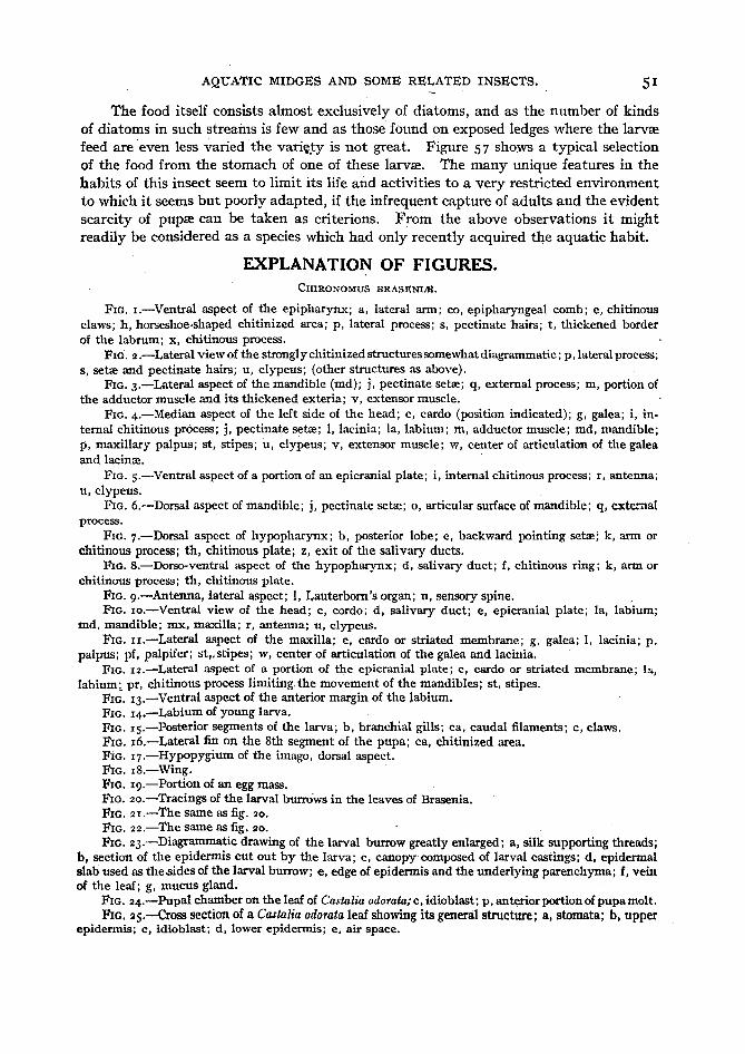
AQUATIC MIDGES AND SOME RELATED INSECTS. 51
The food itself consists almost exclusively of diatoms, and as the number of kindsof diatoms in such streams is few and as those found on exposed ledges where the larvtefeed are even less varied the variety is not great. Figure 57 shows a typical selectionof the food from the stomach of one of these larvse. The many unique features in thehabits of this insect seem to limit its life and activities to a very restricted environmentto which it seems but poorly adapted, if the infrequent capture of adults and the evidentscarcity of pupse can be taken as criterions. From the above observations it mightreadily be considered as a species which had only recently acquired the aquatic habit.
EXPLANATION OF FIGURES.CHIRONOMUS BRASENIJ$.
FIG. r.-Ventral aspect of the epipharynx; a, lateral ann; co, epipharyngeal comb; e, chitinousclaws; h, horseshoe-shaped chitinized area; p, lateral process; s, pectinate 'hairs; t, thickened borderof the labrum; x, chitinous process.
FIG. 2.-Lateral view of the strongl y chitinized structuressomewhat diagrammatic; p, lateral process;s, setse and pectinate hairs; u, clypeus; (other structures as above).
FIG. 3.-Lateral aspect of the mandible (md); j, pectinate setee: q, external process; m, portion ofthe adductor muscle and its thickened exteria; v, extensor muscle.
FIG. 4.-Median aspect of the left side of the head; c, cardo (position indicated); g, galea; i, internal chitinous process; j, pectinate setse: 1, lacinia; la, labium; ttl, adductor muscle; md, mandible;p, maxillary palpus; st, stipes; u, clypeus; v, extensor muscle; w, center of articulation of the galeaand lacinee. '
FIG. s.--Ventral aspect of a portion of an epicranial plate; i, internal chitinous process; r, antenna;u, clypeus.
FIG. 6.-Dorsal aspect of mandible; j, pectinate setee: 0, articular surface of mandible; q, externalprocess.
FIG. 7.-Dorsal aspect of hypopharynx; b, posterior lobe; e. backward pointing setse: k, ann orchitinous process; th, chitinous plate; z, exit of the salivary ducts.
FIG. B.-Dorso-ventral aspect of the hypopharynx; d, salivary duct; f, chitinous ring; k, arm orchitinous process; tho chitinous plate.
FIG. g.-Antenna, lateral aspect; 1, Lauterborn's organ; n, sensory spine.FIG. Io.-Ventral view of the head; c, cordo; d, salivary duct; e, epicranial plate; la, labium;
md, mandible; mx, maxilla; r, antenna; u, clypeus.FIG. Ir.-Lateral aspect of the maxilla; c, cardo or striated membrane; g, galea; 1, lacinia; p,
palpus; pf, palpifer; st.sstipes; w, center of articulation of the galea and lacinia,FIG. 12.-Lateral aspect of a portion of the epicranial plate; c, cardo or striated membrane; Ia,
labium; pr, chitinous process limiting, the movement of the mandibles; st, stipes.FIG. 13.-Ventral aspect of the anterior margin of the labium.FIG. 14.-Labium of young larva.FIG. IS.-Posterior segments of the larva; b , branchial gills; ca, caudal filaments; c, claws.FIG. 16.-Lateral fin on the 8th segment of the pupa; ca, chitinized area.FIG. 17.-HYPopygium of the imago, dorsal aspect.FIG. lB.-Wing.FIG. Ig.-Portion of an egg mass.FIG.20.-Tracings of the larval burrows in the leaves of Brasenia.FIG. 2r.-The same as fig. 20.FIG. 22.-The same as fig. 20.FIG. 23.-Diagrammatic drawing of the larval burrow greatly enlarged; a, silk supporting threads;
b, section of the epidennis cut out by the larva; c, canopy composed of larval castings; d. epidermalslab used as thesides of the larval burrow; e, edge of epidermis and the underlying parenchyma; f. veinof the leaf; g, mucus gland.
FIG. 24.-Pupal chamber on the leaf of Castalia odorata;c, idioblast; p, anterior portion of pupamolt.FIG. 2S.-crOSS section of a Castalia odorata leaf showing its general structure; a, stomata; b. upper
epidermis; c, idioblast; d , lower epidennis; e. air space.

BULLETIN OF THE BUREAU OF FISHERIES.
CmRONOMUS LOBIFSRUS.
FIG. 26.-Diagram of a glass preparation showing the position occupied by the larva in its burrow;lc, lower glass; nt, conical net; t, glass tube; u, upper glass; .~, larva.
FIG. 27.-Diagram of the contracted conical net.FIG. 28.-Broken labial plate.
ORTHOCLADIUS SP.FIG. 29.-Labium.FIG. 30.-Mandible.
PRODIAMSSA SP.
FIG. 3I.-Maxilla; ar, articular process; c, vibrissee; 1, lacinia; la, labium; p, palpus; st, stipes.FIG. 32.-Ventral aspect of a portion of the head; ar, articular process; c, vibrissre; la, labium.
CHIRONOMUS LOBIFERUS.
FIG. 33.-Claw of posterior proleg.FIG. 34.-Spines of anterior proleg; a, serrate spine; b, c, hooked spines; d, hairlike spine.FIG. 3s.-Left atiterior proleg; a, hairlike spines; b, hooked spines; c, serrate spines.FIG. 36.-An enlarged silken holdfast attached to a glass surface; g, a process on the side of a larval
burrow.FIG. 37.-Silken network between two larval burrows ( g, h); i, interlacing silk fibers.
ORPHNEPHILA TESTACEA.
FIG. 52.-Side view of entire larva showing characteristic color pattern. .FIG. 53.-Side view of the head and proleg; ey, eye spot; la, labium; lb, labrum; md, mandible;
DDt, maxilla; pr, proleg; r, antenna; sp, spines.FIG. S4.-Frontal view of the head; cl, elypial plate; Ia, labium; lb, labrum; md, mandible;
DDt, maxilla; rd , rod attached to the mandible.FIG. 5s.-Ventral aspect of the mandible.FIG. S6.-Dorsal-lateral aspect of the hypopharynx.FIG. S7.-Miscellaneous diatoms from the stomach of Orphnephila.
ORPHNEPHILA AMERICANA.
FIG. 38.-View of habitat where larvse were found, west bank of Six Mile Creek, Ithaca, N. Y.;F, water level of the main stream; 1, ledges kept moist by water flowing in a small stream shown justabove; w, water flowing over ledges.
TANYPus DYARI.
FIG. 39.-Labrum; ch, chitinous plate; cp, chitinized process; se, sensory process.FIG. 40.-Chitinous structures of the maxilla; 1, lacinia; p, palpus.FIG. 4I.-Ventral aspect of the head; hy, hypopharynx; la, labial papilla; md, mandible; mx,
maxilla; p, maxillary palpus; r, antenna.FIG. 42.-Dorsal view of the labium and hypopharynx; ar, articular plate; cb , chitinous band;
hy, hypopharynx; la, labial process; lp, lateral hypopharyngeal process; pr, sensory process; th,chitinous plate of the hypopharynx.
FIG. 43.-Lateral view of the same. .FIG. 44.-Dorsal view of the entire larva; br, branchial gill; c, cardiac chamber; cr, crop or pro.
. ventriculus; g, salivary gland; m, malpighian tubule; r, retractile antenna; st, stomach or ventriculus.
CULICOIDES SP.
FIG. 4s.-Dorsal view of entire larva; f, caudal filaments; g, branchial gills.FIG. 46. Lateral view of the head; d, salivary duct; ep , epipharynx; ey, eye spot; hy, hypo
pharynx; la, labium; lb, labrum; md, mandible; mx, maxilla.FIG. 47.-Ventral view of the head; structures the same as shown in fig. 46.FIG. 48.-Dorsal view of the posterior end of the larva considerably enlarged; f, caudal filaments;
ft, accessory caudal filaments; g, branchial gills. Larva from sap flow in elms resembling Culicoideshieroglyphicus.
FIG. 49.-The entire larva, dorsal view; ch , chitinous rod.FIG. So.-Posterior end of the same greatly enlarged; c, caudal processes; g, branchial gills; s,
setre resembling caudal filaments.FIG. 5I.-Mandible.

AQUATIC MIDGES AND SOME RELATED INSEC'fS. S3
8
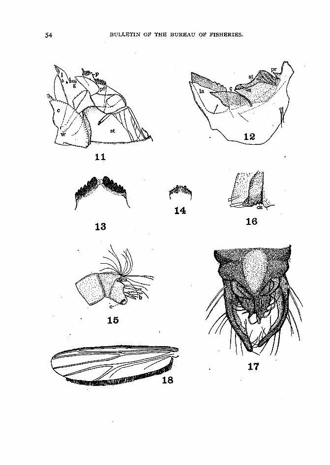
54 BULLETIN OF THE BUREAU OF FISHERIES.
11
13
'~
1416
15
17

AQUATIC MIDGES AND SOME RELATED INSECTS. 55
.: .
. "":\><>" ."...... ....... ·0'
::I.~ :1.'1
,',',"....:.. ,."." : t....
:~::.~. 23,.. .25
24
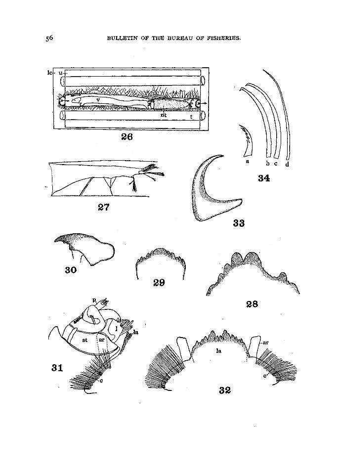
BULLETIN OF THE BUREAU OF FISHERIES.
bed
34
a
26
tit
v
27
..... '''''.
lcl- u· ..-:-I--------------~-IT'
29
28

B ULL. U. S. B . H', .. J9 2 1- 2 2 .
35
36 37
3850

AQUATIC MIDGES AND SOME RELATED INSECTS. 57
39
43 er
41
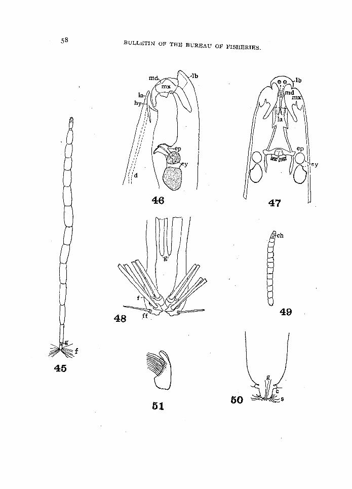
BULLETIN OF THE BUREAU OF FISHERIES.
45
46
51
ch
49

AQUA'l'IC MIDGES AND SOME REI.ATED INSECTS.
52
59
/ 53
55
54
56
57

60 BULLETIN OF THE BUREAU OF FISHERIES.
BIBLIOGRAPHY.DUFOUR, LEON.
1845. Observations sur les metamorphoses du Ceratopoqon. genuulatus Guerin. Annales de laSociete entomologique de France, 2" ser., Tome III, p. 215-223, pl. III, No. n. Paris.
GOETGHEBUER, M.1911. Chironomides de Belgique. Annales de la Societe entomologique de Belgique, Tome 55,
p. 95-II3. Bruxelles. .1913. Etudes sur les Chironomides de Belgique. Memoires de l 'Academie royale des sciences,
des lettres et des beaux-arts de Belgique, 3 fasc., 6 Deer., p. 1-26, pl. I-V. Bruxelles.GUERIN, F. E.
1833. Notice sur les metamorphoses des Ceratapogons, et description de deux especes nouvelles. de ce genre, decouvertes aux environs de Paris. Annales de la Societe entomologiqna de
France, Tome II, p. 161-167, PI. VIII. Paris.HEEGER, ERNST.
1856. Neue metamorphosen einiger Dipteren. Sitzungsberichte der Kaiserlichen Akademie derWissenschaften. Bd. XX, p. 335-350, pl. 4. Wien.
HOLMGREN, N.1904. Zur Morphologie des Insektenkopfes. 1. Zum metameren Aufbau des Kopfes der Chireno- .
mouslarve. Zeitschrift fur wissenschaftliche ' Zoologie, Bd, LXXVI, p. 43lr477, pl-. XXVII, XXVIII. Leipzig.
JOHANNSEN, O. A.1903. Aquatic nematocerous Diptera [of New York]. Bulletin of the New York State Museum,
University of the State of New York, LXVIII,p. 328-441, PI. XXXIII-'L. Albany, N. Y.
1905. Aquatic nematocerous Diptera, II: Chironomidse, New York State Education Department, New York State Museum Annual Report, Bulletin LXXXVI, p. 76-327, PI. XVIXXXVII. Albany, N. Y.
KELLOGG, V. L. •1902. The development and homologies of the mouth parts of insects. American Naturalist, Vol.
XXXVI, No. 429, p. 683-706, 26 fig. The Athenseum Press, Ginn & Co., Boston.1905. American insects. 674 p., 812 fig., 13 pI. New York.
KIEFFER, J. J., and A. THIENEMANN.1908. Neue und bekannte Chironomiden und ihre Metamorphose. I. Neue und bekannte Chiro
nomiden, von Kieffer. II. Chironomidernetamorphosen, von Thienemann. Zeitschriftfur wissenschaftliche Insektenbiologie, Ed. 4, p. 1-10, 33-39, 78-84, 124-128, 184-19°,214-219, 256-259, 277-286. Berlin.
KNAB, FREDERICK.1905. A chironomid inhabitant of Sarracenia purpurea, Metriocnemus knabi Coq. Journal of the
New York Entomological Society, vol. XIII, p. 6lr73, pI. VI. New York, N. Y.1914. Ceratopagoninae sucking the blood of caterpillars. Proceedings, Entomological Society of
Washington, Vol. XVI, p. 63-{)6. Washington, D. C.1914!t. Ceratopagoninae sucking the blood of other insects. Ibid., p. 13<)-141.
LABOULB;£NE, ALEX.1869. Histoire des metamorphoses du Ceratopoqon dufouri. Annales de la Societe entomologique
de France, 4" ser., Tome IX, p. 157-166, pl. 7. Paris.LAUTERBORN, R.
1905. Zur Kenntnis der Chironomiden-Larven. Zoologischer Anzeiger, Bd. X~IX, p. 207-217,15 fig. Leipzig.
LoNG, WILLIAM HENRY, JR.1902. New species of Ceratopogon. Biological Bulletin, Marine Biological Laboratory, Woods.
Hole, Mass., Vol. III, p. 3-14. Press of New Era Printing Co., Lancaster, Pa.LYONET, PIETER. .
1832. Recherches sur 1'anatomie et les metamorphoses de differentes especes d'insectes. Ouvrageposthume. Publie par M. W. De Haan. 580 p., 54 pl. Paris, Roret.
AQUATIC MIDGES AND SOME RELATED INSECTS. 61
MALLOCH, JOHN R.1915. The Chironomidae, or midges, of Illinois, with particular reference to the species occurring
in the Illinois River. Bulletin, Illinois State Laboratory of Natural History, Vol. X,Art. VI, p. 275-543. Urbana, Ill.
MnINnRT, FR.1882. Om retractile antenner hos en dipter-larve, Tanypus. Entomologisk Tidskrift, Argangen 3,
p. 83-86. Stockholm.1886. De eucephale Myggelarver. K. Videnskabernes Selskabs Skrifter, Argangen III, p. 373
493, PIs. I-IV. Copenhagen.MIALL, Lours COMPTON.
1895. The natural history of aquatic insects. 8 vo., 395 p., II6 fig. Macmillan and Co., Londonand New York. Reprinted 1903, 1912.
MIALL, LOUIS COMPTON, and A. R. HAMMOND.1900. The structure and life-history of the harlequin fly (Chironomous). 196 p., 129 fig. Oxford.
MUNDY, ARTHUR TERRY.:r909. The anatomy, habits, and psychology of Chironomus p-usio Meigen (the early stages), with
notes on various other invertebrates, chiefly Chironomidae. 56 p., VIII pl.' Leicester,England.
NnnDHAM, JAMns G.1906. The egg-laying of Chironomous annularis. Science, New Series, Vol. XXIV, p. 299. The
Macmillan Co., New York.1908. Notes on the aquatic insects of Walnut Lake, with especial reference to a few species of
considerable importance as fish food. In A biological survey of Walnut Lake, Mich., byThos. L. Hankinson, p. 252-271, I pI. Lansing, Mich.
NnnDHAM, JAMES G., and J. T. LLOYD.1916. The life of inland waters. 8 vo., 438 p., 244 figs. Ithaca, N. Y.
PnRRIS, EDOUARD.1847. Notes pour servir a I 'histoire des Ceratopogon. Annales de la Societe entomologique de
France. 2e ser., Tome V, p. 555-569, pI. 9, No. III. Paris.PnnIT, R. H.
, 1900. A leaf-miner, Cheironomous sp., in water lilies. Michigan Academy of Science, First Report,p. IIo-III. Lansing.
TAYLOR, T. H.190 5. Notice of a chironomous larva. Zoologischer Anzeiger, Bd, XXIX, p, 4S:r-452. Leipzig.
THInNnMANN, AUGUST.1909· Orphnephila testacea Macq. Ein Beitrag zur Kenntnis der fauna hygropetrica. Annales
de Biologie Lacustre, Tome 4, p. 53-87, pls, viii, Ix, Bruxelles.'I'n.BURY, MARY RUTH.
1913. Notes on the feeding and rearing of the midge Chironomus cayugae Johannsen. Journal ofthe New York Entomological Society, Vol. 21, p. 305-308. New York.
Wn.LnM, VICTOR.1908. Larves de chironomides vivant dans des feuilles. Bulletin de la classe des sciences de
I'Academie royale des sciences, des lettres et des beaux arts de Belgique, p. 697-704, pl •. Bruxelles.
ZnBROWSKA, A.1914. Recherches sur les larves de chironomides du Lac Leman. Lausanne.