ecmo nomenclature · while the term ecmo refers to a particular extracorporeal configuration and...
TRANSCRIPT

The Extracorporeal Life Support Organization Maastricht Treaty for Nomenclature in Extracorporeal Life Support
A Position Paper of the Extracorporeal Life Support Organization
Steven A. Conrad1, L. Mikael Broman2, Fabio S. Taccone3, Roberto Lorusso4, Maximilian V. Malfertheiner5, Federico Pappalardo6, Matteo Di Nardo7, Mirko Belliato8, Lorenzo Grazioli9, Ryan P. Barbaro10, D. Michael McMullan11, Vincent Pellegrino12, Daniel Brodie13, Melania M. Bembea14, Eddy Fan15, Malaika Mendonca16, Rodrigo Diaz17, Robert H. Bartlett18
1Departments of Medicine, Emergency Medicine and Pediatrics, Louisiana State University Health Sciences Center Shreveport, Louisiana, USA
2ECMO Center, Karolinska University Hospital, and Department of Physiology and Pharmacology, Karolinska Institutet Stockholm, Sweden
3Department of Intensive Care Hôpital Erasme Brussels, Belgium
4Department of Cardiothoracic Surgery Maastricht University Medical Centre Maastricht, The Netherlands
5Department of Internal Medicine II University Medical Center Regensburg Regensburg, Germany 6Department of Cardiothoracic Anesthesia and Intensive Care San Raffaele Hospital Milan, Italy
7Pediatric Intensive Care Unit Children’s Hospital Bambino Gesù, IRCCS Rome, Italy 8U.O.C. Anestesia e Rianimazione 1 Fondazione IRCCS Policlinico San Matteo Pavia, Italy
9Department of Anesthesiology ASST Papa Giovanni XXIII Bergamo, Italy
Page 1 of 36 AJRCCM Articles in Press. Published on 03-April-2018 as 10.1164/rccm.201710-2130CP
Copyright © 2018 by the American Thoracic Society

10Department of Pediatrics/Intensive Care University of Michigan Ann Arbor, Michigan, USA
11Department of Surgery/Cardiothoracic Surgery University of Washington Seattle, Washington, USA
12Department of Intensive Care Alfred Hospital Melbourne, Victoria, Australia
13Department of Medicine/Pulmonary, Allergy and Critical Care Medicine Columbia University College of Physicians and Surgeons New York, New York, USA
14Department of Anesthesiology and Critical Care Johns Hopkins University School of Medicine Baltimore, Maryland 21287, USA
15Interdepartmental Division of Critical Care Medicine University of Toronto Toronto, Ontario, Canada
16Division of Pediatric Critical Care Sheikh Khalifa Medical City Abu Dhabi, UAE
17Division of Cardiovascular Anesthesiology Clinica las Condes Santiago, Chile
18Department of Surgery University of Michigan. Ann Arbor, Michigan, USA
Author contributions: Drafting of the article: SAC and LMB. Critical revision of the article for important intellectual content: FST, RL, MVM, FP, MDN, MB, LG, RPB, DMM, VP, DB, MMB, EF, MM, RD, RHB. Final approval of the article: All authors
Page 2 of 36 AJRCCM Articles in Press. Published on 03-April-2018 as 10.1164/rccm.201710-2130CP
Copyright © 2018 by the American Thoracic Society

Correspondence to:
Steven A. Conrad, MD PhD Louisiana State University Health Sciences Center 1501 Kings Highway Shreveport, Louisiana 71103, USA Voice +1 318 626 2325 Fax +1 318 626 3892 [email protected]
Keywords: Nomenclature; terminology; extracorporeal life support; extracorporeal membrane oxygenation; extracorporeal carbon dioxide removal; extracorporeal circulatory support
Grant support: Melania Bembea - The National Institute of Neurological Disorders and Stroke of the National Institutes of Health under Award Number K23NS076674
Running title: Terminology for extracorporeal life support
Manuscript descriptor: 2.01 Adherence/Compliance/Self-Regulation
Word count: 1495 (targeting Critical Care Perspective)
Current scientific knowledge: Extracorporeal life support (ECLS) is a rapidly expanding field, with the number of research publications growing accordingly. While there is a generally accepted terminology for ECLS, a consistent guide for terminology and abbreviations does not exist.
What does this manuscript add? This manuscript provides a consistent nomenclature and abbreviations for the description of the practice of ECLS and associated devices and techniques.
Page 3 of 36 AJRCCM Articles in Press. Published on 03-April-2018 as 10.1164/rccm.201710-2130CP
Copyright © 2018 by the American Thoracic Society

1
Abstract
Extracorporeal life support (ECLS) was developed more than 50 years ago initially with
venoarterial (VA) and subsequently venovenous (VV) configurations. As the technique of ECLS
has significantly improved and newer skills developed, complexity in terminology and advances
in cannula design led to some misunderstanding and inconsistency in definitions both in clinical
practice and scientific research. This document is a consensus of multispecialty international
representatives of the Extracorporeal Life Support Organization, including the North American,
Latin American, European, South and West Asian, and Asian-Pacific chapters, imparting a
global perspective on ECLS. The goal is to provide a consistent and unambiguous nomenclature
for ECLS and to overcome the inconsistent use of abbreviations for ECLS cannulation.
Secondary benefits are ease of multicenter collaboration in research and improved registry data
quality and clear communication among practitioners and researchers in the field.
Word count: 137
MeSH keywords: Terminology; Extracorporeal membrane oxygenation; Oygenators, Membrane;
Extracorporeal Circulation; Cannula
Page 4 of 36 AJRCCM Articles in Press. Published on 03-April-2018 as 10.1164/rccm.201710-2130CP
Copyright © 2018 by the American Thoracic Society

2
Introduction
Extracorporeal therapies for temporary, non-intraoperative support of patients with cardiac
and/or pulmonary dysfunction are an outgrowth of cardiopulmonary bypass . The success of
cardiopulmonary bypass beginning in 1953 for short term circulatory support did not directly
translate to more prolonged support due in large part to the lack of biocompatibility of devices of
the time (1). Long term extracorporeal support would await the development of newer
technologies and approaches over the ensuing decades. The result is a diversity of approaches to
temporary support that is rapidly becoming mainstream in the management of severely ill and
injured patients
This diversity has led to the use of terms and abbreviations marked by inconsistency and
ambiguity. The first reported application of extracorporeal support in the intensive care unit
setting (2) employed extracorporeal membrane oxygenation (ECMO) in post-traumatic acute
respiratory failure. While the term ECMO refers to a particular extracorporeal configuration and
application for support of cardiopulmonary dysfunction, it became synonymous with the use of
any extracorporeal system other than surgical cardiopulmonary bypass. A number of
extracorporeal applications have emerged that are not considered ECMO, such as extracorporeal
carbon dioxide removal (ECCO2R) for managing hypercapnic respiratory failure or supporting
ultraprotective ventilation in acute respiratory distress syndrome, extracorporeal
cardiopulmonary resuscitation (ECPR) for maintaining systemic perfusion during cardiac arrest,
and extracorporeal interval support for organ recovery (EISOR) for providing perfusion of
organs awaiting recovery following declaration of cardiac death. The term extracorporeal life
support (ECLS) has emerged to describe the entire family of extracorporeal support modalities
for long-term support.
Page 5 of 36 AJRCCM Articles in Press. Published on 03-April-2018 as 10.1164/rccm.201710-2130CP
Copyright © 2018 by the American Thoracic Society
3
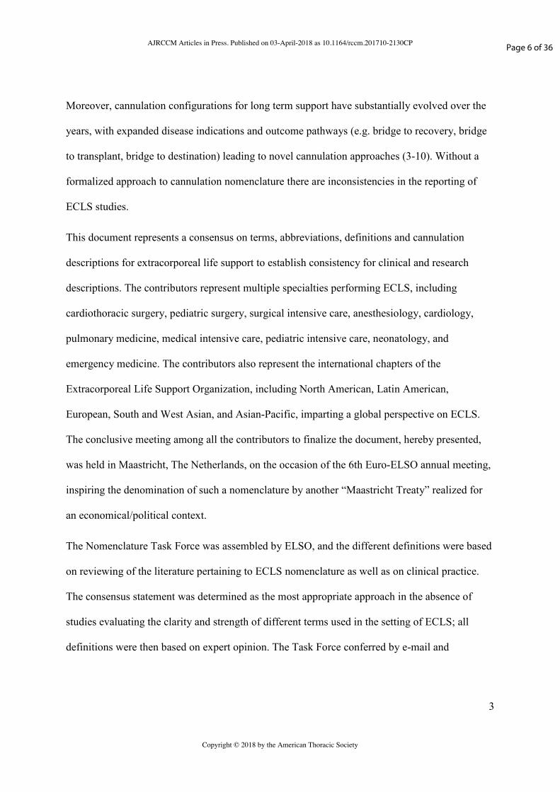
Moreover, cannulation configurations for long term support have substantially evolved over the
years, with expanded disease indications and outcome pathways (e.g. bridge to recovery, bridge
to transplant, bridge to destination) leading to novel cannulation approaches (3-10). Without a
formalized approach to cannulation nomenclature there are inconsistencies in the reporting of
ECLS studies.
This document represents a consensus on terms, abbreviations, definitions and cannulation
descriptions for extracorporeal life support to establish consistency for clinical and research
descriptions. The contributors represent multiple specialties performing ECLS, including
cardiothoracic surgery, pediatric surgery, surgical intensive care, anesthesiology, cardiology,
pulmonary medicine, medical intensive care, pediatric intensive care, neonatology, and
emergency medicine. The contributors also represent the international chapters of the
Extracorporeal Life Support Organization, including North American, Latin American,
European, South and West Asian, and Asian-Pacific, imparting a global perspective on ECLS.
The conclusive meeting among all the contributors to finalize the document, hereby presented,
was held in Maastricht, The Netherlands, on the occasion of the 6th Euro-ELSO annual meeting,
inspiring the denomination of such a nomenclature by another “Maastricht Treaty” realized for
an economical/political context.
The Nomenclature Task Force was assembled by ELSO, and the different definitions were based
on reviewing of the literature pertaining to ECLS nomenclature as well as on clinical practice.
The consensus statement was determined as the most appropriate approach in the absence of
studies evaluating the clarity and strength of different terms used in the setting of ECLS; all
definitions were then based on expert opinion. The Task Force conferred by e-mail and
Page 6 of 36 AJRCCM Articles in Press. Published on 03-April-2018 as 10.1164/rccm.201710-2130CP
Copyright © 2018 by the American Thoracic Society
4
agreements were achieved through iterative discussion and debate. Recommendations were
unanimously agreed and then approved by the Task Force.
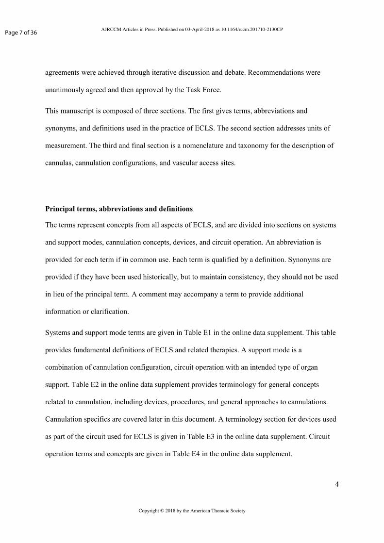
This manuscript is composed of three sections. The first gives terms, abbreviations and
synonyms, and definitions used in the practice of ECLS. The second section addresses units of
measurement. The third and final section is a nomenclature and taxonomy for the description of
cannulas, cannulation configurations, and vascular access sites.
Principal terms, abbreviations and definitions
The terms represent concepts from all aspects of ECLS, and are divided into sections on systems
and support modes, cannulation concepts, devices, and circuit operation. An abbreviation is
provided for each term if in common use. Each term is qualified by a definition. Synonyms are
provided if they have been used historically, but to maintain consistency, they should not be used
in lieu of the principal term. A comment may accompany a term to provide additional
information or clarification.
Systems and support mode terms are given in Table E1 in the online data supplement. This table
provides fundamental definitions of ECLS and related therapies. A support mode is a
combination of cannulation configuration, circuit operation with an intended type of organ
support. Table E2 in the online data supplement provides terminology for general concepts
related to cannulation, including devices, procedures, and general approaches to cannulations.
Cannulation specifics are covered later in this document. A terminology section for devices used
as part of the circuit used for ECLS is given in Table E3 in the online data supplement. Circuit
operation terms and concepts are given in Table E4 in the online data supplement.
Page 7 of 36 AJRCCM Articles in Press. Published on 03-April-2018 as 10.1164/rccm.201710-2130CP
Copyright © 2018 by the American Thoracic Society

5
Units of measurement
Several units of measurement are used for devices and patient management during ECLS. Table
E5 in the online data supplement provides the preferred measurement unit systems for ECLS.
Système international d'unités (SI) are preferred over Imperial units and used in most cases,
except where manufacturer specifications dictate the unit system.
Configurations for peripheral cannulation
Peripheral cannulation configurations vary in complexity, from simple two cannula
configurations for traditional venovenous and venoarterial extracorporeal membrane oxygenation
(ECMO) to more configurations, for example, with multiple cannulas and multiple drainage or
return sites. There is a need to be able to provide basic cannulation information for clinical
purposes that conveys the essential configuration. To meet these objectives, a two-level
classification system with increasing levels of descriptive information was developed.
Fundamental to all cannulation abbreviations is the use of a hyphen to distinguish drainage
cannulas, on the left of the hyphen, and return cannulas, on the right of the hyphen, with the
membrane lung, represented by the hyphen itself. In this approach, the presence of a hyphen
differentiates a cannulation configuration from the support modes introduced above.
Level one: Cannula hierarchy
All cannulas contributing to the primary (major) draining and return circuit flow are written in
upper case letters, e.g. ‘V-V’ representing venous drainage and venous return for venovenous
support (Table E6 in the online data supplement). All cannulas with minor flow for secondary
drainage, unloading of specific anatomical location, or to promote distal perfusion are written in
lower case letters after the major flow cannula to which side it belongs, e.g. ‘V-Aa’ representing
venous drainage, arterial return and secondary arterial return such as for distal perfusion. The use
Page 8 of 36 AJRCCM Articles in Press. Published on 03-April-2018 as 10.1164/rccm.201710-2130CP
Copyright © 2018 by the American Thoracic Society
6
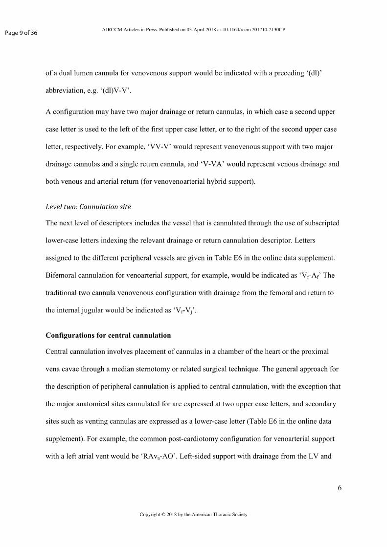
of a dual lumen cannula for venovenous support would be indicated with a preceding ‘(dl)’
abbreviation, e.g. ‘(dl)V-V’.
A configuration may have two major drainage or return cannulas, in which case a second upper
case letter is used to the left of the first upper case letter, or to the right of the second upper case
letter, respectively. For example, ‘VV-V’ would represent venovenous support with two major
drainage cannulas and a single return cannula, and ‘V-VA’ would represent venous drainage and
both venous and arterial return (for venovenoarterial hybrid support).
Level two: Cannulation site
The next level of descriptors includes the vessel that is cannulated through the use of subscripted
lower-case letters indexing the relevant drainage or return cannulation descriptor. Letters
assigned to the different peripheral vessels are given in Table E6 in the online data supplement.
Bifemoral cannulation for venoarterial support, for example, would be indicated as ‘Vf-Af’ The
traditional two cannula venovenous configuration with drainage from the femoral and return to
the internal jugular would be indicated as ‘Vf-Vj’.
Configurations for central cannulation
Central cannulation involves placement of cannulas in a chamber of the heart or the proximal
vena cavae through a median sternotomy or related surgical technique. The general approach for
the description of peripheral cannulation is applied to central cannulation, with the exception that
the major anatomical sites cannulated for are expressed at two upper case letters, and secondary
sites such as venting cannulas are expressed as a lower-case letter (Table E6 in the online data
supplement). For example, the common post-cardiotomy configuration for venoarterial support
with a left atrial vent would be ‘RAva-AO’. Left-sided support with drainage from the LV and
Page 9 of 36 AJRCCM Articles in Press. Published on 03-April-2018 as 10.1164/rccm.201710-2130CP
Copyright © 2018 by the American Thoracic Society

7
aortic return would be ‘LV-AO’, and right-sided support from the right atrium to the pulmonary
artery would be ‘RA-PA’.
Conclusions
This classification system for ECLS nomenclature provides a standardized foundation for the
description of ECLS application, decreasing ambiguity and providing a consistency for the
comparison of clinical reports. It includes an extensive terminology of systems, support modes,
devices, units of measurement and cannulation configurations. Given the hierarchical structure of
cannulation description, it provides for the inclusion of only as much detail as needed for a given
purpose. Based on a defined system, it maintains flexibility to adapt to (many if not all) future
developments in cannulation approaches as it supports extensibility.
This nomenclature has limitations. While adequate for supporting descriptions for most clinical
applications, it may not meet the needs for research applications where more detail for
cannulation configurations such as location of the cannula tip, additional cannulation sites, or
non-traditional cannulation configurations, would be desirable. Given its systematic basis,
however, it could be extended for such a purpose.
Page 10 of 36 AJRCCM Articles in Press. Published on 03-April-2018 as 10.1164/rccm.201710-2130CP
Copyright © 2018 by the American Thoracic Society

8
References
1. Fortenberry JD, Lorusso R. The history and development of extracorporeal support. In:
Brogan TV, Lequier L, Lorusso R, MacLaren G, Peek G, editors. Extracorporeal Life
Support: The ELSO Red Book, 5 ed. Ann Arbor, MI: Extracorporeal Life Support
Organization; 2017.
2. Hill JD, O'Brien TG, Murray JJ, Dontigny L, Bramson ML, Osborn JJ, Gerbode F.
Prolonged extracorporeal oxygenation for acute post-traumatic respiratory failure
(shock-lung syndrome). Use of the Bramson membrane lung. N Engl J Med 1972; 286:
629-634.
3. Javidfar J, Brodie D, Wang D, Ibrahimiye AN, Yang J, Zwischenberger JB, Sonett J,
Bacchetta M. Use of bicaval dual-lumen catheter for adult venovenous extracorporeal
membrane oxygenation. Ann Thorac Surg 2011; 91: 1763-1768; discussion 1769.
4. Palmer O, Palmer K, Hultman J, Broman M. Cannula design and recirculation during
venovenous extracorporeal membrane oxygenation. ASAIO J 2016; 62: 737-742.
5. Biscotti M, Lee A, Basner RC, Agerstrand C, Abrams D, Brodie D, Bacchetta M. Hybrid
configurations via percutaneous access for extracorporeal membrane oxygenation: a
single-center experience. ASAIO J 2014; 60: 635-642.
6. Madershahian N, Nagib R, Wippermann J, Strauch J, Wahlers T. A simple technique of
distal limb perfusion during prolonged femoro-femoral cannulation. J Card Surg 2006;
21: 168-169.
7. Le Guyader A, Lacroix P, Ferrat P, Laskar M. Venous leg congestion treated with distal
venous drainage during peripheral extracorporeal membrane oxygenation. Artif Organs
2006; 30: 633-635.
8. Skarsgard ED, Salt DR, Lee SK, Extracorporeal Life Support Organization R.
Venovenous extracorporeal membrane oxygenation in neonatal respiratory failure: does
routine, cephalad jugular drainage improve outcome? J Pediatr Surg 2004; 39: 672-676.
9. Avalli L, Maggioni E, Sangalli F, Favini G, Formica F, Fumagalli R. Percutaneous left-
heart decompression during extracorporeal membrane oxygenation: an alternative to
surgical and transeptal venting in adult patients. ASAIO J 2011; 57: 38-40.
Page 11 of 36 AJRCCM Articles in Press. Published on 03-April-2018 as 10.1164/rccm.201710-2130CP
Copyright © 2018 by the American Thoracic Society

9
10. Kim HE, Jung JW, Shin YR, Park HK, Park YH, Shin HJ. Left Atrial Decompression by
Percutaneous Left Atrial Venting Cannula Insertion during Venoarterial Extracorporeal
Membrane Oxygenation Support. Korean J Thorac Cardiovasc Surg 2016; 49: 203-206.
Page 12 of 36 AJRCCM Articles in Press. Published on 03-April-2018 as 10.1164/rccm.201710-2130CP
Copyright © 2018 by the American Thoracic Society
10
Figure Captions
Figure 1 Relationship between ECLS systems, support modes and applications. ECLS
extracorporeal life support, ECMO extracorporeal membrane oxygenation, VA ECMO
venoarterial extracorporeal membrane oxygenation, VVA ECMO venovenoarterial
extracorporeal membrane oxygenation, VV ECMO venovenous extracorporeal membrane
oxygenation, ECPR extracorporeal cardiopulmonary resuscitation, EISOR extracorporeal
interval support for organ retrieval, ECCO2R extracorporeal carbon dioxide removal, VV
ECCO2R venovenous extracorporeal carbon dioxide removal, AV ECCO2R arteriovenous
extracorporeal carbon dioxide removal.
Figure 2 Schematic of venoarterial (VA) ECMO (A) and venovenous (VV) ECMO (B)
showing typical cannulation sites and direction of blood flow.
Figure 3 Schematic of venovenoarterial (VVA) ECMO (A) and pumpless arteriovenous
(AV) ECCO2R showing typical cannulation sites and direction of blood flow.
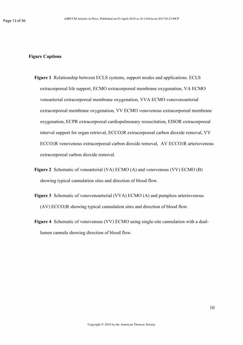
Figure 4 Schematic of venovenous (VV) ECMO using single-site cannulation with a dual-
lumen cannula showing direction of blood flow.
Page 13 of 36 AJRCCM Articles in Press. Published on 03-April-2018 as 10.1164/rccm.201710-2130CP
Copyright © 2018 by the American Thoracic Society

Figure 1. Relationship between ECLS systems, support modes and applications. ECLS extracorporeal life support, ECMO extracorporeal membrane oxygenation, VA ECMO venoarterial extracorporeal membrane oxygenation, VVA ECMO venovenoarterial extracorporeal membrane oxygenation, VV ECMO venovenous
extracorporeal membrane oxygenation, ECPR extracorporeal cardiopulmonary resuscitation, EISOR extracorporeal interval support for organ retrieval, ECCO2R extracorporeal carbon dioxide removal, VV ECCO2R venovenous extracorporeal carbon dioxide removal, AV ECCO2R arteriovenous extracorporeal
carbon dioxide removal.
137x92mm (300 x 300 DPI)
Page 14 of 36 AJRCCM Articles in Press. Published on 03-April-2018 as 10.1164/rccm.201710-2130CP
Copyright © 2018 by the American Thoracic Society

Figure 2. Schematic of venoarterial (VA) ECMO (A) and venovenous (VV) ECMO (B) showing typical cannulation sites and direction of blood flow.
138x91mm (300 x 300 DPI)
Page 15 of 36 AJRCCM Articles in Press. Published on 03-April-2018 as 10.1164/rccm.201710-2130CP
Copyright © 2018 by the American Thoracic Society

Figure 3. Schematic of venovenoarterial (VVA) ECMO (A) and pumpless arteriovenous (AV) ECCO2R showing typical cannulation sites and direction of blood flow.
139x94mm (300 x 300 DPI)
Page 16 of 36 AJRCCM Articles in Press. Published on 03-April-2018 as 10.1164/rccm.201710-2130CP
Copyright © 2018 by the American Thoracic Society

Figure 4. Schematic of venovenous (VV) ECMO using single-site cannulation with a dual-lumen cannula showing direction of blood flow.
137x187mm (300 x 300 DPI)
Page 17 of 36 AJRCCM Articles in Press. Published on 03-April-2018 as 10.1164/rccm.201710-2130CP
Copyright © 2018 by the American Thoracic Society

The Extracorporeal Life Support Organization Maastricht Treaty for Nomenclature in Extracorporeal Life Support (Online Data Supplement) A Position Paper of the Extracorporeal Life Support Organization
Steven A. Conrad, L. Mikael Broman, Fabio S. Taccone, Roberto Lorusso, Maximilian V. Malfertheiner, Federico Pappalardo, Matteo Di Nardo, Mirko Belliato, Lorenzo Grazioli, Ryan P. Barbaro, D. Michael McMullan, Vincent Pellegrino, Daniel Brodie, Melania M. Bembea, Eddy Fan, Malaika Mendonca, Rodrigo Diaz, Robert H. Bartlett
American Journal of Respiratory and Critical Care Medicine, 2018
Page 18 of 36 AJRCCM Articles in Press. Published on 03-April-2018 as 10.1164/rccm.201710-2130CP
Copyright © 2018 by the American Thoracic Society

S1
Table E1. Terminology for Systems and Support Modes
Term (Abbreviation) Definition
Extracorporeal life support (ECLS) A collective term for extracorporeal therapies used for the support of various presentations of cardiac and/or pulmonary failure through the use of an extracorporeal circuit (E1).
ECLS includes therapies focusing on oxygenation, carbon dioxide removal, cardiac support, or a combination thereof. It excludes cardiopulmonary bypass for cardiothoracic or vascular surgical procedures.
Extracorporeal membrane oxygenation (ECMO)
ECMO is the provision of oxygen and carbon dioxide exchange through the use of an extracorporeal circuit consisting minimally of a blood pump, artificial lung, and vascular access cannulae, using blood flows sufficient to support oxygenation and concomitantly enhance carbon dioxide removal (E2, E3).
The term ECLS has been used interchangeably with the term ECMO, but ECMO is the preferred term when the goal is oxygen and carbon dioxide exchange by means of a pumped extracorporeal circuit.
Venoarterial (VA) support VA support is the application of extracorporeal circulation primarily for cardiac or circulatory support, in which the extracorporeal circuit drains blood from the venous system and returns into the systemic arterial system. Without qualification, VA support refers to support that returns blood to the systemic arterial system, operating in parallel with and providing partial or complete bypass of, the heart and lungs (E4, E5). Although used primarily for cardiac support, in selected circumstances VA support is used for respiratory or combined cardiac and respiratory support.
VA can be used to qualify the application of ECMO (VA ECMO).
Venovenous (VV) support VV support is the application of extracorporeal circulation primarily for respiratory support, in which the extracorporeal circuit drains blood from the venous system and reinfuses into the venous system. VV support operates in series with the heart and lungs, and does not provide bypass of these organs (E6, E7).
VV can be used to qualify the application of ECMO (VV ECMO). A variation of VV support is the use of a dual-lumen cannula inserted across the tricuspid valve into the pulmonary artery that provides support of right ventricular function in addition to gas exchange.
Page 19 of 36 AJRCCM Articles in Press. Published on 03-April-2018 as 10.1164/rccm.201710-2130CP
Copyright © 2018 by the American Thoracic Society

S2
Venovenoarterial (VVA) support VVA is a hybrid configuration of VV and VA extracorporeal support in which the extracorporeal circuit drains blood from the venous system and reinfuses into both the venous and systemic arterial systems (E8, E9, E10). VVA ECMO provides both pulmonary (VV component) and cardiac support (VA component) in patients with combined cardiopulmonary failure.
VVA can be used to qualify the application of ECMO (VVA ECMO). The abbreviation VVA is preferred over VAV since it is a contraction of ‘VV’ and ‘VA’, and is established in the literature (E8).
Extracorporeal cardiopulmonary resuscitation (ECPR)
ECPR is the application of rapid-deployment venoarterial extracorporeal membrane oxygenation, usually by peripheral cannulation, to provide circulatory support in patients in whom conventional cardiopulmonary resuscitation (CPR) is unsuccessful in achieving sustained return of spontaneous circulation (sustained ROSC) (E11, E12, E13). Sustained ROSC is deemed to have occurred when chest compressions are not required for 20 consecutive minutes and signs of circulation persist (E14).
ECPR implies the application of ECLS during conventional CPR. Use of ECLS initiated for low cardiac output following sustained ROSC is considered venoarterial (VA) ECMO.
Extracorporeal carbon dioxide removal (ECCO2R)
ECCO2R is the provision of carbon dioxide exchange through the use of an extracorporeal circuit consisting minimally of an optional blood pump artificial lung and vascular access cannulas using blood flows lower than required for oxygenation support (E15).
Peripheral cannulation for venovenous access using a blood pump would be the most common mode.
Venovenous extracorporeal carbon dioxide removal (VV ECCO2R)
VV ECCO2R is the provision of carbon dioxide exchange through the use of an extracorporeal circuit consisting of a blood pump, artificial lung, and venovenous vascular access cannulas using lower blood flows (E16).
Arteriovenous extracorporeal carbon dioxide removal ( AV ECCO2R)
AV ECCO2R is the provision of pumpless carbon dioxide exchange through the use of an extracorporeal circuit consisting of an artificial lung, and venous and arterial vascular access cannulas using lower blood flows. Blood flow is driven by the patient’s arterio-venous pressure gradient (E17).
Synonym: AVCO2R , pECLA (pumpless extracorporeal lung assist), iLA (interventional lung assist)
Page 20 of 36 AJRCCM Articles in Press. Published on 03-April-2018 as 10.1164/rccm.201710-2130CP
Copyright © 2018 by the American Thoracic Society

S3
Cardiac ECMO (cECMO) The use of extracorporeal membrane oxygenation with a primary indication for support of left and/or right ventricular failure by providing cardiac and gas exchange support. Does not imply any specific ECLS mode or cannulation configuration.
Synonym: cECLS
cECMO is an abbreviation that can be used in a generic context when describing the type of organ support as cardiac.
Respiratory ECMO (rECMO) The use of extracorporeal membrane oxygenation with a primary indication for support of respiratory failure by providing gas exchange support. Does not imply any specific ECLS mode or cannulation configuration.
Synonym: rECLS
rECMO is an abbreviation that can be used in a generic context when describing the type of organ support as respiratory.
Extracorporeal interval support for organ retrieval (EISOR)
The use of venoarterial extracorporeal membrane oxygenation to provide organ perfusion in non-heart-beating organ donors in the interval between declaration of death and organ retrieval (E18).
Synonym: Organ-preserving extracorporeal membrane oxygenation (OP-ECMO) (E19)
EISOR has been described in the context of death due to cessation of cardiorespiratory function, and OP-ECMO in the context of brain death, but both are intended to preserve organ function prior to retrieval.
Prolonged ECLS A continuous episode of extracorporeal support with duration more than 28 days. It does not indicate type or mode of ECLS.
Synonym: Prolonged ECMO, when used in context of ECMO
Page 21 of 36 AJRCCM Articles in Press. Published on 03-April-2018 as 10.1164/rccm.201710-2130CP
Copyright © 2018 by the American Thoracic Society

S4
Table E2. Terminology for Cannulation Concepts
Term Definition
Vascular cannula A plastic tube, inserted into the vascular system for drainage or reinfusion of blood, typically over a trocar or dilator and optionally using a guidewire. Vascular cannulas may have metal reinforcement to assist in preserving its shape and prevent kinking.
Synonyms: Cannula (when the context of vascular cannula is understood)
Often used interchangeably, ‘cannula’ is preferred over ‘catheter’ since the latter does not typically involve a trocar or loading dilator, and the former is fully established in cardiovascular surgery and extracorporeal life support.
Vascular cannulation The procedure of insertion of a vascular cannula into a patient for purposes of extracorporeal circulation.
Synonym: Cannulation
‘Cannulation’ is preferred over ‘catheterization’
Decannulation The procedure of removal of a vascular cannula following termination of extracorporeal life support
Unplanned decannulation The unintended or accidental partial or complete removal of a vascular cannula during extracorporeal life support prior to intended termination.
Synonym: Accidental decannulation
Percutaneous cannulation Minimally invasive placement of a cannula into a vessel through the skin aided by the use of a minimal skin incision, placement of a guidewire and sequential dilation of the insertion tract, based on the Seldinger technique (E20).
Percutaneous cannulation may be aided by the use of ultrasound and/or fluoroscopic imaging.
Semi-percutaneous cannulation A variation of percutaneous cannulation in which a surgical incision is made to expose the vessel, with placement of the guidewire and cannula through the skin using the Seldinger technique and guided into the vessel under direct visualization.
Synonyms: Semi-open cannulation, semi-Seldinger cannulation
Page 22 of 36 AJRCCM Articles in Press. Published on 03-April-2018 as 10.1164/rccm.201710-2130CP
Copyright © 2018 by the American Thoracic Society

S5
Surgical cannulation Placement of a cannula into a vessel under direct vision following incision of the skin, surgical exposure of the vessel(s), venotomy or/or arteriotomy, and placement of the cannula(s).
Synonym: Open cannulation
Central cannulation Direct cannulation of the cardiac chambers (e.g. right atrium) or central vessels (e.g. aorta) through a thoracic incision, usually a median sternotomy.
Central cannulation may include cannulation through an open or partially closed sternum, or through the use of tunneled cannula with a closed sternum.
Peripheral cannulation Cannulation of a vessel accessible by percutaneous or direct surgical access without entering the thoracic or abdominal cavities.
Single-lumen cannula A cannula with a single internal lumen intended for placement in a major vein, the right atrium, or major artery.
A minimum of two single-lumen cannulas are required for extracorporeal support.
Dual-lumen cannula A cannula with two internal lumens intended for placement in a major vein, one or both vena cavae, the right atrium, and/or the pulmonary artery.
Synonym: Double-lumen cannula.
Most dual-lumen cannulas are designed for venovenous support. The alternative application for dual-lumen cannulas is venoarterial for right ventricular support.
Bi-caval cannula A dual-lumen cannula designed for placement in, and drainage of, both the superior and inferior vena cavae.
Used in the context of dual-lumen cannulation for venovenous support, with reinfusion into the right atrium.
Cavo-atrial cannula A dual-lumen cannula designed for placement in, and drainage of, the superior vena cava and right atrium.
Used in the context of dual-lumen cannulation for venovenous support, with reinfusion into the right atrium
Page 23 of 36 AJRCCM Articles in Press. Published on 03-April-2018 as 10.1164/rccm.201710-2130CP
Copyright © 2018 by the American Thoracic Society

S6
Distal cannula A secondary cannula placed distal to a primary cannula used for distal arterial perfusion or distal venous drainage of limb vessels where cannulation is performed.
Synonym: Distal perfusion cannula (when used for arterial perfusion), or distal drainage cannula (when used for venous drainage)
A distal cannula is typically connected to the associated primary cannula.
Page 24 of 36 AJRCCM Articles in Press. Published on 03-April-2018 as 10.1164/rccm.201710-2130CP
Copyright © 2018 by the American Thoracic Society

S7
Table E3. Terminology for Devices
Term (Abbreviation) Definition
Blood pump A mechanical device, typically powered by an electric drive motor, that produces blood flow by creating a hydrodynamic pressure gradient between an inlet and outlet port.
Centrifugal blood pump An axisymmetric blood pump that produces a hydrodynamic pressure gradient through rotational kinetic energy through the use of an impeller assembly. The impeller is sealed within an operating chamber and magnetically coupled to the drive motor.
Roller blood pump A peristaltic blood pump that produces a hydrodynamic pressure gradient through compression of a circular segment of tubing with a roller (wiper) that rotates and positively displaces the fluid in the tube.
Membrane lung An extracorporeal gas exchange device for transfer of oxygen and carbon dioxide by diffusion across a membrane between a blood phase and a gas phase.
Synonyms: Artificial lung, membrane oxygenator
The term membrane lung is preferred since it describes the fundamental gas exchange interface (membrane) and the analogy with the natural lungs for exchange of both oxygen and carbon dioxide.
Hollow fiber membrane lung A membrane lung in which the membrane is formed into capillaries, or hollow fibers. Modern hollow fiber membrane lungs use extracapillary blood flow, in which blood flows in the region exterior to the fibers and gas flows in the region interior to the fibers.
Sweep gas The gas applied to the gas phase of the membrane lung.
Oxygen, air, or air blended with oxygen are used for the sweep gas. In specialized circumstances, other gases such as carbon dioxide, volatile anesthetics or nitric oxide may be added.
Sweep gas flow The volumetric flow rate of sweep gas applied to the membrane lung.
Synonym: Sweep flow
The volumetric sweep gas flow is controlled by an external flowmeter.
Page 25 of 36 AJRCCM Articles in Press. Published on 03-April-2018 as 10.1164/rccm.201710-2130CP
Copyright © 2018 by the American Thoracic Society

S8
Sweep gas:blood flow ratio (QG/QB)
The ratio of sweep gas flow to blood flow in a membrane lung, usually expressed relative to unit blood flow.
A sweep gas:blood flow ratio of 1:1 indicates a volumetric sweep gas flow equal to the volumetric blood flow.
Sweep gas inlet oxygen fraction (FSO2)
The oxygen fraction of the sweep gas supplied to the membrane lung, usually ranging from 0.21 to 1.0. The fraction is controlled by a gas blender.
Synonyms: FDO2 (device inlet oxygen fraction)
The abbreviation is intended to distinguish it from FIO2, which is the inspired oxygen fraction provided to the patient through the airway.
Device inlet carbon dioxide fraction (FSCO2)
The carbon dioxide fraction of the sweep gas supplied to the membrane lung, usually ranging from 0.01 to 0.05. The fraction is controlled by a rotameter or gas blender.
Synonyms: FDCO2 (device inlet carbon dioxide fraction)
Carbon dioxide may be mixed into the sweep gas in a physiologic concentration to manage respiratory alkalosis not amenable to sweep gas flow reduction, or as a means to assess the native lung’s capacity for carbon dioxide clearance.
Heat exchanger A device which transfers heat between a recirculating water phase and the blood phase of the ECLS circuit, separated by a heat exchanging material, usually metal or plastic.
Modern artificial membrane lungs have heat exchangers integrated into their design.
Heater-cooler unit A device which provides recirculating water at a controlled specified temperature to the heat exchanger.
Bridge A segment of circuit tubing component inserted between the drainage and reinfusion limbs near the cannulation connections, acting as a shunt for recirculating circuit blood when the cannulas are clamped or disconnected. It is commonly used to facilitate weaning from VA ECLS (E21).
The term ‘bridge’ is historically well established and appears unique to extracorporeal life support.
Page 26 of 36 AJRCCM Articles in Press. Published on 03-April-2018 as 10.1164/rccm.201710-2130CP
Copyright © 2018 by the American Thoracic Society

S9
Loop A segment of narrow-diameter tubing inserted between the reinfusion and drainage limbs close to the inlet side of the pump and the outflow side of the membrane lung, acting as a low-flow shunt with the purpose of monitoring, blood sampling and administration of pharmaceuticals. The loop is continuously open for low-flow oxygenator-to-pump recirculation.
The loop allows for outlet blood gases assessments without risk for emboli being injected into the circuit after the membrane lung.
Bladder An optional venous reservoir in an ECLS circuit characterized by 1) small size, 2) completely enclosed design and 3) absence of air-blood interface.
The term ‘bladder’ is historically well established and appears unique to extracorporeal life support.
Arterial filter A filter placed in the blood phase, typically as the last component in the circuit, that can capture particulates such as micro blood and gas emboli and prevent their infusion into the patient.
Surface modification The application of compounds to the blood-contacting surfaces of an extracorporeal circuit or circuit component for purposes of improving biocompatibility during extracorporeal support.
Synonym: Surface coating, coated circuit
Page 27 of 36 AJRCCM Articles in Press. Published on 03-April-2018 as 10.1164/rccm.201710-2130CP
Copyright © 2018 by the American Thoracic Society

S10
Table E4. Terminology for Circuit Operation
Term (Abbreviation) Definition
Circuit prime The physiologic solution introduced into the ECLS circuit prior to initiating support, displacing all air in the circuit.
Crystalloid prime A circuit prime consisting of isotonic, usually balanced electrolyte, crystalloid solutions, optionally with additional electrolytes such as calcium. It is free of albumin or banked blood products.
Synonym: Clear prime
Albumin prime A crystalloid circuit prime with added albumin. It is free of banked blood products.
Synonym: Colloid prime
Blood prime A crystalloid or albumin circuit prime with added banked red blood cells, optionally with plasma.
Drainage Drainage is the process of removal of blood from the vascular system for purposes of extracorporeal circulation. The cannula(s) used to effect drainage are referred to as drainage cannulas.
The term drainage is preferred over alternative terms such as ‘venous limb’ or ‘venous cannula’ since drainage may occur from cannulation of either a vein or an artery.
Gravity drainage The use of a hydrostatic column of blood in the venous drainage limb to assist venous drainage. It is achieved by elevating the patient relative to the level of the blood pump.
Gravity drainage produces a positive pressure at the pump inlet relative to ambient pressure. It can be used with any blood pump technology, but is the only drainage assist that can be used with roller blood pumps.
Kinetic drainage The use of a pump-generated controlled suction on the venous drainage limb of the circuit to assist venous drainage.
At present this is only a capability of centrifugal blood pumps, and is achieved and controlled through the rotational speed of the blood pump.
Page 28 of 36 AJRCCM Articles in Press. Published on 03-April-2018 as 10.1164/rccm.201710-2130CP
Copyright © 2018 by the American Thoracic Society

S11
Return Return is the process of returning blood from the extracorporeal circuit to the vascular system for purposes of extracorporeal circulation. The cannula(s) used to effect blood return are referred to as return or reinfusion cannulas.
Synonym: Reinfusion
The term return is preferred over alternative terms such as ‘arterial limb’ or ‘arterial cannula’ since return may occur using cannulation of either an artery or a vein.
Recirculation Recirculation is the phenomenon observed during venovenous ECLS in which a portion of the reinfused blood from the circuit is returned to the circuit through the drainage cannula instead of flowing to the patient (E22, E23).
Recirculation is limited to venovenous (or venovenoarterial) cannulation only, since the other modes reinfuse into a separate portion of the circulation.
Recirculation fraction (Rf) The fraction of total extracorporeal flow (QEC, as measured by reinfusion flow to the patient) that is recirculated (QREC, flowing directly from the reinfusion cannula into the drainage cannula), calculated as Rf = QREC/QEC (E24).
Effective extracorporeal flow (QEFF)
The fraction of total extracorporeal blood flow that contributes to oxygen delivery to the patient, calculated as QEFF = QEC(1-Rf), representing the recirculation flow subtracted from total extracorporeal flow.
Extracorporeal flow fraction (EFF)
The fraction of total systemic blood flow captured by the extracorporeal circuit.
Cavitation The occurrence of vapor cavities or voids in the blood phase that result from a rapid decrease in fluid pressure. In the ECLS circuit these voids can occur where negative pressures develop, such as near or at the blood pump inlet, and are usually transient (inertial cavitation).
Rated flow An industry-standard rating of membrane lung oxygen transfer performance. It is defined as the blood flow at which a specified inlet blood saturation (typically 75%) is raised to a specified outlet blood saturation (typically) 95% under conditions of a specified hemoglobin (typically) 12 g/dl, a sweep gas:blood flow ratio of 1:1 (E25, E26).
Inlet saturation (SPREO2) The oxygen saturation of hemoglobin measured at the inlet of the membrane lung.
Synonym: drainage saturation, pre-membrane lung saturation, pre-oxygenator saturation
Page 29 of 36 AJRCCM Articles in Press. Published on 03-April-2018 as 10.1164/rccm.201710-2130CP
Copyright © 2018 by the American Thoracic Society

S12
Outlet saturation (SPOSTO2) The oxygen saturation of hemoglobin measured at the outlet of the membrane lung.
Synonym: post-membrane lung saturation, post-oxygenator saturation
Pump inlet pressure (PINLET) The fluid pressure in the blood phase before the roller or centrifugal pump.
Synonym: Pre-pump pressure
Pre-membrane pressure (PPRE) The fluid pressure in the blood phase at the inlet to the membrane lung.
Post-membrane pressure (PPOST) The fluid pressure in the blood phase at the outlet of the membrane lung.
Membrane pressure drop (∆P) The pressure gradient between the inlet and outlet of the membrane lung, calculated as the difference between the post-membrane and pre-membrane pressures (PPRE - PPOST).
Synonym: Delta P
The term transmembrane pressure has been used in this context, but this technically refers the pressure gradient across the diffusion membrane itself between the gas and blood phases.
Differential hypoxemia A pattern of hypoxemia in which arterial saturation differs between circulatory beds and is low in at least one of the regions, usually between the lower body and all or part of the upper body (E27, E28).
Synonyms: Regional hypoxemia, Dual-circuit circulation on VA ECMO, North-South phenomena, Harlequin phenomena
This pattern of hypoxemia is exclusively associated with VA ECMO via peripheral, and in particular femoral, cannulation.
Global hypoxemia A pattern of hypoxemia in which arterial saturation is low and consistent throughout the arterial circulation.
Synonym: Systemic hypoxemia
This pattern of hypoxemia is usually associated with VV ECMO.
Page 30 of 36 AJRCCM Articles in Press. Published on 03-April-2018 as 10.1164/rccm.201710-2130CP
Copyright © 2018 by the American Thoracic Society

S13
Left ventricular venting Drainage of blood flow from the left heart during ECLS through a route other than the primary drainage cannula. Venting techniques include left atrial venting catheter placement, atrial septostomy, pulmonary valve stenting, pulmonary artery drainage catheter placement, and left ventricular apical drainage. Venting procedures drain blood into the circuit or right heart, but do not return blood to the aorta.
Left ventricular unloading A procedure intended to mechanically assist ventricular ejection into the aorta during ECLS. Unloading procedures include intra-aortic balloon counterpulsation and trans-aortic axial pump support.
ECLS initiation Initiation of extracorporeal circuit flow to the patient following cannulation and circuit connection to cannulas.
ECLS implantation The procedure encompassing both cannulation and initiation of extracorporeal circuit flow.
Weaning trial The procedure of a reduction in circuit blood flow and/or sweep gas flow with assessment of patient response with the intention of discontinuation of ECLS.
Discontinuation trial The procedure of temporarily removing a patient from extracorporeal support for the purpose of assessing continued need for support.
Synonym: Trial off
The discontinuation trial procedures will vary depending on the type of support, type of blood pump, and presence or absence of a bridge.
Pump-controlled retrograde discontinuation trial
A discontinuation trial during VA support in which the centrifugal blood pump speed is reduced to allow a small, controlled amount of retrograde blood flow (E29).
This type of discontinuation trial requires a non-occlusive (centrifugal) blood pump.
ECLS discontinuation Removal of extracorporeal circuit flow to the patient with circuit disconnection from cannulas
ECLS explantation The procedure encompassing both discontinuation of extracorporeal support and decannulation.
Page 31 of 36 AJRCCM Articles in Press. Published on 03-April-2018 as 10.1164/rccm.201710-2130CP
Copyright © 2018 by the American Thoracic Society

S14
Table E5. Units of Measurement
Physical concept Unit Definition
Pressure Millimeters of mercury (mmHg) Preferred unit of pressure for ECLS, applied to absolute fluid pressures within the ECLS circuit and to partial pressures of gases in blood.
Volumetric flow Liters per minute (L/min) Preferred unit of volumetric flow for ECLS, applied to both blood flow and sweep gas flow.
Length Centimeter (cm) Preferred unit for cannula length, cannula insertion depth, and device dimension measurements.
Meter (m) Preferred unit for circuit tubing length and other lengths that exceed approximately 50 cm
Inch (in) Preferred unit for circuit tubing diameter.
French (Fr) Preferred unit for cannula diameter.
Area Square meter (m2) Preferred unit for surface area, such as artificial membrane lung surface area.
Temperature Degrees Centigrade (C°) Preferred unit for temperature, including body temperature, blood temperature, and circuit component temperatures.
Page 32 of 36 AJRCCM Articles in Press. Published on 03-April-2018 as 10.1164/rccm.201710-2130CP
Copyright © 2018 by the American Thoracic Society

S15
Table E6. Abbreviations for Peripheral Cannulation
Level Abbreviation Definition
Primary or secondary access A or a Systemic artery
V Systemic vein
P Pulmonary artery
Cannulation site c Carotid artery
f Femoral vessel
j Jugular vein
s Subclavian vessel
Central cannulation sites RA Right atrium
LA Left atrium
LV Left ventricle
AO Aorta
PA Pulmonary artery
va Left atrial vent
Page 33 of 36 AJRCCM Articles in Press. Published on 03-April-2018 as 10.1164/rccm.201710-2130CP
Copyright © 2018 by the American Thoracic Society
S16
References
!"#$ %&'()*(($+,-$./0'&1$23#$45*$65789/)/:7$/;$*<('&=/'6/'*&)$)9;*$8>66/'(#$?0 $%'/:&0$4!-$"*#>9*'$"-$"/'>88/$+-$$&="&'*0$%-$&**'$%-$*19(/'8#$!<('&=/'6/'*&)$"9;*$2>66/'( $45*$!"2($+*1$%//'-$)$*1#$300$3'*/'-$$? $!<('&=/'6/'*&)$"9;*$2>66/'($(':&09+&(9/0,$-."/#$
!-#$ 3*'&08$1-$./0**8$3-$%'/19*$1#$!<('&=/'6/'*&)$0*0*'&0*$/<7:*0&(9/0$90$=&'19/6>)0/0&'7$198*&8*$90$&1>)(8#$!"#$"%&''"%()*+&'"-."2,$34 $-/356-//7#$
!4#$ 8'*0='0*'$%#$!<('&=/'6/'*&)$0*0*'&0*$/<7:*0&(9/0 $&$*'*&'(5'/>:5$;/'$'*869'&(/'7$;&9)>'*#$!",-./)-"0/*"-."),$-/7 $)736)57#$
!2#$ %9))*$9&-$%&:09*:8'9$3$#$4*0$7*&'8$/;$>8*$/;$*<('&=/'6/'*&)$0*0*'&0*$/<7:*0&(9/0$;!.$(<$90$(5*$('*&(0*0($/;$&=>(*$'*869'&(/'7$908>;;9=9*0=7$;3+?<#$1)(-2"#$"3&4"#).+5",-./)-"6)7(-2""5/3,$-- $".-6".5#$
!)#$ =&6/)$>$-$2091*'$$4-$2=50*91*'$+.#$!<('&=/'6/'*&)$0*0*'&0*$/<7:*0&(9/0$;/'$&=>(*$'*869'&(/'7$;&9)>'*#$#-/2.8/2+&'&79""5//,$23 $-/-6-7)#$
!3#$ 301'*:8$38-$4//0&89&0$9-$('&0$3-$%&'()*(($+,#$4/(&)$'*869'&(/'7$8>66/'($:9(5$?*0/?*0/>8$;!!<$!.$(#$1)(-2"#$"3&4"#).+5",-./)-"6)7(-2""57-,$-7 $4).64)4#$
!/#$ 301'*:8$38-$@)*90$$1-$4//0&89&0$9$-$+/)/;;$1>-$%&'()*(($+,#$!*0/?*0/>8$*<('&=/'6/'*&)$0*0*'&0*$/<7:*0&(9/0$90$0*/0&(*8$:9(5$'*869'&(/'7$;&9)>'*#$!":/*+(.)"3;)7""574,$"7 $4456423#$
!7#$ 2(A5'$8-$!00*'($$B-$"&=5&($$"-$2(/='*'$+-$$&::9/'909$$-$8&)'$!-$>9)5*)0$$9#$!<('&=/'6/'*&)$0*0*'&0*$/<7:*0&(9/0$;/'$&=>(*$'*869'&(/'7$198('*88$8701'/0* $98$(5*$=/0;9:>'&(9/0$0/1*$&0$906/'(&0($6'*19=(/'$;/'$(5*$/>(=/0*C$,-./)(4."%()*+&<(24"18&)(4"3;)7"-."",$"- $3/3637.#$
!5#$ %98=/((9$$-$"**$3-$%&80*'$+.-$3:*'8('&01$.-$3*'&08$1-$%'/19*$1-$%&==5*((&$$#$,7*'91$=/0;9:>'&(9/08$?9&$6*'=>(&0*/>8$&==*88$;/'$*<('&=/'6/'*&)$0*0*'&0*$/<7:*0&(9/0 $&$890:)*6=*0(*'$*<6*'9*0=*#$#3#,6"!"-."2,$3. $34)632-#$
!".#$ >*'0*'$D"-$./>:5)90$$-$.//)*7$!-$,&;($9>-$,9'8=5)$+%-$%&'()*(($+,-$$7=5&)98'&$%%#$45*$E09?*'89(7$/;$$9=59:&0$*<6*'9*0=*$:9(5$?*0/6?*0/&'(*'9&)$57*'91$0/1*$/;$*<('&=/'6/'*&)$0*0*'&0*$/<7:*0&(9/0#$#3#,6"!"-."3,$3- $)/76)74#$
!""#$ %F)/5)G?*'$9-$.5*0$B6,-$$/'90>'&$D#$!<('&=/'6/'*&)$=&'19/6>)0/0&'7$'*8>8=9(&(9/0$90$&1>)(8#$?0 $%'/:&0$4!-$"*#>9*'$"-$"/'>88/$+-$$&="&'*0$%-$&**'$%-$*19(/'8#$!<('&=/'6/'*&)$"9;*$2>66/'( $45*$!"2($+*1$%//'-$)$*1#$300$3'*/'-$$? $!<('&=/'6/'*&)$"9;*$2>66/'($(':&09+&(9/0,$-."/#$
!"-#$ ./0'&1$23-$+7=>8$&4#$!<('&=/'6/'*&)$0*0*'&0*$/<7:*0&(9/0$;/'$'*;'&=(/'7$=&'19&=$&''*8(#$#--"%()*"#-(/2.8"-."/,$-. $2262".#$
!"4#$ B&0$D-$$=$>))&0$1$#$!<('&=/'6/'*&)$=&'19/6>)0/0&'7$'*8>8=9(&(9/0#$#--"1)(-2'"0/*"-."/,$) $/-#$
!"2#$ 9&=/*8$?-$D&1'&'09$!-$%&5'$9-$%*':$+3-$%9))9$9!-$%/88&*'($"-$.&88&0$&-$.//?&19&$3-$1H!8(*$@-$8900$9-$,&)6*'90$,-$,&01)*7$3-$,*')9(+$9-$,9='*7$+-$?1'98$3-$@)/*='$>-$"&''90$%"-$$&0=909$$!-$$&8/0$&-$$*&'8$%-$$/089*>'8$@-$$/0(:/0*'7$>-$$/')*7$&-$D9=5/)$%-$D/)&0$9-$('&1&$@-$&*')0&0$9-$25>8(*'$$-$2(**0$&3-$2(*'+$8-$49**&))8$9-$490*'0&0$2-$4'>9(($4-$=91*0&0$1-$?0(*'0&(9/0&)$"9&98/0$./009((**$/0$+-$30*'9=&0$,*&'($3-$!>'/6*&0$+*8>8=9(&(9/0$.-$3>8('&)9&0$+*8>8=9(&(9/0$.-$D*:$=*&)&01$+*8>8=9(&(9/0$.-$,*&'(-$2('/'*$8/>01&(9/0$/;$.-$?0(*'30*'9=&0$,*&'($8-$+*8>8=9(&(9/0$./>0=9)8$/;$2/>(5*'0$3-$3''*8($?48/.-$.&'19/6>)0/0&'7$+*8>8=9(&(9/0$(#$.&'19&=$&''*8($&01$=&'19/6>)0/0&'7$
Page 34 of 36 AJRCCM Articles in Press. Published on 03-April-2018 as 10.1164/rccm.201710-2130CP
Copyright © 2018 by the American Thoracic Society

S17
'*8>8=9(&(9/0$/>(=/0*$'*6/'(8 $>61&(*$&01$8906)9;9=&(9/0$/;$(5*$E(8(*90$(*06)&(*8$;/'$'*8>8=9(&(9/0$'*:98('9*8 $&$8(&(*0*0($;/'$5*&)(5=&'*$6'/;*889/0&)8$;'/0$&$(&8'$;/'=*$/;$(5*$?0(*'0&(9/0&)$"9&98/0$./009((**$/0$+*8>8=9(&(9/0$;30*'9=&0$,*&'($388/=9&(9/0-$!>'/6*&0$+*8>8=9(&(9/0$./>0=9)-$3>8('&)9&0$+*8>8=9(&(9/0$./>0=9)-$D*:$=*&)&01$+*8>8=9(&(9/0$./>0=9)-$,*&'($&01$2('/'*$8/>01&(9/0$/;$.&0&1&-$?0(*'30*'9=&0$,*&'($8/>01&(9/0-$+*8>8=9(&(9/0$./>0=9)8$/;$2/>(5*'0$3;'9=&<#$%+)4;'(.+&-"-..2,$"". $447)6445/#$
!")#$ %&((90/09$"-$@/)/*/:$4-$1&09&$%-$3:/8(/09$3-$&*8*0(9$3#$!<('&=/'6/'*&)$=&'*/0$19/<91*$'*0/?&)$;!..(-+< $&$0*:$;/'0$/;$'*869'&(/'7$&8898(&0=*#$,-."!"#).+5"6)7(-2""5/5,$- $"746"7)#$
!"3#$ %&((90/09$"-$@/)/*/:$4-$3:/8(/09$3-$1&09&$%-$&*)9++/)&$3-$+/889$%&-$"&0:*'$$-$2/)=&$$-$.9((*'9/$+-$&*8*0(9$3-$8/<$E-$E+9*)$"#$.)909=&)$&66)9=&(9/0$/;$)/:$;'*#>*0=7$6/89(9?*$6'*88>'*$?*0(9)&(9/0$:9(5$*<('&=/'6/'*&)$.(-$'*0/?&)$;"8&&!6!..(-+<$90$('*&(0*0($/;$&1>)($'*869'&(/'7$198('*88$8701'/0*$;3+12<#$,-."!"#).+5"6)7(-2""5/5,$- $-7-6-74#$
!"/#$ ./0'&1$23-$=:98=5*0**':*'$9%-$%'9*'$"+-$3)6&'1$2@-$%91&09$3#$4/(&)$*<('&=/'6/'*&)$&'(*'9/?*0/>8$=&'*/0$19/<91*$'*0/?&)$90$&=>(*$'*869'&(/'7$;&9)>'* $&$65&8*$?$=)909=&)$8(>17#$,-./-2+</"%()/"0/*"-..",$-/ $"42.6"4)"#$
!"7#$ 1*I/50$.-$=:98=5*0**':*'$9%#$!(59=&)$906)9=&(9/08$/;$*<('&=/'6/'*&)$90(*'?&)$8>66/'($;/'$/':&0$'*('9*?&)$;!?2(+<#$#3#,6"!"-..3,$)- $""56"--#$
!"5#$ 1&))*$3?*$3"-$%&'190*'$1-$25&:$1$#$45*$*(59=8$/;$*<('&=/'6/'*&)$0*0*'&0*$/<7:*0&(9/0$90$*'&9061*&1$6/(*0(9&)$/':&0$1/0/'8#$1)(-2='",-."-."3,$-5 $3"-63"7#$
!-.#$ 2*)190:*'$2?#$.&(5*(*'$'*6)&=*0*0($/;$(5*$0**1)*$90$6*'=>(&0*/>8$&'(*'9/:'&657,$&$0*:$(*=509#>*#$#4.(">(*+&'""5)4,$45 $43764/3#$
!-"#$ %&*&'$=E-$25&'0&$32-$%&0>85=5&'$B$-$1*)0/9I$42-$1/0'*'$1>-$$&*88*0$9%-$>**':901$&>#$30$&'(*'9/6?*0/>8$*'91:*$;/'$:'&1>&)$:*&090:$;'/0$&1>)($?*0/6&'(*'9&)$*<('&=/'6/'*&)$)9;*$8>66/'(#$:/)5;2+&-"-."),$4. $3746377#$
!--#$ 3*'&08$1-$%&==5*((&$$-$%'/19*$1#$+*=9'=>)&(9/0$90$?*0/?*0/>8$*<('&=/'6/'*&)$0*0*'&0*$/<7:*0&(9/0#$#3#,6"!"-."),$3" $"")6"-"#$
!-4#$ %'/0&0$$-$8'*0='0*'$%-$%I&))0&''$3-$%'//0*$$#$+*=9'=>)&(9/0$1>'90:$?*0/6?*0/>8$*<('&6=/'6/'*&)$0*0*'&0*$/<7:*0&(9/066&$890>)&(9/0$8(>17#$,-."!"#).+5"6)7(-2"-."),$47 $-464.#$
!-2#$ &&)0*'$(-$&&)0*'$@-$,>)(0&0$9-$%'/0&0$$#$.&00>)&$1*89:0$&01$'*=9'=>)&(9/0$1>'90:$?*0/?*0/>8$*<('&=/'6/'*&)$0*0*'&0*$/<7:*0&(9/0#$#3#,6"!"-."3,$3- $/4/6/2-#$
!-)#$ ,/)1*;*'$>8-$4'&=7$>%#$45*$>8*$/;$'&(*1$*)//1$;)/:$(/$1*8='9**$(5*$/<7:*0&(90:$=&6&*9)9(7$/;$0*0*'&0*$)>0:8#$#--"18&)(4"3;)7""5/4,$") $")36"3-#$
!-3#$ "*#>9*'$"-$,/'(/0$2%-$$=$>))&0$1$-$%&'()*(($+,#$!<('&=/'6/'*&)$0*0*'&0*$/<7:*0&(9/0$=9'=>9('7#$:/*+(.)"%)+."%()/"0/*"-."4,$"2 $2/6"-#$
!-/#$ @9(&0>'&$$-$259*>7&$$-$@>'95&'&$,-$3'90/(/$4-$!01/$$-$@/7&0&:9$,#$!;;*=(9?*$='/886=9'=>)&(9/0$(*=509#>*$/;$?*0/&'(*'9&)$*76&88$;/'$19;;*'*0(9&)$576/<9&$=/019(9/0#$#).+5"6)7(-2""55/,$-" $/736/77#$
!-7#$ ,/>$J-$B&0:$J-$1>$=-$J90:$9-$"9$,-$99&0:$.-$>&0:$9-$J90:$=-$"9$2-$"9$J-$B&0:$8-$>&0:$,-$=*0:$,#$2>6*'9/'$?*0&$=&?&$1'&90&:*$906'/?*8$>66*'$*/17$/<7:*0&(9/0$1>'90:$?*0/6&'(*'9&)$*<('&=/'6/'*&)$0*0*'&0*$/<7:*0&(9/0$90$85**6#$%)+."%()/"-."),$"5 $37#$
Page 35 of 36 AJRCCM Articles in Press. Published on 03-April-2018 as 10.1164/rccm.201710-2130CP
Copyright © 2018 by the American Thoracic Society

S18
!-5#$ >*8('/6*$.-$,&'?*7$.-$+/*908/0$2-$26*::9/'90$2-$8&>)'0*'$%-$&**'$%9#$&>06$=/0('/))*1$'*('/:'&1*$('9&)$/;;$;'/0$!36!.$(#$#3#,6"!"-."4,$)5 $)"/6)"5#$
Page 36 of 36 AJRCCM Articles in Press. Published on 03-April-2018 as 10.1164/rccm.201710-2130CP
Copyright © 2018 by the American Thoracic Society