echo-phonocardiographic features of regurgitant porcine mitral and tricuspid valves presenting with...
TRANSCRIPT
Echo-phonocardiographic features of regurgitant porcine mitral and tricuspid valves presenting with musical murmurs
Echophonographic findings of three patients with spontaneous degeneration of porcine tricuspid and mitral valves presenting with musical murmurs are reported. Echocardiography in all these patients revealed systolic or diastolic cusp flutter similar in frequency to the musical murmur on simultaneously recorded phonocardiogram. Porcine tricuspid regurgitation is usually well tolerated and can be followed clinically for many years. However, patients with mitral porcine valves usually become symptomatic or present with congestive heart failure and usually require valve surgery soon after clinical or echo-phonocardiographic findings of valve regurgitation appear. (AM HEART J 105456, 1983.)
Mohsin Alam, M.D., Remigio Garcia, M.D., and Sidney Goldstein, M.D. Detroit, Mich.
Severe porcine valve insufficiency resulting from cusp degeneration is usually a late complication occurring 48 months or longer after surgery in adults but earlier in children.‘e5 We have previously described M-mode and two-dimensional echocardio- graphic features of this entity in valves implanted in mitral and aortic positions.6-7 Although clinical studies have also shown porcine valves in the tricus- pid position to undergo severe degeneration, echo- cardiographic features of this have not been report- ed. In this report, we present echo- and phonocar- diographic findings in patients with musical mur- murs associated with regurgitant tricuspid and mitral valves.
METHODS
M-mode and two-dimensional echocardiographic studies were obtained in two patients with regurgitant tricuspid and one with regurgitant mitral porcine valve. The studies were performed with the use of commercially available instruments (Advanced Technological Laborato- ry Mark III or Smith-Kline Instrument 20A and Ekosec- tor 1). In addition, simultaneous echo-phonocardiographic studies were obtained in these patients with the use of an Irex 101 Continutrace recorder. Echocardiographic stud- ies were performed from the left parasternal approach by means of a 2.25 MHz transducer with patients in the 30 to 40-degree left lateral position. The tricuspid cusps were demonstrated and carefully scanned by medial, anterior,
From the Division of Cardiovascular Medicine, Henry Ford Hospital.
Received for publication May 18, 1981; revision received Aug. 3, 1981; accepted Nov. 12, 1981.
Reprint requests: M. Alam, M.D., 2799 W. Grand Blvd., Detroit, MI
48202.
456
and superior angulation of the transducer. The mitral cusps were similarly visualized by medial to lateral angu- lation of the transducer from the aortic root. The porcine cusp echoes were enlarged and recorded on a strip chart at 50 to 100 mm/set paper speed.
RESULTS
The clinical and echocardiographic features of our three patients are summarized below.
Patient 1. ET, 1 72-year-old black woman with severe mitral and tricuspid regurgitation, underwent mitral and tricuspid valve replacement in June, 1972, with porcine (Hancock, size 33 valves). Post- operatively, she improved and was noted to have a grade l/6 diastolic rumble and a midsystolic mur- mur at the lower left sternal border. Fifty-seven months later, on a routine clinic visit, a change in the character and intensity of both murmurs was noted. She now had a rough honking grade 216 diastolic and a grade 216 holosystolic murmur at the lower left sternal border which increased with deep inspiration (Fig. 1). M-mode and two-dimensional echocardiography revealed thin tricuspid valve cusps with diastolic flutter (Fig. 2). The frequency of valve flutter was similar to that of the diastolic murmur on simultaneously recorded echo-phono- cardiography (Fig. 1). The mitral cusp echoes were thin without abnormalities. In the subsequent 45 months of follow-up, the patient has continued to do well (NYHA functional class II) without developing right-sided heart failure.
Patient 2. DT, a 55-year-old white woman with severe mitral and tricuspid valve stenosis, under- went double valve replacement with porcine (Han-

Volume 105
Number 3
EKG
Echophonograms of regurgitant porcine AV valves 457
Stvl - ’ DM 1 ,$M.‘;f&4 . f !I.
$1 .A2 ;$I *A2 31 *
..-t-r+--- I;---~+~-“i---t--t--t--t--.‘~i-:-t- -.-: --. i--- -j
Fig. 1. Echo-phonocardiogram of patient recorded at 50 mm/set paper speed. Note the low-frequency diastolic murmur (DM) and holosystolic murmur (SM) at lower left sternal border (LLSB). The frequency of diastolic murmur is identical to that of the cusp flutter (arrow) by echocardiography. EKG = electro- cardiogram; INSP = inspiration; RESP = respiration; TV = tricuspid valve.
EKG --
Fig. 2. M-mode echocardiogram of the dysfunctioning porcine tricuspid valve of patient 1 recorded at 100 mm/set paper speed. Note dense echoes from the stents (St) and diastolic fluttering of the thin cusps
(arrow). C = cusps; EKG = electrocardiogram; St = stents.
cock, size 31 and 35) valves in March, 1974. Her sternal border. Fifteen months later on a routine exercise tolerance improved postoperatively, and clinic visit, a new musical honking diastolic murmur she was noted to have an opening snap followed by a was heard at the lower left sternal border. M-mode grade l/6 diastolic rumble and a grade l/6 systolic echocardiography revealed thin tricuspid valve murmur at the apex. In addition, a grade 2/6 midsystolic murmur was also noted at the lower left
cusps with diastolic fluttering (Fig. 3). The frequen- cy of the valve flutter was similar to that of the

450 Alam, Garcia, and Goldstein
Fig. 3. M-mode echocardiogram of dysfunctioning porcine tricuspid valve of patient 2 recorded at 50 mm/set paper speed. Note thin cusps fC) with diastolic fluttering (arrortij. For legend, see Fig. 2.
diastolic murmur on simultaneous echo-phonocardi- ography. The mitral valve had normal cusp echoes without abnormalities. The patient has continued to do well (functional class I) without developing fea- tures of right-sided heart failure up to an additional 60 months of follow-up.
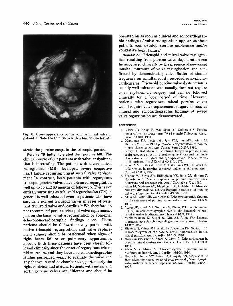
Patient 3. JF, a 50-year-old white woman with severe mitral stenosis, had mitral valve replacement surgery with a porcine (Hancock) valve in March, 1974. She presented 50 months later with sudden onset of shortness of breath, orthopnea, and hearing a “loud noise in her heart.” Physical examination revealed evidence of congestive heart failure. In addition, a new systolic thrill and a grade 616 holosystolic murmur was heard at the apex radiating to the axilla (Fig. 4). Echocardiography revealed systolic cusp fluttering with similar frequency as the holosystolic murmur (Fig. 5). The patient under- went porcine mitral valve replacement and at sur- gery was noted to have a tear in one of the cusps without evidence of thickening (Fig. 6).
DISCUSSION
Porcine valve degeneration. Gross anatomic exami- nation of severe porcine valve degeneration has revealed cusp thickening and calcification with or without cusp tears.3 Depending on which of the pathologic features predominate, the patient clini- cally presents with findings of valve stenosis or more
commonly valve regurgitation or both.5 Echocardi- ography usually reflects the underlying pathology and may demonstrate cusp thickening or fiutter.6 The echocardiographic findings of our patients dem- onstrated tricuspid diastolic and mitral systolic cusp flutter. We postulate that the valve flutter results from the flow of blood across torn and unsupported cusp margins similar to that reported with flail native valves.” This theory is further supported by observing similar frequency of the diastolic and systolic murmurs and the cusp flutter on simulta- neously performed phono-echocardiography. Ven- kataraman et al9 had reported similar echo-phono- cardiographic findings in patients with musical mur- murs and regurgitant native mitral, tricuspid, and aortic valves.
Mitral systolic and tricuspid diastolic porcine cusp flutter. Diastolic fluttering of mitral porcine valves has been reported in the presence of aortic regurgi- tation, atria1 fibrillation, and paravalvular regurgi- tation.‘0-‘2 However, the systolic fluttering observed in our patients has not been reported with any other conditions except for valve regurgitation.6 Diastolic fluttering of a porcine tricuspid valve has not been reported with pulmonary regurgitation, atrial fibril- lation, or paravalvular regurgitation, but it is con- ceivable that this probably does occur on the same basis as with mitral porcine valves. None of our patients had clinical evidence of pulmonary regurgi-
Volume 105
Number 3 Echophonograms of regurgitant porcine AV valves 459
: , i/
_-_ __ ___*_.-- . - . - _ _c __c^c* “ - - . - -+ . .+T . . I l - . f . - . ^ - *_ I . . ” _I - _ . . ”
I
EKG/” r! - Ir -
Apex
’ i , i ; I I
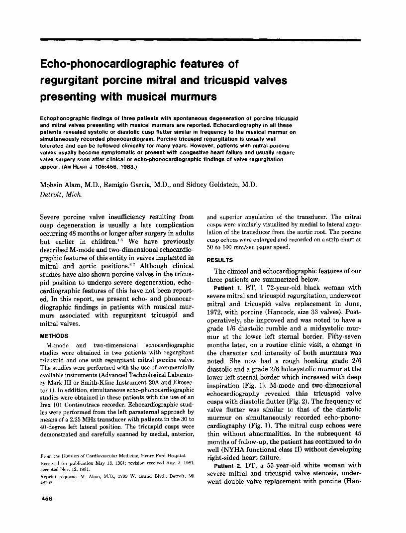
iliii~i~~lilliiiiiii~~i~~iitlIIiiii~ Fig. 4. Phonocardiogram of patient 3. Note the (musical) holosystolic murmur at the apex. C = carotid pulse; EKG = electrocardiogram.
Fig. 5. Echocardiogram of regurgitant and thin mitral porcine valve of patient 3 recorded at 100 mm/set paper speed. Note the systolic fluttering of the cusps (arrow) with similar frequency as that of holosystolic musical murmur depicted in Fig. 4.
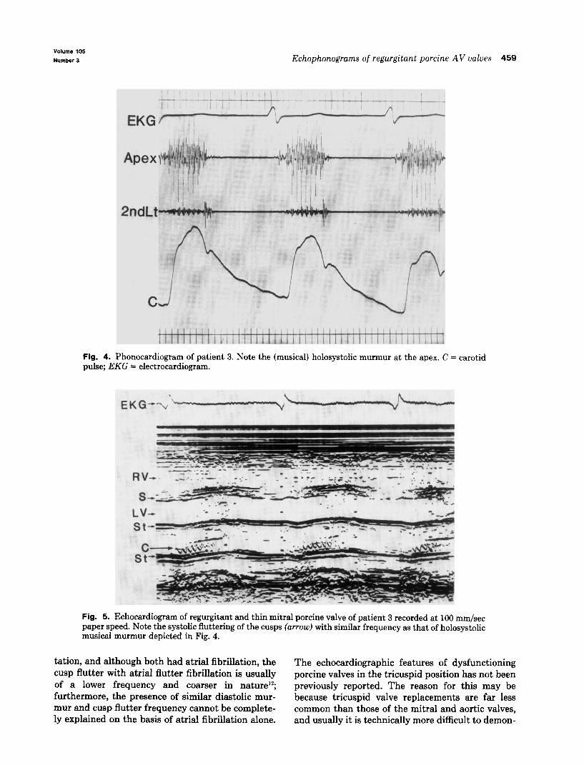
tation, and although both had atria1 fibrillation, the The echocardiographic features of dysfunctioning cusp flutter with atrial flutter fibrillation is usually porcine valves in the tricuspid position has not been of a lower frequency and coarser in nature12; previously reported. The reason for this may be furthermore, the presence of similar diastolic mur- because tricuspid valve replacements are far less mur and cusp flutter frequency cannot be complete- common than those of the mitral and aortic valves, ly explained on the basis of atria1 fibrillation alone. and usually it is technically more difficult to demon-
460 Alam, Garcia, and Goldstein March, 1983
American Heart Journal
Fig. 6. Gross appearance of the porcine mitral valve of patient 3. Note the thin cusps with a tear in one leaflet.
skate the porcine cusps in the tricuspid position. Porcine TR better tolerated than porcine MR. The
clinical course of our patients with valvular dysfunc- tion is interesting. The patient with severe mitral regurgitation (MR) developed severe congestive heart failure requiring urgent mitral valve replace- ment In contrast, both patients with regurgitant tricuspid porcine valves have tolerated regurgitation well up to 45 and 60 months of follow-up. This is not entirely surprising as tricuspid regurgitation (TR) in general is well tolerated even in patients who have surgically excised tricuspid valves in cases of resis- tant tricuspid valve endocarditis.13 We therefore do not recommend porcine tricuspid valve replacement just on the basis of valve regurgitation or abnormal echo-phonocardiographic findings alone. These patients should be followed as any patient with native tricuspid regurgitation, and valve replace- ment surgery should be performed when signs of right heart failure or pulmonary hypertension appear. Both these patients have been closely fol- lowed clinically since the onset of regurgitant tricus- pid murmurs, and they have had echocardiographic studies performed yearly to evaluate the valve and any change in cardiac chamber size, particularly the right ventricle and atrium. Patients with mitral and aortic porcine valves are different and should be
operated on as soon as clinical and echocardiograp- hit findings of valve regurgitation appear, as these patients soon develop exercise intolerance and/or congestive heart failure.”
Conclusion. Tricuspid and mitral valve regurgita- tion resulting from porcine valve degeneration can be recognized clinically by the presence of new-onset musical murmurs of valve regurgitation and con- firmed by demonstrating valve flutter of similar frequency on simultaneously recorded echo-phono- cardiograms. Tricuspid porcine valve dysfunction is usually well tolerated and usually does not require valve replacement surgery and can be followed clinically for a long period of time. However, patients with regurgitant mitral porcine valves would require valve replacement surgery as soon as clinical and echocardiographic findings of severe valve regurgitation are demonstrated.
REFERENCES
1.
2.
:i.
4.
5.
6.
7.
8.
9.
10.
Lakier JB, Khaja F, Magilligan DJ, Goldstein S: Porcine xenograft valves. Long-term 16089 month) Follow-up. Circu- lation 62:313. 1980. Magilligan DJ, Lewis JW. .Jara FM, Lee MW, Alam M. Riddle JM, Stein PD: Spontaneous degeneration of porcine bioprosthetic valves. Ann Thorac Surg 30:259, 1980. Sorav TL. Roberts WC: Structural changes in porcine xeno- grafts used as a substitute cardiac valve. Gross and histologic observations in 51 glutaraldehyde-preserved Hancock valves in 41 patients. .4m J Cardiol 40:319, 1977. Silver MM, Pollak J, Silver MD, Williams WG, Trusler GA: Calcification in porcine xenograft valves in children. Am .J Cardiol 45:685, 1980. Ferrans VJ, Boyce SW, Billingham ME, dones M, lshihara T, Roberts WC: Calcific deposits in porcine bioprostheses: Structure and pathogenesis. Am J Cardiol 46:721, 1980. Alam M, Madrazo AC, Magilligan DJ, Goldstein S: M-mode and two-dimensional echocardiographic features of porcine valve dysfunction. Am J Cardiol 43:502, 1979. Alam M. Lakier JB. Goldstein S: Echocardioaraphic changes in the thickness of’porcine valves with tim; Chest 79:663, 1981. Meyer JF, Frank MJ, Goldberg S, Cheng TO: Systolic mitral flutter, an echocardiographic clue to the diagnosis of rup- tured chordae tendineae. AM HEART J 93:3, 1977. Venkataraman K, Siegel R, Kim SJ, Allen JW: Musical murmurs: An echo-phonocardiographic study. Am ,J Cardiol 41:952, 1978. Bloch WN, Felner JM, Wicklitfe C. Symbas PN, Schlant RC: Echocardiogram of the porcine aortic bioprosthesis in the mitral position. Am J Cardiol 38:293, 1976.
11. Harrison EE. Sbar S. Suoto E. Clark P: Echocardiogram in porcine mitral dysfunction (letter). Am J Cardiol -45908, 1980.
12. Alam M, Goldstein S: Echocardiogram in porcine mitral dysfunction (reply). Am J Cardiol 45908, 1980.
19. Robin E, Thorns NW, Arbulu A, Ganguly SN, Magnisalis K: Hemodynamic consequences of total removal of the tricuspid valve without prosthetic replacement. Am J Cardiol 35481, 1975.