echinoderm calcite: single crystal or polycrystalline aggregate
TRANSCRIPT
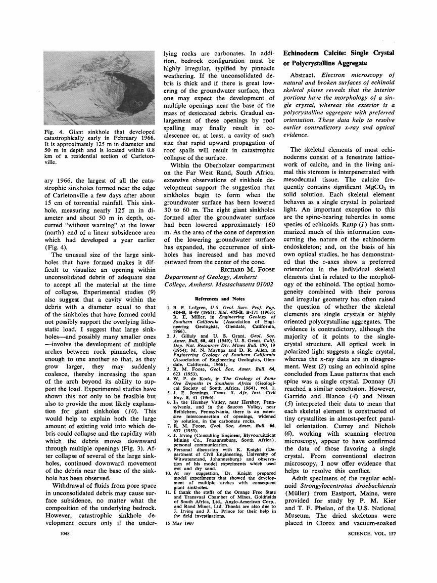
Fig. 4. Giant sinkhole that developed catastrophically early in February 1966. It is approximately 125 m in diameter and 50 m in depth and is located within 0.8 km of a residential section of Carleton- ville.
ary 1966, the largest of all the cata- strophic sinkholes formed near the edge of Carletonville a few days after about 15 cm of torrential rainfall. This sink- hole, measuring nearly 125 m in di- ameter and about 50 m in depth, oc- curred "without warning" at the lower (north) end of a linear subsidence area which had developed a year earlier (Fig. 4).
The unusual size of the large sink- holes that have formed makes it dif- ficult to visualize an opening within unconsolidated debris of adequate size to accept all the material at the time of collapse. Experimental studies (9) also suggest that a cavity within the debris with a diameter equal to that of the sinkholes that have formed could not possibly support the overlying litho- static load. I suggest that large sink- holes-and possibly many smaller ones -involve the development of multiple arches between rock pinnacles, close enough to one another so that, as they grow larger, they may suddenly coalesce, thereby increasing the span of the arch beyond its ability to sup- port the load. Experimental studies have shown this not only to be feasible but also to provide the most likely explana- tion for giant sinkholes (10). This would help to explain both the large amount of existing void into which de- bris could collapse and the rapidity with which the debris moves downward through multiple openings (Fig. 3). Af- ter collapse of several of the large sink- holes, continued downward movement of the debris near the base of the sink- hole has been observed.
Withdrawal of fluids from pore space in unconsolidated debris may cause sur- face subsidence, no matter what the composition of the underlying bedrock. However, catastrophic sinkhole de- velopment occurs only if the under-
1048
lying rocks are carbonates. In addi- tion, bedrock configuration must be highly irregular, typified by pinnacle weathering. If the unconsolidated de- bris is thick and if there is great low- ering of the groundwater surface, then one may expect the development of multiple openings near the base of the mass of desiccated debris. Gradual en- largement of these openings by roof spalling may finally result in co- alescence or, at least, a cavity of such size that rapid upward propagation of roof spalls will result in catastrophic collapse of the surface.
Within the Oberholzer compartment on the Far West Rand, South Africa, extensive observations of sinkhole de- velopment support the suggestion that sinkholes begin to form when the groundwater surface has been lowered 30 to 60 m. The eight giant sinkholes formed after the groundwater surface had been lowered approximately 160 m. As the area of the cone of depression of the lowering groundwater surface has expanded, the occurrence of sink= holes has increased and has moved outward from the center of the cone.
RICHARD M. FoosF8E
Department of Geology, Amherst College, Amherst, Massachusetts 01002
References and Notes
1. B. E. Lofgren, U.S. Geol. Surlv. Prof. Pap. 424-B, B-49 (1961); ibid. 475-B, B-171 (1963); R. E. Miller, in Engineering Geology of Southern California (Association of Engi- neering Geologists, Glendale, California, 1966).
2. J. Gilluly and U. S. Grant, Geol. Soc. Amer. Bull. 60, 461 (1949); U. S. Grant, Calif, Dep. Nat. Resources Div. Mines Bull. 170, 19 (1954); M. N. Mayuga and D. R. Allen, in Engineering Geology of Southern California (Association of Engineering Geologists, Glen- dale, California, 1966).
3. R. M. Foose, Geol. Soc. Amer, Bull, 64, 623 (1953).
4. W. P. de Kock, in The Geology of Some Ore Deposits in Southern Africa (Geologi- cal Society of South Africa, 1964), vol. 1.
5. J. E. Jennings, Trans. S. Aft. Inst. Civil Eng. 8, 41 (1966).
6. In the Hershey Valley, near Hershey, Pernl- sylvania, and in the Saucon Valley, near Bethlehem, Pennsylvania, there is an exten sive interconnection of openings, widened by solution, in the carbonate rocks.
7. R. M. Foose, Geol. Soc. Amer. Bull. 64, 637 (1953).
8. J. Irving (Consulting Engineer, Blyvooruitzicht Mining Co., Johannesburg, South Africa), personal communication.
9. Personal discussion with K. Knight (De- partment of Civil Engineering, University of Witwatersrand, Johannesburg) and observa- tion of his model experiments which used wet and dry sand.
10. At my suggestion, Dr. Knight prepared model experiments that showed the develop- ment of multiple arches with consequent giant sinkholes.
11. I thank the staffs of the Orange Free State and Transvaal Chamber of Mines, Goldfields
lying rocks are carbonates. In addi- tion, bedrock configuration must be highly irregular, typified by pinnacle weathering. If the unconsolidated de- bris is thick and if there is great low- ering of the groundwater surface, then one may expect the development of multiple openings near the base of the mass of desiccated debris. Gradual en- largement of these openings by roof spalling may finally result in co- alescence or, at least, a cavity of such size that rapid upward propagation of roof spalls will result in catastrophic collapse of the surface.
Within the Oberholzer compartment on the Far West Rand, South Africa, extensive observations of sinkhole de- velopment support the suggestion that sinkholes begin to form when the groundwater surface has been lowered 30 to 60 m. The eight giant sinkholes formed after the groundwater surface had been lowered approximately 160 m. As the area of the cone of depression of the lowering groundwater surface has expanded, the occurrence of sink= holes has increased and has moved outward from the center of the cone.
RICHARD M. FoosF8E
Department of Geology, Amherst College, Amherst, Massachusetts 01002
References and Notes
1. B. E. Lofgren, U.S. Geol. Surlv. Prof. Pap. 424-B, B-49 (1961); ibid. 475-B, B-171 (1963); R. E. Miller, in Engineering Geology of Southern California (Association of Engi- neering Geologists, Glendale, California, 1966).
2. J. Gilluly and U. S. Grant, Geol. Soc. Amer. Bull. 60, 461 (1949); U. S. Grant, Calif, Dep. Nat. Resources Div. Mines Bull. 170, 19 (1954); M. N. Mayuga and D. R. Allen, in Engineering Geology of Southern California (Association of Engineering Geologists, Glen- dale, California, 1966).
3. R. M. Foose, Geol. Soc. Amer, Bull, 64, 623 (1953).
4. W. P. de Kock, in The Geology of Some Ore Deposits in Southern Africa (Geologi- cal Society of South Africa, 1964), vol. 1.
5. J. E. Jennings, Trans. S. Aft. Inst. Civil Eng. 8, 41 (1966).
6. In the Hershey Valley, near Hershey, Pernl- sylvania, and in the Saucon Valley, near Bethlehem, Pennsylvania, there is an exten sive interconnection of openings, widened by solution, in the carbonate rocks.
7. R. M. Foose, Geol. Soc. Amer. Bull. 64, 637 (1953).
8. J. Irving (Consulting Engineer, Blyvooruitzicht Mining Co., Johannesburg, South Africa), personal communication.
9. Personal discussion with K. Knight (De- partment of Civil Engineering, University of Witwatersrand, Johannesburg) and observa- tion of his model experiments which used wet and dry sand.
10. At my suggestion, Dr. Knight prepared model experiments that showed the develop- ment of multiple arches with consequent giant sinkholes.
11. I thank the staffs of the Orange Free State and Transvaal Chamber of Mines, Goldfields of South Africa, Ltd., Anglo-American Corp., and Rand Mines, Ltd. Thanks are also due to J. Irving and J. L. Prince for their help in the field investigations.
15 May 1967
of South Africa, Ltd., Anglo-American Corp., and Rand Mines, Ltd. Thanks are also due to J. Irving and J. L. Prince for their help in the field investigations.
15 May 1967
Echinoderm Calcite: Single Crystal or Polycrystalline Aggregate
Abstract. Electron microscopy of ratural and broken surfaces of echinoid skeletal plates reveals that the interior portions have the morphology of a sin- gle crystal, whereas the exterior is a polycrystalline aggregate with preferred orientation. These data help to resolve earlier contradictory x-ray and optical evidence.
The skeletal elements of most echi- noderms consist of a fenestrate lattice- work of calcite, and in the living ani- mal this stereom is interpenetrated with mesodermal tissue. The calcite fre- quently contains significant MgCO3 in solid solution. Each skeletal element behaves as a single crystal in polarized light. An important exception to this are the spine-bearing tubercles in some species of echinoids. Raup (1) has sum- marized much of this information con- cerning the nature of the echinoderm endoskeleton; and, on the basis of his own optical studies, he has demonstrat- ed that the c-axes show a preferred orientation in the individual skeletal elements that is related to the morphol- ogy of the echinoid. The optical homo- geneity combined with their porous and irregular geometry has often raised the question of whether the skeletal elements are single crystals or highly oriented polycrystalline aggregates. The evidence is contradictory, although the majority of it points to the single- crystal structure. All optical work in polarized light suggests a single crystal, whereas the x-ray data are in disagree- ment. West (2) using an echinoid spine concluded from Laue patterns that each spine was a single crystal. Donnay (3) reached a similar conclusion. However, Garrido and Blanco (4) and Nissen (5) interpreted their data to mean that each skeletal element is constructed of tiny crystallites in almost-perfect paral- lel orientation. Currey and Nichols (6), working with scanning electron microscopy, appear to have confirmed the data of those favoring a single crystal. From conventional electron microscopy, I now offer evidence that helps to resolve this conflict.
Adult specimens of the regular echi- noid Strongylocentrotus droebachiensis (Muller) from Eastport, Maine, were
Echinoderm Calcite: Single Crystal or Polycrystalline Aggregate
Abstract. Electron microscopy of ratural and broken surfaces of echinoid skeletal plates reveals that the interior portions have the morphology of a sin- gle crystal, whereas the exterior is a polycrystalline aggregate with preferred orientation. These data help to resolve earlier contradictory x-ray and optical evidence.
The skeletal elements of most echi- noderms consist of a fenestrate lattice- work of calcite, and in the living ani- mal this stereom is interpenetrated with mesodermal tissue. The calcite fre- quently contains significant MgCO3 in solid solution. Each skeletal element behaves as a single crystal in polarized light. An important exception to this are the spine-bearing tubercles in some species of echinoids. Raup (1) has sum- marized much of this information con- cerning the nature of the echinoderm endoskeleton; and, on the basis of his own optical studies, he has demonstrat- ed that the c-axes show a preferred orientation in the individual skeletal elements that is related to the morphol- ogy of the echinoid. The optical homo- geneity combined with their porous and irregular geometry has often raised the question of whether the skeletal elements are single crystals or highly oriented polycrystalline aggregates. The evidence is contradictory, although the majority of it points to the single- crystal structure. All optical work in polarized light suggests a single crystal, whereas the x-ray data are in disagree- ment. West (2) using an echinoid spine concluded from Laue patterns that each spine was a single crystal. Donnay (3) reached a similar conclusion. However, Garrido and Blanco (4) and Nissen (5) interpreted their data to mean that each skeletal element is constructed of tiny crystallites in almost-perfect paral- lel orientation. Currey and Nichols (6), working with scanning electron microscopy, appear to have confirmed the data of those favoring a single crystal. From conventional electron microscopy, I now offer evidence that helps to resolve this conflict.
Adult specimens of the regular echi- noid Strongylocentrotus droebachiensis (Muller) from Eastport, Maine, were provided for study by P. M. Kier and T. F. Phelan, of the U.S. National Museum. The dried skeletons were placed in Clorox and vacuum-soaked
SCIENCE, VOL. 157
provided for study by P. M. Kier and T. F. Phelan, of the U.S. National Museum. The dried skeletons were placed in Clorox and vacuum-soaked
SCIENCE, VOL. 157
overnight to insure removal of the or-
ganic material from within the fenes- trated stereom. Except for the pale green pigment trapped within the cal- cite, most of the organic material is removed by this treatment. Single-stage platinum-carbon replicas were prepared from both the natural and fractured surfaces of plates, tubercles, and spines, and then examined in the electron mi-
croscope. The surface of the calcite within the
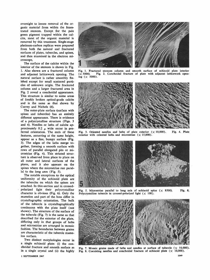
interior of the stereom is shown in Fig. 1. Also shown are a fractured column and adjacent latticework opening. The natural surface is rather smoothly fin- ished except for small scattered parti- cles of unknown origin. The fractured column and a larger fractured area in
Fig. 2 reveal a conchoidal appearance. This structure is similar to some areas of freshly broken optical-grade calcite and is the same as that shown by Currey and Nichols (6).
The outer-plate surface (surface with spines and tubercles) has an entirely different appearance. There is evidence of a polycrystalline structure (Figs. 3 and 4). Needles or laths of calcite ap- proximately 0.2 /p wide occur in pre- ferred orientation. The ends of these features, occurring at the same height, appear as a fine, bumpy surface (Fig. 3). The edges of the laths merge to- gether, forming a smooth surface with rows of parallel elongated pits or mi- crostriae (Fig. 4). This striated struc- ture is observed from place to place on all outer and lateral surfaces of the plates, and it also appears on the
spines where the microstriae run paral- lel to the long axes (Fig. 5).
The notable exceptions to the optical uniformity of the echinoid plate are the tubercles on which the spines are attached. In thin-section and in crossed- polarized light their polycrystalline character is obvious (Fig. 6). Only the mamelon and part of the boss differ in crystallographic orientation. The bulk of the tubercle is crystallographically continuous with the plate itself (not shown). The structure of the surface of the tubercle (Fig. 7) is the same as that described for the exterior of the plate, differing only in that groups of laths and microstriae are arranged in mosaic fashion. The boundaries between grains are characteristic of the tubercle mame- lon surface.
Two distinct morphologies occur in a single echinoid plate: (i) the con- choidal fracture and smooth surface as in a single crystal and (ii) the highly
1 SEPTEMBER 1967
Fig. 1. Fractured stereom column and smooth surface of echinoid plate interior (X 5000). Fig. 2. Conchoidal fracture of plate with adjacent latticework open- ing (X 5000).
Fig. 3. Oriented needles and laths of plate exterior (X 10,000). Fig. 4. Plate exterior with oriented laths and microstriae (X 15,000).
Fig. 5. Microstriae parallel to long axis of echinoid spine (X 8500). Fig. 6. Polycrystalline tubercle in crossed-polarized light (X 100).
Fig. 7. Mosaic grains made of laths and needles at surface of tubercle (X 10,000). Fig. 8. Coexisting needles and conchoidal fracture of echinoid plate (X 10,000).
1049
I
preferred orientation of crystallites as in a polycrystalline aggregate. In some photographs, these two types coexist
(Fig. 8). This may be indicative of the method of development of the echino- derm skeletal element-a process of oriented polycrystalline growth on, or in, an organic matrix followed by maturation which appears to involve recrystallization by continual fusion and coalescence.
Such an hypothesis allows for the absence of crystal faces within the ma- ture stereom. A recrystallization in the solid state produces a pseudomorph, the shape of which is determined by the polycrystalline aggregate and the original organic framework. Most of the skeletal stereom occurs in the ma- ture state, and, except for the tuber- cles, only the actively growing surfaces are polycrystalline. Thus, the echino- derm skeletal elements are neither sin-
identical sequence in the last 105 amino
Development of the concept that the light chains of immunoglobulins have a variable NH2-terminal half and an essentially invariant COOH-terminal half began in 1963 with the suggestion of Putnam et al. (1) based on peptide maps that "All Bence Jones proteins of the same antigenic type share a fixed portion of their sequence and also have a mutable part." The con- cept was explicitly proposed on the basis of partial analysis of the amino acid sequence of K-type Bence Jones proteins (2) and has received strong support as a result of complete amino acid sequence analysis of one K-type (3) and one A-type (4, 5) Bence Jones protein of man, and of extensive se- quence analysis of two K-type Bence Jones proteins from the mouse (6). We have now verified this concept for human A-chains by amino acid se- quence analysis of two more A-type Bence Jones proteins reported herein. Further support is offered by the con- firmation (7) of the sequence we have reported for the COOH-terminal half of human X-chains (5).
I050
gle crystals nor polycrystalline aggre- gates, but they are a combination of both. These data help to reconcile the contradictions in some of the studies of earlier workers.
KENNETH M. TOWE
Department of Paleobiology, Smithsonian Institution,
Washington, D.C. 20560
References and Notes
1. D. M. Raup, in Physiology of Echinodermata, R. A. Boolootian, Ed. (Interscience, New York, 1966), p. 379.
2. C. D. West, J. Paleontol. 11, 458 (1937). 3. G. Donnay, Carnegie Inst. Yearbook 55, 205
(1956). 4. J. Garrido and J. Blanco, Com7pt. Rend. 224,
485 (1947). 5. H. Nissen, Nentes Jahrb. Mineral. Abhand.
117, 230 (1963). 6. J. D. Currey and D. Nichols, Nature 214, 81
(1967). 7. G. Donnay, J. Wyatt Durham, P. M. Kier
and D. M. Raup provided helpful suggestions and criticism. I thank G. Hamilton, J. Merida and T. Phelan for assistance.
7 June 1967 u
We now report the complete amino acid sequence of two human A-type Bence Jones proteins (designated Ha and Bo, respectively) in comparison to the complete sequence we have pub- lished for the human A-type protein designated Sh (5). In Fig. 1, a dif- ferent numbering system is employed for the Ha and Bo A-chains since their length is greater than that of the Sh A-chain. This is given underneath the sequence for protein Ha. However, all citations in the following text refer to the Sh numbering system given over protein Sh in Fig. 1. [This practice is analogous to that introduced by Braunitzer et al. (8) for sequence com- parison of the a-chains and /-chains of hemoglobin, which also are of unequal length.] With this alignment the amino acid sequence of all human A-chains is presumably identical, from Gln-109 through the COOH-terminal residue Ser-213 (9) with the exception that in the Oz(+) serological subtype the ar- ginine at position 190 is replaced by lysine (10). Hence, the amino acid se- quence is given in Fig. 1 only for the
We now report the complete amino acid sequence of two human A-type Bence Jones proteins (designated Ha and Bo, respectively) in comparison to the complete sequence we have pub- lished for the human A-type protein designated Sh (5). In Fig. 1, a dif- ferent numbering system is employed for the Ha and Bo A-chains since their length is greater than that of the Sh A-chain. This is given underneath the sequence for protein Ha. However, all citations in the following text refer to the Sh numbering system given over protein Sh in Fig. 1. [This practice is analogous to that introduced by Braunitzer et al. (8) for sequence com- parison of the a-chains and /-chains of hemoglobin, which also are of unequal length.] With this alignment the amino acid sequence of all human A-chains is presumably identical, from Gln-109 through the COOH-terminal residue Ser-213 (9) with the exception that in the Oz(+) serological subtype the ar- ginine at position 190 is replaced by lysine (10). Hence, the amino acid se- quence is given in Fig. 1 only for the
NH2-terminal portions of proteins Ha and Bo through the residue denoted Gly-108 in the Sh numbering system.
The conclusion that the sequence of the last 105 residues in human light X-chains is essentially invariant is based on the following findings. By methods already described (4, 5), we have iso- lated from proteins Ha and Bo tryptic peptides corresponding to all those in protein Sh, beginning with Ala-112 through the COOH-terminal residue Ser-213. Without exception, all the cor- responding tryptic peptides for this re- gion of the three proteins [namely peptides TlX, T2, T18, T12, T1, T20, T7, T11, Tj9, and T14 of Wikler et al., (5)] are identical in amino acid compo- sition, in NH-terminal and COOH- terminal groups (in those cases deter- mined), and in such portions of their se- quence as we have already completed. These peptides from proteins Ha and Bo also have the same elution position in ion-exchange chromatography and the same electrophoretic mobility at pH 3.7 as the corresponding peptides iso- lated from the Sh protein. Insofar as the data have been obtained, the same statements apply to the chymotryptic peptides we have isolated from this portion of the three A-chains. Further- more, the published sequence of our A- chain Sh is identical in every detail (5), except for the position of one amide group, with a sequence from Gln-109 through the COOH-terminus Ser-213 for another A-type Bence Jones protein (X) (7). Thus, residues 109 to 213 are identical in all four A-type proteins (Sh, Bo, Ha, and X). On the other hand, the position corresponding to Gly-108 in protein Sh is occupied by arginine in proteins Ha and Bo (Fig. 1) and by serine in protein X.
Like K-chains (3), human A-chains differ in many positions in the NH.- terminal portion of the molecule. Ex- actly half of the 108 positions in the variable part of the lambda-type pro- tein Sh are identical with correspond- ing positions in the lambda-type pro- teins Bo and Ha (Table 1). However, the remaining 54 positions in this por- tion of the A-chain Sh differ from one or both of the other A-chains (Fig. 1). The positions of variation are distrib- uted in a seemingly random fashion throughout the first half of the A- chains; yet, there are two short pep-
NH2-terminal portions of proteins Ha and Bo through the residue denoted Gly-108 in the Sh numbering system.
The conclusion that the sequence of the last 105 residues in human light X-chains is essentially invariant is based on the following findings. By methods already described (4, 5), we have iso- lated from proteins Ha and Bo tryptic peptides corresponding to all those in protein Sh, beginning with Ala-112 through the COOH-terminal residue Ser-213. Without exception, all the cor- responding tryptic peptides for this re- gion of the three proteins [namely peptides TlX, T2, T18, T12, T1, T20, T7, T11, Tj9, and T14 of Wikler et al., (5)] are identical in amino acid compo- sition, in NH-terminal and COOH- terminal groups (in those cases deter- mined), and in such portions of their se- quence as we have already completed. These peptides from proteins Ha and Bo also have the same elution position in ion-exchange chromatography and the same electrophoretic mobility at pH 3.7 as the corresponding peptides iso- lated from the Sh protein. Insofar as the data have been obtained, the same statements apply to the chymotryptic peptides we have isolated from this portion of the three A-chains. Further- more, the published sequence of our A- chain Sh is identical in every detail (5), except for the position of one amide group, with a sequence from Gln-109 through the COOH-terminus Ser-213 for another A-type Bence Jones protein (X) (7). Thus, residues 109 to 213 are identical in all four A-type proteins (Sh, Bo, Ha, and X). On the other hand, the position corresponding to Gly-108 in protein Sh is occupied by arginine in proteins Ha and Bo (Fig. 1) and by serine in protein X.
Like K-chains (3), human A-chains differ in many positions in the NH.- terminal portion of the molecule. Ex- actly half of the 108 positions in the variable part of the lambda-type pro- tein Sh are identical with correspond- ing positions in the lambda-type pro- teins Bo and Ha (Table 1). However, the remaining 54 positions in this por- tion of the A-chain Sh differ from one or both of the other A-chains (Fig. 1). The positions of variation are distrib- uted in a seemingly random fashion throughout the first half of the A- chains; yet, there are two short pep- tide stretches where the amino acids differ in all three A-chains (residues 48 to 51 and 91 to 95). Conversely, up to residue 108 there are four tetra-
SCIENCE, VOL. 157
tide stretches where the amino acids differ in all three A-chains (residues 48 to 51 and 91 to 95). Conversely, up to residue 108 there are four tetra-
SCIENCE, VOL. 157
Immunoglobulin Structure: Variation in Amino Acid
Sequence and Length of Human Lambda Light Chains
Abstract. Variation and conservation in the primary structure of human lambda light chains is revealed by complete amino acid sequence of three Bence Jones proteins. These proteins differ in amino acid sequence in from 38 to 48 positions; they are of unequal length in the amino-terminal half of the chain but have