echinococcus granulosus antigen b binds to … open access echinococcus granulosus antigen b binds...
TRANSCRIPT

RESEARCH Open Access
Echinococcus granulosus Antigen B binds tomonocytes and macrophages modulatingcell response to inflammationValeria Silva-Álvarez1,2,3†, Ana Maite Folle1†, Ana Lía Ramos1, Eduardo S. Kitano4, Leo K. Iwai4, Inés Corraliza5,Betina Córsico2,3 and Ana María Ferreira1*
Abstract
Background: Antigen B (EgAgB) is an abundant lipoprotein released by the larva of the cestode Echinococcusgranulosus into the host tissues. Its protein moiety belongs to the cestode-specific family known as hydrophobicligand binding protein (HLBP), and is encoded by five gene subfamilies (EgAgB8/1-EgAgB8/5). The functions ofEgAgB in parasite biology remain unclear. It may play a role in the parasite’s lipid metabolism since it carries hostlipids that E. granulosus is unable to synthesise. On the other hand, there is evidence supporting immuno-modulating activities in EgAgB, particularly on innate immune cells. Both hypothetical functions might involveEgAgB interactions with monocytes and macrophages, which have not been formally analysed yet.
Methods: EgAgB binding to monocytes and macrophages was studied by flow cytometry using inflammation-recruited peritoneal cells and the THP-1 cell line. Involvement of the protein and phospholipid moieties in EgAgBbinding to cells was analysed employing lipid-free recombinant EgAgB subunits and phospholipase D treated-EgAgB (lacking the polar head of phospholipids). Competition binding assays with plasma lipoproteins and ligandsfor lipoprotein receptors were performed to gain information about the putative EgAgB receptor(s) in these cells.Arginase-I induction and PMA/LPS-triggered IL-1β, TNF-α and IL-10 secretion were examined to investigate theoutcome of EgAgB binding on macrophage response.
Results: Monocytes and macrophages bound native EgAgB specifically; this binding was also found with lipid-freerEgAgB8/1 and rEgAgB8/3, but not rEgAgB8/2 subunits. EgAgB phospholipase D-treatment, but not thecompetition with phospholipid vesicles, caused a strong inhibition of EgAgB binding activity, suggesting an indirectcontribution of phospholipids to EgAgB-cell interaction. Furthermore, competition binding assays indicated that thisinteraction may involve receptors with affinity for plasma lipoproteins. At functional level, the exposure ofmacrophages to EgAgB induced a very modest arginase-I response and inhibited PMA/LPS-mediated IL-1β andTNF-α secretion in an IL-10-independent manner.
Conclusion: EgAgB and, particularly its predominant EgAgB8/1 apolipoprotein, are potential ligands for monocyteand macrophage receptors. These receptors may also be involved in plasma lipoprotein recognition and induce ananti-inflammatory phenotype in macrophages upon recognition of EgAgB.
Keywords: Echinococcus granulosus, Antigen B, HLBP, Cell binding, Lipoproteins, Monocytes, Macrophages
* Correspondence: [email protected]†Equal contributors1Cátedra de Inmunología, Facultad de Ciencias/Facultad de Química,Universidad de la República (UdelaR), Montevideo, UruguayFull list of author information is available at the end of the article
© 2016 Silva-Álvarez et al. Open Access This article is distributed under the terms of the Creative Commons Attribution 4.0International License (http://creativecommons.org/licenses/by/4.0/), which permits unrestricted use, distribution, andreproduction in any medium, provided you give appropriate credit to the original author(s) and the source, provide a link tothe Creative Commons license, and indicate if changes were made. The Creative Commons Public Domain Dedication waiver(http://creativecommons.org/publicdomain/zero/1.0/) applies to the data made available in this article, unless otherwise stated.
Silva-Álvarez et al. Parasites & Vectors (2016) 9:69 DOI 10.1186/s13071-016-1350-7

BackgroundCystic echinococcosis is a worldwide zoonotic infectioncaused by the larval stage of Echinococcus granulosussensu lato species complex, which includes at least sevenspecies that affects humans and livestock with significanteconomic and public health impact [1–5]. The larvae(metacestodes) of these species are fluid-filled, bladder-like structures that establish and gradually grow in theparenchyma of host internal organs, most commonlyliver and lungs. They are usually called hydatid cysts, al-though strictly the term cyst includes a fibrous adventitiallayer generated as a consequence of the host inflammatoryreaction. Once the larva matures and reaches fertility, itgenerates protoscolex (PE), which are the parasite formscapable of developing into the adult worm in the definitivehost (usually dogs). The fluid contained within the cyst,known as hydatid cyst fluid (HF), collects a variety ofproducts excreted or secreted by the cellular, germinallayer (GL) of the cyst wall, as well as by protoscoleces. Inaddition, HF collects a variety of host plasma proteins(mostly albumin and immunoglobulins) which cross thecyst wall by unidentified mechanisms. This work refers toE. granulosus sensu lato, but for simplicity we will use theterm E. granulosus.One of the major HF components synthesised in abun-
dance by the larva is a lipoprotein named Antigen B(EgAgB) [6]. The composition and antigenicity of EgAgBhas been extensively studied, due to the fact that it con-stitutes the most immunogenic and specific Echinococ-cus-genus antigen for human serodiagnosis [7–10].Molecular studies for characterising EgAgB protein moi-ety showed that it is encoded by a polymorphic and mul-tigenic family that comprises five subfamilies namedEgAgB1 to EgAgB5 [11–17]. These genes are differen-tially expressed in single lifecycle stages of the parasite,as well as within distinct tissues of a given developmen-tal stage. EgAgB1 to EgAgB4 are expressed in the meta-cestode stage whereas EgAgB5 seems to be mostlyexpressed in the adult stage. Regarding the metacestode,the expression of all genes was detected in GL, beingEgAgB1 and EgAgB3 the most abundant, while in PEEgAgB3 seems to be over-represented [18].The mature protein products of EgAgB genes are α-
helix rich polypeptides of 8 kDa, referred to as EgAgB8/1 to EgAgB8/5 subunits or apolipoproteins. Interestingly,EgAgB was found to belong to a novel cestode-specificfamily known as Hydrophobic Ligand Binding Proteins(HLBPs) [19], which have emerged by independent geneexpansion events in different species [18]. More recently,our group has made substantial progress in the bio-chemical characterisation of EgAgB by developing novelmethodological tools for purifying and characterising thelipid-free EgAgB8 apolipoproteins [20], and by determin-ing which are its native lipid components [21]. We
showed that in vitro lipid-free EgAgB8 subunits oligo-merise; which agrees with their electrostatic profile pre-dicted by structure modelling [22, 23]. They boundlipids selectively, particularly phospholipids and fattyacids rather than cholesterol [20], confirming previousobservations [19]. Binding of lipids may enhance theoligomerisation of EgAgB8 subunits, favouring the for-mation of large lipoprotein complexes. We showed thatthe native antigen is a large (about 230 kDa in mass)lipoprotein particle, in which lipids account for aboutone-half of EgAgB total mass, comprising a heteroge-neous mixture of neutral (mainly triacylglycerides,sterols and sterol esters) and polar lipids (mainly phos-phatidylcholine) [21]. In sum, EgAgB may adopt a struc-tural organisation similar to that of vertebrate highdensity lipoprotein (HDL), in which around a dozenEgAgB8 apolipoproteins would be embedded in an outer,hydrophilic phospholipid layer that surrounds the hydro-phobic core of the lipoprotein particle [23].We have thus achieved a better knowledge of EgAgB
chemical composition and physicochemical properties.However, the understanding of the role of EgAgB in theparasite adaptation to host environment is still very rudi-mentary. It is uncertain how and when EgAgB is trans-ported outside of the hydatid cyst; its apparent absencein the laminar layer as well as in the apical side of thetegumental syncytium of the GL does not support theexistence of active secretion mechanisms in GL for thistransport [24, 25]. Nevertheless, the fact that the hostmounts a strong specific antibody response against EgAgBreveals that it reaches host tissues. Furthermore, an EgAgBortholog in Taenia solium (TsM 150 kDa HLBP) waslocalised within the granuloma, adjacent to host paren-chyma cells [26]. EgAgB contains lipids that E. granulosusis not able to synthesise [27, 28], such as fatty acids andcholesterol, and this supports the idea that EgAgB partici-pates in parasite’s lipid metabolism (reviewed by [23]). Al-ternatively, EgAgB effects on various innate immune cells,including neutrophils, monocytes and dendritic cells [29–32], have led to the concept that this antigen is capable ofmodulating local inflammation and the subsequent mech-anisms that activate T lymphocytes, favouring the gener-ation of a Th2-type specific response [31]. Nevertheless,this Th2-biasing activity needs to be verified using the na-tive lipoprotein, since it was described using denaturedEgAgB (purified using electroelution [28] or after heatingat 100 °C [31]). In any case, a putative EgAgB role in para-site mechanisms associated with lipid metabolism orimmunomodulation would involve a direct interaction be-tween EgAgB and host cells, likely mediated by cell recep-tors, and these interactions have not been examined yet.In this work, we studied the ability of monocytes and
macrophages to bind EgAgB selectively as well as theimpact of EgAgB binding on the activation phenotype of
Silva-Álvarez et al. Parasites & Vectors (2016) 9:69 Page 2 of 17

macrophages. Our results show that EgAgB, particularlyits apolipoprotein component, binds to monocytes andmacrophages specifically, using receptors shared withplasma lipoproteins. Furthermore, EgAgB receptors inmacrophages seem to induce signalling events involvedin the regulation of inflammatory pathways.
MethodsReagentsAntibiotic solutions, 3,5-di-tert-butyl–4-hydroxytoluene(BHT), cell culture reagents, dimethyl sulfoxide (DMSO),inorganic salts, ovalbumin (OVA), L-arginine, lipopolysac-charides (LPS) from Escherichia coli, phorbol 12-myristate13-acetate (PMA), reduced glutathione, RPMI 1640 cul-ture medium, streptavidin-peroxidase, 3,3′,5,5′-tetra-methylbenzidine (TMB), thrombin from human plasmaand urea were acquired from Sigma Chemicals (USA).Ethylenediaminetetraacetic acid (EDTA) and potassiumbromide were obtained from Applichem (Germany).Acetonitrile was purchased from JT Baker (USA) and tri-fluoroacetic acid from Merck (Germany). GluthathioneSepharose 4B and Q-Sepharose resins were acquired fromGE Healthcare Life Sciences (Sweden). C8-bonded silicacolumn was purchased from Vydac (USA). Humanmonocyte-like THP-1 cell line was obtained from Ameri-can Type Culture Collection (ATCC, USA). Fetal bovineserum (FS) was purchased from Gibco (USA) and N-Hydroxysuccinimide-Biotin (NHS-Biotin) was acquiredfrom Pierce (USA). Phospholipase D (PLD) was obtainedfrom Calbiochem (Germany). Monoclonal antibody (mab)against EgAgB8/1 subunit (EB7 mab) was generouslydonated by Dr. Gualberto González (Cátedra de Inmuno-logía, Facultad de Química, UdelaR). Flow cytometry anti-bodies were purchased from BD Biosciencies or Biolegend(USA). Phosphatidylcholine (PC) and phosphatidylserine(PS) were obtained from Avanti Polar Lipids (USA).
MiceWild type C57BL/6J and BALB/c mice as well as LDL-receptor (LDLr) deficient mice on the C57BL/6J back-ground (B6.129S7-Ldlrtm1Her) were acquired fromPasteur Institute of Montevideo (Uruguay). Animal ma-nipulation and husbandry were done in accordance withthe ethical committee guidelines of the Honorary Commis-sion of Animal Experimentation (CHEA) from UdelaR.
Parasite materialE. granulosus bovine hydatid cysts were obtained fromliver and lungs of naturally infected animals, collectedduring the routine work of local abattoirs inMontevideo (Uruguay). Infertile HF were obtained byaseptic aspiration of the cyst content, preserved byaddition of 5 mM EDTA and 20 μM BHT, and main-tained at −20 °C until use.
Purification of native EgAgBPurification of native EgAgB was achieved employing atwo-step procedure. A pool of individual HF (at least avolume of 2 L, collected from an average of 15 hydatidcysts) was prepared and clarified by centrifugation at10000 g followed by filtration through 0.45 μm filtermembrane (Millipore). The clarified HF was thenfractionated by anion exchange chromatography on a Q-Sepharose column, using 20 mM phosphate, pH 7.5 con-taining 200 mM NaCl, 5 mM EDTA and 20 μM BHT asequilibration buffer, and changing the ionic strength to500 mM NaCl in a single step for elution. The elutedfraction (fQS) was concentrated 10-fold using a SavantSpeedVac System and equilibrated in phosphate bufferedsaline (PBS) containing 5 mM EDTA and 20 μM BHT(PBSEB) with a PD-10 desalting column (Amersham,Biosciences). EgAgB was then purified by ultracentrifu-gation of fQS in a KBr density gradient. 2.45 g of KBrwere dissolved in 5 ml of fQS in an ultracentrifuge tubeand slowly covered with a solution containing 0.15 MNaCl and 0.42 M KBr. After ultracentrifugation (4 h at332000 g) two yellowish bands were obtained. EgAgBwas recovered in the yellow-brown, low density fraction.The homogeneity of EgAgB preparation was analysed bySDS-PAGE on 15 % polyacrylamide gels followed byCoomassie staining. At least two consecutive ultracentri-fugation rounds were performed to achieve a good-quality EgAgB preparation (higher than 95 % purity).Finally, EgAgB was equilibrated in PBSEB and main-tained at 4 °C under N2 atmosphere until use. Five dif-ferent EgAgB preparations were used during this work.The apolipoprotein composition of native EgAgB wasdetermined by LC-MS/MS, using an LTQ-OrbitrapVelos mass spectrometer (Thermo Scientific). Proteinswere identified using MaxQuant software (v.1.5.3.8) bysearching MS and MS/MS data against the Echinococcusdatabase; this database was built in house comprising allEchinococcus granulosus G1 sequences (published inwww.genedb.org as EGU_proteins_29042013_products.fa)plus a total of 107 sequences, including polymorphic vari-ations at the level of the mature products as well as theorthologous products in E. granulosus s.l. and E. mul-tilocularis (available on NCBInr). Statistical analysisfor protein identification was performed using Perseus(v. 1.4.0.11) on the basis of unique peptide MS inten-sities and the presence of a minimum of two uniquepeptides. For evaluating the abundance of EgAgB8subunits in samples, intensity-based absolute quantifi-cation (iBAQ) was used since it has been reported asa useful label-free quantification method provided byMaxQuant [33]. The relative abundance of a particu-lar EgAgB8 subunit was then estimated as the per-centage of the total iBAQ intensity corresponding toEgAgB apolipoproteins.
Silva-Álvarez et al. Parasites & Vectors (2016) 9:69 Page 3 of 17
For functional assays on macrophages native EgAgBwas purified by immunoaffinity chromatography employ-ing the EB7 mab as described by González et al. [13](immunopurified EgAgB) to exclude pyrogens which mayaffect cell functions even at trace levels.
Purification of recombinant lipid-free EgAgB8 subunitsPurification of recombinant lipid-free EgAgB8 subunits(rEgAgB8) was undertaken employing a protocol pre-viously described [20]. Briefly, rEgAgB8/1, rEgAgB8/2and rEgAgB8/3 subunits (corresponding to [Uniprot:Q9UA06, Q27275 and Q95NW6, respectively]) werepurified as Glutathione-S-Transferase fusion proteinsby affinity chromatography on immobilised glutathi-one and recovered by thrombin cleavage. Delipidationof rEgAgB8 subunits was achieved by reversed-phasehigh-performance liquid chromatography (RP-HPLC)in an HPLC System (Merck-Hitachi, Japan) with C8-bonded silica as stationary phase and water/acetonitrile/trifluoroacetic acid mobile phase. The purity of lipid-freerEgAgB8 subunits was monitored by SDS-PAGE on15 % polyacrylamide gels followed by Coomassiestaining. Lipid-free rEgAgB8 subunits were maintainedat −80 °C until use.
Biotin labelling of EgAgBBiotinylation of native EgAgB was carried out by adding80 μL of a fresh solution of NHS-Biotin (5 mg/mL inDMSO) per mg of protein in sodium carbonate 0.1 M,pH 9.0. In the case of lipid-free rEgAgB81, rEgAgB8/2and rEgAgB8/3 subunits, we used an NHS-Biotin con-centration sufficient to label a maximum of six lysinesper molecule in each subunit. The mixture was incu-bated during 4 h at room temperature under continuousstirring. Excess NHS-Biotin was removed by extensivedialysis against PBSEB. Biotinylated rabbit IgG, as well asOVA labelled under the conditions described above wereemployed as controls. Protein biotinylation was moni-tored by adsorbing the biotinylated proteins on anELISA microplate and using streptavidin-peroxidaseand TMB for developing. Under these labelling condi-tions, the extent of biotinylation was similar betweenEgAgB (native or recombinant subunits) and the pro-tein control (OVA), as wells as among rEgAgB8 sub-units (Additional file 1).
EgAgB treatment with phospholipase DNative EgAgB was treated with PLD (10 U PLD/mgEgAgB) for 24 h at 37 °C in 30 mM Tris–HCl, 2 mMCaCl2, 100 mM NaCl, pH 8.0. After treatment, PLD wasremoved by ultracentrifugation of EgAgB particles in aKBr gradient as described above (EgAgBPLD+). Inparallel EgAgB was treated without adding PLD(EgAgBPLD-) as a control of treatment. Effective PLD
treatment was analysed by lipid extraction and highperformance thin layer chromatography (HPTLC) aspreviously described [21].
Purification of plasma lipoproteinsLDL and HDL were isolated from normal human plasmaobtained from healthy volunteers after informed con-sent. Isolation was performed following a described pro-cedure based on ultracentrifugation in a KBr densitygradient [34]. After ultracentrifugation, LDL and HDLwere extensively dialysed against PBSEB, maintained at4 °C and under N2 atmosphere, and immediately used incompetition binding assays as described below.
Large unilamellar vesicles preparationLUVs composed of PC (PC-LUVs) or PC and PS(50:50 mol:mol, PC/PS-LUVs) were prepared by extru-sion through polycarbonate membranes of 100 nm porediameter, employing mini extruder equipment (AvantiPolar Lipids), as previously described [35]. They weremaintained at 4 °C under N2 atmosphere and imme-diately used in competition binding assays as de-scribed below.
Isolation of mouse peritoneal cellsIsolation of mouse inflammatory cells was achieved fol-lowing a procedure previously described [36, 37]. Briefly,C57BL/6J or B6.129S7-Ldlrtm1Her (LDLr−/−) mice wereinjected with 100 μL of Freund’s incomplete adjuvant toinduce an appropriate recruitment of inflammatory cellsinto the peritoneal cavity. After 48 h mice were eutha-nized by cervical dislocation under anaesthesia and cellswere obtained by peritoneal washes using PBS contain-ing 2 mM EDTA and 2 % (v/v) FS. Mouse peritonealcells were then immediately used in binding assays asdescribed below.
Cell cultureThe human monocyte-like cell line THP-1 (AmericanType Culture Collection, USA) was maintained in RPMI1640 culture medium containing 10 mM HEPES, 1.5 g/Lsodium bicarbonate, 1 mM sodium pyruvate, 2 mM glu-tamine, penicillin/streptomycin/amphotericin B (100U/mL, 0.1 mg/mL, 250 ng/mL, respectively) and 10 %(v/v) FS. Cells were maintained in a humidified 37 °Cincubator with 5 % (v/v) CO2 and subcultured every3–4 days to maintain density between 0.2–1.0 × 10⁶
cells/mL. For macrophage differentiation, cells werestimulated with PMA (50 ng/mL) for 72 h. Character-isation of PMA-differentiated macrophages was carriedout by flow cytometry using specific antibodies forcell receptors in the conditions recommended by themanufacturer (BD Bioscience). THP-1 monocytes wereCD14lowCD32highCD64high and they became CD14
Silva-Álvarez et al. Parasites & Vectors (2016) 9:69 Page 4 of 17

−CD64medCD32med after PMA-differentiation as expectedfor macrophages. THP-1 monocytes and THP-1-derived macrophages were used in binding assays asdescribed below.
Binding of biotinylated proteins to cellsBinding assays were performed by flow cytometry usingbiotinylated proteins and mouse peritoneal cells, THP-1monocytes or THP-1 derived macrophages. For THP-1macrophages, cells were firstly detached using cold PBScontaining 1 mM EDTA. All incubations and washingsteps were carried out in binding buffer (BB, PBS con-taining 1 % (v/v) FS and 0.1 % NaN3). Cells were dis-pensed in 96-well conical bottom plates at 0.5, 0.75 or1.0 × 106 cells/well for THP-1 monocytes, macrophagesand peritoneal cells respectively. Cells were incubatedfor 1 h at 4 °C with increasing concentrations of the bio-tinylated protein in duplicates or triplicates. After threewashing steps with BB, protein binding was detected byincubation with an excess concentration of streptavidin-FITC for 45 min at 4 °C. In parallel, cells were incubatedwith biotinylated OVA for controlling unspecific bind-ing. After washing, cells were examined using a FACS-Calibur flow cytometer (BD Biosciences, USA) and datawas analysed using Cell Quest software or the FlowJo™package. In the case of peritoneal cells, cells were co-stained with anti-mouse F4/80 antibody conjugated tophycoerythrin (or its corresponding isotype control)which allow to analyse the binding to mouse peritonealmonocytes and macrophages by gating on F4/80+ cells.Lymphocytes were defined on the basis of their size (for-ward scatter), cell complexity (side scatter) and stain forF4/80 expression (negative cells). For comparison, thebinding of biotinylated proteins was expressed as bind-ing index, which corresponds to the ratio of the fluores-cence intensity (geometric mean) of the sample relativeto the control (cells incubated with BB).
Binding assays using the EB7 monoclonal antibodySimilar binding assays were performed using THP-1monocytes and macrophages, and the EB7 mab for de-veloping. Briefly, cells were incubated with unlabellednative EgAgB, EgAgBPLD+, EgAgBPLD- or lipid-freerEgAgB8/1 subunit for 1 h at 4 °C in duplicate or tripli-cate. Protein binding was detected by incubation withEB7 mab for 45 min at 4 °C, followed by incubation withgoat anti-mouse IgG/IgM conjugated to FITC (1/50) for45 min at 4 °C. In parallel, controls were carried out byadding BB instead of EgAgB, or mouse IgG1 kappa iso-type control instead of EB7 mab. After washing, cellswere analysed by flow cytometry as stated above. Thebinding of EgAgB to the cells was expressed as bindingindex, as previously described.
Competition binding assaysCompetition binding assays were performed by co-incubating biotinylated and unlabelled EgAgB to exam-ine binding specificity. All incubations were performed at4 °C and using BB. For these experiments, we used THP-1monocytes or macrophages, the minimum concentrationof biotinylated EgAgB needed for saturation, and a ratio ofunlabelled:biotinylated EgAgB ranging from 0.5:1 to 4:1.Co-incubation using unlabelled OVA and biotinylatedEgAgB (in a mass ratio of 4:1, which corresponds to about20-fold molar excess) was undertaken as a control. Forcomparison similar competition assays were carried outwith unlabelled and biotinylated OVA. In addition, com-petition binding assays were performed using LUVs,plasma lipoproteins (HDL and LDL) and ligands of lipo-protein receptors (lactoferrin and polyinosinic acid) in anexcess concentration as indicated. In the latter competi-tion assays cells were not co-incubated with EgAgB andthe competitor to prevent possible interactions betweenEgAgB particles and LUVs or lipoproteins; instead, after apre-incubation with the competitor (30 min), cells werecentrifuged and subsequently re-suspended in BB contain-ing the biotinylated EgAgB. All assays were carried out induplicate or triplicate.
Cytokine secretion assaysFor cytokine secretion assays, THP-1 monocytes (0.5 ×10⁶ cells/well) were differentiated with PMA for 72 hand then cultured in medium without PMA for 24 h.The resultant macrophages were stimulated with PMA(50 ng/mL) or LPS (0.1 μg/mL) in the absence or pres-ence of increasing concentrations of immunopurifiedEgAgB (1, 10 or 20 μg/mL) for 12 h at 37 °C with 5 %(v/v) CO2. In parallel cells were incubated with PBSEB orimmunopurified EgAgB (20 μg/mL) as controls. Thelevels of IL-1β, TNFα and IL-10 in culture supernatantswere determined by capture ELISA employing OptEIAkits (BD Biosciences), according to the manufacturer’sinstructions.
Arginase activity assayArginase activity was evaluated in macrophage culturesemploying the method described by Corraliza and col-laborators [38]. Briefly, bone marrow-derived macro-phages (BMDM) were generated by differentiation ofbone marrow precursors from 8 to 10 week old BALB/cmice for 7 days in the presence of conditioned mediumof the M-CSF-secreting L929 cell line. BMDM werecultured in the presence of immunopurified EgAgB(0.2–20 μg/mL), murine IL-4 (2.5 ng/ml, as a control ofalternative activation) or PBSEB, and after 24 h they werelysed with 0.1 % Triton X-100. Cells were scraped into10 mM MnCl2, 50 mM Tris–HCl, pH 7.5 and heated to56 °C for 10 min to activate arginase-I. Substrate
Silva-Álvarez et al. Parasites & Vectors (2016) 9:69 Page 5 of 17

hydrolysis was performed by adding 0.5 M L-arginine,pH 9.7 to the cell lysate followed by 60 min incubationat 37 °C. The reaction was stopped by adding an acidmixture containing H2SO4, H3PO4 and H2O (1:3:7).After adding α-isonitrosopropriophenone, samples wereheated to 110 °C for 30 min, and urea content was thenmeasured spectrophotometrically at 540 nm. Enzymaticactivity (U) was determined as μmoles of urea generatedper minute, employing a urea calibration curve. Arginaseactivity was then expressed as mU per million ofBMDM. In parallel, nitrite levels in BMDM culture su-pernatants were determined employing Griess assay ac-cording to Green and collaborators [39].
Statistical analysisFor all studies, data were obtained from at least three in-dependent experiments and expressed as means ± stand-ard error of the mean (SEM). Most statistical analyseswere undertaken employing Graph Prism software. Inmost studies one-way analysis of variance (ANOVA)followed by the indicated post hoc test (Tukey’s or Dun-nett’s test) was used to evaluate the binding ability ofEgAgB (native, the recombinant subunits or PLD-treatedEgAgB) to cells. Two-way ANOVA followed by Bonfer-oni post hoc test was used for comparing the bindingindex exhibited by EgAgB at different doses. For compe-tition assays one way ANOVA and restricted maximumlikelihood (REML) test were used, employing JMP soft-ware. Significance was defined as p < 0.05 and was indi-cated in each figure.
Results and discussionInfertile bovine cysts constitute the main parasite mater-ial to which we have access in our country. DespiteEgAgB being the most abundant parasite component ofHF, we only recovered between 1.5 and 2.5 mg of EgAgB
per L of infertile HF. Studies described in this work re-quired preparation of five independent batches of nativeEgAgB, each batch from a representative number of in-dividual cysts (at least 10 cysts, 15 cysts on average).Due to the high diversity of EgAgB family, we firstlycharacterised the protein moiety of native EgAgB, inorder to know which EgAgB8 apolipoproteins are rele-vant for binding studies. Characterisation by LC-MS/MSanalysis of these batches demonstrated the presence ofEgAgB8/1 to EgAgB8/5 (Table 1), where EgAgB8/1 wasthe predominant subunit according to the iBAQ param-eter, reaching a relative abundance of 96 %. Interestingly,we found similar results when the protein compositionof native EgAgB present in fertile HF was analysed byLC-MS/MS; although all EgAgB8 subunits were present,EgAgB8/1 was over-represented (Ana Maite Folle, un-published observations). Taken together, these resultssupport that EgAgB8/1 constitutes the bulk of the apoli-poprotein component found in EgAgB derived from HFand it is mainly synthesised by the GL.
Native EgAgB binds to F4/80+ mouse peritoneal cellsAs noted above, EgAgB function may be linked to theparasite adaptation response to host environment, bymediating lipid mobilisation from host to parasite tissues[21, 23] and/or by contributing to modulate the effectormechanisms displayed by immune cells [29–32]. Becauseevidence of EgAgB effects on myeloid cells exists, we ini-tially attempted to examine EgAgB interactions withinflammatory cells. For that purpose, we performedfluorescence binding assays employing mouse peritonealcells, recovered after adjuvant-induced acute inflamma-tion, biotinylated EgAgB and streptavidin-FITC for de-velopment. Employing biotinylated EgAgB offered theadvantage to examine the binding of EgAgB independ-ently of the apolipoprotein composition of individual
Table 1 Identification of EgAgB8 apolipoproteins in Infertile-fQS by LC-MS/MS
Apolipoprotein Uniprot AccessionNumber
MolecularMass (Da)
pI Intensity(% coverage)
iBAQ Unique peptides
EgAgB8/1 Q5EKQ4 7589.9 8.31 29.7 (53.5) 28.1 DDGLTSTSR, DPLGQKVVDLLK, DPLGQKVVDLLKELEEVFQLLR, ELEEVFQLLR,ELEEVFQLLRK, VVDLLKELEEVFQLLR, VVDLLKELEEVFQLLRK, YFFERDPLGQK
Q9UA06a 7555.9 8.31
EgAgB8/2 Q5EKP1 7906.2 9.42 21.1 (37.7) 19.5 AHMGQVVK, AHMGQVVKKDFFRNDPLGQR, LVALGNDLTAICQK,NDPLGQR, NLVEEKDDDSK, YVKNLVEEK, YVKNLVEEKDDDSK
Q27275b 8193.5 9.37
EgAgB8/3 Q95NW6c 7858.2 8.02 24.0 (33.9) 22.4 DDDDDEVTK, DVASVCEMVR, HFFQSDPLGK, HFFQSDPLGKK
A0A068X006–1 6712.8 6.78
EgAgB8/4 Q6Q0H5 8353.66 6.20 24.1 (27.3) 22.6 DFFRSDPLGQR, DLLEEEEEEDDSK, DLTAICQK, YVKDLLEEEEEEDDSK
EgAgB8/5 Q5EKP9 7657.07 9.56 21.0 (14.7) 19.4 EVASVCQMVR
The following proteins (Uniprot accession numbers) generate identical mature products to that indicated with a superscript letter: a U6JQF4 and Q5S577,b Q5EKN4, C1KBK4, Q6Q0H3 and Q6Q0I3, c Q5EKQ8, Q5EKR1, Q5EKR3 and Q95W92Molecular mass and isoelectric point (pI) of mature proteins were calculated using the “compute pI/MW” Expasy tool (http://web.expasy.org/compute_pi/)Intensity values correspond to log2 of summed XIC (extracted ion current) of all isotope clusters associated to the corresponded protein and are given as medianof quintuplicates. The iBAQ values were also log2-transformed and their relative standard deviation was ≤ 1.1 %Coverage values correspond to the percentage of the protein sequence that is covered by the identified peptides
Silva-Álvarez et al. Parasites & Vectors (2016) 9:69 Page 6 of 17

particles, since all EgAgB8 isoforms contain various ly-sine residues [22] to allow easy protein biotinylation. Allincubations were performed at 4 °C in the presence ofNaN3 to avoid EgAgB endocytosis. We used the bindingindex, defined as the increment of the fluorescence rela-tive to the control, in order to compare the binding ac-tivity of EgAgB and OVA. We found that EgAgB bindingindex to inflammatory F4/80+ cells (including mainlymacrophages, but also inflammatory monocytes) wasmore than 20-fold higher than that observed for OVAused as control, and this difference was statisticallysignificant (Fig. 1). Furthermore, EgAgB binding to
inflammatory F4/80+ cells was significantly higher thanthat to inflammatory lymphocytes. Altogether, these re-sults indicated that monocytes and macrophages re-cruited in an inflammatory site are able to interact withnative EgAgB (Fig. 1).
Native EgAgB binds to THP-1 monocytes andmacrophagesSince E. granulosus hydatid cyst is extremely welladapted to host inflammation [40], resident immunecells in the cyst vicinity may not be exposed to a strongpro-inflammatory environment once the parasite has
Fig. 1 Binding of native EgAgB to mouse inflammatory cells. Binding of native EgAgB to mouse inflammatory cells was evaluated using peritonealinflammatory cells and biotinylated EgAgB (20, 50 and 100 μg/mL). Biotinylated OVA was used as a control. Protein binding was detected byincubation with an excess concentration of streptavidin-FITC. Macrophages and monocytes were selected by co-staining with anti-F4/80 antibodyconjugated to phycoerythrin. Lymphocytes were identified on the basis of their size (FSC), complexity (SSC) and negative stain for F4/80. a Histogramswith the distribution of cell population as function of FITC fluorescence for controls (grey) and EgAgB-treated cells (red). Histograms are representativeof three independent experiments for monocytes/macrophages (F4/80+) and lymphocytes (F4/80−). b EgAgB binding to monocytes/macrophages orlymphocytes are shown as binding index (increment of the fluorescence relative to the control with BB), corresponding to the mean values ± SEM ofthree independent experiments. Binding of OVA (grey) and native EgAgB (red) is shown for monocytes/macrophages (empty bars) and forlymphocytes (filled bars). Asterisks (*) denote significant differences with respect to the control (one way ANOVA followed by Dunnett’s post-test,p < 0.05), while number signs (#) denotes significant differences when comparing the binding to monocytes/macrophage to that to lymphocytes(one-way ANOVA analysis, followed by Tukey’s post-test (p < 0.05)
Silva-Álvarez et al. Parasites & Vectors (2016) 9:69 Page 7 of 17

been established. Thus we studied the EgAgB bindingactivity to non-activated macrophages and monocytesusing similar fluorescence binding assays and themonocyte-like THP-1 cell line of human origin. Whencomparing the binding index at the same protein dose,and in a wide range of ligand concentrations (20 –350 μg/ml), we found that biotinylated EgAgB showed ahigher binding index to THP-1 monocytes and macro-phages than biotinylated OVA (Fig. 2a and b, respect-ively). In addition, when the fluorescence intensity wasplotted vs. EgAgB concentration saturation curves wereobtained (Fig. 2c and d), suggesting that EgAgB bindingto monocytes and macrophages involved specific inter-actions. The apparent dissociation constant (Kd) deter-mined from both curves was similar (83 ± 5 and 79 ±
5 μg/ml for monocytes and macrophages, respectively),which corresponds to ~3 ×10⁻⁷ M, considering an aver-age molecular mass of 230 kDa for the native lipoprotein[21]. In comparison, in the conditions of our assay OVAshowed a lower affinity for THP-1 cells (with a Kd of175 ± 25 and 327 ± 92 μg/ml for monocytes and macro-phages, respectively), corresponding to ~10−6 M, anorder of magnitude lower than for EgAgB. Taken to-gether, these results showed that EgAgB binds specific-ally to THP-1 monocytes and macrophages, with a Kd
~10−7 M. This value likely represents an average of theaffinities of EgAgB, since the native lipoprotein com-prises a heterogeneous mixture of particles, with mo-lecular masses between 400 and 200 kDa [21]. As analternative to using biotinylated ligand, EgAgB binding
Fig. 2 Binding of native EgAgB to THP-1 derived monocytes and macrophages. Binding of native EgAgB to THP-1 monocytes and macrophageswas evaluated using biotinylated EgAgB or biotinylated OVA as control (20–350 μg/ml). In (a) and (b) Binding indexes of OVA (grey) or nativeEgAgB (red) for monocytes and macrophages, respectively; data are expressed as mean values ± SEM of triplicates. Asterisks (*) denote significantdifferences in binding indexes of native EgAgB with respect to the control (two-way ANOVA followed by Bonferroni’s post-test, p < 0.05). (c), (d)Titration curves obtained for OVA (▲) or EgAgB (●) binding to monocytes or macrophages, respectively. Several fitting models of Graph Prismsoftware were tested and the “binding saturation with one side-specific binding” model showed the best R2 (0.992). The solid line corresponds tothe theoretical binding curve obtained for each protein. One representative experiment of three is shown in each panel
Silva-Álvarez et al. Parasites & Vectors (2016) 9:69 Page 8 of 17

to monocytes and macrophages was examined by devel-oping the interaction with an anti-EgAgB monoclonalantibody (EB7 mab), which recognizes EgAgB8/1 -thepredominant subunit found in the native antigen-, butnot EgAgB8/2 [13] or EgAgB8/3 (Additional file 2). Nodata is available in relation to the recognition of EB7mab for EgAgB8/4, but it is unlikely since EgAgB8/4 ismore similar to EgAgB8/2 than to EgAgB8/1 [18]. Usingthis approach, we found that the binding index was sig-nificantly higher for native EgAgB than for the control(mouse IgG) and increased in a dose-dependent manner(Additional file 3). Overall, results show that monocytesand macrophages are able to bind native EgAgB, in par-ticular those lipoprotein particles containing EgAgB8/1.In order to confirm a specific interaction of monocytes
and macrophages with EgAgB, we performed a competi-tion assay in which cells were co-incubated with bio-tinylated and unlabelled EgAgB. For this assay we usedthe minimum concentration of biotinylated EgAgBneeded to achieve saturation and employed a ratio ran-ging from 0.5:1 to 4:1 of unlabelled:biotinylated protein.In parallel, cells were co-incubated with biotinylatedEgAgB and OVA as a control. We found that unlabelledEgAgB was able to compete with biotinylated EgAgB forthe binding of monocytes and macrophages (Fig. 3a andb, respectively). In contrast, the binding of biotinylatedOVA was not inhibited by unlabelled OVA. Moreover,no inhibition of EgAgB binding to monocytes and mac-rophages was observed when an excess of OVA was usedfor competition (Fig. 3a and b). Overall, these resultsindicated that THP-1 monocytes and macrophages areable to specifically bind native EgAgB, suggesting thatthese cells possess at least one surface receptor forEgAgB.
EgAgB apolipoproteins directly contribute to EgAgB bindingto cellsNative EgAgB is a complex lipoprotein particle com-posed of around a dozen EgAgB8 subunits, which arelikely exposed at the outer hydrophilic surface of thelipoprotein [21]. The involvement of these subunits inEgAgB binding to monocytes and macrophages wasexamined using biotinylated lipid-free rEgAgB8/1,rEgAgB8/2 and rEgAgB8/3 subunits. For that purpose,rEgAgB8 subunits were labelled with biotin in conditionsto generate a maximum of six biotinylated lysines resi-dues per molecule, attempting to avoid a significant per-turbation of the protein structure. Since the yield ofrEgAgB8/1 expression and delipidation procedures wasmuch lower in comparison with that obtained forrEgAgB8/2 and rEgAgB8/3, some assays were carriedout only with the latter subunits. It is relevant here thatfrom the amino acid sequence, EgAgB8/3 is more similarto EgAgB8/1 than to EgAgB8/2 [18].
When analysing the binding of rEgAgB8 subunits toperitoneal F4/80+ macrophages we found that rEgAgB8/3 behaved similarly to native EgAgB; rEgAgB8/3 binding
Fig. 3 Competition binding assays for studying binding specificity.In these assays the binding of biotinylated EgAgB (in a fixedconcentration 250 μg/ml) was competed out by the unlabelledlipoprotein (at concentrations between 125 and 1000 μg/ml) or OVA(1000 μg/ml) as a control (red bars). In parallel, similar competitionassays were carried out using biotinylated OVA (grey bars). a Assaysfor THP-1 monocytes. b Assays for THP-1 macrophages. Results areexpressed as binding index, and correspond to mean values ± SEMof triplicates. Asterisks (*) indicate significant differences with respectto the binding of biotinylated OVA used as a control, while numbersigns (#) indicate significant differences with respect to biotinylatedEgAgB incubated in the absence of the unlabelled lipoprotein(one-way ANOVA followed by Tukey’s post-test, p < 0.05). Onerepresentative experiment of two is shown in each panel
Silva-Álvarez et al. Parasites & Vectors (2016) 9:69 Page 9 of 17

index increased in a dose-dependent manner, being sig-nificantly higher than that for OVA (Fig. 4a). Moreover,rEgAgB8/3 binding to macrophages was significantlyhigher than to lymphocytes (Fig. 4a). On the other hand,binding of rEgAgB8/2 to macrophages was not observed;the binding index showed a trend to increase in a dosedependent manner, but this trend did not reach statis-tical significance (Fig. 4a). The different behaviour be-tween rEgAgB8/2 and rEgAgB8/3 was also found whenwe used THP-1 cells for binding studies (Fig. 4b and c).rEgAgB8/3, but also rEgAgB8/1 were capable of bindingto both monocytes and macrophages in a dosedependent manner. In contrast, rEgAgB8/2 did not bind
to monocytes and showed a slight binding to macro-phages. Taken into account that the extent of biotinyl-ation of all subunits was similar (even a bit lower forrEgAgB8/3, Additional file 1), these results show thatrEgAgB8/2 has a lower ability to interact with mono-cytes and macrophages than rEgAgB8/1 and EgAgB8/3,supporting the existence of differences in the biologicalproperties of EgAgB subfamilies. However, it cannot bediscarded that biotinylation would affect the bindingproperties of rEgAgB8/2 in a greater extent than thoseof EgAgB8/1 and EgAgB8/3. The capacity of rEgAgB8/1to bind to THP-1 monocytes and macrophages was con-firmed using EB7 mab to analyse the interaction
Fig. 4 Analysis of the ability of monocytes and macrophages to bind rEgAgB8 subunits. Binding assays were performed using biotinylated lipid-free rEgAgB8/1, rEgAgB8/2 and rEgAgB8/3. a Binding index of rEgAgB8 subunits (20, 50 and 100 μg/ml) to mouse peritoneal inflammatory cells isshown. Data are expressed as mean ± SEM of three independent experiments. Asterisks (*) indicate significant differences with respect to thecontrol (OVA) according to the analysis by one-way ANOVA followed by Dunnett’s post-test (p < 0.05). Number signs (#) denotes significantdifferences between binding to monocytes/macrophage and binding to lymphocytes, according to one-way ANOVA analysis, followed by Tukey’spost-test (p < 0.05). (b), (c) Binding indexes of rEgAgB8 subunits to THP-1 derived monocytes and macrophages, respectively. Data are expressedas mean ± SEM of three independent experiments. Asterisks (*) indicate significant differences with respect to the control (OVA) (one-way ANOVAfollowed by Dunnett’s post-test, p < 0.05)
Silva-Álvarez et al. Parasites & Vectors (2016) 9:69 Page 10 of 17
(Additional file 4). The observed similarities and differ-ences in cell interaction among EgAgB subunits are inaccordance with the degree of identity between membersof EgAgB family; as stated above EgAgB8/1 andEgAgB8/3 are more similar to each other than toEgAgB8/2. Taking into account that EgAgB8/1 was thepredominant apolipoprotein found in the native EgAgBpresent in the HF, our results indicate that monocytesand macrophages may be mainly capable of recognisingnative EgAgB through EgAgB8/1. EgAgB8/3 might con-tribute to this recognition as well; although this subunitis poorly represented in the native EgAgB purified fromHF, we cannot rule out that the composition of EgAgBreleased from the cyst towards host tissues may vary atdistinct time points during infection or between differentintermediate hosts.
EgAgB phospholipids provide an adequate environmentfor EgAgB binding to cellsIn accordance with the structural organisation proposedfor native EgAgB, phospholipids may form the outerlayer of the lipoprotein particle in which EgAgB subunitsare embedded. Therefore, phospholipids may play a rele-vant role in EgAgB binding to target cells by contribut-ing to the exposure of apolipoprotein domains towardsthe hydrophilic milieu or by making direct contacts withcell receptors. To analyse the involvement of EgAgBphospholipids in the binding of the native lipoprotein tomonocytes and macrophages, we treated EgAgB withPLD to remove the polar head group from phospho-lipids, yielding phosphatidic acid. PLD-treated EgAgBwas then repurified by ultracentrifugation in a KBr gra-dient to ensure the recovery of the whole lipoproteinparticle after treatment. Analysis by HPTLC of the lipidmoiety of EgAgBPLD+ showed that PLD-treatmentworked efficiently since it caused a strong alteration inthe phospholipid composition of EgAgB. All phospho-lipid classes present in the mock control (EgAgBPLD-)disappeared after PLD treatment being substituted by aunique component that exhibited a migration patterncompatible with phosphatidic acid in the assayed chro-matographic conditions (Fig. 5a, dotted black arrow)[41]. The analysis of EgAgBPLD- and EgAgBPLD+ bindingto THP-1 monocytes and macrophages revealed signifi-cant differences between these lipoprotein particles.EgAgBPLD- bound to monocytes and macrophages to asimilar extent to native EgAgB, whereas EgAgBPLD+ lostaround 80 % of the binding capacity (Fig. 5b and c). Itcannot be excluded that this detrimental effect causedby PLD treatment was a consequence of the negativecharge generated by the formation of phosphatidic acid.Therefore, our results suggest that phospholipids may bedirectly involved in EgAgB-cell interactions or they maycontribute indirectly, by generating an adequate
framework for the recognition of EgAgB8 subunits inthe surface of the lipoprotein particle. In order to evalu-ate whether phospholipids participate directly in EgAgBbinding to cells, we performed competition assaysemploying PC-LUVs, which may model the PCenriched-phospholipid layer exposed on EgAgB, and PC/PS-LUVs for comparison. We found that pre-incubationof monocytes with PC/PS-LUVs, using a mass ratio of5:1 LUVs:EgAgB, did not alter EgAgB binding to cells.PC-LUVs however caused a slight reduction (about10 %) in EgAgB binding (Fig. 5d), which was not com-parable to that caused by PLD treatment. These resultssuggest that EgAgB binding to monocytes may not bedependent on cell interactions with phospholipids ex-posed at the surface of EgAgB particle. On the otherhand, the fact that PC-LUVs, but not PC/PS-LUVscaused a modest, but significant inhibition on EgAgBbinding, suggests that electrostatic forces may play a rolein EgAgB-cell interactions.In summary, in accordance with the results described
above, monocytes and macrophages are able to recog-nise specifically native EgAgB, through motifs belongingto EgAgB8 apolipoproteins rather than to the lipid com-ponents. In particular, the binding activity was found inEgAgB8/1, which is the most abundant apolipoproteinof the native EgAgB present in HF. Electrostatic interac-tions between EgAgB8 subunits and phospholipids maycontribute to an adequate assembly and/or exposure ofEgAgB subunits in the particle surface, which agreeswith the predicted formation of negative and positivecharge regions in all apolipoproteins by in silico struc-ture modelling [23]. On the other hand, the fact that thebinding activity motifs seem to be present in rEgAgB8apolipoproteins suggests that the intact lipoproteinstructure is not essential for cell binding. This might ex-plain why denatured EgAgB preparations showed modu-latory effects on immune cells [30, 31]. In any case,these protein motifs seem to be resistant to hightemperature, SDS and electroelution. Nevertheless, thebiological effects triggered by these motifs on immunecells may be partially different to that caused by the na-tive EgAgB, as the latter might bind to a different set ofcell receptors, imprinting different signals into the cell.
Lipoprotein receptors are likely involved in EgAgBbinding to cellsSince EgAgB lipoprotein share physicochemical proper-ties with plasma lipoproteins, particularly with HDL par-ticles [21], we performed competition experiments withHDL and LDL in order to assess whether lipoprotein re-ceptors are involved in EgAgB-cell interactions. Forthese experiments, THP-1 monocytes were pre-incubatedwith HDL and LDL, centrifuged and then incubated withbiotinylated EgAgB. We did not undertake a co-
Silva-Álvarez et al. Parasites & Vectors (2016) 9:69 Page 11 of 17

Fig. 5 Contribution of phospholipid moiety in EgAgB binding to monocytes and macrophages. EgAgB was treated with PLD to remove the polarhead group from phospholipids. a HPTLC analysis of the lipid fractions extracted from EgAgBPLD- and EgAgBPLD+. Lipid bands were visualisedusing iodine vapour and identified by comparison with the standards (STD). The solid arrow indicates the band corresponding to phosphatidylcholine,whereas the dotted arrow indicates the band corresponding to phosphatidic acid. (b), (c) Binding of EgAgBPLD- and EgAgBPLD+ to monocytes andmacrophages, respectively. Binding was detected employing EB7 mab followed by incubation with anti-IgG/IgM-FITC. Results are expressed aspercentage of binding (% binding), where 100 % corresponds to the binding of native EgAgB without any treatment. Data are expressed as mean ±SEM of three independent experiments. Asterisks (*) indicate significant differences with respect to the control (EgAgBPLD-) according to one-wayANOVA analysis followed by Dunnett’s post-test (p < 0.05). d Competition binding assays employing PC-LUVs and PC/PS-LUVs. Results are expressed aspercentage of binding (% binding), where 100 % corresponds to the binding of native EgAgB in the absence of LUVs. Data are expressed as mean ±SEM of three independent experiments. Asterisks (*) indicate significant differences with respect to the 100 % of binding (without LUVs.) in accordancewith one-way ANOVA analysis followed by Dunnett’s post-test (p < 0.05). Abbreviations: Cho (cholesterol); FA (free fatty acids); DAG (diacylglycerols); SE(sterol esters); TAG (triacylglycerols); PC (phosphatidylcholine); CL (cardiolipin) and PE (phsophatidylethanolamine)
Silva-Álvarez et al. Parasites & Vectors (2016) 9:69 Page 12 of 17
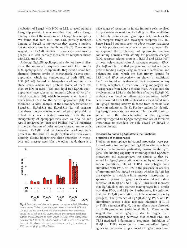
incubation of EgAgB with HDL or LDL to avoid putativeEgAgB-lipoprotein interactions that may reduce EgAgBbinding without the involvement of lipoprotein receptors.We found that both HDL and LDL partially inhibitedbinding of EgAgB to monocytes; they caused a modest,but statistically significant inhibition (Fig. 6). These resultssuggest that EgAgB binding to monocytes and macro-phages is at least partially mediated by receptors sharedwith LDL and HDL.Although EgAgB8 apolipoproteins do not have similar-
ity at the amino acid sequence level with HDL and/orLDL apolipoprotein components, they exhibit some bio-chemical features similar to exchangeable plasma apoli-poproteins, which are components of both HDL andLDL [42, 43]. Indeed, exchangeable apolipoproteins in-clude small, α-helix rich proteins (some of them lessthan 10 kDa in mass) [42], and, lipid-free EgAgB apoli-poproteins have substantial amounts (about 40 %) of α-helical structure [20], which increases when bound tolipids (about 65 % for the native lipoprotein) [44]. Fur-thermore, in silico analysis of the secondary structure ofEgAgB8/1, EgAgB8/2 and EgAgB8/3 [22, 44] suggeststhat these apolipoproteins would adopt amphipathic α-helical structures, a feature associated with the ex-changeability of apolipoproteins such as Apo AI andApo E (reviewed by Jonas and Phillips, [42]). Similaritiesin the distribution of polar and/or charged amino acidsbetween EgAgB8 and exchangeable apolipoproteinspresent in HDL and LDL might explain why these evolu-tionarily distant lipoproteins share receptors in mono-cyte and macrophages. On the other hand, there is a
wide range of receptors in innate immune cells involvedin lipoprotein recognition, including families exhibitinga relatively promiscuous ligand specificity, such as theLDL-receptor family and scavenger receptors [45, 46].Since EgAgB8 subunits seem to adopt tertiary structuresin which positive and negative charges are grouped [23],we explored the involvement of lipoprotein receptorscontaining domains with affinity for positively-charged(LDL receptor related protein 1 [LRP1] and LDLr [45])or negatively-charged (class A scavenger receptor [SR-A][45, 46]) motifs. For that purpose we carried out com-petitive binding assays using an excess of lactoferrin andpolyinosinic acid, which are high-affinity ligands forLRP-1 and SR-A respectively. As shown in Additionalfile 5, we found no evidence of the involvement of anyof these receptors. Furthermore, using monocytes andmacrophages from LDLr-deficient mice, we explored theinvolvement of LDLr in the binding of native EgAgB. Noevidence was found as inflammatory peritoneal F4/80+
cells recovered from LDLr-deficient mice exhibited simi-lar EgAgB binding activity to those from controls (alsoshown in Additional file 5). Further studies for identify-ing EgAgB receptor(s) are needed. This identification to-gether with the characterisation of the signallingpathway triggered by EgAgB recognition are of foremostimportance to elucidate the role played by EgAgB inparasite biology.
Exposure to native EgAgB affects the functionalproperties of macrophagesStudies on macrophage functional properties were per-formed using immunopurified EgAgB to eliminate tracelevels of contaminants, particularly environmental pyro-gens. The binding capacity of immunopurified EgAgB tomonocytes and macrophages was similar to that ob-served for EgAgB preparations obtained by ultracentrifu-gation (Additional file 6). THP-1 macrophages werestimulated with PMA or LPS in the presence or absenceof immunopurified EgAgB to assess whether EgAgB hasthe capacity to modulate inflammatory macrophage re-sponses. Exposure to EgAgB on its own did not inducesecretion of IL-1β or TNFα (Fig. 7). This result indicatesthat EgAgB does not activate macrophages in a similarway than PMA and LPS do. Furthermore, it confirmedthat the EgAgB preparation exclude contamination bypyrogens. The presence of EgAgB during PMA or LPSstimulation caused a dose–response inhibition of IL-1βor TNFα secretion (Fig. 7), but no effects were observedon IL-10 production (Additional file 7). These resultssuggest that native EgAgB is able to trigger IL-10independent-signalling pathways that control PKC andTLR4-mediated inflammatory responses. Inhibition ofIL-1β or TNFα secretion by immunopurified EgAgBagrees with a previous report in which EgAgB was found
Fig. 6 Participation of plasma lipoprotein receptors in EgAgB bindingto monocytes. THP-1 monocytes were pre-incubated with HDL andLDL (500 μg/ml), centrifuged and then incubated with biotinylatedEgAgB (20, 50 100 and 250 μg/ml). Results are expressed as bindingindexes and correspond to mean values ± SEM of three independentexperiments. Asterisks (*) indicate significant differences with respect tothe control incubated without lipoproteins, according to analysis byREML test employing JMP software
Silva-Álvarez et al. Parasites & Vectors (2016) 9:69 Page 13 of 17

to modulate the differentiation as well as the LPSinduced-activation of immature dendritic cells [31]. Incontrast with this report, in macrophages IL-10 secre-tion was not affected by exposure to EgAgB, suggest-ing signalling differences between cell types and/orEgAgB preparations. Activation of macrophages to analternative-activated phenotype characterised by ex-pression of arginase-I, was also investigated. For thatpurpose, BMDM were used since THP-1 macrophagesare not appropriate to analyse arginase activity.BMDM were incubated with different doses of EgAgBfor 24 h and arginase activity was determined incell lysates. Since nitric oxide synthase 2 (NOS2)
competes with arginase-I for their common substrateL-arginine, we examined NOS2 induction by measur-ing the production of nitrite in cell supernatants. Inline with results obtained for cytokine secretion,EgAgB exposure to its own did not generate detect-able levels of nitrite (lower than 3.125 μM), support-ing that inflammatory pathways linked to NOS2induction were not stimulated by EgAgB. On theother hand, in comparison with IL-4, exposure toEgAgB induced a slight increase in arginase activity(Additional file 8), suggesting that EgAgB is not a po-tent inducer of the alternative activation pathway inmacrophages.
Fig. 7 Effects of EgAgB on pro-inflammatory cytokine production by macrophages. THP-1 macrophages were stimulated with PMA (50 ng/ml) orLPS (0.1 ng/ml) for 12 h in the absence or presence of increasing concentrations of EgAgB. Secretion of IL-1β (a, b) and TNFα (c, d) were determinedby ELISA in cell culture supernatant. IL-1β or TNFα levels are expressed as the fold-increase related to the basal secretion (normalised against themedium condition). Results correspond to mean values ± SEM of at least two independent experiments. Asterisks (*) indicate significant differenceswith respect to PMA or LPS-stimulated macrophages according to one-way ANOVA analysis followed by Tukey’s post-test (p < 0.05)
Silva-Álvarez et al. Parasites & Vectors (2016) 9:69 Page 14 of 17

ConclusionsThe results we present in this work support the conceptthat once EgAgB reaches host tissues it may bind to tis-sue resident macrophages and inflammatory monocytesrecruited during infection, through cell receptors likelyinvolved in the recognition of plasma lipoproteins.EgAgB signalling through these receptors seems to in-duce a non-inflammatory phenotype in macrophages. Infact, this signalling could limit TLR4-mediated cytokineproduction that may occur during cyst growth, as a re-sult of degradation of extracellular matrix [47]. Further-more, it cannot be ruled out that EgAgB signallingwould contribute to drive macrophage differentiation to-wards an alternative activation-like phenotype, which isconsistent with the Th2-regulated immune responsefound in helminth infections, including echinococossis[40, 48]. Identification of cell receptor(s) and signallingpathways involved in EgAgB recognition by innate cellswill be of foremost importance for understanding EgAgBrole in parasite adaptation to host. Regarding EgAgB cellreceptor(s), they likely recognise electrostatic motifs ofEgAgB8/3 and EgAgB8/1, the latter constitutes the pre-dominant protein component of native EgAgB presentin HF. The fact that EgAgB cell receptors would alsobind host HDL and LDL, together with the non-inflammatory phenotype observed after macrophage ex-posure to EgAgB, suggest a cross-talk between metabolicand inflammatory pathways [49], which requires furtherinvestigation.
Additional files
Additional file 1: Comparison of the extent of biotinylationobtained for native EgAgB, rEgAgB8 subunits and OVA. Proteinswere adsorbed on ELISA microplates in a wide range of concentrationsand the presence of biotin was determined using streptavidin-peroxidaseand TMB/H2O2 for development. The absorbance at 450 nm was plottedvs. the protein concentration. (A) Comparison of the extent of biotinylationbetween OVA and native EgAgB; data are expressed as the mean values ±SEM of three analytical replicates B) Comparison of the extent ofbiotinylation between rEgAgB8 subunits; data are expressed as themean values ± SEM of three analytical replicates. All preparations ofbiotinylated native EgAgB used in this work were controlled inrespect to a preparation of OVA biotinylated in parallel. (TIF 6094 kb)
Additional file 2: Analysis of the ability of EB7 mab to recognizerEgAgB8 subunits. EB7 rEgAgB8/1, rEgAgB8/2 and rEgAgB8/3 wereadsorbed on ELISA microplates to assess by ELISA the ability of Mab EB7to recognize them. The binding of mab EB7 was developed using aperoxidase conjugated anti-mouse IgG followed by TMB/H2O2. Theabsorbance at 450 nm was plotted vs. the protein concentration. Datacorrespond to mean values ± SEM of analytical triplicates. As observed inthe plot, mab EB7 recognises only EgAgB8/1. (TIF 62 kb)
Additional file 3: Binding of native EgAgB to THP-1 derivedmonocytes and macrophages employing EB7 mab. Binding of nativeEgAgB to THP-1 derived monocytes and macrophages was analysedemploying EB7 mab followed by incubation with goat anti-mouse IgG/IgM antibody conjugated to FITC. As control, cells were incubated withbinding buffer instead of native EgAgB (PBS), without adding EB7 mab(w/EB7) or using mouse IgG1 kappa isotype control instead of EB7 mab
(IC). In left panels figure shows the histograms with the distribution ofcell population as function of FITC fluorescence for controls (grey)and EgAgB-treated cells (red). Histograms are representative of fourindependent experiments for each cell type. In right panels bindingindexes were plotted vs. native EgAgB concentrations (1, 5 and10 μg/ml) for monocytes (upper panels) and macrophages (bottompanels). Data are expressed as mean ± SEM of four independent experiments.Asterisks (*) indicate significant differences with respect to the control withBB according to the analysis by t-test (p < 0.05), while number signs(#) indicate significant differences in the binding indexes obtained at1 and 10 μg/ml native EgAgB (one-way ANOVA followed by Tukey’spost-test, p < 0.05). (TIF 11902 kb)
Additional file 4: Binding of recombinant EgAgB8/1 to THP-1derived monocytes and macrophages employing EB7 mab. Bindingof rEgAgB8/1 to THP-1 derived monocytes and macrophages was analysedemploying EB7 mab followed by incubation with goat anti-mouse IgG/IgMantibody conjugated to FITC. As control, cells were incubated with bindingbuffer instead of native EgAgB (PBS), or in the absence of EB7 mab (w/EB7),or using mouse IgG1 kappa isotype control instead of EB7 mab (IC).Histograms (cell numbers vs. FITC fluorescence) corresponding tocontrol (grey) and rEgAgB8/1-treated cells (blue) are shown on theleft; they are representative of three independent experiments foreach cell type. On the right, the binding indexes were plotted vs.rEgAgB8/1 concentration for monocytes (upper panel) and macrophages(bottom panel). Data are expressed as mean ± SEM of three independentexperiments. Asterisks (*) indicate significant differences with respect to thecontrol (PBS) according to the analysis by t-test (p < 0.05), while numbersigns (#) indicate significant differences in the binding indexes obtained atdifferent native EgAgB concentrations (one-way ANOVA followed by Tukey’spost-test, p < 0.05). (TIF 12381 kb)
Additional file 5: Involvement of plasma lipoprotein receptors inEgAgB binding to monocytes and macrophages. (A) Cells werepre-incubated with ligands of LRP1 and SR-A receptors (lactoferrin andpoly-inosinic acid [poli-I], respectively), centrifuged and then incubated withEgAgB in a fixed concentration (100 μg/ml). As control, BSA was usedat the maximum lactoferrin and poli-I concentration employed. Thebinding index is expressed as mean ± SEM of analytical triplicates. (B)Binding analysis of native EgAgB to monocytes/macrophages F4/80+isolated from control (WT, empty bars) or LDLr−/− mice (filled bars). Statis-tical analysis was undertaken by one-way ANOVA followed by Tukey’s post-test, no significant differences were observed. One representative experi-ment of two is shown in each panel. (TIF 302 kb)
Additional file 6: Binding of immunopurified EgAgB to THP-1derived monocytes and macrophages employing EB7 mab. Bindingof immunopurified EgAgB to THP-1 derived monocytes and macro-phages was analysed employing EB7 mab followed by incubation withgoat anti-mouse IgG/IgM antibody conjugated to FITC. As control, cellswere incubated with binding buffer instead of native EgAgB (PBS), withoutadding EB7 mab (w/EB7) or using mouse IgG1 kappa isotype controlinstead of EB7 mab (IC). In left panels figure shows the histogramswith the distribution of cell population as function of FITC fluorescence forcontrols (grey) and EgAgB-treated cells (red). Histograms are representativeof three independent experiments for each cell type. In right panels bindingindexes were plotted vs. native EgAgB concentrations (1 and 10 μg/ml) formonocytes (upper panels) and macrophages (bottom panels). Data areexpressed as mean ± SEM of three independent experiments. Asterisks(*) indicate significant differences with respect to the control with BBaccording to the analysis by t-test (p < 0.05), while number signs (#)indicate significant differences in the binding indexes obtained at 1and 10 μg/ml native EgAgB (one-way ANOVA followed by Tukey’spost-test, p < 0.05). (TIF 11689 kb)
Additional file 7: Effects of EgAgB on IL-10 production bymacrophages. THP-1 macrophages were stimulated with LPS (0.1 ng/ml) for 12 h in absence or presence of increasing concentrations ofEgAgB. Secretion of IL-10 was determined by ELISA in cell culturesupernatant and expressed as the fold-increase related to the basalsecretion (normalised against the medium condition). Resultscorrespond to mean values ± SEM of two independent experiments.Number signs (#) indicate significant differences with respect to the
Silva-Álvarez et al. Parasites & Vectors (2016) 9:69 Page 15 of 17

medium condition according to one-way ANOVA analysis followed byTukey’s post-test (p < 0.05). (TIF 24 kb)
Additional file 8: Determination of arginase activity in bonemarrow-derived macrophages stimulated with EgAgB. Arginaseactivity was evaluated in BMDM incubated with PBSEB (control),immunopurified EgAgB (0.2–20 μg/mL), or murine IL-4 (2.5 ng/ml) as acontrol of macrophage alternative activation. Arginase activity wasexpressed as mU per million of BMDM. The graph is representative ofthree independent experiments. Bars indicate SEM of analytical triplicates.Asterisks (*) indicate significant differences with respect to the control(medium) according to one-way ANOVA analysis followed by Tukey’spost-test (p < 0.05). (TIF 27 kb)
AbbreviationsBB: Binding buffer (PBS containing 1 % (v/v) FS and 0.1 % NaN3);BMDM: Bone marrow derived macrophages; EgAgB: Antigen B fromEchinococcus granulosus; EgAgBPLD-: Control EgAgB for treatment with PLD;EgAgBPLD+: EgAgB treated with PLD; fQS: Q-Sepharose retained fraction;GL: Germinal layer; HDL: High-density lipoprotein; HF: Hydatid cyst fluid;HLBPs: Hydrophobic Ligand Binding Proteins; HPTLC: High performance thinlayer chromatography; iBAQ: Intensity-based absolute quantification; LC-MS/MS: Liquid chromatography–mass spectrometry; LDL: Low-densitylipoprotein; LDLr: LDL receptor; LPS: Lipopolysaccharides; LRP1: LDL receptorrelated protein 1; MALDI-TOF/TOF: Matrix-assisted laser desorption/ionizationtime-of-flight; OVA: Ovalbumin; PBSEB: PBS containing 5 mM EDTA and20 μM BHT; PC: Phosphatidylcholine; PC/PS-LUVs: Large unilamellar vesiclescomposed of PC and PS (50:50 mol:mol); PC-LUVs: Large unilamellar vesiclescomposed of PC; PLD: Phospholipase D; PS: Phosphatidylserine;rEgAgB8: recombinant lipid-free EgAgB8 subunits; RP-HPLC: Reversed-phasehigh performance liquid chromatography; SR-A: Class A scavenger receptor.
Competing interestsThe authors declare that they have no competing interests.
Authors’ contributionsAMF conceived of the study and participated in its design and coordination.AMFolle and VSA prepared native EgAgB and lipid-free EgAgB subunits,respectively. AMFolle and ESK carried out the characterisation of nativeEgAgB protein moiety by LC-MS/MS. AMFolle, ESK, LKI and AMF analysedand discussed LC-MS/MS data. VSA, AMFolle and ALR performed bindingexperiments. VSA, AMFolle and AMF processed and analysed the bindingdata. ALR and VSA performed cytokine secretion assays. ALR, IC and AMFcarried out arginase assays and analysed data. BC discussed results andcritically revised the paper. VSA and AMFolle prepared figures and tables.AMFolle helped to draft the manuscript. VSA and AMF wrote the paper. Allauthors read and approved the final manuscript.
AcknowledgmentsThe authors gratefully acknowledge Sandra Ferreira (Cátedra de Inmunología,Facultad de Ciencias/Facultad de Química, UdelaR) for her careful workcollecting HF from E. granulosus hydatid cysts, Dr. Carlos Batthyány and MSc.Analía Lima for their assistance with two-dimensional electrophoresis andmass spectrometry analysis, and Dr. Robert B. Sim for critical reading of themanuscript. VSA and AMFolle are very grateful to Consejo Nacional deInvestigaciones Científicas y Técnicas (CONICET, Argentina), Agencia Nacionalde Investigación e Innovación (ANII, Uruguay) and Comisión Sectorial deInvestigación Científica (CSIC, Uruguay) for their postgraduate fellowships.This work was supported by a national grant from the CSIC (Programa I + D,Grant 023–348 and Programa I + D Grupos, 2014).
Author details1Cátedra de Inmunología, Facultad de Ciencias/Facultad de Química,Universidad de la República (UdelaR), Montevideo, Uruguay. 2Instituto deInvestigaciones Bioquímicas de La Plata (INIBIOLP), Facultad de CienciasMédicas, Universidad Nacional de La Plata (UNLP), La Plata, Argentina.3Consejo Nacional de Investigaciones Científicas y Técnicas (CONICET),Ciudad Autónoma de Buenos Aires, Argentina. 4Laboratório Especial deToxinologia Aplicada, Center of Toxins, Immune-Response and Cell Signalling(CeTICS), Instituto Butantan, São Paulo, Brazil. 5Departamento de Bioquímica,
Biología Molecular y Genética, Facultad de Veterinaria, Universidad deExtremadura (UNEX), Cáceres, España.
Received: 22 July 2015 Accepted: 28 January 2016
References1. Thompson RCA. The Biology of Echinococcus and Hydatid Disease. London:
George Allen & Unwin; 1986.2. Budke CM, Deplazes P, Torgerson PR. Global socioeconomic impact of cystic
echinococcosis. Emerg Infect Dis. 2006;12:296–303.3. Cardona GA, Carmena D. A review of the global prevalence, molecular
epidemiology and economics of cystic echinococcosis in productionanimals. Vet Parasitol. 2013;192:10–32.
4. Nakao M, Lavikainen A, Yanagida T, Ito A. Phylogenetic systematics of thegenus Echinococcus (Cestoda: Taeniidae). Int J Parasitol. 2013;43:1017–29.
5. Alvarez Rojas CA, Romig T, Lightowlers MW. Echinococcus granulosus sensulato genotypes infecting humans–review of current knowledge. Int JParasitol. 2014;44:9–18.
6. Oriol R, Williams JF, Pérez Esandi M, Oriol C. Purification of lipoproteinantigens of Echinococcus granulosus from sheep hydatid fluid. Am J TropMed Hyg. 1971;20:569–74.
7. Ioppolo S, Notargiacomo S, Profumo E, Franchi C, Ortona E, Riganò R, et al.Immunological responses to antigen B from Echinococcus granulosus cystfluid in hydatid patients. Parasite Immunol. 1996;18:571–8.
8. Ortona E, Siracusano A, Castro A, Riganò R, Mühlschlegel F, Ioppolo S, et al.Use of a monoclonal antibody against the antigen B of Echinococcusgranulosus for purification and detection of antigen B. Appl Parasitol.1995;36:220–5.
9. Virginio V, Hernández A, Rott M, Monteiro K, Zandonai A, Nieto A, et al. Aset of recombinant antigens from Echinococcus granulosus with potentialfor use in the immunodiagnosis of human cystic hydatid disease. Clin ExpImmunol. 2003;132:309–15.
10. Carmena D, Benito A, Eraso E. Antigens for the immunodiagnosis ofEchinococcus granulosus infection: An update. Acta Trop. 2006;98:74–86.
11. Frosch P, Hartmann M, Mühlschlegel F, Frosch M. Sequence heterogeneityof the echinococcal antigen B. Mol Biochem Parasitol. 1994;64:171–5.
12. Fernández V, Ferreira H, Fernández C, Zaha A, Nieto A. Molecularcharacterisation of a novel 8-kDa subunit of Echinococcus granulosusantigen B. Mol Biochem Parasitol. 1996;77:247–50.
13. González G, Nieto A, Fernández C, Orn A, Wernstedt C, Hellman U. Twodifferent 8 kDa monomers are involved in the oligomeric organization ofthe native Echinococcus granulosus antigen B. Parasite Immunol.1996;18:587–96.
14. Chemale G, Haag K, Ferreira H, Zaha A. Echinococcus granulosus antigen B isencoded by a gene family. Mol Biochem Parasitol. 2001;116:233–7.
15. Arend AC, Zaha A, Ayala FJ, Haag KL. The Echinococcus granulosus antigen Bshows a high degree of genetic variability. Exp Parasitol. 2004;108:76–80.
16. Kamenetzky L, Muzulin PM, Gutierrez AM, Angel SO, Zaha A, Guarnera EA,et al. High polymorphism in genes encoding antigen B from humaninfecting strains of Echinococcus granulosus. Parasitology.2005;131(Pt 6):805–15.
17. Muzulin PPM, Kamenetzky L, Gutierrez AM, Guarnera EA, Rosenzvit MC.Echinococcus granulosus antigen B gene family: further studies of strainpolymorphism at the genomic and transcriptional levels. Exp Parasitol.2008;118:156–64.
18. Zhang W, Li J, Jones MK, Zhang Z, Zhao L, Blair D, et al. The Echinococcusgranulosus antigen B gene family comprises at least 10 unique genes in fivesubclasses which are differentially expressed. PLoS Negl Trop Dis. 2010;4:e784.
19. Chemale G, Ferreira HB, Barrett J, Brophy PM, Zaha A. Echinococcusgranulosus antigen B hydrophobic ligand binding properties. BiochimBiophys Acta. 2005;1747:189–94.
20. Silva-Álvarez V, Franchini GR, Pórfido JL, Kennedy MW, Ferreira AM, CórsicoB. Lipid-Free Antigen B Subunits from Echinococcus granulosus:oligomerization, ligand binding, and membrane interaction properties. PLoSNegl Trop Dis. 2015;9:e0003552.
21. Obal G, Ramos AL, Silva V, Lima A, Batthyany C, Bessio MI, et al.Characterisation of the native lipid moiety of Echinococcus granulosusantigen B. PLoS Negl Trop Dis. 2012;6:e1642.
22. Monteiro KM, Scapin SMN, Navarro MVAS, Zanchin NIT, Cardoso MB, daSilveira NP, et al. Self-assembly and structural characterization of
Silva-Álvarez et al. Parasites & Vectors (2016) 9:69 Page 16 of 17
Echinococcus granulosus antigen B recombinant subunit oligomers. BiochimBiophys Acta. 2007;1774:278–85.
23. Silva-Álvarez V, Folle AM, Ramos AL, Zamarreño F, Costabel MD, García-Zepeda E, et al. Echinococcus granulosus antigen B: a hydrophobic ligandbinding protein at the host–parasite interface. Prostaglandins Leukot EssentFat Acids. 2015;93:17–23.
24. Sánchez F, March F, Mercader M, Coll P, Muñoz C, Prats G. Immunochemicallocalization of major hydatid fluid antigens in protoscoleces and cysts ofEchinococcus granulosus from human origin. Parasite Immunol.1991;13:583–92.
25. Sánchez F, Garcia J, March F, Cardeñosa N, Coll P, Muñoz C, et al.Ultrastructural localization of major hydatid fluid antigens in brood capsulesand protoscoleces of Echinococcus granulosus of human origin. ParasiteImmunol. 1993;15:441–7.
26. Lee E-GE, Kim SS-H, Bae YY-A, Chung J-YJ, Suh M, Na B-K, et al. Ahydrophobic ligand‐binding protein of the Taenia solium metacestodemediates uptake of the host lipid: Implication for the maintenance ofparasitic cellular. Proteomics. 2007;7:4016–30.
27. Tsai IJ, Zarowiecki M, Holroyd N, Garciarrubio A, Sanchez-Flores A, Brooks KL,et al. The genomes of four tapeworm species reveal adaptations toparasitism. Nature. 2013;496:57–63.
28. Zheng H, Zhang W, Zhang L, Zhang Z, Li J, Lu G, et al. The genome of thehydatid tapeworm Echinococcus granulosus. Nat Genet. 2013;45:1168–77.
29. Shepherd JC, Aitken A, McManus DP. A protein secreted in vivo byEchinococcus granulosus inhibits elastase activity and neutrophil chemotaxis.Mol Biochem Parasitol. 1991;44:81–90.
30. Riganò R, Profumo E, Bruschi F, Carulli G, Azzarà A, Ioppolo S, et al.Modulation of human immune response by Echinococcus granulosusantigen B and its possible role in evading host defenses. Infect Immun.2001;69:288–96.
31. Riganò R, Buttari B, Profumo E, Ortona E, Delunardo F, Margutti P, et al.Echinococcus granulosus antigen B impairs human dendritic celldifferentiation and polarizes immature dendritic cell maturation towards aTh2 cell response. Infect Immun. 2007;75:1667–78.
32. Siracusano A, Margutti P, Delunardo F, Profumo E, Riganò R, Buttari B, et al.Molecular cross-talk in host–parasite relationships: the intriguingimmunomodulatory role of Echinococcus antigen B in cystic echinococcosis.Int J Parasitol. 2008;38:1371–6.
33. Arike L, Valgepea K, Peil L, Nahku R, Adamberg K, Vilu R. Comparison andapplications of label-free absolute proteome quantification methods onEscherichia coli. J Proteomics. 2012;75:5437–48.
34. Bejta F, Moore EH, Avella M, Gough PJ, Suckling KE, Botham KM. Oxidationof chylomicron remnant-like particles inhibits their uptake by THP-1macrophages by apolipoprotein E-dependent processes. Biochim BiophysActa. 2007;1771:901–10.
35. Kleinfeld AM, Storch J. Transfer of long-chain fluorescent fatty acidsbetween small and large unilamellar vesicles. Biochemistry. 1993;32:2053–61.
36. Zhang X, Goncalves R, Mosser DM. The isolation and characterization ofmurine macrophages. Curr Protoc Immunol. 2008;Chapter 14:Unit 14.1.
37. Ray A, Dittel BN. Isolation of mouse peritoneal cavity cells. J Vis Exp.2010;35:1–3.
38. Corraliza IM, Campo ML, Soler G, Modolell M. Determination of arginaseactivity in macrophages: a micromethod. J Immunol Methods.1994;174:231–5.
39. Green LC, Wagner DA, Glogowski J, Skipper PL, Wishnok JS, TannenbaumSR. Analysis of nitrate, nitrite, and [15 N]nitrate in biological fluids. AnalBiochem. 1982;126:131–8.
40. Díaz A, Casaravilla C, Allen JE, Sim RB, Ferreira AM. Understanding thelaminated layer of larval Echinococcus II: immunology. Trends Parasitol.2011;27:264–73.
41. Henderson RJ, Tocher DR. Thin-layer chromatography. In: Hamilton RJ,Hamilton S, editors. Lipid Anal A Pract Approach. Oxford, UK: IRL Press;1992. p. 65–111.
42. Jonas A, Phillips MC. Lipoprotein structure. In: Vance DEV and JE. Elsevier B.V, editor. Biochem Lipids, Lipoproteins Membr. 5th ed. 2008. p. 485–506.
43. Sun H-Y, Chen S-F, Lai M-D, Chang T-T, Chen T-L, Li P-Y, et al. Comparativeproteomic profiling of plasma very-low-density and low-densitylipoproteins. Clin Chim Acta. 2010;411:336–44.
44. González-Sapienza G, Cachau RE. Identification of critical residues of animmunodominant region of Echinococcus granulosus antigen B. J BiolChem. 2003;278:20179–84.
45. Schneider WJ. Lipoprotein receptors. In: Dennis E. Vance and Jean E. Vance.Elsevier B.V, editor. Biochem Lipids, Lipoproteins Membr. 5th ed. 2008.p. 555–78.
46. Prabhudas M, Bowdish D, Drickamer K, Febbraio M, Herz J, Kobzik L, et al.Standardizing scavenger receptor nomenclature. J Immunol.2014;192:1997–2006.
47. Brunn GJ, Bungum MK, Johnson GB, Platt JL. Conditional signaling byToll-like receptor 4. FASEB J. 2005;19:872–4.
48. Díaz A, Casaravilla C, Barrios AA, Ferreira AM. Parasite molecules and hostresponses in cystic echinococcosis. Parasite Immunol 2015. In press doi:10.1111/pim.12282.
49. Odegaard JI, Chawla A. The immune system as a sensor of the metabolicstate. Immunity. 2013;38:644–54.
• We accept pre-submission inquiries
• Our selector tool helps you to find the most relevant journal
• We provide round the clock customer support
• Convenient online submission
• Thorough peer review
• Inclusion in PubMed and all major indexing services
• Maximum visibility for your research
Submit your manuscript atwww.biomedcentral.com/submit
Submit your next manuscript to BioMed Central and we will help you at every step:
Silva-Álvarez et al. Parasites & Vectors (2016) 9:69 Page 17 of 17