ecg of epicardial arrhythmias
DESCRIPTION
E l e c t ro c a rd i o g r a p h i c Recognition o f E p i c a rdi a l ArrhythmiasYoav Michowitz, MDa,b,*, Bernard Belhassen, MDaKEYWORDS Epicardial ablation Epicardial origin Ventricular arrhythmias Atrial tachycardiasIn the current electrophysiologic practice, ablation of most cardiac arrhythmias is performed, with high success rates, using an endocardial approach. However, in some cases, endocardial mapping techniques fail to identify the origin of a focal tachyarrhythmia or theTRANSCRIPT

ElectrocardiographicRecognitionof EpicardialArrhythmias
Yoav Michowitz, MDa,b,*, Bernard Belhassen, MDaKEYWORDS
� Epicardial ablation � Epicardial origin� Ventricular arrhythmias � Atrial tachycardias
In the current electrophysiologic practice, ablationof most cardiac arrhythmias is performed, withhigh success rates, using an endocardialapproach. However, in some cases, endocardialmapping techniques fail to identify the origin ofa focal tachyarrhythmia or the critical site of a reen-trant circuit and an epicardial origin is suspected.1
Although successful ablation will depend ultimatelyon the results of detailed intracardiac mapping,early suspicion of the epicardial origin of anarrhythmia by standard 12-lead ECG is very useful.
Epicardial ventricular tachyarrhythmias aremore common in certain patient populations withstructural heart disease such as chagasic heartdisease, nonischemic dilated cardiomyopathyand arrhythmogenic right ventricular dysplasia.1–3
However, they may also occur in patients withischemic cardiomyopathy—most frequently afterinferior myocardial infarction.4 In patients withstructurally normal heart, an epicardial origin hasbeen described in the outflow tract (OT) region,sometimes in proximity to perivascular sites andin the crux of the heart.5,6
Most cases of epicardial ablation have involvedventricular tachyarrhythmias, as the thickness ofthe ventricular myocardium makes it more difficultfor endocardial ablation lesions to penetratethrough the wall and reach an epicardial site. Thereare also some reports of epicardial radiofrequency
a The Department of Cardiology, Tel Aviv Sourasky MedAviv University, 6 Weizman Street, Tel Aviv 64239, Israelb UCLA Cardiac Arrhythmia Center, Ronald Reagan UCLAUCLA, A2-237 CHS, 10833 Le Conte Avenue, Los Angeles* Corresponding author. UCLA Cardiac Arrhythmia CenteSchool of Medicine at UCLA, A2-237 CHS, 10833 Le ContE-mail address: [email protected] (Y. Michowitz).
Card Electrophysiol Clin 2 (2010) 25–33doi:10.1016/j.ccep.2009.11.0081877-9182/10/$ – see front matter ª 2010 Published by E
ablation of atrial arrhythmias in the left atrialappendage area where the thick cords ofpectinate muscle may preclude endocardialablation success.1,7–9 Also, epicardial accessoryatrioventricular pathways have been described inthe area of the right atrial appendage and coronarysinus (CS) tributaries.10–14 The term epicardialorigin is used indifferently throughout this articleregardless of the mechanism of the arrhythmia(reentry vs focal). Since this term only refers tothe arrhythmia exit site, it is not excluded thatsome reentrant mechanisms exhibiting an epicar-dial exit site actually may have critical components(isthmus, entrance site) located at an endocardialsite.
VENTRICULAR ARRHYTHMIAS
Several criteria have been proposed to differen-tiate epicardial from endocardial origin of ventric-ular tachycardia (VT). These include generalcriteria,5,15 and site-specific criteria.16,17 Thegeneral criteria are related to the morphology ofthe QRS. They are all based on the presumedslower conduction during the initial part of theQRS as the wave of depolarization traversesslowly from the epicardium to the endocardiumand then spreads faster using the endocardialPurkinje fibers.
ical Center and the Sackler Faculty of Medicine, Tel
Medical Center, David Geffen School of Medicine at, CA 90095, USAr, Ronald Reagan UCLA Medical Center, David Geffene Avenue, Los Angeles, CA 90095.
lsevier Inc. card
iacE
P.th
ecli
nics
.com
Michowitz & Belhassen26
GENERAL CRITERIAPseudodelta Wave
Pseudodelta wave is defined as the interval fromthe earliest QRS activation to the earliest fastdeflection in the precordial leads (Fig. 1). Berruezoand colleagues15 studied patients with VT and aright bundle branch block (RBBB) morphologyassociated with ischemic or dilated cardiomyop-athy. They analyzed the ECG pattern in VTssuccessfully ablated from the epicardium aftera failed endocardial approach and in VTs success-fully ablated from the endocardium. They demon-strated that a pseudodelta wave cut-off greaterthan or equal to 34 msec has a sensitivity andspecificity of 83% and 95%, respectively, inpredicting an epicardial origin. Bazan andcolleagues16 tested this parameter in a group of15 patients (9 with structural heart disease, butno history of myocardial infarction, and 6 patientswithout heart disease) with refractory ventriculararrhythmias. They compared the QRS morphologyduring pace-mapping from multiple endocardial
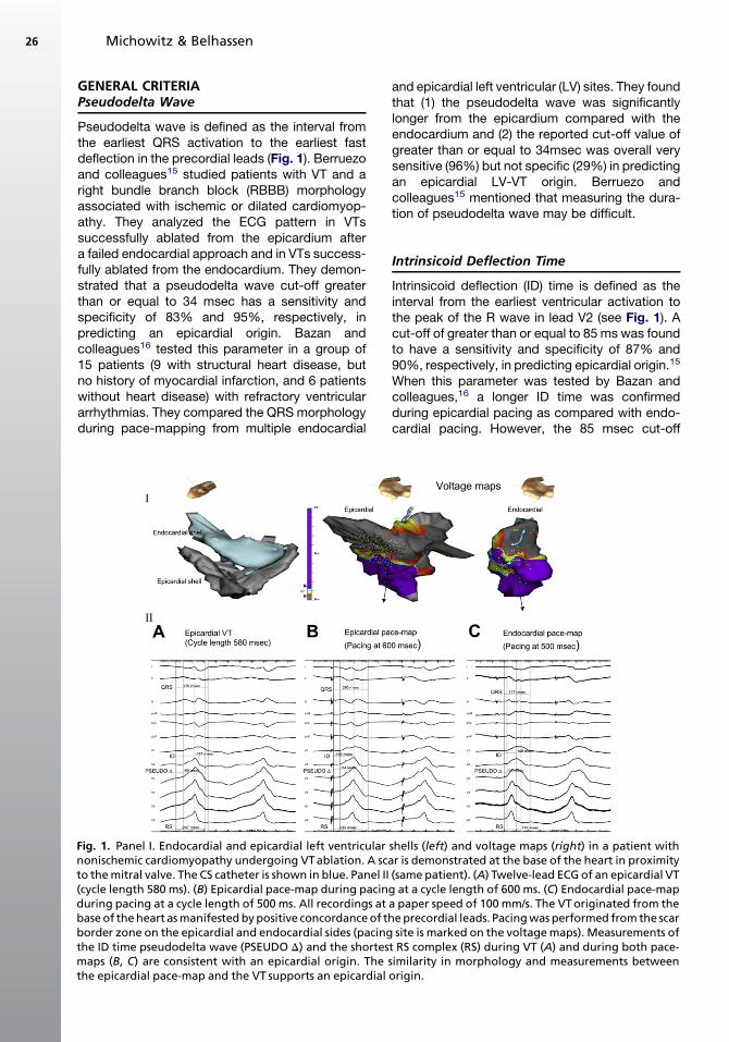
Fig. 1. Panel I. Endocardial and epicardial left ventricularnonischemic cardiomyopathy undergoing VT ablation. A scato the mitral valve. The CS catheter is shown in blue. Panel II(cycle length 580 ms). (B) Epicardial pace-map during pacinduring pacing at a cycle length of 500 ms. All recordings atbase of the heart as manifested by positive concordance of thborder zone on the epicardial and endocardial sides (pacingthe ID time pseudodelta wave (PSEUDO D) and the shortesmaps (B, C) are consistent with an epicardial origin. Thethe epicardial pace-map and the VT supports an epicardial
and epicardial left ventricular (LV) sites. They foundthat (1) the pseudodelta wave was significantlylonger from the epicardium compared with theendocardium and (2) the reported cut-off value ofgreater than or equal to 34msec was overall verysensitive (96%) but not specific (29%) in predictingan epicardial LV-VT origin. Berruezo andcolleagues15 mentioned that measuring the dura-tion of pseudodelta wave may be difficult.
Intrinsicoid Deflection Time
Intrinsicoid deflection (ID) time is defined as theinterval from the earliest ventricular activation tothe peak of the R wave in lead V2 (see Fig. 1). Acut-off of greater than or equal to 85 ms was foundto have a sensitivity and specificity of 87% and90%, respectively, in predicting epicardial origin.15
When this parameter was tested by Bazan andcolleagues,16 a longer ID time was confirmedduring epicardial pacing as compared with endo-cardial pacing. However, the 85 msec cut-off
shells (left) and voltage maps (right) in a patient withr is demonstrated at the base of the heart in proximity(same patient). (A) Twelve-lead ECG of an epicardial VTg at a cycle length of 600 ms. (C) Endocardial pace-mapa paper speed of 100 mm/s. The VT originated from thee precordial leads. Pacing was performed from the scarsite is marked on the voltage maps). Measurements of
t RS complex (RS) during VT (A) and during both pace-similarity in morphology and measurements betweenorigin.

Recognition of Epicardial Arrhythmias 27
reached sensitivity and specificity of only 39% and24%, respectively.16
Shortest Precordial Complex
Shortest RS complex is measured from the earliestventricular activation to the nadir of the first S wavein any precordial lead (see Fig. 1).
A cutoff of greater than or equal to 121 msyielded specificity and sensitivity of 76% and85%, respectively, by Berruezo and colleagues15;and lower values of 53% and 79%, respectively,by Bazan and colleagues.16
QRS-Complex Duration
The QRS complex duration is defined as theinterval measured from the earliest ventricular acti-vation (from the stimulation artifact in pacedpatients) to the offset of QRS in the precordialleads. The QRS duration is longer when pacing isperformed from the epicardium, as comparedwith endocardial pacing at a similar LV site16
(due to the presence of pseudodelta wave).However, due to considerable overlap in QRSduration during VT, an exact cut-off value forQRS width could not be identified to differentiatebetween epicardial and endocardial VTs.16
Precordial Maximum Deflection Index
Maximum deflection index (MDI) is defined as thetime from the beginning of the QRS to the earliestmaximal deflection (largest amplitude deflectioneither above or below the baseline) in any of theprecordial leads divided by the maximal QRSduration.5 A cut-off value of 0.55 had high sensi-tivity and specificity to predict epicardial VT originin patients with idiopathic left VTs arising close tothe aortic sinus of Valsalva5 and near the crux ofthe heart.6 MDI was not tested systematically inpatients with structural heart disease.
Precordial Pattern Break
Another finding that may point to an epicardialorigin is the pattern break or R wave regressionor progression sign (Marchlinski, 2nd annual VTsymposium, Philadelphia, PA, unpublished data,2007) manifested as an abrupt loss of R wave inV2 with resumption in V3-V6.18 According to Haq-qani and colleagues,18 this criterion was not testedin patients with structural heart disease although itis likely to be helpful in this patient group as well.
SITE-SPECIFIC CRITERIA
Several articles have examined region-specificECG features of epicardial ventricular arrhythmia,including the LV,16 right ventricle (RV),17 the OT
area5,17,19 and the crux of the heart.6 The differ-ence between epicardial and endocardial electro-grams may be related to two factors: slowed initialconduction in the epicardial layer and differencesin propagation of the depolarizing wave. QRSmorphology in leads reflecting initial activationwill manifest as a Q wave (activation movingaway from the electrode) when the origin is epicar-dial. However, when the origin is endocardial it willmanifest as an rS complex as part of the activation(endocardial to epicardial) moves toward therecording electrode.
LV
Bazan and colleagues16 divided the LV into fiveparts: basal superior, basal inferior, apical supe-rior, apical inferior, and LV apex.
Basal LV areaVT originating from the base of the heart can berecognized by positive concordance in the precor-dial leads.20 The presence of a Q wave in lead I andthe absence of Q in leads II, III, and AVF are goodpredictors of an epicardial origin for the basalsuperior LV region.16 Conversely, the presence ofQ wave in inferior leads is a good predictor of anepicardial origin in the basal inferior LV area.16
Apical LV areaApical superior VT will have a negative concor-dance in the precordial leads and right inferioraxis.20 The presence of Q wave in lead I supportsan epicardial location at the superior area, whilethe presence of Q wave in inferior leads (asopposed to rS pattern) supports an epicardiallocation at the inferior part.16
LV apexVT arising from the LV apex suggested by negativeprecordial concordance and QS pattern from V4-V6.20 An initial Q wave in lead V2 was significantlyassociated with epicardial sites but this findingwas not confirmed in the matched pace-mappinganalysis.16
These site-specific criteria were developed inpatients without myocardial infarction; obviously,infarct-related Q waves will likely affect the abilityof this criterion to differentiate epicardial fromendocardial foci.
RV
A region-specific study with RV epicardial andendocardial pace-mapping demonstrated severalfindings17: pacing from the epicardial anterior RVis more likely to manifest Q or QS complex inlead I and V2, whereas inferior wall epicardialpacing will manifest Q wave in inferior leads.
Michowitz & Belhassen28
Cut-off values for the general criteria described forthe LV (pseudodelta, ID time, shortest precordialRS complex, and QRS duration) were not appli-cable to the RV, though some significant differ-ences between epicardial and endocardialpacing were observed.
These findings were explained by two factors17:the thinner RV wall and the less abundant Purkinjenetwork over the RV free wall making difference inconduction velocity between epicardial and endo-cardial pacing less robust. As this study includedonly two patients with ischemic heart disease,the results cannot be extrapolated to this patientsubgroup. In addition, there are no studies inpatients with RV hypertrophy.
OT
VT arising from the OT typically has a left bundlebranch block (LBBB) pattern and an inferior axis.Criteria for differentiating right- from left-sidedorigin have been described.21–23 No pathogno-monic differences in ECG morphology were notedbetween epicardial and endocardial pacing in theRVOT region.17
Daniels and colleagues5 described a series ofpatients with epicardial tachycardia originatingfrom LV sites close to the coronary vessels. Tachy-cardias from the anterior interventricular vein (AIV)region exhibit a LBBB pattern in V1 with precordialtransition beyond V2.24 As the AIV or great cardiacvein (GCV) junction lies immediately lateral to theleft coronary cusp (LCC),25 VTs from this locationhave a pattern similar to those originating fromthe aortic sinus of Valsalva (ie, R/S amplitude index>0.5 and R wave duration index >0.3 in V1 or V2).Whether it has M or W pattern in V1 like LCC VThas not been described. VTs originating moredistally in the AIV had narrower QRS complexand were similar to RVOT VTs.
Obel and colleagues19 described a series of fivepatients with epicardial VTs originating in the distalGCV region, all patients had slurring of the precor-dial R wave, four or five had RBBB pattern in V1and 1 had transition in V3.
MDI, which was first introduced by Daniels andcolleagues,5 had a sensitivity of 100% and speci-ficity of 98.7% in identifying epicardial VTs. Itoand colleagues22 developed an algorithm to locateorigin of OT VT. In patients with ECG compatiblewith LVOT VTs, an aVL to aVR Q ratio greaterthan 1.4, or an S wave in V1 greater than 1.2 mVdifferentiated epicardial from endocardial origin.Another study from the same group26 reportedfindings of ECGs from 10 patients with LVOT peri-vascular epicardial VTs. Using either their algo-rithm or MDI they could correctly identify 6 out of
10 ECG with each method separately. However,using both they could identify 9 out of 10 ECGs.
Crux of the Heart
Another recent study described epicardial VTsoriginating from the crux of the heart6 (whichcorresponds to the junction between the CS andmiddle cardiac vein). All four patients had MDIgreater than or equal to 55 (pseudodelta R34and ID R85). However, only one had shortest RSless than 121. These VTs had left superior axisand demonstrated an abrupt precordial transitionin V2. Their main differential diagnosis is VT fromthe posterior mitral annulus. Deep negative deltoidwave in the inferior leads with very positive V2 andMDI greater than or equal to 55 can be helpful indifferentiating the two sites.
SUMMARY ALGORITHM
An algorithm summarizing the current evidenceand approach to ECG recognition of epicardialVT is presented in Tables 1 and 2. None of thecriteria is absolute and all were tested in a limitednumber of patients. In addition, the effect ofslowed conduction by antiarrhythmic drugs onsurface ECG characteristics was not tested.Representative ECG of presumed epicardial VTin a patient with structural heart disease ispresented in Fig. 1. An example of OT epicardialVT is presented in Fig. 2.
ACCESSORY PATHWAYS
Epicardial accessory pathways have beendescribed mainly in two anatomic locations: (1) inthe CS tributaries and (2) the area between theatrial appendages and ventricular myocardium,mainly on the right side.10–13
The CS has extensive electrical connectionswith the left and right atria. It may have a connec-tion to the ventricles along the middle cardiac veinand posterior cardiac vein (related to CS divertic-ulum in 30%) creating the substrate for an acces-sory pathway between the ventricles and theatria.12 These connections manifest electrocardio-graphically as posteroseptal or left posterior path-ways. Their distinguishing features are animmediately negative delta wave in lead II,12,13
a steep positive delta wave in aVR and a deep Swave in lead V6 (R wave % S wave). Accordingto Takahashi and colleagues,13 an immediatenegative delta wave in lead II has the highestsensitivity (found in 70%–87% of cases). However,steep positive delta wave in aVR has the highestspecificity (98%) and positive predictive value

Table 1An algorithm summarizing the current evidence and approach to ECG recognition of epicardial VTs in patients with structural heart disease
Ischemic Heart Disease (RBBB VT) Dilated Cardiomyopathy (RBBB VT)Arrhythmogenic Right VentricularDysplasiad (LBBB VT)
General criteria� Pseudo D R 34 ms� ID R 85 ms� RS R 121 ms� MDI R 55%a
� Precordial pattern breaka
General criteria� Pseudo D R 34msb
� ID R 85msb
� RS R 121msb
� MDI R 55%a
� Precordial pattern breaka
General criteria� Pseudo D, ID, RS—may be higher from
epi pacingc
� MDI—not tested
Site specific criteria� In case of non Q MI,a Q-wave in leads
reflecting the VT exit region may favoran epicardial exit� Differential epi- or endo-pacingCompare QRS duration and morphology
to the clinical VT
Site specific criteria� Q in inferior leads for inferior VT� Q in lead I in basal superior or apical
superior VT� Absence of Q in inferior leads in basal
superior VT� Differential epi- or endo-pacingCompare QRS duration and morphology
to the clinical VT
Site specific criteria� Q in leads that reflect local activation� Differential epi- or endo-pacing� QRS duration—not helpful
If any of the above criteria is found, an epicardial VT is suggested.Abbreviations: AIV, anterior interventricular vein; ARVD, arrhythmogenic right ventricular dysplasia; CMP, cardiomyopathy; GCV, great cardiac vein; HD, heart disease; ID, intrinsi-
coid deflection time; LBBB, left bundle branch block; LV, left ventricle; LVOT, left ventricular outflow tract; MDI, maximal deflection index; Pseudo D, pseudodelta wave; RBBB, rightbundle branch block; RV, right ventricle; RVOT, right ventricular outflow tract; SHD, structural heart disease.
a Not tested in this patient subgroup.b Sensitivity and specificity vary in different articles.c Cutoff reported for LV VT are not applicable to RV V.d Or other structural heart disease patient with RV VT.
Reco
gn
ition
of
Ep
icard
ial
Arrh
ythm
ias
29

Table 2An algorithm summarizing the current evidence and approach to ECG recognition of epicardial VTs in patients with structurally normal heart
RVOT LVOTa CRUX
(VT with inferior axis and precordial R/Stransition beyond V3)
(VT with inferior axis and early precordial R/Stransition)
(VT with left superior axis and abrupt precordialtransition in V2)
Possible epicardial location: distal AIV Possible epicardial location: AIV/GCV Junction-precordial R transition beyond V2
Distal GCV—RBBB pattern in V1
� MDI R 55%� No pathognomonic differences between
epicardial and endocardial pacing� Pseudo D, ID, RS—may be higher from
epicardial pacingb
� MDI R 55%� aVL/aVR Q ratio >1.4 or an S wave in V1 R1.2 mV� Pseudo D, ID, RS—Not tested in this group,
may prove useful
� MDI R 55%� Pseudo D R 34� ID R 85� RS—not useful
If any of the above criteria is found, an epicardial VT is suggested.Abbreviations: AIV, anterior interventricular vein; ARVD, arrhythmogenic right ventricular dysplasia; CMP, cardiomyopathy; GCV, great cardiac vein; HD, heart disease; ID, intrinsi-
coid deflection time; LBBB, left bundle branch block; LV, left ventricle; LVOT, left ventricular outflow tract; MDI, maximal deflection index; Pseudo D, pseudodelta wave; RBBB, rightbundle branch block; RV, right ventricle; RVOT, right ventricular outflow tract; SHD, structural heart disease.
a Other ECG Criteria for LVOT VT include: R-wave duration index R50% and R/S-wave amplitude ratio R30% in V1 or V2.18
b Cutoffs reported for LV VT are not applicable to RV VT.
Mich
ow
itz&
Belh
asse
n30

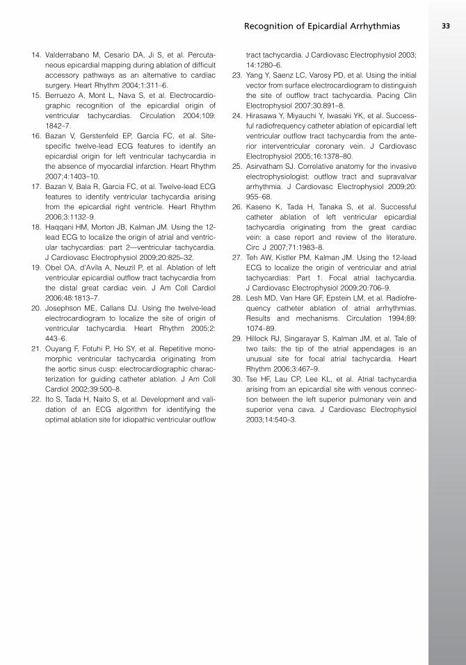
Fig. 2. (A) OT ventricular arrhythmias. Panel I. A patient with ventricular premature complex resembling left OTVT (paper speed 50 mm/s). Endocardial ablation failed to ablate the arrhythmia. MDI above 55% supports anepicardial origin (paper speed of 200 mm/s). Panel II. Earliest ventricular activation during the tachycardia wasrecorded in the distal CS (23 ms before the surface QRS), which supports an origin in the GCV area. On theleft, catheter position during recordings and contrast injection to the left main (LM) coronary artery. The endo-cardial ablation catheter was located endocardially in the LVOT through retrograde aortic approach and the CScatheter was pushed deep into the coronary sinus. (B) ECG of a patient with RVOT VT ablated from epicardialapproach. An MDI of 55% suggests an epicardial location.
Recognition of Epicardial Arrhythmias 31
especially when combined with deep S wave inlead V6.
Epicardial connections between the right atrialappendage and the RV have been described.They can be located 10 mm away from the
annulus. Surface ECG features of these accessorypathways correspond to the anatomic location ofappendage and thus may be similar to those ofright anterior or anterolateral accessory pathways.A relatively long ventriculoatrial conduction time

Michowitz & Belhassen32
during orthodromic atrioventricular reentranttachycardia, due to the long epicardial course,has also been suggested.
ATRIAL TACHYCARDIAS
Atrial tachycardias uncommonly originate from theatrial appendages,27–29 mainly left. In addition, anepicardial AT originating near the left atrialappendage from a venous connection betweenthe left superior pulmonary vein and superiorvena cava has been described.30 The P wavemorphology of left atrial appendage tachycardiawas negative in I and aVL, and positive in V1.The inferior leads demonstrate a broad andnotched P wave due to superior location.27 Nospecific ECG criteria have been described thatdifferentiate endocardial from epicardial atrialtachycardia origin. An epicardial origin should beconsidered when an appendage AT is notamenable to endocardial ablation.
Finally, cases of inappropriate sinus node tachy-cardia requiring epicardial ablation for proceduralsuccess have been described.1 However, as thesinus node is an epicardial structure, no uniqueECG criteria have been described.
SUMMARY
An epicardial origin of ventricular arrhythmia canbe suggested by general and site-specific ECGcriteria. Different features have been shown usefulin different patient population and myocardiallocations. ECG-based criteria have also beendeveloped for recognition of epicardial accessorypathways. In atrial tachycardias, no specific ECGcriteria have been described. However, ina subgroup of patients, despite the use of ECGcriteria, it may not be possible to differentiateepicardial from endocardial origin and intracardiacmapping is needed.
ACKNOWLEDGMENTS
The authors thank Dr Roderick Tung for his helpin preparing the figures.
REFERENCES
1. Schweikert RA, Saliba WI, Tomassoni G, et al. Percu-
taneous pericardial instrumentation for endo-epicar-
dial mapping of previously failed ablations.
Circulation 2003;108:1329–35.
2. Cesario DA, Vaseghi M, Boyle NG, et al. Value of
high-density endocardial and epicardial mapping
for catheter ablation of hemodynamically unstable
ventricular tachycardia. Heart Rhythm 2006;3:1–10.
3. Garcia F, Bazan V, Erica S, et al. Epicardial substrate
and outcome with epicardial ablation of ventricular
tachycardia in arrhythmogenic right ventricular
cardiomyopathy/dysplasia. Circulation 2009;120:
366–75.
4. Aliot EM, Stevenson WG, Almendral-Garrote JM,
et al. EHRA/HRS Expert Consensus on Catheter
Ablation of Ventricular Arrhythmias: developed in
a partnership with the European Heart Rhythm Asso-
ciation (EHRA), a Registered Branch of the Euro-
pean Society of Cardiology (ESC), and the Heart
Rhythm Society (HRS); in collaboration with the
American College of Cardiology (ACC) and the
American Heart Association (AHA). Heart Rhythm
2009;6:886–933.
5. Daniels DV, Lu YY, Morton JB, et al. Idiopathic
epicardial left ventricular tachycardia originating
remote from the sinus of Valsalva: electrophysiolog-
ical characteristics, catheter ablation, and identifica-
tion from the 12-lead electrocardiogram. Circulation
2006;113:1659–66.
6. Doppalapudi H, Yamada T, Ramaswamy K, et al.
Idiopathic focal epicardial ventricular tachycardia
originating from the crux of the heart. Heart Rhythm
2009;6:44–50.
7. Phillips KP, Natale A, Sterba R, et al. Percutaneous
pericardial instrumentation for catheter ablation of
focal atrial tachycardias arising from the left atrial
appendage. J Cardiovasc Electrophysiol 2008;19:
430–3.
8. Di Biase L, Saliba WI, Natale A. Successful ablation
of epicardial arrhythmias with cryoenergy after failed
attempts with radiofrequency energy. Heart Rhythm
2009;6:109–12.
9. McGarvey JR, Schwartzman D, Ota T, et al. Mini-
mally invasive epicardial left atrial ablation and
appendectomy for refractory atrial tachycardia.
Ann Thorac Surg 2008;86:1375–7.
10. Lam C, Schweikert R, Kanagaratnam L, et al. Radio-
frequency ablation of a right atrial appendage-
ventricular accessory pathway by transcutaneous
epicardial instrumentation. J Cardiovasc Electrophy-
siol 2000;11:1170–3.
11. Haghjoo M, Mahmoodi E, Fazelifar AF, et al. Electro-
cardiographic and electrophysiologic predictors of
successful ablation site in patients with manifest
posteroseptal accessory pathway. Pacing Clin
Electrophysiol 2008;3:103–11.
12. Sun Y, Arruda M, Otomo K, et al. Coronary sinus-
ventricular accessory connections producing
posteroseptal and left posterior accessory path-
ways: incidence and electrophysiological identifica-
tion. Circulation 2002;106:1362–7.
13. Takahashi A, Shah DC, Jais P, et al. Specific electro-
cardiographic features of manifest coronary vein
posteroseptal accessory pathways. J Cardiovasc
Electrophysiol 1998;9:1015–25.
Recognition of Epicardial Arrhythmias 33
14. Valderrabano M, Cesario DA, Ji S, et al. Percuta-
neous epicardial mapping during ablation of difficult
accessory pathways as an alternative to cardiac
surgery. Heart Rhythm 2004;1:311–6.
15. Berruezo A, Mont L, Nava S, et al. Electrocardio-
graphic recognition of the epicardial origin of
ventricular tachycardias. Circulation 2004;109:
1842–7.
16. Bazan V, Gerstenfeld EP, Garcia FC, et al. Site-
specific twelve-lead ECG features to identify an
epicardial origin for left ventricular tachycardia in
the absence of myocardial infarction. Heart Rhythm
2007;4:1403–10.
17. Bazan V, Bala R, Garcia FC, et al. Twelve-lead ECG
features to identify ventricular tachycardia arising
from the epicardial right ventricle. Heart Rhythm
2006;3:1132–9.
18. Haqqani HM, Morton JB, Kalman JM. Using the 12-
lead ECG to localize the origin of atrial and ventric-
ular tachycardias: part 2—ventricular tachycardia.
J Cardiovasc Electrophysiol 2009;20:825–32.
19. Obel OA, d’Avila A, Neuzil P, et al. Ablation of left
ventricular epicardial outflow tract tachycardia from
the distal great cardiac vein. J Am Coll Cardiol
2006;48:1813–7.
20. Josephson ME, Callans DJ. Using the twelve-lead
electrocardiogram to localize the site of origin of
ventricular tachycardia. Heart Rhythm 2005;2:
443–6.
21. Ouyang F, Fotuhi P, Ho SY, et al. Repetitive mono-
morphic ventricular tachycardia originating from
the aortic sinus cusp: electrocardiographic charac-
terization for guiding catheter ablation. J Am Coll
Cardiol 2002;39:500–8.
22. Ito S, Tada H, Naito S, et al. Development and vali-
dation of an ECG algorithm for identifying the
optimal ablation site for idiopathic ventricular outflow
tract tachycardia. J Cardiovasc Electrophysiol 2003;
14:1280–6.
23. Yang Y, Saenz LC, Varosy PD, et al. Using the initial
vector from surface electrocardiogram to distinguish
the site of outflow tract tachycardia. Pacing Clin
Electrophysiol 2007;30:891–8.
24. Hirasawa Y, Miyauchi Y, Iwasaki YK, et al. Success-
ful radiofrequency catheter ablation of epicardial left
ventricular outflow tract tachycardia from the ante-
rior interventricular coronary vein. J Cardiovasc
Electrophysiol 2005;16:1378–80.
25. Asirvatham SJ. Correlative anatomy for the invasive
electrophysiologist: outflow tract and supravalvar
arrhythmia. J Cardiovasc Electrophysiol 2009;20:
955–68.
26. Kaseno K, Tada H, Tanaka S, et al. Successful
catheter ablation of left ventricular epicardial
tachycardia originating from the great cardiac
vein: a case report and review of the literature.
Circ J 2007;71:1983–8.
27. Teh AW, Kistler PM, Kalman JM. Using the 12-lead
ECG to localize the origin of ventricular and atrial
tachycardias: Part 1. Focal atrial tachycardia.
J Cardiovasc Electrophysiol 2009;20:706–9.
28. Lesh MD, Van Hare GF, Epstein LM, et al. Radiofre-
quency catheter ablation of atrial arrhythmias.
Results and mechanisms. Circulation 1994;89:
1074–89.
29. Hillock RJ, Singarayar S, Kalman JM, et al. Tale of
two tails: the tip of the atrial appendages is an
unusual site for focal atrial tachycardia. Heart
Rhythm 2006;3:467–9.
30. Tse HF, Lau CP, Lee KL, et al. Atrial tachycardia
arising from an epicardial site with venous connec-
tion between the left superior pulmonary vein and
superior vena cava. J Cardiovasc Electrophysiol
2003;14:540–3.