dyspnea 2005dyspneasociety.com/dyspnea_society/meetings_files/dyspnea_2005_program.…program day 1...
TRANSCRIPT
DYSPNEA 2005
‘
University of San Diego, May 29th-30
Program
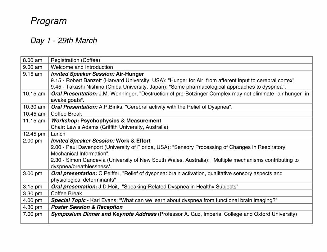
Day 1 - 29th March
8.00 am Registration (Coffee) 9.00 am Welcome and Introduction 9.15 am Invited Speaker Session: Air-Hunger
9.15 - Robert Banzett (Harvard University, USA): "Hunger for Air: from afferent input to cerebral cortex". 9.45 - Takashi Nishino (Chiba University, Japan): "Some pharmacological approaches to dyspnea".
10.15 am Oral Presentation: J.M. Wenninger, "Destruction of pre-Bötzinger Complex may not eliminate "air hunger" in awake goats".
10.30 am Oral Presentation: A.P.Binks, "Cerebral activity with the Relief of Dyspnea". 10.45 am Coffee Break 11.15 am Workshop: Psychophysics & Measurement
Chair: Lewis Adams (Griffith University, Australia) 12.45 pm Lunch 2.00 pm Invited Speaker Session: Work & Effort
2.00 - Paul Davenport (University of Florida, USA): "Sensory Processing of Changes in Respiratory Mechanical Information". 2.30 - Simon Gandevia (University of New South Wales, Australia): 'Multiple mechanisms contributing to dyspnea/breathlessness'.
3.00 pm Oral presentation: C.Peiffer, "Relief of dyspnea: brain activation, qualitative sensory aspects and physiological determinants"
3.15 pm Oral presentation: J.D.Hoit, "Speaking-Related Dyspnea in Healthy Subjects" 3.30 pm Coffee Break 4.00 pm Special Topic - Karl Evans: “What can we learn about dyspnea from functional brain imaging?” 4.30 pm Poster Session & Reception 7.00 pm Symposium Dinner and Keynote Address (Professor A. Guz, Imperial College and Oxford University)
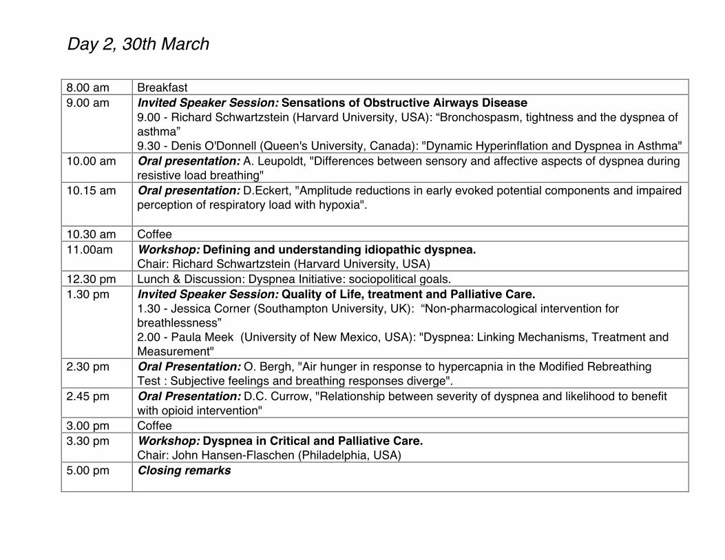
Day 2, 30th March 8.00 am Breakfast 9.00 am Invited Speaker Session: Sensations of Obstructive Airways Disease
9.00 - Richard Schwartzstein (Harvard University, USA): “Bronchospasm, tightness and the dyspnea of asthma” 9.30 - Denis O'Donnell (Queen's University, Canada): "Dynamic Hyperinflation and Dyspnea in Asthma"
10.00 am Oral presentation: A. Leupoldt, "Differences between sensory and affective aspects of dyspnea during resistive load breathing"
10.15 am Oral presentation: D.Eckert, "Amplitude reductions in early evoked potential components and impaired perception of respiratory load with hypoxia".
10.30 am Coffee 11.00am Workshop: Defining and understanding idiopathic dyspnea.
Chair: Richard Schwartzstein (Harvard University, USA) 12.30 pm Lunch & Discussion: Dyspnea Initiative: sociopolitical goals. 1.30 pm Invited Speaker Session: Quality of Life, treatment and Palliative Care.
1.30 - Jessica Corner (Southampton University, UK): “Non-pharmacological intervention for breathlessness” 2.00 - Paula Meek (University of New Mexico, USA): "Dyspnea: Linking Mechanisms, Treatment and Measurement"
2.30 pm Oral Presentation: O. Bergh, "Air hunger in response to hypercapnia in the Modified Rebreathing Test : Subjective feelings and breathing responses diverge".
2.45 pm Oral Presentation: D.C. Currow, "Relationship between severity of dyspnea and likelihood to benefit with opioid intervention"
3.00 pm Coffee 3.30 pm Workshop: Dyspnea in Critical and Palliative Care.
Chair: John Hansen-Flaschen (Philadelphia, USA) 5.00 pm Closing remarks

Invited Speaker Presentations
"Hunger for Air: from Afferent Input to Cerebral Cortex"
Robert Banzett
Physiology Program, Harvard School of Public Health &
Div. of Pulmonary Critical Care Med. Beth Israel Deaconess Medical Center, Harvard Medical School
Invited Speaker Presentation : 9.15 am, 29th March
‘Air hunger’ is the conscious perception of the urge to breathe, a fundamental biological drive. It is one kind of dyspnea, others include 'work/effort' and 'tightness'. Air hunger arises when pulmonary minute ventilation is insufficient. It is described as 'not getting enough air', 'uncomfortable urge to breathe' and is the sensation felt at the end of a long breath hold. Subjects often comment that intense air hunger is a threatening or frightening sensation. Air hunger, like pain, thirst, nausea, and hunger for food, alerts the conscious brain to disturbed physiological state; these perceptions guide adaptive behaviors, and are often important and disabling symptoms of disease. Ordinarily, breathing is matched to metabolic demand by reflex motor responses that are integrated in the brainstem; forebrain input is not required. Thus, ‘routine’ adjustments of breathing do not require perception. Air hunger, in contrast, is essential when threats to respiratory homeostasis require behavioral responses more complex than increasing respiratory muscle activation. The genesis of air hunger requires comparison of inputs that report the demand for respiration versus inputs that report the present level of respiration. Although the afferent mechanisms are not completely understood, our current thinking is that air hunger arises from perception of a 'corollary' copy of brainstem motor output that is transmitted to the forebrain, and is relieved by an increase in mechanoreceptor afferent traffic consequent to increased pulmonary minute ventilation. Thus, if arterial PCO2 is increased (or PO2 is decreased), and breathing is not allowed to increase, the subject experiences air hunger; likewise if PCO2 is held constant and tidal volume is decreased, the subject experiences air hunger. This mechanoreceptor input provides a ‘feed-forward’ mechanism, that warns of immediate and imminently lethal danger. Prominent air hunger-related activations in the forebrain include paralimbic areas such as anterior insula/operculum, anterior cingulate (both also implicated in pain and thirst perception), and the amygdala (implicated in fear responses).
"Treatment of dyspnea with inhaled furosemide: Lessons from breath-holding trials"
T. Nishino
Department of Anesthesiology, Graduate School of Medicine, Chiba University.
Invited Speaker Presentation : 9.45 am, 29th March
Breath-holding is not only a powerful method to induce dyspneic sensation but also give us vital information on the onset and endurance of dyspnea. In conscious subjects, immediately after the start of breathholding at functional residual capacity (FRC), there is a certain period of no particular respiratory sensation lasting for 20-30 seconds, which is designated “no respiratory sensation period”. This period is terminated by the onset of dyspnea and followed by progressive increase in the intensity of dyspnea until the breaking point of breath-holding. The measurement of the period of no respiratory sensation provides us with information on the onset of dyspneic sensation whereas the measurement of the total breath-holding time is a behavioral measure of the tolerable limit of dyspneic sensation. The behavioral measure of tolerable limit of dyspnea can permit the study of dyspnea even in anesthetized animals while observing escape behavior in response to airway occlusion. Inhaled furosemide causes prolongation of both the period of no respiration sensation and total breath-holding time in conscious subjects. Inhaled furosemide also suppresses the behavioral response to airway occlusion but does not affect the behavioral response to noxious somatic stimulus in lightly anesthetized cats. These findings indicate that inhaled furosemide alleviates experimentally-induced dyspnea. The purpose of this presentation is to emphasize the usefulness of breath-holding test as a tool for evaluation of dyspnea. Furthermore, the possible mechanisms of alleviation of dyspnea with inhaled furosemide are highlighted.
"Sensory Processing of Changes in Respiratory Mechanical Information."
Paul W. Davenport
Department of Physiological Sciences, University of Florida, Gainesville, FL
Invited Speaker Presentation : 2.00 pm, 29th March Eupneic breathing normally is not consciously perceived. This suggests that during normal ventilation, respiratory sensory information is gated-out of cognitive centers. When ventilation is obstructed, stimulated, challenged or attended to, cognitive awareness of breathing occurs. This cognitive awareness ranges from an awareness of breathing to a highly distressing sensation of breathlessness. Obstruction of the airways and related increased mechanical work of breathing is one common cause of respiratory distressing sensations. The ability to sense an increased inspiratory load is an essential component of the early recognition of an obstructive event. It is hypothesized that respiratory somatosensation is a gated process that is a function of multiple sensory inputs to higher brain centers. The respiratory related evoked potential (RREP) peak amplitude is correlated with the perception of an increased load to breathing. The P1 peak of the RREP must be present for the late cognitive peaks to occur. In addition, the P1 and P300 peaks were present only if detectable inspiratory loads were presented. An R load with a Weber fraction (_R/Ro) greater than 0.5, elicited the P1, Nf, N1 and P300 peaks. The background resistance was increased to make the Weber fraction less than 0.15 for same _R load. This _R no longer exceeded the detection threshold and the RREP peaks were not present. The RREP response to inspiratory loads was studied in patients with both a double lung transplant and tracheostomy. These patients could respire through both their mouth and tracheostomy. The RREP peaks were present with both mouth breathing and tracheostomy breathing, demonstrated that the RREP can be elicited in the absence of afferent feedback from the lung and upper airways. These results suggest a gating process. Eliciting a cognitive respiratory sensation is dependent on the integration of respiratory afferent activity, respiratory motor drive, affective state, attention, experience and learning. These neural parameters input to a hypothesized gating center that has an output that elicits a cognitive neural response if the combined input exceeds the threshold for gating the respiratory sensation, a gated comparator
"Multiple mechanisms contributing to dyspnea"
Simon Gandevia
University of New South Wales, Australia
Invited Speaker Presentation : 2.30 pm, 29th March
Evolution has built in mechanisms at many levels from the subcellular to the whole-body to preserve oxygen consumption and ventilation. While multiple mechanisms can signal dyspnea (or difficulty with breathing), some signals may have particular significance. Despite initial studies by Campbell, there is now overwhelming evidence that signals related to hypercapnea and hypoxia generate dyspnea. Some of these studies have required complete neuromuscular paralysis. Another basic signaling system involves unmyelinated pulmonary C fibres, a role proposed by Paintal, and by Coleridge and colleagues. Artificial activation of these fibres generates potent respiratory sensations. Other classes of pulmonary afferent probably also mediate specific sensations. Recent studies examined the capacity of pulmonary C fibres to reflexly limit locomotion and voluntary movement (the J reflex proposed by Paintal). Unlike the responses in many experimental animals, conscious humans do not develop motoneuronal inhibition during activation of this system (Butler et al., 2001, J Physiol, 534: 583). This potent viscero-somatic reflex has probably been brought under forebrain control. In addition, the “length-tension” hypothesis of Campbell remains tenable, in which a disparity between achieved and required ventilation is perceived. An alternative, in the same style, focuses on the disparity between achieved and ‘commanded’ ventilation. Signals from specialized receptors in muscle, joints, skin as well as from the lung and upper airways can all contribute. Signals of central motor command may also bias judgements. Recently, neuroimaging studies have revealed the central structures activated during breathlessness. Activation of evolutionarily old regions of the cortex indicates the importance of mounting the correct behavioural response to any challenge to ventilation (Banzett et al., 2000, Neuroreport 11: 2117).
"Bronchospasm, tightness and the dyspnea of asthma"
Richard M. Schwartzstein
Beth Israel and Deaconess Medical Center, Harvard Medical School, USA
Invited Speaker Presentation : 9.00 am, 30th March Since the early 1980’s, the sensation of tightness has been characterized as one of the “elemental sensations” that underlie breathing discomfort. Tightness is particularly evident in association with bronchoconstriction, whether produced experimentally in the laboratory or occurring in the clinical context of an acute asthma attack. The sensation is described by patients with asthma in different cultures and among those who speak widely disparate languages. A prospective study of patients presenting to pulmonary specialists for the evaluation of breathlessness has shown that the sensation of tightness has a high positive predictive value for the diagnosis of asthma. The physiological origins of the sensation of tightness are subject to debate. Data supporting the hypothesis that the sensation arises as the consequence of the stimulation of pulmonary receptors, mediated via vagal afferents, come from studies that utilize inhaled lidocaine to anesthetize the airways, as well as investigations in which bronchoconstriction is provoked in subjects who are breathing passively on a mechanical ventilator. An alternative hypothesis, that tightness arises from hyperinflation, has been proposed. This hypothesis is based primarily upon the correlation of the intensity of breathlessness with reduced inspiratory capacity in experimentally produced bronchoconstriction. These findings are confounded by the observation that patients with asthma have multiple types of respiratory discomfort that evolve as the severity of airway obstruction increases (with tightness present prior to the development of significant hyperinflation), and by the recognition that passively induced hyperinflation as well as hyperinflation associated with emphysema typically do not produce chest tightness. The sensation of chest tightness is one of the earliest and most consistent symptoms associated with acute bronchospasm. The sensation most likely arises from stimulation of pulmonary receptors.
"Dynamic Hyperinflation and Dyspnea in Asthma"
Denis E.M. O'Donnell
Queen's University, Ontario, Canada
Invited Speaker Presentation : 9.30 am, 30th March Dyspnea, the perception of respiratory discomfort, is a primary symptom of asthma. Our understanding of the source and mechanisms of this symptom continues to grow. Earlier psychophysical studies, which have used mechanical loading to simulate asthma have provided insights into causation. However, it can be argued that externally applied resistive loading poorly simulates the fundamental pathophysiological abnormalities of asthmatic attacks namely: expiratory flow limitation and lung hyperinflation. Our approach has been to examine sensory-mechanical relations during high dose methacholine provocation: this allowed us to study sensation (both intensity and quality) over the broad range of physiological perturbations from minor bronchoconstriction to severe lung hyperinflation. We have found that perceived inspiratory difficulty is often more prominently reported than expiratory difficulty during progressive bronchoprovocation. Moreover, the intensity of dyspnea correlates strongly with the extent of lung hyperinflation (as measured by the reduction of inspiratory capacity) that accompanies expiratory flow limitation. We found that significant lung hyperinflation was measurable during relatively minor bronchconstriction when FEV1.0 had declined by only 20%. This makes it difficult to partition the sensory effects of airway narrowing, per se, from that of the attendant elastic loading of the inspiratory muscles. The main qualitative descriptor clusters of dyspnea during bronchoprovocation allude to inspiratory difficulty and unsatisfied inspiratory effort. Both these sensations are reported during minor bronchoconstriction but are more frequently selected at maximal FEV1.0 decline (to <50% of baseline). Hyperinflation results in elastic and threshold loading of the inspiratory muscles already burdened with increased resistive work. We found that continuous positive airway pressure (CPAP), which counterbalances the negative effects of inspiratory threshold loading (i.e., the intrinsic PEEP effect), effectively alleviates the dyspnea of induced asthma. To the extent that unsatisfied inspiration ultimately reflects the disparity that arises between inspiratory effort and the blunted mechanical response of the respiratory system, then CPAP restores more harmonious neuro-mechanical coupling by negating the threshold load at the beginning of each inspiration. Intensity of dyspnea during bronchoconstriction also correlates strongly with the ratio of inspiratory effort (measured by esophageal pressure) to tidal volume displacement, a crude index of neuro-mechanical dissociation. In summary our studies to date have indicated that mechanical factors importantly shape the expression of dyspnea during acute bronchoconstriction in the absence of gas exchange abnormalities. Acute lung hyperinflation which constrains tidal volume expansion in the face of increasing inspiratory effort likely contributes to the intensity and quality of respiratory discomfort of asthma.
"Non-pharmacological intervention for breathlessness"
Jessica Corner
University of Southampton, UK
Invited Speaker Presentation : 1.30 pm, 30th March Non-pharmacological interventions for respiratory problems in palliative care could not as yet be described as a solid, coherent area of study. It is only comparatively recently that the symptoms breathlessness in the context of life-threatening illness has received significant, indeed any attention. The palliative care community has been slow to acknowledge the prevalence of respiratory problems among patients or that the problem of breathlessness warrants significant theoretical or clinical attention. Fortunately the situation is changing, although as yet there is little in the way of research funds to support programmes of research into the problem of breathlessness. The term ‘non pharmacological intervention’ is used to refer to a broader approach to symptom management that suggests not only the need to understand symptoms as a manifestation of disease but also the problems that arise from experiencing symptoms, the meanings symptoms engender, and the facets of life that are interfered with as a result. Intervention therefore is not only directed at breathlessness as a ‘physiological’ event but also at the affect breathlessness has on daily life. A review will be presented of studies of the problem of breathlessness in life-limiting illness and into the effectiveness of interventions tailored to assist individuals adapt to the practical and emotional implications of breathlessness. Goals for future research will be proposed.
"Dyspnea: Linking Mechanisms, Treatment and Measurement"
Paula M Meek
College of Nursing, University of New Mexico
Invited Speaker Presentation : 2.00 pm, 30th March
Dyspnea is the most common symptom experienced by patients with Chronic Pulmonary Disease and is often associated with activity level. Typically dyspnea is first reported in association with activities and can progress over time to also occur at rest. With chronic pulmonary disease, continued daily activity limitations and increases in dyspnea intensity results in an inter-relationship of dyspnea affecting activities, and vice versa, often referred to as the “dyspnea spiral”. This cycle is a complex interchange between physiologic mechanisms, psychosocial issues, mood changes and reduction in function. Implementation of many treatments used with individuals who have chronic pulmonary disease, requires measurement and evaluation of dyspnea as an important part of its assessment. Assessing dyspnea as an outcome may be murky, as frequently, regardless of the type of tool used to measure dyspnea there is a general perception that a reduction in dyspnea intensity or frequency has occurred, despite the fact that the instrument may not actually quantify intensity or frequency nor be linked to the associated physiologic mechanisms. This presentation will review evidence of some misapplication of dyspnea measures and present possible ways to link mechanisms, measurement and treatment in ways that will increase our understanding.

Oral Presentations
“What can we learn about dyspnea from functional brain imaging?”
Karl Evans
Massachussetts General Hospital, Harvard Medical School
Special Topic Presentation : 4.00 pm, 29th March For more than a decade, functional neuroimaging methods have proven to be powerful tools that have advanced models of sensorimotor, cognitive and affective brain function. Both positron emission tomography (PET) and functional magnetic resonance imaging (fMRI) have been used to investigate hypotheses related to the control of breathing and dyspnea perception. While there is human neuroimaging evidence for cortical activation of sensorimotor and supplementary motor regions during volitional breathing, recent PET and fMRI studies have correlated activation of limbic and paralimbic regions with dyspnea perception in healthy humans. Given that neuroimaging techniques are sensitive to dynamic ventilatory associated changes in cerebral blood gases that accompany typical laboratory maneuvers of dyspnea provocation, relatively few dyspnea imaging studies have been published. Air hunger (an uncomfortable urge to breathe) has been successfully provoked in healthy humans during neuroimaging via several different methods. Across methods of provocation the converging lines of evidence support the anterior insular and anterior cingulate cortices as the likely neural substrates for air hunger perception. As numerous other neuroimaging studies of pain and other somatic alarm stimuli have implicated the insular cortex, we believe this region to be essential to air hunger perception.
"Destruction of pre-Bötzinger Complex may not eliminate "air hunger" in awake goats"
J.M. Wenninger, H.V. Forster, L.G. Pan, and R. Banzett
Department of Physiology, Medical College of Wisconsin, Zablocki VA, and Department of
Physical Therapy, Marquette University, Milwaukee, WI 53226. Physiology Program, Harvard School of Public Health & Department of Medicine, Harvard Medical School, MA 02115
Oral Presentation : 10.15 am, 29th March
The primary objective was to determine whether breathing would be sustained in awake goats after bilateral destruction of the pre-Bötzinger Complex (pre-BötzC). Through chronically implanted microtubles, we first bilaterally injected the neurotoxin saporin conjugated to Substance P followed in two weeks by bilateral injection in the awake state of the neurotoxin ibotenic acid (IA, 50 mM, 10µl). With the IA injection, a marked tachypnea commenced within minutes and was sustained for over an hour, after which breathing rapidly declined, leading to terminal apnea in 3 airway intact goats. In four other tracheostomized goats, mechanical ventilation was initiated shortly after the peak tachypnea to avoid severe hypoxia and hypercapnia. While ventilated over the next 4 hours, there was no phasic respiratory muscle activity as long as arterial blood gases were normal. Periodically, the goats were briefly removed from the ventilator, which resulted in severe hypercapnia and hypoxia that was accompanied by phasic abdominal and, on occasion, diaphragm muscle activity. These goats preferred not to or were unable to maintain a standing posture. Once recumbent, some goats maintained the normal sternal posture for all or part of the time. The most prevalent behavioral response was a catatonic-like motor reaction, characterized by: a) lying on one side and resisting attempts to change posture, b) over-all diminished motor initiative with long periods of inactivity broken by tonic-clonic whole body muscle contractions occurring always but not restricted to periods of hypoxemia and hypercapnia, and c) general restlessness and staring,bulging eyes. On the basis of long experience with goat behavior, we interpret these signs as indicators of discomfort. We can find no reasons these goats should have experienced pain; thus, we speculate that some of these informally observed behaviors resulted from dyspnea, specifically "air hunger." Accordingly, since the nuclei hypothesized responsible for eupneic respiratory rhythm were ablated and phasic diaphragm activity was absent during these periods, it seems these data do not support the hypothesis that air hunger results from projection of a copy of respiratory center motor activity to forebrain perceptional areas. In addition, the data indicate the pre-BötzC is required for eupneic breathing and in the absence of the pre-BötzC an abdominal muscle rhythm generator sustains a low level of breathing.
"Cerebral activity with the Relief of Dyspnea"
Andrew Binks & Andrea Vovk
Physiology Program, Harvard School of Public Health, Boston, MA, USA.
Oral Presentation : 10.30 am, 29th March Using fMRI, we have investigated the areas of the brain that respond to the rapid relief of air hunger. Subjects (n=6) were mechanically ventilated at comfortable levels (V∞E = 12 liters•min-1) and rated their air hunger continuously on a visual analog scale (VAS). At the onset of the image acquisition the tidal volume was reduced to a baseline value of 6 liters•min-1 to induce ‘moderate’ air hunger (~60% of VAS). Once at steady-state, 2 or 3 large breaths (volume = 1.5 liters) were delivered to rapidly relieve the air hunger. The relief breaths reduced air hunger to ‘none’ or ‘slight’ within 20 seconds (10-20% of VAS). The ventilation was then returned to baseline to increase air hunger and the process was repeated. In a 10min imaging trial we performed ten rapid drops in air hunger. End-tidal PCO2 was maintained consent throughout the study (~45mmHg) by adjusting inspired PCO2 levels. Event related analysis looked for brain areas that showed a sharp decrease in activity coincident with the sudden relief of air hunger (i.e. positively related to air hunger); fMRI (BOLD) data showed the anterior insula and the anterior cingulate. Regions that were activated when breathlessness was relieved (i.e negatively correlated with air hunger) included the thalamus and the putamen. This study was supported by the Breathlessness Relief Charitable Trust and NIH HL46690.
"Relief of dyspnea: brain activation, qualitative sensory aspects and physiological determinants"
C. Peiffer 1, 2, N. Costes 3, Ph. Herve 2 & L. Garcia-Larrea 3
Oral Presentation : 3.00 pm, 29th March
Several important aspects of dyspnea still await elucidation as for example its relief i.e. the specific sensation of breathing becoming easier. While this sensory experience is commonly used in daily clinical practice to assess the subjective effect of therapeutical intervention, its underlying mechanisms, and more specifically, its central processing remain largely unknown. We studied induced dyspnea relief in terms of physiological and sensory characteristics and associated brain activation in 10 healthy male volunteers. Relief of dyspnea was induced by acute decrease of an important component of dyspnea, perception of loaded breathing. The latter was experimentally induced by a strong resistive load consisting of an inflatable ring which was inserted in an external breathing device. This device enabled us to induce relief of dyspnea during imaging by deflating the ring leading to a rapid but smooth decrease of the resistive load. Brain imaging was performed either during a period of dyspnea induced by a resistive load of constant intensity (cD) or during relief of a dyspnea (RD) of similar intensity than cD. At the end of each scan, the subjects rated the perceived intensity of either dyspnea or the sensation of RD on a Borg scale, and the qualitative aspect by choosing appropriate word descriptors out of a list. Relief of dyspnea was characterized by a sensation of high intensity (similar to that of cD) and by specific word descriptors involving for most of them a clearly positive valence such as “pleasure associated with breathing” and ”easier and/or more comfortable to breathe than during normal breathing”. Sensory intensity of RD was highly determined by the decrease in amplitude of mouth pressure swings (∆P) (which were highly increased during the just preceding dyspnea) and by the parallel increase in minute ventilation (Ve). The best predictor of perceived intensity of RD was however the combination of both parameters namely the decrease in ∆P/Ve which reflects a decrease in neuro-mechanical uncoupling. RD was also associated with specific brain activation whose main activation area was located in the anterior left cingulate gyrus (BA 32) extending in the prefrontal cortex, a region which has been previously shown to be involved in relief of pain. Brain deactivation associated with acute load-induced dyspnea showed a non significant activation area in the left frontal lobe (BA 6) and thus was located in a more posterior and higher part of frontal lobe than dyspnea relief associated activation. In summary, our results suggest that relief of acutely load-induced dyspnea, is a strong, specific, basically pleasant sensory experience that involves a specific neural network which is partly different from that subserving dyspnea.
"Speaking-Related Dyspnea in Healthy Subjects"
Jeannette D. Hoit, Robert W. Lansing, Kristen E. Perona, Robert B. Banzett*
University of Arizona & *Harvard School of Public Health
Oral Presentation : 3.15 pm, 29th March The act of speaking frequently exacerbates dyspnea, sometimes causing patients to withdraw from communication. Little is known about the qualities of dyspnea evoked by speaking, yet knowledge of these qualities could help us understand the underlying physiological mechanisms. We sought to identify the qualities of speaking-related dyspnea by studying 11 healthy young adults while they spoke at different levels of hypercapnia. The CO2 stimulus (2 to 8%) was introduced through a facemask that allowed comfortable breathing and speaking. Flow was adjusted to prevent rebreathing. We recorded chest wall surface motions (converted to volumes), end-tidal PCO2, transcutaneous PCO2, SpO2, and the acoustic speech signal as subjects read aloud a standard text. After the first period of reading when hypercapnic (average end-tidal PCO2 = 50.9 Torr), subjects provided unguided descriptions of their experience and then selected dyspnea-related descriptors. Subjects' responses were later categorized as air hunger, physical exertion, mental effort, and speech observations. Next, we gave subjects definitions of air hunger ("uncomfortable sensation of needing to breathe more, to get more air than you are getting"), physical exertion ("sensation of exerting muscular effort or force in the act of breathing"), and mental effort ("experience of having to pay attention to your breathing, having to think about it, having to control it"). We then asked them to provide visual analog ratings of each quality after reading at different hypercapnic levels (average end-tidal PCO2 for the three levels: 43.7, 47.0, and 49.9 Torr). Rating magnitude increased with stimulus strength. Subjects reported that they were rating distinctly different sensations; however, the three quality ratings were not significantly different from one another (average percent of scale for the three stimulus levels: 16.6, 25.4, and 46.5%). As hypercapnia increased, ventilation, inspired volume, end-inspiratory and end-expiratory volumes, expiratory flow during speaking, and frequency of nonspeech expirations increased. We concluded that speaking-related dyspnea is characterized by two of the qualities associated with the dyspnea of spontaneous breathing: air hunger and physical exertion (work/effort). A third quality, mental effort, appeared to be related to the demands of balancing ventilatory demands and speech requirements. How these qualities might differ with speaking task and communication context has yet to be studied.
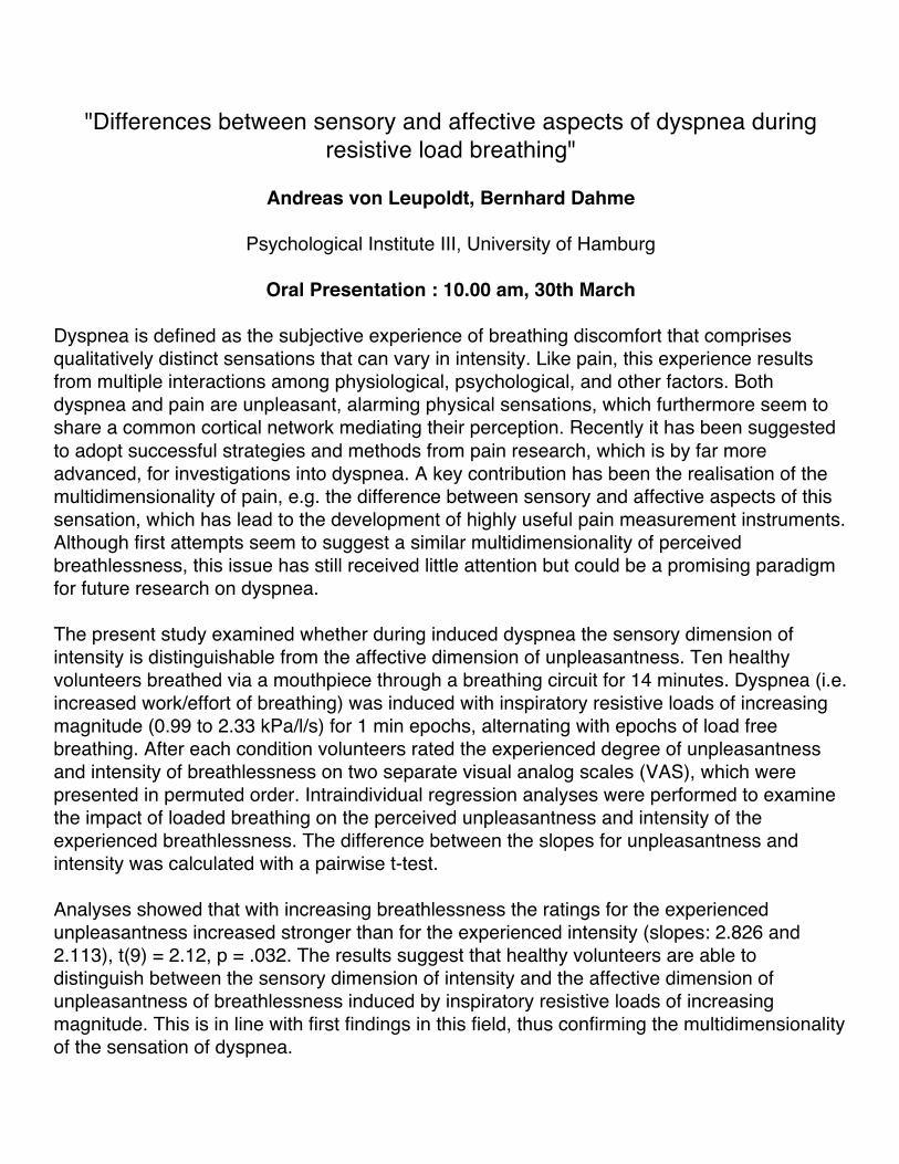
"Differences between sensory and affective aspects of dyspnea during resistive load breathing"
Andreas von Leupoldt, Bernhard Dahme
Psychological Institute III, University of Hamburg
Oral Presentation : 10.00 am, 30th March
Dyspnea is defined as the subjective experience of breathing discomfort that comprises qualitatively distinct sensations that can vary in intensity. Like pain, this experience results from multiple interactions among physiological, psychological, and other factors. Both dyspnea and pain are unpleasant, alarming physical sensations, which furthermore seem to share a common cortical network mediating their perception. Recently it has been suggested to adopt successful strategies and methods from pain research, which is by far more advanced, for investigations into dyspnea. A key contribution has been the realisation of the multidimensionality of pain, e.g. the difference between sensory and affective aspects of this sensation, which has lead to the development of highly useful pain measurement instruments. Although first attempts seem to suggest a similar multidimensionality of perceived breathlessness, this issue has still received little attention but could be a promising paradigm for future research on dyspnea. The present study examined whether during induced dyspnea the sensory dimension of intensity is distinguishable from the affective dimension of unpleasantness. Ten healthy volunteers breathed via a mouthpiece through a breathing circuit for 14 minutes. Dyspnea (i.e. increased work/effort of breathing) was induced with inspiratory resistive loads of increasing magnitude (0.99 to 2.33 kPa/l/s) for 1 min epochs, alternating with epochs of load free breathing. After each condition volunteers rated the experienced degree of unpleasantness and intensity of breathlessness on two separate visual analog scales (VAS), which were presented in permuted order. Intraindividual regression analyses were performed to examine the impact of loaded breathing on the perceived unpleasantness and intensity of the experienced breathlessness. The difference between the slopes for unpleasantness and intensity was calculated with a pairwise t-test. Analyses showed that with increasing breathlessness the ratings for the experienced unpleasantness increased stronger than for the experienced intensity (slopes: 2.826 and 2.113), t(9) = 2.12, p = .032. The results suggest that healthy volunteers are able to distinguish between the sensory dimension of intensity and the affective dimension of unpleasantness of breathlessness induced by inspiratory resistive loads of increasing magnitude. This is in line with first findings in this field, thus confirming the multidimensionality of the sensation of dyspnea.
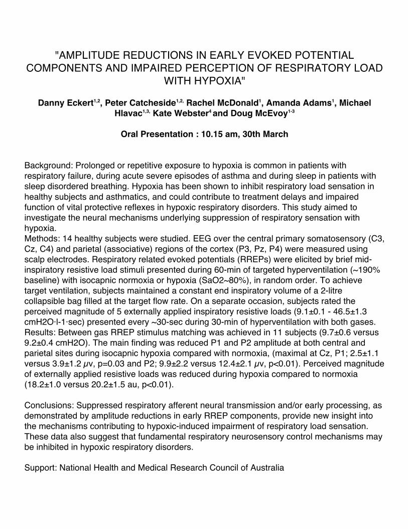
"AMPLITUDE REDUCTIONS IN EARLY EVOKED POTENTIAL COMPONENTS AND IMPAIRED PERCEPTION OF RESPIRATORY LOAD
WITH HYPOXIA"
Danny Eckert1,2, Peter Catcheside1,2, Rachel McDonald1, Amanda Adams1, Michael Hlavac1,3, Kate Webster4 and Doug McEvoy1-3
Oral Presentation : 10.15 am, 30th March
Background: Prolonged or repetitive exposure to hypoxia is common in patients with respiratory failure, during acute severe episodes of asthma and during sleep in patients with sleep disordered breathing. Hypoxia has been shown to inhibit respiratory load sensation in healthy subjects and asthmatics, and could contribute to treatment delays and impaired function of vital protective reflexes in hypoxic respiratory disorders. This study aimed to investigate the neural mechanisms underlying suppression of respiratory sensation with hypoxia. Methods: 14 healthy subjects were studied. EEG over the central primary somatosensory (C3, Cz, C4) and parietal (associative) regions of the cortex (P3, Pz, P4) were measured using scalp electrodes. Respiratory related evoked potentials (RREPs) were elicited by brief mid-inspiratory resistive load stimuli presented during 60-min of targeted hyperventilation (~190% baseline) with isocapnic normoxia or hypoxia (SaO2~80%), in random order. To achieve target ventilation, subjects maintained a constant end inspiratory volume of a 2-litre collapsible bag filled at the target flow rate. On a separate occasion, subjects rated the perceived magnitude of 5 externally applied inspiratory resistive loads (9.1±0.1 - 46.5±1.3 cmH2O·l-1·sec) presented every ~30-sec during 30-min of hyperventilation with both gases. Results: Between gas RREP stimulus matching was achieved in 11 subjects (9.7±0.6 versus 9.2±0.4 cmH2O). The main finding was reduced P1 and P2 amplitude at both central and parietal sites during isocapnic hypoxia compared with normoxia, (maximal at Cz, P1; 2.5±1.1 versus 3.9±1.2 µv, p=0.03 and P2; 9.9±2.2 versus 12.4±2.1 µv, p<0.01). Perceived magnitude of externally applied resistive loads was reduced during hypoxia compared to normoxia (18.2±1.0 versus 20.2±1.5 au, p<0.01). Conclusions: Suppressed respiratory afferent neural transmission and/or early processing, as demonstrated by amplitude reductions in early RREP components, provide new insight into the mechanisms contributing to hypoxic-induced impairment of respiratory load sensation. These data also suggest that fundamental respiratory neurosensory control mechanisms may be inhibited in hypoxic respiratory disorders. Support: National Health and Medical Research Council of Australia
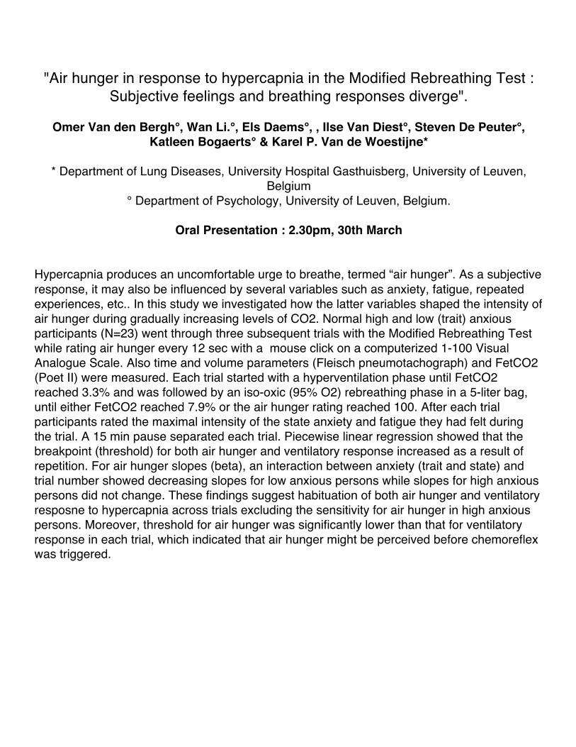
"Air hunger in response to hypercapnia in the Modified Rebreathing Test : Subjective feelings and breathing responses diverge".
Omer Van den Bergh°, Wan Li.°, Els Daems°, , Ilse Van Diest°, Steven De Peuter°,
Katleen Bogaerts° & Karel P. Van de Woestijne*
* Department of Lung Diseases, University Hospital Gasthuisberg, University of Leuven, Belgium
° Department of Psychology, University of Leuven, Belgium.
Oral Presentation : 2.30pm, 30th March Hypercapnia produces an uncomfortable urge to breathe, termed “air hunger”. As a subjective response, it may also be influenced by several variables such as anxiety, fatigue, repeated experiences, etc.. In this study we investigated how the latter variables shaped the intensity of air hunger during gradually increasing levels of CO2. Normal high and low (trait) anxious participants (N=23) went through three subsequent trials with the Modified Rebreathing Test while rating air hunger every 12 sec with a mouse click on a computerized 1-100 Visual Analogue Scale. Also time and volume parameters (Fleisch pneumotachograph) and FetCO2 (Poet II) were measured. Each trial started with a hyperventilation phase until FetCO2 reached 3.3% and was followed by an iso-oxic (95% O2) rebreathing phase in a 5-liter bag, until either FetCO2 reached 7.9% or the air hunger rating reached 100. After each trial participants rated the maximal intensity of the state anxiety and fatigue they had felt during the trial. A 15 min pause separated each trial. Piecewise linear regression showed that the breakpoint (threshold) for both air hunger and ventilatory response increased as a result of repetition. For air hunger slopes (beta), an interaction between anxiety (trait and state) and trial number showed decreasing slopes for low anxious persons while slopes for high anxious persons did not change. These findings suggest habituation of both air hunger and ventilatory resposne to hypercapnia across trials excluding the sensitivity for air hunger in high anxious persons. Moreover, threshold for air hunger was significantly lower than that for ventilatory response in each trial, which indicated that air hunger might be perceived before chemoreflex was triggered.
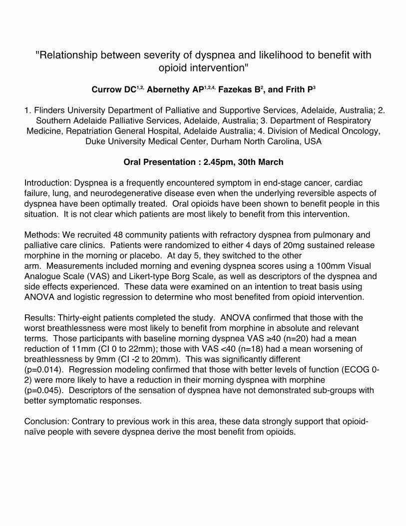
"Relationship between severity of dyspnea and likelihood to benefit with opioid intervention"
Currow DC1,2, Abernethy AP1,2,4, Fazekas B2, and Frith P3
1. Flinders University Department of Palliative and Supportive Services, Adelaide, Australia; 2.
Southern Adelaide Palliative Services, Adelaide, Australia; 3. Department of Respiratory Medicine, Repatriation General Hospital, Adelaide Australia; 4. Division of Medical Oncology,
Duke University Medical Center, Durham North Carolina, USA
Oral Presentation : 2.45pm, 30th March Introduction: Dyspnea is a frequently encountered symptom in end-stage cancer, cardiac failure, lung, and neurodegenerative disease even when the underlying reversible aspects of dyspnea have been optimally treated. Oral opioids have been shown to benefit people in this situation. It is not clear which patients are most likely to benefit from this intervention. Methods: We recruited 48 community patients with refractory dyspnea from pulmonary and palliative care clinics. Patients were randomized to either 4 days of 20mg sustained release morphine in the morning or placebo. At day 5, they switched to the other arm. Measurements included morning and evening dyspnea scores using a 100mm Visual Analogue Scale (VAS) and Likert-type Borg Scale, as well as descriptors of the dyspnea and side effects experienced. These data were examined on an intention to treat basis using ANOVA and logistic regression to determine who most benefited from opioid intervention. Results: Thirty-eight patients completed the study. ANOVA confirmed that those with the worst breathlessness were most likely to benefit from morphine in absolute and relevant terms. Those participants with baseline morning dyspnea VAS ≥40 (n=20) had a mean reduction of 11mm (CI 0 to 22mm); those with VAS <40 (n=18) had a mean worsening of breathlessness by 9mm (CI -2 to 20mm). This was significantly different (p=0.014). Regression modeling confirmed that those with better levels of function (ECOG 0-2) were more likely to have a reduction in their morning dyspnea with morphine (p=0.045). Descriptors of the sensation of dyspnea have not demonstrated sub-groups with better symptomatic responses. Conclusion: Contrary to previous work in this area, these data strongly support that opioid-naïve people with severe dyspnea derive the most benefit from opioids.
"A method for comparing dyspnea and pain in fMRI settings".
Andreas von Leupoldt, Bernhard Dahme
Presented as Poster : Reception 4.30-6.45 pm, 29th March The cortical mechanisms underlying the perception of dyspnea are still far from being known. First neuroimaging studies in this field showed activations of distinct brain areas (e.g. anterior insular, anterior cingulate cortex) which are similar to those involved in pain perception. Both are unpleasant, alarming physical sensations and strongly motivate adaptive behaviour. Because of these similarities a common cortical network has been assumed but not examined within one study yet. The present pilot study examined without imaging techniques whether both sensations can be compared within one experimental context, thus allowing predictions for a study in fMRI settings. Seven healthy volunteers breathed via a mouthpiece through a breathing circuit for 16 minutes. Dyspnea was induced with resistive loads for 1 min epochs. The loads were added to the inspiratory portion of the circuit to prevent rebreathing of CO2, which increases global cerebral blood flow and thus interferes with fMRI measurements. A painful heat stimulus was induced for 1 min epochs with a surface contact thermode (TSA II, Medoc) placed below the sternum. During the 1 min baselines the thermode was set to adaptive 32°C with no inspiratory load added. To obtain comparable stimulus sizes for dyspnea and pain in each individual extensive pre tests were performed. During these, volunteers rated the intensity of different resistive loads and heat stimuli, presented for 1 min each, on a Borg-Scale. Inspiratory load (M=2.45 kPa/l/s) and heat stimulus (M= 46.0°C) had to induce a corresponding Borg value (M=5, strong intensity). During the main test four dyspneic, four painful, and eight baseline conditions were presented in permuted order with a baseline separating two unpleasant conditions. Endtidal Pco2 (PETCO2), inspiratory time (Ti) and breathing frequency (f) were measured continuously. After each condition participants rated the experienced unpleasantness and intensity on visual analog scales (VAS). The quality of dyspnea and heat pain was rated after the test with verbal descriptor scales. ANOVAs confirmed the expected increases in Ti and decreases in f during dyspneic conditions when compared to baseline conditions. No differences between heat pain and baseline conditions in Ti and f were obtained, but higher Ti during dyspneic when compared to painful stimulation. PETCO2 showed slight increases during dyspneic conditions compared to baseline and painful conditions (∆PETCO2= 1.5 and 1.3 mmHg) with the latter showing no differences. ANOVAs for VAS-ratings revealed no difference between dyspnea and heat pain in experienced intensity (MD=4.9 and MH=5.3) and unpleasantness (MD=5.0 and MH= 5.1). Similar verbal descriptors were used to characterise both sensations. The results suggest that dyspnea and heat pain can be examined within one experimental design. Both tonic stimuli can be presented with comparable intensity and unpleasantness, which is the basis for a future comparison within fMRI settings. The change in PETCO2 of 1.5 mmHg between conditions is within the natural range and should still allow a clear differentiation between stimulus induced changes in the fMRI signal and those related to increases in global cerebral blood flow due to increased arterial PCO2.
"Imagined Suffocation as a Trigger for Hyperventilation"
Omer Van den Bergh*, Ilse Van Diest*a, Steven De Peuter*, Elke Wellens*, & Karel P. Van de Woestijne°
*Department of Psychology, University of Leuven, Belgium
°Faculty of Medicine, University of Leuven, Belgium
Presented as Poster : Reception 4.30-6.45 pm, 29th March It was tested whether stronger hyperventilation responses occurred in response to imagined suffocation compared to imagining other fearful situations. Fractional end-tidal CO2-concentration (FETCO2), breathing frequency and inspiratory volume were measured non-intrusively in high (n=24) and low (n=24) trait anxious women during imagery of three fear, one tension, one depressive and three relaxation scripts. The fear scripts were equal in ratings of unpleasantness and arousal, but differed regarding the inclusion of suggested suffocation and entrapment. After each imagery trial, participants rated the emotional dimensions of pleasantness, arousal, and dominance and the vividness of imagery. FETCO2 decreased in in all fear scripts. High trait anxious women showed a stronger reduction in FETCO2 compared to low trait anxious women during the fear script suggesting risk of suffocation, but not during the other fear scripts. This effect was unrelated to any of the self-reported fear ratings. Self-reported fear of entrapment was associated with an overall lower FETCO2, but not with enhanced reactivity to imagined entrapment. The findings suggest that high trait anxiety is associated with stronger respiratory responsivity to imagined risk of suffocation and may constitute a specific vulnerability factor for the development of panic disorder and claustrophobia.
"Cultural specificities of medically unexplained dyspnea in China".
Jiangna Han*, M.D., Ph.D., Yuanjue Zhu*, M.D., Ph.D., Shunwei Li*, M.D., Ph.D., Dongmei Luo*, M.A., Xiansheng Chen*, M.A., Zheng Hu*, M.A., Claudia Put^, Ph.D.,
Karel P. Van de Woestijne^ M.D., Ph.D., & Omer Van den Bergh° Ph.D.
*Peking Union Medical College Hospital, Beijing, China °Department of Psychology, University of Leuven, Belgium
^ Department of Lung Diseases, University Hospital Gasthuisberg, University of Leuven, Belgium
Presented as Poster : Reception 4.30-6.45 pm, 29th March
Sixty-one spontaneously reported descriptors of dyspnea and associated symptoms were elicited in Chinese patients to make a symptom checklist, which was administered to new groups of patients with different cardiopulmonary diseases, to patients with medically unexplained dyspnea and to healthy subjects. Test-retest reliability was satisfactory for most of the descriptors. A principal component analysis on the descriptors yielded eight factors: dyspnea-effort of breathing, dyspnea-affective aspect, wheezing, anxiety, tingling, palpitation, coughing and sputum, and dying experience. The descriptors of dyspnea-effort of breathing resembled western wordings and were shared by patients with a variety of diseases. The descriptors of dyspnea-affective aspect appeared more culturally specific and were primarily linked to the diagnosis of medically unexplained dyspnea. Wheezing was specifically linked to asthma. In conclusion, three factors of breathlessness were found in the Chinese language, as used by patients. The descriptors of dyspnea-effort of breathing and wheezing are similar to Western descriptors, whereas the dyspnea-affective aspect appears more culturally specific.
A multi-site randomized controlled trial of oxygen versus air for the palliative
management of dyspnea
AP Abernethy1, DC Currow2, PA Frith3, AJ Crockett4, BS Fazekas2, S Booth5, CF McDonald6, K Clark7, D Woods8 and IH Young7
Presented as Poster : Reception 4.30-6.45 pm, 29th March An international multi-site randomized controlled trial of oxygen versus medical air for the palliative management of refractory dyspnea in patients with PaO2>55mmHg. Methodology of the O2 Breathe Study. Management of breathlessness is a central challenge in the care of patients with life-limiting illness; the role of oxygen therapy in this setting remains controversial. Oxygen benefits people with chronic obstructive pulmonary disease (COPD) who have severe hypoxemia (PaO2≤55mmHg). When PaO2>55mmHg and the funding criteria for home oxygen are not met, palliative oxygen may be provided on a compassionate basis for people with intractable breathlessness in the setting of life-limiting illness. In these cases the goal is relief of the sensation of breathlessness and improvement in quality of life, yet we do not know how effective or burdensome such treatment is. In response, we are conducting an international multi-center randomized double-blind controlled trial of oxygen versus medical air in the palliative care setting. The specific aims are to test the effectiveness of oxygen versus air in relieving the sensation of breathlessness and improving quality of life, to identify predictors of effect and burdens of the interventions, and to analyze costs. Eligible participants have intractable breathlessness at rest or with minimal exertion due to cancer, COPD or chronic heart disease but PaO2>55mmHg; 240 participants are being recruited from eight sites in the US, UK and Australia. Oxygen or medical air will be delivered continuously from identical appearing concentrators through nasal cannulae for seven days; participants and investigators will be blinded to the gas provided. Outcome measures reflect the specific aims and the primary outcome is breathlessness on a 0-10 numerical rating scale. Recruitment began in October 2004 and is expected to be completed by mid-2006. The results of this study will significantly advance our knowledge of how to better care for this large group of distressed individuals. The results will also advance a long-standing international debate about the role of palliative oxygen for intractable dyspnea and its funding.
"Psycho-emotional Aspects of Dyspnea and Other Breathing Problems: How the Combination of CBT (Cognitive Behavioral Therapy) and YBT
(Yoga Breathing Techniques) Can Help."
Vijai Sharma
Behavioral Medicine Center, Cleveland, TN.
Presented as Poster : Reception 4.30-6.45 pm, 29th March
(modified 05/03/05) Anxiety can cause a dyspnea episode and dyspnea can cause anxiety. The “anxiety-dyspnea circularity” may contribute to a high incidence of anxiety and panic attacks in people with COPD (presumptive estimate). Such symptoms as the shortness of breath, excessive breathing effort, “air hunger,” smothering, chest tightness, nausea, fear of loss of control, death or disability can be symptoms of physiologic Dyspnea, psychogenic anxiety and panic attack or due to combination of both. Dyspnea and anxiety can escalate due to: - Misperception and misinterpretations (cognitive) - Heightened awareness, hypervigilance and scanning (fight-flight response) - Anxious temperament (Anxiety trait) and anxiety state - Anxiety sensitivity to physical symptoms - Reduced tolerance of uncomfortable body and breathing sensations Patients find it difficult to differentiate between an anxiety attack and a dyspnea episode Dyspnea attack caused by organic factors or psychogenic factors may be further compounded by pain episodes in case of co-morbid disorders. CBT techniques for anxiety and chronic panic disorders can be effective in reduction of dyspnea. Specific breathing techniques utilized in Panic Control Therapy and Yogic breathing techniques known for slowing and stabilizing the breath can minimize breath-related anxiety CBT and YBT should be added to the medical management of dyspnea. Vijai Sharma, PhD RYT, clinical psychologist, certified yoga teacher, Director National Emphysema/COPD Association (NECA), COPD-ALERT.
"Structural and functional neural characteristics in a developmental model of impaired awareness to respiratory stimuli"
R. M. Harper, P. M. Macey, R. Kumar, M.A. Woo, R.K. Harper.
Presented as Poster : Reception 4.30-6.45 pm, 29th March
Congenital Central Hypoventilation Syndrome (CCHS) patients show impaired ventilatory responses to hypercapnia and hypoxia, diminished ventilation during sleep, and impaired awareness of low oxygen or high CO2. Perception of exertion to enhanced breathing efforts, however, appears largely intact. To determine the neural structures and functional interactions of brain areas underlying processing of respiratory-related stimuli, we examined functional magnetic resonance (fMRI) blood oxygen level dependent signals in 14 children with CCHS and 14 age- and gender- matched control subjects to hypercapnic, hypoxic and expiratory loaded breathing challenges, and T2 relaxometry differences (n = 12 CCHS, 28 controls) to evaluate structural damage. Functional challenges lasted 120s, and were preceded by 30s baseline. Areas of deficient fMRI responses for each challenge were determined using SPM2 (boxcar model, random effects, threshold p<0.01) and overlaid onto anatomical maps. For structural studies, high resolution T1-weighted images were normalized to Montreal Neurological Institute space templates, and the resulting parameters were applied to T2 maps; these normalized T2 maps were smoothed. Two-sample t-tests were performed at each voxel to compare T2 values of control and CCHS groups, and statistical parametric maps showing regions of significant T2 value changes were displayed (corrected for multiple comparisons; p < 0.05). Both white and gray matter showed damage in CCHS patients, with substantially greater axonal injury on the right side, from the anterior cingulate cortex to the level of the posterior cingulate. Portions of the right cerebellar cortex and deep nuclei, cingulate cortex and cingulum bundle, internal capsule, putamen and globus pallidus, basal forebrain extending to the medial thalamus, mid-hippocampus and ventral prefrontal cortex showed structural damage. Functional responses in CCHS cases that differed significantly from controls emerged in the cerebellar cortex and deep nuclei, amygdala or bed nucleus, right mid-hippocampus and insular cortex to hypoxia, hypercapnia and expiratory loading challenges; cingulate cortex showed deficits over nearly the entire extent to loaded breathing, but only in a restricted area to hypercapnia, and not to hypoxia. The functional responses in amygdala/bed nucleus and insular areas may contribute to disturbances in affective aspects of the ventilatory challenges in CCHS. Structural injuries within the limbic sites, or in projections to the sites, may mediate the impaired affective characteristics to ventilatory stimuli. A principal functional response difference between expiratory loading and hypoxic and hypercapnic challenges emerged in the cingulate cortex, a region which shows structural damage both in gray matter and in fibers of the cingulum bundle. Determination of structural neural injury within CCHS and assessment of selective functional aberrations in the syndrome have the potential to clarify neural mediation of affective properties of ventilatory stimuli.
"Parenteral Opioid Dose Titration and Ventilatory Function"
Bassam Estfan, Declan Walsh, Mellar P. Davis, Susan B. LeGrand, Ruth L. Lagman, Philip Shaheen
The Harry R. Horvitz Center For Palliative Medicine, Taussig Cancer Center, The Cleveland
Clinic Foundation, Cleveland OH
Presented as Poster : Reception 4.30-6.45 pm, 29th March Background: Physicians fear opioid induced respiratory depression, which prevents proper dose titration and leads to under treatment. Hypoventilation causes hypoxemia, reduced respiratory rate, carbon dioxide (CO2) retention and increased end-tidal CO2 (ET-CO2),. ET-CO2 can increase due to reduced tidal volume despite a normal respiratory rate. We evaluated ventilatory function in cancer patients undergoing parenteral opioid titration for severe pain. Method: Patients on intravenous opioids for severe cancer pain on study. Patients on study could not be oxygen dependent and had to have a normal cognitive function by the Bedside Confusion Scale. Informed consent was obtained. ET-CO2, O2 saturation, respiratory rate, vital signs and a delirium scale were done daily during opioid titration and at the time of pain control. This was defined as pain less than 4 (0 - 10) and < 4 rescue doses per day. Spirotomy (FVC) was done on the last day of study. A paired two-sample T-test for equal variance was used to compare the last day ET-CO2 to the pre-study ET-CO2. The paired T-test used a variable of 0 and also 4. The normal variation for repeat ET-CO2 determinations is 4. Results: 27 completed the trial (including FVC) and 3 patients completed the ET-CO2 but not the FVC. 28 maintained a RR > 10, (one transiently dropped to 8 and one to 9). All maintained O2 sats > 92%. The mean ET-CO2 was 32mm Hg (+ 5) before opioid titration and 35mm Hg (+ 6) on the last day (p = 0.13 for 0 variation, p = 0.23 for variation of 4). None had an ET-CO2 > 50mm Hg. Conclusion: Parenteral opioid titration to pain relief is not associated with either hypoxemia nor CO2 retention.
"Dyspnea in Patients with Environmental Chemical Intolerance"
Andrea Vovk
Physiology Program, Harvard School of Public Health
Presented as Poster : Reception 4.30-6.45 pm, 29th March Dyspnea is one of the primary symptoms associated with a class of syndromes without a known “organic” origin called environmental chemical intolerance (ECI). Over 61% of people with ECI report dyspnea. Similarly, dyspnea is the second most reported symptom in patients with diseases that have a known “organic” origin such as COPD, asthma and CHF. While a good deal of literature has focused on elucidating the physiological mechanisms and describing the subjective sensations of dyspnea in these diseases, only scattered reports of dyspnea in ECI exist. This review consolidates the existing information about the demographics and prevalence of dyspnea in this group, subjective reports on the sensation of dyspnea and physiological measurements that might elude to a mechanistic pathway of dyspnea in ECI. This review serves as a preliminary step in guiding scientists to develop future research projects investigating dyspnea in people with ECI.