dynamic positioning of mitotic spindles in yeastlabs.bio.unc.edu/bloom/content/reference...
TRANSCRIPT

Molecular Biology of the CellVol. 11, 3949–3961, November 2000
Dynamic Positioning of Mitotic Spindles in Yeast:Role of Microtubule Motors and Cortical Determinants□V
Elaine Yeh,* Charlie Yang,* Elaine Chin,* Paul Maddox,* E. D. Salmon,*Daniel J. Lew,† and Kerry Bloom*‡
*Department of Biology, University of North Carolina at Chapel Hill, Chapel Hill, North Carolina27599-3280; †Department of Pharmacology and Cancer Biology, Duke University Medical Center,Durham, North Carolina 27709
Submitted June 16, 2000; Revised August 16, 2000; Accepted September 13, 2000Monitoring Editor: J. Richard McIntosh
In the budding yeast Saccharomyces cerevisiae, movement of the mitotic spindle to a predeterminedcleavage plane at the bud neck is essential for partitioning chromosomes into the mother anddaughter cells. Astral microtubule dynamics are critical to the mechanism that ensures nuclearmigration to the bud neck. The nucleus moves in the opposite direction of astral microtubulegrowth in the mother cell, apparently being “pushed” by microtubule contacts at the cortex. Incontrast, microtubules growing toward the neck and within the bud promote nuclear movementin the same direction of microtubule growth, thus “pulling” the nucleus toward the bud neck.Failure of “pulling” is evident in cells lacking Bud6p, Bni1p, Kar9p, or the kinesin homolog,Kip3p. As a consequence, there is a loss of asymmetry in spindle pole body segregation into thebud. The cytoplasmic motor protein, dynein, is not required for nuclear movement to the neck;rather, it has been postulated to contribute to spindle elongation through the neck. In the absenceof KAR9, dynein-dependent spindle oscillations are evident before anaphase onset, as are post-anaphase dynein-dependent pulling forces that exceed the velocity of wild-type spindle elonga-tion threefold. In addition, dynein-mediated forces on astral microtubules are sufficient to segre-gate a 2N chromosome set through the neck in the absence of spindle elongation, but cytoplasmickinesins are not. These observations support a model in which spindle polarity determinants(BUD6, BNI1, KAR9) and cytoplasmic kinesin (KIP3) provide directional cues for spindle orien-tation to the bud while restraining the spindle to the neck. Cytoplasmic dynein is attenuated bythese spindle polarity determinants and kinesin until anaphase onset, when dynein directs spindleelongation to distal points in the mother and bud.
INTRODUCTION
The regulated orientation of cell divisions in response topositional cues is critical for morphogenesis and cell-fatespecification during development in many organisms(Drubin and Nelson, 1996; Gonczy and Hyman, 1996). Posi-tional information, either cell-intrinsic or derived from ex-trinsic cues, directs the alignment of the mitotic spindlealong a specified axis, leading to oriented cell division. In thebudding yeast Saccharomyces cerevisiae, the site of bud emer-gence becomes the mother-bud neck, where cytokinesistakes place (Chant and Pringle, 1995; Pringle et al., 1995).Thus, the mitotic spindle must orient perpendicular to thisprespecified cleavage plane to accurately partition chromo-somes into mother and daughter cells. Spindle orientation is
achieved through interactions between astral microtubules,microtubule-based motor proteins, and cortical determi-nants, as well as the actin cytoskeleton (Palmer et al., 1992;Carminati and Stearns, 1997; Shaw et al., 1997b; Theesfeld etal., 1999; Heil-Chapdelaine et al., 2000).
Cytoplasmic astral microtubules in yeast emanate fromthe spindle pole body (SPB; functionally equivalent to thecentrosome in animal cells), which is embedded in the nu-clear envelope (Peterson and Ris, 1976; Byers, 1981). The SPBis duplicated near the time of the G1/S transition, and in G2the duplicated SPBs separate to opposite sides of the nucleusas the mitotic spindle is formed. Astral microtubules extendfrom each SPB toward the cell cortex, while intranuclearmicrotubules extending between the SPBs and from SPBs tokinetochores form the spindle (as with other fungi, the nu-clear envelope does not break down during mitosis). Beforeanaphase, the spindle is positioned close to the neck suchthat one SPB directs one or more astral microtubule toward
□V Online version of this article contains video material.‡ Corresponding author. E-mail address: [email protected].
© 2000 by The American Society for Cell Biology 3949
the bud cortex and the other SPB nucleates microtubulespredominantly within the mother cell. This feature results inspindle orientation along the mother-bud axis; anaphasespindle elongation then proceeds until the spindle polesreach the distal ends of the budded cell, resulting in accuratesegregation of chromosomes at cell division.
Our understanding of the molecular basis for spindle orien-tation has benefited greatly from genetic and cell biologicalapproaches, in which specific microtubule motors (primarilydynein and the kinesin homologue Kip3p), a putative micro-tubule plus-end capturing protein (Kar9p)(Miller and Rose,1998), and asymmetrically distributed cortical proteins (Kar9p,Num1p, Bni1p, and Bud6p) were all shown to be important forspindle positioning. Cells lacking any one of these proteinsoften contain misaligned spindles, but in all cases this defect isrectified before cytokinesis. In contrast, cells lacking dynein incombination with one of several other proteins (e.g., Kip3p,Kar9p, Bud6p, Bni1p) exhibit conditional phenotypes (thisstudy and Lee et al., 1999; Miller et al., 1999). Two partiallyredundant “pathways” for spindle orientation have been pro-posed, one, involving Kip3p, Kar9p, Bni1p, and Bud6p, thatacts early in the cell cycle to position the preanaphase spindle,and a second involving dynein, that acts late in the cell cycle topromote spindle segregation (Cottingham and Hoyt, 1997;Heil-Chapdelaine et al., 1999; Lee et al., 1999; Miller et al., 1999).
Accurate spindle positioning also depends upon an intactactin cytoskeleton (Palmer et al., 1992). Actin’s role in spindlepositioning versus its essential role in bud growth was de-termined through perturbations of F-actin and use of muta-tions that selectively disrupt actin cables (Pruyne et al., 1998;Theesfeld et al., 1999). Actin is required early in the cell cycle,temporally coincident with the Kip3p-dependent phase, toestablish spindle orientation. Two proteins required for theKip3p-dependent phase (Bni1p, Bud6p) are themselves ac-tin-interacting proteins. Bni1p/She5p contains a formin ho-mology domain, is required for integrity of the actin cy-toskeleton, and is itself related to a protein, Cdc12p, in S.pombe that is transported on microtubules and actin fila-ments (Chang, 1999). Bud6p/Aip3p (Amberg et al., 1997) isdelivered to cortical sites via the secretory machinery, whereit contributes to polarized cell growth (Jin and Amberg,2000). Bud6p/Aip3p and Bni1p/She5p are required for theintegrity of the actin cytoskeleton, localized distribution ofKar9p (Miller et al., 1999), and regulating mating compe-tency in the mother versus the bud (ASH1 mRNA; Beachand Bloom 1999). The pleiotropic phenotype of bud6 andbni1 mutants point to a more general role for these proteinsin polarized events. Bud6p and Bni1p have been proposed tocomprise a cortical scaffold for docking ASH1 mRNA as wellas for providing a target for microtubule capture via Kar9p.The role of the actin cytoskeleton is therefore critical fortimely delivery and/or deposition of these cortical compo-nents (e.g., Bni1p, Bud6p, Kar9p) that in turn facilitate mi-crotubule–cortical interactions.
Microtubule interactions with cortical sites are mediatedin part by Bim1p. Bim1p, a member of the EB1 family ofproteins that interact with the adenomatous polyposis colitumor suppressor, binds the plus ends of microtubules (Tir-nauer et al., 1999) and physically interacts with Kar9p (Ko-rinek et al., 2000; Lee et al., 2000). The capture of microtubuleends at the cell cortex (Adames and Cooper, 2000), togetherwith findings that shortening astral microtubules lose tubu-
lin subunits from the plus ends (Maddox et al., 2000) indicatethat cortical sites of Kar9p promote plus-end disassemblythrough Bim1p, while maintaining contact with the shorten-ing end. Loss of the cortical interactions can be correctedthrough dynein-dependent microtubule sliding (Adamesand Cooper (2000)), consistent with previous findings thatthe early pathway is dispensable (Cottingham and Hoyt,1997; DeZwaan et al., 1997). The proposed functional overlapof cortical capture mechanisms with dynein-dependent slid-ing processes (Adames and Cooper, 2000) was not predictedfrom the temporal separation of these pathways in geneticstudies.
A remarkably small number (3–5) of dynamic astral mi-crotubules provide the basis for nuclear movement through-out the cell cycle. The astral microtubules have an unexpect-edly short average lifetime (, 3.65 min; Maddox et al., 1999),suggesting that spindle positioning may result from multi-ple transient interactions. In addition, preexisting microtu-bules are partitioned unequally upon SPB duplication andseparation. One SPB retains all of the dynein-GFP labeledastral microtubules. The other SPB acquires dynein-GFPafter a delay of ;10 min, during which time intranuclearmicrotubules push the SPBs apart (Shaw et al., 1997b). This“delayed acquisition” of dynein-GFP to one SPB is regulatedby the Cdc28p cyclin-dependent kinase and appears to beimportant for preventing the late-acquiring SPB from nucle-ating bud-directed microtubules (Segal et al., 2000). Theseobservations suggest a simple model in which the stableorientation of one SPB toward the bud and the other SPBtoward the mother comes about through the preferentialassociation of one SPB at the neck with bud-directed astralmicrotubules. The rigid intranuclear spindle predisposes thesecond SPB away from the neck, steering astral microtubulesfrom that SPB away from the bud.
In this study we have examined microtubule and spindlebehavior in living cells that lack individual proteins (or pairsof proteins) involved in spindle orientation. Our resultssuggest that Kip3p, Kar9p, Bni1p, and Bud6p provide direc-tional cues for spindle orientation, as well as oppose theaction of dynein until anaphase onset. Cytoplasmic dyneinis the dominant cytoplasmic force and drives spindle elon-gation to distal reaches in the mother and bud. The hierar-chy of motors, their interaction with each other and transientinteraction with astral microtubules reflect distinct but co-ordinated pathways toward faithful nuclear migration.
MATERIALS AND METHODS
Strains, Media, Growth Conditions, and GeneticTechniquesS. cerevisiae strains used in this study are described in Table 1 andwhere appropriate. YEF473, 1345, 1358, AM102, AM16, AM29, andAM8 were kindly provided by J. Pringle (University of North Caro-lina at Chapel Hill). dhc1::URA3 and dhc1::LEU2 deletions wereconstructed as described previously (Li et al., 1993). kar9::LEU2deletions were constructed by transforming strains with SmaI cutpRM386 (kindly provided by R. Miller and M. Rose). Akip3::KANMX cassette was designed that results in a deletion of theentire KIP3 coding region (kip3::KANMX 59 catattccctgattgcatcgtt-tatctgattatcatttcattgcattcttcagctgaagcttcgtacgc(69mer),kip3::KANMX39 gcgtttaatgctggcggaaagaagttatattcgatagtttacgtaggatagcataggccact-agtggatctg (72mer)). The deletion cassettes were introduced intoDLY2 (DLY3650) and W303 (KBY58 and KBY61) strain back-
E. Yeh et al.
Molecular Biology of the Cell3950
grounds. All three strains displayed similar numbers of binucleatecells in logarithmically growing populations. The aip3/bud6 pheno-type has been reported to vary in different genetic backgrounds(Amberg et al., 1997). The aip3-D3 mutant used herein (1345) anddescribed in Amberg et al. (1997) is resistant to 37°C and containsvery few binucleated cells. Strains derived from YEF473 were usedin the real-time analysis. Microtubules were visualized in live cellseither by transformation with the plasmid pKB701 containing aDHC1-GFP fusion protein on the GAL1 promoter or by integrationof pASF125 containing GFP::TUB1 at URA3 (kindly provided by A.Straight). To visualize DHC1-GFP, cells were grown overnight inSD-URA medium containing 2% glucose and 2% galactose. Yeastcultures were grown at 32°C, except where otherwise noted. Yeastrich medium (YPD) contained 2% glucose, 2% peptone, and 1%yeast extract. Synthetic Dropout (SD) medium lacking particularnutrients (SD-Leu, SD-His) and synthetic sporulation medium sup-plemented with amino acids were made as described (Rose et al.,1991).
Genetic Interactions. dhc1 kar9 and dhc1 bni1 double-mutant strainswere not viable when grown at temperatures outside a narrowpermissive range (DHC1 encodes the dynein heavy chain) as re-ported by (Lee et al., 1999; Miller et al., 1999). In addition to theseinteractions dhc1,bud6 double mutants exhibited a conditionalgrowth phenotype (.75% of double-mutant spore colonies from adhc1 BUD6 X DHC1 bud6 cross were not viable at 37°C, comparedwith , 15% lack of viability for wild-type or single-mutant sporecolonies from the cross). Mutations in other genes affecting actinorganization did not show synthetic growth defects when crossed todhc1 mutants (this group included bem1 [Chenevert et al., 1992],bem2 [Peterson et al., 1994], tpm1 [Liu and Bretscher, 1992], cap2[Amatruda and Cooper, 1992; Karpova et al., 1995], bed1 [Mondesertand Reed, 1996], and spa2 [Snyder, 1989; Zahner et al., 1996]).
Microscopy and QuantitationCells expressing the Dhc1p-GFP fusion protein or GFP-Tub1p wereobserved using time-lapse video microscopy. Strains 1345 and 1358containing plasmid pKB701 were grown overnight to midlogarith-mic phase in glucose/galactose-containing medium at 24°C. Doublemutants (dhc1/bni1 from 1358 x KBY61; dhc1/bud6 from 1345 xKBY58) containing GFP-Tub1 were grown overnight to midloga-rithmic phase in glucose-containing medium at 24°C. Cells wereapplied to microscope slides coated with minimal medium contain-ing 25% gelatin. Coverslips were applied and sealed with valap(1:1:1 vaseline:lanolin:paraffin), and cells were observed with a mi-croscope (Microphot FXA, Nikon, Garden City, NY) using a 100X/1.4NA Plan Apo objective. Metamorph software (Universal ImagingCorp., West Chester, PA) was used to control a Biopoint 99B100filter wheel (Ludl Electronic Products, Hawthorne, NY) (Salmon etal., 1998). A shuttered mercury lamb was used for fluorescenceexcitation (490 nm) at 0.3-s exposures. Images were collected with acooled CCD camera (C4880; Hamamatsu, Photonics, Bridgewater,NJ) at either 30-s or 1-min intervals throughout the course of cellgrowth. A total of five fluorescence images were acquired at 0.75mm intervals through the cell and projected into a single reconstruc-tion. A single differential interference contrast (DIC) image wasmade at the middle z-step by rotating the analyzer into the lightpath and taking a 0.6-s exposure (Shaw et al., 1997a).
RESULTS
Spindle Pole Asymmetry Is Retained in bni1, bud6,and kar9 MutantsObservation of bni1, bud6, or kar9 single-mutant cells con-taining GFP-tagged tubulin or dynein (see MATERIALSAND METHODS) confirmed previous findings that these
Table 1. Yeast strains and plasmids
Yeast strains Genotype
YEF473 MATa ura3-52, trp1D63, leu2D1, lys2-801, his3D200YEF1345 MATa ura3-52, trp1D63, leu2D1, lys2-801, his3D200, aip3D3(bud6)<TRP1YEF1358 MATa ura3-52, trp1D63, leu2D1, lys2-801, his3D200, bni1<HIS3CYY118 MATa ura3-52, trp1D63, leu2D1, lys2-801, his3D200, dhc1D<LEU2KBY474 MATa ura3-52, trp1D63, leu2D1, lys2-801, his3D200, kip3D<KANMXKBY475 MATa ura3-52, trp1D63, leu2D1, lys2-801, his3D200, kar9D<LEU2AM16 MATa ura3-52, trp1D63, leu2D1, lys2-801, his3D200, bud7<HIS3AM29 MATa ura3-52, trp1D63, leu2D1, lys2-801, his3D200, bud3<TRP1AM8 MATa ura3-52, trp1D63, leu2D1, lys2-801, his3D200, bud8<TRP1KBY61 MATa leu2-3,112, ura3-52, trp1, ade2, his3D200, dhc1D7<URA3KBY58 MATa leu2-3,112, ura3-52, trp1, ade2, his3D200, dhc1D<LEU2DLY2 MATa ura3Dns, leu2-3,112, trp1-1a, his2, ade1DLY3650 MATa ura3Dns, leu2-3,112, trp1-1a, his2, ade1, dhc1D<LEU2DLY2719 MATa ura3Dns, leu2-3,112, trp1-1a, his2, ade1, bem2<URA3JMY1011 MATa ura3Dns, leu2-3,112, trp1-1a, his2, ade1, bem1<URA3JMY1020 MATa ura3Dns, leu2-3,112, trp1-1a, his2, ade1, tpm1<URA3JMY1023 MATa ura3Dns, leu2-3,112, trp1-1a, his2, ade1, cap2<URA3JMY1030 MATa ura3Dns, leu2-3,112, trp1-1a, his2, ade1, spa2<URA3JMY1080 MATa ura3Dns, leu2-3,112, trp1-1a, his2, ade1, bed1<URA3JMY2-26 MATa ura3Dns, leu2-3,112, trp1-1a, his2, ade1, myo2-66
Plasmids
pKB701 GALpDHC1-GFP, 2m, URA3pAFS125 TUB1-GFP integrated at URA3pRM386 kar9DLEU2 disruption plasmid
Mechanisms of Spindle Positioning
Vol. 11, November 2000 3951
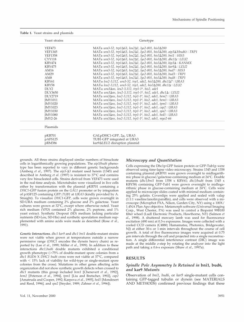
cells frequently misorient the spindle (Miller and Rose, 1998;Lee et al., 1999). Failure to regulate the asymmetric behaviorof spindle pole bodies during spindle assembly also resultsin a spindle position defect in cdc28–4 clb5Dmutants (Segalet al., 2000). To assess spindle pole asymmetry in bni1, bud6,or kar9 cells, we monitored dynein-GFP accumulation dur-ing SPB separation. In every case (.50 examples for eachmutant) there was a delay after visible separation of theSPBs before the second spindle pole body accumulated cy-toplasmic dynein (and astral microtubules), similar in tim-ing to that seen in wild-type cells (e.g., kar9 mutant, Figure1A; quantitative fluorescence accumulation in Figure 1B).Like wild-type cells these mutants are not defective in theasymmetric partitioning and/or acquisition of dynein-GFPduring SPB separation.
Defective Spindle Pole Orientation Toward the Budin bni1, bud6, and kar9 MutantsThe pole that initially acquires astral microtubules in G1orients the nucleus toward the neck and leads spindle mi-gration into the bud upon anaphase onset (Shaw et al.,1997b). While spindle pole asymmetry was maintained inkar9, bud6, and bni1 mutants, neither SPB was predisposed tolead spindle migration into the bud (e.g., migration of lateacquiring SPB into the bud in kar9 mutant, Figure 1A). Insome cases (bni1 mutant, Figure 2), defects in SPB orienta-tion toward the bud occurred through depolymerization ofthe bud-directed microtubule and subsequent penetration ofan astral microtubule from the other SPB into the bud. Inother instances, microtubules from the second pole pene-trated into the bud even though the first pole retained bud-directed microtubules, generating cells with astral microtu-bules from both spindle poles extending into the bud (bud6mutant, Figure 3). Loss of SPB orientation and/or dualmicrotubule penetration was observed in 25–35% of themutant cells (7/28 for bud6, 11/35 for bni1, and 5/14 forkar9). Loss of spindle orientation along the mother-bud axiswas often accompanied by cycles of microtubule polymer-ization/depolymerization in the bud, alternating betweeneach SPB (shown in Figure 2). These transition periods per-sisted for ; 1–8% of the time before anaphase onset (3.7% inbud6, 8.5/230 total minutes; 8.1% in bni1, 50/617 total min;1.6% in kar9, 7.5/464 total min). Neither loss of SPB orien-tation nor penetration of astral microtubules from both polesinto the bud were observed in . 6000 min of image analysisin wild-type or dynein mutant cells (Shaw et al., 1997b).
Despite the frequent appearance of spindles with micro-tubules from both poles penetrating the bud in bni1, bud6, orkar9 mutants, there was not a commensurate migration ofthe entire spindle into the bud (few transient events, ,5 min,observed over the course of 20–30 h of filming) as observedin clb5-cdc28–4 mutants (Segal et al., 2000).
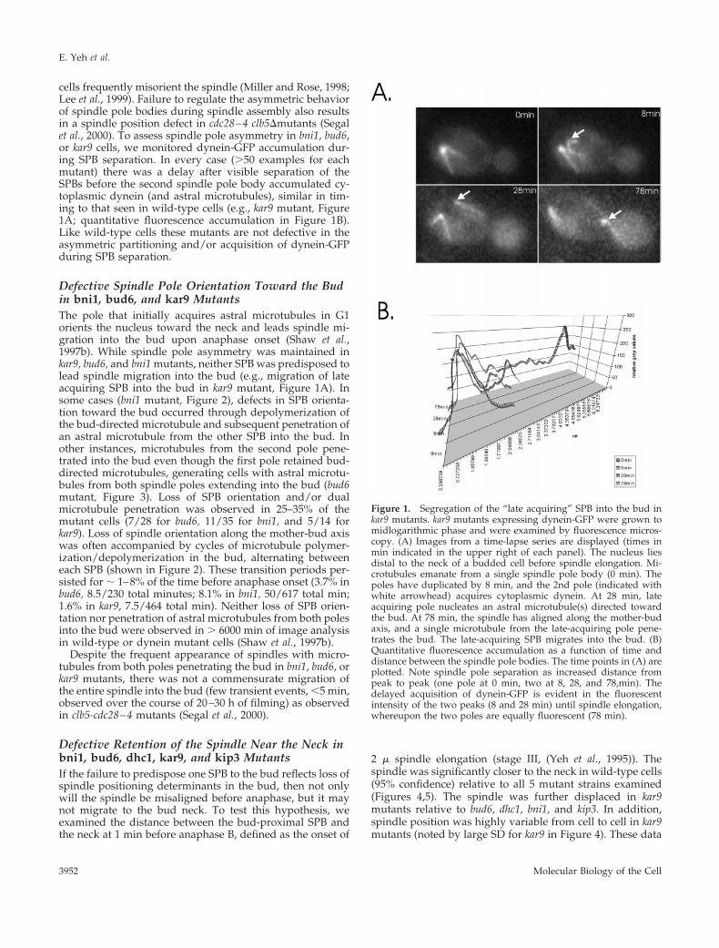
Defective Retention of the Spindle Near the Neck inbni1, bud6, dhc1, kar9, and kip3 MutantsIf the failure to predispose one SPB to the bud reflects loss ofspindle positioning determinants in the bud, then not onlywill the spindle be misaligned before anaphase, but it maynot migrate to the bud neck. To test this hypothesis, weexamined the distance between the bud-proximal SPB andthe neck at 1 min before anaphase B, defined as the onset of
2 m spindle elongation (stage III, (Yeh et al., 1995)). Thespindle was significantly closer to the neck in wild-type cells(95% confidence) relative to all 5 mutant strains examined(Figures 4,5). The spindle was further displaced in kar9mutants relative to bud6, dhc1, bni1, and kip3. In addition,spindle position was highly variable from cell to cell in kar9mutants (noted by large SD for kar9 in Figure 4). These data
Figure 1. Segregation of the “late acquiring” SPB into the bud inkar9 mutants. kar9 mutants expressing dynein-GFP were grown tomidlogarithmic phase and were examined by fluorescence micros-copy. (A) Images from a time-lapse series are displayed (times inmin indicated in the upper right of each panel). The nucleus liesdistal to the neck of a budded cell before spindle elongation. Mi-crotubules emanate from a single spindle pole body (0 min). Thepoles have duplicated by 8 min, and the 2nd pole (indicated withwhite arrowhead) acquires cytoplasmic dynein. At 28 min, lateacquiring pole nucleates an astral microtubule(s) directed towardthe bud. At 78 min, the spindle has aligned along the mother-budaxis, and a single microtubule from the late-acquiring pole pene-trates the bud. The late-acquiring SPB migrates into the bud. (B)Quantitative fluorescence accumulation as a function of time anddistance between the spindle pole bodies. The time points in (A) areplotted. Note spindle pole separation as increased distance frompeak to peak (one pole at 0 min, two at 8, 28, and 78,min). Thedelayed acquisition of dynein-GFP is evident in the fluorescentintensity of the two peaks (8 and 28 min) until spindle elongation,whereupon the two poles are equally fluorescent (78 min).
E. Yeh et al.
Molecular Biology of the Cell3952

indicate a defect in the mechanisms responsible for juxtapo-sition of the SPB to the neck before anaphase onset.
Spindle Alignment and Oscillations BeforeAnaphase in bni1, bud6, dhc1, kar9, and kip3MutantsCytoplasmic forces acting on the preanaphase spindle aremanifested as oscillations of the spindle along the mother/bud axis at constant spindle length (Yeh et al., 1995). Theoscillations begin ; 5–15 min before anaphase onset and
average between 2 and 3 per cell (range 1–5, Figure 5 and(Yeh et al., 1995)). Cells lacking cytoplasmic dynein did notdisplay spindle oscillations: the spindle migrated to within1.0 mm of the neck (as in wild-type cells) and remained static(Figure 5, dhc1 panels), suggesting that the preanaphasespindle oscillations are indeed powered by dynein. Dis-placement of the spindle from the neck in spindle positionmutants shown in Figure 4 could reflect a general loss ofcytoplasmic forces, or specific loss of a bud-directed force.To distinguish between these mechanisms we examinedspindle oscillations before and through the initial stages ofanaphase B.
Spindle oscillations were quantitated using a semiauto-mated tracking system to determine the position of bothSPBs relative to the neck at 30-s intervals. The neck provideda reference point for comparing spindle movement from cellto cell. Loss of spindle alignment along the mother-bud axiswas observed as converging or overlapping points, indicat-ing that the SPBs have rotated perpendicular to the imageplane (e.g., bni1 right panel 10min, kip3 left panel 5, 14min,bud6 right panel 1–5min, 22min). Spindles in wild-type,bud6, bni1, and kip3 cells exhibited a similar number ofoscillation, however the average distance from the neck wasslightly higher in mutant versus wild-type (wild-type 1.04 60.57 mm, bni1 1.24 6 0.60 mm, bud6 1.57 6 0.89 mm, kip31.63 6 0.68 mm). Spindles in bni1 and bud6 were observed totransit into and out of the bud (Figure 5, bni1 left panel35min, right panel 20min; complete spindle transit left panelbud6 42min)(Lee et al., 1999). In kar9 mutants, the few cells
Figure 2. SPB nucleating microtubules that first penetrate the budare not the SPB destined for the bud in bni1 mutants. bni1 mutantsexpressing dynein-GFP were grown to midlogarithmic phase andexamined by fluorescence microscopy. Images from a time-lapseseries are displayed (right, times in min indicated in the upper rightof each panel). Corresponding DIC images are shown to the left.Two spindle pole bodies can be seen in the upper cell (4 o’clock and6 o’clock, respectively). Astral microtubules from the pole at 6o’clock penetrate the bud (0,1 min). At 2 and 3 min, the bud isdevoid of astral microtubules. At 4 and 5 min, the pole at 4 o’clocknucleates an astral microtubule that penetrates the bud. This poleleads the nucleus into the bud at later time points.
Figure 3. Simultaneous penetration of astral microtubules in thebud in bud6 mutants. Bud6 mutants expressing dynein-GFP werevisualized by fluorescence microscopy. The mother cell is in thebottom right quadrant, the daughter to the top left. Astral microtu-bules emanating from spindle pole bodies in the mother (8 and 10o’clock respectively) cross over the neck and penetrate the budsimultaneously. These defects were observed in 3/18 bud6 mutantsfor a total of 3.7 percentage of the time before anaphase onset (8 minof alternating cycles of microtubule polymerization/depolymeriza-tion in the bud emanating from each pole, 30 s where microtubulesfrom both poles penetrate the bud; 230 total minutes). 5/20 bni1mutants exhibited dual penetration from both spindle poles for 8.1%of the time (50/617 total min) and 2/11 kar9 mutants for a totalduration of 1.6% of the time before anaphase onset (7.5/464 totalmin).
Mechanisms of Spindle Positioning
Vol. 11, November 2000 3953
(n 5 5) that contain spindles aligned along the mother/budaxis also displayed frequent oscillations that spanned aneven greater distance (3.7 6 0.89 mm; Figure 5), as well ascomplete transits into the bud (left panel 13 min, right panel35, 40 and 48 min). These spindle oscillations are consistentwith the high variability found in spindle position relative tothe neck (kar9, Figure 4). The increased spindle motilityindicates that spindle displacement from the neck does notreflect a general loss of cytoplasmic forces. KIP3, which hasbeen proposed to act in the same pathway as BNI1 andKAR9, is likewise not required for spindle oscillations beforeanaphase. Spindle oscillations in kip3 precede anaphase by30 min (similar to kar9) and are characterized by loss of poleorientation where the initially distal pole (circles) came to lieproximal to the neck (left panel 10–14 min). However theamplitude of oscillations in kip3 was not as great as kar9.This finding is consistent with results above demonstratingdecreased displacement from the neck in kip3 relative to kar9mutants, and indicates kip3 and kar9 have distinguishablespindle position and motility phenotypes before anaphase
onset. The dependency of these preanaphase spindle oscil-lations on dynein is indicative of early dynein function.Kar9p, Bni1p, and Bud6p are not required for dynein-de-pendent spindle oscillations. Rather, they appear to restrainthe spindle in the vicinity of the neck. This might beachieved either through a role of these proteins in down-regulating dynein activity, or through a role in anchoringthe spindle and/or astral microtubules near the neck in theface of dynein-dependent pulling forces.
Astral Microtubule Length and Orientation in bni1,bud6, kar9, kip3, and dhc1 MutantsOne possible cause of spindle orientation and motility de-fects are alterations in astral microtubule dynamics. To char-acterize the preanaphase astral microtubules we examinedthe length distributions of three classes of astral microtu-bules in preanaphase mitotic cells. The three classes includemicrotubules extending into the mother cell and nucleatedfrom the spindle pole distal to the neck (SPBd mother), those
Figure 4. Distance from SPB proximal to neck 1 min before spindle pole elongation. Wild-type, bud6, bni1, kar9, kip3, and dhc1 mutantscontaining Tub1-GFP were grown to midlogarithmic growth and examined by fluorescence microscopy. Cells containing a 1–1.5 mm spindlewere examined and followed by time-lapse microscopy through spindle elongation. At 1 min before anaphase spindle elongation, a point atthe center of the neck was determined in DIC and was used as the origin when measuring the distance to the center of SPB fluorescence. TrackPoints software in Metamorph (Universal Imaging Corp.) recorded the SPB position at each time point. The distance was determined byconverting pixels to microns using the image of the stage micrometer. Values are the average of ten to twenty cells. Wild-type spindles arestatistically closer to the neck than spindles in bud6, bni1, kar9, and kip3 mutants as determined by 2-tailed unequal variance Students t test(95% confidence). kar9 spindles are significantly further from the neck relative to bud6, bni1, kip3, and wild-type. Spindle position in bud6, bni1,kip3, and dhc1 are statistically indistinguishable.
E. Yeh et al.
Molecular Biology of the Cell3954

extending into the mother cell and nucleated from the spin-dle pole proximal to the neck (SPBp mother), and those ex-tending into the bud and nucleated from the spindle poleproximal to the neck (SPBp bud)(Figure 6). All three classes ofmicrotubules were significantly longer in kip3 mutants
(.95% confidence interval), extending previous reports andconsistent with the interpretation that Kip3p affects micro-tubule dynamics. Interestingly, bud6, bni1, kar9, and dhc1SPB
d mothermicrotubules were only slightly longer than their
wild-type counterpart (20% on average) but not statistically
Figure 5. Metaphase and anaphase spindle move-ments in wild-type and mutant cells. Spindle move-ments in wild-type, bud6, bni1, kar9, kip3, and dhc1mutants. The distances between each spindle polebody and neck were measured (mm) at 30-s intervalsusing Track Points software in Metamorph (Univer-sal Imaging Inc.). The position of the SPB relative tothe mother is plotted as positive numbers, the dis-tance between the neck and the bud is plotted as anegative number. The position of the neck is set aszero. Two examples of each cell type are shown. Thespindle pole that is initially distal to the neck at t 50 is indicated by a circle, the pole proximal to theneck by a diamond. Preanaphase spindle oscillationswere observed in 5/7 wild-type cells, 5/5 kar9, 10/13kip3, 9/16 bni1, 8/15 bud6, and 0/8 dhc1 mutants,respectively.
Mechanisms of Spindle Positioning
Vol. 11, November 2000 3955
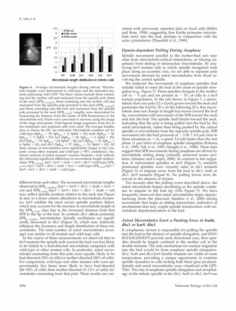
different from each other. The increased microtubule lengthsobserved in SPBp mother (kip3 5 kar9 5 dhc1 . bud6 5 bni1 5wt) and SPBp bud (kip3 5 kar95 bni1 . dhc1 5 bud6 5 wt)may reflect spindle position relative to the neck (see Figure4) and, to a lesser extent, alterations in microtubule dynam-ics. kar9 exhibits the most severe spindle position defect,which may account for the increase in microtubule length ofthe SPBp bud class due to the increased distance from thatSPB to the tip of the bud. In contrast, dhc1 affects primarilySPBp mother microtubules. Spindle oscillations are signifi-cantly decreased in dhc1 (Figure 5), which may indirectlyinfluence the dynamics and length distribution of these mi-crotubules. The total number of astral microtubules (aver-age) was similar in all mutant and wild-type cells.
In the course of these measurements we observed that inkar9 mutants the spindle pole nearest the bud was less likelyto be linked to a bud-directed microtubule compared withwild-type or other mutant cells. In particular, astral micro-tubules emanating from this pole were equally likely to bebud-directed (30% of cells) or mother-directed (30% of cells).For comparison, wild-type and other mutant cells were ap-proximately five times more likely to have bud-directed(40–54% of cells) than mother-directed (5–11% of cells) mi-crotubules emanating from that pole. These results are con-
sistent with previously reported data on fixed cells (Millerand Rose, 1998), suggesting that Kar9p promotes microtu-bule entry into the bud, perhaps in conjunction with theactin cytoskeleton (Theesfeld et al., 1999).
Dynein-dependent Pulling During AnaphaseSpindle movements parallel to the mother-bud axis mayarise from microtubule-cortical interactions, or (during an-aphase) from sliding of intranuclear microtubules. By ana-lyzing kar9 mutant cells in which spindle elongation tookplace along an eccentric axis, we are able to separate polemovements directed by astral microtubules from those in-volving the central spindle.
We analyzed the movements of anaphase spindles thatinitially failed to enter the bud at the onset of spindle elon-gation (e.g., Figure 7). These spindles elongate in the mothercell to ; 5 mm and are present in ; 5% of cells in a kar9mutant population. In the cell shown in Figure 7, a micro-tubule from one pole (12 o’clock) grows toward the neck andpenetrates the bud by 30 s. In the following 30 s, this micro-tubule does not change in length but moves toward the budtip, concomitant with movement of the SPB toward the neckand into the bud. The spindle itself bends toward the neck,indicating that the pole is being pulled into the bud by theastral microtubule, rather than being pushed by the centralspindle or microtubules from the opposite spindle pole. SPBmovement into the bud proceeds at ; 3.58 6 0.8 mm/min inthese situations (n 5 6), a speed 3.6-fold faster than the fastphase (1 mm/min) of anaphase spindle elongation (Kahanaet al., 1995; Yeh et al., 1995; Straight et al., 1998). These datasuggest that SPB movements during anaphase are driven bymicrotubule sliding along cortically anchored motor pro-teins (Adames and Cooper, 2000). In contrast to fast migra-tion of maloriented spindles in kar9 (Figure 7), similarlypositioned spindles were virtually static in dhc1 mutants(Figure 5) or migrate away from the bud in dhc1, bud6 ordhc1, kar9 mutants (Figure 8). No pulling forces were ob-served in the absence of dynein.
Ten seconds after the pulling event described above, theastral microtubule begins shortening as the spindle contin-ues to migrate to the bud tip (120s Figure 7). We havefrequently observed that astral microtubules begin depoly-merizing (from the plus-end; Maddox et al., 2000) duringmovements that begin as sliding interactions, indicative ofmechanisms that may couple spindle translocation with mi-crotubule depolymerization in the bud.
Astral Microtubules Exert a Pushing Force in bud6,dhc1 or kar9, dhc1If cytoplasmic dynein is responsible for pulling the spindleinto the bud in the absence of spindle elongation, and BNI1/BUD6/KAR9/KIP3 provide early directional cues, then spin-dles should be largely confined to the mother cell in thedouble mutants. The only mechanism for nuclear migrationinto the bud would be from anaphase spindle elongation.dhc1, bud6 and dhc1,kar9 double mutants are viable at roomtemperature, providing a unique opportunity to examinespindle dynamics in cells lacking both these gene products.Spindle and astral microtubules were visualized with GFP-Tub1. The rate of anaphase spindle elongation and morphol-ogy of the mitotic spindle in the dhc1, bud6 or dhc1, kar9 was
Figure 6. Average microtubule lengths during mitosis. Microtu-bule lengths were determined in wild-type and the indicated mu-tants expressing Tub1-GFP. The three classes include those extend-ing into the mother cell and nucleated from the spindle pole distalto the neck (SPBd mother), those extending into the mother cell andnucleated from the spindle pole proximal to the neck (SPBp mother),and those extending into the bud and nucleated from the spindlepole proximal to the neck (SPBp bud). Lengths were determined bymeasuring the distance from the center of SPB fluorescence to themicrotubule end. Pixels were converted to microns using the imageof the stage micrometer. Time-lapsed image sequences from five toten metaphase and anaphase cells were used. The average lengths,plus or minus the SD, are indicated. Microtubule numbers are forwild-type (Spbpb 5 39, Spbpm 5 8, Spbd 5 25); bud6 (Spbpb 5 36,Spbpm 5 7, Spbd 5 23); bni1 (Spbpb 5 30, Spbpm 5 3, Spbd 5 25),kar9 (Spbpb 5 25, Spbpm 5 24, Spbd 5 30), kip3 (Spbpb 5 32, Spbpm 56, Spbd 5 42), and dhc1 (Spbpb 5 57, Spbpm 5 10, Spbd 5 43). Allthree classes of microtubules were significantly longer in kip3 mu-tants versus other mutants and wild-type (.95% confidence). Sta-tistical analysis (2-tailed unequal variance Students t test) indicatethe following significant differences in microtubule length relation-ships: SPBd mother kip3. kar9 5 bud6 5 bni15 dhc1.wild-type; SPBpmother kip3 5 kar9 5 dhc1 . bud6 5 bni1 5 wild-type; SPBp bud kip3 5kar95 bni1 . dhc1 5 bud6 5 wild-type.
E. Yeh et al.
Molecular Biology of the Cell3956

typical of wild-type cells (data not shown). Late anaphasespindles were readily observed in the mother cell in thesemutants and exhibited erratic movement and rotation (Fig-ure 8A and B). Microtubules penetrated the bud, and spindlepoles migrated opposite to the direction of astral microtu-bule growth (dhc1, bud6 Figure 8A, 0 -31 min). There was noevidence for pulling forces toward the bud in bud6, dhc1mutants. In contrast, the spindle migrated away from theneck from t 5 0 (1.34 mm) to t 5 31 min (2.01 mm) despite thepenetration of microtubules into the bud. Movement awayfrom the microtubule plus-end was reminiscent of nuclearrotations in G1, in which the nucleus is propelled opposite tothe direction of microtubule growth (Shaw et al., 1997b).Similarly, in dhc1, kar9 double mutants, astral microtubules
penetrated the bud, yet there was no net movement of thespindle toward the bud (Figure 8B). In addition, prean-aphase spindle oscillations (shown in Figure 5) were com-pletely absent in the dhc1, kar9 double mutant (data notshown). Elongation of astral microtubule in the bud andcontact with the bud cortex lead to spindle movement awayfrom the neck (Figure 8B, 42 min). Thus cells lacking Kar9por Bud6p and dynein are devoid of all positional cues fordirecting nuclear movement via astral microtubules.
Dynein Exerts the Dominant Cytoplasmic PullingForce during AnaphaseIn a separate approach, we examined segregation of theSPB containing attached chromosomes in ndc1–1 cells
Figure 7. Spindle translocation in the absence of spindle elongation.Astral microtubules pull the nucleus into the bud in the absence ofKar9p (n 5 9). (A). The spindle has elongated in the mother perpen-dicular to the mother-bud axis. N denotes the position of the neck, anarrow marks the end of the microtubule, and the spindle pole indicatedby an asterisk. An astral microtubule from the SPB at 12 o’clock growsinto the bud in the top three panels (0–60 s). Once the astral microtu-bule interacts with the bud cortex, there is net movement of the spindletoward the bud (90 s). (B). Graph of the microtubule length (opencircles), spindle length (closed diamonds), and SPB movement (opentriangles) as a function of time (sec). Movement of the SPBp into thebud is concomitant with astral microtubule interaction with the budcortex. SPB movement is independent of spindle elongation.
Figure 8. Spindle movement in dhc1,bud6, and dhc1, kar9 mutants.dhc1, bud6 (A), and dhc1, kar9 (B) double mutants containing GFP-Tub1were grown at 24°C. GFP-Tub1 was used to visualize the spindle andastral microtubules. Dynein-GFP could not be used to image astralmicrotubules in the double mutants because dynein-GFP complementsthe heavy chain deletion. The spindle in bud6, dhc1 (A) is visible as thebright bar and remains in the body of the mother cell. The astralmicrotubules originate from the spindle pole body and extend into thebud. The spindle is aligned along the mother/bud axis at t 5 0. Thespindle is 1.91 mm in length and lies 1.34 mm from the neck of thebudded cell. At t 5 31 min, the spindle elongates slightly (2.82 mm) andmigrates away from the neck (2.01 mm from the neck). By 60 min,spindle length 5 5.74 mm and the spindle lies 2.11 mm from the neck,and at 81 min the spindle has disassembled. There is no evidence fornuclear migration toward the bud even though an astral microtubulepenetrates the bud throughout 81 min of continuous observation. Thespindle in kar9, dhc1 (B) is aligned along the mother-bud axis at t 5 0.An astral microtubule extends into the bud at t 5 0 min. The spindledoes not migrate into the bud; in contrast, spindle movement awayfrom the neck is apparent at 42 min.
Mechanisms of Spindle Positioning
Vol. 11, November 2000 3957
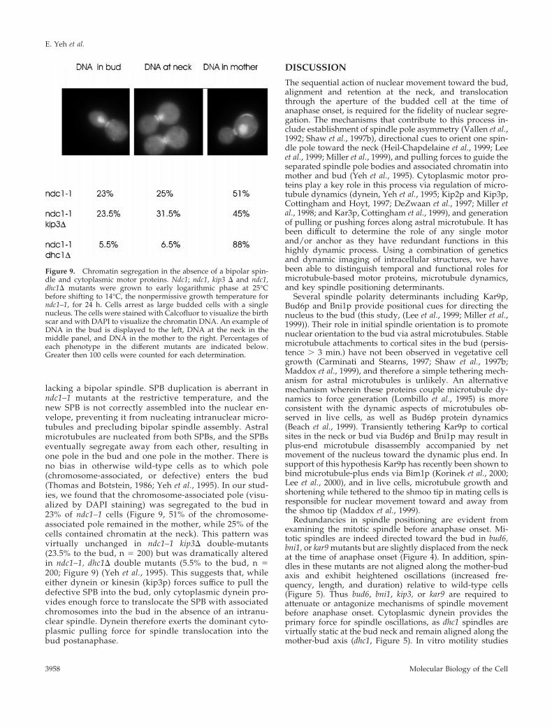
lacking a bipolar spindle. SPB duplication is aberrant inndc1–1 mutants at the restrictive temperature, and thenew SPB is not correctly assembled into the nuclear en-velope, preventing it from nucleating intranuclear micro-tubules and precluding bipolar spindle assembly. Astralmicrotubules are nucleated from both SPBs, and the SPBseventually segregate away from each other, resulting inone pole in the bud and one pole in the mother. There isno bias in otherwise wild-type cells as to which pole(chromosome-associated, or defective) enters the bud(Thomas and Botstein, 1986; Yeh et al., 1995). In our stud-ies, we found that the chromosome-associated pole (visu-alized by DAPI staining) was segregated to the bud in23% of ndc1–1 cells (Figure 9, 51% of the chromosome-associated pole remained in the mother, while 25% of thecells contained chromatin at the neck). This pattern wasvirtually unchanged in ndc1–1 kip3D double-mutants(23.5% to the bud, n 5 200) but was dramatically alteredin ndc1–1, dhc1D double mutants (5.5% to the bud, n 5200; Figure 9) (Yeh et al., 1995). This suggests that, whileeither dynein or kinesin (kip3p) forces suffice to pull thedefective SPB into the bud, only cytoplasmic dynein pro-vides enough force to translocate the SPB with associatedchromosomes into the bud in the absence of an intranu-clear spindle. Dynein therefore exerts the dominant cyto-plasmic pulling force for spindle translocation into thebud postanaphase.
DISCUSSION
The sequential action of nuclear movement toward the bud,alignment and retention at the neck, and translocationthrough the aperture of the budded cell at the time ofanaphase onset, is required for the fidelity of nuclear segre-gation. The mechanisms that contribute to this process in-clude establishment of spindle pole asymmetry (Vallen et al.,1992; Shaw et al., 1997b), directional cues to orient one spin-dle pole toward the neck (Heil-Chapdelaine et al., 1999; Leeet al., 1999; Miller et al., 1999), and pulling forces to guide theseparated spindle pole bodies and associated chromatin intomother and bud (Yeh et al., 1995). Cytoplasmic motor pro-teins play a key role in this process via regulation of micro-tubule dynamics (dynein, Yeh et al., 1995; Kip2p and Kip3p,Cottingham and Hoyt, 1997; DeZwaan et al., 1997; Miller etal., 1998; and Kar3p, Cottingham et al., 1999), and generationof pulling or pushing forces along astral microtubule. It hasbeen difficult to determine the role of any single motorand/or anchor as they have redundant functions in thishighly dynamic process. Using a combination of geneticsand dynamic imaging of intracellular structures, we havebeen able to distinguish temporal and functional roles formicrotubule-based motor proteins, microtubule dynamics,and key spindle positioning determinants.
Several spindle polarity determinants including Kar9p,Bud6p and Bni1p provide positional cues for directing thenucleus to the bud (this study, (Lee et al., 1999; Miller et al.,1999)). Their role in initial spindle orientation is to promotenuclear orientation to the bud via astral microtubules. Stablemicrotubule attachments to cortical sites in the bud (persis-tence . 3 min.) have not been observed in vegetative cellgrowth (Carminati and Stearns, 1997; Shaw et al., 1997b;Maddox et al., 1999), and therefore a simple tethering mech-anism for astral microtubules is unlikely. An alternativemechanism wherein these proteins couple microtubule dy-namics to force generation (Lombillo et al., 1995) is moreconsistent with the dynamic aspects of microtubules ob-served in live cells, as well as Bud6p protein dynamics(Beach et al., 1999). Transiently tethering Kar9p to corticalsites in the neck or bud via Bud6p and Bni1p may result inplus-end microtubule disassembly accompanied by netmovement of the nucleus toward the dynamic plus end. Insupport of this hypothesis Kar9p has recently been shown tobind microtubule-plus ends via Bim1p (Korinek et al., 2000;Lee et al., 2000), and in live cells, microtubule growth andshortening while tethered to the shmoo tip in mating cells isresponsible for nuclear movement toward and away fromthe shmoo tip (Maddox et al., 1999).
Redundancies in spindle positioning are evident fromexamining the mitotic spindle before anaphase onset. Mi-totic spindles are indeed directed toward the bud in bud6,bni1, or kar9 mutants but are slightly displaced from the neckat the time of anaphase onset (Figure 4). In addition, spin-dles in these mutants are not aligned along the mother-budaxis and exhibit heightened oscillations (increased fre-quency, length, and duration) relative to wild-type cells(Figure 5). Thus bud6, bni1, kip3, or kar9 are required toattenuate or antagonize mechanisms of spindle movementbefore anaphase onset. Cytoplasmic dynein provides theprimary force for spindle oscillations, as dhc1 spindles arevirtually static at the bud neck and remain aligned along themother-bud axis (dhc1, Figure 5). In vitro motility studies
Figure 9. Chromatin segregation in the absence of a bipolar spin-dle and cytoplasmic motor proteins. Ndc1; ndc1, kip3 D and ndc1,dhc1D mutants were grown to early logarithmic phase at 25°Cbefore shifting to 14°C, the nonpermissive growth temperature forndc1–1, for 24 h. Cells arrest as large budded cells with a singlenucleus. The cells were stained with Calcofluor to visualize the birthscar and with DAPI to visualize the chromatin DNA. An example ofDNA in the bud is displayed to the left, DNA at the neck in themiddle panel, and DNA in the mother to the right. Percentages ofeach phenotype in the different mutants are indicated below.Greater then 100 cells were counted for each determination.
E. Yeh et al.
Molecular Biology of the Cell3958
indicate that, when conventional kinesin and axonemal dy-nein are loaded at similar concentrations on the same mi-crotubule, microtubule translocation proceeds in a kinesin-directed manner, i.e. conventional kinesin is dominant todynein (Vale et al., 1992). If Kip3p is a conventional kinesin-like protein, it could mask dynein function in vivo as well.Upon deletion of Kip3p or potential effectors of Kip3p(Bud6p, Bni1p, Kar9p), dynein function is revealed in in-creased frequency and duration of spindle oscillations (Fig-ures 5 and 7). Analysis of spindle oscillations revealed twoimportant functional aspects of motor and spindle position-ing determinants. First, cytoplasmic dynein functions beforeanaphase onset, and second, spindle positioning determi-nants (bud6, bni1, and kar9) act to attenuate spindle oscilla-tions, perhaps through regulating cytoplasmic dynein be-fore anaphase onset.
The live cell assay provides an opportunity to measure thevelocity of the dynein-generated pulling force. Translocationinto the bud in the absence of spindle elongation proceeds at3.6 mm/min, considerably greater than the 1 mm/min spin-dle elongation characteristic of the fast phase of anaphase B(Kahana et al., 1995; Yeh et al., 1995). Spindle translocationinto the bud is governed by the rate of spindle elongation,and/or astral microtubule assembly/disassembly at the cellcortex. Nuclear maintenance at the neck is therefore not a“static” phase of the nuclear/spindle cycle. Rather, we pro-pose that early Kip3p function, in addition to providingdirectional cues for initial spindle orientation, antagonizescytoplasmic dynein. Preanaphase spindle oscillations at andthrough the neck reflect this balance between Kip3p anddynein. Upon anaphase onset, forces from spindle elonga-tion and limitations in microtubule dynamics prevent dy-nein from pulling the entire nucleus into the bud.
We have attempted to determine the hierarchy of cyto-plasmic motor function in yeast using mutants that fail toassemble a bipolar spindle but contain two spindle polebodies, each capable of nucleating astral microtubules. Thenewly formed spindle pole body in ndc1–1 mutants is notcorrectly assembled into the nuclear envelope, and thus thebipolar spindle fails to assemble. In ndc1–1 cells at the re-strictive temperature, the nucleus and associated chromatinDNA is either deposited into the bud or remains in themother cell (; 50:50 bud:mother)(Thomas and Botstein,1986; Yeh et al., 1995). In the absence of dynein, the nuclearmass is not translocated to the bud (,3% in bud) (Yeh et al.,1995). In contrast, the nuclear mass is translocated into thebud (23.5%) in the presence or absence of kip3 (Figure 9).Thus dynein is dominant to Kip3p in its ability to pull thenucleus into the bud in the absence of forces from spindleelongation. Spindle retention at the neck may reflect thishierarchy of motor function. The early force directing spin-dle migration to the neck may be weak and lack sufficientpower to pull the spindle through the aperture of buddedcells. Dynein provides the power for getting the nucleusthrough the neck upon anaphase onset.
The pattern of distribution of the asymmetric determi-nants, Bud6p (Amberg et al., 1997; Beach et al., 1999) andBni1p make it unlikely that these proteins interact exclu-sively with elements of the microtubule cytoskeleton. Bud6pand Bni1p more likely recruit or anchor other key compo-nents (e.g., Kar9p) that provide cortical linkage to microtu-bules (Bloom and Beach, 1999). Bni1p was identified as a She
mutant (she5, symmetric HO expression (Bobola et al., 1996),and she mutants are defective in either the formation of anAsh1 mRNA particle or the polar distribution of ASH1mRNA. Beach et al. (1999), using a live cell assay for mRNAdynamics, demonstrated that ASH1 mRNA is distributed tothe bud of large budded cells in bni1 mutants, but ASH1mRNA in the bud is delocalized. Thus bni1 mutants lack anasymmetric determinant that keeps mRNA distributed tothe tip of budded cells (Bloom and Beach, 1999). Bud6 has aslight she phenotype (Beach and Bloom, unpublished), andASH1 mRNA is also delocalized in bud6 mutants (Beach etal., 1999). Bud6p and Bni1p may therefore comprise a mul-tipurpose cortical scaffold that directs several elements (mi-crotubules, mRNA) to sites of polarized growth. Interactionbetween cytoskeletal elements and cortical sites is likely toproceed via individual effector molecules, with Kar9p as theprimary candidate for directing the microtubule cytoskele-ton. In contrast to bni1 and bud6, kar9 mutants show nodefect in ASH1 mRNA localization (Beach and Bloom, un-published results).
The spindle polarity determinants (Bud6p, Bni1p, andKar9p) must recognize sites of polarized growth or otherasymmetries in cell patterning. In yeast, polarized cellgrowth is initialized by the selection of bud-site (Chant andPringle, 1995; Pringle et al., 1995), followed by polarizationof the actin cytoskeleton, spindle orientation, nuclear migra-tion, and other events including ASH1 mRNA deposition.How various cytoskeletal elements interact with a specificlocal at a specific time is central to both cell and develop-mental processes. We posit that construction of a multipur-pose scaffold (defined by Bud6p and Bni1p) that in turnbinds different effectors (Kar9p for microtubules, She pro-teins for mRNA) is one mechanism for attaining specificity.These effectors, at least for nuclear migration, are likely tointeract with the cytoplasmic microtubule-based motor pro-teins that can generate force from transient interactions.
ACKNOWLEDGMENTS
We thank Dale Beach, Doug Thrower, Chad Pearson, and membersof the Bloom and Salmon laboratories for helpful discussions. C.Yang was supported in part by a fellowship for UndergraduateResearch from the University of North Carolina at Chapel Hill.
This work was supported by research grants from the NationalInstitutes of Health (K. Bloom, GM-32238; D.J. Lew, GM-53050; andE.D. Salmon, GM-24364).
REFERENCES
Adames, N.R., and Cooper, J.A. (2000). Microtubule interactionswith the cell cortex causing nuclear movements in Saccharomycescerevisiae. J. Cell Biol. 149, 863–874.
Amatruda, J.F., and Cooper, J.A. (1992). Purification, characteriza-tion, and immunofluorescence localization of Saccharomyces cerevi-siae capping protein. J. Cell. Biol. 117, 1067–1076.
Amberg, D.C., Zahner, J.E., Mulholland, J.W., Pringle, J.R., andBotstein, D. (1997). Aip3p/Bud6p, a Yeast actin-interacting proteinthat is involved in morphogenesis and the selection of bipolarbudding sites. Mol. Biol. Cell 8, 729–753.
Mechanisms of Spindle Positioning
Vol. 11, November 2000 3959

Beach, D.L., Salmon, E.D., and Bloom, K. (1999). Localization andanchoring of mRNA in budding yeast. Curr. Biol. 9, 569–578.
Bloom, K., and Beach, D.L. (1999). mRNA localization: MotilemRNA, asymmetric anchors. Curr. Opin. Microbiol: Genet. Dev. 2,604–609.
Bobola, N., Jansen, R.-P., Shin, T.H., and Nasmyth, K. (1996). Asym-metric accumulation of ash1p in postanaphase nuclei depends on amyosin and restricts yeast mating-type switching to mother cells.Cell 84, 699–709.
Byers, B. (1981). Cytology of the yeast life cycle. In: The MolecularBiology of the Yeast Saccharomyces: Life Cycle and Inheritance. ed.J.N. Strathern, E.W. Jones, and J.R. Broach. Cold Spring Harbor, NY:Cold Spring Harbor Laboratory, 59–96.
Carminati, J.L., and Stearns, T. (1997). Microtubules orient the mi-totic spindle in yeast through dynein-dependent interactions withthe cell cortex. J. Cell. Biol. 138, 629–641.
Chang, F. (1999). Movement of a cytokinesis factor Cdc12p to thesite of cell division. Curr. Biol. 9, 849–852.
Chant, J., and Pringle, J.R. (1995). Patterns of bud-site selection inthe yeast Saccharomyces cerevisiae. J. Cell. Biol. 129, 751–765.
Chenevert, J., Corrado, K., Bender, A., Pringle, J., and Herskowitz, I.(1992). A yeast gene (BEM1) necessary for cell polarization whoseproduct contains two SH3 domains. Nature 356, 77–79.
Cottingham, F.R., Gheber, L., Miller, D.L., and Hoyt, M.A. (1999).Novel roles for Saccharomyces cerevisiae mitotic spindle motors.J. Cell. Biol. 147, 335–349.
Cottingham, F.R., and Hoyt, M.A. (1997). Mitotic spindle position-ing in Saccharomyces cerevisiae is accomplished by antagonisticallyacting microtubule motor proteins. J. Cell. Biol. 138, 1041–1053.
DeZwaan, T.M., Ellingson, E., Pellman, D., and Roof, D.M. (1997).Kinesin-related KIP3 of Saccharomyces cerevisiae is required for adistinct step in nuclear migration. J. Cell Biol. 138, 1023–1040.
Drubin, D.G., and Nelson, W.J. (1996). Origins of cell polarity. Cell84, 335–344.
Gonczy, P., and Hyman, A.A. (1996). Cortical domains and themechanisms of asymmetric cell division. Trends Cell. Biol. 6, 382–387.
Heil-Chapdelaine, R.A., Adames, N.R., and Cooper, J.A. (1999).Formin’ the connection between microtubules and the cell cortex.J. Cell Biol. 144, 809–811.
Heil-Chapdelaine, R.A., Tran, N.K., and Cooper, J.A. (2000). Dy-nein-dependent movements of the mitotic spindle in Saccharomycescerevisiae do not require filamentous actin. Mol. Biol. Cell 11, 863–872.
Jin, H., and Amberg, D.C. (2000). The secretory pathway mediateslocalization of the cell polarity regulator Aip3p/Bud6p. Mol. Biol.Cell 11, 647–661.
Kahana, J.A., Schnapp, B.J., and Silver, P.A. (1995). Kinetics ofspindle pole body separation in budding yeast. Proc. Natl. Acad.Sci. USA 92, 9707–9711.
Karpova, T.S., Tatchell, K., and Cooper, J.A. (1995). Actin filamentsin yeast are unstable in the absence of capping protein or fimbrin.J. Cell Biol. 131, 1483–1493.
Korinek, W.S., Copeland, M.J., Chaudhuri, A., and Chant, J. (2000).Molecular linkage underlying microtubule orientation toward cor-tical sites in yeast. Science 287, 2257–2259.
Lee, L., Klee, S.K., Evangelista, M., Bonne, C., and Pellman, D.(1999). Control of mitotic spindle position by the Saccharomycescerevisiae formin Bni1p. J. Cell Biol. 144, 947–961.
Lee, L., Tirnauer, J.S., Li, J., Schuyler, S.C., Liu, J.Y., and Pellman, D.(2000). Positioning of the mitotic spindle by a cortical-microtubulecapture mechanism. Science 287, 2260–2262.
Li, Y.Y., Yeh, E., Hays, T., and Bloom, K. (1993). Disruption ofmitotic spindle orientation in a yeast dynein mutant. Proc. Natl.Acad. Sci. USA. 90, 10096–10100.
Liu, H., and Bretscher, A. (1992). Characterization of TPM1 dis-rupted yeast cells indicates an involvement of tropomyosin in di-rected vesicular transport. J. Cell Biol. 118, 285–299.
Lombillo, V.A., Stewart, R.J., and McIntosh, J.R. (1995). Minus-end-directed motion of kinesin-coated microspheres driven by microtu-bule depolymerization. Nature 373, 161–164.
Maddox, P., Bloom, K., and Salmon, E.D. (2000). Polarity and dy-namics of microtubule assembly in the budding yeast Saccharomycescerevisiae. Nat. Cell Biol. 2, 36–41.
Maddox, P., Chin, E., Mallavarapu, A., Yeh, E., Salmon, E.D., andBloom, K. (1999). Microtubule dynamics from mating through thefirst zygotic division in the budding yeast Saccharomyces cerevisiae.J. Cell Biol. 144, 977–987.
Miller, R., Matheos, D., and Rose, M. (1999). The cortical localization ofthe microtubule orientation protein, Kar9p, is dependent upon actinand proteins required for polarization. J. Cell Biol. 144, 963–975.
Miller, R.K., Heller, K.K., and Rose, M.D. (1998). The kinesin-relatedproteins, Kip2p and Kip3p, function differently in nuclear migrationin yeast. Mol. Biol. Cell 9, 2051.
Miller, R.K., and Rose, M.D. (1998). Kar9p is a novel cortical proteinrequired for cytoplasmic microtubule orientation in yeast. J. Cell.Biol. 140, 377–390.
Mondesert, G., and Reed, R.I. (1996). BED1, a gene encoding agalactosyltransferase homologue, is required for polarized growthand efficient bud emergence in Saccharomyces cerevisiae. J. Cell Biol.132, 137–143.
Palmer, R.E., Sullivan, D.S., Huffaker, T., and Koshland, D. (1992).Role of astral microtubules and actin in spindle orientation andmigration in the budding yeast, Saccharomyces cerevisiae. J. Cell Biol.119, 583–593.
Peterson, J., Zheng, Y., Bender, L., Myers, A., Cerione, R., andBender, A. (1994). Interactions between the bud emergence proteinsBem1p and Bem2p and Rho-type GTPases in yeast. J. Cell Biol. 127,1395–1406.
Peterson, J.B., and Ris, H. (1976). Electron-microscopic study of thespindle and chromosome movement in the yeast Saccharomyces cer-evisiae. J. Cell Sci. 22, 219–242.
Pringle, J.R., Bi, E., Harkins, H.A., Zahner, J.E., De, V.C., Chant, J.,Corrado, K., and Fares, H. (1995). Establishment of cell polarity inyeast. (Review) (111 refs). Cold Spring Harb. Symp. Quant. Biol. 60,729–744.
Pruyne, D.W., Schott, D.H., and Bretscher, A. (1998). Tropomyosin-containing actin cables direct the Myo2p-dependent polarized deliveryof secretory vesicles in budding yeast. J. Cell Biol. 143, 1931–1945.
Rose, M.D., Winston, F., and Hieter, P. (1991). Methods in yeastgenetics: a laboratory course manual. Cold Spring Harbor, NY: ColdSpring Harbor Laboratory
Salmon, E.D., Shaw, S.L., Waters, J., Waterman-Storer, C.M., Mad-dox, P.S., Yeh, E., and Bloom, K. (1998). A high-resolution multi-mode digital microscope system. Methods Cell Biol. 56, 185–215.
Segal, M., Clarke, D.J., Maddox, P., Salmon, E.D., Bloom, K. and Reed,S.J. (2000). Coordinated spindle assembly and orientation requiresclb5p-dependent kinase in budding yeast. J. Cell Biol. 148, 441–451.
E. Yeh et al.
Molecular Biology of the Cell3960
Shaw, S.L., Yeh, E., Bloom, K., and Salmon, E.D. (1997a). Imaginggreen fluorescent protein fusion proteins in Saccharomyces cerevisiae.Curr. Biol. 7, 701–704.
Shaw, S.L., Yeh, E., Maddox, P., Salmon, E.D., and Bloom, K.(1997b). Astral microtubule dynamics in yeast: A microtubule-basedsearching mechanism for spindle orientation and nuclear migrationinto the bud. J.Cell Biol. 139, 985–994.
Snyder, M. (1989). The SPA2 protein of yeast localizes to sites of cellgrowth. J. Cell Biol. 108, 1419–1429.
Straight, A.F., Sedat, J.W., and Murray, A.W. (1998). Time-lapsemicroscopy reveals unique roles for kinesins during anaphase inbudding yeast. J. Cell Biol. 143, 687.
Theesfeld, C.L., Irazoqui, J.E., Bloom, K., and Lew, D.J. (1999). Therole of actin in spindle orientation changes during the Saccharomycescerevisiae cell cycle. J. Cell Biol. 146, 1019–1032.
Thomas, J.H., and Botstein, D. (1986). A gene required for the separa-tion of chromosomes on the spindle apparatus in yeast. Cell 44, 65–76.
Tirnauer, J.S., O’Toole, E., Berrueta, L., Bierer, B.E., and Pellman, D.(1999). Yeast Bimlp promotes the G1-specific dynamics of microtu-bules. J Cell Biol. 145, 993–1007.
Vale, R.D., Malik, F., and Brown, D. (1992). Directional instability ofmicrotubule transport in the presence of kinesin and dynein, twoopposite polarity motor proteins. J. Cell Biol. 119, 1589–1596.
Vallen, E.A., Scherson, T.Y., Roberts, T., van Zee, K., and Rose, M.D.(1992). Asymmetric mitotic segregation of the yeast spindle polebody. Cell 69, 505–515.
Yeh, E., Skibbens, R.V., Cheng, J.W., Salmon, E.D., and Bloom, K.(1995). Spindle dynamics and cell cycle regulation of dynein inthe budding yeast, Saccharomyces cerevisiae. J. Cell Biol. 130, 687–700.
Zahner, J.E., Harkins, H.A., and Pringle, J.R. (1996). Genetic analysisof the bipolar pattern of bud site selection in the yeast Saccharomycescerevisiae. Mol. Cell. Biol. 16, 1857–1870.
Mechanisms of Spindle Positioning
Vol. 11, November 2000 3961