dynactin targets pavarotti-klp to the central spindle ...dynactin targets pavarotti-klp to the...
TRANSCRIPT
4431Research Article
IntroductionDynein is a microtubule motor that moves toward the minusend of microtubules as part of a large macromolecular complex(Collins and Vallee, 1989; Vallee et al., 1989). This complexhas many functions. It is involved in the transport of moleculesand organelles along microtubules during interphase andnuclear positioning (Karki and Holzbaur, 1999). Cytoplasmicdynein, which is present at the cell cortex, pulls on centrosome-connected microtubules and is responsible for the positioningof the centrosomes in the centre of the cell (Koonce et al., 1999;Vallee and Stehman, 2005). The same principle is used inyeast cells to pull newly duplicated nuclei into the bud and inmitotic vertebrate cultured cells, as well as in Drosophilaand Caenorhabditis elegans somatic tissues, to pull thecentrosomes apart during prophase (Busson et al., 1998;Carminati and Stearns, 1997; Koonce et al., 1999; Vallee andStehman, 2005; Gonczy et al., 1999; Robinson et al., 1999;Schmidt et al., 2005).
The dynein complex can be isolated biochemically withdynactin, a 20S multi-protein complex containing at least tenproteins (Karki and Holzbaur, 1999). In vitro, the dynactincomplex stimulates the dynein motor activity (Gill et al., 1991).It has been suggested that one major function of dynactin is totarget dynein where it is required in the cell (Vallee et al.,1995).
The Glued gene encodes a 150 kDa protein that belongs tothe dynactin complex. In fission yeasts and in humans,p150Glued regulates microtubule dynamics (Niccoli et al.,2004). In human cells, this regulation depends on associationwith EB1 and is essential to maintain the radial arrays ofmicrotubules in interphase cells (Askham et al., 2002;Strickland et al., 2005). A recent study further proposed thatthe interaction between EB1 and p150Glued promotes theelongation of astral microtubules to facilitate furrow ingressionduring cytokinesis (Strickland et al., 2005).
When cells enter mitosis and prophase, dynein facilitatesnuclear envelope breakdown for spindle formation: it pullsnuclear membranes and associated proteins poleward alongastral microtubules leading to nuclear membrane detachment(Salina et al., 2002). During prometaphase, it has been clearlydemonstrated in Drosophila and human cultured cells, that thedynein-dynactin complex (DDC) is continuously recruited tothe kinetochores until they become properly attached to themitotic spindle. Kinetochore dynein then moves towards thespindle poles thereby removing checkpoint proteins andallowing the inactivation of the spindle checkpoint and theanaphase onset (Karess, 2005). In addition, mutations thataffect dynein loading at the kinetochore, lead to asynchronouschromosome segregation and attenuated chromosome motiontoward the poles (Savoian et al., 2000; Sharp et al., 2000). In
The dynactin complex cooperates with the dynein complexin various systems for mitotic completion. Here weanalysed the mitotic phenotype of Drosophila S2 cellsfollowing the knockdown of the dynactin subunit p150Glued.We found that p150Glued-depleted cells were delayed inmetaphase and that the centrosomes were poorly connectedto mitotic spindle poles. In addition, anaphase occurredwith asynchronous chromosome segregation. Althoughcyclin B was degraded in these anaphase cells, Aurora B,MEI-S322 and BubR1 were not released from the non-segregating chromosomes. We also found that the densityand organisation of the central spindle were compromised,with Aurora B and polo kinases absent from the diminishednumber of microtubules. Pavarotti-KLP, a component ofthe centralspindlin complex required for the formation ofstable microtubule bundles, was not immediately targeted
to the plus ends of the microtubules following anaphaseonset as happened in controls. Instead, it accumulatedtransiently at the cell cortex during early anaphase and itstargeting to the central spindle was delayed. These datasuggest that the dynactin complex contributes tocytokinesis by promoting stable targeting of thecentralspindlin complex to microtubule plus ends atanaphase onset. The contribution of the dynein-dynactincomplex to synchronous chromosome segregation andcytokinesis is discussed.
Supplementary material available online athttp://jcs.biologists.org/cgi/content/full/119/21/4431/DC1
Key words: Dynactin, Mitosis, Cytokinesis
Summary
Dynactin targets Pavarotti-KLP to the central spindleduring anaphase and facilitates cytokinesis inDrosophila S2 cellsJean-Guy Delcros, Claude Prigent and Régis Giet*CNRS UMR 6061 ‘Génétique et Développement’, Groupe Cycle Cellulaire, Faculté de Médecine, IFR 140 Génomique Fonctionnelle et Santé,Université de Rennes I, 2 avenue du Pr. Léon Bernard, CS 34317, F-35043 Rennes CEDEX, France*Author for correspondence (e-mail: [email protected])
Accepted 8 August 2006Journal of Cell Science 119, 4431-4441 Published by The Company of Biologists 2006doi:10.1242/jcs.03204
Jour
nal o
f Cel
l Sci
ence
4432
Drosophila, interfering with dynein-dynactin functions usingantibodies or RNAi also induces the detachment of thecentrosomes from the spindle poles (Goshima and Vale, 2003;Goshima and Vale, 2005; Morales-Mulia and Scholey, 2005).
Here, using RNAi for the Glued gene in Drosophila S2 cells,we confirm that Glued is required for centrosome connectionto spindle poles, the metaphase-to-anaphase transition, andsynchronous chromosome-to-pole movement during anaphase.In addition, we report that the dynactin complex also facilitatescytokinesis by targeting Pavarotti-KLP, a component of thecentralspindlin complex, to the plus ends of central spindlemicrotubules.
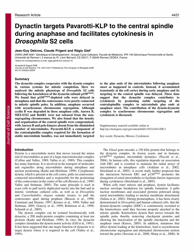
Resultsp150Glued localises to centrosomes, mitotic spindlemicrotubules and kinetochoresTwo antisera raised against the first 148 amino acids of thep150Glued polypeptide were produced in rabbits (Rb1477 andRb1478). These two antisera recognised a common protein at150 kDa, the expected size of the p150Glued protein inDrosophila S2 cell extracts as well as one or more other bands(Fig. 1A). After affinity purification, only a single 150 kDaprotein could be detected (Fig. 1A, lane AP). The p150Glued
protein signal disappeared following Glued RNAi of S2 cellswhereas loading control proteins, cyclin B, aurora A, CP190and dynein intermediate chain (DIC) remained stable (Fig. 1B,compare lanes – and lanes +), further demonstrating thespecificity of our antibodies.
The anti-p150Glued affinity-purified antibody was used tostain cultured Drosophila S2 cells and embryos (Fig. 2).During prophase, the antibody decorated the two asters ofmicrotubules (Fig. 2A, panel p). During metaphase, labellingwas observed on centrosomes/asters as well as on spindlemicrotubule fibres (panel m). From anaphase until cytokinesis,p150Glued was detected on the astral and central spindlemicrotubules (Fig. 2A, panels a,c). In interphase cells, wefound a punctuated p150Glued labelling in the cytoplasm andalong microtubule fibres (Fig. 2B) as observed previously inhuman cells (Vaughan et al., 2002). We further observedpaired-dot-like staining on some metaphase chromosomes.This staining became more evident when microtubules weredepolymerised with colchicine and furthermore, co-localisedwith polo kinase, a known kinetochore component (Fig. 2D).Thus, like the dynein motor protein, p150Glued associates withkinetochores. To further examine the distribution of p150Glued
during mitosis, we stained syncitial embryos (Fig. 2C). Inagreement with our findings in cultured cells, p150Glued firstassociated with prophase asters and the subsequent metaphaseand anaphase spindles.
We next attempted to examine the dynamics of p150Glued inliving cells using a GFP-tagged transgene. However, despitethe use of the ubiquitin promoter to drive expression (see
Journal of Cell Science 119 (21)
Materials and Methods) the fluorescence intensity of GFP-p150Glued was too weak to be detected in live material.Therefore a GFP antibody was used to stain fixed expressingcells. GFP labelling was detected on the centrosomal regionand the spindle microtubules during mitosis, as well as atthe midbody region during cytokinesis (supplementarymaterial Fig. S1). Thus, a combination of GFP-tagging andimmunofluorescence studies demonstrated that the p150Glued
protein shares a common localisation in both S2 cellsand syncitial embryos; it associates with centrosomes,microtubules and kinetochores. In addition, the p150Glued
protein was also detected on the central spindle microtubulesand the midbody during late telophase and cytokinesis.
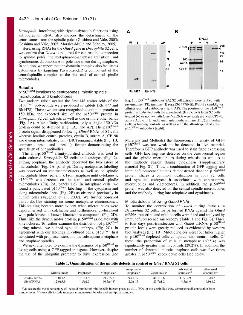
Mitotic defects following Glued RNAiTo monitor the contribution of Glued during mitosis inDrosophila S2 cells, we performed RNAi against the GluedmRNA transcript, and mitotic cells were fixed and analysed byimmunofluorescence microscopy (Table 1 and Fig. 3). Threeto four days post-transfection with Glued dsRNA, p150Glued
protein levels were greatly reduced as evidenced by westernblot analyses (Fig. 1B). Mitotic indices were four times higherin p150Glued-depleted cells compared with control cells. Ofthese, the proportion of cells at metaphase (60.5%) wassignificantly greater than in controls (29.2%). In addition, thenumber of abnormal mitotic anaphase cells was five timesgreater in p150Glued knock down cells (see below).
Fig. 1. p150Glued antibodies. (A) S2 cell extracts were probed withpre-immune (PI), immune (I) sera Rb1477(left), Rb1478 (middle) oraffinity-purified antibodies (right, AP). The position of the p150Glued
protein is indicated with the arrowhead. (B) Extracts from S2 cellstreated (+) or not (–) with Glued dsRNA were analysed with CP190,aurora A, cyclin B and dynein intermediate chain (DIC) antibodies(left) as loading controls, as well as with the affinity-purified anti-p150Glued antibodies (right).
Table 1. Quantification of the mitotic defects in control or Glued RNAi S2 cellsAnaphase + Abnormal Abnormal
Mitotic index Prophase* Metaphase* telophase* Cytokinesis* spindles*,‡ anaphases*
Control RNAi 3.0±1.5 8.1±1.9 29.2±5.3 9.4±1.9 41.1±3.8 11.9±2.9 0.3±0.5Glued RNAi 12.0±3.9 4.5±1.3 60.5±4.8† 2.0±1.7 23.7±1.2 4.5±1.9 4.9±1.2
*Values are the mean percentage of the total number of mitotic cells in each phase (± s.d.). †50% of these spindles show centrosome disconnection fromspindle poles. ‡These cells include monopolar, monoastral bipolar and multipolar spindles.
Jour
nal o
f Cel
l Sci
ence

4433Dynactin is a cytokinesis facilitator
In 50% of the cases, RNAi-treated metaphase cells hadnormal spindle morphology and could not be differentiatedfrom control cells (Fig. 3B, panel 1). However, the other halfof the metaphase cells displayed a weak connection or clearseparation between the centrosomes and the spindle poles (Fig.3B, panel 2), a phenotype also observed after dynein ordynactin interference (Goshima et al., 2005; Morales-Muliaand Scholey, 2005; Siller et al., 2005). The major defectobserved following p150Glued RNAi occurred during anaphase,where 4.9% of cells were abnormal compared with 0.3%of control cells. These defects were manifest as grossmorphological spindle defects and chromosome segregationerrors. In most cases, the spindle appeared abnormallyelongated with some chromosomes located at the spindle poleswhereas others were still at the spindle equator, suggestingasynchronous chromosome separation. (Fig. 3B, lower panels3 and 4). During early anaphase, as judged by the separationof the two chromatid masses, the central spindle microtubuledensity was less robust and lacked the symmetry observed incontrols (Fig. 3B, middle panels 3 and 4). In telophase, a
disorganised region of equatorial microtubules was able toform, but this region was lacking the well-organised stable anti-parallel microtubule bundles seen in control telophase cells(Fig. 3, compare B panel 5 with A panel t). The FACS analysesshowed the absence of polyploid cells in the Glued dsRNA-treated cells, but an increase of apoptotic and/or aneuploid cellswas detected (Fig. 3C, arrow) suggesting that despite theformation of normal anaphase cells, cytokinesis occurrednormally. In all depleted anaphase cells, the chromosomesshowed a dot-like morphology suggesting an overcondensationof chromosomes and hence mitotic arrest, as expected from theelevated mitotic index (Table 1).
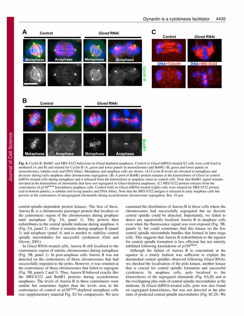
To confirm that these elongated spindles representedanaphase cells and not aberrant prometaphase cells withcongression defects, we monitored the degradation of cyclin Bin control and Glued dsRNA-treated cells at the metaphase-anaphase transition. Cyclin B levels were high in wild-type andGlued dsRNA-treated cells during metaphase and the proteinaccumulated strongly at centrosomes, spindles and, to a lesserextent, at some kinetochores (Fig. 4, left panels A and B).
Fig. 2. p150Glued localises to centrosomes, spindle and interphase microtubules. S2 cells (A and B) or early Drosophila syncitial embryos (C)were fixed and stained with the affinity-purified anti-p150Glued antibodies (red and lower panels in monochrome), the anti-�-tubulin antibody(green and middle panels in monochrome) and DAPI (DNA staining in blue). (A) Mitotic cells. (B) Interphase cells: the right column shows a10� magnified view of the border of a flat S2 cell. Note the punctuate staining along microtubule fibres. a, anaphase; c, cytokinesis; m,metaphase; p, prophase; t, telophase. (D) A mitotic cell treated with colchicine to depolymerise the mitotic spindle was stained for polo kinase(red and upper right panel in monochrome) and p150Glued (green and lower panel on the right in monochrome). The lower left panel shows a10� magnification view of the kinetochore region boxed in the panel above. Bars, 10 �m (A-C); 1 �m (D).
Jour
nal o
f Cel
l Sci
ence
4434
During anaphase, cyclin B levels were strongly reduced incontrol cells but some signal was still detected at centrosomesas previously described (Mathe et al., 2004). We consistentlyfound that cyclin B was degraded in Glued-depleted cells withelongated spindles and aberrantly positioned chromosomes(Fig. 4A, right panels), indicating that these cells hadprogressed into anaphase. In addition, we examined thedistribution of the MEI-S322 antigen (the Drosophilahomologue of mammalian shugosin) (Kerrebrock et al., 1995).This protein was released from the centromeres in controlanaphase cells and was also lacking at polar-proximalchromosomes in Glued dsRNA-treated cells. However, thesignal remained quite strong on those chromosomes lagging atthe equator (Fig. 4C). Finally we investigated the status of thespindle assembly checkpoint in knockdown cells by stainingfor the BubR1 antigen. As expected in control cells, BubR1protein levels were elevated at the kinetochores in earlyprometaphase cells and much lower in metaphase. Theremaining BubR1 protein disappeared following anaphaseentry (Karess, 2005). In those RNAi-treated cells exhibiting
normal metaphase spindles, all kinetochores appeared to belabelled similarly to control cells. During anaphase, thekinetochores of polar-positioned chromosomes on elongatedspindles showed no staining whereas those kinetochores atchromosomes at the centre of the spindle exhibitedfluorescence (Fig. 4B). Taken together, these results reveal thatthe knockdown of p150Glued leads to elongated spindles uponwhich some chromosomes are able to segregate to the polesduring anaphase while others lag at the spindle equator. Theselagging chromosomes retain the MEI-S322 centromericprotein as well as the checkpoint component BubR1. Alongwith these chromosome segregation defects, the centralspindle, which normally forms during late anaphase ortelophase is greatly diminished, suggesting that p150Glued isneeded for central spindle formation or stabilisation.
Aurora B and polo kinases are not recruited to themicrotubules in Glued-depleted anaphase cellsThe failure to form or maintain the central spindle in p150Glued-depleted cells prompted us to investigate the distribution of two
Journal of Cell Science 119 (21)
Fig. 3. Glued RNAi leads to anaphase defects. Control (A) or Glued dsRNA-treated (B) mitotic S2 cells were fixed and stained for �-tubulin(red), �-tubulin (green and middle panels in monochrome) and DNA (blue and lower panels in monochrome). a, anaphase; c, cytokinesis; m,metaphase; p, prophase; t, telophase. In p150Glued-depleted cells, cells are delayed in metaphase (see also Table 1). 50% of these cells showweak centrosome connection to spindle poles (panel 2) whereas others show a normal shape (panel 1). Note the segregation defect and theabsence of microtubule bundling during anaphase (panels 3 and 4). During telophase, a central spindle forms but with strongly disorganisedmicrotubules and a lack of well-defined microtubule bundles (panel 5). (C) FACS analysis of Glued dsRNA-treated cells does not show anyaccumulation of polyploid cells but evidences an increase of apoptotic or aneuploid cells (arrow) compared with control cells. Bar, 10 �m.
Jour
nal o
f Cel
l Sci
ence
4435Dynactin is a cytokinesis facilitator
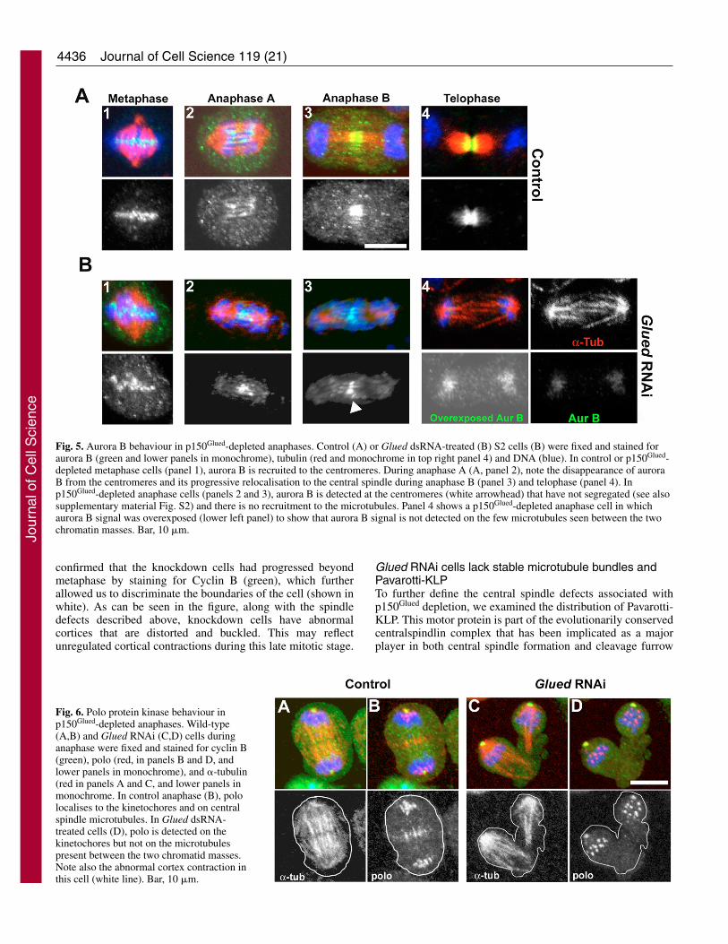
central-spindle-dependent protein kinases. The first of these,Aurora B, is a chromosome passenger protein that localises tothe centromeric region of the chromosomes during prophaseuntil metaphase (Fig. 5A, panel 1). This protein thenredistributes to the central spindle midzone during anaphase A(Fig. 5A, panel 2), where it remains during anaphase B (panel3) and telophase (panel 4) and is needed to stabilise centralspindle microtubules for successful cytokinesis (Giet andGlover, 2001).
In Glued RNAi-treated cells, Aurora B still localised to thecentromeric region of mitotic chromosomes during metaphase(Fig. 5B, panel 1). In post-anaphase cells Aurora B was notdetected on the centromeres of those chromosomes that hadsuccessfully migrated to the poles. However, it was retained atthe centromeres of those chromosomes that failed to segregate(Fig. 5B, panels 2 and 3). Thus, Aurora B behaved exactly likethe MEI-S322 and BubR1 proteins during asynchronousanaphases. The levels of Aurora B in these centromeres weresimilar but sometimes higher than the levels seen in thecentromeres of control or p150Glued-depleted metaphase cells(see supplementary material Fig. S2 for comparison). We next
examined the distribution of Aurora B in those cells where thechromosomes had successfully segregated but no discretecentral spindle could be detected. Importantly, we failed todetect any equatorially localised Aurora B in anaphase cellseven when the fluorescence signal was over-exposed (Fig. 5B,panels 4), but could sometimes find this kinase on the fewcentral spindle microtubule bundles that formed in later-stagecells. This suggests that Aurora B redistribution to the equatorfor central spindle formation is less efficient but not entirelyinhibited following knockdown of p150Glued.
Although the failure of Aurora B to concentrate at theequator in a timely fashion was sufficient to explain thediminished central spindles observed following Glued RNAi,we checked the localisation of the polo kinase, another kinasethat is crucial for central spindle formation and successfulcytokinesis. In anaphase cells, polo localised to thekinetochores of the segregated chomatids (Fig. 6A,B) and atthe overlapping plus ends of central spindle microtubules at themidzone. In Glued dsRNA-treated cells, polo was also foundon segregated kinetochores, but was not detected at the plusends of predicted central spindle microtubules (Fig. 6C,D). We
Fig. 4. Cyclin B, BubR1 and MEI-S322 behaviour in Glued-depleted anaphases. Control or Glued dsRNA-treated S2 cells were cold fixed inmethanol (A and B) and stained for Cyclin B (A, green and lower panels in monochrome) and BubR1 (B, green and lower panels inmonochrome), tubulin (red) and DNA (blue). Metaphase and anaphase cells are shown. (A) Cyclin B levels are elevated in metaphase anddecrease during early anaphase after chromosome segregation. (B) A pool of BubR1 protein remains at the kinetochores of Glued or controldsRNA-treated cells during metaphase and is released from the kinetochore at anaphase onset in control cells. Note that BubR1 signal remainselevated at the kinetochore of chromatids that have not segregated in Glued-depleted anaphases. (C) MEI-S322 protein releases from thecentromeres of p150Glued knockdown anaphase cells. Control (left) or Glued dsRNA-treated (right) cells were stained for MEI-S322 protein(red in bottom panels), �-tubulin (red in top panels) and DNA (blue). Note that the MEI-S322 antigen is released in early anaphase cells butpersists at the centromeres of unsegregated chromatids during asynchronous chromosome segregation. Bar, 10 �m.
Jour
nal o
f Cel
l Sci
ence
4436
confirmed that the knockdown cells had progressed beyondmetaphase by staining for Cyclin B (green), which furtherallowed us to discriminate the boundaries of the cell (shown inwhite). As can be seen in the figure, along with the spindledefects described above, knockdown cells have abnormalcortices that are distorted and buckled. This may reflectunregulated cortical contractions during this late mitotic stage.
Glued RNAi cells lack stable microtubule bundles andPavarotti-KLP To further define the central spindle defects associated withp150Glued depletion, we examined the distribution of Pavarotti-KLP. This motor protein is part of the evolutionarily conservedcentralspindlin complex that has been implicated as a majorplayer in both central spindle formation and cleavage furrow
Journal of Cell Science 119 (21)
Fig. 5. Aurora B behaviour in p150Glued-depleted anaphases. Control (A) or Glued dsRNA-treated (B) S2 cells (B) were fixed and stained foraurora B (green and lower panels in monochrome), tubulin (red and monochrome in top right panel 4) and DNA (blue). In control or p150Glued-depleted metaphase cells (panel 1), aurora B is recruited to the centromeres. During anaphase A (A, panel 2), note the disappearance of auroraB from the centromeres and its progressive relocalisation to the central spindle during anaphase B (panel 3) and telophase (panel 4). Inp150Glued-depleted anaphase cells (panels 2 and 3), aurora B is detected at the centromeres (white arrowhead) that have not segregated (see alsosupplementary material Fig. S2) and there is no recruitment to the microtubules. Panel 4 shows a p150Glued-depleted anaphase cell in whichaurora B signal was overexposed (lower left panel) to show that aurora B signal is not detected on the few microtubules seen between the twochromatin masses. Bar, 10 �m.
Fig. 6. Polo protein kinase behaviour inp150Glued-depleted anaphases. Wild-type(A,B) and Glued RNAi (C,D) cells duringanaphase were fixed and stained for cyclin B(green), polo (red, in panels B and D, andlower panels in monochrome), and �-tubulin(red in panels A and C, and lower panels inmonochrome. In control anaphase (B), pololocalises to the kinetochores and on centralspindle microtubules. In Glued dsRNA-treated cells (D), polo is detected on thekinetochores but not on the microtubulespresent between the two chromatid masses.Note also the abnormal cortex contraction inthis cell (white line). Bar, 10 �m.
Jour
nal o
f Cel
l Sci
ence
4437Dynactin is a cytokinesis facilitator
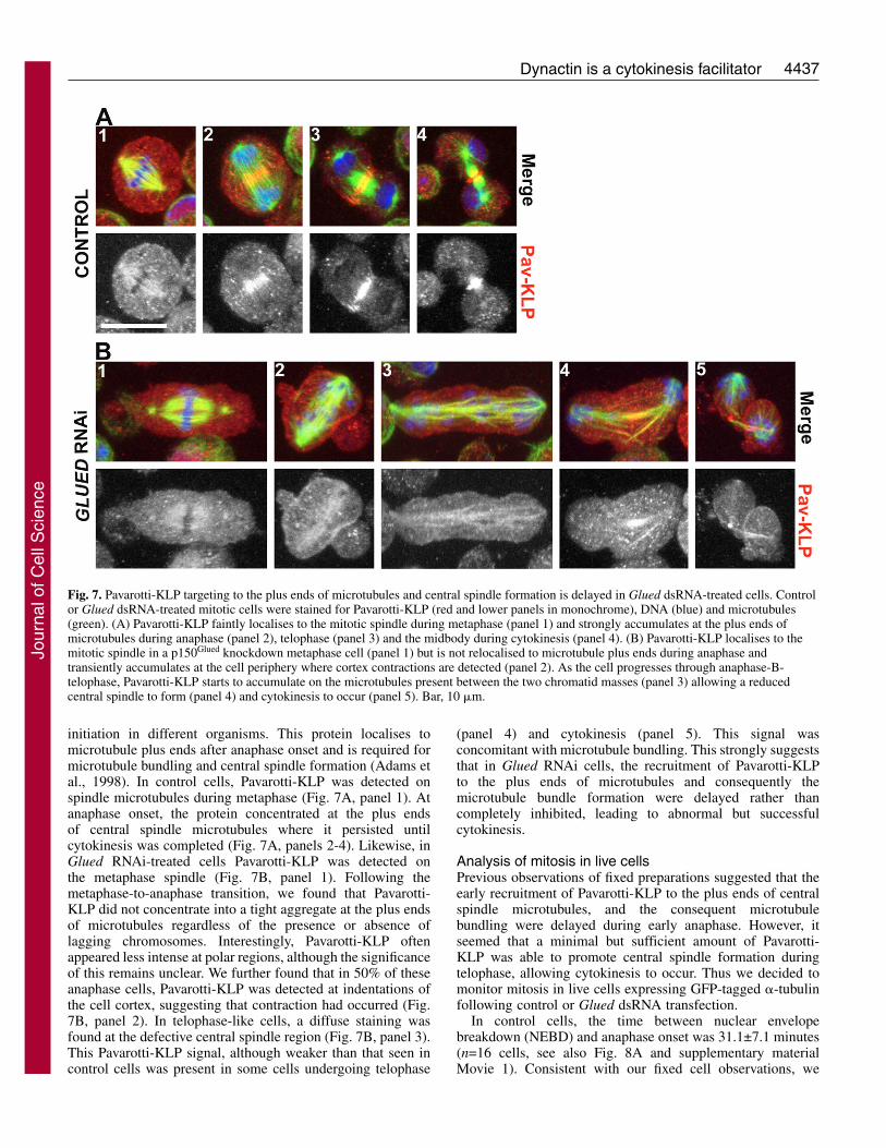
initiation in different organisms. This protein localises tomicrotubule plus ends after anaphase onset and is required formicrotubule bundling and central spindle formation (Adams etal., 1998). In control cells, Pavarotti-KLP was detected onspindle microtubules during metaphase (Fig. 7A, panel 1). Atanaphase onset, the protein concentrated at the plus endsof central spindle microtubules where it persisted untilcytokinesis was completed (Fig. 7A, panels 2-4). Likewise, inGlued RNAi-treated cells Pavarotti-KLP was detected onthe metaphase spindle (Fig. 7B, panel 1). Following themetaphase-to-anaphase transition, we found that Pavarotti-KLP did not concentrate into a tight aggregate at the plus endsof microtubules regardless of the presence or absence oflagging chromosomes. Interestingly, Pavarotti-KLP oftenappeared less intense at polar regions, although the significanceof this remains unclear. We further found that in 50% of theseanaphase cells, Pavarotti-KLP was detected at indentations ofthe cell cortex, suggesting that contraction had occurred (Fig.7B, panel 2). In telophase-like cells, a diffuse staining wasfound at the defective central spindle region (Fig. 7B, panel 3).This Pavarotti-KLP signal, although weaker than that seen incontrol cells was present in some cells undergoing telophase
(panel 4) and cytokinesis (panel 5). This signal wasconcomitant with microtubule bundling. This strongly suggeststhat in Glued RNAi cells, the recruitment of Pavarotti-KLPto the plus ends of microtubules and consequently themicrotubule bundle formation were delayed rather thancompletely inhibited, leading to abnormal but successfulcytokinesis.
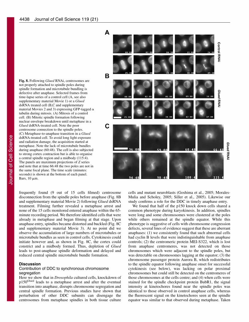
Analysis of mitosis in live cellsPrevious observations of fixed preparations suggested that theearly recruitment of Pavarotti-KLP to the plus ends of centralspindle microtubules, and the consequent microtubulebundling were delayed during early anaphase. However, itseemed that a minimal but sufficient amount of Pavarotti-KLP was able to promote central spindle formation duringtelophase, allowing cytokinesis to occur. Thus we decided tomonitor mitosis in live cells expressing GFP-tagged �-tubulinfollowing control or Glued dsRNA transfection.
In control cells, the time between nuclear envelopebreakdown (NEBD) and anaphase onset was 31.1±7.1 minutes(n=16 cells, see also Fig. 8A and supplementary materialMovie 1). Consistent with our fixed cell observations, we
Fig. 7. Pavarotti-KLP targeting to the plus ends of microtubules and central spindle formation is delayed in Glued dsRNA-treated cells. Controlor Glued dsRNA-treated mitotic cells were stained for Pavarotti-KLP (red and lower panels in monochrome), DNA (blue) and microtubules(green). (A) Pavarotti-KLP faintly localises to the mitotic spindle during metaphase (panel 1) and strongly accumulates at the plus ends ofmicrotubules during anaphase (panel 2), telophase (panel 3) and the midbody during cytokinesis (panel 4). (B) Pavarotti-KLP localises to themitotic spindle in a p150Glued knockdown metaphase cell (panel 1) but is not relocalised to microtubule plus ends during anaphase andtransiently accumulates at the cell periphery where cortex contractions are detected (panel 2). As the cell progresses through anaphase-B-telophase, Pavarotti-KLP starts to accumulate on the microtubules present between the two chromatid masses (panel 3) allowing a reducedcentral spindle to form (panel 4) and cytokinesis to occur (panel 5). Bar, 10 �m.
Jour
nal o
f Cel
l Sci
ence
4438
frequently found (9 out of 15 cells filmed) centrosomedisconnection from the spindle poles before anaphase (Fig. 8Band supplementary material Movie 2) following Glued dsRNAtreatment. Filming further revealed a metaphase arrest andnone of the 15 cells monitored entered anaphase within the 65-minute recording period. We therefore identified cells that werealready in metaphase and began filming at that stage. Uponanaphase entry, spindles became distorted and buckled (Fig. 8Cand supplementary material Movie 3). At no point did weobserve the accumulation of large numbers of microtubules ormicrotubule bundles as seen in control cells. Cytokinesis couldinitiate however and, as shown in Fig. 8C, the cortex couldconstrict and a midbody formed. Thus, depletion of Gluedleads to post-anaphase spindle deformation and delayed andreduced central spindle microtubule bundle formation.
DiscussionContribution of DDC to synchronous chromosomesegregationHere we show that in Drosophila cultured cells, knockdown ofp150Glued leads to a metaphase arrest and after the eventualtransition into anaphase, disrupts chromosome segregation andcentral spindle formation. Previous studies have shown thatperturbation of other DDC subunits can disengage thecentrosomes from metaphase spindles in both tissue culture
cells and mutant neuroblasts (Goshima et al., 2005; Morales-Mulia and Scholey, 2005; Siller et al., 2005). Likewise ourstudy confirms a role for the DDC in timely anaphase entry.
We found that half of the p150 knock down cells shared acommon phenotype during karyokinesis. In addition, spindleswere long and some chromosomes were clustered at the poleswhile others remained at the spindle equator. While thisphenotype is suggestive of cells with chromosome congressiondefects, several lines of evidence suggest that these are aberrantanaphases: (1) we consistently found that such abnormal cellshad cyclin B levels that were indistinguishable from anaphasecontrols; (2) the centromeric protein MEI-S322, which is lostfrom anaphase centromeres, was not detected on thosechromosomes which were adjacent to the spindle poles, butwas detectable on chromosomes lagging at the equator; (3) thechromosome passenger protein Aurora B, which redistributesto the spindle equator following anaphase onset for successfulcytokinesis (see below), was lacking on polar proximalchromosomes but could still be detected on the centromeres ofthose chromosomes at the cells centre; and (4) when cells werestained for the spindle checkpoint protein BubR1, the signalintensity at kinetochores found near the spindle poles wasidentical to those observed in control anaphase cells, whereasthe fluorescent signal on the kinetochores seen at the spindleequator was similar to that observed during metaphase. Taken
Journal of Cell Science 119 (21)
Fig. 8. Following Glued RNAi, centrosomes arenot properly attached to spindle poles duringspindle formation and microtubule bundling isdefective after anaphase. Selected frames fromtime-lapse series of a control cell (A, see alsosupplementary material Movie 1) or a GlueddsRNA treated cell (B,C and supplementarymaterial Movies 2 and 3) expressing GFP-tagged �tubulin during mitosis. (A) Mitosis of a controlcell. (B) Mitotic spindle formation followingnuclear envelope breakdown until metaphase in aGlued dsRNA-treated cell. Note the poorcentrosome connection to the spindle poles.(C) Metaphase-to-anaphase transition in a GlueddsRNA-treated cell. To avoid long light exposureand radiation damage, the acquisition started atmetaphase. Note the lack of microtubule bundlesduring anaphase (60:48). The cell is also subjectedto strong cortex contraction but is able to organisea central spindle region and a midbody (115:4).The panels are maximum projections of Z seriesand note that at time 66:48 the two poles are not inthe same focal plane. The time scale (minutes:seconds) is shown at the bottom of each panel.Bars, 10 �m.
Jour
nal o
f Cel
l Sci
ence
4439Dynactin is a cytokinesis facilitator
together, these data strongly suggest that the depletion ofp150Glued by RNAi distorts anaphase, allowing somechromosomes to undergo their normal maturation (i.e. loss ofcheckpoint and centromeric proteins followed by disjunctionand segregation to the poles), while others are inhibited frominitiating this series of events.
Our observations indicate that the disappearance of AuroraB, MEI-S322 and BubR1 from the centromeres/innerkinetochores may be not be fully dependent on the global lossof MPF activity (note that most cyclin B is degraded in Gluedanaphase cells). This suggests that DDC does not influence theinitial accumulation of cyclin B on the spindle or kinetochoreregions, and is not involved in the degradation of the spindle-associated pool. However, the retention of Aurora B, MEI-S332 and BubR1 at kinetochores in Glued-depleted cellssuggests that the DDC is needed for removal of these proteinsand for anaphase onset/chromatid disjunction. How the DDCregulates this is unclear. A pool of BubR1 localises to the innerregion of the kinetochore (Buffin et al., 2005; Jablonski et al.,1998) where it interacts with Aurora B (Lampson and Kapoor,2005). In addition, MEI-S322-dependant phosphorylation byaurora B is required for chromatid cohesion (Resnick et al.,2006). Thus, the loss of p150glued and DCC function mayprevent these interactions and the loss or redistribution of keyproteins for chromatid cohesion and subsequent chromosomesegregation. In addition, previous studies have revealed that aprimary function of the DDC, loaded to the kinetochore via theZW10/Rod complex, is to generate at least in part, polewardchromosome movement during anaphase in many organisms(Savoian et al., 2000; Schmidt et al., 2005; Sharp et al., 2000).Thus, depletion of p150Glued may lead to lagging chromosomesby two methods, in the first, the molecules needed forchromatid disjunction fail to be redistributed and second, thereis a failure to secure polewards force generating dynein motormolecules.
Contribution of DDC to central spindle formationAlong with the karyokinetic defects described above, we foundthat the depletion of Glued also disrupted central spindleformation. This microtubule-based structure consists ofoverlapping anti-parallel arrays of bundled microtubules and avariety of associated proteins and is crucial for successfulcytokinesis (D’Avino et al., 2005). Indeed in many instances,only weak indications of a central spindle could be found. Incontrast to control cells which always contained robust centralspindles and stained positive for Aurora B and polo, the earlyforming central spindle structures in Glued RNAi cells lackeddetectable levels of these kinases. These molecules areessential for successful cytokinesis (Adams et al., 1998;D’Avino et al., 2005; Echard et al., 2004; Gatti et al., 2000;Giet and Glover, 2001; Somma et al., 2002), leading to theexpectation that cleavage should fail in Glued RNAi-treatedcells. We failed to detect any octaploid cells or elevatednumbers of binucleated cells following Glued RNAi in ourstudy, even after a double Glued RNAi treatment. In addition,several RNAi screens recently performed in Drosophila S2cells, did not identify any DDC encoding genes essential forcytokinesis (Echard et al., 2004; Eggert et al., 2004) althoughDDC proteins are found in midbody preparations (Skop et al.,2004). It is thus possible that DDC facilitates central spindlestabilisation and contributes to cytokinesis without being
absolutely necessary for this process in flies. Indeed, we foundthat later-staged cells did localise Aurora B and polo to theircentral spindle structures, indicating that the DDC facilitatesthe recruitment of these kinases to the central spindle. Inagreement with our findings, a recent study revealed thatcytokinesis was not abolished but likewise delayed in seaurchin embryos injected with anti-p150Glued antibodies(Strickland et al., 2005).
Our study further revealed that the DDC is needed for therecruitment of Pavarotti-KLP to the spindle midzone. Thiskinesin is involved in central spindle formation, furrowformation and ingression by forming a complex withRacGAP50c to form the centralspindlin complex, a masterregulator of cytokinesis (D’Avino et al., 2005). Pavarotti-KLP,which concentrates at the overlapping plus ends ofmicrotubules at anaphase onset in control cells, failed toconcentrate at the midzone in p150Glued knockdown cells.However, like Aurora B and polo kinases, Pavarotti-KLP didultimately concentrate on the central spindles and midbodiesof later-staged cells. We further observed that Pavarotti-KLPconcentrated at the cell cortex in RNAi-treated cells. This mayexplain the abnormal contractions seen in both fixed and livingpreparations because it has previously been demonstrated thatectopic expression of this motor protein can promote therecruitment of other furrow components for furrow ingression(Minestrini et al., 2003). Thus, the DDC appears to beresponsible for an efficient localisation of Aurora B and polokinases as well as of Pavarotti-KLP to the central spindle fora timely onset of furrowing.
In summary, we report here for the first time that p150Glued,part of the evolutionarily conserved DDC, is needed for thelocalised redistribution of the centromeric and/or kinetochoreproteins MEI-S332, BubR1 and Aurora B for chromatiddisjunction and subsequent segregation. We further show forthe first time that p150Glued is involved in cytokinesis. Althoughnot required for successful cell cleavage, our data reveal thatGlued increases the efficiency of Aurora B and polo kinaserecruitment to the midzone for central spindle formation andstabilisation. We further demonstrate that the DDC is neededfor the efficient recruitment of Pavarotti-KLP, a member of thecentralspindlin complex, which is essential for cell cleavage.
Materials and MethodsdsRNA production and constructsTo generate Glued dsRNA, a cDNA fragment was amplified by PCR using theoligonucleotides 5�-ATGTAATACGACTCACTATAGGGCGAATGTCCGAGAA-AAACCTGAAAGTG-3� and 5�-ATGTAATACGACTCACTATAGGGCGACGA-AGCCTGAGCACCCAT-3� containing a T7 promoter sequence at each end. The1100 bp PCR product was used as a template to generate RNA using the Megascriptkit (Promega). After isolation, the RNAs were boiled for 20 minutes and annealedby slow cooling overnight at room temperature. dsRNA was analysed by agarosegel electrophoresis and aliquoted at –80°C before use in RNAi experiments.
To generate a construct for expressing GFP-tagged p150Glued in Drosophila cells,a PCR fragment was amplified using the oligonucleotides 5�-AAGTCGACG-TACATCAGTTATACCCAC-3� and 5�-TTGGTACCATTTACCTTTAATATATA-ATATAC-3� and cloned into pKS-Ub-GFP using SalI and KpnI restriction sites togenerate pKS-Ub-GFP-p150Glued (Minestrini et al., 2002).
To produce a recombinant protein used to generate an antibody against thep150Glued protein, a ~450 base pair DNA fragment, encoding the 148 N-terminaldomain of p150Glued was amplified by PCR using the oligonucleotides 5�-AAG-AATTCCATGTCCGAGAAAAACCTGAAAGTG-3� and 5�-AAAAGCTTTTGC-GGCGCCAAAGATTT-3� and cloned into pET23b using the EcoRI and HindIIIrestriction sites to generate pET23b-p150Glued-Nter(His)6 expression construct.
Production of recombinant proteins and antibody purificationp150Glued-Nter(His)6 protein was expressed in E.coli BL21(DE3)pLysS (Novagen) for
Jour
nal o
f Cel
l Sci
ence
4440
4 hours at 25°C. The protein was purified on a Ni-NTA-agarose column (Qiagen)following the manufacturer’s instructions. The purified protein was dialysedovernight against PBS (136 mM NaCl, 26 mM KCl, 2 mM Na2HPO4, 2 mMKH2PO4, pH 7.2) and used to immunise rabbits to generate Rb1477 and Rb1478antisera.
Rabbit anti-p150Glued antibodies were affinity purified on nitrocellulosemembrane. 1 mg p150Glued-Nter(His)6 protein was immobilised on a nitrocellulosemembrane. The membrane was incubated for 10 minutes at room temperature in100 mM glycine-HCl pH 3 to remove unbound protein, then 2 hours in PBScontaining 5% BSA and 0.5% Tween 20 (PBST-BSA) for blocking. The membranewas incubated overnight in 20 ml PBST-BSA containing 10% antisera Rb1477 ANDRb1478. After extensive washings in PBST-BSA, specific anti-p150Glued IgG wereeluted with 2 ml of 100 mM Glycine-HCl pH 3 for 10 minutes, neutralised with200 �l of 1M Tris solution, concentrated using a centricon 30 (PAL) and stored at–80°C at 1 �g/ml.
RNAi, transfections, drug treatment and stable line generationDrosophila S2 cells were grown and processed for RNAi as described previously(Clemens et al., 2000). Briefly, 106 cells were incubated with 10 �g/ml dsRNA inserum-free medium. Alternatively, 10 �g Transfast transfection reagent were addedtogether with 3 �g dsRNA following the manufacturer’s instructions (Promega).After 1 hour, fresh medium was added to the cells. At 4 days post transfection, thecells were fixed and analysed for mitotic defects (Giet and Glover, 2001). 100-200mitoses were scored and analysed per experiment, and each experiment wasrepeated at least three times.
To depolymerise microtubules, S2 cells were treated for 1 hour with 10 �g/mlcolchicine (Sigma) before analysis.
To prepare stable lines expressing GFP-tagged 150Glued or GFP-tagged �-tubulin,10 �g plasmid pKS-Ub-GFP-p150Glued or pAc-GFP-tub (Rogers et al., 2002) wasco-transfected with 1 �g pIB/V5-His/CAT or pCoHygro in S2 cells. Stable lineswere selected and expanded in medium containing 25 �g/ml blasticidin S or 200�g/ml hygromycin following the manufacturer’s instructions (Life Technologies).
Immunofluorescence analysisS2 cells were fixed in PHEM buffer (60 mM PIPES, 25 mM HEPES, 10 mM EGTA,4 mM MgCl2) containing 5% formaldehyde and 0.1% Triton X-100 at roomtemperature for 10 minutes. For Cyclin B immunofluorescence analyses, the cellswere fixed in methanol at –20°C for 10 minutes. The fixed cells were briefly washedin PBS, and blocked for 1 hour in PBS containing 0.1% Triton X-100 and 1% BSA(PBSX-BSA). Primary antibodies were incubated overnight at 4°C and secondaryantibodies were incubated for 2 hours at room temperature in PBSX-BSA. DNAwas stained with Hoechst 33258. Slides were mounted in Vectashield (VectorLaboratories) and observed with a DMRXA2 microscope (Leica). Images wereacquired with a CoolSnapsHQ camera (Photometrics) and processed withMetamorph software (Universal Imaging). Alternatively, images were acquired witha Leica DMIRE2 inverted confocal microscope.
FACS analysisS2 cells were fixed in PBS containing 70% ethanol and stored at –20°C untilanalysis. For FACS analysis, S2 ethanol-fixed cells were washed twice in PBS andincubated with PBS containing 40 �g/ml RNAse A and 10 �g/ml propidium iodidefor 30 minutes. S2 cells were analysed using a Coulter Epics Elite Flow Cytometer(Beckman Coulter).
Live cell imagingAll time-lapse imaging was performed on a Leica DMIRBE inverted microscopecontrolled by the Metamorph software using an oil 63� objective (NA 1.40). Zseries (0.3 �m steps) were acquired at 30 or 60 second intervals with a CoolSnapEScamera (Roper Scientific) using a bin of 2. Cells were bathed in a chamberedcoverglass system (Lab-Tek) and maintained at 25°C.
Antibodies and western blottingThe YL1/2 rat anti-tyrosinated tubulin antibody (dilution 1:100) was from Seralab.The GTU-88 mouse anti-�-tubulin, the anti-dynein intermediate chain and the anti-GFP monoclonal antibodies (dilution 1:1000) were from Sigma, Chemicon andRoche respectively. Antibodies against Pavarotti-KLP (1:1000), BubR1 (1:3000)MEI-S322 (dilution 1:2000), Aurora B (1:1000), Cyclin B (1:5000) and anti-polomonoclonal antibody (dilution 1:40) were kind gifts from the indicated authors(Adams et al., 1998; Giet and Glover, 2001; Heuer et al., 1995; Kerrebrock et al.,1995; Logarinho et al., 2004; Whitfield et al., 1995). The affinity-purified anti-p150Glued antibody was used at 0.2 �g/ml. Peroxidase-, FITC-, Texas Red- and Cy5-conjugated anti-rabbit, anti-sheep, anti-mouse or anti-rat secondary antibodies werefrom Jackson, and the Alexa Fluor 488-conjugated antibodies from MolecularProbes. For western blotting, ECL reagent was purchased from Pierce.
We would like to acknowledge S. Dutertre for help with microscopywork, L. Rault for some immunostaining analyses and G. Goshima,
D. M. Glover, T. C. Kaufman, T. L. Orr-Weaver, C. Sunkel forreagents. Thanks are also due to M. S. Savoian for his very helpfulcritical readings of this manuscript and to R. Karess and unknownreferees for helpful suggestions. Rennes Métropole supported thiswork.
ReferencesAdams, R. R., Tavares, A. A., Salzberg, A., Bellen, H. J. and Glover, D. M. (1998).
pavarotti encodes a kinesin-like protein required to organize the central spindle andcontractile ring for cytokinesis. Genes Dev. 12, 1483-1494.
Askham, J. M., Vaughan, K. T., Goodson, H. V. and Morrison, E. E. (2002). Evidencethat an interaction between EB1 and p150(Glued) is required for the formation andmaintenance of a radial microtubule array anchored at the centrosome. Mol. Biol. Cell13, 3627-3645.
Buffin, E., Lefebvre, C., Huang, J., Gagou, M. E. and Karess, R. E. (2005).Recruitment of Mad2 to the kinetochore requires the Rod/Zw10 complex. Curr. Biol.15, 856-861.
Busson, S., Dujardin, D., Moreau, A., Dompierre, J. and De Mey, J. R. (1998). Dyneinand dynactin are localized to astral microtubules and at cortical sites in mitoticepithelial cells. Curr. Biol. 8, 541-544.
Carminati, J. L. and Stearns, T. (1997). Microtubules orient the mitotic spindle in yeastthrough dynein-dependent interactions with the cell cortex. J. Cell Biol. 138, 629-641.
Clemens, J. C., Worby, C. A., Simonson-Leff, N., Muda, M., Maehama, T.,Hemmings, B. A. and Dixon, J. E. (2000). Use of double-stranded RNA interferencein Drosophila cell lines to dissect signal transduction pathways. Proc. Natl. Acad. Sci.USA 97, 6499-6503.
Collins, C. A. and Vallee, R. B. (1989). Preparation of microtubules from rat liver andtestis: cytoplasmic dynein is a major microtubule associated protein. Cell Motil.Cytoskeleton 14, 491-500.
D’Avino, P. P., Savoian, M. S. and Glover, D. M. (2005). Cleavage furrow formationand ingression during animal cytokinesis: a microtubule legacy. J. Cell Sci. 118, 1549-1558.
Echard, A., Hickson, G. R., Foley, E. and O’Farrell, P. H. (2004). Terminal cytokinesisevents uncovered after an RNAi screen. Curr. Biol. 14, 1685-1693.
Eggert, U. S., Kiger, A. A., Richter, C., Perlman, Z. E., Perrimon, N., Mitchison, T.J. and Field, C. M. (2004). Parallel chemical genetic and genome-wide RNAi screensidentify cytokinesis inhibitors and targets. PLoS Biol. 2, e379.
Gatti, M., Giansanti, M. G. and Bonaccorsi, S. (2000). Relationships between thecentral spindle and the contractile ring during cytokinesis in animal cells. Microsc. Res.Tech. 49, 202-208.
Giet, R. and Glover, D. M. (2001). Drosophila aurora B kinase is required for histoneH3 phosphorylation and condensin recruitment during chromosome condensation andto organize the central spindle during cytokinesis. J. Cell Biol. 152, 669-682.
Gill, S. R., Schroer, T. A., Szilak, I., Steuer, E. R., Sheetz, M. P. and Cleveland, D.W. (1991). Dynactin, a conserved, ubiquitously expressed component of an activatorof vesicle motility mediated by cytoplasmic dynein. J. Cell Biol. 115, 1639-1650.
Gonczy, P., Pichler, S., Kirkham, M. and Hyman, A. A. (1999). Cytoplasmic dyneinis required for distinct aspects of MTOC positioning, including centrosome separation,in the one cell stage Caenorhabditis elegans embryo. J. Cell Biol. 147, 135-150.
Goshima, G. and Vale, R. D. (2003). The roles of microtubule-based motor proteins inmitosis: comprehensive RNAi analysis in the Drosophila S2 cell line. J. Cell Biol. 162,1003-1016.
Goshima, G. and Vale, R. D. (2005). Cell cycle-dependent dynamics and regulation ofmitotic kinesins in Drosophila S2 cells. Mol. Biol. Cell 16, 3896-3907.
Goshima, G., Nedelec, F. and Vale, R. D. (2005). Mechanisms for focusing mitoticspindle poles by minus end-directed motor proteins. J. Cell Biol. 171, 229-240.
Heuer, J. G., Li, K. and Kaufman, T. C. (1995). The Drosophila homeotic target genecentrosomin (cnn) encodes a novel centrosomal protein with leucine zippers and mapsto a genomic region required for midgut morphogenesis. Development 121, 3861-3876.
Jablonski, S. A., Chan, G. K., Cooke, C. A., Earnshaw, W. C. and Yen, T. J. (1998).The hBUB1 and hBUBR1 kinases sequentially assemble onto kinetochores duringprophase with hBUBR1 concentrating at the kinetochore plates in mitosis.Chromosoma 107, 386-396.
Karess, R. (2005). Rod-Zw10-Zwilch: a key player in the spindle checkpoint. Trends CellBiol. 15, 386-392.
Karki, S. and Holzbaur, E. L. (1999). Cytoplasmic dynein and dynactin in cell divisionand intracellular transport. Curr. Opin. Cell Biol. 11, 45-53.
Kerrebrock, A. W., Moore, D. P., Wu, J. S. and Orr-Weaver, T. L. (1995). Mei-S332,a Drosophila protein required for sister-chromatid cohesion, can localize to meioticcentromere regions. Cell 83, 247-256.
Koonce, M. P., Kohler, J., Neujahr, R., Schwartz, J. M., Tikhonenko, I. and Gerisch,G. (1999). Dynein motor regulation stabilizes interphase microtubule arrays anddetermines centrosome position. EMBO J. 18, 6786-6792.
Lampson, M. A. and Kapoor, T. M. (2005). The human mitotic checkpoint proteinBubR1 regulates chromosome-spindle attachments. Nat. Cell Biol. 7, 93-98.
Logarinho, E., Bousbaa, H., Dias, J. M., Lopes, C., Amorim, I., Antunes-Martins, A.and Sunkel, C. E. (2004). Different spindle checkpoint proteins monitor microtubuleattachment and tension at kinetochores in Drosophila cells. J. Cell Sci. 117, 1757-1771.
Mathe, E., Kraft, C., Giet, R., Deak, P., Peters, J. M. and Glover, D. M. (2004). TheE2-C vihar is required for the correct spatiotemporal proteolysis of cyclin B and itselfundergoes cyclical degradation. Curr. Biol. 14, 1723-1733.
Minestrini, G., Mathe, E. and Glover, D. M. (2002). Domains of the Pavarotti kinesin-
Journal of Cell Science 119 (21)
Jour
nal o
f Cel
l Sci
ence
4441Dynactin is a cytokinesis facilitator
like protein that direct its subcellular distribution: effects of mislocalisation on thetubulin and actin cytoskeleton during Drosophila oogenesis. J. Cell Sci. 115, 725-736.
Minestrini, G., Harley, A. S. and Glover, D. M. (2003). Localization of Pavarotti-KLPin living Drosophila embryos suggests roles in reorganizing the cortical cytoskeletonduring the mitotic cycle. Mol. Biol. Cell 14, 4028-4038.
Morales-Mulia, S. and Scholey, J. M. (2005). Spindle pole organization in DrosophilaS2 cells by Dynein, abnormal spindle protein (Asp), and KLP10A. Mol. Biol. Cell 16,3176-3186.
Niccoli, T., Yamashita, A., Nurse, P. and Yamamoto, M. (2004). The p150-GluedSsm4p regulates microtubular dynamics and nuclear movement in fission yeast. J. CellSci. 117, 5543-5556.
Resnick, T. D., Satinover, D. L., Macisaac, F., Stukenberg, P. T., Earnshaw, W. C.,Orr-Weaver, T. L. and Carmena, M. (2006). INCENP and Aurora B promote meioticsister chromatid cohesion through localization of the shugoshin MEI-S332 inDrosophila. Dev. Cell 11, 57-68.
Robinson, J. T., Wojcik, E. J., Sanders, M. A., McGrail, M. and Hays, T. S. (1999).Cytoplasmic dynein is required for the nuclear attachment and migration ofcentrosomes during mitosis in Drosophila. J. Cell Biol. 146, 597-608.
Salina, D., Bodoor, K., Eckley, D. M., Schroer, T. A., Rattner, J. B. and Burke, B.(2002). Cytoplasmic dynein as a facilitator of nuclear envelope breakdown. Cell 108,97-107.
Savoian, M. S., Goldberg, M. L. and Rieder, C. L. (2000). The rate of polewardchromosome motion is attenuated in Drosophila zw10 and rod mutants. Nat. Cell Biol.2, 948-952.
Schmidt, D. J., Rose, D. J., Saxton, W. M. and Strome, S. (2005). Functional analysisof cytoplasmic dynein heavy chain in Caenorhabditis elegans with fast-actingtemperature-sensitive mutations. Mol. Biol. Cell 16, 1200-1212.
Sharp, D. J., Rogers, G. C. and Scholey, J. M. (2000). Cytoplasmic dynein is required
for poleward chromosome movement during mitosis in Drosophila embryos. Nat. CellBiol. 2, 922-930.
Siller, K. H., Serr, M., Steward, R., Hays, T. S. and Doe, C. Q. (2005). Live imagingof Drosophila brain neuroblasts reveals a role for Lis1/dynactin in spindle assemblyand mitotic checkpoint control. Mol. Biol. Cell 16, 5127-5140.
Skop, A. R., Liu, H., Yates, J., 3rd, Meyer, B. J. and Heald, R. (2004). Dissection ofthe mammalian midbody proteome reveals conserved cytokinesis mechanisms. Science305, 61-66.
Somma, M. P., Fasulo, B., Cenci, G., Cundari, E. and Gatti, M. (2002). Moleculardissection of cytokinesis by RNA interference in Drosophila cultured cells. Mol. Biol.Cell 13, 2448-2460.
Strickland, L. I., Wen, Y., Gundersen, G. G. and Burgess, D. R. (2005). Interactionbetween EB1 and p150(glued) is required for anaphase astral microtubule elongationand stimulation of cytokinesis. Curr. Biol. 15, 2249-2255.
Vallee, R. B. and Stehman, S. A. (2005). How dynein helps the cell find its center: aservomechanical model. Trends Cell Biol. 15, 288-294.
Vallee, R. B., Shpetner, H. S. and Paschal, B. M. (1989). The role of dynein and othermicrotubule-activated ATPases in mitosis. Prog. Clin. Biol. Res. 318, 205-215.
Vallee, R. B., Vaughan, K. T. and Echeverri, C. J. (1995). Targeting of cytoplasmicdynein to membranous organelles and kinetochores via dynactin. Cold Spring Harb.Symp. Quant. Biol. 60, 803-811.
Vaughan, P. S., Miura, P., Henderson, M., Byrne, B. and Vaughan, K. T. (2002). Arole for regulated binding of p150(Glued) to microtubule plus ends in organelletransport. J. Cell Biol. 158, 305-319.
Whitfield, W. G., Chaplin, M. A., Oegema, K., Parry, H. and Glover, D. M. (1995).The 190 kDa centrosome-associated protein of Drosophila melanogaster contains fourzinc finger motifs and binds to specific sites on polytene chromosomes. J. Cell Sci.108, 3377-3387.
Jour
nal o
f Cel
l Sci
ence