durable response to anti-pd-1 immunotherapy in epithelioid
TRANSCRIPT

CASE REPORT Open Access
Durable response to anti-PD-1immunotherapy in epithelioidangiomyolipoma: a report on the successfultreatment of a rare malignancyMichael Lattanzi1, Fang-Ming Deng2,3,5, Luis A. Chiriboga2, Alisa N. Femia4, Shane A. Meehan2,4, Gopa Iyer6,Martin H. Voss6, Yuliya Sundatova5,7, William C. Huang3,5 and Arjun V. Balar1,5,7*
Abstract
Background: Malignant angiomyolipoma is an uncommon tumor of the class of perivasciular epithelioid cellneoplasms (PEComas). These tumors are characteristically driven by deleterious mutations in the tumor suppressorsTSC1 and TSC2, whose gene products typically act to inhibit mTOR. There are several cases of malignantangiomyolipoma which exhibit transient responses to mTOR inhibitors, forming the basis of current practiceguidelines in malignant PEComa. However the tumors ultimately acquire resistance, and there is no well-establishedsecond-line option. Despite the increasing prevalence of immunotherapy across a wide range of solid tumors, littleis known about the immune infiltrate and PD-L1 expression of angiomyolipoma. Furthermore, there is no reportedcase on the treatment of malignant angiomyolipoma with an immune checkpoint inhibitor.
Case presentation: A 38 year-old man presented with gross hematuria and was diagnosed with renal epithelioidangiomyolipoma. Despite surgical resection, the tumor recurred and metastasized. Targeted genomic sequencingrevealed a deleterious mutation in TSC2, and the patient was treated with the mTOR inihbitor everolimus. Thepatient went on to have a partial response but ultimately progressed. He was then treated with the anti-PD-1immune checkpoint inhibitor nivolumab, and achieved a durable near-complete response which is ongoing aftertwo years of treatment. Immunohistochemical staining of tumor tissue revealed strong PD-L1 expression and a briskT-cell infiltrate.
Conclusions: We report on the first durable systemic treatment of malignant epithelioid angiomyolipoima with theuse of PD-1 antibody nivolumab. Given the absence of prospective clinical trials in this exceedingly rare disease,particularly in the second-line setting, immune checkpoint inhibitors like nivolumab should be considered.
Keywords: Immunotherapy, Angiomyolipoma, PEComa, Nivolumab, PD-1, PD-L1, Tuberous sclerosis, TSC2, mTOR,Everolimus
* Correspondence: [email protected] of Medicine, NYU Langone Health, New York, NY, USA5Laura and Isaac Perlmutter Cancer Center, NYU Langone Health, New York,NY, USAFull list of author information is available at the end of the article
© The Author(s). 2018 Open Access This article is distributed under the terms of the Creative Commons Attribution 4.0International License (http://creativecommons.org/licenses/by/4.0/), which permits unrestricted use, distribution, andreproduction in any medium, provided you give appropriate credit to the original author(s) and the source, provide a link tothe Creative Commons license, and indicate if changes were made. The Creative Commons Public Domain Dedication waiver(http://creativecommons.org/publicdomain/zero/1.0/) applies to the data made available in this article, unless otherwise stated.
Lattanzi et al. Journal for ImmunoTherapy of Cancer (2018) 6:97 https://doi.org/10.1186/s40425-018-0415-x

BackgroundAngiomyolipomas (AMLs) are neoplasms thought to arisefrom pericites, hence belonging to the family of perivasci-ular epithelioid cell neoplasms (PEComas). Though asso-ciated with tuberous sclerosis complex (TSC), most AMLsarise de novo, in the absence of germline TSC1 or TSC2mutations [1]. There exist two well-described histologicvariants: classical and epithelioid. Although the vast ma-jority of AMLs are benign, a minority of epithelioid AML(EAML) may become malignant and have been reportedto metastasize [2]. The exact prevalence of malignantEAML is not well characterized; however, it likely fallsbelow 1:300,000 [3, 4]. Given its rarity, there is no estab-lished treatment for unresectable or metastatic EAML.TSC is a genetic syndrome characterized by multisystem
tumor development, including renal angiomyolipoma.Most patients harbor pathogenic germline loss-of-functionmutations in TSC1 or TSC2 [5], whose wild-type geneproducts inhibit mammalian target of rapamycin (mTOR)complex 1 (mTORC1) [6, 7]. This observation prompted aphase III placebo-controlled trial demonstrating significanttumor regression of TSC-associated AML with everolimus[8], a United States Food and Drug Administration(FDA)-approved allosteric inhibitor of mTORC1. Somaticmutations in TSC1 and TSC2 also contribute to tumorgrowth via unopposed mTOR signaling, and sporadicAML is similarly characterized by somatic loss-of-functionalterations in TSC2 [9]. Multiple reports detail responsesto mTOR inhibitors among tumors harboring TSC1 orTSC2 mutations [7, 10], including PEComa, not otherwisespecified [11], and sporadic AML [12], though DNAsequencing was not reported. Based on these responses,clinical practice guidelines for malignant PEComa cur-rently emphasize the use of mTOR inhibitors such aseverolimus [13]. However, despite an initial response torapalog therapy, virtually all patients ultimately developprogressive disease, and there is no well-establishedsecond-line treatment.Nivolumab is a fully humanized monoclonal IgG4
antibody that targets the programmed death 1 (PD-1)receptor, an immune checkpoint expressed on exhaustedeffector T lymphocytes, and prevents binding by itsactivating ligand PD-L1, leading to reinvigoration ofanti-tumor immunity [14]. Nivolumab is FDA-approvedfor melanoma, renal cell carcinoma, and urothelial blad-der cancer, among other solid tumors. Although tumorPD-L1 expression is associated with response [15], nobiomarker of response has been rigorously validated.Additionally, immune checkpoint inhibitors are associ-ated with the development and/or exacerbation of auto-immunity [15], and such immune-related toxicities maycorrelate with enhanced clinical efficacy [16].Given the lack of data concerning the treatment of this
rare cancer, we report a case of metastatic EAML
harboring a deleterious TSC2 mutation. The patientexhibited a transient response to everolimus, but ultim-ately progressed. He subsequently achieved a significantand durable response to nivolumab. To the best of ourknowledge, this is the first report on the treatment ofmalignant EAML with immunotherapy.
Case presentationA 38 year-old man with vitiligo and hypothyroidism ini-tially presented in 2011 with gross hematuria. Diagnosticimaging (Fig. 1a) revealed a 6-cm renal mass concerningfor malignancy, for which he underwent a right radicalnephrectomy at the recommendation of his treatingurologic oncologist (WCH). Gross pathology (Fig. 1b)revealed a 6 × 5-cm encapsulated hilar mass withhemorrhage and central necrosis. The mass was limitedto the renal parenchyma, without evidence of renal sinusor vascular invasion, and surgical margins were negativefor tumor cells. Histologic sections (Fig. 1c) demon-strated sheets of epithelioid cells with sarcomatoid andrhabdoid features as well as round, polygonal cells withpleomorphic nuclei and prominent nucleoli. Mitoticfigures were visualized at a rate of approximately threeper high-powered field. Immunohistochemical staining(Fig. 1d-e) revealed tumor cell positivity for: HMB45,melan-A, carbonic anhydrase IX, and to a lesser extent,Cam5.2, vimentin and SMA (cytoplasmic), and negativityfor: EMA, keratins (AE1/3), CK7, CK20, P63, Pax-2,AMACAR, S-100, and CD10. Based on these histo-pathologic features, the patient was diagnosed with pri-mary EAML.The patient had an uneventful course for the next 3
years until April, 2014, when surveillance imaging de-tected an asymptomatic 13-cm renal fossa mass for whichhe underwent repeat surgical resection. Surgical pathologyconfirmed recurrent EAML, again with negative margins.The patient’s tumor recurred again in October, 2014,prompting a third surgical resection. Pathologic evaluationthis time demonstrated indeterminate margins, promptedreferral to medical oncology for further management.December, 2014 surveillance imaging obtained by the
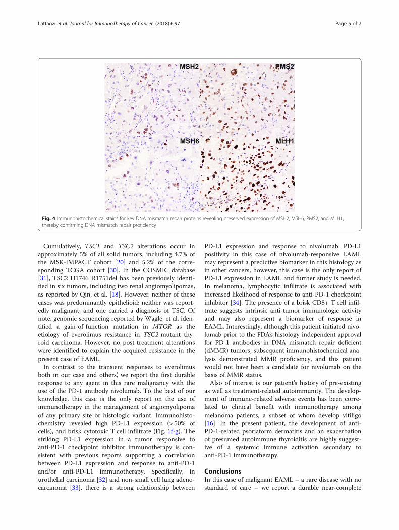
treating medical oncologist (AVB) demonstrated newretroperitoneal and pelvic implants consistent with meta-static EAML. The patient’s tumor DNA was subjected toFoundationOne® targeted next-generation sequencing[17], which revealed four oncogenic alterations: truncatingmutations in TP53 and APC, a frameshift mutation inATRX, and a deletion in TSC2, specifically, TSC2H1746_R1751del, which has been reported both as asomatic variant in AML [18] and as a germline mutationin TSC [19]. Of note, the FoundationOne® assay demon-strated no genomic alterations in the four genes en-coding key DNA mismatch repair proteins: MSH2,MSH6, PMS2, or MLH1.
Lattanzi et al. Journal for ImmunoTherapy of Cancer (2018) 6:97 Page 2 of 7

Based on the TSC2 deletion, the patient was initiatedon everolimus in January, 2015. Imaging at 3 months(Fig. 2) demonstrated marked decrease in size of themajority of the soft tissue masses throughout the rightnephrectomy bed, retroperitoneum, and mesentery, andno new sites of disease. The patient remained clinicallyasymptomatic for 8 months, until he noted unintendedweight loss in September, 2015. Imaging demonstratedslight enlargement of the dominant right renal fossamass (Fig. 2), which in the context of progressive
anemia, was interpreted as disease progression. Everoli-mus was discontinued, and the patient was referred for atreatment-directed biopsy for consideration of a clinicaltrial.He underwent a biopsy of the dominant 6 cm retro-
peritoneal mass, from which DNA was isolated andsubjected to paired tumor-germline next-generationsequencing via MSK-IMPACT [20], which confirmed theabsence of a TSC2 germline mutation. However, no newsomatic variants were identified to explain the tumors’
A C F
D G
E
B
Fig. 1 a CT Urogram demonstrating the primary right renal mass, (b) Gross pathology demonstrating the resected perihilar tumor with centralnecrosis, (c) H&E stain demonstrating angiomyolipoma with a substantial epithelial component, (d) Immunohistochemical stain negative forcytokeratin AE1/AE3, (e) Immunohistochemical stain positive for HMB-45, (f) Immunohistochemical stain positive for PD-L1 (> 50% of cells),(f) Immunohistochemical stain positive for T lymphocyte marker CD8
Fig. 2 Schematic representation of radiographic tumor burden over time indicating (left-to-right): baseline pre-everolimus scan, best response toeverolimus, progression on everolimus and baseline pre-nivolumab scan, initial post-nivolumab response, and continued response at the time ofnivolumab discontinuation
Lattanzi et al. Journal for ImmunoTherapy of Cancer (2018) 6:97 Page 3 of 7

acquired resistance, and he was not eligible for anyclinical trials. Given the well-known activity of anti-PD-1checkpoint inhibition across a range of advanced solidtumors, including renal cell carcinoma [21], the patientwas offered a trial of off-label nivolumab via theBristol-Myers Squibb Expanded Access program, and hebegan treatment in October, 2015. After two cycles ofnivolumab (administered at 3 mg/kg IV every 2 weeks),the patient felt well, and resolution of his anemia sug-gested possible clinical benefit. Imaging after 5 cyclesdemonstrated responding disease (Fig. 2).Nivolumab was well-tolerated, with the exception
of immune-related exacerbation of pre-existinghypothyroidism (Fig. 3a-b) after 2 months of therapy,and immune-related pruritic cutaneous eruption pre-dominantly within areas of pre-existing vitiligo (Fig. 3c-d),occurring after 18 months of treatment. Over the courseof nivolumab therapy, the patient required increasingdoses of levothyroxine to maintain a euthyroid state.An archival thyroid ultrasound reveals an enlargedheterogenous thyroid gland suggestive of possible auto-immune thyroiditis. With regard to cutaneous toxicity, thepatient was referred to dermatology (ANF), and a skinbiopsy was obtained of an involved area on the upperback. Histologic evaluation by the dermatopathologist(SAM) revealed a thin granular layer, confluent para-keratosis with collections of neutrophils, and dilatedcapillaries throughout the papillary dermis, consistentwith psoriasis (Fig. 3e-f ).Although these toxicities were not dose-limiting, the
patient had already completed 2 years of therapy [22],
and nivolumab was therefore discontinued. The mostrecent imaging at the time of discontinuation in Novem-ber, 2017 demonstrates continued response and intervalcalcification of his intra-abdominal tumors (Fig. 2).Archival tissue derived from the initial surgical resectionwas analyzed for PD-L1 and CD8 expression via immu-nohistochemistry (Fig. 1f-g) utilizing a modified Agilent/Dako 22C3 pharmaDX kit, revealing high PD-L1 expres-sion (> 50% of cells) and a brisk CD8+ T cell infiltrate.Finally, immunohistochemical analysis demonstratedpreserved expression of key DNA mismatch repair(MMR) proteins MSH2, MSH6, PMS2, and MLH1 [23],thus confirming MMR proficiency (Fig. 4).
DiscussionCurrent approaches to treatment for the sporadic form ofrecurrent or metastatic EAML are derived from anecdotalreports of efficacy with mTOR inhibitors [11, 24–27]. Fur-thermore, although Wagner, et al. [11] reported TSC1 lossin PEComa, genomic sequencing of TSC1 and TSC2 wasnot performed in these cases, as they predate the wide-spread use of next-generation sequencing, and the histo-logic subtype (e.g. angiomyolipoma) was not specified.Nonetheless, despite an initial response to rapalog therapy,virtually all tumors ultimately developed resistance. Inaddition to mTOR inhibitors, tumor responses have beenobserved with the use of chemotherapy. Cibas, et al.reported a partial response to doxorubicin [28], and othershave reported various combinations of cisplatin, cyclo-phosphamide, dacarbazine, and ifosfamide [29], with mod-est results and no reported long-term responses.
A
B
C
D
E
F
Fig. 3 a Graph of the patient’s prescribed daily dose of levothyroxine and serum thyroid stimulating hormone (TSH) over time relative toinitiation of nivolumab, (b) Archival thyroid ultrasound demonstrating enlarged, heterogeneous thyroid gland suggestive of chronic thyroiditis,(c) Distribution of psoriasis involving the patient’s back, (d) Representative scaly, well-demarcated erythematous papules overlying the right lowerabdomen predominantly within an area of vitiligo, (e) Low-resolution and (f) High-resolution histologic sections of cutaneous biopsy with H&Estaining demonstrating a thin granular layer, confluent parakeratosis with collections of neutrophils, and dilated capillaries throughout thepapillary dermis, consistent with psoriasis
Lattanzi et al. Journal for ImmunoTherapy of Cancer (2018) 6:97 Page 4 of 7
Cumulatively, TSC1 and TSC2 alterations occur inapproximately 5% of all solid tumors, including 4.7% ofthe MSK-IMPACT cohort [20] and 5.2% of the corre-sponding TCGA cohort [30]. In the COSMIC database[31], TSC2 H1746_R1751del has been previously identi-fied in six tumors, including two renal angiomyolipomas,as reported by Qin, et al. [18]. However, neither of thesecases was predominantly epithelioid; neither was report-edly malignant; and one carried a diagnosis of TSC. Ofnote, genomic sequencing reported by Wagle, et al. iden-tified a gain-of-function mutation in MTOR as theetiology of everolimus resistance in TSC2-mutant thy-roid carcinoma. However, no post-treatment alterationswere identified to explain the acquired resistance in thepresent case of EAML.In contrast to the transient responses to everolimus
both in our case and others’, we report the first durableresponse to any agent in this rare malignancy with theuse of the PD-1 antibody nivolumab. To the best of ourknowledge, this case is the only report on the use ofimmunotherapy in the management of angiomyolipomaof any primary site or histologic variant. Immunohisto-chemistry revealed high PD-L1 expression (> 50% ofcells), and brisk cytotoxic T cell infiltrate (Fig. 1f-g). Thestriking PD-L1 expression in a tumor responsive toanti-PD-1 checkpoint inhibitor immunotherapy is con-sistent with previous reports supporting a correlationbetween PD-L1 expression and response to anti-PD-1and/or anti-PD-L1 immunotherapy. Specifically, inurothelial carcinoma [32] and non-small cell lung adeno-carcinoma [33], there is a strong relationship between
PD-L1 expression and response to nivolumab. PD-L1positivity in this case of nivolumab-responsive EAMLmay represent a predictive biomarker in this histology asin other cancers, however, this case is the only report ofPD-L1 expression in EAML and further study is needed.In melanoma, lymphocytic infiltrate is associated withincreased likelihood of response to anti-PD-1 checkpointinhibitor [34]. The presence of a brisk CD8+ T cell infil-trate suggests intrinsic anti-tumor immunologic activityand may also represent a biomarker of response inEAML. Interestingly, although this patient initiated nivo-lumab prior to the FDA’s histology-independent approvalfor PD-1 antibodies in DNA mismatch repair deficient(dMMR) tumors, subsequent immunohistochemical ana-lysis demonstrated MMR proficiency, and this patientwould not have been a candidate for nivolumab on thebasis of MMR status.Also of interest is our patient’s history of pre-existing
as well as treatment-related autoimmunity. The develop-ment of immune-related adverse events has been corre-lated to clinical benefit with immunotherapy amongmelanoma patients, a subset of whom develop vitiligo[16]. In the present patient, the development of anti-PD-1-related psoriaform dermatitis and an exacerbationof presumed autoimmune thyroiditis are highly suggest-ive of a systemic immune activation secondary toanti-PD-1 immunotherapy.
ConclusionsIn this case of malignant EAML – a rare disease with nostandard of care – we report a durable near-complete
Fig. 4 Immunohistochemical stains for key DNA mismatch repair proteins revealing preserved expression of MSH2, MSH6, PMS2, and MLH1,thereby confirming DNA mismatch repair proficiency
Lattanzi et al. Journal for ImmunoTherapy of Cancer (2018) 6:97 Page 5 of 7

response to the anti-PD-1 immune checkpoint inhibitor,nivolumab. This case highlights the cross-histologic effi-cacy of immune checkpoint inhibition, particularly intumors with high PD-L1 expression and brisk lympho-cytic infiltrate. In the absence of prospective clinicaltrials, nivolumab should be considered for use in otherpatients with recurrent or metastatic EAML who haveexhausted traditional therapeutic options.
AbbreviationsAML: Angiomyolipoma; EAML: Epithelioid angiomyolipoma; FDA: Food andDrug Administration; MMR: Mismatch repair; MTOR: Mammalian target ofrapamycin; PD-1: Programmed death receptor 1; PD-L1: Programmed deathreceptor ligand 1; TSC: Tuberous sclerosis complex
FundingThis study was not supported by any external funding.
Availability of data and materialsIdentifying patient information must remain confidential; however additionaldata may be available from upon reasonable request at the discretion of thecorresponding author.
Authors’ contributionsML is a research coordinator for the NYU genitourinary oncology tumorregistry and compiled all patient data. FMD is the attending genitourinarypathologist who diagnosed the case. LAC is the director of theimmunohistochemistry core facility and performed all correlative staining.ANF is the attending dermatologist who performed the cutaneous biopsy.SAM is the attending dermatopathologist who diagnosed the cutaneoustoxicity. GI and MHV are medical oncologists who consulted on the case andorchestrated the treatment-directed biopsy and subsequent genomicsequencing. YS is the nurse practitioner who oversaw the patient’s medicaloncology care. WCH is the attending urologic oncologist who performed allsurgical resections. AVB is the attending genitourinary medical oncologistwho treated the patient and directed all study-related activities. All authorsread and approved the final manuscript.
Ethics approval and consent to participateThe present study was conducted in accordance with all accepted standards forthe ethical conduct of human subjects research. The patient’s data and tissuespecimens are stored according to NYU IRB-approved protocol s15–01579. Thepatient provided written informed consent at the time of enrollment.
Consent for publicationThe patient provided verbal and written consent to publish all pertinentdemongraphic, clinical, and biologic information. The signed consent formwill be made available to the editorial staff upon request.
Competing interestsMHV reports consulting fees from Novartis as well as research funding fromGenentech and Bristol-Myers Squibb. All other authors report no disclosures.
Publisher’s NoteSpringer Nature remains neutral with regard to jurisdictional claims inpublished maps and institutional affiliations.
Author details1Department of Medicine, NYU Langone Health, New York, NY, USA.2Department of Pathology, NYU Langone Health, New York, NY, USA.3Department of Urology, NYU Langone Health, New York, NY, USA. 4RonaldO. Perelman Department of Dermatology, NYU Langone Health, New York,NY, USA. 5Laura and Isaac Perlmutter Cancer Center, NYU Langone Health,New York, NY, USA. 6Department of Medicine, Memorial Sloan-KetteringCancer Center, New York, NY, USA. 7Genitourinary Medical OncologyProgram, NYU School of Medicine, Laura and Isaac Perlmutter Cancer Center,NYU Langone Health, 160 East 34th Street, 10th Floor, New York, NY 10016,USA.
Received: 15 June 2018 Accepted: 18 September 2018
References1. Flum AS, Hamoui N, Said MA, Yang XJ, Casalino DD, McGuire BB, et al.
Update on the diagnosis and Management of Renal Angiomyolipoma.J Urol. 2016;195(4 Pt 1):834–46.
2. Aydin H, Magi-Galluzzi C, Lane BR, Sercia L, Lopez JI, Rini BI, et al. Renalangiomyolipoma: clinicopathologic study of 194 cases with emphasis onthe epithelioid histology and tuberous sclerosis association. Am J SurgPathol. 2009;33(2):289–97.
3. He W, Cheville JC, Sadow PM, Gopalan A, Fine SW, Al-Ahmadie HA, et al.Epithelioid angiomyolipoma of the kidney: pathological features and clinicaloutcome in a series of consecutively resected tumors. Mod Pathol. 2013;26(10):1355–64.
4. Fujii Y, Ajima J, Oka K, Tosaka A, Takehara Y. Benign renal tumors detectedamong healthy adults by abdominal ultrasonography. Eur Urol. 1995;27(2):124–7.
5. Northrup H, Krueger DA. International tuberous sclerosis complex consensusG. tuberous sclerosis complex diagnostic criteria update: recommendationsof the 2012 Iinternational tuberous sclerosis complex consensus conference.Pediatr Neurol. 2013;49(4):243–54.
6. El-Hashemite N, Zhang H, Henske EP, Kwiatkowski DJ. Mutation in TSC2 andactivation of mammalian target of rapamycin signalling pathway in renalangiomyolipoma. Lancet. 2003;361(9366):1348–9.
7. Iyer G, Hanrahan AJ, Milowsky MI, Al-Ahmadie H, Scott SN, Janakiraman M,et al. Genome sequencing identifies a basis for everolimus sensitivity.Science. 2012;338(6104):221.
8. Bissler JJ, Kingswood JC, Radzikowska E, Zonnenberg BA, Frost M, BelousovaE, et al. Everolimus for angiomyolipoma associated with tuberous sclerosiscomplex or sporadic lymphangioleiomyomatosis (EXIST-2): a multicentre,randomised, double-blind, placebo-controlled trial. Lancet. 2013;381(9869):817–24.
9. Smolarek TA, Wessner LL, McCormack FX, Mylet JC, Menon AG, Henske EP.Evidence that lymphangiomyomatosis is caused by TSC2 mutations:chromosome 16p13 loss of heterozygosity in angiomyolipomas and lymphnodes from women with lymphangiomyomatosis. Am J Hum Genet. 1998;62(4):810–5.
10. Wagle N, Grabiner BC, Van Allen EM, Amin-Mansour A, Taylor-Weiner A,Rosenberg M, et al. Response and acquired resistance to everolimus inanaplastic thyroid cancer. N Engl J Med. 2014;371(15):1426–33.
11. Wagner AJ, Malinowska-Kolodziej I, Morgan JA, Qin W, Fletcher CD, Vena N,et al. Clinical activity of mTOR inhibition with sirolimus in malignantperivascular epithelioid cell tumors: targeting the pathogenic activation ofmTORC1 in tumors. J Clin Oncol. 2010;28(5):835–40.
12. Bissler JJ, McCormack FX, Young LR, Elwing JM, Chuck G, Leonard JM, et al.Sirolimus for angiomyolipoma in tuberous sclerosis complex orlymphangioleiomyomatosis. N Engl J Med. 2008;358(2):140–51.
13. von Mehren M, Randall RL, Benjamin RS, Boles S, Bui MM, Conrad EU 3rd,et al. Soft tissue sarcoma, version 2.2016, NCCN clinical practice guidelinesin oncology. J Natl Compr Cancer Netw. 2016;14(6):758–86.
14. Wolchok JD, Kluger H, Callahan MK, Postow MA, Rizvi NA, Lesokhin AM,et al. Nivolumab plus ipilimumab in advanced melanoma. N Engl J Med.2013;369(2):122–33.
15. Topalian SL, Hodi FS, Brahmer JR, Gettinger SN, Smith DC, McDermott DF,et al. Safety, activity, and immune correlates of anti-PD-1 antibody in cancer.N Engl J Med. 2012;366(26):2443–54.
16. Teulings HE, Limpens J, Jansen SN, Zwinderman AH, Reitsma JB, Spuls PI,et al. Vitiligo-like depigmentation in patients with stage III-IV melanomareceiving immunotherapy and its association with survival: a systematicreview and meta-analysis. J Clin Oncol. 2015;33(7):773–81.
17. Frampton GM, Fichtenholtz A, Otto GA, Wang K, Downing SR, He J, et al.Development and validation of a clinical cancer genomic profiling testbased on massively parallel DNA sequencing. Nat Biotechnol. 2013;31(11):1023–31.
18. Qin W, Bajaj V, Malinowska I, Lu X, MacConaill L, Wu CL, et al. Angiomyolipomahave common mutations in TSC2 but no other common genetic events. PLoSOne. 2011;6(9):e24919.
19. Roberts PS, Dabora S, Thiele EA, Franz DN, Jozwiak S, Kwiatkowski DJ.Somatic mosaicism is rare in unaffected parents of patients with sporadictuberous sclerosis. J Med Genet. 2004;41(5):e69.
Lattanzi et al. Journal for ImmunoTherapy of Cancer (2018) 6:97 Page 6 of 7

20. Zehir A, Benayed R, Shah RH, Syed A, Middha S, Kim HR, et al. Mutationallandscape of metastatic cancer revealed from prospective clinicalsequencing of 10,000 patients. Nat Med. 2017;23(6):703–13.
21. Motzer RJ, Escudier B, McDermott DF, George S, Hammers HJ, Srinivas S,et al. Nivolumab versus Everolimus in advanced renal-cell carcinoma. N EnglJ Med. 2015;373(19):1803–13.
22. Robert C, Schachter J, Long GV, Arance A, Grob JJ, Mortier L, et al.Pembrolizumab versus Ipilimumab in advanced melanoma. N Engl J Med.2015;372(26):2521–32.
23. Le DT, Durham JN, Smith KN, Wang H, Bartlett BR, Aulakh LK, et al.Mismatch repair deficiency predicts response of solid tumors to PD-1blockade. Science. 2017;357(6349):409–13.
24. Italiano A, Delcambre C, Hostein I, Cazeau AL, Marty M, Avril A, et al.Treatment with the mTOR inhibitor temsirolimus in patients with malignantPEComa. Ann Oncol. 2010;21(5):1135–7.
25. Gennatas C, Michalaki V, Kairi PV, Kondi-Paphiti A, Voros D. Successfultreatment with the mTOR inhibitor everolimus in a patient with perivascularepithelioid cell tumor. World J Surg Oncol. 2012;10:181.
26. Benson C, Vitfell-Rasmussen J, Maruzzo M, Fisher C, Tunariu N, Mitchell S,et al. A retrospective study of patients with malignant PEComa receivingtreatment with sirolimus or temsirolimus: the Royal Marsden Hospitalexperience. Anticancer Res. 2014;34(7):3663–8.
27. Kohno J, Matsui Y, Yamasaki T, Shibasaki N, Kamba T, Yoshimura K, et al.Role of mammalian target of rapamycin inhibitor in the treatment ofmetastatic epithelioid angiomyolipoma: a case report. Int J Urol.2013;20(9):938–41.
28. Cibas ES, Goss GA, Kulke MH, Demetri GD, Fletcher CD. Malignantepithelioid angiomyolipoma (‘sarcoma ex angiomyolipoma’) of the kidney: acase report and review of the literature. Am J Surg Pathol. 2001;25(1):121–6.
29. Park HK, Zhang S, Wong MK, Kim HL. Clinical presentation of epithelioidangiomyolipoma. Int J Urol. 2007;14(1):21–5.
30. Gao J, Aksoy BA, Dogrusoz U, Dresdner G, Gross B, Sumer SO, et al.Integrative analysis of complex cancer genomics and clinical profiles usingthe cBioPortal. Sci Signal. 2013;6(269):pl1.
31. Forbes SA, Beare D, Boutselakis H, Bamford S, Bindal N, Tate J, et al.COSMIC: somatic cancer genetics at high-resolution. Nucleic Acids Res.2017;45(D1):D777–D83.
32. Sharma P, Retz M, Siefker-Radtke A, Baron A, Necchi A, Bedke J, et al.Nivolumab in metastatic urothelial carcinoma after platinum therapy(CheckMate 275): a multicentre, single-arm, phase 2 trial. Lancet Oncol.2017;18(3):312–22.
33. Borghaei H, Paz-Ares L, Horn L, Spigel DR, Steins M, Ready NE, et al.Nivolumab versus docetaxel in advanced nonsquamous non-small-cell lungCancer. N Engl J Med. 2015;373(17):1627–39.
34. Tumeh PC, Harview CL, Yearley JH, Shintaku IP, Taylor EJ, Robert L, et al.PD-1 blockade induces responses by inhibiting adaptive immune resistance.Nature. 2014;515(7528):568–71.
Lattanzi et al. Journal for ImmunoTherapy of Cancer (2018) 6:97 Page 7 of 7