duck hepatitis b virus infection of muscovy duck hepatocytes and nature of virus resistance in vivo
TRANSCRIPT

JOURNAL OF VIROLOGY, Apr. 1994, p. 2487-2494 Vol. 68, No. 40022-538X/94/$04.00+0Copyright ©D 1994, American Society for Microbiology
Duck Hepatitis B Virus Infection of Muscovy Duck Hepatocytesand Nature of Virus Resistance In Vivo
JOHN C. PUGH* AND HEIDI SIMMONS
Fox Chase Cancer Center, Philadelphia, Pennsylvania 19111
Received 1 December 1993/Accepted 17 January 1994
To test the hypothesis that in vivo resistance to hepadnavirus infection was due to resistance of hosthepatocytes, we isolated hepatocytes from Muscovy ducklings and chickens, birds that have been shown to beresistant to duck hepatitis B virus (DHBV) infection, and attempted to infect them in vitro with virus fromcongenitally infected Pekin ducks. Chicken hepatocytes were resistant to infection, but we were able to infectapproximately 1% of Muscovy duck hepatocytes in culture. Infection requires prolonged incubation with virusat 37°C. Virus spread occurs in the Muscovy cultures, resulting in 5 to 10% DHBV-infected hepatocytes by 3weeks after infection. The relatively low rate of accumulation of DHBV DNA in infected Muscovy hepatocytecultures is most likely due to inefficient spread of virus infection; in the absence of virus spread, the rates ofDHBV replication in Pekin and Muscovy hepatocyte cultures are similar. 5-Azacytidine treatment can inducesusceptibility to DHBV infection in resistant primary Pekin hepatocytes but appears to have no similar effectin Muscovy cultures. The relatively inefficient infection of Muscovy duck hepatocytes that we have describedmay account for the absence of a detectable viremia in Muscovy ducklings experimentally infected with DHBV.
An important factor in determining whether a cell becomesinfected with a given virus is the availability of suitable host cellreceptors to facilitate virus binding and uptake. The impor-tance of virus receptors in determining the very restricted hostand tissue range displayed by hepatitis B virus (HBV) is largelyinferred, as a receptor has not been identified for any memberof the hepadnavirus family. Duck hepatitis B virus (DHBV) isthe best characterized of the avian members of the hepadna-virus family. The virus was originally isolated from the domes-tic Pekin duck (Anas domesticus) but can be successfullytransmitted to other closely related ducks and to geese (4).Marion and coworkers have shown that DHBV cannot bereadily transmitted to Muscovy (Cairina moschata) ducks or tochickens, as indicated by the absence of detectable DHBVDNA in the sera and livers of birds challenged with virusduring the first few days after hatching (4). The Muscovy is aspecies of domesticated duck that is distinct from all otherdomesticated ducks, which were originally derived from themallard (Anas platyrhzynchos). In this study, we have askedwhether resistance of Muscovy ducks and chickens to in vivoinfection with DHBV can be attributed solely to resistance ofhepatocytes to infection.To determine resistance to DHBV, we isolated primary
hepatocytes from Muscovy ducklings and chickens and in-fected them in vitro with DHBV. We report that primaryhepatocytes isolated from Muscovy ducklings can be infectedwith DHBV but that this infection is less efficient and thecourse of infection is retarded as compared with that in Pekinduck hepatocytes. Chicken hepatocytes appear to be com-pletely resistant to DHBV infection in vitro, however.
MATERIALS AND METHODS
Experimental animals, virus stocks, and cell culture. Pekinducks were purchased as 1-day-old ducklings from MetzerFarms, Gonzales, California. Muscovy ducklings were pur-
* Corresponding author. Mailing address: Fox Chase Cancer Cen-ter, 7701 Burholme Ave., Philadelphia, PA 19111. Phone: (215)728-4780. Fax: (215) 728-3574.
chased as 1-day-old ducklings from Hoffman Hatchery, Gratz,Pa. Ducklings were tested for DHBV infection by dot blothybridization of serum, and any infected birds were isolated.The source of DHBV used for infections of primary hepa-
tocytes was a pool of sera isolated from 3-week-old ducklingsfrom a congenitally infected flock maintained by the Fox ChaseLaboratory Animal Facility. The serum was stored in aliquotsat - 700C.
Primary hepatocytes were prepared from 1- to 2-week-oldducklings by collagenase perfusion of the liver. The procedureis based upon that described by Berry and Friend (la) and isdescribed in detail elsewhere (6, 13). Approximately 5 x 10⁶cells were plated on each 60-mm-diameter dish (4 ml of a 1%[vol/vol] suspension of cells). Cells were maintained in Liebo-vitz-15 (L-15) medium (Gibco BRL) supplemented with insu-lin (Sigma), hydrocortisone-hemisuccinate (Sigma), HEPES(N-2-hydroxyethylpiperazine-N'-2-ethanesulfonic acid), andantibiotics, as previously described (13), but without fetalbovine serum, dimethyl sulfoxide, or additional sodium bicar-bonate or glucose supplements unless otherwise indicated (6).Fresh medium was added to cells every 1 to 2 days. 5-Azacy-tidine (5-aza) (Sigma) was added to cultures from a sterile 0.1M stock in water. Suramin (Bayer) was added to cells at a finalconcentration of 100 [Lg/ml.
Hepatocytes were infected for 16 h at 370C with a 1:5dilution of DHBV duck serum (approximately 10i DHBVDNA-containing particles per ml) in L-15, unless otherwisestated. No attempt was made to remove the great excess ofnoninfectious surface antigen particles from the duck serumused for infections.
Staining for DHBV core and envelope proteins by immuno-fluorescence. DHBV core protein was detected by stainingcells fixed on tissue culture plastic with ethanol-glacial aceticacid (95:5), by using a specific rabbit antiserum (a gift ofWilliam Mason) followed by fluorescein isothiocyanate-conju-gated goat anti-rabbit immunoglobulin G (Cappell) (13). Forstaining of DHBV envelope proteins, cells were fixed inmethanol-acetone (1:1) and stained with a mixture of two
mouse monoclonal antibodies, 1H.1 and 7C.12, that are spe-cific for the pre-S and S domains of the DHBV envelope,
2487
Dow
nloa
ded
from
http
s://j
ourn
als.
asm
.org
/jour
nal/j
vi o
n 02
Jan
uary
202
2 by
59.
148.
221.
218.

2488 PUGH AND SIMMONS
1:5 DHBV 1:500 DHBV
Day 8p.i.
FIG. 1. Titration of infectivity of a DHBV serum stock on Pekin duck hepatocytes. Primary hepatocytes were plated on 60-mm dishes andinfected 2 days after plating with 2 ml of a 1:5 or 1:500 dilution of duck serum in L-15 medium for 16 h at 37°C. Infected cultures were maintainedin L-15 medium for 8 days and then fixed and stained for DHBV envelope protein (see Materials and Methods). The bar in the right panelrepresents approximately 200 urm. p.i., postinfection.
respectively (5a). Antibody binding was detected by using a
fluorescein isothiocyanate-conjugated goat anti-mouse immu-noglobulin G (Cappell). Cells were photographed with a NikonDiaphot fluorescence microscope.Assay of infected hepatocyte cultures for DHBV DNA. Total
cellular nucleic acids were isolated from infected hepatocytecultures as follows. After brief washing of the monolayer withphosphate-buffered saline (PBS), 1 ml of lysis buffer (20 mMTris-hydrochloride [pH 7.5], 10 mM EDTA, 0.2 M NaCl, 0.2%sodium dodecyl sulfate (SDS), 0.5 mg of pronase per ml) was
added to each 60-mm dish, and cells were incubated for 1 h at37°C. The lysate was extracted once with an equal volume ofphenol, and nucleic acids were precipitated with 2 volumes of100% ethanol. The nucleic acid precipitate was dissolved in100 RI of TE (10 mM Tris-hydrochloride [pH 7.5], 1 mMEDTA). A third of each DNA sample was loaded ontoindividual lanes of a 1.5% (wt/vol) agarose gel, and afterelectrophoresis DNA was denatured and transferred to a
Hybond N (Amersham) nylon filter. DNA was immobilized byUV cross-linking of the filter in a Stratagene Stratalinker 1800,and DHBV DNA was detected by using an in vitro-synthesized32P-labelled RNA complementary to DHBV minus strandDNA (13).To prepare nucleic acids enriched for DHBV covalently
closed circular (CCC) DNA, cells were lysed in the bufferdescribed above (excluding pronase) for 30 min at 37°C, andthen KCl was added to a 0.5 M final concentration. Themixture was vortexed briefly and stored at room temperaturefor 30 min. The protein-detergent complex was removed bycentrifugation, and the supernatant, which contains CCCDNA, was extracted with an equal volume of phenol. Nucleicacids were recovered by ethanol precipitation, and DHBVCCC DNA was detected by Southern blot hybridization as
described above (10, 13). The hybridization standard on all gelswas 10 pg of linear cloned DHBV DNA.
RESULTS
Optimal conditions for DHBV infection of Muscovy duckhepatocytes. In order to determine whether Muscovy duckhepatocytes could be infected in vitro with DHBV, we initially
used conditions which had previously been shown to result inmaximal infection of primary Pekin duck hepatocyte cultures(6). Optimal infection of hepatocytes requires incubation witha high-titer virus inoculum (approximately 109 DHBV particlesper ml) for several hours at 37°C. Staining of Pekin duckcultures for DHBV envelope protein 8 days after infectionunder these conditions reveals that almost all cells are infected(Fig. 1, left panel [1:5 dilution]).To compare the relative amounts of infection in Pekin duck
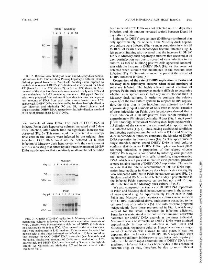
and Muscovy duck hepatocyte cultures following infectionunder identical conditions, hepatocytes isolated from bothbirds were infected with equivalent amounts of DHBV at 4 and37°C. The relative amounts of DHBV DNA in cultures 8 daysafter infection were determined by Southern hybridization(Fig. 2). The much greater amount of DHBV DNA in infectedPekin cultures suggests that many more hepatocytes becomeinfected in the Pekin than in the Muscovy duck culturesfollowing incubation with the same amount of virus. Theresults also indicate that prolonged incubation at 370C with ahigh-titer virus inoculum is required for optimal infection ofMuscovy duck hepatocytes. Suramin was added to cells imme-diately after infection in these experiments to limit the spreadof virus infection. However, it appears that uptake of boundvirus is sensitive to the action of suramin and that incubationfor several hours following virus adsorption is required to allowvirus to bypass the suramin-sensitive step (5). This accounts forthe absence of detectable DHBV DNA after infection at 40C,at which temperature no uptake of bound virus should occur,and for the much larger amount of DHBV DNA in culturesincubated with virus for 5 h at 37°C (Fig. 2, lanes 3) comparedwith 1 h (Fig. 2, lanes 2) before addition of suramin.
Kinetics of DHBV replication following infection of Mus-covy duck hepatocytes. The appearance of CCC DHBV DNAwas assayed following infection of Muscovy and Pekin duckhepatocytes under the conditions described above (Fig. 3). Thenumber of CCC DNA molecules in DHBV-infected hepato-cytes is tightly regulated (10, 12); thus, CCC DNA serves as anindicator of the relative number of infected cells. CCC DNAcould be detected in cells 24 h after addition of the virusinoculum to Pekin duck hepatocyte cultures, indicating thatmost cells in the culture had taken up and converted at least
J. VIROL.
Dow
nloa
ded
from
http
s://j
ourn
als.
asm
.org
/jour
nal/j
vi o
n 02
Jan
uary
202
2 by
59.
148.
221.
218.
AVIAN HEPADNAVIRUS HOST RANGE 2489
Pekin Muscovy1 2 3 1 2 3 hs
RC - t
s
FIG. 2. Relative susceptibility of Pekin and Muscovy duck hepato-cyte cultures to DHBV infection. Primary hepatocyte cultures (60-mmdishes) prepared from 1- to 2-week-old ducklings were exposed toequivalent amounts of DHBV (1:5 dilution of stock serum) for 1 h at4°C (lanes 1), 1 h at 37°C (lanes 2), or 5 h at 37°C (lanes 3). Afterremoval of the virus inoculum, cells were washed briefly with PBS andthen maintained in L-15 containing suramin at 100 p.g/ml. Nucleicacids were prepared from cultures 8 days after infection, and equiva-lent amounts of DNA from each culture were loaded on a 1.5%
agarose gel. DHBV DNA was detected by Southern blot hybridization(see Materials and Methods). RC and SS, relaxed circular and
single-stranded DHBV DNA, respectively; hs, hybridization standard
of 10 pg of cloned linear DHBV DNA.
one molecule of virus DNA. The level of CCC DNA ininfected Pekin duck hepatocyte cultures increased until 6 daysafter infection, after which time no significant increase was
observed (Fig. 3). This result would be expected if all suscep-tible cells in the culture were infected by the original virus
inoculum. CCC DNA could not be detected 24 h afterinfection of Muscovy duck hepatocytes with the same amountof virus, indicating that either uptake and conversion of DHBVDNA was delayed or that a relatively small number of cells had
Muscovydayp.i. 1 3 101316 2024hs
RC_ ___Z
ccc-4
Pekin
dayp.i. 1 3 4 6 8 10 1316hs
RC
FIG. 3. Kinetics of DHBV replication in Muscovy and Pekin duck
hepatocyte cultures following infection with equivalent amounts of
virus. Cultures were infected with a high titer of DHBV (1:5 dilution
of stock serum) for 16 h at 37C. After removal of the virus inoculum,
cells were maintained in L-15 medium. Cultures were harvested for
nucleic acids at the times indicated postinfection (p.i.) by a procedurethat enriches for CCC DHBV DNA molecules (see Materials and
Methods). Equivalent amounts of DNA were loaded on a 1.5%
agarose gel, and DHBV DNA was detected by Southern blot hybrid-ization (see Materials and Methods). RC and hs are defined in the
legend to Fig. 2.
been infected. CCC DNA was not detected until 10 days afterinfection, and this amount increased tenfold between 13 and 16days after infection.
Staining for DHBV core antigen (DHBcAg) confirmed thatonly approximately 1% of cells in the Muscovy duck hepato-cyte culture were infected (Fig. 4) under conditions in which 80to 100% of Pekin duck hepatocytes become infected (Fig. 1,left panel). Staining also revealed that the increase in DHBVDNA in Muscovy duck hepatocyte cultures that occurred at 16days postinfection was due to spread of virus infection in theculture, as foci of DHBcAg-positive cells appeared concomi-tant with the increase in DHBV DNA (Fig. 4). Foci were notdetected when suramin was maintained in the medium afterinfection (Fig. 4). Suramin is known to prevent the spread ofDHBV infection in vitro (5).Comparison of the rate of DHBV replication in Pekin and
Muscovy duck hepatocyte cultures when equal numbers ofcells are infected. The highly efficient initial infection ofprimary Pekin duck hepatocytes made it difficult to determinewhether virus spread was in fact any more efficient than inMuscovy duck cultures. In order to compare the relativecapacity of the two culture systems to support DHBV replica-tion, the virus titer in the inoculum was adjusted such thatapproximately equal numbers of cells were infected. Titrationof virus infectivity on Pekin duck hepatocytes showed that a
1:500 dilution of a DHBV-positive duck serum resulted inapproximately 1% infected cells after 8 days (Fig. 1, right panel[1:500 dilution]). Infection of Muscovy duck hepatocytes with a
1:5 dilution of the same serum also resulted in approximately1% infected cells (Fig. 4). Thus, having established conditionsfor infecting equivalent numbers of cells in Pekin and Muscovyduck hepatocyte cultures, we compared the kinetics of DHBVDNA replication in these cultures (Fig. 5). The appearance ofsingle-stranded, minus strand DHBV DNA in both culturesconfirms that de novo DHBV DNA replication takes placefollowing infection. A proportion of the relaxed circularDHBV DNA signal is contributed by infecting virus particlesthat remain associated with cells; therefore, single-strandedDNA, which is not present in mature virus particles, providesa more reliable marker of DHBV DNA replication. The resultsindicate that the rate of accumulation of DHBV DNA repli-cation intermediates in Muscovy duck hepatocytes was slightlyslow compared with that in Pekin hepatocyte cultures (Fig. 5).Single-stranded DNA can be detected at day 6 postinfection inthe infected Pekin hepatocyte culture but not until 15 daysafter infection in the Muscovy duck culture (Fig. 5).We also compared the kinetics of DHBV DNA replication
in Pekin and Muscovy duck hepatocyte cultures in the absenceof virus spread (Fig. 6). Approximately 1% of cells in bothPekin and Muscovy duck hepatocyte cultures were infectedwith DHBV, as described above, and suramin was added to thecultures I day after infection (5). The cultures were preparedindependently from those represented in Fig. 5, which mayaccount for the small differences in infection efficiency.Suramin was maintained in the culture medium until cells wereharvested for DHBV DNA analysis at the times indicated.Maximum levels of intracellular DHBV DNA were attainedapproximately 14 days after infection in both Pekin andMuscovy duck hepatocyte cultures. Hence, when only a singleround of infection was allowed to take place, it was not
apparent that the kinetics of DHBV DNA replication were
significantly different in Muscovy versus Pekin duck hepatocytecultures. The more rapid accumulation of DHBV DNA inter-mediates in infected Pekin duck hepatocytes in the absence ofsuramin (Fig. 5) may, therefore, be due to more efficient
VO)L. 68, 1994
Dow
nloa
ded
from
http
s://j
ourn
als.
asm
.org
/jour
nal/j
vi o
n 02
Jan
uary
202
2 by
59.
148.
221.
218.

2490 PUGH AND SIMMONS
plussuramin
minussuramin
Day 15 p..
Day 26 p.i.
I
FIG. 4. Spread of DHBV infection in Muscovy duck hepatocytes infected in vitro. Muscovy duck cultures were infected with DHBV and stainedfor DHBV core protein at 15 or 26 days after infection (see Materials and Methods). Following infection, cells were maintained in L-15 plussuramin (100 ,ug/ml) or in L-15 medium alone, as indicated. The bar represents approximately 120 pum. p.i., postinfection.
spread of DHBV in Pekin duck hepatocyte cultures ratherthan to an increased rate of DHBV DNA replication.
Passage of DHBV from infected Muscovy duck hepatocytecultures. An alternative explanation for the apparently morerapid rate of accumulation of DHBV DNA intermediates inPekin duck hepatocytes might be that virus is released moreefficiently from Muscovy duck hepatocytes. To test this hypoth-esis, we monitored the release of infectious DHBV fromMuscovy duck cells by transferring virus to highly susceptiblecultures of Pekin duck hepatocytes. Figure 7A shows the rateat which DHBV DNA accumulated in the infected Muscovyduck hepatocyte cultures. DHBV was recovered from mediumharvested from the Muscovy duck cells and used to infectfreshly prepared Pekin duck hepatocytes. The Pekin duck cellswere harvested 8 days after infection, and the relative amountof DHBV DNA in infected cultures was determined bySouthern hybridization (Fig. 7B). The results show that themaximum amount of infectious DHBV is released from theMuscovy duck cells between 16 and 19 days after infection(Fig. 7B). For reasons that are not clear, single-strandedDHBV DNA was not recovered efficiently from the infectedPekin duck cultures in this experiment. Maximal release ofinfectious virus coincides with a marked increase in the amount
Muscovy Pekin
dayp.L. 2 4 6 8 10 13 15 1718 20hs 2 4 6 8 10
RC -
SS-
FIG. 5. Kinetics of DHBV replication in Muscovy and Pekin duckhepatocyte cultures following infection of equivalent numbers of cells.Primary Muscovy and Pekin duck hepatocyte cultures were infectedfor 16 h at 370C with a virus inoculum determined to infect equalnumbers (approximately 1%) of cells in both cultures (a 1:5 dilution ofDHBV duck serum for Muscovy duck hepatocytes and a 1:500 dilutionfor Pekin duck hepatocytes). Cells were maintained in L-15, andinfected cultures were harvested for total nucleic acids at the timesshown postinfection (p.i.). DHBV DNA was detected by Southern blothybridization (see Materials and Methods and Fig. 2). RC, SS, and hsare defined in the legend to Fig. 2.
J. VIROL.
Dow
nloa
ded
from
http
s://j
ourn
als.
asm
.org
/jour
nal/j
vi o
n 02
Jan
uary
202
2 by
59.
148.
221.
218.

AVIAN HEPADNAVIRUS HOST RANGE 2491
Muscovy
dayp.i. hs 6 9 12 15 20 23 27
Pekin
day p.i. hs 2 4 6 8 10 12 1416
RC- _
SsS
FIG. 6. Kinetics of DHBV replication in Muscovy and Pekin duckhepatocytes when virus spread is inhibited by suramin. PrimaryMuscovy and Pekin duck hepatocyte cultures were infected as de-scribed in the legend to Fig. 5 in order to infect approximatelyequivalent numbers of cells in both cultures. After removal of the virusinoculum, cells were maintained in L-15 containing suramin at 100p.g/ml to inhibit spread of virus infection. Total nucleic acids wereprepared from cultures at the times shown postinfection (p.i.), andDHBV DNA was detected by Southern blot hybridization (see Mate-rials and Methods and Fig. 2). RC, SS, and hs are defined in the legendto Fig. 2.
of intracellular DHBV DNA (Fig. 7A), which we have shownis associated with the appearance of virus spread in thecultures (Fig. 4). Maximal release of DHBV from Pekin duckhepatocytes is also coincident with an increase in intracellularDHBV DNA and spread of virus infection (data not shown).Thus, it appears that DHBV is released from hepatocytesisolated from both Pekin and Muscovy ducks at similar stagesduring the virus infectious cycle. The results also indicate thata single round of replication in Muscovy duck cells does notappear to reduce the infectivity of DHBV on cells isolatedfrom the normal permissive host.When virus released into the medium from a DHBV-
infected Muscovy duck hepatocyte culture was passaged to asecond Muscovy culture, a time course similar to that in theoriginal infected culture was observed (results not shown).Very few cells were infected, however, presumably because ofboth the relatively low amounts of virus released from theoriginal infected culture and the small number of susceptiblecells. Hence, it appears that in vitro infection of Muscovy duckhepatocytes has not selected for rare DHBV variants with thecapacity to infect these cells, as infection at the second passmight then be expected to be more efficient.Chicken hepatocytes are resistant to DHBV infection in
vitro. We were concerned that infection of the small number ofcells in the Muscovy duck culture might result from uptake ofvirus by some route that did not involve a specific virus-receptor interaction. If this were the case, we would expect asimilar proportion of primary chicken hepatocytes to be sus-ceptible to DHBV infection. Chickens are resistant to DHBV
A
dayp.l. 1 3 6 9 1215 202327 hs
RC -
Ss-
Bdayp.i. 3 5 7 9 12 14 1619 21 hs
RC -
Ss-
FIG. 7. Time course to assay release of infectious DHBV from invitro-infected Muscovy duck hepatocytes (p.i., postinfection). PrimaryMuscovy duck hepatocytes were infected with a 1:5 dilution of DHBVduck serum in L-15 for 16 h at 37°C. One set of infected cultures (nine60-mm dishes) was used for nucleic acid analysis to assay the accumu-lation of DHBV DNA intermediates in infected Muscovy duck cells(A). A pair of infected Muscovy duck cultures (100-mm dishes)infected under the same conditions was used to assay the release ofvirus. Medium was harvested from these cells every 2 to 3 days afterinfection and stored at -70°C. Virus was concentrated from themedium by precipitation with 10% (wt/vol) PEG 8000 (Sigma), and thevirus pellet was dissolved in 2 ml of L-15 medium and used to infect60-mm dishes of Pekin duck hepatocytes for 16 h at 37°C. Eight daysafter infection (p.i.), total nucleic acids were prepared from theinfected Pekin duck cultures, equivalent amounts ofDNA were loadedon a 1.5% agarose gel, and DHBV DNA was detected by Southernhybridization (B). RC, SS, and hs are defined in the legend to Fig. 2.
infection (4), but a cell line derived from transformed chickenhepatocytes has been shown to support highly efficient repli-cation of DHBV (2). Hence, if DHBV DNA can be deliveredto the nuclei of chicken hepatocytes, replication might initiateefficiently. We prepared chicken hepatocytes and infectedthem under conditions that gave rise to maximal infection ofprimary Pekin duck hepatocytes. We were unable to detectDHBV replication by Southern hybridization or by staining forDHBcAg in cultures harvested at 48-h intervals between 2 and20 days after infection (Fig. 8). Hence, we conclude that fewerthan 0.001% of primary chicken hepatocytes were susceptibleto DHBV infection. This result suggests that chicken hepato-cytes are resistant to DHBV because of the absence of virusreceptors and that the limited infection we observed in Mus-covy duck cultures was indeed receptor mediated. The pres-ence of DHBV relaxed circular DNA indicates that virusremained associated with cells for the 20 days followinginfection. Whether this represents trapping of virus at theplasma membrane or specific binding to a hepatocyte cellsurface molecule is not clear.
5-aza can transform primary Pekin duck hepatocytes from aDHBV-resistant to a DHBV-susceptible phenotype. We havepreviously shown that primary Pekin duck hepatocytes pro-gressively lose susceptibility to DHBV infection in culture; thiseffect is especially pronounced in cultures maintained inmedium supplemented with fetal bovine serum (6). The ap-
VOL. 68, 1994
Dow
nloa
ded
from
http
s://j
ourn
als.
asm
.org
/jour
nal/j
vi o
n 02
Jan
uary
202
2 by
59.
148.
221.
218.

2492 PUGH AND SIMMONS
Chicken
day p.l.
RC-
Ss-
FIG. 8. Time course of DHBV infection of primary chicken hepa-tocytes. Primary hepatocytes prepared from a week-old chicken wereplated in 60-mm dishes and infected 2 days after plating with a 1:5dilution of DHBV duck serum in L-15. Cells were incubated with virusfor 16 h at 37°C. Total nucleic acids were prepared from infectedcultures at the times shown postinfection (p.i.), and equivalentamounts ofDNA were loaded on a 1.5% agarose gel. DHBV DNA wasdetected by Southern blot hybridization (see Materials and Methodsand Fig. 2). RC, SS, and hs are defined in the legend to Fig. 2.
Pekin5-aza pre- 5-aza post-Infection Infection
0153060120 0153060120
RC-
Ss -
L15 alone
Muscovy5-aza pre-Infection
0 20
.^-
40 hs-k_*
___ w
_s%ms X
__Xi_w_w s
o .. w.sls,ls. v .S.; Q
0153060120 0153060 120 hS
RC- U
pearance of resistance to DHBV infection probably correlateswith the gradual loss of liver-specific gene expression inhepatocytes maintained in culture.
5-aza has been shown to induce expression of a variety ofquiescent genes (3, 8). We were interested, therefore, to seewhether the gradual appearance of virus resistance in Pekinhepatocyte cultures could be reversed by treatment with 5-aza.Cultures maintained for 12 days after plating in either L-15alone or L-15 plus 5% fetal bovine serum (Fig. 9, left panels)were treated with various concentrations of 5-aza for approx-imately 16 h before infection with DHBV. Cultures weremaintained for 6 to 8 days after infection and then analyzed forthe presence of DHBV DNA (Fig. 9; see also Materials andMethods). Drug treatment resulted in a 5- to 10-fold increasein the amount of virus DNA in the cultures. Staining ofcultures for the presence of DHBV core protein showed thatthis increase in the amount of DHBV DNA corresponded toan increase in the number of infected cells (Fig. 10). Treatmentof cultures with 5-aza after infection did not result in themarked increase in DHBV DNA that was observed when thedrug was added before infection (Fig. 9). 5-aza had no effect onthe level of DHBV DNA in primary hepatocytes isolated fromducklings congenitally infected with DHBV (results notshown), suggesting that the drug does not affect DHBV DNAreplication directly.Treatment of primary Muscovy duck hepatocytes with 5-aza
does not induce susceptibility to DHBV infection. The resultsdescribed above indicate that 5-aza may induce expression ofone or more proteins required early during DHBV infection ofPekin duck hepatocytes. We hypothesized that the relativeresistance of Muscovy duck hepatocytes to infection withDHBV might be due to repressed expression of host cell genesrequired for DHBV infection. Hence, we hoped that treatmentwith 5-aza might remove the block to expression and result ina relative increase in the number of cells susceptible to DHBVinfection. However, we found that 5-aza added at concentra-tions that induce susceptibility to infection with DHBV inPekin duck hepatocyte cultures (Fig. 9, left panels) did notenhance the susceptibility of Muscovy duck cells, as indicatedby the absence of any detectable increase in the amount ofDHBV DNA in treated versus untreated cells (Fig. 9, rightpanel).
ss-
FIG. 9. 5-aza induces susceptibility to DHBV infection in Pekinduck hepatocytes. Pekin duck hepatocytes were maintained in L-15medium (top panel) or L-15 plus 5% (vol/vol) fetal bovine serum(bottom panel) for 12 days, at which time cells were incubated with 0,15, 30, 60, or 120 p.M 5-aza in L-15 medium for 16 h at 37°C. Afterremoval of 5-aza, cultures were infected with a 1:10 dilution of DHBVduck serum in L-15 for 2 h at 37°C. A duplicate set of infected cultureswere incubated with 5-aza immediately after infection, as describedabove. Cultures were harvested 8 days after infection for nucleic acidpreparation, and DHBV DNA was analyzed by Southern blot hybrid-ization (see Materials and Methods). Duplicate 60-mm dishes ofMuscovy duck hepatocytes (top right panel) were maintained in L-15medium for 12 days before incubation with 0, 20, and 40 ,uM 5-aza inL-15 for 16 h at 37°C. Cultures were infected immediately following5-aza treatment, as described above. Ten days after infection, cultureswere harvested for DHBV DNA analysis (see Materials and Methods).RC, SS, and hs are defined in the legend to Fig. 2.
DISCUSSION
Hepadnaviruses are characterized by a very limited host andtissue range. Experiments involving transfection of liver celllines with cloned hepadnavirus DNA have shown that hepad-naviruses will replicate efficiently in liver cells isolated fromhosts that are not the normal hosts for virus infection (2, 7).However, no cell line has been clearly demonstrated to besusceptible to infection with any hepadnavirus. A recent reportindicates that HepG2 cells can be infected with HBV undercertain conditions, however (1). Resistance to HBV infectionexhibited by cells that are competent to support HBV replica-tion suggests that the principal factor limiting host range acts atthe level of virus uptake.The present study has investigated the nature of resistance
to DHBV infection in the Muscovy duck and the chicken. Ourinitial hypothesis was that resistance of Muscovy ducklings toinfection could be explained by resistance of hepatocytes tovirus infection. We have shown, however, that virus resistancein vivo cannot be fully accounted for by the presence of
J. VIROL.
Dow
nloa
ded
from
http
s://j
ourn
als.
asm
.org
/jour
nal/j
vi o
n 02
Jan
uary
202
2 by
59.
148.
221.
218.

AVIAN HEPADNAVIRUS HOST RANGE 2493
A
FIG. 10. 5-aza induces susceptibility to DHBV infection in Pekin
duck hepatocytes. Primary Pekin duck hepatocytes were maintained
for 12 days after plating before infection with DHBV. Cells were
incubated for 16 h before infection with 20 jiM 5-aza in L-15 (B) or
with L-15 medium alone (A). Cultures were infected with a 1:10
dilution of DHBV duck serum in L-15 for 2 h at 370C. Eight days after
infection, cells were stained for the presence of DHBcAg (see Mate-
rials and Methods). The bar in panel A represents approximately 100
m.
resistant hepatocytes, as we are able to infect a significant
proportion of primary hepatocytes isolated from Muscovy
ducklings. Virus infection spreads to neighboring cells in the
Muscovy duck cultures, indicating that most cells are suscep-
tible to infection but that virus in the inoculum binds with low
efficiency. We were not able to infect primary chicken hepato-
cytes in vitro, which indicates that resistance of chicken
hepatocytes to DHBV is at the level of virus adsorption and
uptake. More importantly, resistance of chicken hepatocytes to
DHBV infection~indicates that the infection of Muscovy duck
hepatocytes we have described takes place via specific virus-
receptor interactions.
We have shown that 5-aza can transform DHBV-resistant
Pekin duck hepatocytes to a susceptible phenotype. The prin-
cipal mechanism of action of 5-aza is to prevent methylation of
newly replicated DNA and thus allow expression of genes that
are repressed by methylation (3, 8). It is not clear how the drug
may be acting in this instance, as primary duck hepatocytes do
not actively divide after a few days in culture. However, we
would postulate that 5-aza is inducing expression of a hepato-
cyte protein(s) that is essential for virus infection. The resis-
tance of Muscovy duck hepatocytes to this induction indicates
that the low level of DHBV infection in these cells is not dueto repressed expression of a virus receptor gene that can beinduced with 5-aza.The following two models could both adequately account for
the limited infection of Muscovy duck hepatocytes which wehave described. According to the first model, Muscovy duckhepatocytes may express the same receptor for DHBV as ispresent on Pekin duck hepatocytes, though at much reducedlevels. Our results would support a model in which very fewunoccupied receptors are present on Muscovy duck hepato-cytes and infectious DHBV must compete for available recep-tors with the huge excess of noninfectious DHBV surfaceantigen particles. The second model proposes that DHBVinfects Muscovy duck hepatocytes via binding to a receptordistinct from that present on Pekin duck hepatocytes. If thisreceptor had a much lower affinity for binding to DHBV, itmight account for the prolonged incubation at 37°C in thepresence of virus required to achieve successful infection ofMuscovy duck cells.The rate at which replicative DHBV DNA accumulates in
infected Muscovy duck hepatocytes and the period before virusis released from infected cells are delayed compared with thecourse of virus replication in Pekin duck hepatocytes. How-ever, infection of equivalent numbers of cells in Pekin andMuscovy duck cultures, under conditions in which spread ofvirus infection was inhibited, indicated that the kinetics ofDHBV DNA replication were similar in hepatocytes from bothbirds. Hence, it would appear that a reduced rate of spread ofvirus infection in Muscovy duck hepatocyte cultures mayaccount for the relatively low rate of accumulation of DHBVDNA.We should consider how we define resistance to in vivo
infection for hepadnaviruses. Experimental infection of Pekinducklings usually leads to massive infection, with virus titers ashigh as 1010 DNA-containing DHBV particles per ml of serum.If hepadnavirus infection fails to give rise to a detectableviremia, we conclude that infection did not become establishedand that the host animal may be considered resistant. Infectionof 1-day-old Muscovy ducklings with DHBV does not producea virus titer that can be detected in serum by dot blothybridization 1 to 3 weeks after infection. It must be empha-sized that this assay will fail to detect virus if there are fewerthan about 106 DHBV particles per ml of serum. It is possible,in light of our results, that infection of Muscovy ducklings withDHBV does result in infection of a small number of cells in theliver but that this infection is insufficient to give rise to adetectable level of viremia. Alternatively, the relatively ineffi-cient infection of Muscovy duck hepatocytes may allow thedeveloping immune system in young ducklings to rapidly clearDHBV-infected cells from the liver, such that a viremia isnever achieved.
In contrast to DHBV infection of Pekin ducklings, in vivoinfection of ground squirrels or chipmunks with ground squir-rel hepatitis B virus results in a latency period of severalmonths before viremia can be detected (9, 11). Our results withthe Muscovy duck may provide a possible explanation for howsuch a long latent period could occur. If only a very minorsubset of hepatocytes become infected by the initial virusinoculum and spread of virus to other cells in the liver isrestricted because of inefficient binding to available virusreceptors, or perhaps because of low receptor density, then along latent period before detectable viremia might be pre-dicted.
VOL. 68, 1994
Dow
nloa
ded
from
http
s://j
ourn
als.
asm
.org
/jour
nal/j
vi o
n 02
Jan
uary
202
2 by
59.
148.
221.
218.

2494 PUGH AND SIMMONS
ACKNOWLEDGMENTS
This work was supported by USPHS grants CA-40737 and CA-06927and by an appropriation from the Commonwealth of Pennsylvania.We thank Maureen Climaldi for secretarial assistance, the Fox
Chase Special Services department for photographic work, and JeffSaputelli for help with the ducks. We are grateful to William Masonand Jesse Summers for helpful comments on the manuscript.
REFERENCES1. Bchini, R., F. Capel, C. Dauguet, S. Dubanchet, and M. A. Petit.
1990. In vitro infection of human hepatoma (HepG2) cells withhepatitis B virus. J. Virol. 64:3025-3032.
la.Berry, M. N., and D. S. Friend. 1969. High-yield preparation ofisolated rat liver parenchymal cells. J. Cell Biol. 43:506-520.
2. Condreay, L. D., C. E. Aldrich, L. Coates, W. S. Mason, and T. T.Wu. 1990. Efficient duck hepatitis B virus production by an avianliver tumor cell line. J. Virol. 64:3249-3258.
3. Jones, P. A., and S. M. Taylor. 1980. Cellular differentiation,cytidine analogs and DNA methylation. Cell 20:85-93.
4. Marion, P. L., J. M. Cullen, R. R. Azcarraga, D. M. Van, and W. S.Robinson. 1987. Experimental transmission of duck hepatitis Bvirus to Pekin ducks and to domestic geese. Hepatology 7:724-731.
5. Petcu, D., C. Aldrich, L. Coates, J. Taylor, and W. Mason. 1988.Suramin inhibits in vitro infection by duck hepatitis B virus, Roussarcoma virus, and hepatitis Delta virus. Virology 167:385-392.
5a.Pugh, J. C. Unpublished data.
6. Pugh, J. C., and J. W. Summers. 1989. Infection and uptake ofduck hepatitis B virus by duck hepatocytes maintained in thepresence of dimethyl sulfoxide. Virology 172:564-572.
7. Pugh, J. C., K. Yaginuma, K. Koike, and J. Summers. 1988. Duckhepatitis B virus (DHBV) particles produced by transient expres-sion of DHBV DNA in a human hepatoma cell line are infectiousin vitro. J. Virol. 62:3513-3516.
8. Razin, A., and A. R. Riggs. 1980. DNA methylation and genefunction. Science 210:604-610.
9. Seeger, C., P. L. Marion, D. Ganem, and H. E. Varmus. 1987. Invitro recombinants of ground squirrel and woodchuck hepatitisviral DNAs produce infectious virus in squirrels. J. Virol. 61:3241-3247.
10. Summers, J., P. M. Smith, and A. L. Horwich. 1990. Hepadnavirusenvelope proteins regulate covalently closed circular DNA ampli-fication. J. Virol. 64:2819-2824.
11. Trueba, D., M. Phelan, J. Nelson, F. Beck, B. S. Pecha, R. J.Brown, H. E. Varmus, and D. Ganem. 1985. Transmission ofground squirrel hepatitis virus to homologous and heterologoushosts. Hepatology 5:435-439.
12. Tuttleman, J. S., C. Pourcel, and J. Summers. 1986. Formation ofthe pool of covalently closed circular viral DNA in hepadnavirus-infected cells. Cell 47:451-460.
13. Tuttleman, J. S., J. C. Pugh, and J. W. Summers. 1986. In vitroexperimental infection of primary duck hepatocyte cultures withduck hepatitis B virus. J. Virol. 58:17-25.
J. VIROL.
Dow
nloa
ded
from
http
s://j
ourn
als.
asm
.org
/jour
nal/j
vi o
n 02
Jan
uary
202
2 by
59.
148.
221.
218.