drug-induced acid-base disorders - home - springer · therapy or citrate administration; and (5)...
TRANSCRIPT

EDUCATIONAL REVIEW
Drug-induced acid-base disorders
Daniel Kitterer & Matthias Schwab & M. DominikAlscher & Niko Braun & Joerg Latus
Received: 19 April 2014 /Revised: 28 August 2014 /Accepted: 3 September 2014 /Published online: 5 November 2014# IPNA 2014
Abstract The incidence of acid-base disorders (ABDs) ishigh, especially in hospitalized patients. ABDs are often indi-cators for severe systemic disorders. In everyday clinicalpractice, analysis of ABDs must be performed in a standard-ized manner. Highly sensitive diagnostic tools to distinguishthe various ABDs include the anion gap and the serum osmo-lar gap. Drug-induced ABDs can be classified into five differ-ent categories in terms of their pathophysiology: (1) metabolicacidosis caused by acid overload, which may occur throughaccumulation of acids by endogenous (e.g., lactic acidosis bybiguanides, propofol-related syndrome) or exogenous (e.g.,glycol-dependant drugs, such as diazepam or salicylates)mechanisms or by decreased renal acid excretion (e.g., distalrenal tubular acidosis by amphotericin B, nonsteroidal anti-inflammatory drugs, vitamin D); (2) base loss: proximal renaltubular acidosis by drugs (e.g., ifosfamide, aminoglycosides,carbonic anhydrase inhibitors, antiretrovirals, oxaliplatin orcisplatin) in the context of Fanconi syndrome; (3) alkalosisresulting from acid and/or chloride loss by renal (e.g., di-uretics, penicillins, aminoglycosides) or extrarenal (e.g., laxa-tive drugs) mechanisms; (4) exogenous bicarbonate loads:milk–alkali syndrome, overshoot alkalosis after bicarbonatetherapy or citrate administration; and (5) respiratory acidosisor alkalosis resulting from drug-induced depression of therespiratory center or neuromuscular impairment (e.g., anes-thetics, sedatives) or hyperventilation (e.g., salicylates, epi-nephrine, nicotine).
Keywords Acid-base disorders . Drug-induced . Anion gap .
Osmolar gap . Chloride depletion alkalosis
Introduction
Acid-base disorders (ABDs) are frequently present in hospi-talized patients and are often a manifestation of systemicdisorders. Analysis of ABDs must be performed in a stan-dardizedmanner. Calculating the serum anion gap (AG) is thefirst step to differentiate between ABDs [1–5]. The AG mustbe corrected for serum albumin levels, and it must be consid-ered that several factors (e.g., paraproteinemia, lithium, andbromide intoxication; hypercalcemia; hypermagnesemia; syn-drome of inappropriate antidiuretic hormone secretion(SIADH); severe hyperphosphatemia), as well as the labora-tory measurement method used [1], could interfere with thecalculation. Clinical information, including medical historyand laboratory data, must be obtained from the patient, espe-cially in differentiating possible mixed acid-base disturbances.The urine AG (UAG) is a useful tool for differentiating ABDs,especially in patients with metabolic acidosis with normalserumAG. UAG can be used as a parameter for acid excretionby the kidney: ([urine sodium ions (Na+)]+[urine potassiumions (K+)]) − [urine chloride (Cl−)]), normal range −10 to +10 mmol/L). A negative UAG (average ≥15 mmol/l) indicatesan increased ammonium (NH4
+) excretion (e.g., diarrhea) inmetabolic acidosis with normal serum AG. In such cases,positive UAG (>20 mmol/l) indicates a low urinary NH4
+
excretion [altered distal urinary acidification, e.g., alteredrenal tubular acidosis (RTA)]. It should be noted that UAGis influenced by exogenous anions (ketonuria, penicillins, andhigh doses of acetylsalicylic acid). In patients with positiveUAG, determining urine pH could help distinguish betweenthe different types of RTA: type 1 is characterized by a fixedurine pH of >5.5 and decreased or normal serum K+ levels;
D. Kitterer (*) :M. D. Alscher :N. Braun : J. LatusDepartment of Internal Medicine, Division of Nephrology, RobertBosch Hospital, Auerbachstr. 110, 70376 Stuttgart, Germanye-mail: [email protected]
M. SchwabDr. Margarete Fischer–Bosch-Institute of Clinical Pharmacology,Stuttgart, Germany, and Department of Clinical Pharmacology,University Hospital, Tuebingen, Germany
Pediatr Nephrol (2015) 30:1407–1423DOI 10.1007/s00467-014-2958-5

type 2 by urine pH levels <5.5; and type 4 commonly byhyperkalemia and urine pH levels <5.5 (Fig. 1).
The AG can be calculated using the simplified formula[Na+]− ([Cl−]+[bicarbonate (HCO3
−)]) (normal range, 3–11 mEq/L), measured with ion-selective electrodes, up to18 mEq/L in newborns; if serum K+ is included in AGmeasurement, normal range is ~4 mEq/L higher [1, 6–9]. Itis noteworthy that the AG depends on plasma albumin levels,and hypoalbuminemia is a common finding in hospitalizedpatients. A decrease of 1.0 g/dl (from 4.5 g/dl) of albuminconcentration decreases the AG by roughly 2.5 mEq/L[10–12]. Increased AG indicates acid overload caused byketoacidosis, lactic acidosis, uremia, salicylates, methanol,or ethylene glycol intoxication. If there is an increased AG,the osmolar gap (OG), defined as the difference betweenmeasured and calculated serum osmolality, should be calcu-lated to detect methanol or ethylene glycol intoxication, whichwill result in an increased OG ([2 Na+glucose]/18)+(bloodurea nitrogen [BUN/2.81]); correction factors for calculatingthe OG are only required in if nonstandard units (i.e., mg/dl)are used. However, simple alcohol (ethanol) intoxication withlactate acidosis can resemble changes [13]. The normal OGrange is wide in children (from+8.9 to −13.5 mOsm/L), butintoxication must be considered when the OG is positive(>10 mOsm) [14]. Furthermore, in high-AG metabolic acido-sis, the change in AG should correlate with the change inserum HCO3
− concentration. A further hint to the presence ofmixed metabolic ABD is the relationship between the increasein AG (ΔAG) and decrease in HCO3
− (ΔHCO3). It has beensuggested that mixed ABD should be considered if theΔAG/ΔHCO3
− ratio is <0.8 or >1.2 [15–17]. Additionally, calcu-lating the UAG could be useful to differentiate ABDs. Anegative UAG indicates increased NH4
+ excretion (e.g., diar-rhea) of the kidney. In metabolic acidosis without elevatedAG, positive UAG is associated with low urinary NH4
+ (e.g.,RTA) [18–20].
Drug-induced ABDs (DABDs) are very common in every-day clinical practice. Renal clearance of the drug and/or itsmetabolite(s) is an important mechanism of drug elimination.Renal drug clearance can be reduced in patients with acute andchronic kidney injury, and the drug and/or its metabolites cancause tubular necrosis or interstitial nephritis. In this review,we focus on direct drug-induced effects on the proximaltubule cell, the thick ascending limb cell, the collecting ductprincipal cell and the collecting duct intercalated cell. Mech-anisms of acute kidney failure due to tubular necrosis andinterstitial nephritis caused by antibiotics are beyond the scopeof this review and are reviewed elsewhere [21, 22]. First, wegive a short summary of the pathophysiology of the differentdisorders of acid-base homeostasis based on the differentfindings in analysis of arterial or, if not available, venousblood gases, which is usually the first diagnostic step ineveryday clinical practice (Fig. 1). Second, we go into detail
about the selected drugs and their potential to cause ABDs,especially in children.
Metabolic acidosis
Increased acid production (normochloremic metabolicacidosis with elevated anion gap)
Pathophysiology
In metabolic acidosis with increased acid production, thearterial partial pressure of carbon dioxide (pCO2) is appropri-ately decreased because of respiratory compensation. Further-more, increased acid production leads to a decrease in HCO3.In patients without appropriately decreased pCO2, an addi-tional respiratory acidosis must be considered. If pCO2 islower as expected, a further respiratory alkalosis might bepresent. Next step, AG must be calculated to detect additionof acids (Fig. 1) and must be adjusted for serum albuminlevels. In patients with otherwise (e.g., lactic acidosis, uremia,or ketoacidosis), unexplained high-AGmetabolic acidosis, theserum OG should be calculated. In patients with elevated AGand high OG intoxication of methanol, ethylene glycol oringestion of propylene-glycol-containing drugs must be con-sidered. However, it should be noted that ethanol itself leads toan elevated OG without resulting necessarily in metabolicacidosis. Furthermore, severe ethanol ingestion might causeketoacidosis. Other considerations in regard to normochloremicmetabolic acidosis with elevated AG are drugs such aslinezolid, propofol infusion syndrome (PRIS), metformin-associated lactic acidosis (MALA), and—rarely—penicillins(Table 1).
Linezolid Linezolid is the first member of the oxazolidinonedrug family. The efficacy of linezolid in children is similar tothose of vancomycin and cefadroxil. The most common sideeffects are diarrhea, nausea, vomiting, and thrombocytopenia.Linezolid is used in children to treat infections, mainly thosecaused by resistant Gram-positive organisms or drug-resistanttuberculosis [23–26]. Linezolid can cause lactic acidosis in asmall proportion of patients [27]. The risk for linezolid-induced lactic acidosis is associated with duration of therapy[28], but early linezolid-induced lactic acidosis has also beenreported [29]. Some studies using cell-culture experimentshave suggested that linezolid toxicity is caused by inhibitionof mitochondrial protein synthesis [30–32]. There is someevidence that polymorphisms in the mitochondrial 16S ribo-somal RNA (rRNA) (e.g., A1036G) may contribute to severelinezolid-associated lactic acidosis in adults [33, 34]. Theprognosis of linezolid-induced lactic acidosis is unclear; insome cases, the outcome was excellent, but there are alsosome reports of patients with fatal outcome [28, 35].
1408 Pediatr Nephrol (2015) 30:1407–1423

Pediatr Nephrol (2015) 30:1407–1423 1409

Propofol infusion syndrome Propofol is a short-acting intra-venously administered anesthetic agent widely used in bothadults and children for sedation or anesthesia [36, 37]. PRIS isa rare complication, which is associated with high doses(>4 mg/kg/h) and long-term use (>48 h) of propofol, concom-itant steroid therapy, high consumption of vasopressors, andpatient age <18 years [38, 39]. The incidence of PRIS is ~1 %in adult patients [40]. There are no valid data for children, but
propofol use in children in pediatric intensive care units(PICUs) appeared to be safe when doses did not exceed4 mg/kg/h and use was restricted to <24 h [41, 42]. Patientsshould be monitored closely for the presence of elevatedtriglycerides and lactic acidosis [43]. PRIS leads to character-istic symptoms and clinical signs, including hepatomegaly,severe metabolic acidosis, rhabdomyolysis, hyperkalemia,acute kidney injury, dyslipidemia, and progressive myocardialfailure with dysrhythmias [44]. Most patients with PRISpresent with severe metabolic acidosis with elevated lactatelevels and metabolic alkalosis. Elevated lactate levels in pa-tients receiving propofol infusion, which cannot be explainedotherwise, might be an early marker of PRIS [45]. There isexperimental evidence that in states of increased metabolicdemand with reduced glycogen reserves and increased fatty-acid oxidation, propofol affects mitochondrial fatty-acid
�Fig. 1 Standardized approach for analyzing disturbances in acid-basephysiology. AG anion gap, HCO3
− bicarbonate, K+ potassium ions, Na+
sodium ions, Cl− chloride ions, BUN blood urea nitrogen, ABD acid-basedisorders, GI gastrointestinal tract, EABVeffective arterial blood volume,CDA chloride depletion alkalosis, RTA renal tubular acidosis, fK+efractional K+ excretion, N normal, I increased, D decreased
Table 1 Acid-base disturbances caused by drugs
Clinical disturbance and relevant drugs Frequency Mechanism
Normochloremic metabolic acidosis with elevated AG
Linezolid Rare Mitochondrial toxicity
IV drugs containing propylene glycol Rare Glycolic and oxalic acid accumulation
Propofol infusion syndrome (PRIS) Rare Mitochondrial toxicity
Biguanide Rare Mitochondrial toxicity
Penicillins Rare Disturbance of the gamma glutamyl cycle
Hyperchloremic metabolic acidosis with normal AG
NSAIDs, heparin, LMWH Rare Mineralocorticoid deficiency with RTA type 4
Spironolactone, eplerenone, Frequent Inhibition of the Na+ reabsorption (ENaC)
Amiloride, triamterene Frequent Inhibition of the Na+ reabsorption (ENaC)
Trimethoprim Rare Inhibition of the Na+ reabsorption (ENaC)
Pentamidine Rare Inhibition of the Na+ reabsorption (ENaC)
Amphotericin B Rare RTA type 1 with hypokalemia by increasing membrane permeability in the collectingduct
Foscarnet Rare Mitochondrial dysfunction
Ifosfamide Frequent CAA toxicity with Fanconi syndrome
Oxaliplatin and cisplatin Rare Fanconi syndrome
Acetazolamide (CA) Frequent Inhibition of CA IV
Antiretrovirals Rare Mitochondrial toxicity
Valproic acid Rare Unclear
Aminoglycosides Rare Fanconi syndrome
Tetracyclines Rare Fanconi syndrome
CDA
Loop diuretics Frequent CDA
Thiazide diuretics Rare CDA
Penicillins Frequent Nonreabsorbable anion
Aminoglycosides Frequent Bartter-like syndrome by CaSR stimulation
Non-CDA
Calcium-alkali syndrome (milk–alkalisyndrome)
Rare Activation of the CaSR
Na+ sodium ion, ENaC epithelial sodium channel, RTA renal tubular acidosis,CAA chloroacetaldehyde,CA carbonic anhydrase,CDA chloride depletionalkalosis, CaSR calcium-sensing receptor
1410 Pediatr Nephrol (2015) 30:1407–1423
oxidation, leading to PRIS, and inhibits the level of mitochon-drial oxidative phosphorylation and lipid metabolism [46, 47].
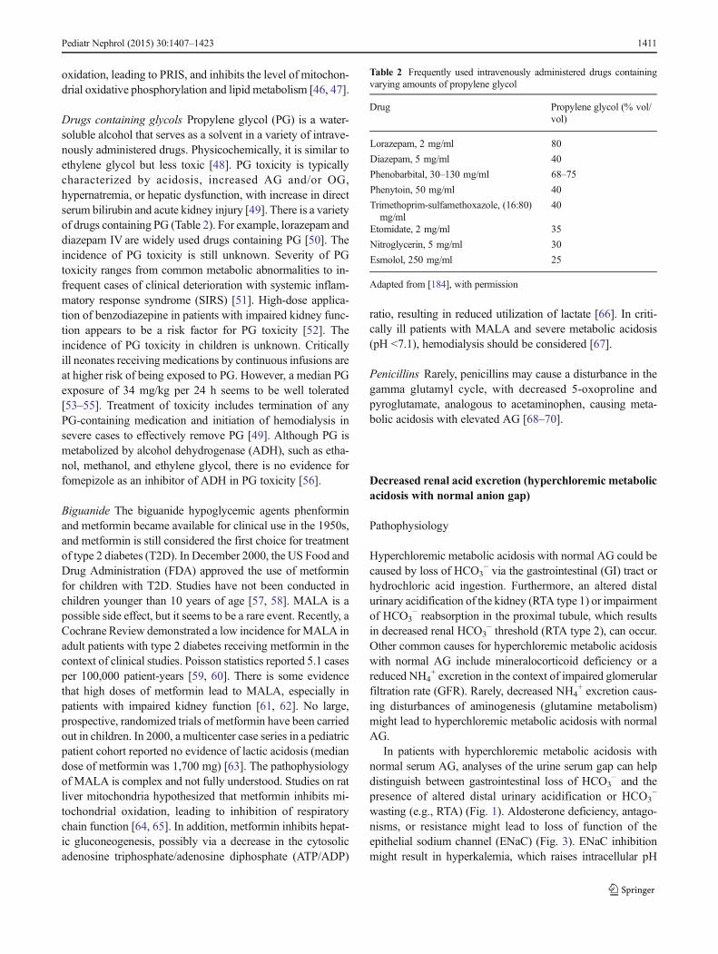
Drugs containing glycols Propylene glycol (PG) is a water-soluble alcohol that serves as a solvent in a variety of intrave-nously administered drugs. Physicochemically, it is similar toethylene glycol but less toxic [48]. PG toxicity is typicallycharacterized by acidosis, increased AG and/or OG,hypernatremia, or hepatic dysfunction, with increase in directserum bilirubin and acute kidney injury [49]. There is a varietyof drugs containing PG (Table 2). For example, lorazepam anddiazepam IV are widely used drugs containing PG [50]. Theincidence of PG toxicity is still unknown. Severity of PGtoxicity ranges from common metabolic abnormalities to in-frequent cases of clinical deterioration with systemic inflam-matory response syndrome (SIRS) [51]. High-dose applica-tion of benzodiazepine in patients with impaired kidney func-tion appears to be a risk factor for PG toxicity [52]. Theincidence of PG toxicity in children is unknown. Criticallyill neonates receiving medications by continuous infusions areat higher risk of being exposed to PG. However, a median PGexposure of 34 mg/kg per 24 h seems to be well tolerated[53–55]. Treatment of toxicity includes termination of anyPG-containing medication and initiation of hemodialysis insevere cases to effectively remove PG [49]. Although PG ismetabolized by alcohol dehydrogenase (ADH), such as etha-nol, methanol, and ethylene glycol, there is no evidence forfomepizole as an inhibitor of ADH in PG toxicity [56].
Biguanide The biguanide hypoglycemic agents phenforminand metformin became available for clinical use in the 1950s,and metformin is still considered the first choice for treatmentof type 2 diabetes (T2D). In December 2000, the US Food andDrug Administration (FDA) approved the use of metforminfor children with T2D. Studies have not been conducted inchildren younger than 10 years of age [57, 58]. MALA is apossible side effect, but it seems to be a rare event. Recently, aCochrane Review demonstrated a low incidence for MALA inadult patients with type 2 diabetes receiving metformin in thecontext of clinical studies. Poisson statistics reported 5.1 casesper 100,000 patient-years [59, 60]. There is some evidencethat high doses of metformin lead to MALA, especially inpatients with impaired kidney function [61, 62]. No large,prospective, randomized trials of metformin have been carriedout in children. In 2000, a multicenter case series in a pediatricpatient cohort reported no evidence of lactic acidosis (mediandose of metformin was 1,700 mg) [63]. The pathophysiologyof MALA is complex and not fully understood. Studies on ratliver mitochondria hypothesized that metformin inhibits mi-tochondrial oxidation, leading to inhibition of respiratorychain function [64, 65]. In addition, metformin inhibits hepat-ic gluconeogenesis, possibly via a decrease in the cytosolicadenosine triphosphate/adenosine diphosphate (ATP/ADP)
ratio, resulting in reduced utilization of lactate [66]. In criti-cally ill patients with MALA and severe metabolic acidosis(pH <7.1), hemodialysis should be considered [67].
Penicillins Rarely, penicillins may cause a disturbance in thegamma glutamyl cycle, with decreased 5-oxoproline andpyroglutamate, analogous to acetaminophen, causing meta-bolic acidosis with elevated AG [68–70].
Decreased renal acid excretion (hyperchloremic metabolicacidosis with normal anion gap)
Pathophysiology
Hyperchloremic metabolic acidosis with normal AG could becaused by loss of HCO3
− via the gastrointestinal (GI) tract orhydrochloric acid ingestion. Furthermore, an altered distalurinary acidification of the kidney (RTA type 1) or impairmentof HCO3
− reabsorption in the proximal tubule, which resultsin decreased renal HCO3
− threshold (RTA type 2), can occur.Other common causes for hyperchloremic metabolic acidosiswith normal AG include mineralocorticoid deficiency or areduced NH4
+ excretion in the context of impaired glomerularfiltration rate (GFR). Rarely, decreased NH4
+ excretion caus-ing disturbances of aminogenesis (glutamine metabolism)might lead to hyperchloremic metabolic acidosis with normalAG.
In patients with hyperchloremic metabolic acidosis withnormal serum AG, analyses of the urine serum gap can helpdistinguish between gastrointestinal loss of HCO3
− and thepresence of altered distal urinary acidification or HCO3
−
wasting (e.g., RTA) (Fig. 1). Aldosterone deficiency, antago-nisms, or resistance might lead to loss of function of theepithelial sodium channel (ENaC) (Fig. 3). ENaC inhibitionmight result in hyperkalemia, which raises intracellular pH
Table 2 Frequently used intravenously administered drugs containingvarying amounts of propylene glycol
Drug Propylene glycol (% vol/vol)
Lorazepam, 2 mg/ml 80
Diazepam, 5 mg/ml 40
Phenobarbital, 30–130 mg/ml 68–75
Phenytoin, 50 mg/ml 40
Trimethoprim-sulfamethoxazole, (16:80)mg/ml
40
Etomidate, 2 mg/ml 35
Nitroglycerin, 5 mg/ml 30
Esmolol, 250 mg/ml 25
Adapted from [184], with permission
Pediatr Nephrol (2015) 30:1407–1423 1411

and could interfere with enzymes involved in aminogenesis,leading to decreased NH4
+ excretion. Reduced NH4+ excre-
tion might lead to impaired hydeogen ion (H+) secretion, withhyperkalemic RTA (type 4 distal RTA) [71, 72].
Angiotensin-converting enzyme (ACE) and angiotensin IIreceptor antagonists (AT2RA) decrease Na+ reabsorption andcould induce hyperkalemic RTA via the aldosterone axis.Furthermore, drugs interfering with Na+- channel functioncan cause hyperkalemic RTA due to decreased distal H+
secretion.
Inhibition of the aldosterone axis (angiotensin inhibitionand heparin-induced selective aldosterone deficiency)
Aldosterone deficiency or antagonism with renal tubularacidosis type 4 (mineralocorticoid deficiency)
Renin inhibition can be triggered by simultaneous use ofcyclooxygenase inhibitors (nonsteroidal anti-inflammatorydrugs; NSAIDs), leading to hyperkalemia and metabolichyperchloremic acidosis [73, 74]. Heparin impairs aldosteronesynthesis as a result of direct toxicity to the zona glomerulosa,with inhibition of aldosterone synthase (adrenal 18-hydroxy-lase). Additionally, heparin can decrease the number andaffinity of angiotensin II receptors in the adrenal zonaglomerulosa [75, 76]. There is also an effect of low-molecular-weight heparin on K+ levels [77, 78]. ACE inhib-itors and AT2RA can cause hyperkalemia and acidosis, par-ticularly in patients with advanced renal insufficiency[79–81]. The risk of hyperkalemia is increased by simulta-neous administration of ACE inhibitors and heparin [82].
Secondary to drugs that interfere with Na+-channel function
K+-sparing diuretics and mineralocorticoid receptor antago-nists (e.g., spironolactone, eplerenone, amiloride, triamterene)can lead to moderate hyperchloremic metabolic acidosis. Thisis the result of inhibition of Na+-reabsorbing ENaC in thecollecting duct, which impairs creation of the lumen-negative voltage gradient and inhibits renal electrogenic distalH+ secretion into the tubular lumen . In addition, the impairedK+ secretion by principal cells results in an increase in serumK+ levels [83–85]. Coexisting hyperkalemia impairs renalammoniagenesis [86], tand These two effects reduce distalH+ secretion.
Figure 2 demonstrates how drug interplay affects proximaltubule cells and the thick ascending limb of the loop of Henle.In this depiction, gentamicin enters the proximal tubule cellvia the megalin–cubilin system. In the thick ascending, limbaminoglycosides can stimulate the calcium-sensing receptor(CaSR). As a consequence, the function of mitochondrial
ribosomes and conversion of ADP to ATP can be disturbedand the Na+/K+-ATPase pump is inhibited. CaSR activation inthe thick ascending limb cell could impair electrolyte transportvia inhibition of four different pathways: the kidney-specificNa+/K+/Cl− symporter (NKCC2), renal outer medullary po-tassium channel (ROMK), and Na+/K+-ATPase and/orparacellular diffusion. This results in increased urinary excre-tion of Na+, K+, magnesium (Mg2+) and Ca2+. Tetracyclinescould enter the proximal tubule cell through apical orbasolateral organic anion transporters (OATs) and inhibit theNa+/K+-ATPase pump.
Figure 3 demonstrateshow drugs that interfere withcollecting-duct principal and intercalated cells. Trimethoprimand amiloride inhibit ENaC, causing Na+ wasting, reducingkaliuresis, and causing distal RTA via a decrease in H+ excre-tion. Penicillin acts as a nonresorbable anion, leading to aluminal negative charge if aldosterone-driven Na+ reabsorp-tion is increased. The consequences are K+ secretion withhypokalemia. Demeclocycline (a tetracycline) andamphotericin B could cause nephrogenic diabetes insipidusby inhibiting vasopressin-stimulated vasopressin-2–aquapo-rin-2 (V2–AQP2) signaling. Demeclocycline enters the cellvia hOAT1 or 3. The mechanism of V2–AQP2 signalinginterruption is still unknown. Amphotericin B might lead topores in collecting duct cell membranes. This results in K+
waste via inhibition of adenylate cyclase and a back-flux of H+
into the cells, which inhibits urinary H+ excretion, resulting indistal RTA.
Trimethoprim
Similarly, trimethoprim, commonly administered in combina-tion with sulfamethoxazole as cotrimoxazole, can interferewith the ENaC (Fig. 3) [87].
Pentamidine
Luminal pentamidine directly and reversibly blocks api-cal Na+ channels in the same way as K+-sparing di-uretics, causing a decrease in the electrochemical gradi-ent for both K+ and H+ secretion in the corticalcollecting tubule. This leads to hyperchloremic metabolicacidosis with hyperkalemia [88].
Distal (type 1) renal tubular acidosis with hypokalemia
Pathophysiology
Distal RTA is characterized by impairment of H+ secretion bythe distal nephron, which results in reduced NH4
+ excretion.Primary distal RTA occurs in children, and clinical features
1412 Pediatr Nephrol (2015) 30:1407–1423

include growth impairment, polyuria, hypercalciuria,nephrocalcinosis, nephrolithiasis, and K+ depletion [89].Furthermore, distal RTA is associated with a variety ofgenetic diseases in childhood. In adults, autoimmune andrenal diseases play an important role in secondary distalRTA [90]. Additionally, distal RTA presents asnormochloremic metabolic acidosis with elevated AG. Uri-nary findings are at first glance similar to hyperkalemicRTA (type 4), but serum K+ levels and fractional urinaryK+ excretion (fK+e) are different (Fig. 1). Distal RTA isaccompanied by normal or decreased serum K+ levels andincreased fK+e in the urine (rarely, a small proportion ofpatients with hyperkalemic distal RTA present with de-creased fK+e). Remarkably, drugs might cause distal RTAin a proportion of patients.
Amphotericin B
Amphotericin B can bind to cholesterol in mammalian cellmembranes andmodify their ion permeability. This results in abroad spectrum of renal toxic effects [91]. In addition to theeffects of amphotericin B onmembrane permeability tomono-valent cations, there is a direct effect on tubular function.Amphotericin B induces RTA type 1 by increasing membranepermeability in the collecting duct. This results in back-diffusion of secreted H+ ions and K+ wasting, which leads tohypokalemia [92, 93]. Patients with significant proteinuria(>3 g/l) seem to have a reduced risk of renal tubular toxiceffects caused by amphotericin B [94], which might be relatedto a reduced concentration of free amphotericin B in tubularfluid.
Fig. 2 Interplay of drugs thataffect proximal tubule cells andthick ascending limb of the loopof Henle. Ca2+ calcium ions,CaSR calcium-sensing receptor,Cl− chloride ions, K+ potassiumions, Mg2+ magnesium ions, Na+
sodium ions, NKCC2 kidney-specific Na+//K+/Cl− symporter,ROMK renal outer medullarypotassium channel, H+ hydrogenions, hOAT organic aniontransporter, collecting-ductprincipal and intercalated cells.AC adenylate cyclase, AQP2aquaporin 2, cAMP cyclicadenosine monophosphate(AMP), ENaC epithelial sodiumchannel, hOAT organic aniontransporter, K+ potassium ions,Na+ sodium ions, V2 vasopressin2 receptor. H+ hydrogen ions.Adapted from [185], withpermission
Pediatr Nephrol (2015) 30:1407–1423 1413

RTA is a rare side effect in amphotericin B therapy [95].Polyuria and nephrogenic diabetes insipidus are more frequentfindings in patients during amphotericin B therapy [96]. Of-ten, there is a combination of RTA and nephrogenic diabetesinsipidus (Fig. 3) [97].
Foscarnet
Foscarnet is a structural mimic of the anion pyrophosphate,which selectively inhibits pyrophosphate-binding sites of var-ious viral DNA sequences and inhibits viral nucleic acidsynthesis. The drug needs no prior metabolic activation byviral or cellular enzymes. It is used in immune-compromisedpatients with life-threatening herpes simplex or cytomegalo-virus infections [98, 99]. Intensive IV hydration dramaticallyreduces the incidence of foscarnet-induced acute kidney injury[100]. Foscarnet can lead to acute tubular toxicity; rarely, a
direct effect on tubular function can be detected, which maylead to mitochondrial dysfunction, inducing mitochondrialDNA (mtDNA) in renal tubule cells [101]. This can result indistal RTA [102].
Renal loss of bicarbonate (hyperchloremic metabolicacidosis with normal anion gap)
Proximal renal tubular acidosis in the context of drug-inducedrenal Fanconi syndrome
Pathophysiology
Proximal RTA (type 2) is caused by impairment of HCO3−
reabsorption in the proximal tubule, leading to HCO3−
Fig. 3 Drugs that interfere withframeworks of collecting-ductprincipal and intercalated cells.AC adenylate cyclase, AQP2aquaporin 2, cAMP cyclicadenosine monophosphate(AMP), ENaC epithelial sodiumchannel, hOAT organic aniontransporter, K+ potassium ions,Na+ sodium ions, V2 vasopressin2 receptor. H+ hydrogen ions.Adapted from [185], withpermission
1414 Pediatr Nephrol (2015) 30:1407–1423

wasting. Patients present with hyperchloremic metabolic aci-dosis with normal serum AG. However, distal H+ secretion isstill intact, which leads to urine pH <5.5. If plasma HCO3
−
concentration is normalized by administration of alkali, urinepH is increased. Furthermore, there is a negative urine AG(≥15 mmol/l) in proximal RTA. Diagnosis of proximal RTA isconfirmed after exclusion of GI HCO3
− loss or excessive useof laxatives (Fig. 1). Proximal RTA may be primary [103,104] or accompanied by other proximal tubular defects in thecontext of Fanconi syndrome. Drug therapy often causessecondary proximal RTA and is often associated with drug-induced renal Fanconi syndrome (RFS), with generalizeddysfunction of the proximal tubule system and HCO3
− loss.Furthermore, a variety of clinical entities (e.g., vitamin Ddeficiency, hyperparathyroidism, congenital heart disease,Alport syndrome, corticoresistant nephrotic syndrome, renaltransplantation, amyloidosis, and recurrent nephrolithiasis)could cause proximal RTA. In children with metabolic acido-sis and growth impairment, it is mandatory to differentiatebetween proximal RTA and RFS, because correction of met-abolic acidosis can improve growth in proximal RTA but notin RFS [105].
Ifosfamide
Ifosfamide, a structural analog of the oxazaphosphorine cy-clophosphamide, is an alkylating agent. Since the 1980s,ifosfamide has been the standard therapy for childhood soft-tissue and bone sarcomas and is also used to treat germinaltumors. There are various renal side effects associated withifosfamide. Toxicity involves mainly the proximal—and inrare cases, the distal—renal tubules [106]. It leads toifosfamide-induced proximal tubulopathy, which results in aloss of phosphate and HCO3
− (proximal RTA) [107]. In astudy of 75 patients, the incidence of acidosis was 7 %[108]. In another study of 22 children, proximal tubular,glomerular, and distal tubular impairment (in descending or-der of frequency) was identified [109].
Proximal RTA as an isolated defect in HCO3− reabsorption
is rarely observed. It is characterized by a decrease in HCO3−
reabsorption in the proximal tubule, without any modificationin the transport of other solutes. This disturbance is character-ized by a decrease in the threshold value for HCO3
− reabsorp-tion [90, 110]. A more frequent ABD in patients receivingifosfamide is RFS [111, 112], a disorder of the proximal tubulecell associated with RTA in combination with glycosuria,aminoaciduria, phosphaturia, and normal AG [113]. ProximalRTA in the context of RFS is not rare in patients treated withifosfamide (4.6 % of patients). Risk factors include cumula-tive dose of ifosfamide and younger age [106]. The frequencyof subclinical tubular dysfunction is up to 90 % [114, 115].However, there is also a report of complete recovery aftersevere RFS [116].
There are reports that one metabolite of ifosfamide,chloroacetaldehyde (CAA), can cause renal injury byinhibiting nicotinamide adenine dinucleotide (NADH),resulting in inhibition of ubiquinone oxidoreductase, an im-portant enzyme in the oxidative phosphorylation pathway ofthe cell. Another suspected pathway in rat proximal tubules isendocytosis inhibition resulting from CAA-induced decreasein ATP levels [117].
Oxaplatin and cisplatin
Platin-induced proximal RTA has been described as bothisolated proximal RTA and as part of RFS [118–120].Cisplatin-induced RFS is caused by a direct toxic effect onthe amino acid transporter in the proximal convoluted tubule.This can be caused by an effect similar to that of aminogly-coside toxicity. Aminoglycosides reduce glucose reabsorptionin kidney tissue by reducing mRNA and protein expressionand by disrupting the sodium-dependent glucose transporter(SGLT1) function, which is located in the apical membrane ofthe proximal tubule [121]. This causes a reduction in glucosereabsorption in the kidney [122]. It has been suggested thatone possible mechanism of cisplatin-induced proximal tubuletoxicity is reduced expression of SGLTs [121, 123]. However,in patients with familial renal glucosuria (mutations inSGLT1/SGLT2 coding gene, SLC5A2), no acid-base homeo-stasis disturbances have been reported [124, 125].
Acetazolamide (carbonic anhydrase inhibitors)
Acetazolamide is used to treat idiopathic intracranial hyper-tension (pseudotumor cerebri) in children and particularly inobese women of childbearing age but also adult patients withglaucoma. It is well known that carbonic anhydrase (CA)inhibitors cause isolated proximal RTA [111]. There are twomain isoforms of CA in the kidneys: membrane-bound andcytoplasmic: CA IVand CA II, respectively [126, 127]. Bothisoforms play an important role. CA II is widely distributed inthe various compartments of the kidney and is present inalmost all cell types. The membrane-bound CA IV is mainlylocated in the proximal tubule and is absent from or poorlyexpressed in the collecting duct and the proximal tubule [127].Defects in HCO3
− reabsorption cause inhibition of CA IV inthe apical membrane of the proximal tubule cell. The inhibi-tion of CA IV leads to an isolated blocking of the membrane-bound CA IV isoform, which causes an isolated inhibition ofHCO3
− reabsorption without signs of RFS [128, 129].
Antiretrovirals
Human immune deficiency (HIV) infection represents one ofthe most serious pediatric diseases worldwide, with an esti-mated 3.3 million children younger than 15 years being
Pediatr Nephrol (2015) 30:1407–1423 1415

infected [130]. Currently available antiretroviral therapy(ART) utilizes five major classes of antiretroviral (ARV)drugs: nucleoside/nucleotide analog reverse transcriptase in-hibitors (NRTIs/NtRTIs), nonnucleoside RTIs (NNRTIs), pro-tease inhibitors (PIs), entry and fusion inhibitors, and integraseinhibitors. Combination ART, also defined as highly activeART (HAART), is composed of three ARV drugs from at leasttwo major classes in order to achieve maximal suppression ofHIV replication and preserve immune function disrupted byHIV.
Tenofovir disoproxil fumarate (TDF) is an NtRTI thathas been licensed by the FDA for use in children>2 years of age [131]; TDF can cause FS [132]. Severalmechanisms of TDF nephrotoxicity have been investi-gated: It enters proximal tubule cells through basolateralOATs and exits by using an apical transporter, themultidrug-resistance-associated protein 4 (MRP 4)[133, 134]. TDF toxicity in the proximal tubule cellcan lead to damaged and dysmorphic mitochondria, withloss of matrix cristae; in some cases, it can result ingiant mitochondria. Phosphate is reabsorbed from theglomerular filtrate across the apical proximal tubulemembrane by the Na+–phosphate cotransporter subtype2a (NaPi-IIa). Mitochondrial failure leads to energyfailure and impaired phosphate transport, leading tourinary phosphate wasting.
There are various treatments for TDF toxicity. Probenecidis increases uric acid excretion and is primarily used to treatgout and hyperuricemia. Probenecid inhibits TDF uptake intoproximal tubule cells by OATs. Rosiglitazone binds toperoxisome-proliferator-activated receptor-γ (PPAR-γ) andcauses an increase in NaPi expression. Treatment withrosiglitazone increases expression levels of NaPi-IIa and so-dium–hydrogen exchanger 3 (NHE3), acting against electro-lyte and acid-base disturbances caused by TDF [135, 136].
Valproic acid
In rare cases, long-term treatment with valproic acid can alsocause FS. There are some case reports of FS developing inpatients on long-term valproic acid therapy [137–140]. Themechanism of valproic acid toxicity is not clear. A direct toxiceffect on mitochondria of the proximal tubules has beensuggested [141, 142].
Aminoglycosides and tetracyclines
Aminoglycosides and tetracyclines can affect proximal tubu-lar function and cause fluid and electrolyte disorders. Genta-micin can interfere with specific transport mechanisms in theprimal tubular system or can induce RFS. Usually,aminoglycoside-inducedRFS is seen in patients with impairedrenal function, but some reports of patients with normal
kidney function exist [143]. Gentamicin enters the cellthrough the megalin–cubulin system [41]. Intracellular genta-micin can decrease the function of mitochondrial ribosomes[144] and interfere with the conversion of ADP to ATP [145].Consequently, gentamycin inhibits the Na+/K+-ATPase pump,which is dependent upon ATP, and inhibits NHE-3 and otherNa+-dependent solute transporters, including transporters foramino acids, uric acid, and glucose (Fig. 2) [146].
The second group of antibiotics that causes RFS are thetetracyclines, which can cause proximal tubular damage with-in 1 week after start of therapy [147]. Tetracyclines enter theproximal tubule cell via apical or basolateral OATs.Basolateral transport of tetracyclines into the tubule cell ismediated by OATs 1, 2, and 3, and reabsorption from theluminal side of the tubule cell is mediated by OAT 4 [148].Similar to the toxic mechanism of aminoglycosides, tetracy-clines can impair the function of mitochondrial ribosomes(Fig. 2) [149, 150].
Metabolic alkalosis
Pathophysiology
Metabolic alkalosis is a common but often underdiagnosedABD in hospitalized patients [90]. However, metabolic alka-losis may contribute to increased mortality in hospitalized andcritically ill patients [151–153]. Increased blood pH maydisturb the respiratory center and lead to respiratory depres-sion [154, 155]. Furthermore, metabolic alkalosis leads to ashift in the oxygen dissociation curve of hemoglobin, whichcould cause hypoxemia [156]. Additionally, alkalosis leads tovasoconstriction, and the subsequent decreased blood flowcould compound hypoxemia of peripheral tissue. Appropri-ately, it has been demonstrated in humans that alkalosis mightcause coronary vasoconstriction and angina [156, 157]. Inpatients with metabolic alkalosis, it is mandatory to distin-guish between a disturbed HCO3
− clearance caused by renalfailure and metabolic alkalosis, which is maintained by adecreased effective arterial blood volume (EABV) or in whichalkalosis is maintained by the combination of high aldosteroneand high distal Na+ delivery. The first step in analyzingmetabolic alkalosis is determining EABV. The next step in-volves analyses of urinary electrolytes (Fig. 1). In patientswith decreased EABV and decreased urine Cl−, urine Na+
should be measured to distinguish between the two mostcommon reasons for metabolic alkalosis [chloride-depletionalkalosis (CDA) under remote use of diuretics, and loss of Na+
and Cl− via the gastrointestinal tract]. Furthermore, acid losswith Cl− depletion without loss of Na+ occurs in activevomiting and nasogastric suction. In patients with decreasedEABV and increased urine Cl−, diuretic use with increased
1416 Pediatr Nephrol (2015) 30:1407–1423

natriuresis or Bartter syndrome should be considered. In pa-tients with hypercalcemia, metabolic alkalosis with or withoutrenal failure calcium–alkali syndrome must be taken intoconsideration.
Renal acid and/or chloride and potassium loss (CDA)
Loop and thiazide diuretics
High doses of loop and thiazide diuretics can lead tometabolic alkalosis, which can result in diuretic resis-tance [158]. Severe metabolic alkalosis is rare but isusually associated with high dosages of loop diuretics[159]. Three phases have been distinguished: (1) gener-ation, (2) maintenance, and (3) recovery. In 1965, Can-non et al. [160] established the concept of “contractionalkalosis” in the context of loop diuretics. They sug-gested that distal H+ excretion with renal acid loss andrapid contraction of the extracellular fluid (ECF) volumeat constant extracellular HCO3
− leads to metabolic alka-losis. It was observed that Cl− loss influences HCO3
−
reabsorption [161]. The concept of contraction alkalosisis still widely used, but the preferred term is nowchloride-depletion alkalosis (CDA). Recently, Lukeet al. showed that CDA can be corrected by selectiveCl− administration despite continued or increased nega-tive Na+ or K+ balance, continuing HCO3
− loading, andcontinuing high levels of angiotensin II or aldosterone.CDA is not corrected by Na+ or K+ administrationwithout Cl− repletion [162, 163]. CDA persisted in ratsinfused with albumin that increased ECF by 15 %[163]. CDA was corrected by Cl− infusion and persis-tent volume depletion and decreased GFR [164]. Insummary, Cl− administration without volume expansionis necessary and sufficient to correct CDA, and the termchloride depletion alkalosis should be used instead ofcontraction alkalosis. In the maintenance phase of CDA,pendrin activity is increased but HCO3
− secretion isinhibited due to the reduced Cl− delivery for anionexchange with HCO3
−. From a therapeutic point ofview, at this step, substitution of Cl− is mandatory.After Cl− substitution, HCO3
− secretion increases andmedullary HCO3
− reabsorption decreases [186]. Thisleads to bicarbonaturia, with adjustment of hypochloremicalkalosis (Fig. 4).
Penicillins
Penicillins are frequently used worldwide and have a largetherapeutic window. Hypokalemia has been reported in pa-tients receiving penicillin derivatives [165]. It has been
suspected that penicillins act as nonreabsorbable anions,which maintain a transmembrane potential gradient that isnegative on the lumen side in the cortical collecting duct,while the delivery of Cl− ions in the distal collecting duct isreduced (Fig. 3) [166]. This effect is enhanced by volumedepletion, which leads to increased aldosterone synthesis andK+ secretion. Laboratory findings in patients with hypokale-mia are a low urinary Cl− concentration and decreased K+
levels in urine, in combination with a metabolic alkalosis.Additionally, severe hypokalemia has been reported in pa-tients treated with flucloxacillin: Cl− level in urine was in-creased, suggesting that an additional direct penicillin-inducedtubule toxicity occurs [167].
Aminoglycosides
Aminoglycosides affect the tubular system in the thick as-cending limb of the loop of Henle. It has been hypothesizedthat gentamicin activates the CaSR in this area and in the distalconvoluted tubule [168–170]. Under physiological condi-tions, the increased serum Ca2+ levels stimulate this receptorand decrease the active reabsorption of Na+. This reduces theluminal positive driving force and results in increased urinaryCa excretion [171]. Activation of the CaSR can disrupt elec-trolyte transport via inhibition of four different pathways: (1)renal channel proteins, such as the Na-K-2Cl cotransporter(NKCC2), (2) ROMK, (3) Na+/K+-ATPase, and (4)paracellular diffusion [146]. Inhibition of these transportmechanisms leads to increased urinary excretion of Na+, K+,Mg2+, and Ca2+ (Fig. 2). This results in hypokalemic meta-bolic alkalosis with hypermagnesuria and hypercalciuria and anormal serum creatinine (Bartter-like syndrome) [172, 173].Furthermore, gentamicin might cause Mg2+ wasting; animalexperiments have shown a causal relationship between genta-micin administration and Mg2+ wasting [174]. In healthypeople, gentamicin causes immediate and transient renal cal-cium (Ca2+) and Mg2+ wasting (Fig. 2) [175].
Extrarenal acid and/or chloride loss (chloride-depletionmetabolic alkalosis)
Laxative abuse in adults (e.g., bisacodyl, sodium picosulfate,saline laxatives, lactulose, and cisapride) results in electrolytedisturbances (mainly hypokalemia), metabolic alkalosis, andrenal tubular dysfunction [176]. Laxative abuse occurs inchildren with, for example, Munchausen's syndrome, byproxy and in adolescents with eating disorders [177]. There-fore, this should be especially considered in patients withunexplainable chloride-depletion metabolic alkalosis (CDA).
Pediatr Nephrol (2015) 30:1407–1423 1417

Alkali administration (non-chloride-depletion metabolicalkalosis)
Milk–alkali or calcium–alkali syndrome
Milk–alkali syndrome was a very common cause of hypercal-cemia, metabolic alkalosis, and renal failure at the beginningof the last century. The syndrome was associated with theingestion of large quantities of milk and absorbable alkali totreat peptic ulcer disease [178]. Nowadays, this syndrome iscalled calcium–alkali syndrome (CAS) [179]. The incidenceof CAS in children is not clear. Vitamin D supplementation toprevent vitamin-D-deficient rickets in children is generallyrecommended [180], but there are some reports of children
with hypercalcemia after treatment with vitamin D [181]. Theclassic symptoms consist of hypercalcemia, various degrees ofrenal failure, and metabolic alkalosis [182]. There are severalrisk factors for development of CAS: The use of thiazidediuretics predisposes to CAS by increasing Ca2+ reabsorptionin combination with volume depletion and metabolic alkalo-sis. ACE inhibitors and NSAIDs increase the risk of CAS byreducing GFR, leading to decreased Ca2+ excretion [183]. Theincreased serum Ca2+ levels (via increased intestinal supply orelevated vitamin D levels) lead to activation of the CaSR inthe thick ascending limb of Henle and the medullarycollecting duct and to parathyroid hormone suppression. Con-comitant metabolic alkalosis also increases tubular reabsorp-tion of Ca2+.
Fig. 4 Different phases ofchloride-depletion alkalosis(CDA). K+, potassium ions; Na+,sodium ions; Cl−, chloride ions;H+, hydrogen ions; HCO3
−,bicarbonate; ENaC, epithelialsodium channel; Pn, pendrin(sodium-independent chloride/iodide transporter); AEI, anionexchanger isoform 1. Adaptedfrom [186], with permission
1418 Pediatr Nephrol (2015) 30:1407–1423

Key summary points
– Drug- nduced ABDs are frequently present in hospital-ized patients.
– Analysis of ABDs should be performed systematically ina standardized manner.
– Drug-induced ABDs can be classified into five differentcategories in terms of pathophysiology:
– (1) Metabolic acidosis caused by acid overload– (2) Base loss– (3) Alkalosis resulting from acid loss– (4) Exogenous HCO3
− loads– (5) Respiratory acidosis or alkalosis resulting from drug-
induced depression of the respiratory center or neuromus-cular impairment
– Chloride administration without volume expansion isnecessary and sufficient to correct CDA, and the termchloride-depletion alkalosis should be used instead ofcontraction alkalosis.
Multiple-choice questions (answers are providedfollowing the reference list)
Q1: A newborn developed diarrhea at day 10 after birth,and on the subsequent day, lethargy and refusal to eat. Rapid-ly, he developed diffuse cyanosis, hypotonia, and areflexia.After symptom onset, he was immediately transferred to theintensive care unit. First blood gas analyses showed: pH 6.9,pCO2 29.8 mmHg, pO2 90.1 mmHg (under oxygen therapy),HCO3
− 3 mEq/l, base excess 22, lactic acid 17 mmol/l (nor-mal <2 mmol/l). What kind of disturbance is present?
(a) Metabolic acidosis(b) Respiratory alkalosis(c) Metabolic alkalosis(d) Respiratory acidosis
Q2: A 14-year-old boy receives chronic furosemide treat-ment. The dose of furosemide was accidentally increased for10 days. His medical history was notable for the diagnosis ofsevere heart failure due to myocarditis at the age of 12 years.What acid-base disorder would you expect?
(a) Hyperchloremic metabolic acidosis(b) Hypochloremic metabolic alkalosis(c) Normochloremic respiratory alkalosis(d) Hypochloremic metabolic acidosis
Q3: How would you calculate the AG, and in which caseswould you expect to have an increased AG?
(a) ([Na+]+[K+])+([Cl−]+[HCO3−]): diabetic ketoacidosis
(b) ([Na+]+[K+])−([Cl−] - [HCO3−]): uremia
(c) ([Na+]+[K+])−([Cl−]+[HCO3−]): diabetic ketoacidosis
(d) ([Na+]+[K+])+([Cl−]+[HCO3−]): uremia
Q4: What acid-base disorder would you expected in pa-tients with propofol infusion syndrome?
(a) Metabolic alkalosis with normal lactate levels(b) Metabolic acidosis with elevated lactate levels(c) Respiratory acidosis with normal lactate levels(d) Respiratory alkalosis with normal lactate levels
Q5: Aminoglycosides might cause which of the following?
(a) Hyperkalemic metabolic acidosis(b) Hypokalemic respiratory alkalosis(c) Hyperkalemic metabolic alkalosis(d) Hypokalemic metabolic alkalosis
Acknowledgments MS was supported by the Robert-Bosch Founda-tion, Stuttgart, Germany
References
1. Kraut JA, Madias NE (2007) Serum anion gap: its uses and limita-tions in clinical medicine. Clin J Am Soc Nephrol 2:162–174
2. Palmer BF, Alpern RJ (1997) Metabolic alkalosis. J Am SocNephrol 8:1462–1469
3. Ishihara K, Szerlip HM (1998) Anion gap acidosis. Semin Nephrol18:83–97
4. Gluck SL (1998) Acid-base. Lancet 352:474–4795. Adrogue HJ (2006) Metabolic acidosis: pathophysiology, diagnosis
and management. J Nephrol Nurs 19(Suppl 9):S62–S696. Lorenz JM, Kleinman LI, Markarian K, Oliver M, Fernandez J
(1999) Serum anion gap in the differential diagnosis of metabolicacidosis in critically ill newborns. J Pediatr 135:751–755
7. Winter SD, Pearson JR, Gabow PA, Schultz AL, Lepoff RB (1990)The fall of the serum anion gap. Arch Intern Med 150:311–313
8. Singh RN, Singh NC, Hutchison J, Moses GC (1994) Lower aniongap increases sensitivity in predicting elevated lactate. ClinIntensive Care 5:221–224
9. Sadjadi SA, Manalo R, Jaipaul N, McMillan J (2013) Ion-selectiveelectrode and anion gap range: what should the anion gap be? Int JNephrol Renov Dis 6:101–105
10. Hatherill M, Waggie Z, Purves L, Reynolds L, Argent A (2002)Correction of the anion gap for albumin in order to detect occulttissue anions in shock. Arch Dis Child 87:526–529
11. Feldman M, Soni N, Dickson B (2005) Influence of hypoalbumin-emia or hyperalbuminemia on the serum anion gap. J Lab Clin Med146:317–320
12. Figge J, Jabor A, Kazda A, Fencl V (1998) Anion gap and hypoal-buminemia. Crit Care Med 26:1807–1810
13. Kruse JA, Cadnapaphornchai P (1994) The serum osmole gap. JCrit Care 9:185–197
14. McQuillen KK, Anderson AC (1999) Osmol gaps in the pediatricpopulation. Acad Emerg Med 6:27–30
Pediatr Nephrol (2015) 30:1407–1423 1419

15. Wrenn K (1990) The delta (delta) gap: an approach to mixed acid-base disorders. Ann Emerg Med 19:1310–1313
16. Rastegar A (2007) Use of the DeltaAG/DeltaHCO3- ratio in thediagnosis of mixed acid-base disorders. J Am Soc Nephrol 18:2429–2431
17. Adrogue HJ, Wilson H, Boyd AE 3rd, Suki WN, Eknoyan G(1982) Plasma acid–base patterns in diabetic ketoacidosis. N EnglJ Med 307:1603–1610
18. Goldstein MB, Bear R, Richardson RM,Marsden PA, Halperin ML(1986) The urine anion gap: a clinically useful index of ammoniumexcretion. Am J Med Sci 292:198–202
19. Kirschbaum B, Sica D, Anderson FP (1999) Urine electrolytes andthe urine anion and osmolar gaps. J Lab Clin Med 133:597–604
20. Batlle DC, Hizon M, Cohen E, Gutterman C, Gupta R (1988) Theuse of the urinary anion gap in the diagnosis of hyperchloremicmetabolic acidosis. N Engl J Med 318:594–599
21. Gonzalez E, Gutierrez E, GaleanoC, Chevia C, de Sequera P, BernisC, Parra EG, Delgado R, SanzM, Ortiz M, GoicoecheaM, QueredaC, Olea T, Bouarich H, Hernandez Y, Segovia B, Praga M (2008)Early steroid treatment improves the recovery of renal function inpatients with drug-induced acute interstitial nephritis. Kidney Int 73:940–946
22. Rougier F, Claude D, Maurin M, Maire P (2004) Aminoglycosidenephrotoxicity. Curr Drug Targets CNS Neurol Disord 4:153–162
23. Chiappini E, Conti C, Galli L, de Martino M (2010) Clinicalefficacy and tolerability of linezolid in pediatric patients: a system-atic review. Clin Ther 32:66–88
24. Dotis J, Iosifidis E, IoannidouM, Roilides E (2010) Use of linezolidin pediatrics: a critical review. Int J Infect Dis 14:e638–e648
25. Garcia-Prats AJ, Rose PC, Hesseling AC, Schaaf HS (2014)Linezolid for the treatment of drug-resistant tuberculosis in chil-dren: a review and recommendations. Tuberculosis (Edinb) 94:93–104
26. Saiman L, Goldfarb J, Kaplan SA, Wible K, Edge-Padbury B,Naberhuis-Stehouwer S, Bruss JB (2003) Safety and tolerabilityof linezolid in children. Pediatr Infect Dis J 22:S193–S200
27. Apodaca AA, Rakita RM (2003) Linezolid-induced lactic acidosis.N Engl J Med 348:86–87
28. Su E, Crowley K, Carcillo JA, Michaels MG (2011) Linezolid andlactic acidosis: a role for lactate monitoring with long-term linezoliduse in children. Pediatr Infect Dis J 30:804–806
29. Scotton P, Fuser R, Torresan S, Carniato A, Giobbia M, Rossi C,Inojosa WO, Vaglia A (2008) Early linezolid-associated lactic aci-dosis in a patient treated for tuberculous spondylodiscitis. Infection36:387–388
30. De Vriese AS, Coster RV, Smet J, Seneca S, Lovering A, Van HauteLL, Vanopdenbosch LJ, Martin JJ, Groote CC, Vandecasteele S,Boelaert JR (2006) Linezolid-induced inhibition of mitochondrialprotein synthesis. Clin Infect Dis 42:1111–1117
31. Garrabou G, Soriano A, Lopez S, Guallar JP, Giralt M, Villarroya F,Martinez JA, Casademont J, Cardellach F, Mensa J, Miro O (2007)Reversible inhibition of mitochondrial protein synthesis duringlinezolid-related hyperlactatemia. Antimicrob Agents Chemother51:962–967
32. Soriano A, Miro O, Mensa J (2005) Mitochondrial toxicity associ-ated with linezolid. N Engl J Med 353:2305–2306
33. Palenzuela L, Hahn NM, Nelson RP Jr, Arno JN, Schobert C,Bethel R, Ostrowski LA, Sharma MR, Datta PP, Agrawal RK,Schwartz JE, Hirano M (2005) Does linezolid cause lactic acidosisby inhibiting mitochondrial protein synthesis? Clin Infect Dis 40:e113–e116
34. Carson J, Cerda J, Chae JH, Hirano M, Maggiore P (2007) Severelactic acidosis associated with linezolid use in a patient with themitochondrial DNA A2706G polymorphism. Pharmacotherapy 27:771–774
35. Boutoille D, Grossi O, Depatureaux A, Tattevin P (2009)Fatal lactic acidosis after prolonged linezolid exposure fortreatment of multidrug-resistant tuberculosis. Eur J IntMed 20:e134–e135
36. McKeage K, Perry CM (2003) Propofol: a review of its use inintensive care sedation of adults. CNS Drugs 17:235–272
37. Rigby-Jones AE, Sneyd JR (2011) Propofol and children–what weknow and what we do not know. Paediatr Anaesth 21:247–254
38. Diedrich DA, Brown DR (2011) Analytic reviews: propofol infu-sion syndrome in the ICU. J Intensive Care Med 26:59–72
39. Bonhomme V, Demoitie J, Schaub I, Hans P (2009) Acid–basestatus and hemodynamic stability during propofol andsevoflurane-based anesthesia in patients undergoing uncomplicatedintracranial surgery. J Neurosurg Anesthesiol 21:112–119
40. Roberts RJ, Barletta JF, Fong JJ, Schumaker G, Kuper PJ,Papadopoulos S, Yogaratnam D, Kendall E, Xamplas R, GerlachAT, Szumita PM, Anger KE, Arpino PA, Voils SA, Grgurich P,Ruthazer R, Devlin JW (2009) Incidence of propofol-related infu-sion syndrome in critically ill adults: a prospective, multicenterstudy. Crit Care 13:R169
41. Koriyama H, Duff JP, Guerra GG, Chan AW (2014) Is propofol afriend or a foe of the pediatric intensivist? Description of propofoluse in a PICU*. Pediatr Crit Care Med 15:e66–e71
42. Kruessell MA, Udink ten Cate FE, Kraus AJ, Roth B, TrieschmannU (2012) Use of propofol in pediatric intensive care units: a nationalsurvey in Germany. Pediatr Crit Care Med 13:e150–e154
43. Hatch DJ (1999) Propofol-infusion syndrome in children. Lancet353:1117–1118
44. Kam PC, Cardone D (2007) Propofol infusion syndrome.Anaesthesia 62:690–701
45. Koch M, De Backer D, Vincent JL (2004) Lactic acidosis: an earlymarker of propofol infusion syndrome? Intensive Care Med 30:522
46. Wolf A,Weir P, Segar P, Stone J, Shield J (2001) Impaired fatty acidoxidation in propofol infusion syndrome. Lancet 357:606–607
47. Withington DE, Decell MK, Al Ayed T (2004) A case of propofoltoxicity: further evidence for a causal mechanism. Paediatr Anaesth14:505–508
48. Kraut JA, Kurtz I (2008) Toxic alcohol ingestions: clinical features,diagnosis, and management. Clin J Am Soc Nephrol 3:208–225
49. Zar T, Graeber C, Perazella MA (2007) Recognition, treatment, andprevention of propylene glycol toxicity. Semin Dial 20:217–219
50. Tayar J, Jabbour G, Saggi SJ (2002) Severe hyperosmolar metabolicacidosis due to a large dose of intravenous lorazepam. N Engl JMed346:1253–1254
51. Wilson KC, Reardon C, Theodore AC, Farber HW (2005)Propylene glycol toxicity: a severe iatrogenic illness in ICU patientsreceiving IV benzodiazepines: a case series and prospective, obser-vational pilot study. Chest 128:1674–1681
52. Yahwak JA, Riker RR, Fraser GL, Subak-Sharpe S (2003) Anobservational pilot study of osmolar gap monitoring to detect pro-pylene glycol toxicity in adult icu patients receiving high-doselorazepam. CHEST Journal 124:178S-a
53. Shehab N, Lewis CL, Streetman DD, Donn SM (2009) Exposure tothe pharmaceutical excipients benzyl alcohol and propylene glycolamong critically ill neonates. Pediatr Crit Care Med 10:256–259
54. Kulo A, de Hoon JN, Allegaert K (2012) The propylene glycolresearch project to illustrate the feasibility and difficulties to studytoxicokinetics in neonates. Int J Pharm 435:112–114
55. Allegaert K, Vanhaesebrouck S, Kulo A, Cosaert K, Verbesselt R,Debeer A, de Hoon J (2010) Prospective assessment of short-termpropylene glycol tolerance in neonates. Arch Dis Child 95:1054–1058
56. Mullins ME, Barnes BJ (2002) Hyperosmolar metabolic acidosisand intravenous Lorazepam. N Engl J Med 347:857–858, authorreply 857–858
1420 Pediatr Nephrol (2015) 30:1407–1423

57. Jones KL, Arslanian S, Peterokova VA, Park JS, Tomlinson MJ(2002) Effect ofmetformin in pediatric patients with type 2 diabetes:a randomized controlled trial. Diabetes Care 25:89–94
58. Gaylor AS, Condren ME (2004) Type 2 diabetes mellitus in thepediatric population. Pharmacotherapy 24:871–878
59. Kirpichnikov D, McFarlane SI, Sowers JR (2002) Metformin: anupdate. Ann Intern Med 137:25–33
60. Salpeter SR, Greyber E, Pasternak GA, Salpeter EE (2010) Risk offatal and nonfatal lactic acidosis with metformin use in type 2diabetes mellitus. Cochrane Database Syst Rev CD002967
61. Brown JB, Pedula K, Barzilay J, Herson MK, Latare P (1998) Lacticacidosis rates in type 2 diabetes. Diabetes Care 21:1659–1663
62. Misbin RI, Green L, Stadel BV, Gueriguian JL, Gubbi A, FlemingGA (1998) Lactic acidosis in patients with diabetes treated withmetformin. N Engl J Med 338:265–266
63. Spiller HA, Weber JA, Winter ML, Klein-Schwartz W, Hofman M,Gorman SE, Stork CM, Krenzelok EP (2000) Multicenter caseseries of pediatric metformin ingestion. Ann Pharmacother 34:1385–1388
64. OwenMR, Doran E, Halestrap AP (2000) Evidence that metforminexerts its anti-diabetic effects through inhibition of complex 1 of themitochondrial respiratory chain. Biochem J 348(Pt 3):607–614
65. El-MirMY, Nogueira V, Fontaine E, Averet N, Rigoulet M, LeverveX (2000) Dimethylbiguanide inhibits cell respiration via an indirecteffect targeted on the respiratory chain complex I. J Biol Chem 275:223–228
66. Schafer G, Bojanowski D (1972) Interaction of biguanides withmitochondrial and synthetic membranes. Eur J Biochem 27:364–375
67. Seidowsky A, Nseir S, Houdret N, Fourrier F (2009) Metformin-associated lactic acidosis: a prognostic and therapeutic study. CritCare Med 37:2191–2196
68. Fenves AZ, Kirkpatrick HM 3rd, Patel VV, Sweetman L, EmmettM (2006) Increased anion gap metabolic acidosis as a result of 5-oxoproline (pyroglutamic acid): a role for acetaminophen. Clin JAm Soc Nephrol 1:441–447
69. Peter JV, Rogers N,Murty S, Gerace R,Mackay R, Peake SL (2006)An unusual cause of severe metabolic acidosis. Med J Aust 185:223–225
70. Rolleman EJ, Hoorn EJ, Didden P, Zietse R (2008) Guilty ascharged: unmeasured urinary anions in a case of pyroglutamicacidosis. Neth J Med 66:351–353
71. DuBose TD Jr (1997) Hyperkalemic hyperchloremic metabolicacidosis: pathophysiologic insights. Kidney Int 51:591–602
72. Karet FE (2009) Mechanisms in hyperkalemic renal tubular acido-sis. J Am Soc Nephrol 20:251–254
73. Wang JL, Cheng HF, Harris RC (1999) Cyclooxygenase-2 inhibi-tion decreases renin content and lowers blood pressure in a model ofrenovascular hypertension. Hypertension 34:96–101
74. Harris RC, Breyer MD (2006) Update on cyclooxygenase-2 inhib-itors. Clin J Am Soc Nephrol 1:236–245
75. O'Kelly R, Magee F, McKenna TJ (1983) Routine heparin therapyinhibits adrenal aldosterone production. J Clin Endocrinol Metab56:108–112
76. Sherman RA, Ruddy MC (1986) Suppression of aldosterone pro-duction by low-dose heparin. Am J Nephrol 6:165–168
77. Canova CR, Fischler MP, Reinhart WH (1997) Effect of low-molecular-weight heparin on serum potassium. Lancet 349:1447–1448
78. Wiggam MI, Beringer TR (1997) Effect of low-molecular-weight heparin on serum concentrations of potassium. Lancet350:292–293
79. Henger A, Tutt P, Riesen WF, Hulter HN, Krapf R (2000) Acid-baseand endocrine effects of aldosterone and angiotensin II inhibition inmetabolic acidosis in human patients. J Lab Clin Med 136:379–389
80. Gupta P, Franco-Saenz R, Mulrow PJ (1995) Locally generatedangiotensin II in the adrenal gland regulates basal, corticotropin-,and potassium-stimulated aldosterone secretion. Hypertension 25:443–448
81. Sakemi T, Ohchi N, Sanai T, Rikitake O,Maeda T (1988) Captopril-induced metabolic acidosis with hyperkalemia. Am J Nephrol 8:245–248
82. Durand D, Ader JL, Rey JP, Tran-Van T, Lloveras JJ, Bernadet P,Suc JM (1988) Inducing hyperkalemia by converting enzyme in-hibitors and heparin. Kidney Int Suppl 25:S196–S197
83. Palmer BF (2011) Metabolic complications associated with use ofdiuretics. Semin Nephrol 31:542–552
84. DuBose TD Jr, Good DW (1988) Effects of diuretics on renal acid–base transport. Semin Nephrol 8:282–294
85. Wagner CA, Geibel JP (2002) Acid–base transport in the collectingduct. J Nephrol 15(Suppl 5):S112–S127
86. Sastrasinh S, Tannen RL (1983) Effect of potassium on renal NH3production. Am J Physiol 244:F383–F391
87. Muto S, Tsuruoka S, Miyata Y, Fujimura A, Kusano E (2006) Effectof trimethoprim-sulfamethoxazole on Na and K+transport proper-ties in the rabbit cortical collecting duct perfused in vitro. NephronPhysiol 102:51–60
88. Kleyman TR, Roberts C, Ling BN (1995) A mechanism for pent-amidine-induced hyperkalemia: inhibition of distal nephron sodiumtransport. Ann Intern Med 122:103–106
89. Caldas A, Broyer M, Dechaux M, Kleinknecht C (1992) Primarydistal tubular acidosis in childhood: clinical study and long-termfollow-up of 28 patients. J Pediatr 121:233–241
90. Rodriguez Soriano J (2002) Renal tubular acidosis: the clinicalentity. J Am Soc Nephrol 13:2160–2170
91. Sawaya BP, Briggs JP, Schnermann J (1995) Amphotericin B neph-rotoxicity: the adverse consequences of altered membrane proper-ties. J Am Soc Nephrol 6:154–164
92. Douglas JB, Healy JK (1969) Nephrotoxic effects of amphotericinB, including renal tubular acidosis. Am J Med 46:154–162
93. Burges JL, Birchall R (1972) Nephrotoxicity of amphotericin B,with emphasis on changes in tubular function. Am J Med 53:77–84
94. Steinmetz PR, Lawson LR (1970) Defect in urinary acidificationinduced in vitro by amphotericin B. J Clin Invest 49:596–601
95. Stamm AM, Diasio RB, Dismukes WE, Shadomy S, Cloud GA,Bowles CA, Karam GH, Espinel-Ingroff A (1987) Toxicity ofamphotericin B plus flucytosine in 194 patients with cryptococcalmeningitis. Am J Med 83:236–242
96. Barbour GL, Straub KD, O'Neal BL, Leatherman JW (1979)Vasopressin-resistant nephrogenic diabetes insipidus. A result ofamphotericin B therapy. Arch Intern Med 139:86–88
97. Hoorn EJ, Zietse R (2007) Combined renal tubular acidosis anddiabetes insipidus in hematological disease. Nat Clin Pract Nephrol3:171–175
98. Marshall BC, Koch WC (2009) Antivirals for cytomegalovirusinfection in neonates and infants: focus on pharmacokinetics,formulations, dosing, and adverse events. Paediatr Drugs 11:309–321
99. Hatchette T, Tipples GA, Peters G, Alsuwaidi A, Zhou J, MailmanTL (2008) Foscarnet salvage therapy for acyclovir-resistant varicel-la zoster: report of a novel thymidine kinase mutation and review ofthe literature. Pediatr Infect Dis J 27:75–77
100. Deray G, Martinez F, Katlama C, Levaltier B, Beaufils H, Danis M,RozenheimM, BaumelouA, Dohin E, Gentilini M, Jacobs C (1989)Foscarnet nephrotoxicity: mechanism, incidence and prevention.Am J Nephrol 9:316–321
101. Izzedine H, Launay-Vacher V, Deray G (2005) Antiviral drug-induced nephrotoxicity. Am J Kidney Dis 45:804–817
102. Navarro JF, Quereda C, Quereda C, Gallego N, Antela A, Mora C,Ortuno J (1996) Nephrogenic diabetes insipidus and renal tubular
Pediatr Nephrol (2015) 30:1407–1423 1421

acidosis secondary to foscarnet therapy. Am J Kidney Dis 27:431–434
103. Igarashi T, Ishii T, Watanabe K, Hayakawa H, Horio K, Sone Y,Ohga K (1994) Persistent isolated proximal renal tubular acidosis–asystemic disease with a distinct clinical entity. Pediatr Nephrol 8:70–71
104. Nash MA, Torrado AD, Greifer I, Spitzer A, Edelmann CM Jr(1972) Renal tubular acidosis in infants and children. Clinicalcourse, response to treatment, and prognosis. J Pediatr 80:738–748
105. Hsu SY, Tsai IJ, Tsau YK (2005) Comparison of growth in primaryFanconi syndrome and proximal renal tubular acidosis. PediatrNephrol 20:460–464
106. Stohr W, Paulides M, Bielack S, Jurgens H, Treuner J, Rossi R,Langer T, Beck JD (2007) Ifosfamide-induced nephrotoxicity in593 sarcoma patients: a report from the Late Effects SurveillanceSystem. Pediatr Blood Cancer 48:447–452
107. Pratt CB, Meyer WH, Jenkins JJ, Avery L, McKay CP, Wyatt RJ,Hancock ML (1991) Ifosfamide, Fanconi's syndrome, and rickets. JClin Oncol 9:1495–1499
108. Rossi R, Pleyer J, Schafers P, Kuhn N, Kleta R, Deufel T, Jurgens H(1999) Development of ifosfamide-induced nephrotoxicity: pro-spective follow-up in 75 patients. Med Pediatr Oncol 32:177–182
109. Skinner R, Pearson AD, English MW, Price L, Wyllie RA,Coulthard MG, Craft AW (1996) Risk factors for ifosfamide neph-rotoxicity in children. Lancet 348:578–580
110. Rodriguez Soriano J, Boichis H, Stark H, Edelmann CM Jr (1967)Proximal renal tubular acidosis. A defect in bicarbonate reabsorp-tion with normal urinary acidification. Pediatr Res 1:81–98
111. Izzedine H, Launay-Vacher V, Isnard-Bagnis C, Deray G (2003)Drug-induced Fanconi's syndrome. Am J Kidney Dis 41:292–309
112. Hall AM, Bass P, Unwin RJ (2014) Drug-induced renal Fanconisyndrome. QJM 107:261–269
113. Haque SK, Ariceta G, Batlle D (2012) Proximal renal tubularacidosis: a not so rare disorder of multiple etiologies. Nephrol DialTransplant 27:4273–4287
114. Stevens MC, Brandis M (1993) Incidence and etiology ofifosfamide nephrotoxicity: report of a meeting held in Rhodes,Greece, October 3, 1991, sponsored by Asta Medica, Frankfurt,Germany. Med Pediatr Oncol 21:640–644
115. Skinner R, Cotterill SJ, Stevens MC (2000) Risk factors for neph-rotoxicity after ifosfamide treatment in children: a UKCCSG LateEffects Group study. United Kingdom Children's Cancer StudyGroup. Br J Cancer 82:1636–1645
116. Ashraf MS, Skinner R, English MW, Craft AW, Pearson AD (1997)Late reversibility of chronic ifosfamide-associated nephrotoxicity ina child. Med Pediatr Oncol 28:62–64
117. Yaseen Z, Michoudet C, Baverel G, Dubourg L (2008) Mechanismsof the ifosfamide-induced inhibition of endocytosis in the rat prox-imal kidney tubule. Arch Toxicol 82:607–614
118. Cachat F, Nenadov-Beck M, Guignard JP (1998) Occurrence of anacute Fanconi syndrome following cisplatin chemotherapy. MedPediatr Oncol 31:40–41
119. Negro A, Grasselli C, Galli P (2010) Oxaliplatin-induced proximalrenal tubular acidosis. Intern Emerg Med 5:267–268
120. Linch M, Cunningham D, Mingo O, Stiles A, Farquhar-Smith WP(2007) Renal tubular acidosis due to oxaliplatin. Ann Oncol 18:805–806
121. Portilla D, Li S, NagothuKK,Megyesi J, KaisslingB, SchnackenbergL, Safirstein RL, Beger RD (2006) Metabolomic study of cisplatin-induced nephrotoxicity. Kidney Int 69:2194–2204
122. Takamoto K, Kawada M, Usui T, Ishizuka M, Ikeda D (2003)Aminoglycoside antibiotics reduce glucose reabsorption in kidneythrough down-regulation of SGLT1. Biochem Biophys ResCommun 308:866–871
123. Xu EY, Perlina A, Vu H, Troth SP, Brennan RJ, Aslamkhan AG, XuQ (2008) Integrated pathway analysis of rat urine metabolic profiles
and kidney transcriptomic profiles to elucidate the systems toxicol-ogy of model nephrotoxicants. Chem Res Toxicol 21:1548–1561
124. Scholl-Burgi S, Santer R, Ehrich JH (2004) Long-term outcome ofrenal glucosuria type 0: the original patient and his natural history.Nephrol Dial Transplant 19:2394–2396
125. Santer R, Calado J (2010) Familial renal glucosuria and SGLT2:from a mendelian trait to a therapeutic target. Clin J Am SocNephrol 5:133–141
126. Karlmark B, Agerup B, Wistrand PJ (1979) Renal proximal tubularacidification. Role of brush-border and cytoplasmic carbonicanhydrase. Acta Physiol Scand 106:145–150
127. Schwartz GJ (2002) Physiology and molecular biology of renalcarbonic anhydrase. J Nephrol 15(Suppl 5):S61–S74
128. Winum JY, Poulsen SA, Supuran CT (2009) Therapeutic applica-tions of glycosidic carbonic anhydrase inhibitors. Med Res Rev 29:419–435
129. Tsuruoka S, Schwartz GJ (1998) HCO3- absorption in rabbit outermedullary collecting duct: role of luminal carbonic anhydrase. Am JPhysiol 274:F139–F147
130. World Health Organization (2012) Global epidemic and health careresponse. In: Global summary of the HIV/AIDS epidemic
131. World Health Organization (2012) Technical update on treatmentoptimization: use of tenofovir disoproxil fumarate in HIV-infectedchildren and adolescents aged 2–18 years: a public health perspec-tive. WHO HIV/AIDS Programme
132. Gupta SK (2008) Tenofovir-associated Fanconi syndrome: reviewof the FDA adverse event reporting system. AIDS Patient CareSTDS 22:99–103
133. Hall AM, Hendry BM, Nitsch D, Connolly JO (2011) Tenofovir-associated kidney toxicity in HIV-infected patients: a review of theevidence. Am J Kidney Dis 57:773–780
134. Imaoka T, Kusuhara H, Adachi M, Schuetz JD, Takeuchi K,Sugiyama Y (2007) Functional involvement of multidrug resis-tance-associated protein 4 (MRP4/ABCC4) in the renal eliminationof the antiviral drugs adefovir and tenofovir. Mol Pharmacol 71:619–627
135. Hall AM (2013) Update on tenofovir toxicity in the kidney. PediatrNephrol 28:1011–1023
136. Liborio AB, Andrade L, Pereira LV, Sanches TR, Shimizu MH,Seguro AC (2008) Rosiglitazone reverses tenofovir-induced neph-rotoxicity. Kidney Int 74:910–918
137. Endo A, Fujita Y, Fuchigami T, Takahashi S, Mugishima H (2010)Fanconi syndrome caused by valproic acid. Pediatr Neurol 42:287–290
138. Watanabe T, Yoshikawa H, Yamazaki S, Abe Y, Abe T (2005)Secondary renal Fanconi syndrome caused by valproate therapy.Pediatr Nephrol 20:814–817
139. Yoshikawa H,Watanabe T, Abe T (2002) Fanconi syndrome causedby sodium valproate: report of three severely disabled children. EurJ Paediatr Neurol 6:165–167
140. Knorr M, Schaper J, Harjes M, Mayatepek E, Rosenbaum T (2004)Fanconi syndrome caused by antiepileptic therapy with valproicAcid. Epilepsia 45:868–871
141. Hawkins E, Brewer E (1993) Renal toxicity induced by valproicacid (Depakene). Pediatr Pathol 13:863–868
142. Lenoir GR, Perignon JL, Gubler MC, Broyer M (1981) Valproicacid: a possible cause of proximal tubular renal syndrome. J Pediatr98:503–504
143. Alexandridis G, Liberopoulos E, Elisaf M (2003) Aminoglycoside-induced reversible tubular dysfunction. Pharmacology 67:118–120
144. Bennett WM, Mela-Riker LM, Houghton DC, Gilbert DN, BussWC (1988) Microsomal protein synthesis inhibition: an early man-ifestation of gentamicin nephrotoxicity. Am J Physiol 255:F265–F269
145. Weinberg JM, Harding PG, Humes HD (1980) Mechanisms ofgentamicin-induced dysfunction of renal cortical mitochondria. II.
1422 Pediatr Nephrol (2015) 30:1407–1423

Effects on mitochondrial monovalent cation transport. ArchBiochem Biophys 205:232–239
146. Sassen MC, Kim SW, Kwon TH, Knepper MA, Miller RT, FrokiaerJ, Nielsen S (2006) Dysregulation of renal sodium transporters ingentamicin-treated rats. Kidney Int 70:1026–1037
147. Hemstreet BA (2004) Antimicrobial-associated renal tubular acido-sis. Ann Pharmacother 38:1031–1038
148. Babu E, Takeda M, Narikawa S, Kobayashi Y, Yamamoto T, ChaSH, Sekine T, Sakthisekaran D, Endou H (2002) Human organicanion transporters mediate the transport of tetracycline. Jpn JPharmacol 88:69–76
149. Connell SR, Tracz DM, Nierhaus KH, Taylor DE (2003) Ribosomalprotection proteins and their mechanism of tetracycline resistance.Antimicrob Agents Chemother 47:3675–3681
150. Wirmer J, Westhof E (2006) Molecular contacts between antibioticsand the 30S ribosomal particle. Methods Enzymol 415:180–202
151. Anderson LE, Henrich WL (1987) Alkalemia-associated morbidityand mortality in medical and surgical patients. SouthMed J 80:729–733
152. Wilson RF, Gibson D, Percinel AK, Ali MA, Baker G, LeBlanc LP,Lucas C (1972) Severe alkalosis in critically ill surgical patients.Arch Surg 105:197–203
153. Webster NR, Kulkarni V (1999) Metabolic alkalosis in the criticallyill. Crit Rev Clin Lab Sci 36:497–510
154. Leusen I (1950) Blood pH, cerebrospinal fluid pH and the respira-tory function. Arch Int Physiol 58:115–116
155. Brimioulle S, Kahn RJ (1990) Effects of metabolic alkalosis onpulmonary gas exchange. Am Rev Respir Dis 141:1185–1189
156. Bellingham AJ, Detter JC, Lenfant C (1971) Regulatory mecha-nisms of hemoglobin oxygen affinity in acidosis and alkalosis. JClin Invest 50:700–706
157. Yasue H, Omote S, Takizawa A, Nagao M, Nosaka K, Nakajima H(1981) Alkalosis-induced coronary vasoconstriction: effects of cal-cium, diltiazem, nitroglycerin, and propranolol. Am Heart J 102:206–210
158. Loon NR, Wilcox CS (1998) Mild metabolic alkalosis impairs thenatriuretic response to bumetanide in normal human subjects. ClinSci (Lond) 94:287–292
159. Sica DA (2004) Diuretic-related side effects: development andtreatment. J Clin Hypertens (Greenwich) 6:532–540
160. Cannon PJ, Heinemann HO, Albert MS, Laragh JH, Winters RW(1965) "Contraction" Alkalosis after Diuresis of Edematous Patientswith Ethacrynic Acid. Ann Intern Med 62:979–990
161. Schwartz WB, Cohen JJ (1978) The nature of the renal response tochronic disorders of acid–base equilibrium. Am J Med 64:417–428
162. Cohen JJ (1968) Correction of metabolic alkalosis by the kidneyafter isomertric expansion of extracellular fluid. J Clin Invest 47:1181–1192
163. Luke RG, Galla JH (2012) It is chloride depletion alkalosis, notcontraction alkalosis. J Am Soc Nephrol 23:204–207
164. Galla JH, Bonduris DN, Luke RG (1987) Effects of chloride andextracellular fluid volume on bicarbonate reabsorption along thenephron in metabolic alkalosis in the rat. Reassessment of theclassical hypothesis of the pathogenesis of metabolic alkalosis. JClin Invest 80:41–50
165. Brunner FP, Frick PG (1968) Hypokalaemia, metabolic alkalosis,and hypernatraemia due to "massive" sodium penicillin therapy. BrMed J 4:550–552
166. Lipner HI, Ruzany F, Dasgupta M, Lief PD, Bank N (1975) Thebehavior of carbenicillin as a nonreabsorbable anion. J Lab ClinMed 86:183–194
167. Hoorn EJ, Zietse R (2008) Severe hypokalaemia caused byflucloxacillin. J Antimicrob Chemother 61:1396–1398
168. Ward DT, McLarnon SJ, Riccardi D (2002) Aminoglycosides in-crease intracellular calcium levels and ERK activity in proximaltubular OK cells expressing the extracellular calcium-sensing re-ceptor. J Am Soc Nephrol 13:1481–1489
169. Ward DT, Maldonado-Perez D, Hollins L, Riccardi D (2005)Aminoglycosides induce acute cell signaling and chronic cell deathin renal cells that express the calcium-sensing receptor. J Am SocNephrol 16:1236–1244
170. McLarnon S, Holden D, Ward D, Jones M, Elliott A, Riccardi D(2002) Aminoglycoside antibiotics induce pH-sensitive activationof the calcium-sensing receptor. Biochem Biophys Res Commun297:71–77
171. Riccardi D, Hall AE, Chattopadhyay N, Xu JZ, Brown EM, HebertSC (1998) Localization of the extracellular Ca2+/polyvalent cation-sensing protein in rat kidney. Am J Physiol 274:F611–F622
172. Shiah CJ, Tsai DM, Liao ST, Siauw CP, Lee LS (1994) Acutemuscular paralysis in an adult with subclinical Bartter's syndromeassociated with gentamicin administration. Am J Kidney Dis 24:932–935
173. Szylman P, Better OS, Chaimowitz C, Rosler A (1976) Role ofhyperkalemia in the metabol ic ac idos is of iso latedhypoaldosteronism. N Engl J Med 294:361–365
174. Finton CK, Bjorkland S, Zaloga GP, Uddin DE, Chernow B (1983)Gentamicin-induced hypomagnesemia. Am Surg 49:576–578
175. Elliott C, Newman N, Madan A (2000) Gentamicin effects onurinary electrolyte excretion in healthy subjects. Clin PharmacolTher 67:16–21
176. Muller-Lissner SA (1993) Adverse effects of laxatives: fact andfiction. Pharmacology 47(Suppl 1):138–145
177. Eldridge DL, Van Eyk J, Kornegay C (2007) Pediatric toxicology.Emerg Med Clin North Am 25:283–308, abstract vii-viii
178. Beall DP, Henslee HB, Webb HR, Scofield RH (2006) Milk-alkalisyndrome: a historical review and description of the modern versionof the syndrome. Am J Med Sci 331:233–242
179. Patel AM, Goldfarb S (2010) Got calcium? Welcome to the calci-um-alkali syndrome. J Am Soc Nephrol 21:1440–1443
180. Wagner CL, Greer FR (2008) Prevention of rickets and vitamin Ddeficiency in infants, children, and adolescents. Pediatrics 122:1142–1152
181. Vanstone MB, Oberfield SE, Shader L, Ardeshirpour L, CarpenterTO (2012) Hypercalcemia in children receiving pharmacologicdoses of vitamin D. Pediatrics 129:e1060–e1063
182. Medarov BI (2009) Milk-alkali syndrome. Mayo Clin Proc 84:261–267
183. Felsenfeld AJ, Levine BS (2006) Milk alkali syndrome and thedynamics of calcium homeostasis. Clin J Am Soc Nephrol 1:641–654
184. Arroliga AC, Shehab N, McCarthy K, Gonzales JP (2004)Relationship of continuous infusion lorazepam to serum propyleneglycol concentration in critically ill adults. Crit Care Med 32:1709–1714
185. Zietse R, Zoutendijk R, Hoorn EJ (2009) Fluid, electrolyte andacid–base disorders associated with antibiotic therapy. Nat RevNephrol 5:193–202
186. Galla JH, Rome L, Luke RG (1995) Bicarbonate transport incollecting duct segments during chloride-depletion alkalosis.Kidney Int 48:52–55
Correct answers:1a, 2b, 3c, 4b, 5d
Pediatr Nephrol (2015) 30:1407–1423 1423