drug-eluting stent for svg lesions: useful or deleterious · savage, et al. n eng j med...
TRANSCRIPT
DrugDrug--Eluting Stent for SVG lesions:Eluting Stent for SVG lesions:Useful or Deleterious ?Useful or Deleterious ?
T. SantosoT. SantosoUniv. of Indonesia Medical SchoolUniv. of Indonesia Medical School
Medistra HospitalMedistra HospitalJakarta, IndonesiaJakarta, Indonesia

Natural History of SVGNatural History of SVG
Perc
ent
Time since bypass surgery (years)
Motwani JG, Topol EJ. Circulation 1998;97:916Motwani JG, Topol EJ. Circulation 1998;97:916--31 31
SVG patencyEvent free survival
Up to 1 yr Up to 1 yr ---- 1010--15% occlusion15% occlusion1 1 –– 6 yrs 6 yrs ---- 11--2 % attrition/yr2 % attrition/yr66--10 yrs 10 yrs –– 4 % occlusion/yr4 % occlusion/yr10 yrs 10 yrs –– 4040--50% occluded 50% occluded

SVG InterventionSVG Intervention
The Problem : The Problem : Diffusely Diseased SVGs Diffusely Diseased SVGs

Treatment Options for Diseased SVGs Treatment Options for Diseased SVGs
ReRe--do CABG Surgery:do CABG Surgery:Mortality 6 Mortality 6 –– 8 %8 %Risk of death 3 Risk of death 3 –– 5 x greater than initial 5 x greater than initial procedureprocedure
PCI:PCI:HistoricallyHistorically periperi--procedural complications high procedural complications high & long& long--term outcome suboptimalterm outcome suboptimalTreat the native vesselTreat the native vessel

15,331 consecutive pts between 1994 & 199615,331 consecutive pts between 1994 & 1996
InIn--hospital Mortalityhospital Mortality% pts% pts DeathDeath OROR CI (95%)CI (95%) P valueP value
NativeNative 94.394.3 1.0 %1.0 % 1.01.0
SVGSVG 5.75.7 3.0 %3.0 % 3.03.0 2.0 2.0 ––4.74.7 < 0.001< 0.001
Higher InHigher In--hospital Mortality hospital Mortality In PCI for SVG In PCI for SVG
OO’’ Conor GT et al. JACC 1999;34:681 Conor GT et al. JACC 1999;34:681

Why is SVG PCI unlike Why is SVG PCI unlike Native Vessel Intervention ??Native Vessel Intervention ??
Reason PCI Solution ResultPatient population sicker
-- --
Friable atheroma / ↑ embolic risk
Protection devices ↓ periprocedural MI from 16% to 8%
High target vessel failure
Stent BMS also have a high restenosis rate whenplaced in SVG (12–37% in most studies).
Is DES the logical solutionto the high rate of in-stent restenosis with BMS ?
Technical problems with DES placement:geographical miss, plaque prolapse, progression of nontarget lesions
could mitigate the clinical benefit.Bryan AJ, Curr Opin Cardiol 1994;9:641–9;
Depre C, cs Am J Clin Pathol1998;110:378–84;Van Beusekom H, cs. J Am Coll Cardiol 1993;21:45–54.

Case 1: Case 1: SVGSVG--RCA treated with stenting & RCA treated with stenting & Filter EZ wireFilter EZ wire
Degenerated SVGDegenerated SVG--RCA (LAO)RCA (LAO)Degenerated SVGDegenerated SVG--RCA (RAO)RCA (RAO)
Direct stenting (Driver 4/30 mm)Direct stenting (Driver 4/30 mm)with FilterWire EZwith FilterWire EZ
Direct stenting (Driver 4/12 mm)Direct stenting (Driver 4/12 mm)overlapping with the 1overlapping with the 1stst stentstent

Distal Protection Device: Distal Protection Device: When ?When ?
Not required:Not required:Lesions of proximal or distal anastomosisLesions of proximal or distal anastomosis
Lesions of distal native vessel beyond distal anastomosisLesions of distal native vessel beyond distal anastomosis
InIn--stent restenosis stent restenosis
Lesions of the body of the graft:Lesions of the body of the graft:
Young graftYoung graft
Low degeneration scoreLow degeneration score
Short lesionShort lesion
Single stentSingle stent
No visible thrombusNo visible thrombus
Moderate diameter stenosisModerate diameter stenosis
Required: Required: Old graftOld graft
DegeneratedDegenerated
Long lesionLong lesion
>> 2 stents2 stents
ThrombusThrombus
High grade stenosisHigh grade stenosis
Fajadet J, 2004

Savage, et al. N Eng J Med 1997;337:740-747
Stenting (BMS) of selected SVG lesion resulted in:superior procedural outcomes & a reduction in cardiac event at f-up. no significant benefit in the rate of angiographic restenosis (primary end-point)
37
92
73
7
17
46
69
58
9
26
0
10
20
30
40
50
60
70
80
90
100
Restenosis Proceduralefficacy
Event freesurvival
death at f-up TVR at f-up
Event rate (%)
StentBalloon
RR 0.84 0.8295% CI 0.64-1.11 0.68-0.98
P=0.44
P=0.09
P=0.03P<0.001P=0.24
SStenting (BMS) vs Balloon tenting (BMS) vs Balloon AAngioplasty for ngioplasty for VeVenous Coronary Bypass Stenosis (nous Coronary Bypass Stenosis (SAVED TrialSAVED Trial))

Hanekamp CEE., et al. Cathet Cardiovasc Intervent: 2003;60:452–457
0102030405060708090
100
0.0 0.5 1.0 1.5 2.0 2.5 3.0 3.5 4.0 4.5 5.0
MLD (mm)
%
StentBalloon
0%10%20%30%40%50%60%70%80%90%
100%
0 50 100 150 200 250 300 350 400
Time (Days)
StentBalloon
Compared to balloon angioplasty, elective stent implantation (BMS) in de novo SVG lesions is associated with:
a significantly lower TVR ratea significantly higher event-free survival at 1-yr f-up
Post Procedure
Pre Procedure 6 mm
FU
Balloon Angioplasty & Elective Stent (BMS) Implantation in Venous Bypass Grafts: The Venestent Study

Case 2: ACS, occluded SVGCase 2: ACS, occluded SVG--RCA, RCA, Treated with DES & PercuSurge GuardWireTreated with DES & PercuSurge GuardWire
Baseline: occluded SVGBaseline: occluded SVG--RCARCA(proximal to 2 previously (proximal to 2 previously
implanted stents, 2 yrs before)implanted stents, 2 yrs before)
PercuSurge, aspiration, thenPercuSurge, aspiration, thenpredilatation followed by stenting predilatation followed by stenting
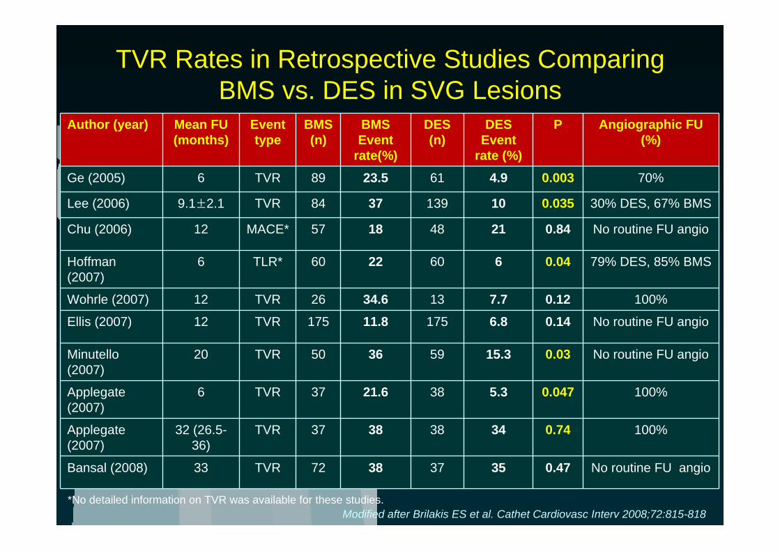
TVR Rates in Retrospective Studies Comparing BMS vs. DES in SVG Lesions
Author (year) Mean FU(months)
Event type
BMS (n)
BMS Event
rate(%)
DES(n)
DESEvent
rate (%)
P Angiographic FU (%)
Ge (2005) 6 TVR 89 23.5 61 4.9 0.003 70%
Lee (2006) 9.1±2.1 TVR 84 37 139 10 0.035 30% DES, 67% BMS
Chu (2006) 12 MACE* 57 18 48 21 0.84 No routine FU angio
Hoffman (2007)
6 TLR* 60 22 60 6 0.04 79% DES, 85% BMS
Wohrle (2007) 12 TVR 26 34.6 13 7.7 0.12 100%
Ellis (2007) 12 TVR 175 11.8 175 6.8 0.14 No routine FU angio
Minutello (2007)
20 TVR 50 36 59 15.3 0.03 No routine FU angio
Applegate (2007)
6 TVR 37 21.6 38 5.3 0.047 100%
Applegate (2007)
32 (26.5-36)
TVR 37 38 38 34 0.74 100%
Bansal (2008) 33 TVR 72 38 37 35 0.47 No routine FU angio
*No detailed information on TVR was available for these studies.Modified after Brilakis ES et al. Cathet Cardiovasc Interv 2008;72:815-818

The Strategic TranscatheterEvaluation of New Therapies (STENT) Group
DES (n=418) BMS (n=281) pLesion length (mm) 18.0 16.2 0.1Vessel diameter (mm) 3.4 3.7 < 0.0001Stent length (mm) 23.7 22.1 0.007Distal location (%) 16 8 0.0007Distal emboli (%) 0.4 3.3 0.003Acute closures (%) 0.4 2.1 0.04Death (%) 5.0 6.8 0.41MI (%) 4.3 8.2 0.005TVR (%) 5.7 8.5 0.17SAT (%) 0.5 1.4 0.23MACE (%) 12.7 20.3 0.008
Adjusted proportional HR for MACE 0.61 (95% CI 0.40, 0.91, p=0.0157) favoring DES. The individual adjusted HR for MI (0.55, 95% CI 0.28, 1.10, p=0.0919)
& TVR (0.60, 95% CI 0.33, 1.11, p=0.1031)
No consistent superior benefits for the use of DES in SVGs
Wilson BH, cs. J Am Coll Cardiol 2007;49 (Suppl B):41B

* Off –Label Use; N= 75 pts with 96 SVG lesions
Sirolimus-Eluting Stenting* in Diseased SVGs:The Reduction of Restenosis In SVGs with Cypher
(Delayed- RRISC) Trial
Vermeersch, P. et al. J Am Coll Cardiol 2007;50:261-267
Increased MortalityIncreased Mortality Delayed TVR Delayed TVR
Surv
ival
Surv
ival
TVR
TVR -
- free
Sur
viva
lfr
ee S
urvi
val
Pts treated with SES showed a significant Pts treated with SES showed a significant increase in total mortalityincrease in total mortality; & the benefit ; & the benefit of SES in terms of reduced TVR shown at 6 months of SES in terms of reduced TVR shown at 6 months was lost at longwas lost at long--term fterm f--upup

* Off –Label Use; N= 75 pts with 96 SVG lesions
Sirolimus-Eluting Stenting* in Diseased SVGs:The Reduction of Restenosis In SVGs with Cypher
(Delayed- RRISC) Trial
Vermeersch, P. et al. J Am Coll Cardiol 2007;50:261-267
After a median follow-up of 32 months:11 deaths occurred in the group receiving SES (29%), but none occurred in the group receiving BMS (p < 0.001). 3 deaths were sudden & 1 was caused by stent thrombosis
Although the findings added to concerns about the long-term safety of DES, the 75-patient study was:
Small, not prospectively designed to show a mortality difference.Analysis is postAnalysis is post--hochocSome pts may have premature antiplatelet discontinuation Some pts may have premature antiplatelet discontinuation

Brilakis E.S., et al. J am Coll Cardiol 2009;53:919-28* Off –Label Use; N= 80 pts with 112 SVG lesions in 88 SVGs
The Stenting Of Saphenous Vein Grafts (SOS) Trial: The Stenting Of Saphenous Vein Grafts (SOS) Trial: In-Stent MLD Cumulative Frequency
Distributions in the BMS & PES Groups
0
25
50
75
100
0 1 2 3 4
Minimum lumen diameter (mm)
%BMSPES
Before intervention
0
25
50
75
100
0 1 2 3 4
In-stent Minimum lumen diameter (mm)
%BMSPES
After intervention(In-stent MLD)
After intervention
Follow-up
Angiographic restenosis was 51% in the BMS group vs. 9% in the PES group (p < .0001, RR 0.18, 95% CI 0.07-0.48)
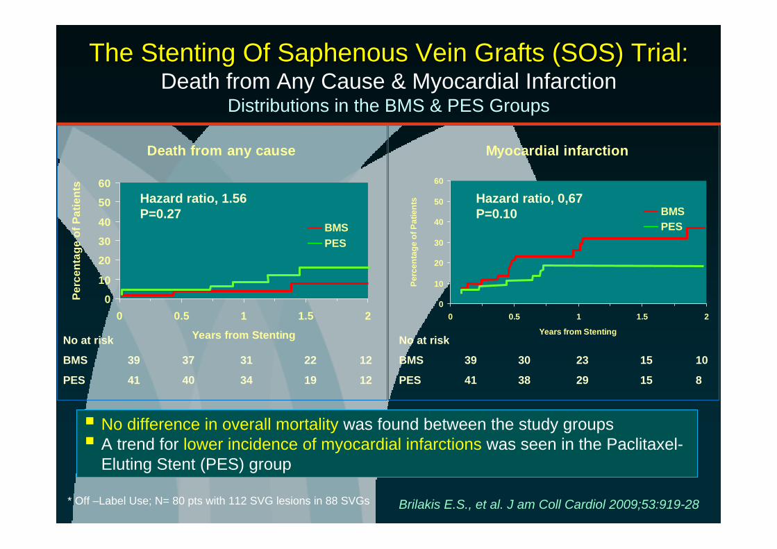
The Stenting Of Saphenous Vein Grafts (SOS) Trial: The Stenting Of Saphenous Vein Grafts (SOS) Trial: Death from Any Cause & Myocardial Infarction
Distributions in the BMS & PES Groups
No difference in overall mortality was found between the study groups A trend for lower incidence of myocardial infarctions was seen in the Paclitaxel-Eluting Stent (PES) group
Brilakis E.S., et al. J am Coll Cardiol 2009;53:919-28* Off –Label Use; N= 80 pts with 112 SVG lesions in 88 SVGs
Death from any cause
0
10
20
30
40
50
60
0 0.5 1 1.5 2
Years from Stenting
Perc
enta
ge o
f Pat
ient
s
BMSPES
No at risk
BMS 39 37 31 22 12
PES 41 40 34 19 12
Myocardial infarction
0
10
20
30
40
50
60
0 0.5 1 1.5 2
Years from Stenting
Perc
enta
ge o
f Pat
ient
s
BMSPES
No at risk
BMS 39 30 23 15 10
PES 41 38 29 15 8
Hazard ratio, 1.56P=0.27
Hazard ratio, 0,67P=0.10

The incidence of TLR & TVF and composite end point of cardiac death, myocardial infarction & TVR, was significantly lower in the PES group than the BMS group
The Stenting Of Saphenous Vein Grafts (SOS) Trial: The Stenting Of Saphenous Vein Grafts (SOS) Trial: Target Lesion Revascularization & Target Vessel Failure
Distributions in the BMS & PES Groups
Brilakis E.S., et al. J am Coll Cardiol 2009;53:919-28* Off –Label Use; N= 80 pts with 112 SVG lesions in 88 SVGs
Target vessel failure
0102030405060
0 0.5 1 1.5 2
Years from Stenting
Perc
enta
ge o
f Pat
ient
s
BMSPES
Target lesion revascularization
0
10
20
30
40
50
60
0 0.5 1 1.5 2
Years from Stenting
Perc
enta
ge o
f Pat
ient
s
BMSPES
No at risk
BMS 39 33 23 13 8
PES 41 40 32 17 10
Hazard ratio, 0.38P=0.003
Hazard ratio, 0.65P=0.03
No at risk
BMS 39 37 31 22 12
PES 41 40 34 19 12

SVG failure post-PCIoften occurs at Non-Target Sites
Ellis SG, cs.. Am J Cardiol 1997;79:1460–4.
Target Sites RestenosisTarget Sites Restenosis NonNon--Target Sites RestenosisTarget Sites Restenosis

Incidence of Early (30-day) Stent Thrombosis in Vein Graft Intervention: AMEthyst Study
Srihari S. Naidu, cs. J Am Coll Cardiol 2009;53:A83 (abstr)
786 pts undergoing SVG stenting:60.2% (n=473) received DES (41.7% Taxus, n=195 & 58.3% Cypher, n=273) 39.8% (n=313) received BMS.
Compared to BMS pts, DES pts had:lower GP 2b/3a receptor inhibitor use (35.5% vs. 51.1%, p<0.001), smaller ref. vessel diameter (3.02 mm vs. 3.61 mm, p<0.001) &lower plaque volume (94.4 mm3 vs. 131.3 mm3, p<0.001).
Early stent thrombosis:for all pts was 0.5%. No differences between DES & BMS pts, either prior to (0.4% vs. 0.7%, OR 0.65, p=0.67) or after adjustment (adjusted OR 1.10, p=0.93). No differences between Taxus & Cypher, either prior to (0.5% vs. 0.4%, OR 1.37, p=0.83) or after adjustment (adjusted OR 0.22, p=0.58).

Use of DES in SVG Lesions:What does the EBM tell us?
DES produce better primary angiographic end points (lower early risk of restenosis) than BMSThis does not mean that DES will always:
produce better clinical outcomes, achieve better angiographic outcomes for all pts with SVG lesions, or even achieve the same angiographic outcomes at different times after stent implantation.
Whether there is a problem of “catch-up phenomenon” & increased risk of very late stent thrombosis which may drive late events is still unknown.Noise due to late target vessel, non-target lesion disease progression

“First do no harm!”Use embolic protection regardless of BMS or DES
Target vessel revascularization in SVGs usually due to progression of disease, rather than target lesion failureThe decision to use DES for SVG lesions remains multifaceted & depends on such factors as graft size, predicted adherence to prolonged dual antiplatelet therapy & the increasingly dominant role of patient preference.Be reminded that prolonged dual antiplatelet therapy for both BMS & DES is necessaryUse DES only in patients who can tolerate prolonged dual antiplatelet therapy
Use of DES in SVG Lesions:What should we do?

Current evidence is based on small, largely retrospective data & only 2 small, prospective trials with only short-term follow-upTo date, the data was underpowered to detect differences in clinical outcomesLarge, RCTs with longer follow-up are needed
Use of DES in SVG Lesions:What do we need?