drug design approaches to manipulate the agonist...
TRANSCRIPT

10
Drug Design Approaches to Manipulate the Agonist-Antagonist Equilibrium
in Steroid Receptors
Scott J. Lusher1,6, Paolo Conti2, Wim Dokter3, Pedro H. Hermkens2,4 and Jacob de Vlieg1,5,6
Departments of 1Molecular Design & Informatics,
2Medicinal Chemistry 3Immune Therapeutics, MSD, Oss
4Institute of Molecules & Materials 5Computational Drug Discovery Group,
Radboud University, Nijmegen, 6Netherlands eScience Center,
The Netherlands
1. Introduction
The steroid hormone receptors, the Androgen Receptor (AR), Estrogen Receptors (ER┙ and
ER┚), Glucocorticoid Receptor (GR), Mineralocorticoid Receptor and Progesterone Receptor
(PR), have been crucial targets for drug discovery even before their existence was known or
understood. The drugs on the market for this sub-class of the nuclear hormone receptors
constitute a significant pharmacopeia for the treatment of a vast array of conditions and
ailments. Despite the breadth of drugs targeted toward this family, they remain an
important target for the pharmaceutical industry.
Key considerations when designing drugs for any family, beyond the on-target
pharmaceutical action and safety, is to ensure specificity against related targets, exploration
of the most appropriate routes of administration and desirable pharmacokinetic (PK)
profiles. Developing non-steroidal modulators for the steroid receptor family has been a key
strategy employed to achieve these goals, although there appears to be growing consensus
that not being steroidal is insufficient to justify new drugs on its own (Hermkens et al, 2006).
Unlike targeting many families, steroid hormone receptor drug discovery also has to
balance the need to elicit either agonistic or antagonistic responses depending on the desired
indication.
The history of drug discovery for the steroid hormone receptors has tended to follow a
common path, beginning with the application of purified endogenous hormone and
followed by the application of the first synthetic analogs with improved PK properties or
selectivity. For some of the receptors this period was followed by the design of antagonists,
including non-steroidal structures. More recently, steroid hormone drug discovery has been
www.intechopen.com

Steroids – Basic Science
222
dominated by the search for ligands characterized by partial agonistic or partial antagonistic
responses, the so called selective modulators. It is hoped and expected that partial agonists
and antagonists for the various receptors will provide improved therapeutic profiles. For
example, selective GR modulators (SGRMs) could provide their anti-inflammatory action
without the undesirable side-effects, including osteoporosis and diabetes, currently
associated with oral glucocorticoids (Hudson, Roach, and Higuchi, 2008). Selective ER
modulators (SERMs) hold the promise of being active on bone but not breast or endometrial
tissue (Shelly et al, 2008;Silverman, 2010), whereas a desirable profile for a selective AR
modulator (SARM) would likely have a greater action in bone and muscle compared to the
prostate (Gao and Dalton, 2007).
2. Molecular basis for partial agonism
The shared domain structure of steroid receptors includes a variable N-terminal domain, a
highly conserved DNA-binding domain and a moderately conserved ligand-binding
domain (LBD). The LBD domain tends to be the primary target for drug-design. The LBD
combines a number of functions, including hormone binding, receptor dimerization and
binding to other co-modulating proteins that play a role in the control of transcription.
These functions have the ability to influence each other, with ligand-binding, as an example;
influencing the pattern of co-modulator recruitment. Specifically, gene activation requires
the recruitment of co-modulating proteins to a region of the surface of the LBD formed by
helices 3/4, 5 and 12. The position of helix-12, as we will discuss, can be influenced by the
nature of the ligand bound to the receptor allowing drugs to influence the binding of co-
modulators and consequently gene activation and the resulting biological effects (Bourguet,
Germain, and Gronemeyer, 2000;Egea, Klaholz, and Moras, 2000;Kumar and Thompson,
1999;Weatherman, Fletterick, and Scanlan, 1999).
Understanding the molecular basis for partial agonism is hampered by the difficulty in
solving the X-ray structures of steroid-receptors in general and specifically complexes
including partial active ligands (Nettles et al, 2008). Full agonists stabilize the receptor, and
specifically helix-12, in a conformation suited to binding co-activating proteins and full
antagonists stabilize the receptor in a conformation suited to binding co-repressing proteins.
The apparent reason for the difficulty in co-crystallizing partial agonists is that they do not
fully stabilize the receptor in either conformation, adopting some degree of equilibrium
between the two (Nettles et al, 2008;Raaijmakers, Versteegh, and Uitdehaag, 2009). This
equilibrium allows partially active compounds to bind unique patterns of co-modulators
compared to full agonists and antagonists, resulting in their potentially interesting
biological effects. Unfortunately as a result it also renders them poorly suited to co-
crystallization studies.
The degree of partial activity (how far from either a full agonist or antagonist response) will
go some way to determining the profile of co-modulators which will bind. Additionally, the
ratio of co-activators compared to co-repressors in each cell type will influence the biological
effect of a partial compound. In cells with a high co-activator concentration we would expect
partial compounds to show a greater degree of agonistic activity compared to the same
ligand in a cell with a high co-repressor concentration. The limitless combination of ligand
partiality and co-modulator distribution appears to be a major contributor to the tissue
selective responses of partial compounds.
www.intechopen.com

Drug Design Approaches to Manipulate the Agonist-Antagonist Equilibrium in Steroid Receptors
223
3. Mechanisms for ligand-induced partial agonist design
In the absence of a complete record of X-ray structures of steroid receptors bound to agonists, antagonists and partially active compounds, we have to fill in the knowledge gaps with mutation studies and ligand-based structure-activity relationships (SAR). Even with this extra information, our understanding of the mechanisms underpinning the repositioning of helix-12 and the resulting spectrum of partial responses remains relatively naive, but there do appear to be a small number of approaches available to the drug designer who wishes to rationally influence the degree of agonism elicited by their compound series. 1. Sterically impede the agonistic orientation of Helix-12 2. Disrupt the function of other indirect stabilizing interactions. 3. Influence the position of Helix-12 by modulating the end of Helix-11 and the loop
between Helices 11 & 12. 4. Reduce the stabilizing interactions between the ligand and Helix-12. 5. Straighten Helix-3, and/or disrupt interactions between Helices 3 & 5. Incorporating these approaches into the optimization of steroid receptor ligands allows the drug-designer to modulate the degree of agonistic and antagonistic response their compounds induce. Pharmacologically it remains difficult to define a priori the precise agonistic or antagonistic efficacy (percentage effect or intrinsic activity) required for any desired indication, but it is now possible to generate a series of ligands with tuned efficacies to cover a broad range and then utilize molecular profiling approaches to select the most desirable. The five basic approaches for generating partially active compounds have been deduced by
numerous studies from all members of the steroid receptors and nuclear receptor family in
total. For the purposes of this review we present a single receptor case study to demonstrate
each of the five mechanisms, but wish to stress that to a greater and lesser degree all
mechanisms should be applicable to all steroid receptors.
3.1 Sterically impede the agonistic orientation of helix-12 3.1.1 Case study: the progesterone receptor
Steroidal anti-progestins are typically differentiated structurally from progestins by the presence of a bulky attachment at their position 11 (Madauss, Stewart, and Williams, 2007). Recent publications of the anti-progestin Mifepristone (Raaijmakers, Versteegh, and Uitdehaag, 2009) and the SPRM Asoprisnil (Madauss et al, 2007) clearly demonstrate that the role of this bulky attachment is to clash with helix-12 and preclude it from adopting its required agonistic position. Both studies also demonstrate an important role specifically for Met909 in the agonism/antagonism balance. Met909 sits within helix-12 at the C-terminal end of the ligand binding domain (LBD), and in the classic agonist conformation of the receptor, is oriented toward the ligand binding pocket. Met909 is typically the only helix-12 residue directly in contact with ligands. The nature of the ligand-Met909 interactions appears to be a key determinant of the receptors function (Petit-Topin et al, 2009). Clashes between Met909 and ligands are likely to destabilize helix-12 (Raaijmakers, Versteegh, and Uitdehaag, 2009), which results in a reduced agonistic response. It has even been suggested that the degree of clash with Met909 might correspond directly to the reduction in agonism (Madauss, Stewart, and Williams, 2007), but this has yet to be shown categorically.
www.intechopen.com

Steroids – Basic Science
224
Introducing bulky groups onto PR modulating non-steroidal scaffolds has also been demonstrated to result in partial agonists on a number of occasions (Jones et al, 2005;Kallander et al, 2010;Thompson et al, 2009;Washburn et al, 2009).
3.1.2 Additional examples
The existence of a clash between antagonists and helix-12 was first demonstrated for ER by studies comparing the X-ray structures of Estradiol to Raloxifene (Brzozowski et al, 1997) and Diethylstilbestrol to Tamoxifen (Shiau et al, 1998). Numerous reviews of these two studies have been published (Hubbard et al, 2000;Kong, Pike, and Hubbard, 2003;Mueller-Fahrnow and Egner, 1999;Pike et al, 2000;Pike, Brzozowski, and Hubbard, 2000) as have many further studies on the X-ray structures of SERMs, full antagonists and full agonists bound to the ERs (Blizzard et al, 2005;Dykstra et al, 2007;Heldring et al, 2007;Kim et al, 2004;Renaud et al, 2003;Renaud et al, 2005;Tan et al, 2005;Vajdos et al, 2007).
Fig. 1. Binding of PR agonist Norethindrone (orange) from X-ray structure compared to PR antagonist Mifepristone (green) demonstrating clash between antagonists and Met909 in helix-12.
The same helix-12 clash has also been demonstrated for AR (Cantin et al, 2007) and GR (Schoch et al, 2010) in recent X-ray structure determination studies. It was also suggested for GR by a mutagenesis study (Hillmann et al, 2002) that showed that mutating Leu753 (equivalent to Met909 in PR) to a phenylalanine results in a receptor defective in transactivation. We can conclude that the reason for this loss of activation is that an increase in the size of the residue at this position prevents helix-12 from adopting its agonistic conformation due to a clash with the ligand.
www.intechopen.com

Drug Design Approaches to Manipulate the Agonist-Antagonist Equilibrium in Steroid Receptors
225
3.2 Disrupt the function of direct stabilizing interactions 3.2.1 Case study: the androgen receptor
The binding of testosterone and dihydrotestosterone to AR demonstrate the existence of crucial receptor stabilizing interactions mediated by agonistic ligands. As we will discuss later, the loop between helix-11 and helix-12 is a key region for mediating partial agonism. As shown in figure 2, AR is stabilized by a ligand mediated hydrogen-bond network from Thr877 in helix-11 to the 17┚-OH group in the endogenous steroidal agonists to Asn705 in helix-3 and finally to the backbone of Asp890 in the loop itself (Matias et al, 2000). Hydroxyflutamide is the active metabolite of the androgen receptor antagonist flutamide. Its antagonism appears to be a result of its inability to complete the entire network of stabilizing hydrogen-bonds (Bohl et al, 2005) also shown in figure 2. The result is that Thr877 is left buried in a predominately hydrophobic pocket, destabilizing the receptor and shifting the agonist-antagonist equilibrium.
Fig. 2. Left shows the X-ray structure of DHT bound to AR including full hydrogen-bond network. Right shows a model of hydroxyflutamide bound to AR based on the X-ray structure of hydroxyflutamide bound to an AR-T877A mutant.
3.2.2 Additional examples
The residue equivalent to AR residue Asn705 in MR is Asn770. Extensive X-ray, SAR and
mutation studies have been conducted on Asn770 which demonstrate clearly the existence
of a ligand-mediated hydrogen bonding network which is critical for the activation of MR in
a similar fashion to the one described for AR (Bledsoe et al, 2005;Hellal-Levy et al, 2000).
Agonistic steroidal ligands for GR and MR are typified by 11┚-hydroxyl groups which
hydrogen bond to Asn564 in GR and Asn770 in MR respectively. Despite the similarity
between MR and PR, the endogenous PR agonist progesterone behaves as an antagonist of
PR. This appears to at least in part be due to a lack of an 11┚-hydroxyl group on
progesterone. It is interesting how the lack of the hydroxyl group doesn’t disturb the
agonistic activity of PR but does MR.
Another important example of disrupting the function of stabilizing interactions can be seen
in the estrogen receptors. In addition to their role in sterically precluding helix-12, SERM
side-chains also contain an important basic amine function which is almost ubiquitous
www.intechopen.com
Steroids – Basic Science
226
amongst this drug class. The role of this nitrogen is to form a salt-bridge to Asp351 in helix-3
of ER┙ (Asp303 in ER┚). The importance of this salt-bridge is that it requires Asp351 to
adopt a new conformation and prevents it from undertaking is usual function of stabilizing
the agonistic position of helix-12 by hydrogen-bonding to backbone residues in the helix. It
also appears that the exact nature of the interaction between the basic amine and Asp351,
including angle, distance and perhaps pKa can influence the biological effect of the ligands.
3.3 Modulate the end of helix-11 & the loop between helices-11 & 12 3.3.1 Case study: the glucocorticoid receptor
Due to the difficulty in crystallizing partial agonists in complex with steroid-receptors much of the evidence to support these mechanisms has to be inferred from other indirect sources. Some of the most valuable evidence comes from mutagenesis studies including those that indicate that the loop between helix-11 and helix-12 is a hotspot that is crucial to the agonism/antagonism balance in GR. Mutation of Ile747, which sits in the middle of the helix-11 to helix-12 loop, to methionine results in GR having a reduced transactivation potential without affecting the binding of classic glucocorticoids (Vottero et al, 2002). Presumably, the increased size of the residue prevents the correct packing of the loop and therefore destabilizes helix-12. Tyr735 at the end of helix-11 is a surface residue whose role is poorly understood, but it has been shown that various mutations (W735F, W735V and W735S) result in a receptor with significant reduction in transactivation activity without affecting ligand binding (Ray et al, 1999;Stevens et al, 2003). Thr739 is the last residue in helix-11 whose mutation to alanine has no effect on the binding of triamcinolone acetonide, but does result in a 16-fold reduction in transactivation (Lind et al, 2000). In addition to these mutation studies, as discussed already, there is also overwhelming evidence across the family to support the hypothesis that Asn564 is crucial for the agonistic activity of GR and related receptors (Bledsoe et al, 2005;Bledsoe, Stewart, and Pearce, 2004;Fagart et al, 1998;Hellal-Levy et al, 2000;Necela and Cidlowski, 2003;Rafestin-Oblin et al, 2002). The role of Asn564 (Asn705 in AR, Asn770 in MR) was previously discussed. Tyr735, Thr739 and Ile747, as shown in Fig 3, are all located at the end of helix-11 or in the following loop. Asn564 has an important role in stabilizing the loop. The studies associated with each of these residues indicate how sensitive this region to influencing the agonism/ antagonism balance and therefore the potential to modify its function by ligand design. The helix-11 to helix-12 loop in steroid-receptors is well suited to drug-design intervention as it forms around the 17┚ group of steroids and is therefore likely to be in close proximity to most ligands. Bledsoe and colleagues recognized the importance of this region when solving the first GR-Dexamethasone structure (Bledsoe et al, 2002;Bledsoe, Stewart, and Pearce,2004) as did the group of Kauppi when solving GR complexed with Dexamethasone and RU486, including noting the flexibility of this loop (Kauppi et al, 2003).
3.3.2 Additional examples
The importance of the loop region between helix-11 and helix-12 has also been demonstrated by X-ray structure studies for ER┙ (Pike et al, 1999;Shiau et al, 1998), and mutagenesis studies on MR also support the conclusion that this region of the steroid-receptors is crucial for the agonism/antagonism balance (Fagart et al, 2005).
www.intechopen.com

Drug Design Approaches to Manipulate the Agonist-Antagonist Equilibrium in Steroid Receptors
227
Fig. 3. The loop between helix-11 and helix-12 illustrating key residues believed to influence GR function
3.4 Reduce the stabilizing interactions between the ligand and helix-12 3.4.1 Case study: the estrogen receptors alpha & beta Methods for antagonizing or reducing the agonism of steroid receptors that do not involve direct steric clashes with the receptor are often referred to as “passive antagonism”. This term was coined by the group of Geoffrey Greene to explain their observations when studying the binding of tetrahydrochyrsene (THC) and its interactions with ER┙ and ER┚ (Shiau et al, 2002). THC is an ER┙ agonist and an ER┚ antagonist. The group of Greene was able to conclude, after generating X-ray structures of both complexes that THC stabilizes ER┙ in its agonist conformation but ER┚ is in an antagonist conformation. This difference on its own is of significant interest, but the study also demonstrated that the reason for ER┚ not adopting an agonist conformation was due to missing stabilizing interactions between the receptor and the ligand. They observe that in ER┚ residues Leu476 and Met479 are not positioned correctly by the ligand to form interactions with relevant residues in helix-12 to stabilize its agonist conformation. The result is a failure of THC to stabilize the agonist conformation of helix-12 and therefore a shift in the agonist-antagonist equilibrium. The fact that THC has such differing effects on two such similar receptors illustrates the challenge when following this or any of the five described approaches in drug-design.
3.5 Straighten helix-3, and/or disrupt interactions between helices-3 & 5 3.5.1 Case study: the mineralocorticoid receptor It is generally accepted that steroid-receptor activation is facilitated by interactions between helix-3 and helix-5. The correct positioning of the basic component of the charge clamp (Lys579 in GR and Lys785 in MR) and the formation of the hydrophobic pocket in which co-
www.intechopen.com
Steroids – Basic Science
228
activators bind is dependent on a bend forming in the middle of helix-3. That bend in helix-3 is induced by a ligand mediated hydrogen bond to helix-5 via the 3-keto group of steroidal ligands. It was initially believed that the importance of the classic interactions between the 3-keto group of steroids and the Glutamine (Glutamate in ER┙ and ER┚) and Arginine residues in steroid hormones was purely to ensure potent binding of the steroids, but the work of Bledsoe (Bledsoe et al, 2005) and Huyet (Huyet et al, 2007) have demonstrated that is also has a role in the agonism-antagonism balance. Huyet et al demonstrated that mutation of either Gln776 or Arg817 in MR to alanine results in previously ligand-mediated agonistic responses being lost. Bledsoe et al have further demonstrated the importance of this bend in helix-3 by characterizing the S810L mutation in MR. This mutation has the effect of stabilizing the agonist conformation of MR, rendering some antagonistic ligands to have an increased agonistic response. Their analysis shows that the role of the S810L mutation is to increase the hydrophobic stabilization between helix-3 and helix-5.
3.5.2 Additional example A recent X-ray structure publication from our group suggests that PR antagonism seen in a compound series can in part be explained by a loss of these same interactions (Lusher et al, 2011).
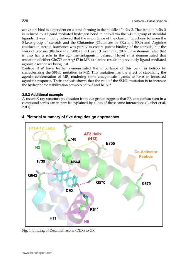
4. Pictorial summary of five drug design approaches
Fig. 4. Binding of Dexamethasone (DEX) to GR
www.intechopen.com

Drug Design Approaches to Manipulate the Agonist-Antagonist Equilibrium in Steroid Receptors
229
4.1 Binding mode of DEX to GR illustrates each of the five design approaches
The binding of Dexamethasone to the Glucocorticoid Receptor (GR) is shown to illustrate the five major routes for reducing agonist efficacy in steroid receptors via the destabilisation of the binding of co-activating proteins. Co-activating proteins bind in a hydrophobic pocket on the surface of the ligand-binding domain (LBD) stabilised by a charge-clamp formed by residues Lys579 in helix-3 and Glu755 in helix-12. [1] Direct clashes between ligands and helix-12 prevent Glu755 from adopting its necessary position and thus prevent the formation of the charge clamp. It has been shown for some receptors that clashes with helix-12 result in the helix adopting a new orientation actually precluding the binding of co-activators by binding in the required hydrophobic pocket. [2] It is probably therefore not a surprise that the positioning of helix-12 can be influenced by the residues that directly precede it. The loop before helix-12 influences its position and is clearly a hotspot that can influence degree of agonism by modifying the ligand. [3] Other interactions also help stabilise helix-12 in its agonist position. For example, in GR, there is a hydrogen-bond network from the ligand to Asn564 in helix-3 to Glu748 in the loop before helix-12. Disruption of this network, by perhaps removing the hydrogen-bonding function in the ligand, can influence the stabilisation of helix-12. [4] In a number of nuclear receptors Helix-12 also makes direct hydrophobic interactions to the ligand. Loss of these interactions, by changing the properties of the ligand, can decrease the stabilisation of helix-12 and therefore alter the agonistic capability of the complex. [5] Finally, the first four approaches are directly or indirectly related to ensuring Glu755, as half of the charge-clamp, is correctly positioned. The second residue in the charge-clamp, Lys579, should not be overlooked. Lys579 is part of helix-3 which itself bends midway along its length. This bend is crucial for ensuring that Lys579 is in the correct position to form the charge-clamp. The bend in helix-3 is partly as a result of its interaction with helix-5. For GR this is largely mediated by a hydrogen-bond network between Gln570 in helix-3, the ligand and Arg611 in helix-5. Disrupting this network by modifying the ligand may influence the distortion in helix-3 and therefore the correct formation of the charge-clamp and therefore co-activator binding.
5. Other structure-based design considerations
In addition to exploring the development of partial agonists, structure-based approaches continue to play an important role in the identification of new ligands via virtual screening approaches and other compound optimization tasks. An important lesson in this regard has been our change in understanding the dynamic nature of the steroid-receptor binding pocket. We have seen examples of extensive induced fits for amongst others the glucocorticoid receptor which is able to bind ligands beyond the conventional confines of its binding pocket whilst remaining in an agonistic conformation (Biggadike et al, 2009;Madauss et al, 2008;Suino-Powell et al, 2008). The pocket, behind the crucial helix-3 and helix-5 binding residues, Gln570 and Arg611, is normally water filled. It has already been demonstrated to be a viable ligand-binding region with the potential to improve ligand potency. An interesting note regarding the exploration of the pocket is that GSK report difficulty in combining the use of this pocket with the maintenance of partial agonism (Biggadike et al, 2009). PR has been shown to adapt to steroids baring bulky 17┙ groups (Madauss et al, 2004) and Trp741 in AR adapts to different ligands, adopting a new position to open an additional channel in the receptor (Bohl et al, 2005).
www.intechopen.com

Steroids – Basic Science
230
6. Conclusion
As we look to the future of rational and structure-based drug design for the steroid receptors there remain key areas and questions that will dominate research in the short to medium term: 1. Is each of the five described methods for generating partial compounds equally
applicable for each of the receptors? It is generally considered true that ER┚ is easier to antagonize than ER┙. This is most likely due to the agonist conformation of ER┚ being less intrinsically stable than ER┙ and therefore ensuring that ER┚ is more sensitive than ER┙ in this respect (Pike et al, 1999).
2. Does the choice of the mechanism for instilling partiality affect the eventual biological activity? Does a compound with a 40% reduction in agonistic activity due to a clash with helix-12 have the same biological effect as a compound with a 40% reduction in agonism due to the loss of other stabilizing interactions?
3. As described earlier, partial agonists and antagonists are often poor candidates for co-crystallization Recently we have seen the first publications describing methods to circumvent this problem, either by introducing stabilizing mutations into the receptor (Bohl et al, 2007;Fagart et al, 2005;Nettles et al, 2008;Sack et al, 2001) or by generating stable crystals of the receptor using a receptor stabilizing ligand and then exchanging this compound with other compounds of interest via soaking (Raaijmakers, Versteegh, and Uitdehaag, 2009). Both approaches have the potential to dramatically increase our understanding of the biological mechanisms underpinning partial agonism.
7. References
Biggadike K, Bledsoe RK, Coe DM, Cooper TW, House D, Iannone MA, Macdonald SJ, Madauss KP, McLay IM, Shipley TJ, Taylor SJ, Tran TB, Uings IJ, Weller V, and Williams SP. 2009. Design and x-ray crystal structures of high-potency nonsteroidal glucocorticoid agonists exploiting a novel binding site on the receptor. Proc. Natl. Acad. Sci. U. S. A 106 (43): 18114-18119.
Bledsoe RK, Madauss KP, Holt JA, Apolito CJ, Lambert MH, Pearce KH, Stanley TB, Stewart EL, Trump RP, Willson TM, and Williams SP. 2005. A ligand-mediated hydrogen bond network required for the activation of the mineralocorticoid receptor. J. Biol. Chem. 280 (35): 31283-31293.
Bledsoe RK, Montana VG, Stanley TB, Delves CJ, Apolito CJ, McKee DD, Consler TG, Parks DJ, Stewart EL, Willson TM, Lambert MH, Moore JT, Pearce KH, and Xu HE. 2002. Crystal structure of the glucocorticoid receptor ligand binding domain reveals a novel mode of receptor dimerization and coactivator recognition. Cell 110 (1): 93-105.
Bledsoe RK, Stewart EL, and Pearce KH. 2004. Structure and function of the glucocorticoid receptor ligand binding domain. Vitam. Horm. 68: 49-91.
Blizzard TA, Dininno F, Morgan JD, Chen HY, Wu JY, Kim S, Chan W, Birzin ET, Yang YT, Pai LY, Fitzgerald PM, Sharma N, Li Y, Zhang Z, Hayes EC, DaSilva CA, Tang W, Rohrer SP, Schaeffer JM, and Hammond ML. 2005. Estrogen receptor ligands. Part 9: Dihydrobenzoxathiin SERAMs with alkyl substituted pyrrolidine side chains and linkers. Bioorg. Med. Chem. Lett. 15 (1): 107-113.
Bohl CE, Gao W, Miller DD, Bell CE, and Dalton JT. 2005. Structural basis for antagonism and resistance of bicalutamide in prostate cancer. Proc. Natl. Acad. Sci. U. S. A 102 (17): 6201-6206.
www.intechopen.com

Drug Design Approaches to Manipulate the Agonist-Antagonist Equilibrium in Steroid Receptors
231
Bohl CE, Miller DD, Chen J, Bell CE, and Dalton JT. 2005. Structural basis for accommodation of nonsteroidal ligands in the androgen receptor. J. Biol. Chem. 280 (45): 37747-37754.
Bohl CE, Wu Z, Miller DD, Bell CE, and Dalton JT. 2007. Crystal structure of the T877A human androgen receptor ligand-binding domain complexed to cyproterone acetate provides insight for ligand-induced conformational changes and structure-based drug design. J. Biol. Chem. 282 (18): 13648-13655.
Bourguet W, Germain P, and Gronemeyer H. 2000. Nuclear receptor ligand-binding domains: three-dimensional structures, molecular interactions and pharmacological implications. Trends Pharmacol. Sci. 21 (10): 381-388.
Brzozowski AM, Pike AC, Dauter Z, Hubbard RE, Bonn T, Engstrom O, Ohman L, Greene GL, Gustafsson JA, and Carlquist M. 1997. Molecular basis of agonism and antagonism in the oestrogen receptor. Nature 389 (6652): 753-758.
Cantin L, Faucher F, Couture JF, de Jesus-Tran KP, Legrand P, Ciobanu LC, Frechette Y, Labrecque R, Singh SM, Labrie F, and Breton R. 2007. Structural characterization of the human androgen receptor ligand-binding domain complexed with EM5744, a rationally designed steroidal ligand bearing a bulky chain directed toward helix 12. J. Biol. Chem. 282 (42): 30910-30919.
Dykstra KD, Guo L, Birzin ET, Chan W, Yang YT, Hayes EC, DaSilva CA, Pai LY, Mosley RT, Kraker B, Fitzgerald PM, Dininno F, Rohrer SP, Schaeffer JM, and Hammond ML. 2007. Estrogen receptor ligands. Part 16: 2-Aryl indoles as highly subtype selective ligands for ERalpha. Bioorg. Med. Chem. Lett. 17 (8): 2322-2328.
Egea PF, Klaholz BP, and Moras D. 2000. Ligand-protein interactions in nuclear receptors of hormones. FEBS Lett. 476 (1-2): 62-67.
Fagart J, Huyet J, Pinon GM, Rochel M, Mayer C, and Rafestin-Oblin ME. 2005. Crystal structure of a mutant mineralocorticoid receptor responsible for hypertension. Nat. Struct. Mol. Biol. 12 (6): 554-555.
Fagart J, Wurtz JM, Souque A, Hellal-Levy C, Moras D, and Rafestin-Oblin ME. 1998. Antagonism in the human mineralocorticoid receptor. EMBO J. 17 (12): 3317-3325.
Gao W, and Dalton JT. 2007. Expanding the therapeutic use of androgens via selective androgen receptor modulators (SARMs). Drug Discov. Today 12 (5-6): 241-248.
Heldring N, Pawson T, McDonnell D, Treuter E, Gustafsson JA, and Pike AC. 2007. Structural insights into corepressor recognition by antagonist-bound estrogen receptors. J. Biol. Chem. 282 (14): 10449-10455.
Hellal-Levy C, Fagart J, Souque A, Wurtz JM, Moras D, and Rafestin-Oblin ME. 2000. Crucial role of the H11-H12 loop in stabilizing the active conformation of the human mineralocorticoid receptor. Mol. Endocrinol. 14 (8): 1210-1221.
Hermkens PH, Kamp S, Lusher S, and Veeneman GH. 2006. Non-steroidal steroid receptor modulators. IDrugs. 9 (7): 488-494.
Hillmann AG, Ramdas J, Multanen K, Norman MR, and Harmon JM. 2000. Glucocorticoid receptor gene mutations in leukemic cells acquired in vitro and in vivo. Cancer Res. 60 (7): 2056-2062.
Hubbard RE, Pike AC, Brzozowski AM, Walton J, Bonn T, Gustafsson JA, and Carlquist M. 2000. Structural insights into the mechanisms of agonism and antagonism in oestrogen receptor isoforms. Eur. J. Cancer 36 Suppl 4: S17-S18.
www.intechopen.com

Steroids – Basic Science
232
Hudson AR, Roach SL, and Higuchi RI. 2008. Recent developments in the discovery of selective glucocorticoid receptor modulators (SGRMs). Curr. Top. Med. Chem. 8 (9): 750-765.
Huyet J, Pinon GM, Fay MR, Fagart J, and Rafestin-Oblin ME. 2007. Structural basis of spirolactone recognition by the mineralocorticoid receptor. Mol. Pharmacol. 72 (3): 563-571.
Jones DG, Liang X, Stewart EL, Noe RA, Kallander LS, Madauss KP, Williams SP, Thompson SK, Gray DW, and Hoekstra WJ. 2005. Discovery of non-steroidal mifepristone mimetics: pyrazoline-based PR antagonists. Bioorg. Med. Chem. Lett. 15 (13): 3203-3206.
Kallander LS, Washburn DG, Hoang TH, Frazee JS, Stoy P, Johnson L, Lu Q, Hammond M, Barton LS, Patterson JR, Azzarano LM, Nagilla R, Madauss KP, Williams SP, Stewart EL, Duraiswami C, Grygielko ET, Xu X, Laping NJ, Bray JD, and Thompson SK. 2010. Improving the developability profile of pyrrolidine progesterone receptor partial agonists. Bioorg. Med. Chem. Lett. 20 (1): 371-374.
Kauppi B, Jakob C, Farnegardh M, Yang J, Ahola H, Alarcon M, Calles K, Engstrom O, Harlan J, Muchmore S, Ramqvist AK, Thorell S, Ohman L, Greer J, Gustafsson JA, Carlstedt-Duke J, and Carlquist M. 2003. The three-dimensional structures of antagonistic and agonistic forms of the glucocorticoid receptor ligand-binding domain: RU-486 induces a transconformation that leads to active antagonism. J. Biol. Chem. 278 (25): 22748-22754.
Kim S, Wu JY, Birzin ET, Frisch K, Chan W, Pai LY, Yang YT, Mosley RT, Fitzgerald PM, Sharma N, Dahllund J, Thorsell AG, Dininno F, Rohrer SP, Schaeffer JM, and Hammond ML. 2004. Estrogen receptor ligands. II. Discovery of benzoxathiins as potent, selective estrogen receptor alpha modulators. J. Med. Chem. 47 (9): 2171-2175.
Kong EH, Pike AC, and Hubbard RE. 2003. Structure and mechanism of the oestrogen receptor. Biochem. Soc. Trans. 31 (Pt 1): 56-59.
Kumar R, and Thompson EB. 1999. The structure of the nuclear hormone receptors. Steroids 64 (5): 310-319.
Lind U, Greenidge P, Gillner M, Koehler KF, Wright A, and Carlstedt-Duke J. 2000. Functional probing of the human glucocorticoid receptor steroid-interacting surface by site-directed mutagenesis. Gln-642 plays an important role in steroid recognition and binding. J. Biol. Chem. 275 (25): 19041-19049.
Lusher SJ. 2011. Structural basis for agonism and antagonism for a set of chemically related progesterone receptor modulators J.Biol. Chem. Publication in Press.
Madauss KP, Bledsoe RK, Mclay I, Stewart EL, Uings IJ, Weingarten G, and Williams SP. 2008. The first X-ray crystal structure of the glucocorticoid receptor bound to a non-steroidal agonist. Bioorg. Med. Chem. Lett. 18 (23): 6097-6099.
Madauss KP, Deng SJ, Austin RJ, Lambert MH, Mclay I, Pritchard J, Short SA, Stewart EL, Uings IJ, and Williams SP. 2004. Progesterone receptor ligand binding pocket flexibility: crystal structures of the norethindrone and mometasone furoate complexes. J. Med. Chem. 47 (13): 3381-3387.
Madauss KP, Grygielko ET, Deng SJ, Sulpizio AC, Stanley TB, Wu C, Short SA, Thompson SK, Stewart EL, Laping NJ, Williams SP, and Bray JD. 2007. A structural and in vitro characterization of asoprisnil: a selective progesterone receptor modulator. Mol. Endocrinol. 21 (5): 1066-1081.
www.intechopen.com

Drug Design Approaches to Manipulate the Agonist-Antagonist Equilibrium in Steroid Receptors
233
Madauss KP, Stewart EL, and Williams SP. 2007. The evolution of progesterone receptor ligands. Med. Res. Rev. 27 (3): 374-400.
Matias PM, Donner P, Coelho R, Thomaz M, Peixoto C, Macedo S, Otto N, Joschko S, Scholz P, Wegg A, Basler S, Schafer M, Egner U, and Carrondo MA. 2000. Structural evidence for ligand specificity in the binding domain of the human androgen receptor. Implications for pathogenic gene mutations. J. Biol. Chem. 275 (34): 26164-26171.
Mueller-Fahrnow A, and Egner U. 1999. Ligand-binding domain of estrogen receptors. Curr. Opin. Biotechnol. 10 (6): 550-556.
Necela BM, and Cidlowski JA. 2003. Crystallization of the human glucocorticoid receptor ligand binding domain: a step towards selective glucocorticoids. Trends Pharmacol. Sci. 24 (2): 58-61.
Nettles KW, Bruning JB, Gil G, Nowak J, Sharma SK, Hahm JB, Kulp K, Hochberg RB, Zhou H, Katzenellenbogen JA, Katzenellenbogen BS, Kim Y, Joachmiak A, and Greene GL. 2008. NFkappaB selectivity of estrogen receptor ligands revealed by comparative crystallographic analyses. Nat. Chem. Biol. 4 (4): 241-247.
Petit-Topin I, Turque N, Fagart J, Fay M, Ulmann A, Gainer E, and Rafestin-Oblin ME. 2009. Met909 plays a key role in the activation of the progesterone receptor and also in the high potency of 13-ethyl progestins. Mol. Pharmacol. 75 (6): 1317-1324.
Pike AC, Brzozowski AM, and Hubbard RE. 2000. A structural biologist's view of the oestrogen receptor. J. Steroid Biochem. Mol. Biol. 74 (5): 261-268.
Pike AC, Brzozowski AM, Hubbard RE, Bonn T, Thorsell AG, Engstrom O, Ljunggren J, Gustafsson JA, and Carlquist M. 1999. Structure of the ligand-binding domain of oestrogen receptor beta in the presence of a partial agonist and a full antagonist. EMBO J. 18 (17): 4608-4618.
Pike AC, Brzozowski AM, Walton J, Hubbard RE, Bonn T, Gustafsson JA, and Carlquist M. 2000. Structural aspects of agonism and antagonism in the oestrogen receptor. Biochem. Soc. Trans. 28 (4): 396-400.
Raaijmakers HC, Versteegh JE, and Uitdehaag JC. 2009. The X-ray structure of RU486 bound to the progesterone receptor in a destabilized agonistic conformation. J. Biol. Chem. 284 (29): 19572-19579.
Rafestin-Oblin ME, Fagart J, Souque A, Seguin C, Bens M, and Vandewalle A. 2002. 11beta-hydroxyprogesterone acts as a mineralocorticoid agonist in stimulating Na+ absorption in mammalian principal cortical collecting duct cells. Mol. Pharmacol. 62 (6): 1306-1313.
Ray DW, Suen CS, Brass A, Soden J, and White A. 1999. Structure/function of the human glucocorticoid receptor: tyrosine 735 is important for transactivation. Mol. Endocrinol. 13 (11): 1855-1863.
Renaud J, Bischoff SF, Buhl T, Floersheim P, Fournier B, Geiser M, Halleux C, Kallen J, Keller H, and Ramage P. 2005. Selective estrogen receptor modulators with conformationally restricted side chains. Synthesis and structure-activity relationship of ERalpha-selective tetrahydroisoquinoline ligands. J. Med. Chem. 48 (2): 364-379.
Renaud J, Bischoff SF, Buhl T, Floersheim P, Fournier B, Halleux C, Kallen J, Keller H, Schlaeppi JM, and Stark W. 2003. Estrogen receptor modulators: identification and structure-activity relationships of potent ERalpha-selective tetrahydroisoquinoline ligands. J. Med. Chem. 46 (14): 2945-2957.
Sack JS, Kish KF, Wang C, Attar RM, Kiefer SE, An Y, Wu GY, Scheffler JE, Salvati ME, Krystek SR, Jr., Weinmann R, and Einspahr HM. 2001. Crystallographic structures
www.intechopen.com

Steroids – Basic Science
234
of the ligand-binding domains of the androgen receptor and its T877A mutant complexed with the natural agonist dihydrotestosterone. Proc. Natl. Acad. Sci. U. S. A 98 (9): 4904-4909.
Schoch GA, D'Arcy B, Stihle M, Burger D, Bar D, Benz J, Thoma R, and Ruf A. 2010. Molecular switch in the glucocorticoid receptor: active and passive antagonist conformations. J. Mol. Biol. 395 (3): 568-577.
Shelly W, Draper MW, Krishnan V, Wong M, and Jaffe RB. 2008. Selective estrogen receptor modulators: an update on recent clinical findings. Obstet. Gynecol. Surv. 63 (3): 163-181.
Shiau AK, Barstad D, Loria PM, Cheng L, Kushner PJ, Agard DA, and Greene GL. 1998. The structural basis of estrogen receptor/coactivator recognition and the antagonism of this interaction by tamoxifen. Cell 95 (7): 927-937.
Shiau AK, Barstad D, Radek JT, Meyers MJ, Nettles KW, Katzenellenbogen BS, Katzenellenbogen JA, Agard DA, and Greene GL. 2002. Structural characterization of a subtype-selective ligand reveals a novel mode of estrogen receptor antagonism. Nat. Struct. Biol. 9 (5): 359-364.
Silverman SL. 2010. New selective estrogen receptor modulators (SERMs) in development. Curr. Osteoporos. Rep. 8 (3): 151-153.
Stevens A, Garside H, Berry A, Waters C, White A, and Ray D. 2003. Dissociation of steroid receptor coactivator 1 and nuclear receptor corepressor recruitment to the human glucocorticoid receptor by modification of the ligand-receptor interface: the role of tyrosine 735. Mol. Endocrinol. 17 (5): 845-859.
Suino-Powell K, Xu Y, Zhang C, Tao YG, Tolbert WD, Simons SS, Jr., and Xu HE. 2008. Doubling the size of the glucocorticoid receptor ligand binding pocket by deacylcortivazol. Mol. Cell Biol. 28 (6): 1915-1923.
Tan Q, Blizzard TA, Morgan JD, Birzin ET, Chan W, Yang YT, Pai LY, Hayes EC, DaSilva CA, Warrier S, Yudkovitz J, Wilkinson HA, Sharma N, Fitzgerald PM, Li S, Colwell L, Fisher JE, Adamski S, Reszka AA, Kimmel D, Dininno F, Rohrer SP, Freedman LP, Schaeffer JM, and Hammond ML. 2005. Estrogen receptor ligands. Part 10: Chromanes: old scaffolds for new SERAMs. Bioorg. Med. Chem. Lett. 15 (6): 1675-1681.
Thompson SK, Washburn DG, Frazee JS, Madauss KP, Hoang TH, Lapinski L, Grygielko ET, Glace LE, Trizna W, Williams SP, Duraiswami C, Bray JD, and Laping NJ. 2009. Rational design of orally-active, pyrrolidine-based progesterone receptor partial agonists. Bioorg. Med. Chem. Lett. 19 (16): 4777-4780.
Vajdos FF, Hoth LR, Geoghegan KF, Simons SP, LeMotte PK, Danley DE, Ammirati MJ, and Pandit J. 2007. The 2.0 A crystal structure of the ERalpha ligand-binding domain complexed with lasofoxifene. Protein Sci. 16 (5): 897-905.
Vottero A, Kino T, Combe H, Lecomte P, and Chrousos GP. 2002. A novel, C-terminal dominant negative mutation of the GR causes familial glucocorticoid resistance through abnormal interactions with p160 steroid receptor coactivators. J. Clin. Endocrinol. Metab 87 (6): 2658-2667.
Washburn DG, Hoang TH, Frazee JS, Johnson L, Hammond M, Manns S, Madauss KP, Williams SP, Duraiswami C, Tran TB, Stewart EL, Grygielko ET, Glace LE, Trizna W, Nagilla R, Bray JD, and Thompson SK. 2009. Discovery of orally active, pyrrolidinone-based progesterone receptor partial agonists. Bioorg. Med. Chem. Lett. 19 (16): 4664-4668.
Weatherman RV, Fletterick RJ, and Scanlan TS. 1999. Nuclear-receptor ligands and ligand-binding domains. Annu. Rev. Biochem. 68: 559-581.
www.intechopen.com

Steroids - Basic ScienceEdited by Prof. Hassan Abduljabbar
ISBN 978-953-307-866-3Hard cover, 234 pagesPublisher InTechPublished online 11, January, 2012Published in print edition January, 2012
InTech EuropeUniversity Campus STeP Ri Slavka Krautzeka 83/A 51000 Rijeka, Croatia Phone: +385 (51) 770 447 Fax: +385 (51) 686 166www.intechopen.com
InTech ChinaUnit 405, Office Block, Hotel Equatorial Shanghai No.65, Yan An Road (West), Shanghai, 200040, China
Phone: +86-21-62489820 Fax: +86-21-62489821
This book explains the basic science of steroids and is targeted towards professionals engaged in healthservices. It should be noted that medical science evolves rapidly and some information like the understandingof steroids and their therapeutic use may change with new concepts quickly. Steroids are either naturallyoccurring or synthetic fat-soluble organic compounds. They are found in plants, animals, and fungi. Theymediate a very diverse set of biological responses. The most widespread steroid in the body is cholesterol, anessential component of cell membranes, and the starting point for the synthesis of other steroids. Since thescience of steroids has an enormous scope, we decided to put the clinical aspects of steroids in a differentbook titled "Steroids-Clinical Aspects". The two books complete each other. We hope that the reader will gainvaluable information from both books and enrich their knowledge about this fascinating topic.
How to referenceIn order to correctly reference this scholarly work, feel free to copy and paste the following:
Scott J. Lusher, Paolo Conti, Wim Dokter, Pedro H. Hermkens and Jacob de Vlieg (2012). Drug DesignApproaches to Manipulate the Agonist-Antagonist Equilibrium in Steroid Receptors, Steroids - Basic Science,Prof. Hassan Abduljabbar (Ed.), ISBN: 978-953-307-866-3, InTech, Available from:http://www.intechopen.com/books/steroids-basic-science/drug-design-approaches-to-manipulate-the-agonist-antagonist-equilibrium-in-steroid-receptors