drug delivery by red blood cells: vascular carriers designed by mother nature
TRANSCRIPT

1. Introduction: red blood cells as
carriers for drug delivery
2. Vascular delivery of drugs
encapsulated into carrier red
blood cells
3. Coupling therapeutics to the
RBC surface
4. Expert opinion
Review
Drug delivery by red blood cells:vascular carriers designed bymother natureVladimir R MuzykantovUniversity of Pennsylvania Medical Center, Department of Pharmacology and Program in Targeted
Therapeutics of the Institute of Translational Medicine and Therapeutics, IFEM, One John Morgan
Building, 3620 Hamilton Walk, Philadelphia, PA 19104-6068, USA
Importance of the field: Vascular delivery of several classes of therapeutic
agents may benefit from carriage by red blood cells (RBC), for example,
drugs that require delivery into phagocytic cells and those that must act
within the vascular lumen. The fact that several protocols of infusion of RBC-
encapsulated drugs are now being explored in patients illustrates a high
biomedical importance for the field.
Areas covered by this review: Two strategies for RBC drug delivery are
discussed: encapsulation into isolated RBC ex vivo followed by infusion in
compatible recipients and coupling therapeutics to the surface of RBC. Studies
of pharmacokinetics and effects in animal models and in human studies of
diverse therapeutic enzymes, antibiotics and other drugs encapsulated in RBC
are described and critically analyzed. Coupling to RBC surface of compounds
regulating immune response and complement, affinity ligands, polyethylene
glycol alleviating immune response to donor RBC and fibrinolytic plasmino-
gen activators are described. Also described is a new, translation-prone
approach for RBC drug delivery by injection of therapeutics conjugated
with fragments of antibodies providing safe anchoring of cargoes to
circulating RBC, without need for ex vivo modification and infusion of RBC.
What the reader will gain: Readers will gain historical perspective, current
status, challenges and perspectives of medical applications of RBC for
drug delivery.
Take home message: RBC represent naturally designed carriers for intra-
vascular drug delivery, characterized by unique longevity in the bloodstream,
biocompatibility and safe physiological mechanisms for metabolism. New
approaches for encapsulating drugs into RBC and coupling to RBC surface
provide promising avenues for safe and widely useful improvement of drug
delivery in the vascular system.
Keywords: carrier, drug delivery, intravascular, red blood cells
Expert Opin. Drug Deliv. (2010) 7(4):403-427
1. Introduction: red blood cells as carriers for drug delivery
The optimization of vascular delivery of drugs is an important biomedical problem.This problem is especially acute in the case of delivery of potent and specific, yetlabile and complex biotherapeutic agents, including enzymes, which in most casesrequire precise localization in the target site. One means to achieve this goal iscoupling drugs to carriers, such as synthetic or natural polymer structures of diversegeometries, phospholipid liposomes, albumin, antibodies or other biological mole-cules [1]. The use of drug carriers promises to enhance the specificity, effectivenessand safety of therapeutic, diagnostic or prophylactic interventions. Functions of drug
10.1517/17425241003610633 © 2010 Informa UK Ltd ISSN 1742-5247 403All rights reserved: reproduction in whole or in part not permitted
Exp
ert O
pin.
Dru
g D
eliv
. Dow
nloa
ded
from
info
rmah
ealth
care
.com
by
RM
IT U
nive
rsity
on
06/0
2/14
For
pers
onal
use
onl
y.

carriers include: i) providing the optimal drug half-life incirculation and clearance mechanism; ii) restriction of un-intended drug uptake by and effects in non-target tissues;iii) targeting to the intended therapeutic site; and iv) providingoptimal timing of the action, that is, its timely initiationand termination.Among other carriers, erythrocytes (red blood cells [RBC],
non-nuclear biconcave discs with a diameter of ~ 7 µm,thickness ~ 2 µm and plasma membrane surface area~ 160 µm2) represent a potentially attractive and, in someaspects, unique carrier for drug delivery (Table 1). Onemicroliter of human blood contains ~ 5 million RBC andthe total number of RBC, the most abundant cellular con-stituent of the blood (> 99%), in the human body approaches30 trillion. Human erythrocytes normally have a lifespan of
100 – 120 days (of note, mouse RBC lifetime is about a thirdof human’s counterpart), travel ~ 250 km through thecardiovascular system and function as natural carriers foroxygen. RBC lifespan and size markedly exceed those of otherdrug delivery systems (e.g., < 10 h and < 100 nm for PEG-modified ‘stealth’ liposomes). Thus, RBC are attractive car-riers for intravascular delivery, for example, for prolongingdrug circulation and restricting unintended extravasation [2-4].
Injected substances interact with RBC, which partition andmetabolize > 50 known drugs (e.g., captopril, sulfanilamide,testosterone, insulin, catecholamines, tacrolimus, antibioticsand other agents) [5]. Therefore, RBC may unintentionallyserve as a natural blood compartment participating in biodis-tribution, pharmacokinetics, slow release, metabolism andaction of diverse drugs, including antitumor agents [6] andgenetic materials [7]. However, the intentional use of RBC asdrug carriers is arguably of even greater interest. This area ofresearch began in the early 1970s, when it was proposed thateffects of certain drugs might benefit from encapsulation intoautologous or immunologically compatible donor RBC thatcan be safely injected in the host, thereby improving circu-lation of a protected drug and its delivery to certain targets inthe body [8]. A prolonged lifetime in the circulation, avail-ability, considerable surface and volume (mean corpuscularvolume of human and mouse RBC is ~ 90 and ~ 50 µm3,respectively), high biocompatibility and natural mechanismsfor safe elimination of RBC represent attractive features of thistentative drug carrier. Erythrocytes are champions amongother drug delivery systems from the standpoint of longevityof circulation, biocompatibility and restriction of unintendedpermeation of drugs into extravascular compartments. Hydro-dynamic forces driving RBC to the main bloodstream in thecirculation and endothelial glycocalyx minimize RBC inter-action with vascular walls [9]. Normally, RBC do not undergoextravasation from the circulation into tissues except hepaticsinuses and interstitium in the spleenic follicles, that is, opento circulation sites of RBC genesis and elimination, part of thereticuloendothelial system (RES). RES macrophages in thespleen and liver rapidly and effectively take up senescent,damaged and modified RBC by means of phagocytosis,leading to their lysosomal degradation.
RBC transfer many substances in the bloodstream. Oxygen-carrying hemoglobin is the most important cargo among othermolecules naturally encapsulated in the interior volume pro-vided by erythrocyte plasma membrane. RBC membrane issupported from within by a complex cytoskeleton comprisinga hexagonal lattice of actin-spectrin network interconnectedwith anchoring integral plasmalemmal proteins by means ofnumerous structural and connector proteins. Dynamic regu-lation and remodeling of this cytoskeleton maintained viaactivity of small GTPases asserts morphological stability,plasticity and deformability of RBC necessary for millionsof repeat travels through tight capillaries [10]. As inother cell types, RBC plasma membrane represents anasymmetrical phospholipid-cholesterol-composed bilayer,
Article highlights.
. RBC is naturally designed as a biocompatible carrier forvascular drug delivery, with prolonged lifetime incirculation and safe mechanisms for eventual elimination,including fixation of complement and uptake bymacrophages. Drug encapsulation into RBC and couplingto RBC surface represent two approaches for RBC drugdelivery.
. Drugs can be encapsulated into isolated RBC usingmethods including hypotonic dialysis and resealing.RBC-encapsulated drugs are separated from blood, butcan exert their activities in circulation if they or theirsubstrates diffuse through RBC membrane. Unintentionaldamage to RBC in the course of encapsulationcompromises their biocompatibility and negativelyimpacts drug delivery, except cases when macrophagesare the target. Undamaging methods for RBCencapsulation provide clinically testable drug deliverysystems that markedly prolong circulation time,bioavailability and effect of cargoes. Delivery ofRBC-encapsulated anti-inflammatory drugs intophagocytic cells and enhanced bioavailability ofdetoxifying enzymes in the bloodstream representattractive examples of this strategy.
. Drugs can be coupled to the RBC surface using a variety ofcovalent and non-covalent crosslinkers, as well asanchored onto circulating naive RBC using recombinantfusion proteins with specific affinity to RBC. Animalstudies show that surface coupling to RBC can be used forthe improvement of antigen delivery, masking of RBCantigens, clearance of pathogens from blood andintravascular delivery of therapeutics that are supposed toact within the vascular lumen. In particular, coupling offibrinolytic plasminogen activators to RBC provides thebasis for a paradigm-shifting prophylactic thrombolysis,an approach that showed very promising results in animalmodels of peripheral, pulmonary and cerebralthromboses.
. RBC represents a versatile, safe and widely useful carrierfor optimization of drug delivery in the vascular system.Several RBC-drug delivery strategies are posed for thetranslation into industrial development and clinical testingand use.
This box summarizes key points contained in the article.
Drug delivery by red blood cells: vascular carriers designed by mother nature
404 Expert Opin. Drug Deliv. (2010) 7(4)
Exp
ert O
pin.
Dru
g D
eliv
. Dow
nloa
ded
from
info
rmah
ealth
care
.com
by
RM
IT U
nive
rsity
on
06/0
2/14
For
pers
onal
use
onl
y.
that is, phosphatidylserine is enriched in the inner leaflet,whereas the outer surface is slightly negatively charged, mostlydue to anionic components of the glycocalyx extendedfrom integral and surface glycoproteins associated withRBC plasmalemma playing the role of transporting (i.e.,ion exchangers and channels), structural and protective ele-ments. Glycophorin A and band 3 represent two majorintegral glycoproteins among > 300 proteins found in RBCplasma membrane [11]. Some of these proteins are expressed atrelatively low and variable levels throughout the humanpopulation, yet exert key functions of protecting RBC fromdamage and elimination from the bloodstream by the immunesystem (see below).
Owing to these unique natural transporting features, RBCmay be an optimal type of carrier for drugs that either need tobe delivered into the RBC-eliminating cells, such as RESmacrophages, or are meant to work in the bloodstream(Figure 1). In the last three decades, significant efforts havebeen invested in order to prove the validity of this paradigmand establish clinically applicable strategies for RBC carriageof drugs. RBC have been tested for drug delivery in numerousanimal [4,12-14] and human studies [14]. Certain challenges andlimitations of using RBC as drug carriers have been identifiedin these studies. Thus, the use of RBC modified ex vivo (e.g.,using donor or autologous blood) limits treatment options tohemotransfusion settings, technically challenging for wide-spread use. Further, unintentional reduction of biocompati-bility of modified RBC represents a serious potential problem.However, from the results of recent animal studies, approacheswere devised to solve or circumvent these problems and boostconfidence that RBC-based drug delivery systems will findmedical utility relatively soon. Indeed, several RBC-drugdelivery approaches are now undergoing clinical testing andindustrial development. This article reviews two strategies for
drug delivery by carrier RBC: loading into RBC and couplingto the RBC surface.
2. Vascular delivery of drugs encapsulated intocarrier red blood cells
From a grossly oversimplified practical viewpoint, an RBCresembles a durable sac-voyage made of elastic material. Thismight help explain why the exploration of RBC transportingcapacities in the drug delivery field started with drug encap-sulation inside RBC ghosts. Since the early 1970s, this area ofresearch has received substantial attention and produced thefirst clinically tested RBC-based drug delivery systems. Morethan 200 papers, monographs and proceedings chapters havebeen published on diverse aspects of drug encapsulation intocarrier RBC; readers are referred to the previous inclusivereviews for more complete literature references as well as amore specific description of methods for encapsulation ofdrugs into carrier RBC [15-17]. Several excellent reviews out-lining the ideology, methodology and outcomes of animal andhuman studies have been published on this subject, includinga recent article in this journal by one of the leading experts,Mauro Magnani [16-20]. It would be impossible to cover insufficient depth all aspects of this strategy in a reasonablyconcise article; hence, a rather cursory overview is providedof the field with a focus on the most recent results andclinical studies.
2.1 Drug loading into carrier red blood cells andelimination of modified red blood cellsOriginally, RBC carriage was proposed to improve delivery ofcargoes into target cells [8,21]. The methods of electricalinsertion and hypotonic RBC loading followed by resealingprovided encapsulation of diverse agents, including
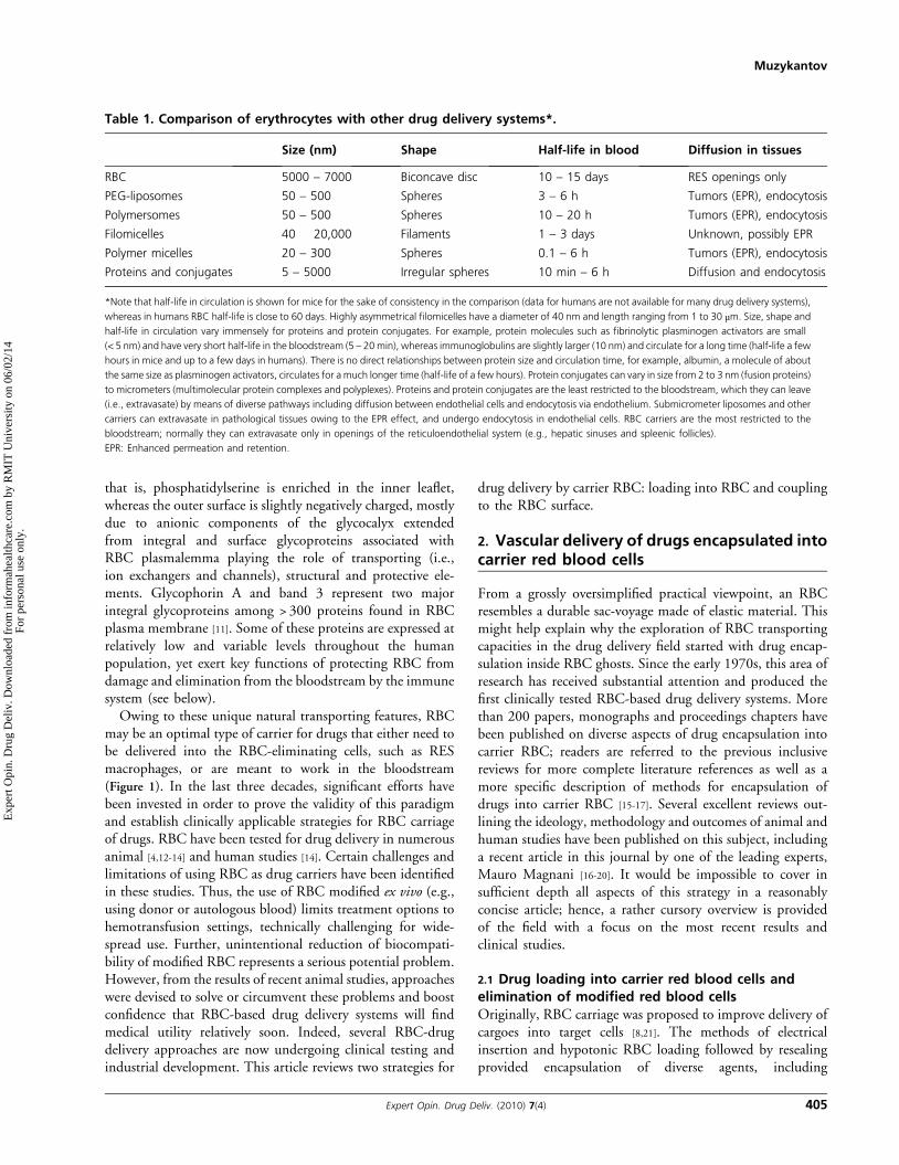
Table 1. Comparison of erythrocytes with other drug delivery systems*.
Size (nm) Shape Half-life in blood Diffusion in tissues
RBC 5000 – 7000 Biconcave disc 10 – 15 days RES openings only
PEG-liposomes 50 – 500 Spheres 3 – 6 h Tumors (EPR), endocytosis
Polymersomes 50 – 500 Spheres 10 – 20 h Tumors (EPR), endocytosis
Filomicelles 40 � 20,000 Filaments 1 – 3 days Unknown, possibly EPR
Polymer micelles 20 – 300 Spheres 0.1 – 6 h Tumors (EPR), endocytosis
Proteins and conjugates 5 – 5000 Irregular spheres 10 min – 6 h Diffusion and endocytosis
*Note that half-life in circulation is shown for mice for the sake of consistency in the comparison (data for humans are not available for many drug delivery systems),
whereas in humans RBC half-life is close to 60 days. Highly asymmetrical filomicelles have a diameter of 40 nm and length ranging from 1 to 30 µm. Size, shape and
half-life in circulation vary immensely for proteins and protein conjugates. For example, protein molecules such as fibrinolytic plasminogen activators are small
(< 5 nm) and have very short half-life in the bloodstream (5 – 20min), whereas immunoglobulins are slightly larger (10 nm) and circulate for a long time (half-life a few
hours in mice and up to a few days in humans). There is no direct relationships between protein size and circulation time, for example, albumin, a molecule of about
the same size as plasminogen activators, circulates for a much longer time (half-life of a few hours). Protein conjugates can vary in size from 2 to 3 nm (fusion proteins)
to micrometers (multimolecular protein complexes and polyplexes). Proteins and protein conjugates are the least restricted to the bloodstream, which they can leave
(i.e., extravasate) by means of diverse pathways including diffusion between endothelial cells and endocytosis via endothelium. Submicrometer liposomes and other
carriers can extravasate in pathological tissues owing to the EPR effect, and undergo endocytosis in endothelial cells. RBC carriers are the most restricted to the
bloodstream; normally they can extravasate only in openings of the reticuloendothelial system (e.g., hepatic sinuses and spleenic follicles).
EPR: Enhanced permeation and retention.
Muzykantov
Expert Opin. Drug Deliv. (2010) 7(4) 405
Exp
ert O
pin.
Dru
g D
eliv
. Dow
nloa
ded
from
info
rmah
ealth
care
.com
by
RM
IT U
nive
rsity
on
06/0
2/14
For
pers
onal
use
onl
y.

antibiotics, steroids, antimicrobial agents, proteins and geneticmaterials, into RBC, with loading efficacy ranging fromreasonable to fair and to excellent (10 to 30% and to 70%,respectively) of drugs retaining their functional activity [22-24].In vitro studies showed that encapsulated agents can bereleased from RBC either slowly (e.g., by means of diffusionthrough RBC plasma membrane [25] and/or its eventualdegradation) [13,26] or rapidly (e.g., by means of lysis of carrierRBC by plasma complement) [27,28]. Early studies also showedthat RBC carriers facilitated intracellular delivery of encap-sulated agents including proteins and DNA into cells inculture [21].Examples of pharmacological and imaging agents encap-
sulated into carrier RBC include diverse enzymes [29,30], fluo-rescent labels [30], erythropoietin for sustained stimulationof hemopoietic potential [31], hemoglobin cofactor inositolhexaphosphate for enhancing RBC oxygen carrying capac-ity [32,33], antithrombotic drugs heparin [34] and thromboly-tics [35], amikacin for antiparasite therapy [36,37], insulin [38],fluoro-AMP [25] and methotrexate for cancer chemother-apy [35], isotopes for imaging of blood pool using gamma-scintigraphy and MRI [39-42], antisense oligonucleotides forgene knockouts [43] and antiviral interventions [44], plasmidDNA for gene therapy [45] and, more recently, magneticnanoparticles [46].Organ distribution, pharmacokinetics and effects of some
of these drug delivery systems have been tested in studies in labanimal species [32,43] and, in a more fragmentary fashion, inhuman patients [42,47-49]. Some of these studies provided rathermixed results, perhaps owing to unintended damage inflictedto carrier RBC during drug loading that compromised theirbiocompatibility, or variability and heterogeneity of RBCloading (noted even within the same batch in the early
studies) [29,30]. For example, RBC-encapsulated and freeerythropoietin showed similar pharmacological propertiesin mice and its activity in plasma was essentially gonewithin < 1 day after injection despite the fact that tracingof 51Cr-label showed relatively prolonged circulation of drug-loaded RBC, with half-life in blood ~ 6 days versus 7 daysfor intact RBC [50]. However, intravenous injection in miceof antisense oligonucleotides encapsulated into opsonizedRBC enhanced hepatic delivery of the cargo [43], whereasinjection of RBC-encapsulated plasmid DNA resulted in arelatively prolonged transfection of the transgene in phago-cytic cells in the RES and blood [45]. A relatively stable MRIsignal was detected in blood samples taken from mice duringthe week post-intraperitoneal injection of RBC-encapsulatedmagnetic nanoparticles [46].
RES macrophages and other professional phagocytes elim-inating senescent and damaged RBC represent a natural targetfor drugs encapsulated in RBC. In fact, it is rather difficult toavoid uptake of carrier RBC by phagocytes and divert deliveryto other targets of interest. Drug loading into RBC inevitablycauses some damage to its membrane and, in some cases,internal content (e.g., depletion of RBC systems for storageand utilization of energy [51] and nitric oxide transport andmetabolism [52]). To avoid these complications, major effortshave been dedicated to studies of mechanisms of RBC bio-compatibility, its damage on RBC modification, and meansfor its preservation for drug delivery applications [53-58].
Conformational changes and abnormal clustering of mem-brane glycoproteins including major components glyco-phorin A and band 3, caused by crosslinking agents andRBC membrane damage by osmotic stress during drug load-ing [53], lead to cytoskeletal dysfunctions (loss of RBC plas-ticity and mechanical stability [55]), sensitize RBC to lysiscaused by oxidants [59] and provoke fixation of immuno-globulins that naturally present in plasma [60]. Exposure ofphosphatidylserine from the inner leaflet of the RBC plasmamembrane [61] and other components normally absent onthe RBC surface augments these processes and predisposesRBC to adhesion to endothelium [62]. Unintentional inacti-vation of specific RBC surface glycoproteins inhibitingcomplement (e.g., Decay Acceleration Factor [DAF] andCD59) [56]) and masking RBC from recognition by phago-cytes (e.g., CD47 [63] and SHPS-1 [64]) compromises RBCbiocompatibility further.
These adverse effects reduce RBC plasticity and resistanceto osmotic and mechanical damage, lead to RBC opsonizationby immunoglobulins and complement promoting phagocy-tosis and directly destroying RBC, which collectively may leadto RBC lysis, aggregation, immune reactions, cellular uptake,adhesion to vascular endothelium and rapid elimination bymeans of phagocytosis and entrapment in the microvascula-ture [30,65-67]. These adverse effects compromise drug deliveryand may cause serious damage typical of intravascular hemo-lysis, including acute vascular, renal and immune reactions tofree hemoglobin, hypoxia and toxic effects of RBC stroma
Vascular injury
WBC
M, LiverSpleen
EC EC
Lymphocytes
Pathogens
Reticuloendothelial System (RES)
Tissues
BloodRBC
Figure 1. Examples of therapeutic sites and targets accessiblefor intravascular drug delivery using RBC carriers. Large, long-circulating RBC have an access and can deliver their cargo to targetsin blood, including other blood cells (lymphocytes, white blood cells[WBC]), and pathogenic agents such as bacteria and toxins, to sitesof vascular injury and endothelial cells lining the vascular lumen(EC), and to diverse components of the reticuloendothelial system(RES), such as macrophages in liver and spleen.
Drug delivery by red blood cells: vascular carriers designed by mother nature
406 Expert Opin. Drug Deliv. (2010) 7(4)
Exp
ert O
pin.
Dru
g D
eliv
. Dow
nloa
ded
from
info
rmah
ealth
care
.com
by
RM
IT U
nive
rsity
on
06/0
2/14
For
pers
onal
use
onl
y.

towards RES macrophages [68]. On the other hand, rapidphagocytosis of altered RBC offers a direct and effective wayfor intracellular delivery, on condition that RBC have notbeen destroyed by complement en route in the circulation.
Major technological advances were made about a decadeago, that is, design of devices allowing semiautomatic high-throughput blood filtration and hypotonic dialysis-basedencapsulation into human RBC of drugs, including steroidsdexamethasone (DEX) and prednisolone, as well as oligonu-cleotides and peptides, producing amounts of loaded RBCsufficient for use in human patients [69,70]. Based on thesedevelopments, several companies embarked on industrialdevelopment of scaled-up production of drug-loaded humanRBC and are now pursuing clinical studies. Below, a briefoverview is given of a few examples of delivery of specific typesof drug encapsulated in carrier RBC in animal and humanstudies. Most of these strategies pursue drug delivery tophagocytic cells in blood and the RES (first of all hepaticand splenic macrophages), or prolonged circulation of RBC-encapsulated enzymes detoxifying various toxic substances inthe bloodstream (Figure 1).
2.2 Red blood cell-mediated drug delivery tomacrophages and other cell typesAs expected, modifications that altered RBC surface antigensand membrane plasticity (e.g., treatment with crosslinkingagents) resulted in their rapid phagocytosis by macrophages inthe RES [71,72], providing a mechanism to deliver encapsulateddrugs into lysosomes in these and other cell types withactive internalization processes, including tumor cells (seebelow) [26,73]. Pilot studies showed that microparticles madefrom RBC ghosts facilitate delivery of cytotoxic agents tomalignant cells [74].
2.2.1 Enzyme replacement therapies for lysosomalstorage diseasesGenetic deficiency of lysosomal acidic hydrolytic enzymesresults in pathological accumulation of their substrates inthe cellular vesicles, leading to lysosomal storage disorders[LSD], manifested by inflammation, neurological, vascular,hepatic and pulmonary disorders [75]. Macrophages and othercells with active lysosomal metabolism represent the primetherapeutic target in LSD management. Pending the devel-opment of effective and safe gene therapy, the only viable andclinically used option for therapeutic management of LSD isperiodic infusion of deficient recombinant enzymes [76], thatis, enzyme replacement therapy (ERT) [77]. However, inacti-vation en route, poor pharmacokinetics and uptake by thetarget cells compromise this approach: only a minute fractionof these protein drugs gets into the lysosomes, hence there is anacute need to improve delivery using drug carriers [78].
Effective uptake and lysosomal addressing of drug-loadedRBC destined for fast phagocytosis due to membrane mod-ification offers a natural mechanism for ERT delivery [4,22]. Infact, initial studies of RBC as carriers for drug delivery were
focused on RBC loading with enzymes for ERT. Hypotonicexchange (i.e., reversible mild poring of RBC membrane in anenzyme suspension) was the most widely accepted and effec-tive among methods for encapsulation in RBC enzymes usedas ERT for treatment of Gaucher’s disease, including galac-tosidase [8], glucuronidase [79] and glucocerebrosidase [4,22].Efficacy of enzyme loading was reversely proportional tothe molecular mass, yet even 180 kDa proteins have beenencapsulated in RBC using hypotonic exchange [8]. RBC-encapsulated enzymes show stable activity [22] and deliverenzyme cargoes into cells in vitro [4]. Animal studiesrevealed relatively more effective lysosomal delivery ofRBC-encapsulated versus free enzymes, despite (or, perhaps,rather in agreement with) very limited circulating capacity ofthe former formulation [79]. Limited clinical studies in the late1970s showed no overt toxicity of RBC-encapsulated gluco-cerebosidase in a patient with advanced adult type Gaucher’sdisease, yet the therapeutic effect was rather modest andtransient [80]. More recently, RBC-loaded enzyme replacementtherapy using thymidine phosphorylase has been testedin a patient with a genetic mitochondrial deficiency ofthis enzyme: treatment improved biochemical readouts,but, unfortunately, the patient’s clinical condition did notimprove [81].
2.2.2 Anti-infectious drugs, antigens and toxic agentsPhagocytosis of RBC intentionally modified in the drugloading process to be taken up by macrophages in the RESserves as a natural delivery path for agents that help toeliminate invaders residing in these cells [73,82]. For example,antiviral drug azidothymidine (AZT) was loaded in carrierRBC [83] that delivered cargo to macrophages and providedenhanced antiviral potency versus free drug in cell culture [26,84]
and in a mouse model of HIV infection [85]. Encouragingresults have been obtained in cells with RBC-mediated deliv-ery of other antiviral agents [86-90], including antisense agentsinhibiting HIV-1 replication in infected cells [91]. Thesestudies provide a basis for a wide-ranging antiviral strat-egy [87,92,93] posed for systematic testing in animal modelsand human studies. It has to be noted, however, that loadingof some antimicrobial drugs into RBC (i.e., antimalarial agentclotrimazole) predisposes RBC to oxidative damage [59],and oxidized RBC are taken up rapidly by hepatic RESmacrophages (i.e., Kupffer cells) by means of scavengerreceptor-mediated phagocytosis [94].
The antigen-presenting function of macrophages and otherimmune cells eliminating altered RBC also offers an attractivenatural mechanism for delivery of antigens to boost theimmune response [95]. In addition to optimized delivery ofantigens, carrier RBC plays the role of the adjuvant, bypresenting multiple copies of antigens and stimulating nonspe-cific immune response [96]. Further, encapsulation of cytokinesincluding interleukins and interferon into RBC facilitatesdelivery of these agents to macrophages [97], resulting instimulation of the immune response in animal models [98].
Muzykantov
Expert Opin. Drug Deliv. (2010) 7(4) 407
Exp
ert O
pin.
Dru
g D
eliv
. Dow
nloa
ded
from
info
rmah
ealth
care
.com
by
RM
IT U
nive
rsity
on
06/0
2/14
For
pers
onal
use
onl
y.

Alternatively, RBC encapsulated with toxic agents, such asricin toxin, kill target cells, first of all, phagocytes [99]. Injectionof a toxin-loaded RBC leads to macrophage depletion [100],providing anti-inflammatory effect and alleviation of acutegraft injury after transplantation [101]. In theory, delivery oftoxic agents by RBC to immune cells may be used for anti-inflammatory interventions, as well as for induction ofantigen tolerance.
2.2.3 Anti-inflammatory agentsDelivery of RBC-loaded anti-inflammatory drugs such asglucocorticoids including DEX to pro-inflammatory cellssuch as macrophages represents a further extension of theprevious approach. It promises to optimize bioavailability ofthese poorly soluble drugs and selectivity of their delivery toactive phagocytes in the RES and other components of theimmune system. Initial studies in vitro developed loading ofDEX derivatives (e.g., more soluble DEX-phosphate) intoRBC; because this procedure predisposes RBC to opsoniza-tion by complement facilitating phagocytosis, RBC-loadedDEX was taken up by cultured macrophages and inhibitedtheir pro-inflammatory response to agonists [70].In a recent series of studies by Magnani’s team, this
approach is tested in human patients with several diseaseconditions associated with and aggravated by inflammation.For example, reinfusion of RBC-loaded DEX in 10 patientssuffering from chronic obstructive pulmonary disease(COPD) has apparently been well tolerated and resulted ina sustained elevation of blood DEX level after a singleinjection [14]. In another lung disease with a major inflam-matory component, cystic fibrosis, reinfusions in the patientsof autologous RBC-loaded DEX at monthly intervals pro-vided a sustained low level of DEX for 28 days in thebloodstream, were well tolerated and provided significantreduction of inflammatory reactions [102]. A similar approachhas been tested in patients with inflammatory bowel disease(IBD, such as Crohn’s disease), and in this study threerepetitive reinfusions of autologous RBC loaded with DEX(every 4 weeks) also provided a prolonged elevation of DEXblood level, permitting the patients to withdraw previouslyprescribed poorly tolerated steroids and still achieve remission,which was maintained for several months [103]. Similar encour-aging results have been observed in a study involving 18pediatric patients treated with monthly infusions of autolo-gous RBC-loaded DEX for 2 years [104]. Finally, a very recentrandomized controlled study of this treatment in 40 IBDpatients refractory to conventional steroids showed that by8 weeks of treatment 75% of 20 patients who received RBC-loaded DEX were in clinical remission (versus 80% in thegroup that received prednisolone and 10% in the controlgroup, respectively), but no adverse effects were detected in theRBC-DEX-treated group, in contrast to 80% of patientsshowing adverse effects of prednisolone [105]. Certainly, thenumber of patients enrolled in these studies (10 – 15) was verysmall on the scale of regular clinical trials. However, these
encouraging pilot results imply that RBC-mediated delivery ofanti-inflammatory agents may find utility in the treatment ofacute and chronic inflammation.
2.2.4 Anticancer agentsLoading anticancer drugs into carriers restricts their toxicity tothe body and improves their delivery to tumors, and relies onseveral mechanisms, both specific (e.g., antibody targeting)and less specific (e.g., enhanced permeation and retentioneffect [EPR], typical of solid tumors). Liposomes, linearpolymers and polymer micelles represent the most popularcarriers for anticancer drugs. However, RBC carriers may finda niche in tumor treatment, for example, by providing for-mulations with prolonged circulation. In support of thisnotion, loading a hydrophobic antitumor agent dequaliniuminto mouse RBC provided much longer half-life in circulationthan PEG-liposomal formulation (5 – 6 days versus 4 h) [106].
Anticancer drugs such as antibiotic doxorubicin havebeen encapsulated into carrier RBC using diverse loadingschemas, including glutaraldehyde crosslinking of RBC mem-brane [13,107]. This modification greatly enhances RBC uptakeby macrophages and other cells exerting active phagocyto-sis [71]. Accordingly, doxorubicin-loaded RBC delivered thecargo into macrophages in culture [108] and accumulated inthe liver (predominantly in macrophages) after intravenousinjection in animals, including dog [109].
This intervention provided encouraging efficacy in thetreatment of lymphoid tumor in dogs without marked cardiactoxicity (a hallmark adverse effect of doxorubicin), yetinflicted unexpected substantial chronic suppression ofmyeloid cells [110]. Nevertheless, a formulation of humanautologous RBC encapsulated with a related anthracyclineantibiotic daunorubicin prepared using a methodology avoid-ing RBC membrane crosslinking has been injected intopatients with acute leukemia and showed a more prolongeddrug level in plasma and fewer side effects than after injectionof free drug [111]. Similarly, doxorubicin-loaded autologousRBC reinfused in patients with lymphomas provided reduc-tion of peak level and extension of drug level in plasma ofpatients, resulting in significant elevation of the area underthe curve and reduction of side effects compared with freedrug [112].
Delivery of drug carriers to solid tumors relies in major parton the EPR effect mediated by abnormally high permeabilityof tumor vasculature and lack of effective lymphatic drainage.In the context of vascular permeability and tumor extravasa-tion by means of the EPR effect, large RBC represent a lesseffective delivery platform than submicrometer carriers suchas liposomes. To address this challenge, antitumor drugs(daunorubicin) have been conjugated with small vesicles(average diameter 100 nm) formed from RBC plasma mem-branes [74]. Soon after intravenous administration (< 30 min)these RBC-based nanovesicles are eliminated from the circu-lation mainly by the RES in liver and spleen [113]. However,injection of this formulation produced more potent antitumor
Drug delivery by red blood cells: vascular carriers designed by mother nature
408 Expert Opin. Drug Deliv. (2010) 7(4)
Exp
ert O
pin.
Dru
g D
eliv
. Dow
nloa
ded
from
info
rmah
ealth
care
.com
by
RM
IT U
nive
rsity
on
06/0
2/14
For
pers
onal
use
onl
y.

effects than free drug in mouse models [114], most probablyowing to slower drug release in the vicinity of tumor cells [115],as these vesicles apparently do not enter target cells [116].
2.2.5 Genetic materialsFinally, RBC ghosts have long been studied as a means forintracellular delivery of genetic materials. Most efforts in thisarea have been focused on cytoplasmic delivery of smalloligonucleotides for interference in protein synthesis. Earlyworks demonstrated proof of principle in cell cultures andprovided initial comparison of this and liposomal antisensedelivery systems [44]. More recent studies confirmed intracel-lular delivery of oligonucleotides [70] and other means ofgenetic interference, such as peptide nucleic acid inactivatingviral RNA replication in culture of human macrophagesinfected with HIV [91]. Several recent studies took this direc-tion in animal studies. Thus, injection of RBC-encapsulatedDNA plasmid led to a prolonged (up to 3 days post-injection)expression of a transgene in blood mononuclear leukocytes [45].Of note, no transgene expression has been reported at thistime in organs, including hepatic Kupffer cells, which in mostanimal studies take the lion’s share of injected materials. Thenature of targeting to blood leukocytes versus RES macro-phages indirectly inferred by this outcome remains enigmatic.By contrast, a very recent study showed that antisense-loadedRBC being opsonized in plasma deliver their cargo to the liver,consistent with known destination of RBC to this organ [43].
2.3 Loading of detoxifying enzymes in RBCThe delivery of RBC-encapsulated drugs discussed above isbased on uptake of modified RBC by macrophages, immuneand tumor cells. By contrast, in theory, minimally alteredRBC loaded with drugs degrading diffusible toxic compoundscould circulate for a prolonged time, avoiding rapid uptake byRES, thereby providing sustained antidotes to toxins.
The design of non-traumatic loading schemas and useof extra precautions (e.g., elimination of subpopulation ofsenescent RBC from the mixture) yielded formulations ofdrug-loaded resealed RBC showing biocompatibility andpharmacokinetics similar to those of unloaded RBC in animalstudies [50,66]. This provided marked prolongation of half-lifein the bloodstream of RBC-encapsulated agents versus freecounterpart injected by means of the same route, includingalcohol oxidase [117], erythropoietin [50], carbonic anhydrase [30]
and other agents [30,46]. Isotope-loaded RBC have been usedto visualize blood pool imaging by gamma-scintigraphy andMRI [41].
Loading capacity of RBC is limited; hence, highly potentand specific enzymes represent more effective cargo for thisapproach than non-enzymatic antidotes such as glutathi-one [118]. More than 10 different detoxifying enzymes havebeen encapsulated into carrier RBC to test this hypothesis,including urikase to eliminate uric acid [119], thiosulfate-cyanide sulfutransferase (AKA rhodanese) converting cyanidesinto less toxic thiocyanates [120-122], phosphothioesterase
to antagonize organophosphorus compounds includingtoxin paraoxon [123-125], alcohol oxidase and alcohol dehydro-genase for elimination of methanol and other toxic alco-hols [117,126,127], L-asparaginase [48,49,128], adenosine aminaseand other enzymes (Table 2). Derivatives of some of theseenzymes modified with polyethylene glycol (PEG-enzymes,partially masked from the immune system) have also beenencapsulated in mouse, human and sheep RBC [129-131].
In most of these studies, encapsulated enzymes retainedactivity and degraded their substrates in vitro. Studies ofcirculation, biodistribution and in vivo stability of enzyme-loaded RBC were rather fragmentary and, in many cases,revealed reduction of circulation time compared with normalRBC [132]. Nevertheless, even this suboptimal approachafforded more prolonged blood level, as well as more potentand sustained detoxifying effects compared with free enzymes,including rhodanese [132] and alcohol oxidase [117]. Infusion ofRBC-encapsulated phosphotriesterase protected mice againstlethal dose of a toxin paraoxon [125]. In some cases, encapsu-lation of enzymes with cofactor molecules facilitated substrateinflux and enzymatic conversion, for example, the kineticsof RBC-encapsulated rhodanese has been improved by co-loading of organic thiosulfonates [122,133]. Such a binarydetoxification system provided significant reduction of cya-nide level [132] and protected against lethal dose of cyanide inmice [134], showing promising efficacy in a mouse model ofcyanide poisoning [135]. Injection of homologous RBC loadedwith PEG-urease/PEG-aldehyde dehydrogenase in sheep pro-vided more prolonged elevation of the enzyme activities inblood than injection of free PEG-modified enzymes (6 versus2 days, respectively) [136].
Several formulations of RBC-encapsulated detoxifyingenzymes have been tested in primates and human patients.In an early study, RBC-encapsulated asparaginase showedmore prolonged circulation (in the range of several days)and more prolonged reduction of asparagine plasmalevel than free enzyme injected in monkeys [128]. In mice,51Cr-labeled RBC-encapsulated asparaginase circulated as acomplex showing the same half-life for RBC carrier and thedrug (although it was shorter than the half-life of controlRBC) [137]. In humans, pharmacokinetics of RBC-loadedasparaginase showed considerable variability between 13tested patients, yet provided a significant prolongation ofhalf-life in the plasma versus free enzyme (27 – 29 days versus8 – 24 h, respectively) and a more profound and prolongedreduction of asparagine level in plasma [48]. Based on thisencouraging outcome, Kravtzoff and co-workers studied tol-erance of RBC-asparaginase in human patients and did notfind any overt harmful effects or antibodies in plasma after asingle injection [49]. Separation of marginal subpopulations ofdrug-loaded RBC (e.g., senescent or unintentionally damagedRBC) using gradient centrifugation helped to minimize dos-ing variability between and within the same batches [138]. Newmethods for asparaginase loading into RBC based on enzymemodification by low-molecular-mass protamine have been
Muzykantov
Expert Opin. Drug Deliv. (2010) 7(4) 409
Exp
ert O
pin.
Dru
g D
eliv
. Dow
nloa
ded
from
info
rmah
ealth
care
.com
by
RM
IT U
nive
rsity
on
06/0
2/14
For
pers
onal
use
onl
y.
proposed recently, yielding formulations with longer half-lifein mice than RBC-loaded asparaginase produced by a hypo-tonic dialysis method and showing promising therapeuticeffect in a mouse model of leukemia dependent on exogenousasparagines [139].Other detoxifying enzymes encapsulated in carrier RBC
include glutamine synthase for ammonia detoxification, pro-viding circulation of detectable enzymatic activity for 48 h andreduction of ammonium level by 50% in mouse blood [140].Alcohol, glutamate and aldehyde dehydrogenases have beenencapsulated into mouse RBC with variable yield and highresultant enzymatic activities (e.g., RBC/GDH effectivelymetabolized high quantities of ammonia) using electropora-tion [127] and hypotonic dialysis [141]. These loading schemasreduced RBC stability, yet injection of RBC-encapsulatedGDH alleviated ammonia toxicity in mice [141]. Isotopetracing of 51Cr-RBC showed that in contrast to controlRBC, a major fraction (> 50%) of electroporated RBC withencapsulated dehydrogenases was eliminated from the
bloodstream within several hours, yet the kinetics of secondphase of RBC/ADH removal was only slightly faster thancontrol RBC (t1/2b approached 4.5 versus 5.3 days,respectively) [142].
Native and PEG-modified adenosine deaminase (ADA) hasbeen encapsulated in human RBC to achieve sustainableelimination of non-metabolized deoxiadenosine that accumu-lates in and inhibits immune cells in the patients with reducedadenosine deaminase level in blood [143]. Human studiesshowed that PEG-ADA-loaded RBC circulate better thannative ADA-loaded RBC, although both formulations havea fairly short half-life (20 versus 12 days, respectively, whichstill was longer than that of PEG-ADA itself, 3 – 6 days) [47].Clinical studies performed by this group involve a very limitednumbers of patients (1 – 2 per study), yet a recent publicationreported a patient who had been treated by RBC-loaded PEG-ADA for correction of a genetic deficiency for 9 years with atotal of 225 infusions every 2 – 3 weeks without overt adverseeffects and with significant, although transient, improvement
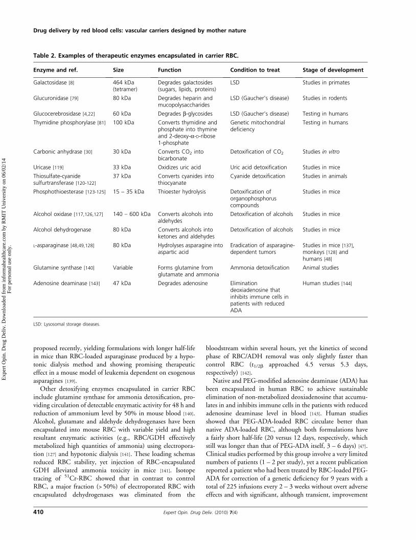
Table 2. Examples of therapeutic enzymes encapsulated in carrier RBC.
Enzyme and ref. Size Function Condition to treat Stage of development
Galactosidase [8] 464 kDa(tetramer)
Degrades galactosides(sugars, lipids, proteins)
LSD Studies in primates
Glucuronidase [79] 80 kDa Degrades heparin andmucopolysaccharides
LSD (Gaucher’s disease) Studies in rodents
Glucocerebrosidase [4,22] 60 kDa Degrades b-glycosides LSD (Gaucher’s disease) Testing in humans
Thymidine phosphorylase [81] 100 kDa Converts thymidine andphosphate into thymineand 2-deoxy-a-D-ribose1-phosphate
Genetic mitochondrialdeficiency
Testing in humans
Carbonic anhydrase [30] 30 kDa Converts CO2 intobicarbonate
Detoxification of CO2 Studies in vitro
Uricase [119] 33 kDa Oxidizes uric acid Uric acid detoxification Studies in mice
Thiosulfate-cyanidesulfurtransferase [120-122]
37 kDa Converts cyanides intothiocyanate
Cyanide detoxification Studies in animals
Phosphothioesterase [123-125] 15 – 35 kDa Thioester hydrolysis Detoxification oforganophosphoruscompounds
Studies in mice
Alcohol oxidase [117,126,127] 140 – 600 kDa Converts alcohols intoaldehydes
Detoxification of alcohols Studies in mice
Alcohol dehydrogenase 80 kDa Converts alcohols intoketones and aldehydes
Detoxification of alcohols Studies in mice
L-asparaginase [48,49,128] 80 kDa Hydrolyses asparagine intoaspartic acid
Eradication of asparagine-dependent tumors
Studies in mice [137],monkeys [128] andhumans [48]
Glutamine synthase [140] Variable Forms glutamine fromglutamate and ammonia
Ammonia detoxification Animal studies
Adenosine deaminase [143] 47 kDa Degrades adenosine Eliminationdeoxiadenosine thatinhibits immune cells inpatients with reducedADA
Human studies [144]
LSD: Lysosomal storage diseases.
Drug delivery by red blood cells: vascular carriers designed by mother nature
410 Expert Opin. Drug Deliv. (2010) 7(4)
Exp
ert O
pin.
Dru
g D
eliv
. Dow
nloa
ded
from
info
rmah
ealth
care
.com
by
RM
IT U
nive
rsity
on
06/0
2/14
For
pers
onal
use
onl
y.

in lymphocyte numbers, sustained level of blood ADA activityand immunoglobulin level, and clinical improvements [144].
3. Coupling therapeutics to the RBC surface
Coupling therapeutics to the surface of carrier RBC representsan alternative to the encapsulation strategies considered above.The RBC membrane provides an extended surface area thatmay be used for anchoring multiple copies of protein or othertherapeutic molecules. Lack of isolation of a drug from blooden route to the therapeutic site represents an obvious downsideof surface coupling versus encapsulation. This concern, how-ever, does not seem to be acute for drugs that are supposed towork in the bloodstream. Furthermore, using pro-drug for-mulations resistant to plasma inhibitors holds promise toresolve issues associated with premature inactivation andside effects en route. On the other hand, surface couplingstrategies avoid damaging encapsulation procedures and there-fore offer theoretical advantages of drug loading withoutcompromising RBC biocompatibility. In addition, couplingof drugs to the RBC surface circumvents issues related to drugrelease (approaches to trigger drug release by using controlledlysis by complement have been suggested, yet practically usefulcontrolled release from carrier RBC remains an elusivegoal) [28]. Of note, coupling to the RBC surface resolvesdiffusional limitations: even enzymes that react with small,membrane-permeable substrate are more active when boundto the RBC surface than when incorporated within thecell [145]. Further, surface coupling offers a unique optionto load drugs on circulating RBC without the technically andlogistically cumbersome need for their extraction necessary fordrug encapsulation and reinfusion.
Generally speaking, techniques for coupling diverse mole-cules to RBC were designed in the 1950s, in the process ofdeveloping reagents for immunological reactions of aggluti-nation. Numerous crosslinking agents and procedures wereapplied to conjugate proteins and other antigens and biolog-ical molecules to RBC of different animal species, includinghumans. Subsequent studies revealed that these conjugationmethods grossly damage RBC membrane, reducing RBCplasticity, resistance to lytic agents and biocompatibility.Nevertheless, reliable methods of biocompatible conjugationmolecules to RBC have been designed recently (see below).
A theoretical paper of mid-1990s provided a speculative yetstimulating analysis of hypothetical RBC membrane anchorsand methods of coupling of therapeutics using chemicalconjugation to tentative affinity ligands and gave a fewexamples of experimental studies in this area that wereavailable at the time [20]. Several practical strategies forcoupling therapeutics to carrier RBC surface have evolvedand have been tested in vitro and in vivo in the last twodecades. These strategies can be divided into three widecategories, described in further detail in the following sections:i) chemical coupling of agents to the RBC surface (eithercovalent or non-covalent); ii) coupling to the RBC membrane
of a receptor that binds a therapeutic agent (and, in somecases, augments its functions); and iii) conjugation of thera-peutics or their receptors with affinity ligands (e.g., antibodiesor their fragments) that bind to RBC thereby anchoringcargoes on RBC (Figure 2). This latter strategy permits loadingdrugs on the RBC surface by injecting these conjugates intothe bloodstream. These strategies provide coupling of anti-bodies, antigens, enzymes, cytokines and other biologicallyactive cargoes to RBC and have been explored for vasculardelivery of several classes of therapeutics, including, morerecently, model polymer nanocarriers anchored to RBC eithernonspecifically [146,147] or by means of affinity peptides [148].Hereafter, surface coupling to RBC is considered for deliveryof the following classes of biotherapeutics: i) antibodies andantigens; ii) affinity ligands for coupling and eliminatingcirculating pathogens; iii) proteins regulating complementsystem; and iv) proteins controlling formation and dissolutionof blood clots.
3.1 Coupling of antigens and antibodies to the surfaceof carrier RBCIn theory, RBC carrying antibodies or antigens on theirsurface can be used to deliver diverse cargoes to intravasculartargets [149] or agents modulating immune response [95],respectively. Since the early 1980s, many methods havebeen tested for coupling protein molecules to RBC forin vitro applications, including the use of nonspecific chemicalcrosslinkers such as tannic acid [150] and chromium chlo-ride [151,152]. These methods have been surpassed by morespecific crosslinkers, for example, a biotin–avidin pair offeringmodular anchoring of diverse biotinylated chemicals includ-ing proteins and nucleic acids to defined reactive groups on theRBC membrane, that is, amino acids [153-155], sulfhydrylgroups [156], sugars [157] and lipids [158,159]. In particular,controlled biotinylation of RBC lysine residues using NHSesters of biotin has become arguably one of the most popularmeans for conjugation cargoes to the RBC surface for a widevariety of applications in vitro and in vivo [69,160-164].
One such application, namely, isolation and tracing ofsubpopulations of RBC in circulation [165,166] as well asmonitoring their survival [167] and volume [168], has founduse in animal [169,170] and human studies involving injection ofnormal, senescent and sickle-cell biotinylated RBC [171,172].Furthermore, methods for direct biotinylation of RBC in thebloodstream using intravascular injection of biotin esters havebeen explored (this method probably yielded promiscuousmodification of diverse blood cells and endothelium) [170,173].On a cautionary note, an excessive biotinylation may reduceRBC biocompatibility and alter RBC antigens [164], andrepetitive injections of biotinylated RBC elicit an immuneresponse [174]. It was also found in the late 1980s that excessivecrosslinking of biotinylated RBC membrane by avidin, strep-tavidin or neutravidin, all of which have four high-affinitybinding sites for biotin and thereby serve for couplingbiotinylated molecules to RBC, causes RBC lysis by
Muzykantov
Expert Opin. Drug Deliv. (2010) 7(4) 411
Exp
ert O
pin.
Dru
g D
eliv
. Dow
nloa
ded
from
info
rmah
ealth
care
.com
by
RM
IT U
nive
rsity
on
06/0
2/14
For
pers
onal
use
onl
y.

complement activated via the alternative pathway [27,175],leading to intravascular hemolysis and rapid elimination ofmodified RBC [176]. Studies of the molecular mechanisms ofthis phenomenon revealed that modification of RBC mem-brane glycoproteins DAF and CD59 (which suppress com-plement activation and assembly of membrane-attackingcomplex in RBC membrane, respectively) is responsible forthis unwanted side effect [56,177,178]. This allowed designof non-damaging means for coupling of molecules toRBC by means of biotin–avidin crosslinker without loss ofRBC biocompatibility [159,179]; thus RBC carrying up to 105
molecules of protein cargo conjugated via streptavidin ina biocompatible manner circulate in animals similarly tonaive RBC, without enhanced clearance, lysis or organuptake [180,181].Several labs explored the use of RBC coated with antigens
and cytokines for stimulation of immune responsein vitro [182,183] and, on a more limited scale, in vivo [184].Antibody-guided RBC formulations have also been tested fordelivery to populations of lymphocytes to achieve selectivemodulation of immune response [152,185]. Further, modelstudies demonstrating proof of principle for targeting anti-body-carrying RBC to selected sites in the vasculature havealso been published [3,149,151]. However, the use of antibody-coated RBC for targeted drug delivery in the vascular systemhas not been actively pursued in the last decade. The need forex vivo modification of RBC for antibody conjugation anddrug loading as well as lack of practically adequate means forcontrolled release of drugs encapsulated into the carrier RBCrepresented a daunting technical challenge. In addition, safetyissues associated with hemotransfusion inevitably involved inthis strategy were viewed unfavorably by the industrial
sector in the age of HIV and other blood-transmitted infec-tions. As a result, synthetic carriers including liposomes andpolymer particles surpassed drug targeting strategies usingantibody-carrying RBC.
3.2 Polyethylene glycol conjugation to red blood cells:an approach to universal donor bloodConjugation of highly hydrophilic PEG with chain length inthe range molecular mass 3 – 10 kDa has evolved since the1970s as a universal ‘stealth’ technology, prolonging circulationand masking from defense systems in the body of liposomes,polymer nanocarriers, proteins and other drug carriers anddrugs themselves. The goal of producing PEGylated ‘stealth’RBC, proposed by Scott and co-workers a decade ago, is toobtain a ‘universal’ donor blood for hemotransfusion by chem-ically camouflaging RBC antigens [186-188]. Indeed, PEG-coatedRBC are less effectively opsonized, taken up by phagocytes andrecognized by antibodies to RBC antigens [189-191]. Maskingagainst immune reaction to mismatched ABO antigens is themost challenging goal; in this setting, PEG-coating did not offerany protection and rather aggravated hemolysis in the initialstudies [191]. Further studies using alternative PEG-couplingtechniques and combinations of PEG with different chainlength produced encouraging results in masking ABO andRh(D) blood group antigens [192-194]. More recently, evenmasking of xeno-antigens on RBC has been attempted, yetthe outcome was not too encouraging [195].
Covalent coupling of cyanuric chloride-activated PEGderivatives to amino acids is the most popular methodfor PEG conjugation to RBC [191,196]. PEG coupling usingsulfo-group chemistry [192,194], insertion of lipid derivativesof PEG [197] and other methods has also been
RBCRBC
YI
YI
A. B. C.
RBC
RBC
PA
PA
PA RuPA
R uPA
R uPA
PA
RBC
R
YI
Red Blood Cell
CR1
Plasminogen activator
Urokinase receptor
uPA Urokinase
Anti-CR1
Legend
PA
PA
Figure 2. Strategies for coupling therapeutic agents to RBC surface. This schema uses plasminogen activators (PA, see Section 3.5) toillustrate methods for coupling drugs to RBC, yet these principles are applicable to a wide variety of therapeutics.A. Direct coupling of drugs(PA) to RBC using covalent or non-covalent crosslinking agents is a prototype paradigm that involves modification of isolated or donor RBCfollowed by infusion in a patient. This paradigm is especially attractive for pursuing a PEG-stealth RBC approach (Section 3.2). B. Non-covalent binding of a drug to its receptor conjugated to RBC may provide a more physiological anchorage for some biotherapeutics andprovide an extra modality for functional regulation of their activity (see an example of coupling urokinase uPA to its receptor suPAR coupledto RBC in Section 3.5.3). C. Conjugation of a drug with fragments of antibodies binding to RBC avoids ex vivomanipulations with RBC, andeases administration and dosing, enhancing clinical feasibility (Sections 3.4 and 3.5.3).
Drug delivery by red blood cells: vascular carriers designed by mother nature
412 Expert Opin. Drug Deliv. (2010) 7(4)
Exp
ert O
pin.
Dru
g D
eliv
. Dow
nloa
ded
from
info
rmah
ealth
care
.com
by
RM
IT U
nive
rsity
on
06/0
2/14
For
pers
onal
use
onl
y.

developed [186,198,199]. The extent of ‘stealth’ feature or mask-ing increases proportionally with PEG length in the range5 – 35 kDa, and with branching and surface density [200].Studies of RBC electrophoresis showed that PEG conjugationincreases apparent length and density of RBC glycocalyx [201]
and that long and especially branched PEG polymers workmore effectively in this respect [202]. Similar results wereobtained when RBC was modified by Pluronic, a tri-blockcopolymer combining two PEG chains at the ends of a lesshydrophilic moiety [203]. Of note, RBC modification usingPEG length and surface density that does not mask comple-ment receptor type 1 (CR1) against antibodies profoundlydecreased binding of immune complexes to RBC, indicatingthat even relatively small steric hindrance may obstruct mul-tivalent interactions of immune complexes with CR1 [204].This observation is important in the context of strategies forCR1-mediated clearance of pathogens discussed below.
PEG modification at the extent that effectively masks RBCfrom recognition by the immune system does not affectfunctions of RBC membrane glycoproteins (e.g., ion exchangefunction of band 3), or RBC stability, plasticity and biocom-patibility [190]. Thus, PEG-RBC circulate with half-life similarto that of non-modified RBC in syngenic animals [186,190].Further fractionation of PEG-coated RBC provides the PEG-RBC subpopulation that is most effectively masked, lessdamaged and circulates equally to native RBC [205]. Further-more, PEG-coated RBC are less susceptible to parasite infec-tion, including malaria Plasmodium [206], owing to maskingtheir receptors [196]. In addition, PEG-modified RBC dem-onstrate reduced tendency to aggregation [203,207] andimproved rheological properties: reduced low shear flowviscosity [189] and enhanced thickness of membrane protectivelayer and plasticity necessary to endure hydrodynamic stress incirculation [196,208,209]. In the last decade, PEG-RBC has beenan active area of research, promising to alleviate acuteproblems of shortage of matched donor blood (see reviewsby Garratty on this subject [210,211]).
3.3 Elimination of circulating pathogens using redblood cell-coupled antibody heteropolymersIt was found several decades ago that one of the RBCtransmembrane glycoproteins, CR1, binds C3b componentof activated complement and immune complexes containingthis protein and that macrophages in the RES safely detachsuch immune complexes, including antibody–pathogen com-plexes from circulating RBC, without damaging the cell [212].Two decades ago, Taylor et al. postulated that this naturalfunction, that is, immune clearance of pathogens from thebloodstream, could be performed as a controlled medicalprocedure that would not require complement activation,by injecting artificial immune complexes called heteropoly-mers that consist of a monoclonal antibody to CR1 conju-gated with an antibody to a pathogen or toxin [213]. Thisstrategy was emulated later by a strategy for a decoy-typeclearance of HIV-1 by RBC carrying electro-inserted CD4 in
their plasma membrane that showed initial promise in in vitrostudies using cell cultures of blood mononuclear cells fromHIV-1 patients [214].
In primates and humans, > 90% of CR1 is expressed onRBC at levels of ~ 500 – 1500 copies per cell, providing ananchorage site for CR1 antibodies and compounds conjugatedwith anti-CR1 (i.e., heteropolymers) injected in blood [215-218].Loading of such heteropolymers on RBC provided effectivebinding of model antigens and bacteria in vitro [213,219,220] andin non-human primates [221] (note, rodents do not expressCR1, hence all in vivo studies of CR1-mediated bloodclearance were performed in monkeys until recent productionof a transgenic mouse expressing human CR1 [222]). CR1represents an immunologically privileged site for antibodybinding to RBC: isotope tracing and fluorescent imagingstudies revealed that binding of neither anti-CR1 nor anti-CR1 conjugates targets carrier RBC for phagocytosis [223], yetphagocytes recognize pathogens and immune complexesbound to CR1 by means of either C3b or anti-CR1 [224],cleave off extracellular domain of CR1 [225] and take up thepathological ‘cargo’ from the RBC surface [226-228]. Studiesin primates demonstrate that diverse ligands can be coupledto circulating RBC using anti-CR1 conjugates and eliminatedby phagocytes without damage or shortening RBCsurvival [213,215,229,230].
This concept has been tested in primates and showedpromising effectiveness of elimination of diverse bacterial [231]and viral [232-235] pathogens using CR1 heteropolymerstargeted to these pathogens, thereby alleviating the infec-tion [235,236]. Further, coupling to RBC of anti-CR1 conju-gated with DNA, a common antigen for autoantibodies inlupus, afforded clearance of such autoantibodies in mon-keys [230,237,238]; the same principle of CR1-targeted hetero-polymers has also been explored for elimination of cytokinesas anti-inflammatory intervention [239], indicating potentiallywide biomedical application of this concept [215].
3.4 Coupling inhibitors of complement to red bloodcells and transfer of red blood cell-anchored proteinsto endotheliumComplement system, consisting of > 20 regulatory andexecuting proteins in blood plasma and cellular membranes,exerts key functions of innate immunity, including opsoni-zation and destruction of pathogens, tumor and foreign cells,and activation of pro-inflammatory cascades in the sites ofinvasion. However, in some pathological conditions, hostcells suffer injury inflicted by overzealous or misguidedactivation of complement. Vascular endothelium and RBCrepresent the most common and vulnerable target for adverseeffects of autologous complement activation, especially insituations of functional deficiency of membrane glycopro-teins inhibiting complement: DAF (CD55 or DAF), CD59and CR1 (CD35, represented in mice by an analogue calledCrry). DAF and CR1 inhibit early stages of complementactivation, thereby protecting cells from lysis and reducing
Muzykantov
Expert Opin. Drug Deliv. (2010) 7(4) 413
Exp
ert O
pin.
Dru
g D
eliv
. Dow
nloa
ded
from
info
rmah
ealth
care
.com
by
RM
IT U
nive
rsity
on
06/0
2/14
For
pers
onal
use
onl
y.

the generation of pro-inflammatory mediators, whereasCD59 blocks the formation of hemolytic pores in theplasmalemma. Complement-mediated hemolysis of RBCdeficient in DAF and CD59 is involved in the mechanismof paroxysmal nocturnal hemoglobinuria (PNH), whereasendothelial damage by complement is involved in acutevascular injury in the ischemia-reperfusion syndrome, suchas in organ transplantation.Several groups pursued targeting complement inhibitors
to RBC, to protect them from hemolysis and inhibit com-plement activation. Of note, natural DAF and CD59 areanchored on the luminal surface of RBC membrane bymeans of a lipid anchor, glycosylphosphatidylinositol(GPI). Accordingly, experimental methods have beendesigned for the insertion of DAF, CD59 and other GPI-linked proteins into RBC plasmalemma using the lipidanchor [240,241]. Insertion of lipid-anchored CD59 in RBCplasmalemma protects against complement-mediated hemo-lysis [242], providing a proof of principle for a new approachfor PNH therapy.The plasma membrane domains that harbor artificially
insertedGPI-linked proteins have not been fully elucidated [243],yet they differ from cholesterol-rich domains that tightly retainnatural GPI-anchored proteins [244]. As a result, GPI-anchoredproteins artificially inserted in membrane (‘painted’) releasemore easily from RBC [241]. Perhaps this is one of the reasonswhy RBC transfer artificially ‘painted’ GPI-linked proteins toother cells in vitro [245] and in vivo, where endotheliumrepresents the preferential acceptor of GPI-inserted proteinsfrom RBC [246]. As a result, injection of RBC carryingGPI-linked DAF and CD59 artificially inserted using genetherapy means alleviates acute ischemia-reperfusion injury in aswine-to-primate model of xenograft transplantation [247].Note, chemical conjugation to RBC plasmalemma is
undesirable for coupling of complement inhibitors becausethis approach would make coupling irreversible, which is notdesirable for protein transfer from RBC to endothelium. Inaddition, it would require RBC isolation, modificationand reinfusion, which inevitably reduce the practical utilityof the approach and RBC resistance to complement. Con-jugation with antibodies also represents a suboptimal meansfor binding complement-regulating proteins to RBC, ascrosslinking RBC membrane by multivalent conjugatescan damage RBC. From the standpoint of safety, as wellas industrial and clinical translation, small monovalentantigen-binding fragments of antibodies, that is, recombi-nant single chain variable fragments (scFv), represent thepreferable targeting moiety. Using modular gene engineeringbiotechnology methods, these recombinant proteins canbe fused by means of short connecting peptides with a varietyof executing proteins. Thus, recombinant scFv fusionstargeted to RBC hold the promise of providing a seriesclinically useful for loading biotherapeutics onto circulatingRBC without the need for RBC isolation, modificationand hemotransfusion.
Expression of scFv in diverse vectors enables large-scale,GMP-quality production of homogeneous monovalentscFv fusion proteins [248,249]. Advantages of scFv include:i) lack of Fc-mediated side effects; ii) lack of crosslinkingof anchoring sites on RBC and RBC aggregation; iii) owingto its smaller size (~ 50 kDa), scFv/PA can be injectedintramuscularly, as other recombinant protein therapies(e.g., insulin); iv) established techniques for humanizationand reduction of immunogenicity of scFv further help tominimize the likelihood of eliciting immune reactions; andv) modular recombinant format of scFv fusion supportssynthesis of diverse fusions [250].
For example, a recombinant fusion protein combiningscFv directed to Rh(D) blood group antigen with humanCR1 binds to CR1-deficient RBC and restores RBC abilityto bind immune complexes [251]. This in vitro study impliesthat this approach may eventually be used either to replenishCR1 functions (i.e., immune clearance and complementinhibition) in CR1-deficient patients (~ 10% of humansare CR1-negative), or to boost CR1 immune clearance ina manner performed by heteropolymers described in theprevious section. Recently, Spitzer and co-workers produceda scFv of a monoclonal antibody TER-119 recognizing amouse analogue of human glycophorin A [252] (GPA), fusedit with DAF [253] and Crry [254], and demonstrated that thismonovalent fusion construct binds to RBC after intravas-cular injection in mice without damaging RBC and, further-more, enhances RBC resistance to lysis by complementin vivo [253,254]. Furthermore, recently this team performeda neonatal in vivo gene transfer of TER-119 scFv/Crry inmice using retroviral gene transfer vector and demonstratedthat prolonged synthesis of this fusion protein in miceleading to its sustained coupling to circulating RBC restoresprotection against excessive complement lysis in geneticallydeficient mice [255]. These studies, showing proof of prin-ciple for in vivo delivery to RBC of therapeutic fusionproteins (injected or encoded by gene therapy means),open exciting applications, some of which are discussed inthe next section.
3.5 RBC carriage of agents regulating formation anddissolution of blood clotsAn idea of intravascular delivery of agents controlling bloodclotting and clot dissolution using carrier RBC was proposeda quarter of century ago, as illustrated by an early workreporting conjugation of fibrinolytic drug streptokinase toRBC targeted to collagen, a thrombogenic component ofextracellular matrix that is exposed to blood in sites ofvascular injury [3]. In theory, coupling drugs to RBC surfacemay favorably alter their pharmacokinetics (i.e., prolonglifetime in circulation) and optimize interaction with com-ponents of blood coagulation and fibrinolytic systems acces-sible from the RBC surface. Attempts in this directionincluded conjugation of heparin to RBC to enhance potencyof anticoagulant thromboprophylaxis in patients predisposed
Drug delivery by red blood cells: vascular carriers designed by mother nature
414 Expert Opin. Drug Deliv. (2010) 7(4)
Exp
ert O
pin.
Dru
g D
eliv
. Dow
nloa
ded
from
info
rmah
ealth
care
.com
by
RM
IT U
nive
rsity
on
06/0
2/14
For
pers
onal
use
onl
y.

to thrombosis [256] and conjugation of pro-thromboticRGD-containing peptide to RBC to design a substitutefor platelet infusion in patients predisposed to hemorrhagicdisorders. [27]
RBC carrying conjugated drugs reported in these studiesexerted good functional activities in vitro [2,3,256]. However,until recently no attempts to test these drug delivery systemsin animal models had been reported, presumably becauseconjugation of drugs grossly compromised RBC biocompat-ibility. In particular, inactivation of complement inhibitorsDAF and CD59 in RBC membrane [257] led to hemolysis,phagocytosis and rapid elimination of RBC [27,175-178,258].Eventually, methods to couple up to ~ 105 copies of ther-apeutic proteins per RBC without RBC damage weredevised [150,159,163,179,180]. In vivo studies using radioisotopetracing showed that 51Cr-RBC/125I-drug complexes circulatesimilarly to control RBC for several days after injection inrats [259] and mice [180,181]. This success brought back on theagenda attempts to couple fibrinolytic agents to RBC and testthe benefit/risk ratio of this approach in animal modelsof thrombosis.
3.5.1 Need for a safer and more effective managementof thrombosisSealing of damaged blood vessels by mural clotsprevents bleeding, whereas pathological vessel occlusion byintravascular clots (thrombosis) causes tissue ischemia anddamage, leading to acute myocardial infarction (AMI), ische-mic stroke, pulmonary embolism and peripheral vasculardisease, among other complications. Thrombosis is the lead-ing cause of mortality and disability in the US [260,261].Thrombi are prone to recur within hours to days after anAMI or stroke and the risk is great after transient ischemicattack or pulmonary embolism and in immobilizedpatients [262-265]. Thrombosis and embolism are also commonand dangerous complications of surgery that are especiallydifficult to manage owing to the risk of acute bleeding at theoperative site. Invasive interventions (e.g., angioplasty, carotidendarterectomy) may be complicated by the formation of clotsthat embolize to the brain and cause neurological disorders.Thus, situations in which patients are at highest short-termrisk for occurrence or recurrence of thrombosis are known.However, antiplatelet and anticoagulant agents providelimited protection and pose considerable risk of bleed-ing [266-269], especially soon after the surgery; in addition,some anticoagulants (e.g., Warfarin) require many hours todevelop an effect, which is inadequate for acute short-termthromboprophylaxis.
Emergency therapy of thrombosis uses injection of plas-minogen activators including tissue type (tPA) and urokinase(uPA), middle-size proteases (molecular mass 30 – 60 kDa)that generate plasmin, which cleave fibrin clots and thusrestore perfusion [249,270]. However, inadequate delivery(blood clearance within < 15 min [271]), inactivation byplasma inhibitors such as PAI-1 [272] and impermeability
of occlusive clots [273] restrict the effectiveness of therapeuticfibrinolysis by PA. Minutes after infusion of mega-doses offibrinolytics (e.g., ~ 100 mg of tPA) needed to overcome itsinefficiency and achieve fibrinolysis locally, excess drugdiffuses into pre-existing hemostatic mural clots predisposedto bleeding and into tissues such as the CNS [274], where itmay cause cerebral hemorrhage, damage to the blood–brainbarrier (BBB) and toxicity within the brain [275]. Owing tothe danger of bleeding and the collateral CNS damage,fibrinolytics cannot be used in > 95% of stroke patientsand in the postoperative period other than as an emergentpotentially life-saving intervention. A more ideal thrombo-prophylactic agent would prevent occlusive thrombi fromforming without lysing hemostatic mural (e.g., postsurgical)clots and causing extravascular toxicity.
Attempts to improve tPA delivery and its benefit/riskratio have not yielded decisively better outcomes [276-279].Ironically, the higher affinity of newly developed PA var-iants for clots promotes drug retention on the clot surface,impeding permeation into the occluding thrombi. The onlycurrent use of tPA, that is, post-thrombosis, is also marredby inevitable delays (time needed for diagnosis, injectionand the lysis proper, slowed by clot impermeability),increasing the risk of ischemia-reperfusion (I/R) injury,which worsens outcome. Prophylactic delivery of a fibrinolyticinto the interior of growing, early nascent thrombi willarrest clot propagation and cause rapid clot lysis. Lysis ofclots from within will be more homogeneous and completethan ‘therapeutic’ external fibrinolysis from the clot surface.This would minimize the incidence of the secondary embo-lism and rethrombosis. Prophylactic use of tPA in patientsat high risk of imminent primary or recurrent thrombosiswill lessen the formation of impermeable occlusive clotsresulting from delays in fibrinolysis. However, fibrinolyticsare not used for prophylaxis because of their rapid clearanceand side effects. Newly designed mutant plasminogenactivators with enhanced potency and fewer side effectsmay improve therapy [279-284], but their diffusion intooccluding clots has yet to be tested and no fibrinolytichas been designed for prophylactic use. All existing plas-minogen activators are short-lived (< 30 min, insufficientfor prophylaxis) and small (< 10 nm) agents, capableof diffusing into hemostatic clots (bleeding) and into theCNS (collateral damage). None is suited for use as pro-phylaxis in postsurgical or other patients at imminent riskof thrombosis.
3.5.2 Coupling plasminogen activators to red bloodcells: a new strategy for short-termthromboprophylaxisRecent animal studies showed that biocompatible coupling toRBC converts plasminogen activators from problematic ther-apeutic agents into effective and safe agents for intravascularthromboprophylaxis. Hemodynamic factors are favorable,propelling RBC towards the blood mainstream [285-287],
Muzykantov
Expert Opin. Drug Deliv. (2010) 7(4) 415
Exp
ert O
pin.
Dru
g D
eliv
. Dow
nloa
ded
from
info
rmah
ealth
care
.com
by
RM
IT U
nive
rsity
on
06/0
2/14
For
pers
onal
use
onl
y.

restricting drug contact with vascular walls and mural clots.Infusion of RBC/tPA complex in rats and mice:
1. does not cause complement activation, hemolysis, phago-cytosis or accelerated clearance of RBC carrying up to 105
tPA molecules stably coupled per RBC [288];2. blocks tPA diffusion into hemostatic clots [288] – in fact,
RBC/tPA does not affect normal hemostasis or causebleeding from fresh postsurgical hemostatic clotssusceptible to free tPA [289];
3. RBC glycocalyx protects tPA from plasma inhibitors [290]
while preserving its fibrin affinity [291] and activation byfibrin [291], which facilitates dissolution of clots.
4. prolongs tPA circulation bymany orders of magnitude [291],permitting its use as prophylaxis [288] – in rats and mice< 20% of RBC/tPA is cleared within 3 h, which projects toa half-life of ~ 2 – 3 days; the half-life of RBC is~ 3 – 4 weeks in mice versus 3 – 4 months in humans,thus the expected prophylactic window of RBC/PA inhumans may vary from hours to many days and weeksdepending on dose.
5. delivers tPA into the interior of intravascular venous andarterial nascent clots, which are then lysed from withinduring clot extension in settings where even a 10-foldhigher dose of soluble tPA is ineffective [288], which isTrojan Horse lysis [291,292].
Furthermore, most recent animal studies showed thatRBC/tPA provides effective and safe thromboprophylaxisin the most challenging and dangerous vascular bed, that is,cerebral circulation, thereby preventing deleterious conse-quences of brain ischemia and stroke in mice [293]. This studyshowed that prophylactic RBC/tPA injection in mice pro-vides dissolution of subsequently formed occlusive cerebro-vascular thrombi, leading to rapid and stable reperfusion,marked alleviation of ischemic brain injury and improvedsurvival, not attainable by even 10-fold higher doses ofsoluble tPA (which in fact greatly enhanced mortality). Incontrast to free tPA, RBC/tPA did not aggravate brainedema, injury and hemorrhage in mice with cerebral ischemiaand a rat model of post-thrombotic intracranial hemorrhage.Moreover, either prophylactic or post-insult injection ofRBC/tPA attenuated disorders of vascular regulation andCNS tissue injury in the brain of pigs subjected to globalischemia/hypoxia, whereas free tPA grossly aggravated thesepathological changes [294].
3.5.3 Advanced approaches for coupling plasminogenactivators with red blood cellsEncouraging outcomes of animal studies using injection ofRBC/tPA complexes preformed ex vivo using chemical cross-linking paved the way for the most recent attempts to advancecoupling techniques. Thus, in addition to coupling to RBCvia streptavidin crosslinker used as a prototype method in allprevious studies of RBC/tPA, urokinase has been coupled toRBC via urokinase receptor chemically conjugated to RBC
without loss of biocompatibility [295]. This approach reducedundesirable interactions of urokinase with its vascular recep-tors and allowed trigger fibrinolysis, because interaction withRBC-coupled receptor activated urokinase [295].
Preformed RBC/tPA conjugates have properties suitable forprophylactic use in settings where transfusion is common, butwould be less practical in other settings and challenging todevelop commercially. To avoid the need to couple tPA toisolated RBC ex vivo followed by transfusion, ways to targettPA to circulating RBC directly have also been designed.Figure 3 illustrates this strategy for coupling plasminogenactivators to carrier RBC.
The first approach capitalized on CR1 anchoring strategy,as agents conjugated with monoclonal antibodies to CR1 [296]
bind to circulating RBC after intravenous injection in animalsincluding primates, circulate with RBC, and bind ligandsin vivo without RBC damage [213,221,297]. Animal studiesshowed that tPA conjugated to a CR1 monoclonal antibodybinds without harm to circulating RBC in mice and affordssafe and effective prophylactic thrombolysis [289] comparableto that provided by infusion of RBC/tPA [288]. To alleviatechallenges associated with translation and application ofchemically produced antibody/tPA conjugates and achievepredictable loading of RBC over a wide range of drug con-centrations, a new recombinant tPA mutant fused to anantigen-binding scFv of TER-119 monoclonal antibody toglycophorin A (anti-GPA scFv) [298,299] was designed veryrecently; pilot animal studies showed that this approach allowsto load up to 20,000 copies of anti-GPA scFv/tPA fusion perRBC after a single intravenous injection of the fusion withoutdetectable changes in behavior of circulating RBC. On anencouraging note, injection of scFv/tPA in mice providedswift dissolution of subsequently formed intravascular clots,which were impervious to injections of equimolar doses ofsoluble tPA [300].
4. Expert opinion
Erythrocytes represent a unique and promising, yet ratherunderdeveloped platform for drug delivery. After an initialburst of interest and research effort three decades ago, drugdelivery by RBC was overshadowed by artificial carriersoffering a wider range of applications, as well as better controlover formulation, storage and utilization of the drug deliverysystem than original RBC-based concepts. Thus, RBC is agood carrier for drug delivery to intravascular and RES targets,but many other important therapeutic targets (e.g., solidtumors, extravascular tissue components, CNS) are normallyinaccessible to RBC. As the drug delivery field originated anddeveloped predominantly in the context of targeted deliveryof toxic anticancer drugs, such limitation of the spectrum ofsuitable targets RBC negatively impacted the developmentof this drug delivery platform.
Studies of RBC drug delivery logically started with in vitroexperimentation using isolated RBC and within two decades
Drug delivery by red blood cells: vascular carriers designed by mother nature
416 Expert Opin. Drug Deliv. (2010) 7(4)
Exp
ert O
pin.
Dru
g D
eliv
. Dow
nloa
ded
from
info
rmah
ealth
care
.com
by
RM
IT U
nive
rsity
on
06/0
2/14
For
pers
onal
use
onl
y.

progressed to extensive animal studies, a necessary step forvalidation of the viability, efficacy and safety of this drugdelivery system, including immune response to RBC-coupledproteins in settings similar to immunization procedures.Of course, certain specific aspects of the RBC drug delivery(e.g., immune response, subtle effects in the CNS and toler-ability of RBC/drug complexes by specific patient popula-tions) can be addressed only in clinical studies. The fact thatseveral clinical trials of RBC drug delivery systems haverecently been initiated instills hope that these translationalbarriers will eventually be overcome.
Among potentially useful biomedical applications, RBC-mediated intracellular delivery of drugs including replacementtherapies, imaging probes and toxic agents into RES macro-phages and other cells exerting robust endocytic uptake seemsto be a reasonably promising goal. Delivery of antimicrobial,immunogenic or toxic agents into defense cells can be used foreradication of intracellular parasites, stimulation or suppres-sion of immune reactions, respectively. It remains to beunderstood better, however, which particular advantages areoffered by RBC in this context versus other drug deliverysystems, all of which are effectively taken up by RES macro-phages and other defense cells (it is more difficult to avoidaddressing this). Further, to translate this concept into theclinical domain, mechanisms of action of RBC-loaded drugsmust be more fully understood and optimized. The pharma-cokinetics, stability and rate of release of drugs in plasma haveto be systematically characterized and effects on diversepopulations of cells in the body defined. This drug deliverysystem is also a bit promiscuous in terms of selectivity oftargeting: many cell types in the body can take up RBC andtheir remnants. For example, phagocytes, immune and tumor
cells all may be targets of antibiotic-loaded RBC. It isconceivable that more precise targeting towards some of thesepopulations may greatly optimize therapeutic effect. It wouldbe undesirable, however, if efforts in this direction were toprovide formulations that were very complex, and difficult toproduce and utilize in medical practice (to be fair, this concernis generally applicable to most previous and current drugdelivery carriers).
Strategies using RBC carriage to augment systemic effectsof drugs in circulation such as sustained effects of detoxifyingenzymes require no targeting and represent one of the uniqueareas of RBC drug delivery that offers clear potential advan-tages over other carriers, owing to unique longevity of RBC.Maintenance of maximal biocompatibility of loaded RBC,yield of enzyme loading and kinetics of transmembranetransport of their substrates and products, as well as controlledrelease and metabolism of encapsulated cargoes, representchallenging, yet theoretically surmountable aspects ofthis approach.
The need for ex vivo manipulations with RBC, a relativelylimited shelf-life and concerns related to the safety of donormatching and blood-borne infections are recognized potentialdownsides of carriage drugs attained by ex vivo RBC loadingand reinfusion. These concerns are shared by all types ofhemotransfusion, cell replacement and stem cell therapies.Note that transfusion of blood and blood products is a verywidely used and generally safe therapeutic intervention world-wide, the safest and most effective type of cell transplantationstrategy. Use of autologous blood (reinfusion) minimizes thesafety concerns.
Drug delivery systems targeted to anchor molecules on theRBC surface (e.g., scFv-fusion proteins) and therefore
Sideeffects
Bleeding
EC
EC
A.
B.
EC
EC
Clearance Injury
RBCRBC
Mural clots
ThrombolysisNascentclots
RBC
Figure 3. A concept of prophylactic fibrinolysis by scFv/tPA targeting to RBC. A. Plasminogen activators (dots) are relativelyineffective, in part owing to rapid uptake by liver, and unsafe owing to bleeding (indiscriminate lysis of hemostatic mural clots), vascular sideeffects (e.g., activation of receptors on endothelial cells [EC]) and injurious effects of tPA diffusing into the CNS. B. Coupling to RBC willdramatically prolong the longevity of scFv/tPA variant. RBC will restrain scFv/tPA binding to cellular receptors, and restrict its access intomural hemostatic clots and the CNS. Propulsion of RBC towards the mainstream will further offset interactions of the pro-drug withhemostatic clots and vascular walls. RBC-bound scFv/tPA will have virtually unlimited access to the interior of nascent pathological thrombiand thereby will dissolve pathological intravascular clots and prevent vascular occlusion.
Muzykantov
Expert Opin. Drug Deliv. (2010) 7(4) 417
Exp
ert O
pin.
Dru
g D
eliv
. Dow
nloa
ded
from
info
rmah
ealth
care
.com
by
RM
IT U
nive
rsity
on
06/0
2/14
For
pers
onal
use
onl
y.

allowing safe and technically simple loading of circulatingRBC hold a promise to solve completely this issue and greatlyexpand general applicability of RBC carriers in medicine. Inparticular, the design of intravascular drug delivery platformsusing coupling to circulating RBC agents that control theimmune system, eliminate pathogens and toxins from thebloodstream and correct pathological aspects of thrombosisand hemostasis seem most exciting and unique utilitiesof RBC drug delivery, promising timely translation intoindustrial development and clinical practice.
Acknowledgments
This work was partially supported by NIH RO1 HL090697.The author thanks J-C Murciano for help in the preparationof the figures.
Declaration of interest
The author states no conflicts of interest and has received nopayment in the preparation of this manuscript.
BibliographyPapers of special note have been highlighted
as either of interest (.) or of considerable
interest (..) to readers.
1. Simone EA, Dziubla TD,
Muzykantov VR. Polymeric carriers: role
of geometry in drug delivery. Expert Opin
Drug Deliv 2008;5:1283-300
2. Coller BS, Springer KT, Beer JH, et al.
Thromboerythrocytes. In vitro studies of a
potential autologous, semi-artificial
alternative to platelet transfusions.
J Clin Invest 1992;89:546-55
3. Muzykantov VR, Sakharov DV,
Smirnov MD, et al. Immunotargeting of
erythrocyte-bound streptokinase provides
local lysis of a fibrin clot.
Biochim Biophys Acta 1986;884:355-62
4. Dale GL, Kuhl W, Beutler E.
Incorporation of glucocerebrosidase into
Gaucher’s disease monocytes in vitro.
Proc Natl Acad Sci USA 1979;76:473-5. This is one of the first papers describing
RBC as a potential drug delivery system.
5. Hinderling PH. Red blood cells: a
neglected compartment in
pharmacokinetics and pharmacodynamics.
Pharmacol Rev 1997;49:279-95
6. Dumez H, Highley M, Guetens G, et al.
Erythrocytes and the transfer of anticancer
drugs and metabolites: a possible
relationship with therapeutic outcome.
Semin Oncol 2001;28:24-8
7. Sakurai F, Nishioka T, Saito H, et al.
Interaction between DNA-cationic
liposome complexes and erythrocytes is an
important factor in systemic gene transfer
via the intravenous route in mice: the role
of the neutral helper lipid. Gene Ther
2001;8:677-86
8. Ihler GM, Glew RH, Schnure FW.
Enzyme loading of erythrocytes. Proc Natl
Acad Sci USA 1973;70:2663-6
9. Vink H, Duling BR. Identification of
distinct luminal domains for
macromolecules, erythrocytes, and
leukocytes within mammalian capillaries.
Circ Res 1996;79:581-9
10. Kalfa TA, Pushkaran S, Mohandas N, et al.
Rac GTPases regulate the morphology and
deformability of the erythrocyte
cytoskeleton. Blood 2006;108:3637-45
11. Pasini EM, Kirkegaard M, Mortensen P,
et al. In-depth analysis of the membrane
and cytosolic proteome of red blood cells.
Blood 2006;108:791-801
12. Ktavtzoff R, Desbois I, Doinel C, et al.
Immunological response to L-asparaginase
loaded into red blood cells. Adv Exp
Med Biol 1992;326:175-82
13. Tonetti M, Astroff B, Satterfield W, et al.
Construction and characterization of
adriamycin-loaded canine red blood cells
as a potential slow delivery system.
Biotechnol Appl Biochem 1990;12:621-9
14. Rossi L, Serafini S, Cenerini L, et al.
Erythrocyte-mediated delivery of
dexamethasone in patients with chronic
obstructive pulmonary disease.
Biotechnol Appl Biochem 2001;33:85-9
15. Hamidi M, Tajerzadeh H. Carrier
erythrocytes: an overview. Drug Deliv
2003;10:9-20
16. Hamidi M, Zarrin A, Foroozesh M,
Mohammadi-Samani S. Applications of
carrier erythrocytes in delivery of
biopharmaceuticals. J Control Release
2007;118:145-60.. This and previous reviews provide a
comprehensive list of drugs encapsulated
into RBC and briefly outline
pharmacokinetic features of selected
RBC–drug complexes in animal studies.
17. Pierige F, Serafini S, Rossi L, Magnani M.
Cell-based drug delivery. Adv Drug
Deliv Rev 2008;60:286-95
18. Patel PD, Dand N, Hirlekar RS,
Kadam VJ. Drug loaded erythrocytes: as
novel drug delivery system.
Curr Pharm Des 2008;14:63-70
19. Rossi L, Serafini S, Pierige F, et al.
Erythrocyte-based drug delivery.
Expert Opin Drug Deliv 2005;2:311-22.. This authoritary review by one of the
leaders of RBC-encapsulation describes
methods for encapsulation and
translational aspects of this drug delivery
strategy, from animal to human studies.
20. Krantz A. Red cell-mediated therapy:
opportunities and challenges. Blood Cells
Mol Dis 1997;23:58-68
21. Schlegel RA, Rechsteiner MC.
Microinjection of thymidine kinase and
bovine serum albumin into mammalian
cells by fusion with red blood cells. Cell
1975;5:371-9
22. Humphreys JD, Ihler G. Enhanced
stability of erythrocyte-entrapped
glucocerebrosidase activity. J Lab
Clin Med 1980;96:682-92
23. Garin M, Rossi L, Luque J, Magnani M.
Lactate catabolism by enzyme-loaded red
blood cells. Biotechnol Appl Biochem
1995;22 (Pt 3):295-303
24. Tajerzadeh H, Hamidi M. Evaluation of
hypotonic preswelling method for
encapsulation of enalaprilat in intact
human erythrocytes. Drug Dev Ind Pharm
2000;26:1247-57
25. Fraternale A, Rossi L, Magnani M.
Encapsulation, metabolism and release of
2-fluoro-ara-AMP from human
erythrocytes. Biochim Biophys Acta
1996;1291:149-54
26. Magnani M, Casabianca A, Fraternale A,
et al. Synthesis and targeted delivery of an
azidothymidine homodinucleotide
conferring protection to macrophages
Drug delivery by red blood cells: vascular carriers designed by mother nature
418 Expert Opin. Drug Deliv. (2010) 7(4)
Exp
ert O
pin.
Dru
g D
eliv
. Dow
nloa
ded
from
info
rmah
ealth
care
.com
by
RM
IT U
nive
rsity
on
06/0
2/14
For
pers
onal
use
onl
y.

against retroviral infection. Proc Natl Acad
Sci USA 1996;93:4403-8. This is one of the first experimental
reports from Magnani’s lab on the use of
RBC for drug delivery to macrophages.
27. Muzykantov VR, Smirnov MD,
Samokhin GP. Avidin attachment to
biotinylated erythrocytes induces
homologous lysis via the alternative
pathway of complement. Blood
1991;78:2611-8
28. Muzykantov VR, Zaltsman AB,
Smirnov MD, et al. Target-sensitive
immunoerythrocytes: interaction of
biotinylated red blood cells with
immobilized avidin induces their lysis by
complement. Biochim Biophys Acta
1996;1279:137-43
29. Perez MT, Alvarez FJ, Garcia-Perez AI,
et al. Heterogeneity of hypotonically
loaded rat erythrocyte populations as
detected by counter-current distribution in
aqueous polymer two-phase systems.
J Chromatogr B Biomed Appl
1996;677:45-51
30. Alvarez FJ, Herraez A, Murciano JC, et al.
In vivo survival and organ uptake of loaded
carrier rat erythrocytes. J Biochem (Tokyo)
1996;120:286-91
31. Garin MI, Lopez RM, Sanz S, et al.
Erythrocytes as carriers for recombinant
human erythropoietin. Pharm Res
1996;13:869-74
32. Teisseire B, Ropars C, Villereal MC,
Nicolau C. Long-term physiological effects
of enhanced O2 release by inositol
hexaphosphate-loaded erythrocytes.
Proc Natl Acad Sci USA 1987;84:6894-8
33. Boucher L, Chassaigne M, Ropars C.
Internalization and distribution of inositol
hexakisphosphate in red blood cells.
Biotechnol Appl Biochem
1996;24 (Pt 1):73-78
34. Eichler HG, Schneider W, Raberger G,
et al. Erythrocytes as carriers for heparin.
Preliminary in vitro and animal studies.
Res Exp Med (Berl) 1986;186:407-12
35. Flynn G, McHale L, McHale AP.
Methotrexate-loaded, photosensitized
erythrocytes: a photo-activatable carrier/
delivery system for use in cancer therapy.
Cancer Lett 1994;82:225-9
36. Millan CG, Castaneda AZ, Lopez FG,
et al. Encapsulation and in vitro evaluation
of amikacin-loaded erythrocytes.
Drug Deliv 2005;12:409-16
37. Gutierrez Millan C, Bax BE,
Castaneda AZ, et al. In vitro studies of
amikacin-loaded human carrier
erythrocytes. Transl Res 2008;152:59-66
38. Al-Achi A, Greenwood R. Erythrocytes as
oral delivery systems for human insulin.
Drug Dev Ind Pharm 1998;24:67-72
39. Kravtzoff R, Urvoase E, Chambon C,
Ropars C. Gd-DOTA loaded into red
blood cells, a new magnetic resonance
imaging contrast agents for vascular
system. Adv Exp Med Biol
1992;326:347-54
40. Doucet D, Urvoas E, Kravtzoff R, et al.
Blood-pool magnetic resonance imaging
contrast agents. New developments.
Invest Radiol 1991;26(Suppl 1):S46-47;
discussion S60-44
41. Johnson KM, Tao JZ, Kennan RP,
Gore JC. Gadolinium-bearing red cells as
blood pool MRI contrast agents.
Magn Reson Med 1998;40:133-42
42. Hambye AS, Verbeke KA,
Vandermeiren RP, et al. Comparison of
modified technetium-99m albumin and
technetium-99m red blood cells for
equilibrium ventriculography. J Nucl Med
1997;38:1521-8
43. Kim SH, Kim EJ, Hou JH, et al.
Opsonized erythrocyte ghosts for
liver-targeted delivery of antisense
oligodeoxynucleotides. Biomaterials
2009;30:959-67
44. Grimaldi S, Lisi A, Pozzi D, Santoro N.
Attempts to use liposomes and RBC ghosts
as vectors in drug and antisense therapy of
virus infection. Res Virol
1997;148:177-80
45. Byun HM, Suh D, Yoon H, et al.
Erythrocyte ghost-mediated gene delivery
for prolonged and blood-targeted
expression. Gene Ther 2004;11:492-6
46. Antonelli A, Sfara C, Mosca L, et al. New
biomimetic constructs for improved
in vivo circulation of superparamagnetic
nanoparticles. J Nanosci Nanotechnol
2008;8:2270-8
47. Bax BE, Bain MD, Fairbanks LD, et al.
In vitro and in vivo studies with human
carrier erythrocytes loaded with
polyethylene glycol-conjugated and native
adenosine deaminase. Br J Haematol
2000;109:549-54
48. Kravtzoff R, Desbois I, Lamagnere JP,
et al. Improved pharmacodynamics of
L-asparaginase-loaded in human red blood
cells. Eur J Clin Pharmacol
1996;49:465-70
49. Kravtzoff R, Colombat PH, Desbois I,
et al. Tolerance evaluation of
L-asparaginase loaded in red blood cells.
Eur J Clin Pharmacol 1996;51:221-5
50. Garin MI, Lopez RM, Luque J.
Pharmacokinetic properties and in-vivo
biological activity of recombinant human
erythropoietin encapsulated in red blood
cells. Cytokine 1997;9:66-71
51. van Wijk R, van Solinge WW. The
energy-less red blood cell is lost:
erythrocyte enzyme abnormalities of
glycolysis. Blood 2005;106:4034-42
52. Dejam A, Hunter CJ, Pelletier MM, et al.
Erythrocytes are the major intravascular
storage sites of nitrite in human blood.
Blood 2005;106:734-9
53. Turrini F, Arese P, Yuan J, Low PS.
Clustering of integral membrane proteins
of the human erythrocyte membrane
stimulates autologous IgG binding,
complement deposition, and phagocytosis.
J Biol Chem 1991;266:23611-7
54. Chiarantini L, Rossi L, Fraternale A,
Magnani M. Modulated red blood cell
survival by membrane protein clustering.
Mol Cell Biochem 1995;144:53-9
55. Paulitschke M, Nash GB, Anstee DJ, et al.
Perturbation of red blood cell membrane
rigidity by extracellular ligands. Blood
1995;86:342-8
56. Zaltzman AB, Van den Berg CW,
Muzykantov VR, Morgan BP. Enhanced
complement susceptibility of
avidin-biotin-treated human erythrocytes
is a consequence of neutralization of the
complement regulators CD59 and decay
accelerating factor. Biochem J
1995;307 (Pt 3):651-656
57. Rancourt C, Robertson MW III, Wang M,
et al. Endothelial cell vehicles for delivery
of cytotoxic genes as a gene therapy
approach for carcinoma of the ovary.
Clin Cancer Res 1998;4:265-70
58. Jordan JA, Alvarez FJ, Lotero LA, et al.
Differential induction of macrophage
recognition of carrier erythrocytes by
treatment with band 3 cross-linkers.
Biotechnol Appl Biochem
1998;27(Pt 2):133-137
59. Lisovskaya IL, Shcherbachenko IM,
Volkova RI, Ataullakhanov FI.
Clotrimazole enhances lysis of human
Muzykantov
Expert Opin. Drug Deliv. (2010) 7(4) 419
Exp
ert O
pin.
Dru
g D
eliv
. Dow
nloa
ded
from
info
rmah
ealth
care
.com
by
RM
IT U
nive
rsity
on
06/0
2/14
For
pers
onal
use
onl
y.

erythrocytes induced by t-BHP.
Chem Biol Interact 2009;180:433-9
60. Turrini F, Mannu F, Arese P, et al.
Characterization of the autologous
antibodies that opsonize erythrocytes with
clustered integral membrane proteins.
Blood 1993;81:3146-52
61. Geldwerth D, Helley D, de Jong K, et al.
Detection of phosphatidylserine surface
exposure on human erythrocytes using
annexin V-ferrofluid. Biochem Biophys
Res Commun 1999;258:199-203
62. Closse C, Dachary-Prigent J,
Boisseau MR. Phosphatidylserine-related
adhesion of human erythrocytes to vascular
endothelium. Br J Haematol
1999;107:300-2
63. Oldenborg PA, Zheleznyak A, Fang YF,
et al. Role of CD47 as a marker of self on
red blood cells. Science 2000;288:2051-4. This paper describes a recently discovered
mechanism preventing uptake of RBC by
host cells.
64. Ishikawa-Sekigami T, Kaneko Y,
Okazawa H, et al. SHPS-1 promotes the
survival of circulating erythrocytes through
inhibition of phagocytosis by splenic
macrophages. Blood 2006;107:341-8
65. Jordan JA, Murciano JC, Lotero A, et al.
In vitro properties and organ uptake of rat
band 3 cross-linked erythrocytes.
Biochimie 1997;79:53-61
66. Chiarantini L, Johnson J, Deloach JR.
Optimized recirculation survival of mouse
carrier erythrocytes. Blood Cells
1991;17:607-617; discussion 618-622
67. Eichler HG, Gasic S, Bauer K, et al. In vivo
clearance of antibody-sensitized human
drug carrier erythrocytes.
Clin Pharmacol Ther 1986;40:300-3
68. Grover GJ, Loegering DJ. Effect of red
blood cell stroma on the
reticuloendothelial system clearance and
killing of Streptococcus pneumoniae.
Circ Shock 1984;14:39-47
69. Magnani M, Rossi L, D’Ascenzo M, et al.
Erythrocyte engineering for drug delivery
and targeting. Biotechnol Appl Biochem
1998;28 (Pt 1):1-6
70. Magnani M, Rossi L, Fraternale A, et al.
Erythrocyte-mediated delivery of drugs,
peptides and modified oligonucleotides.
Gene Ther 2002;9:749-51
71. Alvarez FJ, Jordan JA, Calleja P, et al.
Cross-linking treatment of loaded
erythrocytes increases delivery of
encapsulated substance to macrophages.
Biotechnol Appl Biochem
1998;27(Pt 2):139-143
72. Alvarez FJ, Jordan JA, Herraez A, et al.
Hypotonically loaded rat erythrocytes
deliver encapsulated substances into
peritoneal macrophages. J Biochem
1998;123:233-9
73. Magnani M, Rossi L, Brandi G, et al.
Targeting antiretroviral nucleoside
analogues in phosphorylated form to
macrophages: in vitro and in vivo studies.
Proc Natl Acad Sci USA 1992;89:6477-81
74. Gaudreault RC, Bellemare B, Lacroix J.
Erythrocyte membrane-bound
daunorubicin as a delivery system in
anticancer treatment. Anticancer Res
1989;9:1201-5
75. Schuchman E, Muro S. The development
of enzyme replacement therapy for
lysosomal diseases: Gaucher disease and
beyond. In: Futerman T, Zimran A,
editors, Gaucher disease: lessons learned
about therapy of lysosomal diseases, CRC
Press, Boca Raton, FL; 2006. p. 125-40
76. Schuchman EH, Suchi M, Takahashi T,
et al. Human acid sphingomyelinase.
Isolation, nucleotide sequence and
expression of the full-length and
alternatively spliced cDNAs. J Biol Chem
1991;266:8531-9
77. Schuchman EH. Hematopoietic stem cell
gene therapy for Niemann-Pick disease and
other lysosomal storage diseases.
Chem Phys Lipids 1999;102:179-88
78. Muro S, Schuchman EH,
Muzykantov VR. Lysosomal enzyme
delivery by ICAM-1-targeted nanocarriers
bypassing glycosylation- and
clathrin-dependent endocytosis. Mol Ther
2006;13:135-41
79. Thorpe SR, Fiddler MB, Desnick RJ.
Enzyme therapy. V. In vivo fate of
erythrocyte-entrapped beta-glucuronidase
in beta-glucuronidase-deficient mice.
Pediatr Res 1975;9:918-23
80. Beutler E, Dale GL, Guinto DE, Kuhl W.
Enzyme replacement therapy in Gaucher’s
disease: preliminary clinical trial of a new
enzyme preparation. Proc Natl Acad
Sci USA 1977;74:4620-3
81. Moran NF, Bain MD, Muqit MM,
Bax BE. Carrier erythrocyte entrapped
thymidine phosphorylase therapy for
MNGIE. Neurology 2008;71:686-8. This paper reports the outcome of RBC
drug delivery study in human patients.
82. Magnani M, Rossi L, Casabianca A, et al.
Red blood cells as advanced drug delivery
systems for antiviral nucleoside analogues.
Adv Exp Med Biol 1992;326:239-45
83. Benatti U, Giovine M, Damonte G, et al.
Azidothymidine homodinucleotide-loaded
erythrocytes and bioreactors for slow
delivery of the antiretroviral drug
azidothymidine. Biochem Biophys
Res Commun 1996;220:20-5
84. Rossi L, Brandi G, Schiavano GF, et al.
Macrophage protection against human
immunodeficiency virus or herpes simplex
virus by red blood cell-mediated delivery of
a heterodinucleotide of azidothymidine
and acyclovir. AIDS Res HumRetroviruses
1998;14:435-44
85. Fraternale A, Casabianca A, Orlandi C,
et al. Macrophage protection by addition
of glutathione (GSH)-loaded erythrocytes
to AZT and DDI in a murine AIDS
model. Antiviral Res 2002;56:263-72
86. Chiarantini L, Antonelli A, Rossi L, et al.
Red blood cell phagocytosis following
hexokinase inactivation.
Cell Biochem Funct 1994;12:217-20
87. Magnani M, Casabianca A, Fraternale A,
et al. Inhibition of murine AIDS by a new
azidothymidine homodinucleotide.
J Acquir Immune Defic Syndr
Hum Retrovirol 1998;17:189-95
88. Franchetti P, Cappellacci L, Petrelli R,
et al. Inhibition of HIV-1 replication in
macrophages by red blood cell-mediated
delivery of a heterodinucleotide of
lamivudine and tenofovir.
Nucleosides Nucleotides Nucleic Acids
2007;26:953-7
89. Rossi L, Brandi G, Schiavano GF, et al.
Heterodimer-loaded erythrocytes as
bioreactors for slow delivery of the antiviral
drug azidothymidine and the
antimycobacterial drug ethambutol.
AIDS Res Hum Retroviruses
1999;15:345-53
90. Rossi L, Serafini S, Cappellacci L, et al.
Erythrocyte-mediated delivery of a new
homodinucleotide active against human
immunodeficiency virus and herpes
simplex virus. J Antimicrob Chemother
2001;47:819-27
91. Fraternale A, Paoletti MF, Casabianca A,
et al. Erythrocytes as carriers of antisense
PNA addressed against HIV-1 gag-pol
transframe domain. J Drug Target
2009:1-8
Drug delivery by red blood cells: vascular carriers designed by mother nature
420 Expert Opin. Drug Deliv. (2010) 7(4)
Exp
ert O
pin.
Dru
g D
eliv
. Dow
nloa
ded
from
info
rmah
ealth
care
.com
by
RM
IT U
nive
rsity
on
06/0
2/14
For
pers
onal
use
onl
y.

92. Magnani M, Balestra E, Fraternale A, et al.
Drug-loaded red blood cell-mediated
clearance of HIV-1 macrophage reservoir
by selective inhibition of STAT1
expression. J Leukoc Biol 2003;74:764-71
93. Magnani M, Rossi L, Fraternale A, et al.
Targeting antiviral nucleotide analogues to
macrophages. J Leukoc Biol
1997;62:133-7
94. Terpstra V, van Berkel TJ. Scavenger
receptors on liver Kupffer cells mediate the
in vivo uptake of oxidatively damaged red
blood cells in mice. Blood
2000;95:2157-63
95. Magnani M, Chiarantini L, Vittoria E,
et al. Red blood cells as an antigen-delivery
system. Biotechnol Appl Biochem
1992;16:188-94. This paper describes a strategy of
immune response modulation by
RBC–antigen complexes.
96. Murray AM, Pearson IF, Fairbanks LD,
et al. The mouse immune response to
carrier erythrocyte entrapped antigens.
Vaccine 2006;24:6129-39
97. Olmos G, Lotero LA, Tejedor MC,
Diez JC. Delivery to macrophages of
interleukin 3 loaded in mouse erythrocytes.
Biosci Rep 2000;20:399-410
98. Moyes RB, Kirch H, DeLoach JR.
Enhanced biological activity of human
recombinant interleukin 2 coupled to
mouse red blood cells as evaluated using
the mouse Meth A sarcoma model.
Biotechnol Appl Biochem
1996;23(Pt 1):29-36
99. Chestier N, Kravtzoff R, Canepa S, et al.
Erythrocytes as carriers of ricin A chain:
effects on the erythrophagocytic cells.
Adv Exp Med Biol 1992;326:279-89
100. Rossi L, Serafini S, Antonelli A, et al.
Macrophage depletion induced by
clodronate-loaded erythrocytes.
J Drug Target 2005;13:99-111
101. Rossi L, Migliavacca B, Pierige F, et al.
Prolonged islet allograft survival in diabetic
mice upon macrophage depletion by
clodronate-loaded erythrocytes.
Transplantation 2008;85:648-50
102. Rossi L, Castro M, D’Orio F, et al. Low
doses of dexamethasone constantly
delivered by autologous erythrocytes slow
the progression of lung disease in cystic
fibrosis patients. Blood Cells Mol Dis
2004;33:57-63
103. Annese V, Latiano A, Rossi L, et al.
Erythrocytes-mediated delivery of
dexamethasone in steroid-dependent IBD
patients-a pilot uncontrolled study.
Am J Gastroenterol 2005;100:1370-5
104. Castro M, Rossi L, Papadatou B, et al.
Long-term treatment with autologous red
blood cells loaded with dexamethasone
21-phosphate in pediatric patients affected
by steroid-dependent Crohn disease.
J Pediatr Gastroenterol Nutr
2007;44:423-6
105. Bossa F, Latiano A, Rossi L, et al.
Erythrocyte-mediated delivery of
dexamethasone in patients with
mild-to-moderate ulcerative colitis,
refractory to mesalamine: a randomized,
controlled study. Am J Gastroenterol
2008;103:2509-16
106. Lizano C, Weissig V, Torchilin VP, et al.
In vivo biodistribution of erythrocytes and
polyethyleneglycol-
phosphatidylethanolamine micelles
carrying the antitumour agent
dequalinium. Eur J Pharm Biopharm
2003;56:153-7
107. Ataullakhanov FI, Kulikova EV,
Vitvitsky VM. Reversible binding of
anthracycline antibiotics to erythrocytes
treated with glutaraldehyde.
Biotechnol Appl Biochem
1996;24(Pt 3):241-244
108. Tonetti M, Zocchi E, Guida L, et al. Use of
glutaraldehyde treated autologous human
erythrocytes for hepatic targeting of
doxorubicin. Adv Exp Med Biol
1992;326:307-17
109. Tonetti M, Astroff AB, Satterfield W, et al.
Pharmacokinetic properties of doxorubicin
encapsulated in glutaraldehyde-treated
canine erythrocytes. Am J Vet Res
1991;52:1630-5
110. Matherne CM, Satterfield WC,
Gasparini A, et al. Clinical efficacy and
toxicity of doxorubicin encapsulated in
glutaraldehyde-treated erythrocytes
administered to dogs with lymphosarcoma.
Am J Vet Res 1994;55:847-53
111. Skorokhod OA, Garmaeva T,
Vitvitsky VM, et al. Pharmacokinetics of
erythrocyte-bound daunorubicin in
patients with acute leukemia.
Med Sci Monit 2004;10:PI55-64. This and subsequent paper reports clinical
studies of RBC drug delivery systems.
112. Skorokhod O, Kulikova EV, Galkina NM,
et al. Doxorubicin pharmacokinetics in
lymphoma patients treated with
doxorubicin-loaded eythrocytes.
Haematologica 2007;92:570-1
113. Desilets J, Lejeune A, Mercer J,
Gicquaud C. Nanoerythrosomes, a new
derivative of erythrocyte ghost: IV. Fate of
reinjected nanoerythrosomes.
Anticancer Res 2001;21:1741-7
114. Lejeune A, Moorjani M, Gicquaud C,
et al. Nanoerythrosome, a new derivative
of erythrocyte ghost: preparation and
antineoplastic potential as drug carrier for
daunorubicin. Anticancer Res
1994;14:915-9
115. Moorjani M, Lejeune A, Gicquaud C,
et al. Nanoerythrosomes, a new derivative
of erythrocyte ghost II: identification of the
mechanism of action. Anticancer Res
1996;16:2831-6
116. Lejeune A, Poyet P, Gaudreault RC,
Gicquaud C. Nanoerythrosomes, a new
derivative of erythrocyte ghost: III. Is
phagocytosis involved in the mechanism of
action? Anticancer Res 1997;17:3599-603
117. Magnani M, Fazi A, Mangani F, et al.
Methanol detoxification by enzyme-loaded
erythrocytes. Biotechnol Appl Biochem
1993;18(Pt 3):217-226
118. Fazi A, Mancini U, Piatti E, et al. Human
red blood cells as bioreactors for the
inactivation of harmful xenobiotics.
Biotechnol Appl Biochem 1991;14:60-8
119. Ihler G, Lantzy A, Purpura J, Glew RH.
Enzymatic degradation of uric acid by
uricase-loaded human erythrocytes.
J Clin Invest 1975;56:595-602
120. Way JL, Leung P, Ray L, Sander C.
Erythrocyte encapsulated thiosulfate
sulfurtransferase. Bibl Haematol
1985;51:75-81
121. Leung P, Ray LE, Sander C, et al.
Encapsulation of thiosulfate: cyanide
sulfurtransferase by mouse erythrocytes.
Toxicol Appl Pharmacol 1986;83:101-7
122. Petrikovics I, Pei L, McGuinn WD, et al.
Encapsulation of rhodanese and organic
thiosulfonates by mouse erythrocytes.
Fundam Appl Toxicol 1994;23:70-5
123. McGuinn WD, Cannon EP, Chui CT,
et al. The encapsulation of squid
diisopropylphosphorofluoridate-
hydrolyzing enzyme within mouse
erythrocytes. Fundam Appl Toxicol
1993;21:38-43
124. Pei L, Omburo G, McGuinn WD, et al.
Encapsulation of phosphotriesterase within
Muzykantov
Expert Opin. Drug Deliv. (2010) 7(4) 421
Exp
ert O
pin.
Dru
g D
eliv
. Dow
nloa
ded
from
info
rmah
ealth
care
.com
by
RM
IT U
nive
rsity
on
06/0
2/14
For
pers
onal
use
onl
y.

murine erythrocytes.
Toxicol Appl Pharmacol
1994;124:296-301
125. Pei L, Petrikovics I, Way JL. Antagonism
of the lethal effects of paraoxon by carrier
erythrocytes containing
phosphotriesterase. Fundam Appl Toxicol
1995;28:209-14
126. Sanz S, Pinilla M, Garin M, et al. The
influence of enzyme concentration on the
encapsulation of glutamate dehydrogenase
and alcohol dehydrogenase in red blood
cells. Biotechnol Appl Biochem
1995;22(Pt 2):223-231
127. Lizano C, Sanz S, Luque J, Pinilla M.
In vitro study of alcohol dehydrogenase
and acetaldehyde dehydrogenase
encapsulated into human erythrocytes by
an electroporation procedure.
Biochim Biophys Acta 1998;1425:328-36
128. Updike SJ, Wakamiya RT,
Lightfoot EN Jr. Asparaginase entrapped
in red blood cells: action and survival.
Science 1976;193:681-3
129. Baysal SH, Uslan AH. Encapsulation of
urease and PEG-urease in erythrocyte.
Artif Cells Blood Substit
Immobil Biotechnol 2000;28:263-71
130. Hamarat Baysal S, Uslan AH.
Encapsulation of catalase and PEG-catalase
in erythrocyte. Artif Cells Blood Substit
Immobil Biotechnol 2001;29:359-66
131. Hamarat Baysal S, Uslan AH.
Encapsulation of PEG-urease/
PEG-AlaDH enzyme system in
erythrocyte. Artif Cells Blood Substit
Immobil Biotechnol 2001;29:405-12
132. Leung P, Cannon EP, Petrikovics I, et al.
In vivo studies on rhodanese encapsulation
in mouse carrier erythrocytes.
Toxicol Appl Pharmacol 1991;110:268-74
133. Way JL, Cannon EP, Leung P, et al.
Antagonism of the lethal effects of cyanide
with resealed erythrocytes containing
rhodanese and thiosulfate. Adv Exp
Med Biol 1992;326:159-63
134. Cannon EP, Leung P, Hawkins A, et al.
Antagonism of cyanide intoxication with
murine carrier erythrocytes containing
bovine rhodanese and sodium thiosulfate.
J Toxicol Environ Health 1994;41:267-74
135. Petrikovics I, Cannon EP, McGuinn WD,
et al. Cyanide antagonism with carrier
erythrocytes and organic thiosulfonates.
Fundam Appl Toxicol 1995;24:86-93
136. Baysal SH, Uslan AH, Pala HH,
Tuncoku O. Encapsulation of
PEG-urease/PEG-AlaDH within sheep
erythrocytes and determination of the
system’s activity in lowering blood levels of
urea in animal models. Artif Cells Blood
Substit Immobil Biotechnol
2007;35:391-403
137. Kravtzoff R, Ropars C, Laguerre M, et al.
Erythrocytes as carriers for L-asparaginase.
Methodological and mouse in-vivo studies.
J Pharm Pharmacol 1990;42:473-6
138. Garin MI, Kravtzoff R, Chestier N, et al.
Density gradient separation of
L-asparaginase-loaded human
erythrocytes. Biochem Mol Biol Int
1994;33:807-14
139. Kwon YM, Chung HS, Moon C, et al.
L-Asparaginase encapsulated intact
erythrocytes for treatment of acute
lymphoblastic leukemia (ALL).
J Control Release 2009;139:182-9
140. Kosenko EA, Venediktova NI,
Kudryavtsev AA, et al. Encapsulation of
glutamine synthetase in mouse
erythrocytes: a new procedure for
ammonia detoxification.
Biochem Cell Biol 2008;86:469-76
141. Sanz S, Lizano C, Luque J, Pinilla M.
In vitro and in vivo study of glutamate
dehydrogenase encapsulated into mouse
erythrocytes by a hypotonic dialysis
procedure. Life Sci 1999;65:2781-9
142. Lizano C, Perez MT, Pinilla M. Mouse
erythrocytes as carriers for coencapsulated
alcohol and aldehyde dehydrogenase
obtained by electroporation in vivo survival
rate in circulation, organ distribution and
ethanol degradation. Life Sci
2001;68:2001-16
143. Bax BE, Fairbanks LD, Bain MD, et al.
The entrapment of polyethylene
glycol-bound adenosine deaminase
(Pegademase) in human carrier
erythrocytes. Biochem Soc Trans
1996;24:442S
144. Bax BE, Bain MD, Fairbanks LD, et al. A
9-yr evaluation of carrier erythrocyte
encapsulated adenosine deaminase (ADA)
therapy in a patient with adult-type ADA
deficiency. Eur J Haematol
2007;79:338-48. A longitudinal clinical study of a
patient treatment with a RBC-drug
delivery approach.
145. Magnani M, Mancini U, Bianchi M,
Fazi A. Comparison of uricase-bound and
uricase-loaded erythrocytes as bioreactors
for uric acid degradation. Adv Exp
Med Biol 1992;326:189-94
146. Chambers E, Mitragotri S. Prolonged
circulation of large polymeric nanoparticles
by non-covalent adsorption on
erythrocytes. J Control Release
2004;100:111-9
147. Chambers E, Mitragotri S. Long
circulating nanoparticles via adhesion on
red blood cells: mechanism and extended
circulation. Exp Biol Med (Maywood)
2007;232:958-66
148. Hall SS, Mitragotri S, Daugherty PS.
Identification of peptide ligands facilitating
nanoparticle attachment to erythrocytes.
Biotechnol Prog 2007;23:749-54
149. Smirnov VN, Domogatsky SP,
Dolgov VV, et al. Carrier-directed
targeting of liposomes and erythrocytes to
denuded areas of vessel wall. Proc Natl
Acad Sci USA 1986;83:6603-7
150. Muzykantov VR, Smirnov MD,
Zaltzman AB, Samokhin GP.
Tannin-mediated attachment of avidin
provides complement-resistant
immunoerythrocytes that can be lysed in
the presence of activator of complement.
Anal Biochem 1993;208:338-42
151. Muzykantov VR, Sakharov DV,
Domogatsky SP, et al. Directed targeting
of immunoerythrocytes provides local
protection of endothelial cells from
damage by hydrogen peroxide.
Am J Pathol 1987;128:276-85
152. Chiarantini L, Droleskey R, Magnani M,
DeLoach JR. In vitro targeting of
erythrocytes to cytotoxic T-cells by
coupling of Thy-1.2 monoclonal antibody.
Biotechnol Appl Biochem 1992;15:171-84
153. Orr GA. The use of the
2-iminobiotin-avidin interaction for the
selective retrieval of labeled plasma
membrane components. J Biol Chem
1981;256:761-6
154. Godfrey W, Doe B, Wallace EF, et al.
Affinity targeting of membrane vesicles to
cell surfaces. Exp Cell Res
1981;135:137-45
155. Roffman E, Meromsky L, Ben-Hur H,
et al. Selective labeling of functional groups
on membrane proteins or glycoproteins
using reactive biotin derivatives and
125I-streptavidin. Biochem Biophys
Res Commun 1986;136:80-5
Drug delivery by red blood cells: vascular carriers designed by mother nature
422 Expert Opin. Drug Deliv. (2010) 7(4)
Exp
ert O
pin.
Dru
g D
eliv
. Dow
nloa
ded
from
info
rmah
ealth
care
.com
by
RM
IT U
nive
rsity
on
06/0
2/14
For
pers
onal
use
onl
y.

156. Bayer EA, Safars M, Wilchek M. Selective
labeling of sulfhydryls and disulfides on
blot transfers using avidin-biotin
technology: studies on purified proteins
and erythrocyte membranes.
Anal Biochem 1987;161:262-71
157. Wilchek M, Ben-Hur H, Bayer EA.
p-Diazobenzoyl biocytin–a new
biotinylating reagent for the labeling of
tyrosines and histidines in proteins.
Biochem Biophys Res Commun
1986;138:872-9
158. Samokhin GP, Smirnov MD,
Muzykantov VR, et al. Red blood cell
targeting to collagen-coated surfaces.
FEBS Lett 1983;154:257-61
159. Muzykantov VR, Smirnov MD,
Klibanov AL. Avidin attachment to red
blood cells via a phospholipid derivative of
biotin provides complement-resistant
immunoerythrocytes. J Immunol Methods
1993;158:183-90
160. Smirnov MD, Samokhin GP,
Muzykantov VR, et al. Type I and III
collagens as a possible target for drug
delivery to the injured sites of vascular bed.
Biochem Biophys Res Commun
1983;116:99-105
161. Muzykantov VR, Sakharov DV,
Smirnov MD, et al. Targeting of enzyme
immobilized on erythrocyte membrane to
collagen-coated surface. FEBS Lett
1985;182:62-6
162. Magnani M, Chiarantini L, Mancini U.
Preparation and characterization of
biotinylated red blood cells.
Biotechnol Appl Biochem
1994;20(Pt 3):335-345
163. Muzykantov VR, Taylor RP. Attachment
of biotinylated antibody to red blood cells:
antigen-binding capacity of
immunoerythrocytes and their
susceptibility to lysis by complement.
Anal Biochem 1994;223:142-8
164. Cowley H, Wojda U, Cipolone KM, et al.
Biotinylation modifies red cell antigens.
Transfusion 1999;39:163-8
165. Suzuki T, Dale GL. Biotinylated
erythrocytes: in vivo survival and in vitro
recovery. Blood 1987;70:791-5
166. Suzuki T, Dale GL. Senescent
erythrocytes: isolation of in vivo aged cells
and their biochemical characteristics.
Proc Natl Acad Sci USA 1988;85:1647-51
167. Mock DM, Lankford GL, Widness JA,
et al. Measurement of red cell survival
using biotin-labeled red cells: validation
against 51Cr-labeled red cells. Transfusion
1999;39:156-62
168. Mock DM, Lankford GL, Widness JA,
et al. Measurement of circulating red cell
volume using biotin-labeled red cells:
validation against 51Cr-labeled red cells.
Transfusion 1999;39:149-55
169. Waugh RE, Narla M, Jackson CW, et al.
Rheologic properties of senescent
erythrocytes: loss of surface area and
volume with red blood cell age. Blood
1992;79:1351-8
170. Hoffmann-Fezer G, Mysliwietz J,
Mortlbauer W, et al. Biotin labeling as an
alternative nonradioactive approach to
determination of red cell survival.
Ann Hematol 1993;67:81-7
171. Franco RS, Lohmann J, Silberstein EB,
et al. Time-dependent changes in the
density and hemoglobin F content of
biotin-labeled sickle cells. J Clin Invest
1998;101:2730-40
172. Cavill I, Trevett D, Fisher J, Hoy T. The
measurement of the total volume of red
cells in man: a non-radioactive approach
using biotin. Br J Haematol
1988;70:491-3
173. Hoffmann-Fezer G, Maschke H,
Zeitler HJ, et al. Direct in vivo
biotinylation of erythrocytes as an assay for
red cell survival studies. Ann Hematol
1991;63:214-7
174. Cordle DG, Strauss RG, Lankford G,
Mock DM. Antibodies provoked by the
transfusion of biotin-labeled red cells.
Transfusion 1999;39:1065-9
175. Muzykantov VR, Smirnov MD,
Klibanov AL. Avidin attachment to
biotinylated amino groups of the
erythrocyte membrane eliminates
homologous restriction of both classical
and alternative pathways of the
complement. FEBS Lett 1993;318:108-12
176. Muzykantov VR, Seregina N,
Smirnov MD. Fast lysis by complement
and uptake by liver of avidin-carrying
biotinylated erythrocytes.
Int J Artif Organs 1992;15:622-7
177. Muzykantov VR, Smirnov MD,
Samokhin GP. Streptavidin-induced lysis
of homologous biotinylated erythrocytes.
Evidence against the key role of the avidin
charge in complement activation via the
alternative pathway. FEBS Lett
1991;280:112-4
178. Muzykantov VR, Smirnov MD,
Samokhin GP. Avidin-induced lysis of
biotinylated erythrocytes by homologous
complement via the alternative pathway
depends on avidin’s ability of multipoint
binding with biotinylated membrane.
Biochim Biophys Acta 1992;1107:119-25
179. Muzykantov VR, Smirnov MD,
Samokhin GP. Avidin acylation prevents
the complement-dependent lysis of
avidin-carrying erythrocytes. Biochem J
1991;273(Pt 2):393-7
180. Muzykantov VR, Murciano JC.
Attachment of antibody to biotinylated red
blood cells: immuno-red blood cells
display high affinity to immobilized
antigen and normal biodistribution in rats.
Biotechnol Appl Biochem
1996;24(Pt 1):41-45
181. Muzykantov VR, Murciano JC,
Taylor RP, et al. Regulation of the
complement-mediated elimination of red
blood cells modified with biotin and
streptavidin. Anal Biochem
1996;241:109-19
182. Chiarantini L, Matteucci D, Pistello M,
et al. AIDS vaccination studies using an
ex vivo feline immunodeficiency virus
model: homologous erythrocytes as a
delivery system for preferential
immunization with putative protective
antigens. Clin Diagn Lab Immunol
1998;5:235-41
183. Kirch HJ, Moyes RB, Chiarantini L,
DeLoach JR. Effect of targeted
erythrocytes coated with recombinant
human interleukin 2 on T-lymphocyte
proliferation in vitro.
Biotechnol Appl Biochem
1994;19(Pt 3):331-340
184. Chiarantini L, Argnani R, Zucchini S,
et al. Red blood cells as delivery system for
recombinant HSV-1 glycoprotein B:
immunogenicity and protection in mice.
Vaccine 1997;15:276-80
185. Chiarantini L, Droleskey R, Magnani M,
et al. Targeting of erythrocytes to cytotoxic
T-cells. Adv Exp Med Biol
1992;326:257-67
186. Scott MD, Murad KL, Koumpouras F,
et al. Chemical camouflage of antigenic
determinants: stealth erythrocytes.
Proc Natl Acad Sci USA 1997;94:7566-71.. This paper describes the concept
of masking RBC antigens by
PEG modification.
Muzykantov
Expert Opin. Drug Deliv. (2010) 7(4) 423
Exp
ert O
pin.
Dru
g D
eliv
. Dow
nloa
ded
from
info
rmah
ealth
care
.com
by
RM
IT U
nive
rsity
on
06/0
2/14
For
pers
onal
use
onl
y.

187. Scott MD, Bradley AJ, Murad KL.
Camouflaged blood cells: low-technology
bioengineering for transfusion medicine?
Transfus Med Rev 2000;14:53-63
188. Garratty G. Stealth erythrocytes–a possible
transfusion product for the new century?
Vox Sang 2000;78(Suppl 2):143-147
189. Armstrong JK, Meiselman HJ, Fisher TC.
Covalent binding of poly(ethylene glycol)
(PEG) to the surface of red blood cells
inhibits aggregation and reduces low shear
blood viscosity. Am J Hematol
1997;56:26-8
190. Murad KL, Mahany KL, Brugnara C, et al.
Structural and functional consequences of
antigenic modulation of red blood cells
with methoxypoly(ethylene glycol). Blood
1999;93:2121-7
191. Bradley AJ, Test ST, Murad KL, et al.
Interactions of IgM ABO antibodies and
complement with methoxy-PEG-modified
human RBCs. Transfusion
2001;41:1225-33
192. Nacharaju P, Boctor FN, Manjula BN,
Acharya SA. Surface decoration of red
blood cells with
maleimidophenyl-polyethylene glycol
facilitated by thiolation with
iminothiolane: an approach to mask A, B,
and D antigens to generate universal red
blood cells. Transfusion 2005;45:374-83
193. Tan Y, Qiu Y, Xu H, et al. Decreased
immunorejection in unmatched blood
transfusions by attachment of
methoxypolyethylene glycol on human red
blood cells and the effect on D antigen.
Transfusion 2006;46:2122-7
194. Nacharaju P, Manjula BN, Acharya SA.
Thiolation mediated pegylation platform
to generate functional universal red blood
cells. Artif Cells Blood Substit
Immobil Biotechnol 2007;35:107-18
195. Gundersen SI, Kennedy MS, Palmer AF.
Immune recognition of exposed
xenoantigens on the surface of PEGylated
bovine red blood cells. Biotechnol Bioeng
2008;101:337-44
196. Sabolovic D, Sestier C, Perrotin P, et al.
Covalent binding of polyethylene glycol to
the surface of red blood cells as detected
and followed up by cell electrophoresis and
rheological methods. Electrophoresis
2000;21:301-6
197. Chung HA, Kato K, Itoh C, et al. Casual
cell surface remodeling using
biocompatible lipid-poly(ethylene glycol)
(n): development of stealth cells and
monitoring of cell membrane behavior in
serum-supplemented conditions. J Biomed
Mater Res A 2004;70:179-85
198. Leach JK, Hinman A, O’Rear EA.
Investigation of deformability, viscosity,
and aggregation of mPEG-modified
erythrocytes. Biomed Sci Instrum
2002;38:333-8
199. Hashemi-Najafabadi S,
Vasheghani-Farahani E, Shojaosadati SA,
et al. A method to optimize PEG-coating
of red blood cells. Bioconjug Chem
2006;17:1288-93
200. Bradley AJ, Murad KL, Regan KL,
Scott MD. Biophysical consequences of
linker chemistry and polymer size on
stealth erythrocytes: size does matter.
Biochim Biophys Acta 2002;1561:147-58
201. Neu B, Armstrong JK, Fisher TC, et al.
Electrophoretic mobility of human red
blood cells coated with poly(ethylene
glycol). Biorheology 2001;38:389-403
202. Neu B, Armstrong JK, Fisher TC,
Meiselman HJ. Surface characterization of
poly(ethylene glycol) coated human red
blood cells by particle electrophoresis.
Biorheology 2003;40:477-87
203. Armstrong JK, Meiselman HJ, Wenby RB,
Fisher TC. Modulation of red blood cell
aggregation and blood viscosity by the
covalent attachment of Pluronic
copolymers. Biorheology 2001;38:239-47
204. Bradley AJ, Scott MD. Immune complex
binding by immunocamouflaged [poly
(ethylene glycol)-grafted] erythrocytes.
Am J Hematol 2007;82:970-5
205. Bradley AJ, Scott MD. Separation and
purification of methoxypoly(ethylene
glycol) grafted red blood cells via
two-phase partitioning. J Chromatogr B
Analyt Technol Biomed Life Sci
2004;807:163-8
206. Blackall DP, Armstrong JK,
Meiselman HJ, Fisher TC. Polyethylene
glycol-coated red blood cells fail to bind
glycophorin A-specific antibodies and are
impervious to invasion by the Plasmodium
falciparum malaria parasite. Blood
2001;97:551-6
207. Jeong ST, Byun SM. Decreased
agglutinability of methoxy-polyethylene
glycol attached red blood cells: significance
as a blood substitute. Artif Cells Blood
Substit Immobil Biotechnol
1996;24:503-11
208. Chen PC, Huang W, Stassinopoulos A,
Cheung AT. Effects of pegylated hamster
red blood cells on microcirculation.
Artif Cells Blood Substit
Immobil Biotechnol 2008;36:295-309
209. Jovtchev S, Stoeff S, Arnold K,
Zschornig O. Studies on the aggregation
behaviour of pegylated human red blood
cells with the Zeta sedimentation
technique. Clin Hemorheol Microcirc
2008;39:229-33
210. Garratty G. Modulating the red cell
membrane to produce universal/stealth
donor red cells suitable for transfusion.
Vox Sang 2008;94:87-95
211. Garratty G. Progress in modulating the
RBC membrane to produce transfusable
universal/stealth donor RBCs.
Transfus Med Rev 2004;18:245-56
212. Hess C, Schifferli JA. Immune adherence
revisited: novel players in an old game.
News Physiol Sci 2003;18:104-8
213. Taylor RP, Sutherland WM, Reist CJ,
et al. Use of heteropolymeric monoclonal
antibodies to attach antigens to the C3b
receptor of human erythrocytes:
a potential therapeutic treatment.
Proc Natl Acad Sci USA
1991;88:3305-9
214. Tosi PF, Schwartz D, Sharma U, et al.
Human erythrocytes bearing
electroinserted CD4 neutralize infection
in vitro by primary isolates of human
immunodeficiency virus type 1.
Blood 1996;87:4839-44
215. Lindorfer MA, Hahn CS, Foley PL,
Taylor RP. Heteropolymer-mediated
clearance of immune complexes via
erythrocyte CR1: mechanisms and
applications. Immunol Rev
2001;183:10-24
216. Fearon DT. Identification of the
membrane glycoprotein that is the C3b
receptor of the human erythrocyte,
polymorphonuclear leukocyte, B
lymphocyte, and monocyte.
J Exp Med 1980;152:20-30
217. Nickells M, Hauhart R, Krych M, et al.
Mapping epitopes for 20 monoclonal
antibodies to CR1. Clin Exp Immunol
1998;112:27-33
218. Krych-Goldberg M, Atkinson JP.
Structure-function relationships of
complement receptor type 1.
Immunol Rev 2001;180:112-22
Drug delivery by red blood cells: vascular carriers designed by mother nature
424 Expert Opin. Drug Deliv. (2010) 7(4)
Exp
ert O
pin.
Dru
g D
eliv
. Dow
nloa
ded
from
info
rmah
ealth
care
.com
by
RM
IT U
nive
rsity
on
06/0
2/14
For
pers
onal
use
onl
y.

219. Kuhn SE, Nardin A, Klebba PE,
Taylor RP. Escherichia coli bound to the
primate erythrocyte complement
receptor via bispecific monoclonal
antibodies are transferred to and
phagocytosed by human monocytes
in an in vitro model. J Immunol
1998;160:5088-97
220. Nardin A, Sutherland WM, Hevey M,
et al. Quantitative studies of
heteropolymer-mediated binding of
inactivated Marburg virus to the
complement receptor on primate
erythrocytes. J Immunol Methods
1998;211:21-31
221. Taylor RP, Reist CJ, Sutherland WM,
et al. In vivo binding and clearance of
circulating antigen by bispecific
heteropolymer-mediated binding to
primate erythrocyte complement receptor.
J Immunol 1992;148:2462-8.. This paper introduces the strategy
of pathogen clearance from blood
using CR1-mediated anchoring of
antibody conjugates.
222. Repik A, Pincus SE, Ghiran I, et al. A
transgenic mouse model for studying the
clearance of blood-borne pathogens via
human complement receptor 1 (CR1).
Clin Exp Immunol 2005;140:230-40
223. Reinagel ML, Gezen M, Ferguson PJ, et al.
The primate erythrocyte complement
receptor (CR1) as a privileged site: binding
of immunoglobulin G to erythrocyte CR1
does not target erythrocytes for
phagocytosis. Blood 1997;89:1068-77
224. Gyimesi E, Bankovich AJ, Schuman TA,
et al. Staphylococcus aureus bound to
complement receptor 1 on human
erythrocytes by bispecific monoclonal
antibodies is phagocytosed by acceptor
macrophages. Immunol Lett
2004;95:185-92
225. Nardin A, Lindorfer MA, Taylor RP. How
are immune complexes bound to the
primate erythrocyte complement receptor
transferred to acceptor phagocytic cells?
Mol Immunol 1999;36:827-35
226. Taylor RP, Ferguson PJ, Martin EN, et al.
Immune complexes bound to the primate
erythrocyte complement receptor (CR1)
via anti-CR1 mAbs are cleared
simultaneously with loss of CR1 in a
concerted reaction in a rhesus monkey
model. Clin Immunol Immunopathol
1997;82:49-59
227. Craig ML, Bankovich AJ, Taylor RP.
Visualization of the transfer reaction:
tracking immune complexes from
erythrocyte complement receptor 1 to
macrophages. Clin Immunol
2002;105:36-47
228. Craig ML, Waitumbi JN, Taylor RP.
Processing of C3b-opsonized immune
complexes bound to non-complement
receptor 1 (CR1) sites on red cells:
phagocytosis, transfer, and associations
with CR1. J Immunol 2005;174:3059-66
229. Reist CJ, Liang HY, Denny D, et al.
Cross-linked bispecific monoclonal
antibody heteropolymers facilitate the
clearance of human IgM from the
circulation of squirrel monkeys.
Eur J Immunol 1994;24:2018-25
230. Ferguson PJ, Martin EN, Greene KL, et al.
Antigen-based heteropolymers facilitate,
via primate erythrocyte complement
receptor type 1, rapid erythrocyte binding
of an autoantibody and its clearance from
the circulation in rhesus monkeys.
J Immunol 1995;155:339-47
231. Lindorfer MA, Nardin A, Foley PL, et al.
Targeting of Pseudomonas aeruginosa in
the bloodstream with bispecific
monoclonal antibodies. J Immunol
2001;167:2240-9
232. Taylor RP, Sutherland WM, Martin EN,
et al. Bispecific monoclonal antibody
complexes bound to primate erythrocyte
complement receptor 1 facilitate virus
clearance in a monkey model. J Immunol
1997;158:842-50
233. Taylor RP, Martin EN, Reinagel ML, et al.
Bispecific monoclonal antibody complexes
facilitate erythrocyte binding and liver
clearance of a prototype particulate
pathogen in a monkey model. J Immunol
1997;159:4035-44
234. Hahn CS, French OG, Foley P, et al.
Bispecific monoclonal antibodies mediate
binding of dengue virus to erythrocytes in a
monkey model of passive viremia.
J Immunol 2001;166:1057-65
235. Asher DR, Cerny AM, Finberg RW. The
erythrocyte viral trap: transgenic expression
of viral receptor on erythrocytes attenuates
coxsackievirus B infection. Proc Natl Acad
Sci USA 2005;102:12897-902
236. Craig ML, Reinagel ML, Martin EN, et al.
Infusion of bispecific monoclonal antibody
complexes into monkeys provides
immunologic protection against later
challenge with a model pathogen.
Clin Immunol 1999;92:170-80
237. Ferguson PJ, Reist CJ, Martin EN, et al.
Antigen-based heteropolymers. A potential
therapy for binding and clearing
autoantibodies via erythrocyte CR1.
Arthritis Rheum 1995;38:190-200
238. Pincus SE, Lukacher N, Mohamed N,
et al. Evaluation of antigen-based
heteropolymer for treatment of systemic
lupus erythematosus in a nonhuman
primate model. Clin Immunol
2002;105:141-54
239. Buster BL, Mattes KA, Scheld WM.
Monoclonal antibody-mediated,
complement-independent binding of
human tumor necrosis factor-alpha to
primate erythrocytes via complement
receptor 1. J Infect Dis 1997;176:1041-6
240. Medof ME, Nagarajan S, Tykocinski ML.
Cell-surface engineering with
GPI-anchored proteins. FASEB J
1996;10:574-86
241. Civenni G, Test ST, Brodbeck U,
Butikofer P. In vitro incorporation of
GPI-anchored proteins into human
erythrocytes and their fate in the
membrane. Blood 1998;91:1784-92
242. Hill A, Ridley SH, Esser D, et al.
Protection of erythrocytes from human
complement-mediated lysis by
membrane-targeted recombinant soluble
CD59: a new approach to PNH therapy.
Blood 2006;107:2131-7
243. Chen R, Walter EI, Parker G, et al.
Mammalian glycophosphatidylinositol
anchor transfer to proteins and posttransfer
deacylation. Proc Natl Acad Sci USA
1998;95:9512-7
244. Suzuki K, Okumura Y. GPI-linked
proteins do not transfer spontaneously
from erythrocytes to liposomes. New
aspects of reorganization of the cell
membrane. Biochemistry
2000;39:9477-85
245. Anderson SM, Yu G, Giattina M,
Miller JL. Intercellular transfer of a
glycosylphosphatidylinositol (GPI)-linked
protein: release and uptake of CD4-GPI
from recombinant adeno-associated
virus-transduced HeLa cells. Proc Natl
Acad Sci USA 1996;93:5894-8
246. Kooyman DL, Byrne GW, McClellan S,
et al. In vivo transfer of GPI-linked
complement restriction factors from
Muzykantov
Expert Opin. Drug Deliv. (2010) 7(4) 425
Exp
ert O
pin.
Dru
g D
eliv
. Dow
nloa
ded
from
info
rmah
ealth
care
.com
by
RM
IT U
nive
rsity
on
06/0
2/14
For
pers
onal
use
onl
y.

erythrocytes to the endothelium. Science
1995;269:89-92. This paper introduces the conept of
intercellular transfer of RBC-associated
GPI-anchored proteins in the vasculature.
247. McCurry KR, Kooyman DL,
Alvarado CG, et al. Human complement
regulatory proteins protect
swine-to-primate cardiac xenografts from
humoral injury. Nat Med 1995;1:423-7
248. Runge MS, Quertermous T, Zavodny PJ,
et al. A recombinant chimeric plasminogen
activator with high affinity for fibrin has
increased thrombolytic potency in vitro
and in vivo. Proc Natl Acad Sci USA
1991;88:10337-41
249. Holvoet P, Laroche Y, Stassen JM, et al.
Pharmacokinetic and thrombolytic
properties of chimeric plasminogen
activators consisting of a single-chain Fv
fragment of a fibrin-specific antibody fused
to single-chain urokinase. Blood
1993;81:696-703
250. Ding BS, Gottstein C, Grunow A, et al.
Endothelial targeting of a recombinant
construct fusing a PECAM-1 single-chain
variable antibody fragment (scFv) with
prourokinase facilitates prophylactic
thrombolysis in the pulmonary
vasculature. Blood 2005;106:4191-8
251. Oudin S, Libyh MT, Goossens D, et al. A
soluble recombinant multimeric anti-Rh
(D) single-chain Fv/CR1 molecule restores
the immune complex binding ability of
CR1-deficient erythrocytes. J Immunol
2000;164:1505-13
252. Kina T, Ikuta K, Takayama E, et al. The
monoclonal antibody TER-119 recognizes
a molecule associated with glycophorin A
and specifically marks the late stages of
murine erythroid lineage. Br J Haematol
2000;109:280-7
253. Spitzer D, Unsinger J, Bessler M,
Atkinson JP. ScFv-mediated in vivo
targeting of DAF to erythrocytes inhibits
lysis by complement. Mol Immunol
2004;40:911-9. This paper introduced recombinant fusion
proteins safely anchoring complement
regulating proteins on RBC.
254. Spitzer D, Unsinger J, Mao D, et al.
In vivo correction of complement
regulatory protein deficiency with an
inhibitor targeting the red blood cell
membrane. J Immunol 2005;175:7763-70
255. Spitzer D, Wu X, Ma X, et al. Cutting
edge: treatment of complement regulatory
protein deficiency by retroviral in vivo gene
therapy. J Immunol 2006;177:4953-6
256. Muller M, Buchi L, Woodtli K, et al.
Preparation and characterization of
‘heparinocytes’: erythrocytes with
covalently bound low molecular weight
heparin. FEBS Lett 2000;468:115-9
257. Medof ME, Kinoshita T, Nussenzweig V.
Inhibition of complement activation on
the surface of cells after incorporation of
decay-accelerating factor (DAF) into their
membranes. J ExpMed 1984;160:1558-78
258. Hoffman JF. On red blood cells, hemolysis
and resealed ghosts. Adv Exp Med Biol
1992;326:1-15
259. Muzykantov VR, Murciano JC.
Streptavidin-mediated coupling of
therapeutic proteins to carrier erythrocytes.
In: Magnani M, editor,
Erythrocyte engineering for drug delivery
and targeting. Eurekah.com,
Boston-London; 2001. p. 37-67
260. Jackson MR, Clagett GP. Antithrombotic
therapy in peripheral arterial occlusive
disease. Chest 1998;114:666S-82S
261. Bradberry JC. Peripheral arterial disease:
pathophysiology, risk factors, and role of
antithrombotic therapy. J Am Pharm Assoc
(Wash DC) 2004;44:S37-44; quiz S44-35
262. Albers GW. Expanding the window for
thrombolytic therapy in acute stroke. The
potential role of acute MRI for patient
selection. Stroke 1999;30:2230-7
263. Kang DW, Latour LL, Chalela JA, et al.
Early and late recurrence of ischemic lesion
on MRI: evidence for a prolonged
stroke-prone state? Neurology
2004;63:2261-5
264. Wartenberg KE, Patsalides A, Yepes MS. Is
magnetic resonance spectroscopy superior
to conventional diagnostic tools in
hypoxic-ischemic encephalopathy?
J Neuroimaging 2004;14:180-6
265. Zivin JA. Thrombolytic stroke therapy:
past, present, and future. Neurology
1999;53:14-9
266. Imperiale TF, Speroff T. A meta-analysis
of methods to prevent venous
thromboembolism following total hip
replacement. JAMA 1994;271:1780-5
267. Zlokovic BV. Antithrombotic,
procoagulant, and fibrinolytic mechanisms
in cerebral circulation: implications for
brain injury and protection.
Neurosurg Focus 1997;2:e5
268. Topol EJ, Califf RM, Weisman HF, et al.
Randomised trial of coronary intervention
with antibody against platelet IIb/IIIa
integrin for reduction of clinical restenosis:
results at six months. The EPIC
Investigators. Lancet 1994;343:881-6
269. Konstantopoulos K, Mousa SA.
Antiplatelet therapies: platelet GPIIb/IIIa
antagonists and beyond. Curr Opin
Investig Drugs 2001;2:1086-92
270. Topol EJ, Morris DC, Smalling RW, et al.
A multicenter, randomized,
placebo-controlled trial of a new form of
intravenous recombinant tissue-type
plasminogen activator (activase) in acute
myocardial infarction. J Am Coll Cardiol
1987;9:1205-13
271. Narita M, Bu G, Herz J, Schwartz AL.
Two receptor systems are involved in the
plasma clearance of tissue-type
plasminogen activator (t-PA) in vivo.
J Clin Invest 1995;96:1164-8
272. Reilly CF, Fujita T, Mayer EJ,
Siegfried ME. Both circulating and
clot-bound plasminogen activator
inhibitor-1 inhibit endogenous fibrinolysis
in the rat. Arterioscler Thromb
1991;11:1276-86
273. Rijken DC, Barrett-Bergshoeff MM,
Jie AF, et al. Clot penetration and fibrin
binding of amediplase,a chimeric
plasminogen activator (K2 tu-PA).
Thromb Haemost 2004;91:52-60
274. Wang YF, Tsirka SE, Strickland S, et al.
Tissue plasminogen activator (tPA)
increases neuronal damage after focal
cerebral ischemia in wild-type and
tPA-deficient mice. Nat Med
1998;4:228-31
275. Lo EH, Dalkara T, Moskowitz MA.
Mechanisms, challenges and opportunities
in stroke. Nat Rev Neurosci
2003;4:399-415
276. Collen D, Van Hoef B, Schlott B, et al.
Mechanisms of activation of mammalian
plasma fibrinolytic systems with
streptokinase and with recombinant
staphylokinase. Eur J Biochem
1993;216:307-14
277. Holvoet P, Dewerchin M, Stassen JM,
et al. Thrombolytic profiles of clot-targeted
plasminogen activators. Parameters
determining potency and initial and
maximal rates. Circulation
1993;87:1007-16
278. Muzykantov VR, Barnathan ES,
Atochina EN, et al. Targeting of
Drug delivery by red blood cells: vascular carriers designed by mother nature
426 Expert Opin. Drug Deliv. (2010) 7(4)
Exp
ert O
pin.
Dru
g D
eliv
. Dow
nloa
ded
from
info
rmah
ealth
care
.com
by
RM
IT U
nive
rsity
on
06/0
2/14
For
pers
onal
use
onl
y.

antibody-conjugated plasminogen
activators to the pulmonary vasculature.
J Pharmacol Exp Ther 1996;279:1026-34
279. Runge MS, Harker LA, Bode C, et al.
Enhanced thrombolytic and
antithrombotic potency of a fibrin-targeted
plasminogen activator in baboons.
Circulation 1996;94:1412-22
280. Benedict CR, Refino CJ, Keyt BA, et al.
New variant of human tissue plasminogen
activator (TPA) with enhanced efficacy and
lower incidence of bleeding compared with
recombinant human TPA. Circulation
1995;92:3032-40
281. Chapman DF, Lyden P, Lapchak PA, et al.
Comparison of TNK with wild-type tissue
plasminogen activator in a rabbit embolic
stroke model. Stroke 2001;32:748-52
282. Marder VJ, Stewart D. Towards safer
thrombolytic therapy. Semin Hematol
2002;39:206-16
283. Thomas GR, Thibodeaux H, Errett CJ,
et al. A long-half-life and fibrin-specific
form of tissue plasminogen activator
in rabbit models of embolic
stroke and peripheral bleeding.
Stroke 1994;25:2072-2078;
discussion 2078-2079
284. Zhang RL, Zhang L, Jiang Q, et al.
Postischemic intracarotid treatment with
TNK-tPA reduces infarct volume and
improves neurological deficits in embolic
stroke in the unanesthetized rat. Brain Res
2000;878:64-71
285. Simon SI, Goldsmith HL. Leukocyte
adhesion dynamics in shear flow.
Ann Biomed Eng 2002;30:315-32
286. Graham DA, Huang TC, Keyt BA,
Alevriadou BR. Real-time measurement of
lysis of mural platelet deposits by
fibrinolytic agents under arterial flow.
Ann Biomed Eng 1998;26:712-24
287. Konstantopoulos K, Kukreti S,
McIntire LV. Biomechanics of cell
interactions in shear fields. Adv Drug
Deliv Rev 1998;33:141-64
288. Murciano JC, Medinilla S, Eslin D, et al.
Prophylactic fibrinolysis through selective
dissolution of nascent clots by tPA-carrying
erythrocytes. Nat Biotechnol
2003;21:891-6.. This paper describes safe coupling of
fibrinolytics to RBC and their use for
paradigm-shifting prophylactic
thrombolysis in animal models.
289. Zaitsev S, Danielyan K, Murciano JC,
et al. Human complement receptor
type 1-directed loading of tissue
plasminogen activator on circulating
erythrocytes for prophylactic fibrinolysis.
Blood 2006;108:1895-902
290. Ganguly K, Murciano JC, Westrick R,
et al. The glycocalyx protects
erythrocyte-bound tissue-type
plasminogen activator from enzymatic
inhibition. J Pharmacol Exp Ther
2007;321:158-64
291. Ganguly K, Krasik T, Medinilla S, et al.
Blood clearance and activity of
erythrocyte-coupled fibrinolytics.
J Pharmacol Exp Ther 2005;312:1106-13
292. Ganguly K, Goel MS, Krasik T, et al.
Fibrin affinity of erythrocyte-coupled tPA
resists hemodynamic forces and enhances
fibrinolysis in vivo. J Pharmacol Exp Ther
2006;216:1130-6
293. Danielyan K, Ganguly K, Ding BS, et al.
Cerebrovascular thromboprophylaxis in
mice by erythrocyte-coupled tissue-type
plasminogen activator. Circulation
2008;118:1442-9.. This paper describes advantages of safe
prophylactic thrombolysis attained by
RBC-tPA conjugate in animal models
of stroke.
294. Armstead WM, Ganguly K, Kiessling JW,
et al. Red blood cells-coupled tPA prevents
impairment of cerebral vasodilatory
responses and tissue injury in pediatric
cerebral hypoxia/ischemia through
inhibition of ERK MAPK activation.
J Cereb Blood Flow Metab
2009;29:1463-74
295. Murciano JC, Higazi AA, Cines DB,
Muzykantov VR. Soluble urokinase
receptor conjugated to carrier red blood
cells binds latent pro-urokinase and alters
its functional profile. J Control Release
2009;139:190-6
296. Cornacoff JB, Hebert LA, Smead WL,
et al. Primate erythrocyte-immune
complex-clearing mechanism. J Clin Invest
1983;71:236-47
297. Whipple EC, Shanahan RS, Ditto AH,
et al. Analyses of the in vivo trafficking of
stoichiometric doses of an
anti-complement receptor 1/2 monoclonal
antibody infused intravenously in mice.
J Immunol 2004;173:2297-306
298. Reid ME. Some concepts relating to the
molecular genetic basis of certain MNS
blood group antigens. Transfus Med
1994;4:99-111
299. Chasis JA, Mohandas N. Red blood cell
glycophorins. Blood 1992;80:1869-79
300. Zaitsev S, Spitzer D, Murciano JC, et al.
Targeting of a mutant plasminogen
activator to circulating red blood cells for
prophylactic fibrinolysis. J Pharmacol
Exp Therap 2009. In press
AffiliationVladimir R Muzykantov
University of Pennsylvania Medical Center,
Department of Pharmacology and Program in
Targeted Therapeutics of the Institute of
Translational Medicine and Therapeutics,
IFEM, One John Morgan Building,
3620 Hamilton Walk,
Philadelphia, PA 19104-6068, USA
Tel: +1 215 898 9823; Fax: +1 215 898 0868;
E-mail: [email protected]
Muzykantov
Expert Opin. Drug Deliv. (2010) 7(4) 427
Exp
ert O
pin.
Dru
g D
eliv
. Dow
nloa
ded
from
info
rmah
ealth
care
.com
by
RM
IT U
nive
rsity
on
06/0
2/14
For
pers
onal
use
onl
y.