driver mutations of cancer epigenomes - springer · driver mutations of cancer epigenomes david m....
TRANSCRIPT
REVIEW
Driver mutations of cancer epigenomes
David M. Roy1,2, Logan A. Walsh1, Timothy A. Chan1,3&
1 Human Oncology and Pathogenesis Program, Memorial Sloan-Kettering Cancer Center, New York, NY 10065, USA2 Weill Cornell/Rockefeller/Sloan-Kettering Tri-Institutional MD-PhD Program, New York, NY 10065, USA3 Department of Radiation Oncology, Memorial Sloan-Kettering Cancer Center, New York, NY 10065, USA& Correspondence: [email protected] (T. A. Chan)
Received January 7, 2014 Accepted February 3, 2014
ABSTRACT
Epigenetic alterations are associated with all aspects ofcancer, from tumor initiation to cancer progression andmetastasis. It is now well understood that both lossesand gains of DNA methylation as well as altered chro-matin organization contribute significantly to cancer-associated phenotypes. More recently, new sequencingtechnologies have allowed the identification of drivermutations in epigenetic regulators, providing a mecha-nistic link between the cancer epigenome and geneticalterations. Oncogenic activating mutations are nowknown to occur in a number of epigenetic modifiers (i.e.IDH1/2, EZH2, DNMT3A), pinpointing epigenetic path-ways that are involved in tumorigenesis. Similarly,investigations into the role of inactivating mutations inchromatin modifiers (i.e. KDM6A, CREBBP/EP300,SMARCB1) implicate many of these genes as tumorsuppressors. Intriguingly, a number of neoplasms aredefined by a plethora of mutations in epigenetic regu-lators, including renal, bladder, and adenoid cystic car-cinomas. Particularly striking is the discovery offrequent histone H3.3 mutations in pediatric glioma, aparticularly aggressive neoplasm that has longremained poorly understood. Cancer epigenetics is arelatively new, promising frontier with much potential forimproving cancer outcomes. Already, therapies such as5-azacytidine and decitabine have proven that targetingepigenetic alterations in cancer can lead to tangiblebenefits. Understanding how genetic alterations giverise to the cancer epigenome will offer new possibilitiesfor developing better prognostic and therapeuticstrategies.
KEYWORDS chromatin, cancer, epigenetics, mutations,methylation
INTRODUCTION
Cancer is an evolutionary process through which dysregu-lation in select cellular mechanisms confers a clonaladvantage, leading to tumor growth and eventually, metas-tasis. In fact, nearly all cancers are defined by several“hallmark” capabilities, including resisting cell death, evadinggrowth suppressors, uncontrolled proliferation, neoangio-genesis, invasion/metastasis, and replicative immortality(Hanahan and Weinberg, 2011). Aberrant control of otherprocesses, such as defective differentiation and DNA dam-age repair, is also linked to tumor formation. Research effortshave traditionally focused on genetic abnormalities underly-ing malignant transformation, due to initial technologicalconstraints and our limited understanding of other heritablepatterns of gene regulation. Early studies on these geneticalterations—copy number variations, mutations, gene rear-rangements—have defined mechanisms of oncogenesis, ledto the creation of targeted therapies, and improved patientoutcomes for certain cancers. Only recently has it becomeevident that many genetic alterations in cancer target epi-genetic regulators, causing cancer-associated phenotypesvia epigenetic dysfunction.
Since the first discovery of cancer-associated loss of DNAmethylation, the field of cancer epigenetics has grownremarkably and helped elucidate aspects of cancer biologywhere genetic explanations alone are insufficient (Feinbergand Vogelstein, 1983). Epigenetics is the process by whichcells encode non-genetic, heritable information throughalterations that do not change the DNA sequence. Generally,chromatin exists in two main forms—condensed, transcrip-tional silent heterochromatin and euchromatin, which istranscriptionally active. The functional unit of chromatin isthe nucleosome, which is an octameric structure composedof two histones each of H2A, H2B, H3, and H4 encircled by147 bp of DNA (Margueron and Reinberg, 2010). Regulation
© The Author(s) 2014. This article is published with open access at Springerlink.com and journal.hep.com.cn
Protein Cell 2014, 5(4):265–296DOI 10.1007/s13238-014-0031-6 Protein&Cell
Protein
&Cell
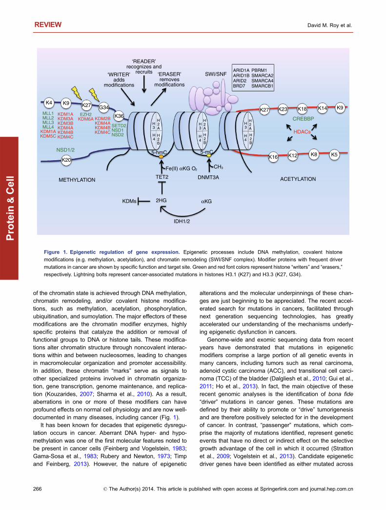
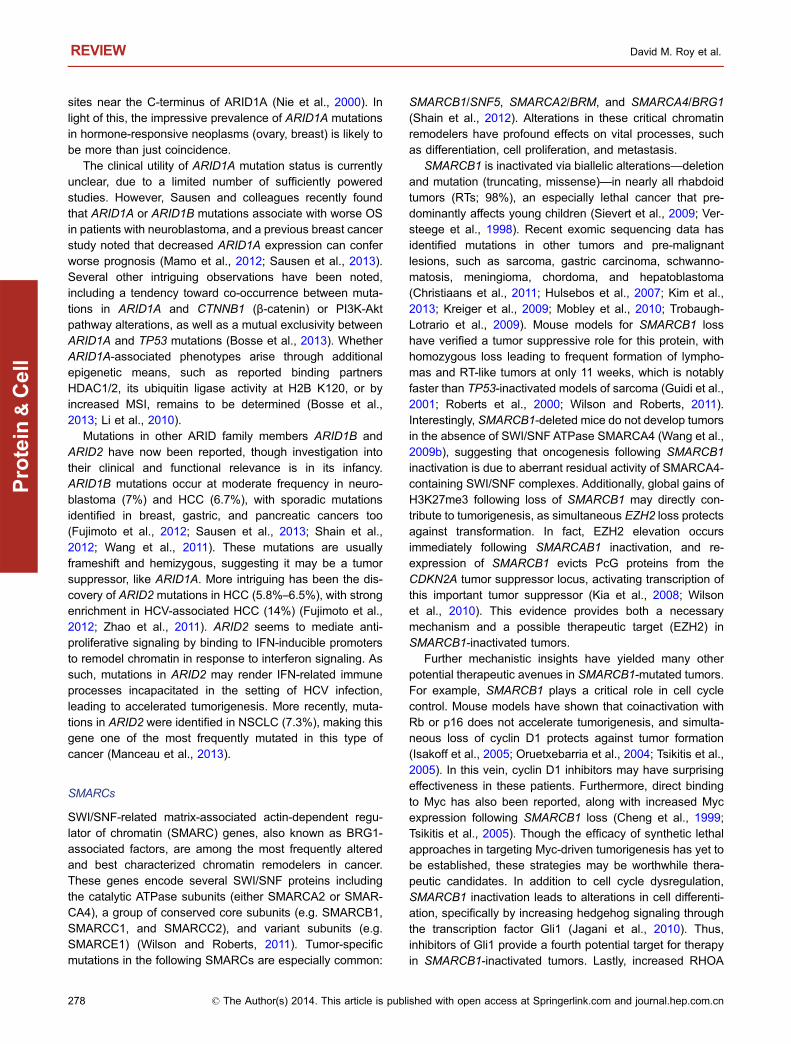
of the chromatin state is achieved through DNA methylation,chromatin remodeling, and/or covalent histone modifica-tions, such as methylation, acetylation, phosphorylation,ubiquitination, and sumoylation. The major effectors of thesemodifications are the chromatin modifier enzymes, highlyspecific proteins that catalyze the addition or removal offunctional groups to DNA or histone tails. These modifica-tions alter chromatin structure through noncovalent interac-tions within and between nucleosomes, leading to changesin macromolecular organization and promoter accessibility.In addition, these chromatin “marks” serve as signals toother specialized proteins involved in chromatin organiza-tion, gene transcription, genome maintenance, and replica-tion (Kouzarides, 2007; Sharma et al., 2010). As a result,aberrations in one or more of these modifiers can haveprofound effects on normal cell physiology and are now well-documented in many diseases, including cancer (Fig. 1).
It has been known for decades that epigenetic dysregu-lation occurs in cancer. Aberrant DNA hyper- and hypo-methylation was one of the first molecular features noted tobe present in cancer cells (Feinberg and Vogelstein, 1983;Gama-Sosa et al., 1983; Rubery and Newton, 1973; Timpand Feinberg, 2013). However, the nature of epigenetic
alterations and the molecular underpinnings of these chan-ges are just beginning to be appreciated. The recent accel-erated search for mutations in cancers, facilitated throughnext generation sequencing technologies, has greatlyaccelerated our understanding of the mechanisms underly-ing epigenetic dysfunction in cancers.
Genome-wide and exomic sequencing data from recentyears have demonstrated that mutations in epigeneticmodifiers comprise a large portion of all genetic events inmany cancers, including tumors such as renal carcinoma,adenoid cystic carcinoma (ACC), and transitional cell carci-noma (TCC) of the bladder (Dalgliesh et al., 2010; Gui et al.,2011; Ho et al., 2013). In fact, the main objective of theserecent genomic analyses is the identification of bona fide“driver” mutations in cancer genes. These mutations aredefined by their ability to promote or “drive” tumorigenesisand are therefore positively selected for in the developmentof cancer. In contrast, “passenger” mutations, which com-prise the majority of mutations identified, represent geneticevents that have no direct or indirect effect on the selectivegrowth advantage of the cell in which it occurred (Strattonet al., 2009; Vogelstein et al., 2013). Candidate epigeneticdriver genes have been identified as either mutated across
CH3
IDH1/2
2HG αKG
TET2
KDMs
K36
K27K9K4
K20
EZH2KDM6A
SETD2NSD1NSD2
NSD1/2
KDM1AKDM3AKDM3BKDM4AKDM4BKDM4C
KDM2BKDM4AKDM4BKDM4C
METHYLATION ACETYLATIONDNMT3A
‘WRITER’adds
modifications
‘ERASER’removes
modifications
‘READER’recognizes and
recruits SWI/SNF
K9K14K18K23
K16
K27
K12 K8 K5
CREBBPMLL1MLL2MLL3MLL4
KDM1AKDM5C
G34
5-mC5-hmC
Fe(II) αKG O2
ARID1AARID1BARID2BRD7
PBRM1SMARCA2SMARCA4SMARCB1
HDACs
H2A
H3
H3
H3
H4
H4
H4
H2A
H2A
H2B
H2B
H2B
Figure 1. Epigenetic regulation of gene expression. Epigenetic processes include DNA methylation, covalent histone
modifications (e.g. methylation, acetylation), and chromatin remodeling (SWI/SNF complex). Modifier proteins with frequent driver
mutations in cancer are shown by specific function and target site. Green and red font colors represent histone “writers” and “erasers,”
respectively. Lightning bolts represent cancer-associated mutations in histones H3.1 (K27) and H3.3 (K27, G34).
REVIEW David M. Roy et al.
266 © The Author(s) 2014. This article is published with open access at Springerlink.com and journal.hep.com.cn
Protein
&Cell
many cancers (e.g. KDM6A in over 12 cancers), havinghighly recurrent mutations (e.g. IDH1 R132), or having highlyprevalent mutations in select tumor histologies (e.g. MLL2 infollicular lymphoma) (You and Jones, 2012). The charac-terization of these driver gene mutations has enhanced ourunderstanding of the mechanisms contributing to oncogen-esis, allowed for improved prognostic assessment, andopened the door for the development of exciting new ther-apies. In this review, we highlight recent developments, newdiscoveries, and therapeutic advances involving cancer-associated mutations in epigenetic regulators.
DNA METHYLATION AND HYDROXYMETHYLATION
DNA methylation plays a well-defined role in both develop-ment and disease, including cancer. First identified in 1975,CpG island (CGI) methylation was shown to function as arelatively stable alteration on DNA that can serve to silencegene transcription (Holliday and Pugh, 1975; Riggs, 1975).We now understand that DNA methylation is much moredynamic and complex, with diverse epigenetic conse-quences linked to varied genomic locations of where thismark occurs. For example, DNA methylation at gene pro-moter CGIs potently blocks the initiation of transcription,whereas methylation within CpG-poor gene bodies mayactually facilitate elongation and influence patterns of alter-nate splicing. In addition, DNA methylation is frequentlyfound in repeat-rich areas of the genome and is vital for bothchromosomal and genomic stability, possibly through therepression of retroviral transposons (Jones, 2012). Still, therole for this epigenetic mark at other regulatory regions, suchas enhancers and insulators, has yet to be determined.Regardless, aberrant methylation in human cancer is adefining feature, with global promoter CGI hypermethylationand non-CGI hypomethylation widely reported (Ehrlich,2002; Sharma et al., 2010). Furthermore, local variations inmethylation at only several key loci have been shown to besufficient for tumorigenesis (Lee et al., 2008; Poage et al.,2011). Importantly, these altered patterns of DNA epigeneticmarks (e.g. 5-mC, 5-hmC) are frequently accompanied by acritical imbalance in transcriptional programs involving dif-ferentiation and stem cell maintenance, thereby initiatingtumorigenesis and sustaining growth (Jones and Baylin,2007).
DNA methylation can function to silence tumor suppres-sor genes along with genetic mutations (Herman and Baylin,2003). For example, in the case of hereditary gastric cancer,methylation of CDH1 (which encodes the E-cadherin tumorsuppressor) can function as a “second hit” and cause gastriccancer when the first allele is mutated (Grady et al., 2000). Insporadic cancers, tumor suppressor genes that are mutatedin hereditary versions of the disease are frequently silencedby DNA methylation instead (Esteller et al., 2001). Forexample, in hereditary nonpolyposis colon cancer (HNPCC),MLH1 inactivation via mutation can lead to microsatellite
instability (MSI) and tumorigenesis, whereas in sporadiccolon cancers, MLH1 is frequently silenced by methylation(Kane et al., 1997; Veigl et al., 1998). These data and othersindicate that aberrant DNA methylation can work along withgenetic alterations to promote tumorigenesis.
DNMT3A
DNA methylation is carried out by the mammalian DNAmethyltransferases (DNMTs), essential enzymes that cata-lyze the addition of a methyl group to cytosine in CpGdinucleotides in DNA (Jones, 2012). The conversion of5-cytosine (5-C) to methyl-cytosine (5-mC) requires thepresence of a methyl donor, S-adenosylmethionine (SAM),and one of the following catalytically active DNMTs: DNMT1,DNMT3A, or DNMT3B (Shen et al., 1992). Although there issome evidence for overlapping roles, DNMT3A andDNMT3B are essential for de novo DNA methylation,whereas DNMT1 “maintains” heritable methylation patternsacross the genome during cell replication (Hsieh, 1999). Infact, the role of DNMT3A in de novo methylation was initiallycharacterized in the context of epigenetic silencing duringdevelopment, including at imprinted loci in germ cells (Kan-eda et al., 2004; Okano et al., 1999). The DNMTs have longbeen suspected to play a role in oncogenesis, as well.Aberrations in DNA methylation—both hyper- and hypo-methylation—have been well-documented in patient tumorsand cell lines for decades (El-Osta, 2004). Additionally, Okaand colleagues first showed that in some cases, DNMT3Aand DNMT3B, not DNMT1, mediate the cytotoxic effects of5-aza-dC, a therapeutic mainstay in the treatment of severalhematopoietic malignancies (Oka et al., 2005; Plimack et al.,2007). Not long after, DNMT3A and DNMT3B were furtherimplicated in both hematopoietic stem cell (HSC) renewaland differentiation, two tightly regulated processes whoseperturbation can lead to carcinogenesis (Challen et al., 2012;Tadokoro et al., 2007).
DNMT3A somatic mutations were first discovered byYamashita and colleagues following sequencing of tissuefrom adult patients with de novo acute myeloid leukemia(AML) (Yamashita et al., 2010). Soon after, DNMT3A muta-tions were reported in AML cohorts from others, with fre-quencies as high as 22.1% (Ley et al., 2010). The majority ofmutations occur at R882 (60%–64%), and almost all areheterozygous (Ley et al., 2010; Thol et al., 2011a). DNMT3Amutations are enriched in AML patients with intermediate-risk cytogenetics and normal karyotype (Lin et al., 2011;Patel et al., 2012). They are also associated with increasedage, M4 and M5 AML subtypes, worse overall survival (OS)and relapse-free survival (RFS), and increased blasts atdiagnosis (Hou et al., 2012; Ley et al., 2010; Lin et al., 2011;Marcucci et al., 2012; Thol et al., 2011a; Yan et al., 2011).There is also evidence that DNTM3A mutations may be aprognostic marker for decreased time to treatment failure(TTF), duration of complete remission (CR), and disease-
Driver mutations of cancer epigenomes REVIEW
© The Author(s) 2014. This article is published with open access at Springerlink.com and journal.hep.com.cn 267
Protein
&Cell

free survival (DFS), at least in cytogenetically normal AML(Marcucci et al., 2012; Thol et al., 2011a; Yan et al., 2011).These studies have also identified several frequently co-occurring mutations, which include NPM1, FLT3, IDH1/2,and less commonly, TET2 (Hou et al., 2012; Ley et al., 2010;Lin et al., 2011; Marcucci et al., 2012; Renneville et al., 2012;Thol et al., 2011a; Yan et al., 2011). As alterations in epi-genetic regulators frequently lead to genomic instability,DNMT3A mutation may further drive progression of the dis-ease itself (Wakita et al., 2013). Further, recent experimentalwork has demonstrated that DNMT3A mutations at R882 areonly found in major clones, suggesting this genetic alterationmay be an initiating lesion in AML (Bisling et al., 2013).
DNMT3A mutations have also been described, albeit atlower frequency, in other myeloid malignancies, such asmyelodysplastic syndromes (MDS; 3%–8%) and myelopro-liferative neoplasms (MPNs; 2%–10%), as well as earlyT-cell precursor acute lymphoblastic leukemia (ETP-ALL;16%–18%) (Brecqueville et al., 2011; Ewalt et al., 2011;Grossmann et al., 2013; Neumann et al., 2013; Stegelmannet al., 2011; Thol et al., 2011b; Traina et al., 2013; Walteret al., 2011). Similar to what is observed in de novo AML,R882 is most frequently targeted for mutation (60%) in theseneoplasms (Thol et al., 2011b). Clinically, DNMT3A muta-tions also correlated with increased age and predictedprognosis in all types, including worse OS, event-free sur-vival (EFS), and AML-free survival (Lin et al., 2011; Neu-mann et al., 2013; Renneville et al., 2012; Thol et al., 2011b;Walter et al., 2011). Paradoxically, univariate and multivari-ate analysis of 92 patients with MDS revealed DNMT3Amutations were correlated with better overall response rate(ORR) and progression-free survival (PFS) (Traina et al.,2013). Interestingly, similar to AML, MPNs showed anassociation between DNMT3A alterations and mutation inJAK2, IDH1/2, and ASXL1 but not TET2 (Stegelmann et al.,2011). However, studies in MDS and ETP-ALL have foundno association between DNMT3A mutations and otherknown leukemogenic drivers, including FLT3 (Neumannet al., 2013; Thol et al., 2011b).
DNMT3A is a 102 kDa protein with three highly conservedfunctional domains: An N-terminal PWWP domain, a cys-teine-rich PHD zinc finger domain, and a C-terminal catalyticdomain (Hermann et al., 2004). DNMT3A mutations in can-cer have been reported in all three domains, with mostoccurring in the catalytic domain, including the R882 muta-tion (60%) (Ley et al., 2010). Still, many cancer-specificmutations occur in non-catalytic domains. The PWWPdomain is essential in localizing DNMT3A to heterochromaticregions of DNA during interphase, though it is unclear if thisis related to its reported ability to bind DNA directly (Bach-man et al., 2001; Ge et al., 2004; Purdy et al., 2010; Suetakeet al., 2011). Alternatively, the PHD domain has also beenshown to mediate regional specificity and repression throughits interactions with transcriptional repressor RP58,HP1beta, histone deacetylatases (HDACs), and SUV39H1
(Datta et al., 2003; Fuks et al., 2001; Fuks et al., 2003).Exactly how these mutations disrupt protein function is ofgreat interest, although a single unifying mechanism isunlikely to exist. Likewise, a recent clinical study found thatR882 mutations confer poor prognosis in older populations,whereas non-R882 mutations confer poor prognosis inyounger patients (Marcucci et al., 2012).
Whether these reported mutations are loss-of-function,gain-of-function, or act via a dominant-negative mechanismhas also been debated. A strong case can be made thatDNMT3A is an oncogene, as it is overexpressed in severalcancers, depletion results in decreased proliferation andmetastasis, and 5-aza-dC causes apoptosis through directinhibition of DNMT3A (Oka et al., 2005). In regards tomutations themselves, the R882H DNMT3A mutant wassufficient to promote tumorigenesis in an IL-3 dependenttransformation assay in leukemic 32D cells (Yan et al., 2011).Alternatively, some studies have demonstrated that mutantDNMT3A (R882H) has a markedly reduced catalytic ability(∼50%) in methyltransferase assays and decreased DNA-binding capacity in vitro, implying a possible loss-of-functionphenotype via a dominant-negative mechanism (Jia et al.,2007; Ley et al., 2010; Yamashita et al., 2010). Clearly,additional studies are necessary to understand the exactnature by which these cancer-associated mutations aretransforming.
Characterizing larger DNMT3A-induced changes in thecancer methylome has proven quite challenging. Ley andcolleagues showed no changes in genome-wide methylationaccording to DNMT3A status in AML, and although 182specific regions showed increased hypomethylation inmutant samples, this did not correlate with gene expression(Ley et al., 2010). However, in a more recent cohort, a total of3878 genomic regions were found to have significantly dif-ferent methylation patterns using MeDIP-chip and differ-ences of expression levels in 889 of 20,723 annotated geneswas observed via an Affymetrix microarray (Yan et al., 2011).Further, this group found during RT-PCR validation that theexpression of several HOX family genes significantlyincreased in DNMT3A mutant samples compared to wild-type.
Recent experiments may offer some insight behind theconflicting methylation and functional data. Protein binding atthe DNMT3A tetramerization interface is important formethylation patterning, inducing processive methylation ofclustered sites (Jia et al., 2007). Most mutations, includingR882, occur within this tetramer interface. Therefore, differ-ences between oligomerization states can explain howDNMT3A mutations alter epigenetic silencing and lead totransformation, without global changes in DNA methylation(Holz-Schietinger et al., 2011; Holz-Schietinger et al., 2012).Although the commonly occurring R882H mutation does notdisrupt DNMT3A association with required cofactor DNMT3Lin vitro, the latter is only expressed in early development(Chedin et al., 2002; Jia et al., 2007; Webster et al., 2005;
REVIEW David M. Roy et al.
268 © The Author(s) 2014. This article is published with open access at Springerlink.com and journal.hep.com.cn
Protein
&Cell

Yamashita et al., 2010). However, other binding proteins,such as thymine-DNA glycosylase and ecotropic viral inter-gration site 1, can adhere to sites within the DNMT3A cat-alytic domain and may explain altered mutant DNMT3Aactivity (Li et al., 2007; Senyuk et al., 2011). In addition, non-catalytic mutations may disrupt protein binding to otherdomains, as described above. Although the exact mecha-nism remains elusive, these mutations result in decreasedmethylation processivity and altered localization, possibly toeuchromatic regions of DNA (Jurkowska et al., 2011). Thismay help explain how DNMT3A mutations drive tumor for-mation in hematopoietic malignancies, even in the absenceof larger global methylation changes.
TET2
The ten-eleven translocation (TET) family proteins were firstdiscovered in cancer with the fusion of TET1 to MLL in selectAML patients with t(10;11)(q22;q23) (Lorsbach et al., 2003).Mechanistic studies then showed the TET proteins aredioxygenases that depend on 2-oxoglutarate, oxygen, Fe(II),and ascorbic acid to catalyze the conversion of 5-mC to5-hydroxymethylcytosine (5-hmC) at CpG regions in DNA(Blaschke et al., 2013; Ito et al., 2010; Minor et al., 2013;Tahiliani et al., 2009; Yin et al., 2013). TET enzymes may beresponsible for DNA demethylation through both passiveand active means. For example, CpG dinucleotides that are“marked” with 5-hmC are not recognized by DNMT1 andtherefore, methylation is passively lost at these sites throughrepeated cycles of cell division (Valinluck and Sowers,2007). Alternatively, active demethylation can proceed fol-lowing placement of 5-hmC via the activation-induced cyti-dine deaminase (AID)-APOBEC DNA repair pathway (Guoet al., 2011). More recently, an even greater role for TETenzymes in active demethylation was shown in vitro, withTET enzymes proving sufficient for converting 5-mC to5-hmC, 5-hmC to 5-formylcytosine (5-fC), and finally 5-fC to5-carboxylcytosine (5-caC). 5-caC is then targeted by baseexcision repair enzymes to complete the demethylationprocess (He et al., 2011; Ito et al., 2011).
TET2 was first suspected to have a role in cancer whensix patients with either secondary AML (sAML) or MDS werenoted to have minimal deletions via FISH on chromosome4q24 (Viguie et al., 2005). Soon after, the first TET2 somaticmutations were identified in 25 patients (14%) with MPNs(Delhommeau et al., 2008). Delhommeau and colleagesthen sequenced TET2 in patient tumor samples, becomingthe first group to identify TET2 mutations in multiple myeloidneoplasms, including MDS (19%), MPNs (12%–14%), andsAML (24%). They concluded that TET2 was a novel bonafide tumor suppressor, noting that the majority of mutationsare heterozygous (55%) and that TET2 defects precede thewell-known JAK2 V617F driver mutation in MPN HSCs(Delhommeau et al., 2009). More recently, Schaub and col-leagues disputed this result using colony formation assays to
show that TET2 mutations can either precede (4 of 8patients), follow (2 of 8), or occur independently (2 of 8) ofJAK2 V617F mutations in MPN patient samples (Schaubet al., 2010). Although the temporal relationship betweenTET2 mutations and other leukemogenic drivers is stillunclear, the frequency and ubiquitous nature of thesemutations in cancer is quite revealing. In addition to MDS,sAML, and MPNs, TET2 mutations have now been descri-bed in other myeloid neoplasms, such as de novo AML(12%) and chronic myelomonocytic leukemia (CMML; 42%–
46%) (Abdel-Wahab et al., 2009; Smith et al., 2010).Unlike DNMT3A mutations, TET2 alterations seem to
hold limited prognostic utility in leukemia. The overwhelmingmajority of studies published to date have found no changein OS or any other prognostic tools between patients har-boring TET2mutations and those who are not. However, onestudy in a cohort of 96 MDS patients reported that TET2mutations conferred an OS, EFS, and AML-free survivaladvantage (Kosmider et al., 2009). Paradoxically, Abdel-Wahab and colleagues found that TET2 mutations werelinked to worse OS in 93 patients with de novo AML, and arecent whole-exome sequencing study reported worse EFSin AML patients with TET2 mutation (Abdel-Wahab et al.,2009; Weissmann et al., 2012). Further, Nibourel and col-leagues reported no association with OS or DFS in theircohort of de novo AML patients, though the prevalence ofTET2 mutations in patients who failed to achieve completeremission (CR) trended higher (27% vs. 17%) (Nibourelet al., 2010). Despite questionable association to patientoutcomes, TET2 mutations are linked with other clinicalfeatures, including monocytosis, leukocytosis, and advancedage at diagnosis (Jankowska et al., 2009; Smith et al., 2010;Tefferi et al., 2009a; Tefferi et al., 2009c). Although TET2mutations show little association to known myeloid leuke-mogenic drivers—FLT3-ITD, RUNX1, CEBPA—they doassociate with NPM1 and ASXL1 mutations and infrequentlyco-occur with IDH1 or IDH2 mutations (Chou et al., 2011a;Weissmann et al., 2012). Lastly, despite some data indicat-ing no association between cytogenetics and TET2 status inMPN, TET2 mutations occur more often in the presence ofnormal karyotype and intermediate-risk AML (Hussein et al.,2010; Weissmann et al., 2012). In this cytogenetic setting,TET2 mutations do predict significantly worse OS in AML(Chou et al., 2011a).
Although few TET2 recurrent mutations have beenreported, many mutations result in a frameshift or early stopcodon and are therefore inactivating (Tefferi et al., 2009b). Infact, the largest proportion of nonsense mutations occur inexon 3, resulting in a truncated protein lacking the C-terminalcatalytic domain (Moran-Crusio et al., 2011). Additionally,several missense mutations have been characterized asloss-of-function, with Ko and colleagues reporting impairedhydroxylation of 5-mC when mutant TET2 was overexpres-sed in HEK-293Tcells. Furthermore, TET2mutation status issignificantly correlated with decreased global 5-hmC in
Driver mutations of cancer epigenomes REVIEW
© The Author(s) 2014. This article is published with open access at Springerlink.com and journal.hep.com.cn 269
Protein
&Cell
myeloid tumors (Ko et al., 2010; Konstandin et al., 2011).Functional studies manipulating TET2 have also been ableto recapitulate phenotypes that are characteristic of myeloidneoplasms, suggesting that TET2 loss may be a key event inleukemic transformation. For example, a conditional mousemodel for TET2 loss in the hematopoietic compartmentresulted in increased HSC self-renewal and myeloprolifera-tion including splenomegaly, monocytosis, and extramedul-lary hematopoiesis (Moran-Crusio et al., 2011). This isconsistent with other studies showing that TET2 inactivationleads to decreased 5-hmC in HSCs, amplification of the stemcell population, and may skew HSCs toward a myeloidlineage (Pronier et al., 2011; Quivoron et al., 2011).
The true effect of TET2 mutations on DNA methylationstatus has been difficult to ascertain. Despite an expectedincrease in 5-mC following TET2 inactivation, several stud-ies have reported a global decrease in methylation (Ko et al.,2010; Yamazaki et al., 2012). However, analysis of specificgene promoters shows mixed results in TET2 mutant sam-ples, frequently exhibiting promoter-specific hypermethyla-tion in spite of global hypomethylation (Perez et al., 2012;Wu et al., 2011; Yamazaki et al., 2012). Still, Ko and col-leagues noted that several AML patients with wild-type TET2had 5-hmC levels very similar to those patients with mutantTET2 (Ko et al., 2010). This suggests a more complexrelationship between TET2, DNA methylation status, andmalignant transformation.
In the past year, exciting new evidence has emerged tosuggest a more diverse role for TET2 in epigenetic regula-tion. In addition to known associations with polycombrepressive complex (PRC) regulator SIN3A and NURDcomplex member MBD3 (Wu et al., 2011; Yildirim et al.,2011), TET2 was recently identified as a direct bindingpartner with O-linked beta-N-acetylglucosamine transferase(OGT), an enzyme that marks histone H2B S112 at activetranscription start sites (TSS) (Chen et al., 2013b). AlthoughOGT doesn’t influence TET2 activity in functional assays,TET2 seems to actively target OGT to unmethylated pro-moters and activate transcription via other means (Vellaet al., 2013). Furthermore, Deplus and colleagues showedthat TET2 and OGT co-localize at active promoters markedby H3K4me3 through a direct interaction with host-cell factor1 (HCF1) and that knockdown of TET2 leads to globaldecreases of H3K4me3 and GlcNAcylation (Deplus et al.,2013). Another direct interaction has been describedbetween TET2 and EBF1, a transcription factor that isassociated with transcriptional activation and “poised” chro-matin (Guilhamon et al., 2013; Treiber et al., 2010). Addi-tional binding partners have also been reported within thepast year, including NANOG and IDAX (Costa et al., 2013;Ko et al., 2013). Lastly, several novel miRNAs were dis-covered to negatively regulate TET2 expression, offering apossible explanation for TET-associated transformation inthe absence of any genomic alterations (Cheng et al., 2013;Fu et al., 2013; Song et al., 2013). Collectively, evidence is
mounting that TET2 inactivation in cancer may alter morethan just DNA methylation; in fact, transformation may resultconsiderably from disrupted interactions with other epige-netic regulators and development-associated transcriptionfactors.
IDH1/2
Isocitrate dehydrogenase 1 (IDH1) and isocitrate dehydro-genase 2 (IDH2) are two homodimeric metabolic enzymesthat convert isocitrate to α-ketoglutarate (α-KG) whilereducing NADP+ to NADPH. IDH1 is present in the cytosoland peroxisomes whereas IDH2 is located exclusively inmitochondria (Geisbrecht and Gould, 1999; Winkler et al.,1986; Xu et al., 2004). Frequent recurrent mutations in IDH1were initially discovered in GBM (12%) following whole-ex-ome sequencing of 22 patient tumor samples (Parsons et al.,2008). Further sequencing efforts revealed that mutationsare most prevalent in WHO grade II/III gliomas (71%) andsecondary GBMs (88%) but less common in primary GBMs(7%) (Balss et al., 2008). Subsequent studies have shownthat IDH2 mutations are also enriched in WHO grade II/IIIgliomas, albeit less frequently, and that IDH1/2 mutationsoccur in a mutually exclusive manner (Hartmann et al.,2009). These data indicate IDH mutation is an early event inglioma oncogenesis, frequently preceding known alterationslike TP53 mutation and 1p/19q loss (Watanabe et al., 2009).Interestingly, recent data suggest IDH1 and IDH2 mutationsmay actually differentially associate with astrocytoma andoligodendrogliomas, respectively (Hartmann et al., 2009).IDH mutations are associated with MGMT promoter hyper-methylation, TP53 mutation, 1p/19q codeletion, ATRX inac-tivation, younger age, and improved prognosis while beinginversely correlated with EGFR amplification in glioma (Chouet al., 2010; Wiestler et al., 2013; Yan et al., 2009; Zou et al.,2013). Further, although early studies could not find any IDHmutations in other types of solid tumors (Bleeker et al., 2009;Kang et al., 2009), recurrent mutations have since beenidentified in chondrosarcoma (56%), cholangiocarcinoma(23%), melanoma (10%), and prostate cancer (2%) (Amaryet al., 2011; Borger et al., 2012; Ghiam et al., 2012; Shibataet al., 2011).
Soon after the discovery of IDH mutations in glioma,recurrent mutations of IDH were also identified in AML(Green and Beer, 2010; Mardis et al., 2009). Similar to gli-oma, IDH1 and IDH2 mutations are mutually exclusive,though the mutational frequency of IDH in AML is muchlower (23%) (Chou et al., 2011b; Ward et al., 2010). Incontrast, the utility of IDH mutation status as an independentprognostic marker in AML is less clear. In a convincingcohort of 493 adult patients with AML, Chou and colleaguesfound that IDH1 mutation had no impact on OS (Chou et al.,2010). Still, other studies have suggested a more disparaterole, with mutation in IDH1 and IDH2 conferring poor andimproved prognosis, respectively (Chou et al., 2011b;
REVIEW David M. Roy et al.
270 © The Author(s) 2014. This article is published with open access at Springerlink.com and journal.hep.com.cn
Protein
&Cell
Schnittger et al., 2010). Notably, although another largecohort of 805 patients found that IDH mutation did not cor-relate with prognosis, a subset of patients with IDH mutationand CN-AML, NPM1 mutation, and no FLT3-ITD did showsignificantly reduced OS and RFS (Paschka et al., 2010).Therefore, it is likely that the use of prognostic subsets—IDHstatus along with other genetic markers—may improve theutility of IDH status as a biomarker in AML. Other featuresthat correlate with IDH mutation status include normalkaryotype, intermediate-risk cytogenetics, NPM1 mutation,and M1 AML subtype (Chou et al., 2010; Schnittger et al.,2010).
To date, practically all IDH mutations found in cancer areheterozygous and highly recurrent. Amino acid substitutionsat residues IDH1-R132, IDH2-R172 and IDH2-R140Q arethe most common, with considerable variability at R132 (R/H, R/C, R/S, R/G, R/L, R/V) and R172 (R/G, R/M, R/K)(Balss et al., 2008; Yan et al., 2009). The remarkableabsence of any frameshift or nonsense mutations, deletions,or epigenetic silencing provided early evidence that IDHmutations were activating (Flanagan et al., 2012; Zhao et al.,2009). The R132 residue is located in the active site of IDH1where it forms 2 hydrogen bonds with α- and β- carboxylateof isocitrate, its substrate (Nekrutenko et al., 1998; Xu et al.,2004). Initially, it was believed that these mutations may beloss-of-function or dominant-negative, as mutant IDHshowed a reduced affinity for isocitrate and producedmarkedly less α-KG and NADPH in vitro (Yan et al., 2009;Zhao et al., 2009). However, an in vitro metabolite screenrevealed that IDH mutations are neomorphic, producing thenovel oncometabolite 2-hydroxyglutarate (2-HG) throughheterodimer formation with the remaining wild-type IDH1.This was also verified in patient samples, with a strongcorrelation between the amount of 2-HG in tumor tissue andIDH1/2 mutation status (Dang et al., 2009; Ward et al.,2010). In fact, 2-HG levels are increased 10–100 fold inpatient sera and can be used to reliably diagnose IDH statusand monitor response to therapy, though this applicationmay be restricted to myeloid neoplasms (Capper et al., 2012;DiNardo et al., 2013; Ward et al., 2010).
The effects of mutant IDH are pleiotropic and affectnumerous cell processes including DNA methylation, histonemethylation, HIF1a signaling, collagen synthesis, andmetabolism (Cairns and Mak, 2013). Remarkably, α-KGlevels are unchanged in mutant IDH AML and glioma (Danget al., 2009; Gross et al., 2010), and it is now clear that 2-HG-mediated inhibition of 2-OG-dependent dioxygenases is thedominant mechanism by which IDHmutations are oncogenic(Xu et al., 2011). Early data from the Cancer Genome Atlasproject first identified the glioma hypermethylator phenotype(G-CIMP) in GBM and its association with IDH mutations(Laffaire et al., 2011; Noushmehr et al., 2010). Following this,Figueroa and colleagues demonstrated that 2-HG inhibitionof the α-KG-dependent enzyme TET2 actively generates thehypermethylator phenotype in AML. Further, they showed
that TET2 and IDH mutations are mutually exclusive in AML,result in overlapping methylation signatures, and impair HSCdifferentiation in 32D myeloid cells (Figueroa et al., 2010).Similarly, work from our lab demonstrated that IDH1mutationdirectly causes the G-CIMP phenotype, reduces global5-hmC through TET2 inhibition, results in hypermethylationof the repressive histone marks H3K9 and H3K27, andblocks differentiation (Turcan et al., 2012). It is important tohighlight that widespread loss of 5-hmC is an additionalepigenetic hallmark in IDH or TET2 mutated cancers,including melanoma, and that reestablishment of the 5-hmClandscape can suppress tumor invasion and growth in bothmelanoma cells and a zebrafish model (Lian et al., 2012).Interestingly, IDH-associated increases in histone methyla-tion are likely due to 2-HG-mediated inhibition of the JumonjiC (JmjC)-domain-containing histone demethylases (Lu et al.,2012). Still, others have proposed additional mechanisms inIDH-mutated cancers such as HIF1a stabilization throughPHD inhibition, altered ECM structure due to decreasedhydroxylation of collagen, and possible metabolic shifts inNADP/NADPH ratio (Sasaki et al., 2012; Zhao et al., 2009).
Recently, several exciting studies have shed light onnovel mechanisms by which IDH mutations initiate malignanttransformation and how underlying mechanisms may beexploited for therapeutic gain. In mouse models of leukemiaand melanoma, IDH mutants accelerated cell cycle transitionby activation of the MAPK/ERK pathway and repression oftumor suppressors CDKN2A and CDKN2B (Chaturvediet al., 2013; Shibata et al., 2011). Although several studieshave noted that IDH mutations cause increased colony for-mation in soft agar and enhanced proliferation, two mousemodels for leukemia found that IDH mutation primes cells byinducing an MDS- or MPN-like state. However, combiningIDH1 mutants with HOXA9, or IDH2 mutants with FLT3 orNRAS, was sufficient to initiate transformation (Chaturvediet al., 2013; Chen et al., 2013a; Xu et al., 2011). This may becancer-specific though, as mutant IDH2 alone was recentlyshown to be sufficient to induce sarcoma formation in mice,at least in one model system (Lu et al., 2013). Regardless,the primary mechanism underlying IDH-induced oncogene-sis in several model systems is a block in cell differentiation(Pirozzi et al., 2013). Both groups showed restoration ofdifferentiation and increased apoptosis following treatmentwith IDH inhibitor HMS-101 or Brd4 inhibitor JQ1. Similarly,Losman and colleagues showed that IDH mutant leukemictransformation is specific to the (R)-enantiomer of 2-HG,which can independently promote cytokine independenceand block differentiation. Again, this transformation wasreversible with IDH inhibitor AGI-5198 (Losman et al., 2013).Lastly, IDH inhibition can reverse novel EMT-associatedexpression patterns, though a lengthy delay to phenotypicchange suggests more stable epigenetic alterations may beto blame (Grassian et al., 2012).
Despite the targeted nature of IDH inhibitors, IDH muta-tion likely unleashes epigenetic marks that are themselves
Driver mutations of cancer epigenomes REVIEW
© The Author(s) 2014. This article is published with open access at Springerlink.com and journal.hep.com.cn 271
Protein
&Cell
selectable, such as DNA methylation. There is thus interestin the therapeutic potential of 5-azacytidine (5-aza) anddecitabine (DAC), due to the complex downstream effects ofIDH mutations on the cancer methylome. In order to deter-mine if broader epigenetic therapies could “unlock” gliomainitiating cells (GIC) from a dedifferentiated state, we treatedboth wild-type and mutant IDH cell lines with DAC and/orIDH inhibitor AGI-5198. Along with Borodovsky and col-leagues, our group found that hypomethylating agentspotently induce differentiation, impair colony formation, andsuppress in vivo growth in IDH mutant cells only (Borodov-sky et al., 2013; Turcan et al., 2013). This demonstrates thatmutant IDH-induced DNA methylation likely plays a role inmaintaining the self-renewal capacity of glioma tumor initi-ating cells. Similar effectiveness has also been seen inleukemia and chondrosarcoma (Chaturvedi et al., 2013;Chen et al., 2013a; Lu et al., 2013). Interestingly, IDH inhi-bition using the mutant IDH1 inhibitor AGI-5198 was notnearly as effective, which suggests that broader epigenetictherapies may be necessary to reverse more permanentchanges induced by long exposure to mutant IDH. In addi-tion, combination therapy with other IDH affected processessuch as histone hypermethylation may have a role andwarrant further investigation.
HISTONE METHYLATION
Histone methylation is a reversible process that takes placeat the amino acid side chains of lysine, arginine, and histi-dine residues. Lysine methylation on histones H3 and H4 isthe best characterized and catalyzed by the lysine methyl-transferases (KMTs) through the required methyl groupdonor SAM. All of the KMTs except DOTL1/KMT4 have acatalytically active SET domain and are highly specific toboth histone residue and degree of methylation (mono- vs.di- vs. tri-methylation) (Feng et al., 2002; Rea et al., 2000).Generally, methylation at H3K4, H3K36, and H3K79 corre-sponds to euchromatic or transcriptionally active regions ofthe genome, whereas methylation at H3K9, H3K27, andH4K20, is associated with heterochromatic regions and genesilencing. In addition, each residue is capable of four meth-ylated states: unmethylated or mono-/di-/tri-methylated. Thisprovides further regulatory diversity in the histone code. Forexample, H3K4me2/3 is found at TSSs of active genes,whereas H3K4me1 tends to localize to enhancer regions(Greer and Shi, 2012; Kampranis and Tsichlis, 2009).
In contrast, the lysine-specific demethylases (KDMs)work in opposition to the KMTs through the catalytic removalof methylation marks on histone tails. The two families ofKDMs responsible are the (FAD)-dependent amine oxidasesand the larger JmjC-containing family of α-KG/Fe(II)-iondependent oxygenases (Shi and Whetstine, 2007). KDM1A/B(LSD1/2) and KDM5A-D (JARID1A-D) catalyze demethyla-tion at H3K4, whereas KDM2A/B (JHDM1A/B) and KDM4A-C (JMJD2A-C) target H3K36, leading to repressed gene
transcription at these sites. Alternatively, transcriptionalactivation is induced in part by demethylation at H3K9 andH3K27 by KDM1A, KDM3A-C (JHDM2A-C), or KDM4A-Dand KDM6A/B (UTX/JMJD3), respectively (Varier and Tim-mers, 2011). Due to the broad and essential nature of theseepigenetic marks across the genome, genetic aberrations ofhistone modifiers have powerful effects on vital cellularprocesses such as differentiation and cell cycle control,among others.
Writers (KMTs)
EZH2
EZH2/KMT6 is the enzymatic component of the polycombrepressor complex 2 (PRC2), which is responsible formethylation at H3K27 and subsequent gene silencing (Ki-rmizis et al., 2004). Other essential subunits of the PRC2complex through which EZH2 interacts include embryonicectoderm development (EED), suppressor of zeste 12homologue (SUZ12), and RBAP48/RBBP4. Collectively,these polycomb group (PcG) proteins have been shown toregulate vital cellular processes including differentiation, cellidentity, stem-cell plasticity, and proliferation (Margueron andReinberg, 2011; Shih et al., 2012). As a result, aberrations inany PRC2 component can have powerful physiologic con-sequences on the cell.
Alterations in EZH2 were first discovered in breast andprostate cancer, where amplification and overexpression firstimplied it may function as an oncogene (Kleer et al., 2003;Varambally et al., 2002; Yang and Yu, 2013). This findingwas validated both in vitro and in vivo, with EZH2 overex-pression proving sufficient to drive proliferation in cancercells and transform primary fibroblasts (Bracken et al., 2003;Croonquist and Van Ness, 2005). Recent sequencing stud-ies have identified numerous mutations of EZH2 in a varietyof leukemias and lymphomas, including follicular lymphoma(FL; 7%–22%), diffuse large B-cell lymphoma (DLBCL; 14%–
21.7%), high grade B-cell lymphoma (18%), MDS/MPN (6%–
13%), CMML (11.1%), T-ALL, and AML (Abdel-Wahab et al.,2011; Bodor et al., 2011; Capello et al., 2011; Ernst et al.,2010; Grossmann et al., 2011; Lohr et al., 2012; Makishimaet al., 2010; Morin et al., 2010; Nikoloski et al., 2010; Ryanet al., 2011; Zhang et al., 2012). Interestingly, frequent mis-sense and truncating mutations were observed, which gen-erated some confusion in the field about whether EZH2could possess both pro- and anti-oncogenic functions.Clinically, EZH2 mutations seem to commonly predict poorprognosis—worse OS/leukemia-free survival (LFS), high-risk IPSS score—especially in myeloid malignancies (Gug-lielmelli et al., 2011; Khan et al., 2013; Nikoloski et al., 2010).Additionally, EZH2 mutation associates with BCL2 rear-rangement in FL and germinal center B-cell like DLBCL(GCB-DLBCL) and is notably absent from activated B-celllike DLBCL (ABC-DLBCL) (Beguelin et al., 2013; Morinet al., 2010; Ryan et al., 2011).
REVIEW David M. Roy et al.
272 © The Author(s) 2014. This article is published with open access at Springerlink.com and journal.hep.com.cn
Protein
&Cell
One landmark finding that emerged from the wealth ofrecent sequencing data was the identification of the highlyrecurrent heterozygous Y641 mutation (Y/F, Y/N, Y/H, Y/C)in FL, DLBCL, and other lymphoid neoplasms (Bodor et al.,2013; Bodor et al., 2011; Morin et al., 2010). Initially thoughtto be inactivating due to reduced catalytic ability against ashort H3-like peptide in vitro, Y641 mutant EZH2 exhibited apowerful gain-of-function phenotype when incubated againstthe entire nucleosomal unit (Sneeringer et al., 2010). Inaddition, Sneeringer and others have observed a powerfulsynergy between the wild-type and Y641 forms of EZH2 bothin vitro and in vivo. Whereas EZH2 is very efficient at cata-lyzing monomethylation of H3K27 but not di-/tri-methylation,EZH2 Y641 shows enhanced ability for di-/tri- methylation atH3K27 (Ryan et al., 2011; Wigle et al., 2011; Yap et al.,2011). Similar findings have also been observed with A677Gand A687V mutant EZH2, though these are far less pre-valent in cancer (Majer et al., 2012; McCabe et al., 2012a).Since almost all EZH2 gain-of-function mutations are het-erozygous, the overall consequence of these mutations is acooperative and thereby efficient silencing of genes associ-ated with the repressive H3K27 mark.
Recent studies have shown that mutant EZH2-driventumors can be effectively targeted with small moleculeinhibitors. Knutson and colleagues were the first to describepotent phenotypic effects in lymphoma cell lines, followingtreatment with the SAM-competitive EZH2 inhibitor(EPZ005687) (Knutson et al., 2012). This inhibitor was highlyselective, inducing cell death in mutant EZH2-expressingcells only. As expected, these cells showed global reductionof the H3K27me2/me3 histone mark and significant enrich-ment of cell cycle gene sets by GSEA. The more recentEZH2 inhibitor GSK126 was also highly selective for mutantEZH2 lymphoma cells in vivo and led to increased activationof known EZH2 target genes, such as TXNIP andTNFRSF21 (McCabe et al., 2012b). Although EZH2 muta-tion alone may be insufficient to induce development ofB-cell lymphoma, new evidence suggests it functions as amaster regulator of GCB phenotype through repression ofCDKN1A, IRF4, and PRDM1 (Beguelin et al., 2013). Due tofrequent activation of EZH2 in lymphoma, these new tar-geted therapies hold exciting promise in the clinic.
SETD2
The major KMT responsible for H3K36 trimethylation isSETD2/KMT3A, which is a novel candidate tumor suppres-sor gene (TSG) (Edmunds et al., 2008). Gene deletions inclear cell renal cell carcinoma (ccRCC)-derived cell lines arecommon, reduced expression is seen in breast tumors, andloss is associated with decreased H3K36 trimethylation(Duns et al., 2012; Duns et al., 2010; Newbold and Mokbel,2010). SETD2 mutations are quite common in ccRCC(7.4%–11.6%), pediatric high-grade glioma (HGG; 15%), andadult HGG (8%) (Cancer Genome Atlas Research Network,
2013; Dalgliesh et al., 2010; Duns et al., 2010; Fontebassoet al., 2013; Hakimi et al., 2013b; Varela et al., 2011). Almostall of the mutations characterized so far are frameshift ornonsense and therefore truncating, further suggestingSETD2 may be an important TSG in select malignancies(Hakimi et al., 2013a). Furthermore, Hakimi and colleaguesfound that SETD2 mutations were significantly associatedwith worse cancer-specific survival (CSS) in ccRCC (Hakimiet al., 2013b). Though phenotypic effects of SETD2 inacti-vation have not been clarified, recent research showed thatSETD2 loss triggers MSI and can increase genome-widemutation rates through alterations in H3K36 methylation (Liet al., 2013a; Schmidt and Jackson, 2013).
MLLs
The mammalian mixed lineage leukemia (MLL) family ofgenes encodes a series of active (MLL1–4/KMT2A–D) andinactive (MLL5/KMT2E) KMTs, which have all been impli-cated in cancer. MLL1–4 are responsible for methylation atH3K4 and share a common core formed by WDR5, RbBP5,Dpy-30, and Ash2L (Varier and Timmers, 2011). Notably,MLL1–2 form a complex along with the menin (MEN1) tumorsuppressor and recent evidence shows that H3K27demethylase KDM6A/UTX can complex with MLL2–4(Hughes et al., 2004; Yokoyama and Cleary, 2008; Yokoy-ama et al., 2005).
The earliest known alterations in MLL family genesinvolved frequent rearrangements of MLL1 at 11q23, withrecombination involving more than 40 different partner genesand occurring in 60%–80% of infants with ALL or AML (Di-martino and Cleary, 1999; Pais et al., 2005; Thirman et al.,1993). Since then, several missense and truncating muta-tions have been identified in MLL1 in bladder, lung, andbreast cancer (Gui et al., 2011; Kan et al., 2010). AlthoughMLL1 acts as a dominant oncogene in liquid tumors, thesenew discoveries suggest a different recessive role for MLL1in some solid tumors (Krivtsov and Armstrong, 2007). Infre-quent truncating mutations have also been noted in MLL4 inmedulloblastoma and head and neck squamous cell carci-noma (HNSCC), suggesting a minor but alternative role forthis family member as well (Pugh et al., 2012; Stransky et al.,2011).
Notably, recently, many new mutations have been iden-tified in both MLL2 and MLL3, showing diversity of bothmutation and tumor type. MLL2 and MLL3 mutations arefrequently nonsense or frameshift, resulting in a truncatedprotein lacking the active SET domain (Morin et al., 2011;Parsons et al., 2011; Pasqualucci et al., 2011b; Pugh et al.,2012; Stransky et al., 2011). Along with MLL1/4, MLL2/3mutations suggest a dual role for MLL family proteins inoncogenesis, which may depend heavily on cellular context.In addition, mutations have been discovered in numerouscancers, occasionally at high frequency, including colon(MLL3, 14%–17%), DLBCL (MLL2, 24%–32%), FL (MLL2,
Driver mutations of cancer epigenomes REVIEW
© The Author(s) 2014. This article is published with open access at Springerlink.com and journal.hep.com.cn 273
Protein
&Cell
89%), AML, breast, GBM, RCC, prostate, pancreatic, blad-der, medulloblastoma, and HNSCC (Balakrishnan et al.,2007; Dalgliesh et al., 2010; Gui et al., 2011; Li et al., 2013b;Lindberg et al., 2013b; Mann et al., 2012; Morin et al., 2011;Parsons et al., 2008; Parsons et al., 2011; Pasqualucci et al.,2011b; Pugh et al., 2012; Sjoblom et al., 2006; Stranskyet al., 2011; Vakoc et al., 2009; Watanabe et al., 2011).Unfortunately, functional data remains sparse and theimportance of these mutations has yet to be characterized.Watanabe and colleagues made the interesting observationthat in colorectal carcinoma, MLL3 mutations were associ-ated with increased MSI, though no mechanism has beenproposed (Watanabe et al., 2011). Regardless, they areintriguing candidates for further study in cancer, especiallysince MLL family proteins are important regulators of HOXproteins and differentiation (Wang et al., 2009a).
NSD1/2
Histone-lysine N-methyltransferase NSD2/MMSET was firstimplicated in cancer as a target for rearrangement [t(4;14)(p16.3;q32)] in 15%–20% of multiple myeloma (MM) patients(Chesi et al., 1998). This translocation results in aberrantupregulation of NSD2, which first suggested that it may bean oncogene. Subsequent work has shown that knockdownin MM KMS11 cells leads to apoptosis and re-expression ofwild-type NSD2 causes increased proliferation (Martinez-Garcia et al., 2011). Furthermore, overexpression of wild-type NSD2 is sufficient to transform NSD2-/- cancer cellsin vivo and in mouse embryonic fibroblasts (MEFs) (Kuoet al., 2011). Functionally, interactions with HDAC1/2 andcatalytic activity at H3K4 and H4K20 have been proposed,though these may be minor (Marango et al., 2008). It is nowclear that the NSD2-catalyzed conversion of unmethylatedH3K36 to mono- or di-methylated forms, with concomitantdecreases in H3K27me3, is the dominant mechanism drivingNSD2-associated oncogenic reprogramming (Kuo et al.,2011; Li et al., 2009). In fact, Kuo and colleagues demon-strated that NSD2 SET catalytic activity is required for tran-scriptional activation at several oncogenic loci (TGFA, MET,PAK1, RRAS2). Pathway analyses have also identified thefollowing as significantly altered in mutant NSD2 tumors:TP53 pathway, cell cycle, DNA repair, focal adhesion, andWnt (Kuo et al., 2011; Martinez-Garcia et al., 2011).
Recently, sequencing projects revealed the presence of ahighly recurrent mutation in NSD2 (E1099K), which is pres-ent in 7.5% of pediatric B-ALL and other lymphoid neo-plasms (Jaffe et al., 2013; Oyer et al., 2013). These studiesshowed that NSD2 E1099K leads to enhanced colony for-mation in soft agar and expected increases and decreases inH3K36me2 and H3K27me3, respectively. This new discov-ery has exciting therapeutic potential—similar to the acti-vating mutations in EZH2 described above. Additionally, therelated KMT NSD1 was recently discovered to harbor pointmutations in multiple cancers, including HNSCC and AML
(Dolnik et al., 2012; Yan et al., 2011). If these mutationsprove to be similarly activating, both NSD members willrepresent completely novel areas of epigenetic regulationthrough which small molecule targeted inhibition could beuseful.
Erasers (KDMs)
KDM6A
Among the first cancer-associated mutations in KDMs thatwere identified were those in KDM6A following sequencingof 1,390 patient tumor samples (van Haaften et al., 2009).Remarkably, KDM6A mutations were found to be wide-spread across both solid and liquid tumors, including AML,chronic myelogenous leukemia (CML), T-ALL, MM, Hodg-kin’s lymphoma (HL), TCC, breast, colon, esophageal,pancreas, endometrial, GBM, small cell lung cancer (SCLC),non-small cell lung cancer (NSCLC), and RCC (Dalglieshet al., 2010; Gui et al., 2011; Mann et al., 2012; Mar et al.,2012; Ross et al., 2013; van Haaften et al., 2009). Sincethen, KDM6A mutations have also been discovered in othertumors such as prostate cancer, medulloblastoma, andadenoid cystic carcinoma (Ho et al., 2013; Lindberg et al.,2013a; Robinson et al., 2012). Although these mutationsoccur at low frequency in most cancers, KDM6Amutations inbladder carcinoma are quite common (20%–29%) (Gui et al.,2011; Poon et al., 2013; Ross et al., 2013). Furthermore,KDM6A mutations in bladder carcinoma associate withearlier grade and are inversely correlated with stage (Guiet al., 2011). Therefore, KDM6A inactivation may be apowerful driver and early event in bladder oncogenesis.However, whether KDM6A mutation status holds significantprognostic value in cancer is yet to be determined.
Despite infrequent inactivation in many cancers, mutationand functional data have established that KDM6A is a bonafide tumor suppressor gene. KDM6A is a 1401 amino acidprotein with several N-terminal tetratricopeptide-repeat(TPR) domains and a single C-terminal Jumonji C (JmjC)domain (Shpargel et al., 2012). Early sequencing effortsrevealed that a majority of KDM6A mutations are eitherframeshift or nonsense, and since most occur before theactive JmjC demethylase domain, they are most likelyinactivating. To test this hypothesis, van Haaften and col-leagues re-expressed wild-type KDM6A in KDM6A-deletedcell lines and observed markedly reduced proliferation (vanHaaften et al., 2009). Similarly, we recently showed thatcancer-specific missense mutations in the JmjC domain canabrogate this growth suppressive effect and may evencontribute to a dominant proliferative phenotype. Further-more, following overexpression, these tumor-specificmutants exhibited reduced demethylase activity at therepressive chromatin mark H3K27me3 (Ho et al., 2013).Dysregulation of methylation at H3K27 may have importantconsequences in cancer, as demethylation of H3K27 at HOXgenes is required for proper differentiation. Interestingly, it
REVIEW David M. Roy et al.
274 © The Author(s) 2014. This article is published with open access at Springerlink.com and journal.hep.com.cn
Protein
&Cell

has also been reported that KDM6A binds directly to theHOXB1 locus and that its activation requires KDM6A cata-lytic activity (Christensen et al., 2007; Morales Torres et al.,2013). In addition to regulation through HOX gene targets,KDM6A catalytic activity also activates RB pathway genesthrough HBP1 to further influence differentiation and cellcycle control (Herz et al., 2010; Wang et al., 2010).
In addition to contributing to tumor suppression via itscatalytic domain, KDM6A also has important demethylase-independent roles in cancer. A recent study found thatconditional inactivation of KDM6A in a mouse model did notchange global levels of H3K27me3, though it did contributeto a MDS-like phenotype and reduced migration of HSCs(Thieme et al., 2013). Furthermore, it is also known thatKDM6A can bind to KMTs MLL2–4 to promote H3K4 meth-ylation independent of its catalytic domain and thatdemethylase-inactive KDM6A is sufficient to induce differ-entiation (Cho et al., 2007; Issaeva et al., 2007; Lee et al.,2007; Morales Torres et al., 2013). In fact, Wang and col-leagues mapped the chromatin occupancy of KDM6A,H3K4me2, and H3K27me3 in primary human fibroblasts anddiscovered that 62% of KDM6A target genes are enriched forunivalent H3K4me2 (Wang et al., 2010). Therefore, KDM6Ahas at least two independent, yet complimentary, mecha-nisms for shaping the epigenetic landscape in cancer.Indeed, one study found that inactivating mutations in thecatalytic JmjC domain caused increased growth yet also ledto simultaneous increases and decreases in H3K27me3 andH3K4me1, respectively (Herz et al., 2010). As a result, it islikely that the KDM6A-associated phenotypes in cancer arediverse and are linked to the type and location of eachdriving mutation.
Other KDMs
Although KDM6A mutations are the most prevalent and bestcharacterized among the KDMs, several others have beenidentified as significantly mutated across cancer, albeit at lowfrequency (Cerami et al., 2012; Gao et al., 2013; Parsonset al., 2011; Pasqualucci et al., 2011b). Uniquely, ccRCCharbors mutations in many of the KDMs, including KDM1A,KDM2B, KDM3A, KDM3B, KDM4A/B, and KDM5C (Dalg-liesh et al., 2010; Hakimi et al., 2013a; Larkin et al., 2012;Shi et al., 2011). The natural function of these KDMs is stillbeing determined, but these studies and others suggestKDM1A, KDM4A–C, and KDM5B are putative oncogenes,whereas KDM6A/B and KDM3B/C are tumor suppressors.Also, KDM2A/B and KDM5A seem likely to be both pro- andanti-oncogenic, depending on context (Rotili and Mai, 2011).More recently, Niu and colleagues provided the first in vivoevidence that KDM5C serves as a tumor suppressor fol-lowing VHL loss in ccRCC and that cancer-specific muta-tions were inactivating. Furthermore, they demonstrated thatHIF2a binds directly to KDM5C, targeting KDM5C to deme-thylate H3K4me3 at HIF-repressed gene loci (Niu et al.,
2012). It will be exciting to determine if some of thesemutations are true oncogenic drivers. Additionally, if gain-of-function mutants are identified in KDM oncoproteins, thesemay be prime candidates for existing KDM inhibitors or newtargeted therapies (Rotili and Mai, 2011).
HISTONE ACETYLATION
Lysine residues on histone tails may also undergo anotherform of covalent modification through the addition of anacetyl functional group. This process uniquely results in theneutralization of charge normally associated with lysineresidues, which weakens the electrostatic interactionbetween histones and negatively charged DNA. As a result,it is believed that histone acetylation primarily results in amore “open” chromatin configuration, serving as a “mark” ofactive gene transcription. Several ChIP-seq studies havenow confirmed this, showing localization of acetylated his-tones at enhancers, promoters, and even throughout thetranscribed region of active genes (Dawson and Kouzarides,2012; Heintzman et al., 2007; Wang et al., 2008). In additionto altering the chromatin state directly, these specific histone“marks” further act to recruit other remodelers containing“reader” bromodomains and tandem plant homeodomain(PHD) fingers (Taverna et al., 2007).
The process of histone acetylation is carried out by thelysine acetyltransferase (KAT) enzymes, of which there aretwo major classes: Type-A, which are usually found in thenucleus and Type-B, which are cytoplasmic and act on freehistones. Dynamic regulation of acetylation is also catalyzedby the histone deacetylase (HDAC) enzymes, which opposethe actions of KATs and remove acetyl groups from histonetails. Interestingly, these enzymes are capable of modifyingother non-histone proteins—including p53, Rb, and MYC—and have additional roles as transcriptional cofactors, whichhas led to many challenges in determining their specific rolesin cancer and other disease processes (Dawson and Ko-uzarides, 2012; Iyer et al., 2004).
Writers (CREBBP and EP300)
CREB-binding protein (CREBBP) and E1A binding proteinp300 (EP300) are structurally distinct from other KATs andhave unique broad substrate specificity, including the abilityto acetylate all four histones in vitro. In fact, both proteins arehighly conserved, with 75% similarity across their entirelength and 63% homology at the amino-acid level (Iyer et al.,2004; Shiama, 1997). Not surprisingly, many functionalsimilarities exist. Both proteins engage in several diversefunctions, including chromatin remodeling via KAT activity,acetylation of association proteins (p53, Rb, E2F), and theability to act as scaffolds for transcription factors and othertranscriptional machinery (Bannister and Kouzarides, 1996;Gu and Roeder, 1997; Nakajima et al., 1997). Despite beingsome of the earliest epigenetic modifiers identified, their
Driver mutations of cancer epigenomes REVIEW
© The Author(s) 2014. This article is published with open access at Springerlink.com and journal.hep.com.cn 275
Protein
&Cell

roles in both normal physiology and disease are just begin-ning to be appreciated.
Both CREBBP and EP300 have long been linked tocancer, though the specific roles they play have been harderto elucidate. Suspicion that CREBBP may be a tumor sup-pressor first arose in the mid-1990s, when heterozygousgermline mutations were identified in the setting of Rubin-stein-Taybi syndrome, a developmental disorder with anincreased prevalence of cancer, including leukemia andlymphoma (Petrij et al., 1995). Around the same time, EP300was first shown to bind to the E1A viral oncoprotein, sug-gesting it may also function as a tumor suppressor (Eckneret al., 1994). Soon after these discoveries, the first geneticalteration of CREBBP in cancer was identified in M4/M5AML subtypes, albeit a rare t(8,16)(p11,p13) translocationthat fuses the MOZ gene with the N-terminus of CREBBP(Borrow et al., 1996; Panagopoulos et al., 2001). Interest-ingly, reports of MOZ-EP300 translocations do exist, thoughthese events may be exceedingly rare (Lai et al., 1985).Though a few early studies identified low frequency EP300mutations in colorectal carcinoma, breast cancer, and gastriccancer, the full spectrum of mutational inactivation ofCREBBP and EP300 would not be fully evident until thegenomics era.
Genomic and exomic sequencing data from the pastseveral years have revealed that CREBBP and EP300inactivation via mutation is more widespread and frequentthan previously thought. For example, CREBBP mutationshave now been described in NHL (21%), DLBCL (29%), FL(32.6%), TCC (13%), ACC (7%), and relapsed ALL (18.3%),with EP300 mutations occurring slightly less frequently inNHL (7%), DLBCL (10%), FL (8.7%), TCC (13%), ACC, andrelapsed ALL (Gui et al., 2011; Ho et al., 2013; Morin et al.,2011; Mullighan et al., 2011; Pasqualucci et al., 2011a).Additionally, CREBBP and EP300 are collectively mutated inup to 18% of SCLC (Peifer et al., 2012). Interestingly,mutations in both genes are mutually exclusive, suggestingfunctional equivalency, at least in part. Additionally, themajority of mutations are heterozygous, indicating that bothgenes most likely function as haploinsufficient tumor sup-pressors. In line with this, an earlier mouse study showedthat heterozygous CREBBP loss led to increased neoplasiaover wild-type mice (Kung et al., 2000). In almost all of thesestudies, mutations strongly clustered in the catalytic KATdomain, several of which exhibit reduced acetyltransferaseability in vitro at H3K18 and in the non-histone substratesBcl6 and p53 (Mullighan et al., 2011; Pasqualucci et al.,2011a; Peifer et al., 2012). Though the targets of CREBBP/p300 are diverse, it seems likely that disruption of acetyl-transferase ability can be a main contributor to tumorformation.
Despite the identification and initial functional character-ization of these tumor-specific mutations, the physiologicconsequences leading to oncogenesis remain obscure. Forexample, in SCLC, CREBBP/EP300 mutations do not lead
to any obvious concerted shifts in gene expression, eventhough several reported mutations are inactivating in vitro(Peifer et al., 2012). Due to the diverse roles of CREBBP andp300, it is possible that some inactivating mutations exertphenotypic consequences through other non-epigeneticmechanisms. For example, a more recent discovery showedthat both proteins exhibit acetyltransferase specificity forhistones H3 and H4 at double-strand break (DSB) sites,facilitating the recruitment of the SWI/SNF chromatinremodeling complex (Ogiwara et al., 2011). Also, CREBBPKAT mutations favor constitutive activity of the Bcl6 onco-gene over p53, which may be alone sufficient to promotetumorigenesis. Lastly, paradoxical roles for CREBBP/EP300have been described recently. For example, EP300 isactually upregulated in melanoma cell lines, and inhibition ofKAT function in vitro reduces melanoma tumor cell growth(Yan et al., 2013). Additional experiments will be necessaryto help identify functional consequences of these mutationsand in what specific contexts these two KATs function astumor suppressors.
Erasers (HDACs)
In contrast to acetyltransferases, the 18 member HDACfamily is responsible for the removal of acetyl groups fromlysine residues on histone tails. Similar to KATs, HDACshave a wide range of protein targets, and are also known todeacetylate nonhistone substrates (Ellis et al., 2009). TheHDAC family has four major classes, with class I (nucleus), II(nucleus and cytoplasm), and IV requiring the zinc ion forcatalytic activity. Class III (sirtuins) are catalytically active inthe absence of zinc and share almost no homology with theother HDACs (New et al., 2012). Notably, several HDACshave been implicated in cancer. Specifically, functionalexperiments have revealed that these HDACs are pro-oncogenic, with increased apoptosis and reduced prolifera-tion (Class I) or reduced angiogenesis and cell migration(Class II) following specific HDAC knockdown (Ellis et al.,2009). Most importantly, this role is congruent with theiracetyltransferase counterparts, the KATs, which have beencharacterized as tumor suppressors (see above).
In the past decade, much work has been done to developtargeted inhibitors of HDACs, since they are now establishedoncogenes in many cellular contexts. These HDAC inhibitors(HDACi) work to prevent histone deacetylation, facilitating amore open chromatin configuration and leading to increasedgene transcription. Vorinostat, the first FDA approvedHDACi, is already in use for select neoplasms, and othermore selective HDACi show promising efficacy in early trialsat reducing cell growth and increasing apoptosis (Witt et al.,2009). Despite the substantial therapeutic potential forHDACi, there is a need to determine biomarkers for treat-ment response and resistance.
Sequencing data from the past several years has identi-fied inactivating mutations in several HDACs, which may
REVIEW David M. Roy et al.
276 © The Author(s) 2014. This article is published with open access at Springerlink.com and journal.hep.com.cn
Protein
&Cell

influence the effectiveness of HDAC inhibitor therapy andpredict overall response in patients (You and Jones, 2012).Specifically, mutations have now been reported in HDAC2(colon), HDAC4 (breast), and HDAC9 (prostate) (Bergeret al., 2011; Ropero et al., 2006; Sjoblom et al., 2006). Ofthese, the recurrent frameshift mutation of HDAC2 in exon 1has been the most extensively characterized. This mutationis incredibly common in colon cancer (21%), resulting in apremature stop codon and loss of measurable HDAC2expression in 83% of mutant tumors (Ropero et al., 2006).Further, HDAC2 mutations are enriched in MSI colon can-cers (43%) (Hanigan et al., 2008). Importantly, in vitro workshowed that HDAC2-deficient cells were resistant to HDACivia trichostatin A, exhibiting no hyperacetylation at targethistones H3 and H4 and no reduction in proliferation com-pared to wild-type HDAC2-expressing cells (Ropero et al.,2006). Recent work has identified the pro-apoptotic geneAPAF1 as a likely target for repression by HDAC2, providinga specific mechanism for both HDACi efficacy and resis-tance in HDAC2 mutant cells, where HDACi does notappreciably alter APAF1 levels (Hanigan et al., 2008). Lastly,although there is an important role for using HDAC mutationstatus as a predictor of HDACi treatment response, Roperoand colleagues have further shown that the HDAC2mutationitself can cause changes in gene expression, actively lead-ing to increased levels of multiple pro-tumorigenic proteins(Ropero et al., 2008).
CHROMATIN REMODELING
In addition to gene regulation via covalent histone tail modifi-cations, the ATP-dependent chromatin remodelers also shapechromatin structure and thereby affect gene expressionpatterns. Several multi-unit effectors share this responsibility,including SWI/SNF, ISWI, INO80, SWR1, and NURD/Mi2/CDHcomplexes. In thepast several years, protein componentsof theSWI/SNF complex have been found to be frequently inactivatedin cancer, and subsequent work has solidified their status asbona fide epigenetic tumor suppressors (Wilson and Roberts,2011).
SWI/SNF complex
The SWI/SNF complex consists of one or two mutuallyexclusive catalytic ATPases (SMARCA2/BRM or SMARCA4/BRG1), a group of conserved core subunits (SMARCB1/SNF5, SMARCC1/BAF155, SMARCC2/BAF170), and othervariant subunits (Wilson and Roberts, 2011). Two importantSWI/SNF complexes implicated in cancer are the BAF andPBAF complexes, which contain the mutually exclusiveARID1A or ARID1B subunits and PBRM1 or BRD7 subunits,respectively (Reisman et al., 2009; Wang et al., 2013).Collectively, SWI/SNF complexes remodel chromatinthrough the mobilization of nucleosomes both by sliding andby the ejection/insertion of histone octomers (Saha et al.,
2006). Through these mechanisms, the SWI/SNF com-plexes have powerful effects on transcriptional regulation,serving an important role in development through the coor-dinate activation and repression of critical gene expressionprograms. Importantly, specificity is most likely achievedthrough the unique combinatorial assembly of the SWI/SNFcomplex, facilitated by the sheer size and diversity of theprotein subunit repertoire (Wang et al., 1996).
ARIDs
The AT-rich interactive-containing domain (ARID) genesuperfamily consists of seven members (ARID1–5), of whichthe following have now been implicated in cancer: ARID1A/BAF250a, ARID1B/250b, and ARID2/BAF200. Mutations inARID1A are the most widely reported in the literature, withremarkable frequency, first reported in ovarian clear cellcarcinoma (OCCC; 50%) and endometrioid carcinoma (30%)(Bosse et al., 2013; Jones et al., 2010; Wiegand et al., 2010).Mutations in other cancers exist, including medulloblastoma,breast, lung adenocarcinoma, ACC, hepatocellular carci-noma (HCC), gastric, pancreatic, and neuroblastoma (Fu-jimoto et al., 2012; Ho et al., 2013; Sausen et al., 2013; Wuand Roberts, 2013; Zang et al., 2012). Interestingly, themajority of mutations are heterozygous, truncating, andevenly spread along the protein, suggesting a possible roleas a haploinsufficient tumor suppressor. Functional studieshave confirmed this, noting increased proliferation and col-ony formation, impaired differentiation, and decreasedapoptosis following partial ARID1A knockdown (Gao et al.,2008; Luo et al., 2008; Nagl et al., 2007; Zang et al., 2012).Correspondingly, re-expression of ARID1A decreases cellproliferation (Zang et al., 2012). In addition, a role in differ-entiation seems likely, though conflicting data on the specificconsequences of ARID1A inactivation has been complicatedthrough varying technical approaches and model systems(Wu and Roberts, 2013).
Little is currently known about how ARID1A inactivationleads to malignant transformation through SWI/SNF chro-matin remodeling, though several intriguing possibilitiesexist. Both ARID1A and ARID1B provide unique and mutu-ally exclusive specificities for SWI/SNF recruitment to chro-matin (Wilson and Roberts, 2011). Interestingly, withARID1A, this process is at least partially independent of itsARID domain, which binds DNA in a non-specific manneronly (Dallas et al., 2000). Instead, ARID1A likely contributesto specific recruitment of SWI/SNF by binding transcriptionfactors and transcriptional coactivator/corepressor com-plexes, including nuclear hormone receptors (Nie et al.,2000; Trotter and Archer, 2004). In fact, Inoue and col-leagues showed that re-expression of ARID1A in a breastcancer cell line augments transcriptional activation throughglucocorticoid receptors, estrogen receptor, and androgenreceptor (Inoue et al., 2002). This specificity is likely due tothe presence of several nuclear hormone receptor binding
Driver mutations of cancer epigenomes REVIEW
© The Author(s) 2014. This article is published with open access at Springerlink.com and journal.hep.com.cn 277
Protein
&Cell
sites near the C-terminus of ARID1A (Nie et al., 2000). Inlight of this, the impressive prevalence of ARID1A mutationsin hormone-responsive neoplasms (ovary, breast) is likely tobe more than just coincidence.
The clinical utility of ARID1A mutation status is currentlyunclear, due to a limited number of sufficiently poweredstudies. However, Sausen and colleagues recently foundthat ARID1A or ARID1B mutations associate with worse OSin patients with neuroblastoma, and a previous breast cancerstudy noted that decreased ARID1A expression can conferworse prognosis (Mamo et al., 2012; Sausen et al., 2013).Several other intriguing observations have been noted,including a tendency toward co-occurrence between muta-tions in ARID1A and CTNNB1 (β-catenin) or PI3K-Aktpathway alterations, as well as a mutual exclusivity betweenARID1A and TP53 mutations (Bosse et al., 2013). WhetherARID1A-associated phenotypes arise through additionalepigenetic means, such as reported binding partnersHDAC1/2, its ubiquitin ligase activity at H2B K120, or byincreased MSI, remains to be determined (Bosse et al.,2013; Li et al., 2010).
Mutations in other ARID family members ARID1B andARID2 have now been reported, though investigation intotheir clinical and functional relevance is in its infancy.ARID1B mutations occur at moderate frequency in neuro-blastoma (7%) and HCC (6.7%), with sporadic mutationsidentified in breast, gastric, and pancreatic cancers too(Fujimoto et al., 2012; Sausen et al., 2013; Shain et al.,2012; Wang et al., 2011). These mutations are usuallyframeshift and hemizygous, suggesting it may be a tumorsuppressor, like ARID1A. More intriguing has been the dis-covery of ARID2 mutations in HCC (5.8%–6.5%), with strongenrichment in HCV-associated HCC (14%) (Fujimoto et al.,2012; Zhao et al., 2011). ARID2 seems to mediate anti-proliferative signaling by binding to IFN-inducible promotersto remodel chromatin in response to interferon signaling. Assuch, mutations in ARID2 may render IFN-related immuneprocesses incapacitated in the setting of HCV infection,leading to accelerated tumorigenesis. More recently, muta-tions in ARID2 were identified in NSCLC (7.3%), making thisgene one of the most frequently mutated in this type ofcancer (Manceau et al., 2013).
SMARCs
SWI/SNF-related matrix-associated actin-dependent regu-lator of chromatin (SMARC) genes, also known as BRG1-associated factors, are among the most frequently alteredand best characterized chromatin remodelers in cancer.These genes encode several SWI/SNF proteins includingthe catalytic ATPase subunits (either SMARCA2 or SMAR-CA4), a group of conserved core subunits (e.g. SMARCB1,SMARCC1, and SMARCC2), and variant subunits (e.g.SMARCE1) (Wilson and Roberts, 2011). Tumor-specificmutations in the following SMARCs are especially common:
SMARCB1/SNF5, SMARCA2/BRM, and SMARCA4/BRG1(Shain et al., 2012). Alterations in these critical chromatinremodelers have profound effects on vital processes, suchas differentiation, cell proliferation, and metastasis.
SMARCB1 is inactivated via biallelic alterations—deletionand mutation (truncating, missense)—in nearly all rhabdoidtumors (RTs; 98%), an especially lethal cancer that pre-dominantly affects young children (Sievert et al., 2009; Ver-steege et al., 1998). Recent exomic sequencing data hasidentified mutations in other tumors and pre-malignantlesions, such as sarcoma, gastric carcinoma, schwanno-matosis, meningioma, chordoma, and hepatoblastoma(Christiaans et al., 2011; Hulsebos et al., 2007; Kim et al.,2013; Kreiger et al., 2009; Mobley et al., 2010; Trobaugh-Lotrario et al., 2009). Mouse models for SMARCB1 losshave verified a tumor suppressive role for this protein, withhomozygous loss leading to frequent formation of lympho-mas and RT-like tumors at only 11 weeks, which is notablyfaster than TP53-inactivated models of sarcoma (Guidi et al.,2001; Roberts et al., 2000; Wilson and Roberts, 2011).Interestingly, SMARCB1-deleted mice do not develop tumorsin the absence of SWI/SNF ATPase SMARCA4 (Wang et al.,2009b), suggesting that oncogenesis following SMARCB1inactivation is due to aberrant residual activity of SMARCA4-containing SWI/SNF complexes. Additionally, global gains ofH3K27me3 following loss of SMARCB1 may directly con-tribute to tumorigenesis, as simultaneous EZH2 loss protectsagainst transformation. In fact, EZH2 elevation occursimmediately following SMARCAB1 inactivation, and re-expression of SMARCB1 evicts PcG proteins from theCDKN2A tumor suppressor locus, activating transcription ofthis important tumor suppressor (Kia et al., 2008; Wilsonet al., 2010). This evidence provides both a necessarymechanism and a possible therapeutic target (EZH2) inSMARCB1-inactivated tumors.
Further mechanistic insights have yielded many otherpotential therapeutic avenues in SMARCB1-mutated tumors.For example, SMARCB1 plays a critical role in cell cyclecontrol. Mouse models have shown that coinactivation withRb or p16 does not accelerate tumorigenesis, and simulta-neous loss of cyclin D1 protects against tumor formation(Isakoff et al., 2005; Oruetxebarria et al., 2004; Tsikitis et al.,2005). In this vein, cyclin D1 inhibitors may have surprisingeffectiveness in these patients. Furthermore, direct bindingto Myc has also been reported, along with increased Mycexpression following SMARCB1 loss (Cheng et al., 1999;Tsikitis et al., 2005). Though the efficacy of synthetic lethalapproaches in targeting Myc-driven tumorigenesis has yet tobe established, these strategies may be worthwhile thera-peutic candidates. In addition to cell cycle dysregulation,SMARCB1 inactivation leads to alterations in cell differenti-ation, specifically by increasing hedgehog signaling throughthe transcription factor Gli1 (Jagani et al., 2010). Thus,inhibitors of Gli1 provide a fourth potential target for therapyin SMARCB1-inactivated tumors. Lastly, increased RHOA
REVIEW David M. Roy et al.
278 © The Author(s) 2014. This article is published with open access at Springerlink.com and journal.hep.com.cn
Protein
&Cell

activity and subsequent cell migration was observed fol-lowing knockdown of SMARCB1, though therapeutic possi-bilities of this mechanism remain unexplored (Caramel et al.,2008).
SMARCA4, one of two SWI/SNF ATPase subunits, wasfirst identified as a potential tumor suppressor in NSCLC,where expression is lost in 15%–50% of all tumors (Fukuokaet al., 2004; Reisman et al., 2003). SMARCA4 mutationshave also been noted in 35% of NSCLC cell lines, as well asin medulloblastoma, Burkitt lymphoma, melanoma, HCC,ccRCC, HNSCC, RT, pancreatic, breast, and prostate can-cer (Cancer Genome Atlas Research Network, 2013; Endoet al., 2013; Love et al., 2012; Medina et al., 2008; Oikeet al., 2013; Shain and Pollack, 2013). The majority ofmutations and deletions are homozygous, though loss ofonly one allele may be sufficient to drive tumorigenesis. Forinstance, 10% of mice heterozygous for SMARCA4 developmammary tumors without spontaneous loss of the secondallele (Bultman et al., 2000; Bultman et al., 2008). Functionalstudies have so far identified dual roles for SMARCA4 inboth differentiation and cell adhesion/migration. In embry-onic stem cells (ESCs), inactivation of SMARCA4 leads todefective self-renewal and promotes differentiation, whileoverexpression enhances the epigenetic reprogramming offibroblasts into induced pluripotent stem (iPS) cells, possiblythrough increased OCT4 binding to target genes (Singhalet al., 2010). In addition, SMARCA4 has been shown topromote osteoblast differentiation (Flowers et al., 2009).Alternatively, SMARCA4 overexpression in cervical cancercell lines leads to increased ROCK1 and stress fiber for-mation, which is reversible upon SMARCA4 knockdown(Asp et al., 2002). Through this latter mechanism, SMAR-CA4 may serve to modulate cell migration and reduce thepotential for invasion.
SMARCA2, the other ATPase subunit of the SWI/SNFcomplex, is also a bona fide tumor suppressor with frequentloss of expression via epigenetic silencing and morerecently, mutational inactivation (Oike et al., 2013). Mousemodels have shown that SMARCA2 deficiency results inproliferative abnormalities, including increased overallweight and tissue-specific increased growth (Reyes et al.,1998). These mice exhibit increased cell proliferation in theprostate as well as androgen independence (Shen et al.,2008). In a similar lung model, heterozygous or homozygousloss of SMARCA2 led to increased tumor formation (Glaroset al., 2007). Other studies have proposed another mecha-nism for enhanced tumorigenicity following SMARCA2 orSMARCA4 loss, specifically through the induction of anepithelial-mesenchymal transition (EMT) phenotype via thetranscription factor ZEB1 (Matsubara et al., 2013; Sanchez-Tillo et al., 2010). Clinically, SMARCA2 inactivation maypredict poor prognosis, as loss of expression in both NSCLCand HCC correlates with worse OS (Endo et al., 2013; Fu-kuoka et al., 2004; Reisman et al., 2003). Recently, weidentified somatic nonsynonymous mutations in SMARCA2
(5%) following whole-exome and whole-genome sequencingof 60 patients with ACC (Ho et al., 2013). All of thesemutations were located in a region encoding the Helicase Cdomain, which is involved in regulating gene transcription.Other SMARCA2 mutations have also been reported inanother cohort of patients with melanoma (Nikolaev et al.,2012). Despite epigenetic silencing being the dominantmechanism for SMARCA2 inactivation, specific mutationsmay also prove important in the pathogenesis of certaincancers.
PBRM1
Mutations in chromatin state regulators are very common inccRCC, with PBRM1/BAF180 mutations noted in 29%–41%of all ccRCCs (Hakimi et al., 2013a; Varela et al., 2011). Infact, the only gene altered more frequently in this malignancyis VHL, a well-characterized driver of these cancers. Inaddition, lower frequency mutation of PBRM1 has also beendescribed in many other neoplasms, including DLBCL,HNSCC, chronic lymphoid leukemia (CLL), gastric, pancre-atic, and breast cancer (Shain and Pollack, 2013; Xia et al.,2008). Many of these mutations are truncating—frameshiftand nonsense—though a sizable number are also missense,evenly distributed across the protein (Hakimi et al., 2013a). Itis most likely that these mutations are inactivating, as loss ofPBRM1 expression and gene deletion are noted in severalcancers and cell lines (Wang et al., 2012). Further, functionalstudies have shown that re-expression of PBRM1 inPBRM1-deficient cells induces cell growth arrest in G1
phase, specifically through binding of the CDKN1A promoter,leading to induction of p21 (Xia et al., 2008). PBRM1 con-tains six tandem bromodomains that bind acetylated histone,two BAH domains that mediate protein-protein interactions,and a HMG domain to bind nucleosomal DNA, all of whichprovide functional specificity to SWI/SNF remodelers (Wil-son and Roberts, 2011).
Exactly how these mutations affect epigenetic reprogram-ming has yet to be determined, though they are likely pleio-tropic, diverse, and dependent on location and type ofmutation. It is clear thatPBRM1 inactivation does have clinicalconsequences, at least in ccRCC. For example, PBRM1mutations have been shown to correlate to worse clinicaloutcomes (Pawlowski et al., 2013). Also, patientswithPBRM1inactivation are more likely to present with stage III or IV dis-ease, increased tumor size, low differentiation grade,increased perinephric or lymphatic invasion, and are lesslikely to have an “incidentaloma” at diagnosis (da Costa et al.,2013; Hakimi et al., 2013a; Pawlowski et al., 2013). In addi-tion, smaller tumors (<4 cm) with PBRM1mutations are morelikely to exhibit stage II pathologic features. Most interestingly,PBRM1 and BAP1 mutations are mutually exclusive inccRCC, and in a side-by-side comparison, PBRM1 mutationcorrelates with improved OS over BAP1 alterations (Kapuret al., 2013; Pena-Llopis et al., 2013).
Driver mutations of cancer epigenomes REVIEW
© The Author(s) 2014. This article is published with open access at Springerlink.com and journal.hep.com.cn 279
Protein
&Cell
BRD7
BRD7 is a PBAF-specific chromatin remodeler that is fre-quently deleted in breast cancer. Recently, low frequencymutations in this gene have been identified in several can-cers (Cerami et al., 2012; Forbes et al., 2011). For sometime, BRD7 was believed to be an important SWI/SNF tumorsuppressor that functions mainly as a cofactor for p53 toactivate oncogene-induced senescence (OIS) (Burrowset al., 2010). In fact, BRD7 was first identified in a loss-of-function screen for genes required for p53-dependent OIS.Drost and colleagues showed that p53 required BRD7 forOIS—and vice versa—and that p53 directly interacts with theN-terminus of BRD7, upstream of its bromodomain. Fur-thermore, BRD7 was required for induction of several p53target genes, including CDKN1A and MDM2, and wascapable of direct binding to their associated promoters(Drost et al., 2010). This mechanism likely explains thereported ability of BRD7 to inhibit cell cycle progression fromG1 to S phase, though direct binding at the E2F3 promotermay also facilitate this (Peng et al., 2006).
Though this mechanism is likely important in certaincancers, the diversity of BRD7 phenotypes may be depen-dent on the unique molecular architecture found within dif-ferent tissues. For example, a recent study focusing onepithelial ovarian carcinoma showed that BRD7 can act as atumor suppressor independent of p53 activity, possibly bysequestering β-catenin in the cytoplasm. These data werefurther supported by a β-catenin responsive TCF-reporterassay in vitro and the presence of decreased BRD7expression in high-grade epithelial ovarian serous carci-noma clinical specimens (Bae et al., 2013). Regardless ofthe mechanism, overexpression of BRD7 has powerfultumor suppressive effects in cancer cell lines, resulting indecreased cell viability and reduced invasion/migration,independent of p53 status. Lastly, BRD7 may also influencecovalent histone modifications themselves, with knockdownresulting in decreased acetylation at H3K9 and direct bindingobserved in vitro at histone residue H3K14 (Bae et al., 2013;Peng et al., 2006). Future studies are necessary to deter-mine which mutations have functional consequences andwhether any associated therapeutic vulnerabilities can beexploited.
HISTONES
An exciting development in the past few years has been theidentification of recurrent mutations in genes encoding thehistones themselves. Histone mutations in cancer were firstdiscovered following massive exomic sequencing efforts incolon cancer (HIST1H1B, 4%) and NHL (HIST1H1C, 7%),though frequencies were low and no recurrent mutations wereidentified (Morin et al., 2011; Sjoblom et al., 2006). However, arecent pair of pediatric glioma studies revealed highly recur-rent heterozygous mutations inHIST1H3B andH3F3A, which
encode histonesH3.1 andH3.3, respectively. Collectively,Wuet al. and Schwartzentruber et al. reported common H3F3Amutations in diffuse intrinsic pontine glioma (DIPG; 78%) andpediatric GBM (22%–31%), withHIST1H3Bmutations almostexclusive to DIPG (18%). Most striking is the location of thesemutations at amino acid residues K27M and G34 (G/R, G/V),which are at or near critical sites for post-translational modi-fication viamethylation and acetylation on the histone tail (e.g.H3K27, H3K36) (Schwartzentruber et al., 2012; Wu et al.,2012). This observation, alongwith themutually exclusive andmonoallelic nature of the K27 and G34 mutations, led tosubsequent investigations into the clinical and biologic con-sequences of these potentially gain-of-function mutations.
Currently, K27M and G34R/V mutations have been foundalmost exclusively in pediatric HGGs, including CNS-PNET(11%, G34R), though sporadic mutations at these residueshave been recently noted in osteosarcoma (G34W) andMDS (K27N) (Attieh et al., 2013; Gessi et al., 2013; Josephet al., 2013). In fact, the remarkable prevalence of thesemutations in pediatric HGGs has enabled a thoroughexamination into the clinical significance of these events. Forexample, despite the mutually exclusive nature of K27M andG34 mutations, which implies a common pathway or bio-logical process, there are important differences. First, sev-eral studies have noted that the K27M mutation predicts bothyounger age and worse OS as compared to G34 and wild-type H3.3 (Chan et al., 2013a; Khuong-Quang et al., 2012;Sturm et al., 2012). In one study, G34 mutations wereactually associated with improved OS when compared towild-type H3.3 (Sturm et al., 2012). Second, H3F3A muta-tions are frequently exclusive with IDH1 mutations andcommonly co-occur with ATRX/DAXX, PDGFRA, and TP53alterations, especially in G34 mutant tumors (Schwartzentr-uber et al., 2012; Sturm et al., 2012). Overlap betweenalterations in H3F3A and ATRX/DAXX is particularly inter-esting, since both have been linked to increased CNVs andalternative lengthening of telomeres (ALT) in tumor speci-mens (Yuen and Knoepfler, 2013). Third, H3F3A mutationspredict tumor anatomic location—K27M and G34 mutantGBMs occur in midline and hemispheric areas of the brain,respectively (Chan et al., 2013a; Schwartzentruber et al.,2012). Likewise, a groundbreaking study by Sturm and col-leagues revealed distinct epigenetic subtypes of GBM, withK27 and G34 mutant tumor samples exhibiting independentgene expression and methylation profiles (Bjerke et al.,2013; Schwartzentruber et al., 2012; Sturm et al., 2012).Interestingly, these profiles may also explain the anatomicpreferences; the G34 mutant transcriptomic signatureresembles embryonic regions of the neocortex and striatum,whereas the K27M signature is more similar to embryonicregions of the striatum and thalamus. Lastly, these epige-netic signatures harbor important differences in markergenes, with DNA hypermethylation and associated silencingat FOXG1 (K27M), MICA (K27M), or OLIG1/2 (G34) andelevated expression of PDGFRA (K27M) (Bender et al.,
REVIEW David M. Roy et al.
280 © The Author(s) 2014. This article is published with open access at Springerlink.com and journal.hep.com.cn
Protein
&Cell
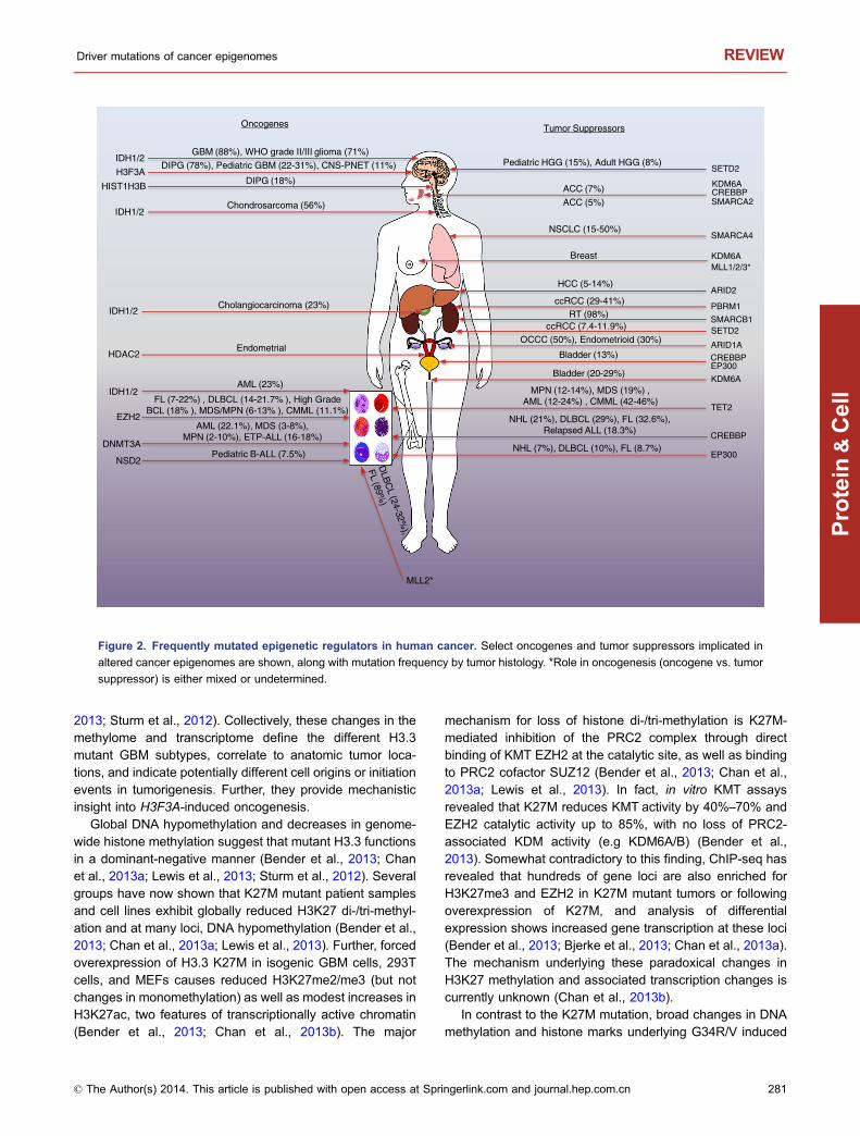
2013; Sturm et al., 2012). Collectively, these changes in themethylome and transcriptome define the different H3.3mutant GBM subtypes, correlate to anatomic tumor loca-tions, and indicate potentially different cell origins or initiationevents in tumorigenesis. Further, they provide mechanisticinsight into H3F3A-induced oncogenesis.
Global DNA hypomethylation and decreases in genome-wide histone methylation suggest that mutant H3.3 functionsin a dominant-negative manner (Bender et al., 2013; Chanet al., 2013a; Lewis et al., 2013; Sturm et al., 2012). Severalgroups have now shown that K27M mutant patient samplesand cell lines exhibit globally reduced H3K27 di-/tri-methyl-ation and at many loci, DNA hypomethylation (Bender et al.,2013; Chan et al., 2013a; Lewis et al., 2013). Further, forcedoverexpression of H3.3 K27M in isogenic GBM cells, 293Tcells, and MEFs causes reduced H3K27me2/me3 (but notchanges in monomethylation) as well as modest increases inH3K27ac, two features of transcriptionally active chromatin(Bender et al., 2013; Chan et al., 2013b). The major
mechanism for loss of histone di-/tri-methylation is K27M-mediated inhibition of the PRC2 complex through directbinding of KMT EZH2 at the catalytic site, as well as bindingto PRC2 cofactor SUZ12 (Bender et al., 2013; Chan et al.,2013a; Lewis et al., 2013). In fact, in vitro KMT assaysrevealed that K27M reduces KMT activity by 40%–70% andEZH2 catalytic activity up to 85%, with no loss of PRC2-associated KDM activity (e.g KDM6A/B) (Bender et al.,2013). Somewhat contradictory to this finding, ChIP-seq hasrevealed that hundreds of gene loci are also enriched forH3K27me3 and EZH2 in K27M mutant tumors or followingoverexpression of K27M, and analysis of differentialexpression shows increased gene transcription at these loci(Bender et al., 2013; Bjerke et al., 2013; Chan et al., 2013a).The mechanism underlying these paradoxical changes inH3K27 methylation and associated transcription changes iscurrently unknown (Chan et al., 2013b).
In contrast to the K27M mutation, broad changes in DNAmethylation and histone marks underlying G34R/V induced
FL (7-22%) , DLBCL (14-21.7% ), High Grade BCL (18% ), MDS/MPN (6-13% ), CMML (11.1%)
MPN (12-14%), MDS (19%) , AML (12-24%) , CMML (42-46%)
Oncogenes Tumor Suppressors
CREBBP
DIPG (78%), Pediatric GBM (22-31%), CNS-PNET (11%)
IDH1/2
Bladder (20-29%)
ccRCC (7.4-11.9%)
ARID1A
MLL2*
ARID2
KDM6A MLL1/2/3*
NSCLC (15-50%)
Pediatric HGG (15%), Adult HGG (8%)
Chondrosarcoma (56%)
Cholangiocarcinoma (23%)
AML (23%)
EZH2
EP300
TET2
IDH1/2
HCC (5-14%)
DNMT3A
AML (22.1%), MDS (3-8%), MPN (2-10%), ETP-ALL (16-18%)
GBM (88%), WHO grade II/III glioma (71%) IDH1/2
SETD2
SETD2
Pediatric B-ALL (7.5%) NSD2
KDM6A
CREBBP KDM6A ACC (7%)
ACC (5%) SMARCA2
NHL (21%), DLBCL (29%), FL (32.6%), Relapsed ALL (18.3%)
Bladder (13%) CREBBP EP300
NHL (7%), DLBCL (10%), FL (8.7%)
OCCC (50%), Endometrioid (30%)
SMARCB1 RT (98%)
SMARCA4
ccRCC (29-41%) PBRM1
DIPG (18%) HIST1H3B
H3F3A
Breast
HDAC2 Endometrial
IDH1/2
Figure 2. Frequently mutated epigenetic regulators in human cancer. Select oncogenes and tumor suppressors implicated in
altered cancer epigenomes are shown, along with mutation frequency by tumor histology. *Role in oncogenesis (oncogene vs. tumor
suppressor) is either mixed or undetermined.
Driver mutations of cancer epigenomes REVIEW
© The Author(s) 2014. This article is published with open access at Springerlink.com and journal.hep.com.cn 281
Protein
&Cell
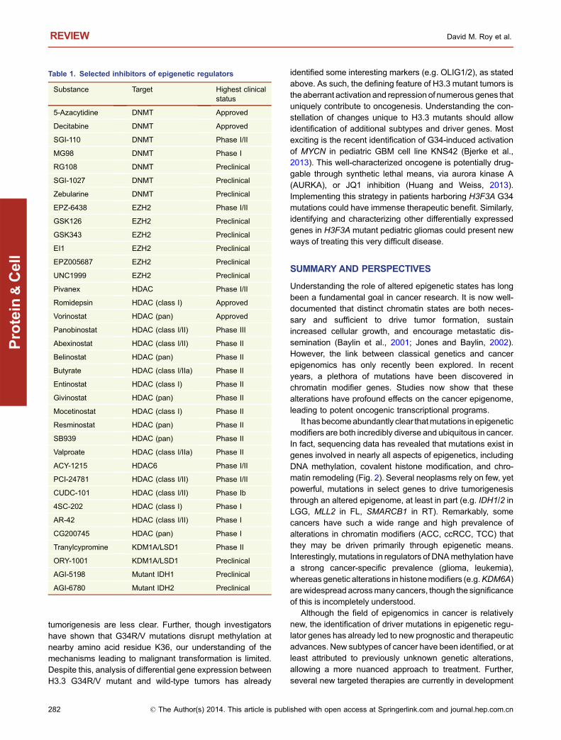
tumorigenesis are less clear. Further, though investigatorshave shown that G34R/V mutations disrupt methylation atnearby amino acid residue K36, our understanding of themechanisms leading to malignant transformation is limited.Despite this, analysis of differential gene expression betweenH3.3 G34R/V mutant and wild-type tumors has already
identified some interesting markers (e.g. OLIG1/2), as statedabove. As such, the defining feature of H3.3 mutant tumors isthe aberrant activation and repression of numerous genes thatuniquely contribute to oncogenesis. Understanding the con-stellation of changes unique to H3.3 mutants should allowidentification of additional subtypes and driver genes. Mostexciting is the recent identification of G34-induced activationof MYCN in pediatric GBM cell line KNS42 (Bjerke et al.,2013). This well-characterized oncogene is potentially drug-gable through synthetic lethal means, via aurora kinase A(AURKA), or JQ1 inhibition (Huang and Weiss, 2013).Implementing this strategy in patients harboring H3F3A G34mutations could have immense therapeutic benefit. Similarly,identifying and characterizing other differentially expressedgenes in H3F3A mutant pediatric gliomas could present newways of treating this very difficult disease.
SUMMARY AND PERSPECTIVES
Understanding the role of altered epigenetic states has longbeen a fundamental goal in cancer research. It is now well-documented that distinct chromatin states are both neces-sary and sufficient to drive tumor formation, sustainincreased cellular growth, and encourage metastatic dis-semination (Baylin et al., 2001; Jones and Baylin, 2002).However, the link between classical genetics and cancerepigenomics has only recently been explored. In recentyears, a plethora of mutations have been discovered inchromatin modifier genes. Studies now show that thesealterations have profound effects on the cancer epigenome,leading to potent oncogenic transcriptional programs.
It hasbecomeabundantly clear thatmutations in epigeneticmodifiers are both incredibly diverse and ubiquitous in cancer.In fact, sequencing data has revealed that mutations exist ingenes involved in nearly all aspects of epigenetics, includingDNA methylation, covalent histone modification, and chro-matin remodeling (Fig. 2). Several neoplasms rely on few, yetpowerful, mutations in select genes to drive tumorigenesisthrough an altered epigenome, at least in part (e.g. IDH1/2 inLGG, MLL2 in FL, SMARCB1 in RT). Remarkably, somecancers have such a wide range and high prevalence ofalterations in chromatin modifiers (ACC, ccRCC, TCC) thatthey may be driven primarily through epigenetic means.Interestingly, mutations in regulators of DNAmethylation havea strong cancer-specific prevalence (glioma, leukemia),whereas genetic alterations in histonemodifiers (e.g.KDM6A)arewidespread acrossmany cancers, though the significanceof this is incompletely understood.
Although the field of epigenomics in cancer is relativelynew, the identification of driver mutations in epigenetic regu-lator genes has already led to new prognostic and therapeuticadvances. New subtypes of cancer have been identified, or atleast attributed to previously unknown genetic alterations,allowing a more nuanced approach to treatment. Further,several new targeted therapies are currently in development
Table 1. Selected inhibitors of epigenetic regulators
Substance Target Highest clinicalstatus
5-Azacytidine DNMT Approved
Decitabine DNMT Approved
SGI-110 DNMT Phase I/II
MG98 DNMT Phase I
RG108 DNMT Preclinical
SGI-1027 DNMT Preclinical
Zebularine DNMT Preclinical
EPZ-6438 EZH2 Phase I/II
GSK126 EZH2 Preclinical
GSK343 EZH2 Preclinical
EI1 EZH2 Preclinical
EPZ005687 EZH2 Preclinical
UNC1999 EZH2 Preclinical
Pivanex HDAC Phase I/II
Romidepsin HDAC (class I) Approved
Vorinostat HDAC (pan) Approved
Panobinostat HDAC (class I/II) Phase III
Abexinostat HDAC (class I/II) Phase II
Belinostat HDAC (pan) Phase II
Butyrate HDAC (class I/IIa) Phase II
Entinostat HDAC (class I) Phase II
Givinostat HDAC (pan) Phase II
Mocetinostat HDAC (class I) Phase II
Resminostat HDAC (pan) Phase II
SB939 HDAC (pan) Phase II
Valproate HDAC (class I/IIa) Phase II
ACY-1215 HDAC6 Phase I/II
PCI-24781 HDAC (class I/II) Phase I/II
CUDC-101 HDAC (class I/II) Phase Ib
4SC-202 HDAC (class I) Phase I
AR-42 HDAC (class I/II) Phase I
CG200745 HDAC (pan) Phase I
Tranylcypromine KDM1A/LSD1 Phase II
ORY-1001 KDM1A/LSD1 Preclinical
AGI-5198 Mutant IDH1 Preclinical
AGI-6780 Mutant IDH2 Preclinical
REVIEW David M. Roy et al.
282 © The Author(s) 2014. This article is published with open access at Springerlink.com and journal.hep.com.cn
Protein
&Cell
and show great promise at reversing phenotypes caused byactivating mutations in certain genes (e.g. IDH1, EZH2). Inaddition, understanding the epigenetic mechanisms thatunderlie these alterations has also provided new therapeuticpotential, even if the driver mutations cannot be targeteddirectly (e.g. HDACi, DNMT inhibitors) (Table 1) (Arrowsmithet al., 2012; Foulks et al., 2012; Helin and Dhanak, 2013; Newet al., 2012; Plass et al., 2013).
There is nodoubt thatweare in themidst of an excitingera inboth cancer genomics and epigenetics. Investigations focusedon the intersectionof these twofieldshaveprovided remarkableclarity into numerous aspects of cancer biology and novelmechanisms of oncogenesis. Already, great strides have beenmade to translate these early discoveries into clinical practice,and the characterization of novel mutations in chromatin mod-ifier genes is rapidly accelerating. With the ongoing develop-ment of new epigenetic therapies, it is possible that muchimproved outcomes, at least in certain cancers, may be pos-sible in the near future. Further, an enhanced understanding ofthe epigenomic alterations associated with these driver genemutations could lead to the discovery of novel pathwaysinvolved in tumorigenesis, which may themselves possessunique vulnerabilities for therapeutic intervention.
ACKNOWLEDGEMENTS
We apologize to the many authors whose work we could not cite
because of space constraints. We would like to thank Deepa Ra-
maswami for help in reviewing the manuscript. D.M.R. was sup-
ported by the HHMI Medical Research Fellows Program and NIH
MSTP grant T32GM007739. L.A.W. was supported by The Cana-
dian Institutes of Health Research PDF. T.A.C. is a Department of
Defense Era of Hope Scholar and supported in part by the Frederick
Adler Fund.
ABBREVIATIONS
ACC, adenoid cystic carcinoma; AML, acute myeloid leukemia; CGI,
CpG island; HNPCC, hereditary nonpolyposis colon cancer; HDACs,
histone deacetylatases; MSI, microsatellite instability; TCC,
transitional cell carcinoma.
COMPLIANCE WITH ETHICS GUIDELINES
Authors David Roy, Logan Walsh, and Timothy Chan declare that
they have no conflict of interest.
OPEN ACCESS
This article is distributed under the terms of the Creative Commons
Attribution License which permits any use, distribution, and
reproduction in any medium, provided the original author(s) and
the source are credited.
REFERENCES
Abdel-Wahab O, Mullally A, Hedvat C, Garcia-Manero G, Patel J,
Wadleigh M, Malinge S, Yao J, Kilpivaara O, Bhat R et al (2009)
Genetic characterization of TET1, TET2, and TET3 alterations in
myeloid malignancies. Blood 114:144–147
Abdel-Wahab O, Pardanani A, Patel J, Wadleigh M, Lasho T, Heguy
A, Beran M, Gilliland DG, Levine RL, Tefferi A (2011) Concom-
itant analysis of EZH2 and ASXL1 mutations in myelofibrosis,
chronic myelomonocytic leukemia and blast-phase myeloprolif-
erative neoplasms. Leukemia 25:1200–1202
Amary MF, Bacsi K, Maggiani F, Damato S, Halai D, Berisha F,
Pollock R, O’Donnell P, Grigoriadis A, Diss T et al (2011) IDH1
and IDH2 mutations are frequent events in central chondrosar-
coma and central and periosteal chondromas but not in other
mesenchymal tumours. J Pathol 224:334–343
Arrowsmith CH, Bountra C, Fish PV, Lee K, Schapira M (2012)
Epigenetic protein families: a new frontier for drug discovery. Nat
Rev Drug Discov 11:384–400
Asp P, Wihlborg M, Karlen M, Farrants AK (2002) Expression of
BRG1, a human SWI/SNF component, affects the organisation of
actin filaments through the RhoA signalling pathway. J Cell Sci
115:2735–2746
Attieh Y, Geng QR, Dinardo CD, Zheng H, Jia Y, Fang ZH, Ganan-
Gomez I, Yang H, Wei Y, Kantarjian H et al (2013) Low frequency
of H3.3 mutations and upregulated DAXX expression in MDS.
Blood 121:4009–4011
Bachman KE, Rountree MR, Baylin SB (2001) Dnmt3a and Dnmt3b
are transcriptional repressors that exhibit unique localization
properties to heterochromatin. J Biol Chem 276:32282–32287
Bae DS, Park YA, Lee JW, Kim HS, Lee YY, Kim TJ, Choi C, Choi JJ,
Jeon HK, Cho YJ et al (2013) Tumor suppressive effects of
bromodomain containing protein 7 (BRD7) in epithelial ovarian
carcinoma. Clin Cancer Res
Balakrishnan A, Bleeker FE, Lamba S, Rodolfo M, Daniotti M,
Scarpa A, van Tilborg AA, Leenstra S, Zanon C, Bardelli A (2007)
Novel somatic and germline mutations in cancer candidate genes
in glioblastoma, melanoma, and pancreatic carcinoma. Cancer
Res 67:3545–3550
Balss J, Meyer J, Mueller W, Korshunov A, Hartmann C, von
Deimling A (2008) Analysis of the IDH1 codon 132 mutation in
brain tumors. Acta Neuropathol 116:597–602
Bannister AJ, Kouzarides T (1996) The CBP co-activator is a histone
acetyltransferase. Nature 384:641–643
Baylin SB, Esteller M, Rountree MR, Bachman KE, Schuebel K,
Herman JG (2001) Aberrant patterns of DNA methylation,
chromatin formation and gene expression in cancer. Hum Mol
Genet 10:687–692
BeguelinW,PopovicR, TeaterM, JiangY, BuntingKL,RosenM,Shen
H, Yang SN, Wang L, Ezponda Tet al (2013) EZH2 is required for
germinal center formation and somatic EZH2 mutations promote
lymphoid transformation. Cancer Cell 23:677–692
Bender S, Tang Y, Lindroth AM, Hovestadt V, Jones DT, Kool M,
Zapatka M, Northcott PA, Sturm D, Wang W et al (2013) Reduced
H3K27me3 and DNA hypomethylation are major drivers of gene
expression in K27M mutant pediatric high-grade gliomas. Cancer
Cell 24:660–672
Berger MF, Lawrence MS, Demichelis F, Drier Y, Cibulskis K,
Sivachenko AY, Sboner A, Esgueva R, Pflueger D, Sougnez C
et al (2011) The genomic complexity of primary human prostate
cancer. Nature 470:214–220
Driver mutations of cancer epigenomes REVIEW
© The Author(s) 2014. This article is published with open access at Springerlink.com and journal.hep.com.cn 283
Protein
&Cell

Bisling KE, Brewin JN, McGovern AP, Horne GA, Rider T, Stewart
HJ, Ramsahoye BH, Chevassut TJ (2013) DNMT3A mutations at
R882 hotspot are only found in major clones of acute myeloid
leukemia. Leuk Lymphoma 1:4
Bjerke L, Mackay A, Nandhabalan M, Burford A, Jury A, Popov S,
Bax DA, Carvalho D, Taylor KR, Vinci M et al (2013) Histone H3.3
mutations drive pediatric glioblastoma through upregulation of
MYCN. Cancer Discov 3:512–519
Blaschke K, Ebata KT, Karimi MM, Zepeda-Martinez JA, Goyal P,
Mahapatra S, Tam A, Laird DJ, Hirst M, Rao A et al (2013)
Vitamin C induces Tet-dependent DNA demethylation and a
blastocyst-like state in ES cells. Nature 500:222–226
Bleeker FE, Lamba S, Leenstra S, Troost D, Hulsebos T, Vandertop
WP, Frattini M, Molinari F, Knowles M, Cerrato A et al (2009) IDH1
mutations at residue p. R132 (IDH1(R132)) occur frequently in
high-grade gliomas but not in other solid tumors. Hum Mutat
30:7–11
Bodor C, O’Riain C, Wrench D, Matthews J, Iyengar S, Tayyib H,
Calaminici M, Clear A, Iqbal S, Quentmeier H et al (2011) EZH2
Y641 mutations in follicular lymphoma. Leukemia 25:726–729
Bodor C, Grossmann V, Popov N, Okosun J, O’Riain C, Tan K,
Marzec J, Araf S, Wang J, Lee AM et al (2013) EZH2 mutations
are frequent and represent an early event in follicular lymphoma.
Blood 122:3165–3168
Borger DR, Tanabe KK, Fan KC, Lopez HU, Fantin VR, Straley KS,
Schenkein DP, Hezel AF, Ancukiewicz M, Liebman HM et al
(2012) Frequent mutation of isocitrate dehydrogenase (IDH)1
and IDH2 in cholangiocarcinoma identified through broad-based
tumor genotyping. Oncologist 17:72–79
Borodovsky A, Salmasi V, Turcan S, Fabius AW, Baia GS, Eberhart
CG, Weingart JD, Gallia GL, Baylin SB, Chan TA et al (2013)
5-azacytidine reduces methylation, promotes differentiation and
induces tumor regression in a patient-derived IDH1 mutant
glioma xenograft. Oncotarget 4:1737–1747
Borrow J, Stanton VP Jr, Andresen JM, Becher R, Behm FG,
Chaganti RS, Civin CI, Disteche C, Dube I, Frischauf AM et al
(1996) The translocation t(8;16)(p11;p13) of acute myeloid
leukaemia fuses a putative acetyltransferase to the CREB-
binding protein. Nat Genet 14:33–41
Bosse T, Ter Haar NT, Seeber LM, Diest PJ, Hes FJ, Vasen HF, Nout
RA, Creutzberg CL, Morreau H, Smit VT (2013) Loss of ARID1A
expression and its relationship with PI3K-Akt pathway alterations,
TP53 and microsatellite instability in endometrial cancer. Mod
Pathol 26:1525–1535
Bracken AP, Pasini D, Capra M, Prosperini E, Colli E, Helin K (2003)
EZH2 is downstream of the pRB-E2F pathway, essential for
proliferation and amplified in cancer. EMBO J 22:5323–5335
Brecqueville M, Cervera N, Gelsi-Boyer V, Murati A, Adelaide J,
Chaffanet M, Rey J, Vey N, Mozziconacci MJ, Birnbaum D (2011)
Rare mutations in DNMT3A in myeloproliferative neoplasms and
myelodysplastic syndromes. Blood Cancer J 1:e18
Bultman S, Gebuhr T, Yee D, La Mantia C, Nicholson J, Gilliam A,
Randazzo F, Metzger D, Chambon P, Crabtree G et al (2000) A
Brg1 null mutation in the mouse reveals functional differences
among mammalian SWI/SNF complexes. Mol Cell 6:1287–
1295
Bultman SJ, Herschkowitz JI, Godfrey V, Gebuhr TC, Yaniv M,
Perou CM, Magnuson T (2008) Characterization of mammary
tumors from Brg1 heterozygous mice. Oncogene 27:460–468
Burrows AE, Smogorzewska A, Elledge SJ (2010) Polybromo-associ-
ated BRG1-associated factor components BRD7 and BAF180 are
critical regulators of p53 required for induction of replicative
senescence. Proc Natl Acad Sci USA 107:14280–14285
Cairns RA, Mak TW (2013) Oncogenic isocitrate dehydrogenase
mutations: mechanisms, models, and clinical opportunities.
Cancer Discov 3:730–741
Cancer Genome Atlas Research Network (2013) Comprehensive
molecular characterization of clear cell renal cell carcinoma.
Nature 499:43–49
Capello D, Gloghini A, Martini M, Spina M, Tirelli U, Bertoni F, Rinaldi
A, Morra E, Rambaldi A, Sinigaglia F et al (2011) Mutations of
CD79A, CD79B and EZH2 genes in immunodeficiency-related
non-Hodgkin lymphomas. Br J Haematol 152:777–780
Capper D, Simon M, Langhans CD, Okun JG, Tonn JC, Weller M,
von Deimling A, Hartmann C (2012) 2-Hydroxyglutarate concen-
tration in serum from patients with gliomas does not correlate with
IDH1/2 mutation status or tumor size. Int J Cancer 131:766–768
Caramel J, Quignon F, Delattre O (2008) RhoA-dependent regula-
tion of cell migration by the tumor suppressor hSNF5/INI1.
Cancer Res 68:6154–6161
Cerami E, Gao J, Dogrusoz U, Gross BE, Sumer SO, Aksoy BA,
Jacobsen A, Byrne CJ, Heuer ML, Larsson E et al (2012) The cBio
cancer genomics portal: an open platform for exploring multidi-
mensional cancer genomics data. Cancer Discov 2:401–404
Challen GA, Sun D, Jeong M, Luo M, Jelinek J, Berg JS, Bock C,
Vasanthakumar A, Gu H, Xi Y et al (2012) Dnmt3a is essential for
hematopoietic stem cell differentiation. Nat Genet 44:23–31
Chan KM, Fang D, Gan H, Hashizume R, Yu C, Schroeder M, Gupta
N, Mueller S, James CD, Jenkins R et al (2013a) The histone
H3.3K27M mutation in pediatric glioma reprograms H3K27
methylation and gene expression. Genes Dev 27:985–990
Chan KM, Han J, Fang D, Gan H, Zhang Z (2013b) A lesson learned
from the H3.3K27M mutation found in pediatric glioma: a new
approach to the study of the function of histone modifications
in vivo? Cell Cycle 12:2546–2552
Chaturvedi A, Araujo Cruz MM, Jyotsana N, Sharma A, Yun H,
Gorlich K, Wichmann M, Schwarzer A, Preller M, Thol F et al
(2013) Mutant IDH1 promotes leukemogenesis in vivo and can
be specifically targeted in human AML. Blood 122:2877–2887
Chedin F, Lieber MR, Hsieh CL (2002) The DNA methyltransferase-
like protein DNMT3L stimulates de novo methylation by Dnmt3a.
Proc Natl Acad Sci USA 99:16916–16921
Chen C, Liu Y, Lu C, Cross JR, Morris JPt, Shroff AS, Ward PS,
Bradner JE, Thompson C, Lowe SW (2013a) Cancer-associated
IDH2 mutants drive an acute myeloid leukemia that is susceptible
to Brd4 inhibition. Genes Dev 27:1974–1985
Chen Q, Chen Y, Bian C, Fujiki R, Yu X (2013b) TET2 promotes
histone O-GlcNAcylation during gene transcription. Nature
493:561–564
Cheng SW, Davies KP, Yung E, Beltran RJ, Yu J, Kalpana GV
(1999) c-MYC interacts with INI1/hSNF5 and requires the SWI/
SNF complex for transactivation function. Nat Genet 22:102–105
REVIEW David M. Roy et al.
284 © The Author(s) 2014. This article is published with open access at Springerlink.com and journal.hep.com.cn
Protein
&Cell

Cheng J, Guo S, Chen S, Mastriano SJ, Liu C, D’Alessio AC, Hysolli
E, Guo Y, Yao H, Megyola CM et al (2013) An extensive network
of TET2-targeting microRNAs regulates malignant hematopoie-
sis. Cell Rep 5:471–481
Chesi M, Nardini E, Lim RS, Smith KD, Kuehl WM, Bergsagel PL
(1998) The t(4;14) translocation in myeloma dysregulates both
FGFR3 and a novel gene, MMSET, resulting in IgH/MMSET
hybrid transcripts. Blood 92:3025–3034
Cho YW, Hong T, Hong S, Guo H, Yu H, Kim D, Guszczynski T,
Dressler GR, Copeland TD, Kalkum M et al (2007) PTIP
associates with MLL3- and MLL4-containing histone H3 lysine
4 methyltransferase complex. J Biol Chem 282:20395–20406
Chou WC, Hou HA, Chen CY, Tang JL, Yao M, Tsay W, Ko BS, Wu
SJ, Huang SY, Hsu SC et al (2010) Distinct clinical and biologic
characteristics in adult acute myeloid leukemia bearing the
isocitrate dehydrogenase 1 mutation. Blood 115:2749–2754
Chou WC, Chou SC, Liu CY, Chen CY, Hou HA, Kuo YY, Lee MC,
Ko BS, Tang JL, Yao M et al (2011a) TET2 mutation is an
unfavorable prognostic factor in acute myeloid leukemia patients
with intermediate-risk cytogenetics. Blood 118:3803–3810
Chou WC, Lei WC, Ko BS, Hou HA, Chen CY, Tang JL, Yao M, Tsay
W, Wu SJ, Huang SY et al (2011b) The prognostic impact and
stability of isocitrate dehydrogenase 2 mutation in adult patients
with acute myeloid leukemia. Leukemia 25:246–253
Christensen J, Agger K, Cloos PA, Pasini D, Rose S, Sennels L,
Rappsilber J, Hansen KH, Salcini AE, Helin K (2007) RBP2
belongs to a family of demethylases, specific for tri-and dime-
thylated lysine 4 on histone 3. Cell 128:1063–1076
Christiaans I, Kenter SB, Brink HC, van Os TA, Baas F, van den
Munckhof P, Kidd AM, Hulsebos TJ (2011) Germline SMARCB1
mutation and somatic NF2 mutations in familial multiple menin-
giomas. J Med Genet 48:93–97
Costa Y, Ding J, Theunissen TW, Faiola F, Hore TA, Shliaha PV,
Fidalgo M, Saunders A, Lawrence M, Dietmann S et al (2013)
NANOG-dependent function of TET1 and TET2 in establishment
of pluripotency. Nature 495:370–374
Croonquist PA, Van Ness B (2005) The polycomb group protein
enhancer of zeste homolog 2 (EZH 2) is an oncogene that
influences myeloma cell growth and the mutant ras phenotype.
Oncogene 24:6269–6280
da Costa WH, Rezende M, Rocha RM, da Cunha IW, Carraro DM,
Guimaraes GC, de Cassio Zequi S (2013) PBRM1, a SWI/SNF
complex subunit is a prognostic marker in clear cell renal cell
carcinoma. BJU Int
Dalgliesh GL, Furge K, Greenman C, Chen L, Bignell G, Butler A,
Davies H, Edkins S, Hardy C, Latimer C et al (2010) Systematic
sequencing of renal carcinoma reveals inactivation of histone
modifying genes. Nature 463:360–363
Dallas PB, Pacchione S, Wilsker D, Bowrin V, Kobayashi R, Moran E
(2000) The human SWI-SNF complex protein p270 is an ARID
family member with non-sequence-specific DNA binding activity.
Mol Cell Biol 20:3137–3146
Dang L, White DW, Gross S, Bennett BD, Bittinger MA, Driggers EM,
Fantin VR, Jang HG, Jin S, Keenan MC et al (2009) Cancer-
associated IDH1 mutations produce 2-hydroxyglutarate. Nature
462:739–744
Datta J, Ghoshal K, Sharma SM, Tajima S, Jacob ST (2003)
Biochemical fractionation reveals association of DNA methyl-
transferase (Dnmt) 3b with Dnmt1 and that of Dnmt 3a with a
histone H3 methyltransferase and Hdac1. J Cell Biochem
88:855–864
Dawson MA, Kouzarides T (2012) Cancer epigenetics: from mech-
anism to therapy. Cell 150:12–27
Delhommeau F, Dupont S, James C, Masse A, le Couedic J, Valle V
(2008) TET2 is a novel tumor suppressor gene inactivated in
myeloproliferative neoplasms: identification of a pre-JAK2 V617F
event. Blood 112:11
Delhommeau F, Dupont S, Della Valle V, James C, Trannoy S,
Masse A, Kosmider O, Le Couedic JP, Robert F, Alberdi A et al
(2009) Mutation in TET2 in myeloid cancers. N Engl J Med
360:2289–2301
Deplus R, Delatte B, Schwinn MK, Defrance M, Mendez J, Murphy
N, Dawson MA, Volkmar M, Putmans P, Calonne E et al (2013)
TET2 and TET3 regulate GlcNAcylation and H3K4 methylation
through OGT and SET1/COMPASS. EMBO J 32:645–655
Dimartino JF, Cleary ML (1999) Mll rearrangements in haematolog-
ical malignancies: lessons from clinical and biological studies. Br
J Haematol 106:614–626
DiNardo CD, Propert KJ, Loren AW, Paietta E, Sun Z, Levine RL,
Straley KS, Yen K, Patel JP, Agresta S et al (2013) Serum
2-hydroxyglutarate levels predict isocitrate dehydrogenase muta-
tions and clinical outcome in acute myeloid leukemia. Blood
121:4917–4924
Dolnik A, Engelmann JC, Scharfenberger-Schmeer M, Mauch J,
Kelkenberg-Schade S, Haldemann B, Fries T, Kronke J, Kuhn
MW, Paschka P et al (2012) Commonly altered genomic regions
in acute myeloid leukemia are enriched for somatic mutations
involved in chromatin remodeling and splicing. Blood 120:e83–
e92
Drost J, Mantovani F, Tocco F, Elkon R, Comel A, Holstege H,
Kerkhoven R, Jonkers J, Voorhoeve PM, Agami R et al (2010)
BRD7 is a candidate tumour suppressor gene required for p53
function. Nat Cell Biol 12:380–389
Duns G, van den Berg E, van Duivenbode I, Osinga J, Hollema H,
Hofstra RM, Kok K (2010) Histone methyltransferase gene
SETD2 is a novel tumor suppressor gene in clear cell renal cell
carcinoma. Cancer Res 70:4287–4291
Duns G, Hofstra RM, Sietzema JG, Hollema H, van Duivenbode I,
Kuik A, Giezen C, Jan O, Bergsma JJ, Bijnen H et al (2012)
Targeted exome sequencing in clear cell renal cell carcinoma
tumors suggests aberrant chromatin regulation as a crucial step
in ccRCC development. Hum Mutat 33:1059–1062
Eckner R, Ewen ME, Newsome D, Gerdes M, DeCaprio JA,
Lawrence JB, Livingston DM (1994) Molecular cloning and
functional analysis of the adenovirus E1A-associated 300-kD
protein (p300) reveals a protein with properties of a transcrip-
tional adaptor. Genes Dev 8:869–884
Edmunds JW, Mahadevan LC, Clayton AL (2008) Dynamic histone
H3 methylation during gene induction: HYPB/Setd2 mediates all
H3K36 trimethylation. EMBO J 27:406–420
Ehrlich M (2002) DNA methylation in cancer: too much, but also too
little. Oncogene 21:5400–5413
Driver mutations of cancer epigenomes REVIEW
© The Author(s) 2014. This article is published with open access at Springerlink.com and journal.hep.com.cn 285
Protein
&Cell

Ellis L, Atadja PW, Johnstone RW (2009) Epigenetics in cancer:
targeting chromatin modifications. Mol Cancer Ther 8:1409–1420
El-Osta A (2004) The rise and fall of genomic methylation in cancer.
Leukemia 18:233–237
Endo M, Yasui K, Zen Y, Gen Y, Zen K, Tsuji K, Dohi O, Mitsuyoshi
H, Tanaka S, Taniwaki M et al (2013) Alterations of the SWI/SNF
chromatin remodelling subunit-BRG1 and BRM in hepatocellular
carcinoma. Liver Int 33:105–117
Ernst T, Chase AJ, Score J, Hidalgo-Curtis CE, Bryant C, Jones AV,
Waghorn K, Zoi K, Ross FM, Reiter A et al (2010) Inactivating
mutations of the histone methyltransferase gene EZH2 in myeloid
disorders. Nat Genet 42:722–726
Esteller M, Fraga MF, Guo M, Garcia-Foncillas J, Hedenfalk I,
Godwin AK, Trojan J, Vaurs-Barriere C, Bignon YJ, Ramus S et al
(2001) DNA methylation patterns in hereditary human cancers
mimic sporadic tumorigenesis. Hum Mol Genet 10:3001–3007
Ewalt M, Galili NG, Mumtaz M, Churchill M, Rivera S, Borot F, Raza
A, Mukherjee S (2011) DNMT3a mutations in high-risk myelo-
dysplastic syndrome parallel those found in acute myeloid
leukemia. Blood Cancer J 1:e9
Feinberg AP, Vogelstein B (1983) Hypomethylation distinguishes
genes of some human cancers from their normal counterparts.
Nature 301:89–92
Feng Q, Wang H, Ng HH, Erdjument-Bromage H, Tempst P, Struhl K,
Zhang Y (2002) Methylation of H3-lysine 79 is mediated by a new
family of HMTases without a SET domain. Curr Biol 12:1052–
1058
Figueroa ME, Abdel-Wahab O, Lu C, Ward PS, Patel J, Shih A, Li Y,
Bhagwat N, Vasanthakumar A, Fernandez HF et al (2010)
Leukemic IDH1 and IDH2 mutations result in a hypermethylation
phenotype, disrupt TET2 function, and impair hematopoietic
differentiation. Cancer Cell 18:553–567
Flanagan S, Lee M, Li CC, Suter CM, Buckland ME (2012) Promoter
methylation analysis of IDH genes in human gliomas. Front Oncol
2:193
Flowers S, Nagl NG Jr, Beck GR Jr, Moran E (2009) Antagonistic
roles for BRM and BRG1 SWI/SNF complexes in differentiation.
J Biol Chem 284:10067–10075
Fontebasso AM, Schwartzentruber J, Khuong-Quang DA, Liu XY,
Sturm D, Korshunov A, Jones DT, Witt H, Kool M, Albrecht S et al
(2013) Mutations in SETD2 and genes affecting histone H3K36
methylation target hemispheric high-grade gliomas. Acta Neuro-
pathol 125:659–669
Forbes SA, Bindal N, Bamford S, Cole C, Kok CY, Beare D, Jia M,
Shepherd R, Leung K, Menzies A et al (2011) COSMIC: mining
complete cancer genomes in the catalogue of somatic mutations
in cancer. Nucleic Acids Res 39:D945–D950
Foulks JM, Parnell KM, Nix RN, Chau S, Swierczek K, Saunders M,
Wright K, Hendrickson TF, Ho KK, McCullar MV et al (2012)
Epigenetic drug discovery: targeting DNA methyltransferases.
J Biomol Screen 17:2–17
Fu X, Jin L, Wang X, Luo A, Hu J, Zheng X, Tsark WM, Riggs AD, Ku
HT, Huang W (2013) MicroRNA-26a targets ten eleven translo-
cation enzymes and is regulated during pancreatic cell differen-
tiation. Proc Natl Acad Sci USA 110:17892–17897
Fujimoto A, Totoki Y, Abe T, Boroevich KA, Hosoda F, Nguyen HH,
Aoki M, Hosono N, Kubo M, Miya F et al (2012) Whole-genome
sequencing of liver cancers identifies etiological influences on
mutation patterns and recurrent mutations in chromatin regula-
tors. Nat Genet 44:760–764
Fuks F, Burgers WA, Godin N, Kasai M, Kouzarides T (2001)
Dnmt3a binds deacetylases and is recruited by a sequence-
specific repressor to silence transcription. EMBO J 20:2536–
2544
Fuks F, Hurd PJ, Deplus R, Kouzarides T (2003) The DNA
methyltransferases associate with HP1 and the SUV39H1
histone methyltransferase. Nucleic Acids Res 31:2305–2312
Fukuoka J, Fujii T, Shih JH, Dracheva T,MeerzamanD, Player A,Hong
K, SettnekS, Gupta A, BuetowK et al (2004)Chromatin remodeling
factors and BRM/BRG1 expression as prognostic indicators in non-
small cell lung cancer. Clin Cancer Res 10:4314–4324
Gama-Sosa MA, Slagel VA, Trewyn RW, Oxenhandler R, Kuo KC,
Gehrke CW, Ehrlich M (1983) The 5-methylcytosine content of
DNA from human tumors. Nucleic Acids Res 11:6883–6894
Gao X, Tate P, Hu P, Tjian R, Skarnes WC, Wang Z (2008) ES cell
pluripotency and germ-layer formation require the SWI/SNF
chromatin remodeling component BAF250a. Proc Natl Acad Sci
USA 105:6656–6661
Gao J, Aksoy BA, Dogrusoz U, Dresdner G, Gross B, Sumer SO,
Sun Y, Jacobsen A, Sinha R, Larsson E et al (2013) Integrative
analysis of complex cancer genomics and clinical profiles using
the cBioPortal. Sci Signal 6:pl1
Ge YZ, Pu MT, Gowher H, Wu HP, Ding JP, Jeltsch A, Xu GL (2004)
Chromatin targeting of de novo DNA methyltransferases by the
PWWP domain. J Biol Chem 279:25447–25454
Geisbrecht BV, Gould SJ (1999) The human PICD gene encodes a
cytoplasmic and peroxisomal NADP(+)-dependent isocitrate
dehydrogenase. J Biol Chem 274:30527–30533
Gessi M, Gielen GH, Hammes J, Dorner E, Muhlen AZ, Waha A,
Pietsch T (2013) H3.3 G34R mutations in pediatric primitive
neuroectodermal tumors of central nervous system (CNS-PNET)
and pediatric glioblastomas: possible diagnostic and therapeutic
implications? J Neurooncol 112:67–72
Ghiam AF, Cairns RA, Thoms J, Dal Pra A, Ahmed O, Meng A, Mak
TW, Bristow RG (2012) IDH mutation status in prostate cancer.
Oncogene 31:3826
Glaros S, Cirrincione GM, Muchardt C, Kleer CG, Michael CW,
Reisman D (2007) The reversible epigenetic silencing of BRM:
implications for clinical targeted therapy. Oncogene 26:7058–
7066
Grady WM, Willis J, Guilford PJ, Dunbier AK, Toro TT, Lynch H,
Wiesner G, Ferguson K, Eng C, Park JG et al (2000) Methylation
of the CDH1 promoter as the second genetic hit in hereditary
diffuse gastric cancer. Nat Genet 26:16–17
Grassian AR, Lin F, Barrett R, Liu Y, Jiang W, Korpal M, Astley H,
Gitterman D, Henley T, Howes R et al (2012) Isocitrate dehydro-
genase (IDH) mutations promote a reversible ZEB1/microRNA
(miR)-200-dependent epithelial-mesenchymal transition (EMT).
J Biol Chem 287:42180–42194
Green A, Beer P (2010) Somatic mutations of IDH1 and IDH2 in the
leukemic transformation of myeloproliferative neoplasms. N Engl
J Med 362:369–370
Greer EL, Shi Y (2012) Histone methylation: a dynamic mark in
health, disease and inheritance. Nat Rev Genet 13:343–357
REVIEW David M. Roy et al.
286 © The Author(s) 2014. This article is published with open access at Springerlink.com and journal.hep.com.cn
Protein
&Cell
Gross S, Cairns RA, Minden MD, Driggers EM, Bittinger MA, Jang
HG, Sasaki M, Jin S, Schenkein DP, Su SM et al (2010) Cancer-
associated metabolite 2-hydroxyglutarate accumulates in acute
myelogenous leukemia with isocitrate dehydrogenase 1 and 2
mutations. J Exp Med 207:339–344
Grossmann V, Kohlmann A, Eder C, Haferlach C, Kern W, Cross
NC, Haferlach T, Schnittger S (2011) Molecular profiling of
chronic myelomonocytic leukemia reveals diverse mutations in
>80% of patients with TET2 and EZH2 being of high prognostic
relevance. Leukemia 25:877–879
Grossmann V, Haferlach C, Weissmann S, Roller A, Schindela S,
Poetzinger F, Stadler K, Bellos F, Kern W, Haferlach Tet al (2013)
The molecular profile of adult T-cell acute lymphoblastic leuke-
mia: mutations in RUNX1 and DNMT3A are associated with poor
prognosis in T-ALL. Genes Chromosom Cancer 52:410–422
Gu W, Roeder RG (1997) Activation of p53 sequence-specific DNA
binding by acetylation of the p53 C-terminal domain. Cell 90:595–
606
Guglielmelli P, Biamonte F, Score J, Hidalgo-Curtis C, Cervantes F,
Maffioli M, Fanelli T, Ernst T, Winkelman N, Jones AV et al (2011)
EZH2 mutational status predicts poor survival in myelofibrosis.
Blood 118:5227–5234
Gui Y, Guo G, Huang Y, Hu X, Tang A, Gao S, Wu R, Chen C, Li X,
Zhou L et al (2011) Frequent mutations of chromatin remodeling
genes in transitional cell carcinoma of the bladder. Nat Genet
43:875–878
Guidi CJ, Sands AT, Zambrowicz BP, Turner TK, Demers DA,
Webster W, Smith TW, Imbalzano AN, Jones SN (2001)
Disruption of Ini1 leads to peri-implantation lethality and tumor-
igenesis in mice. Mol Cell Biol 21:3598–3603
Guilhamon P, Eskandarpour M, Halai D, Wilson GA, Feber A,
Teschendorff AE, Gomez V, Hergovich A, Tirabosco R, Fernanda
Amary M et al (2013) Meta-analysis of IDH-mutant cancers
identifies EBF1 as an interaction partner for TET2. Nat Commun
4:2166
Guo JU, Su Y, Zhong C, Ming GL, Song H (2011) Hydroxylation of
5-methylcytosine by TET1 promotes active DNA demethylation in
the adult brain. Cell 145:423–434
Hakimi AA, Chen YB, Wren J, Gonen M, Abdel-Wahab O, Heguy A,
Liu H, Takeda S, Tickoo SK, Reuter VE et al (2013a) Clinical and
pathologic impact of select chromatin-modulating tumor suppres-
sors in clear cell renal cell carcinoma. Eur Urol 63:848–854
Hakimi AA, Ostrovnaya I, Reva B, Schultz N, Chen YB, Gonen M,
Liu H, Takeda S, Voss MH, Tickoo SK et al (2013b) Adverse
outcomes in clear cell renal cell carcinoma with mutations of 3p21
epigenetic regulators BAP1 and SETD2: a report by MSKCC and
the KIRC TCGA research network. Clin Cancer Res 19:3259–
3267
Hanahan D, Weinberg RA (2011) Hallmarks of cancer: the next
generation. Cell 144:646–674
Hanigan CL, Van Engeland M, De Bruine AP, Wouters KA,
Weijenberg MP, Eshleman JR, Herman JG (2008) An inactivating
mutation in HDAC2 leads to dysregulation of apoptosis mediated
by APAF1. Gastroenterology 135(1654–1664):e1652
Hartmann C, Meyer J, Balss J, Capper D, Mueller W, Christians A,
Felsberg J, Wolter M, Mawrin C, Wick W et al (2009) Type and
frequency of IDH1 and IDH2 mutations are related to astrocytic
and oligodendroglial differentiation and age: a study of 1,010
diffuse gliomas. Acta Neuropathol 118:469–474
He YF, Li BZ, Li Z, Liu P, Wang Y, Tang Q, Ding J, Jia Y, Chen Z, Li L
et al (2011) Tet-mediated formation of 5-carboxylcytosine and its
excision by TDG in mammalian DNA. Science 333:1303–1307
Heintzman ND, Stuart RK, Hon G, Fu Y, Ching CW, Hawkins RD,
Barrera LO, Van Calcar S, Qu C, Ching KA et al (2007)
Distinct and predictive chromatin signatures of transcriptional
promoters and enhancers in the human genome. Nat Genet
39:311–318
Helin K, Dhanak D (2013) Chromatin proteins and modifications as
drug targets. Nature 502:480–488
Herman JG, Baylin SB (2003) Gene silencing in cancer in associ-
ation with promoter hypermethylation. N Engl J Med 349:2042–
2054
Hermann A, Gowher H, Jeltsch A (2004) Biochemistry and biology of
mammalian DNA methyltransferases. CMLS 61:2571–2587
Herz HM, Madden LD, Chen Z, Bolduc C, Buff E, Gupta R, Davuluri
R, Shilatifard A, Hariharan IK, Bergmann A (2010) The
H3K27me3 demethylase dUTX is a suppressor of Notch- and
Rb-dependent tumors in Drosophila. Mol Cell Biol 30:2485–2497
Ho AS, Kannan K, Roy DM, Morris LG, Ganly I, Katabi N,
Ramaswami D, Walsh LA, Eng S, Huse JT et al (2013) The
mutational landscape of adenoid cystic carcinoma. Nat Genet
45:791–798
Holliday R, Pugh JE (1975) DNA modification mechanisms and
gene activity during development. Science 187:226–232
Holz-Schietinger C, Matje DM, Harrison MF, Reich NO (2011)
Oligomerization of DNMT3A controls the mechanism of de novo
DNA methylation. J Biol Chem 286:41479–41488
Holz-Schietinger C, Matje DM, Reich NO (2012) Mutations in DNA
methyltransferase (DNMT3A) observed in acute myeloid leuke-
mia patients disrupt processive methylation. J Biol Chem
287:30941–30951
Hou HA, Kuo YY, Liu CY, Chou WC, Lee MC, Chen CY, Lin LI, Tseng
MH, Huang CF, Chiang YC et al (2012) DNMT3A mutations in
acute myeloid leukemia: stability during disease evolution and
clinical implications. Blood 119:559–568
Hsieh CL (1999) In vivo activity of murine de novo methyltransfer-
ases, Dnmt3a and Dnmt3b. Mol Cell Biol 19:8211–8218
Huang M, Weiss WA (2013) G34, another connection between
MYCN and a pediatric tumor. Cancer Discov 3:484–486
Hughes CM, Rozenblatt-Rosen O, Milne TA, Copeland TD, Levine
SS, Lee JC, Hayes DN, Shanmugam KS, Bhattacharjee A,
Biondi CA et al (2004) Menin associates with a trithorax family
histone methyltransferase complex and with the hoxc8 locus. Mol
Cell 13:587–597
Hulsebos TJ, Plomp AS, Wolterman RA, Robanus-Maandag EC,
Baas F, Wesseling P (2007) Germline mutation of INI1/
SMARCB1 in familial schwannomatosis. Am J Hum Genet
80:805–810
Hussein K, Abdel-Wahab O, Lasho TL, Van Dyke DL, Levine RL,
Hanson CA, Pardanani A, Tefferi A (2010) Cytogenetic correlates
of TET2 mutations in 199 patients with myeloproliferative
neoplasms. Am J Hematol 85:81–83
Inoue H, Furukawa T, Giannakopoulos S, Zhou S, King DS, Tanese
N (2002) Largest subunits of the human SWI/SNF chromatin-
Driver mutations of cancer epigenomes REVIEW
© The Author(s) 2014. This article is published with open access at Springerlink.com and journal.hep.com.cn 287
Protein
&Cell

remodeling complex promote transcriptional activation by steroid
hormone receptors. J Biol Chem 277:41674–41685
Isakoff MS, Sansam CG, Tamayo P, Subramanian A, Evans JA,
Fillmore CM, Wang X, Biegel JA, Pomeroy SL, Mesirov JP et al
(2005) Inactivation of the Snf5 tumor suppressor stimulates cell
cycle progression and cooperates with p53 loss in oncogenic
transformation. Proc Natl Acad Sci USA 102:17745–17750
Issaeva I, Zonis Y, Rozovskaia T, Orlovsky K, Croce CM, Nakamura
T, Mazo A, Eisenbach L, Canaani E (2007) Knockdown of ALR
(MLL2) reveals ALR target genes and leads to alterations in cell
adhesion and growth. Mol Cell Biol 27:1889–1903
Ito S,D’Alessio AC, TaranovaOV, HongK, Sowers LC, ZhangY (2010)
Role of Tet proteins in 5mC to 5hmC conversion, ES-cell self-
renewal and inner cell mass specification. Nature 466:1129–1133
Ito S, Shen L, Dai Q, Wu SC, Collins LB, Swenberg JA, He C, Zhang
Y (2011) Tet proteins can convert 5-methylcytosine to 5-formyl-
cytosine and 5-carboxylcytosine. Science 333:1300–1303
Iyer NG, Ozdag H, Caldas C (2004) p300/CBP and cancer.
Oncogene 23:4225–4231
Jaffe JD, Wang Y, Chan HM, Zhang J, Huether R, Kryukov GV,
Bhang HE, Taylor JE, Hu M, Englund NP et al (2013) Global
chromatin profiling reveals NSD2 mutations in pediatric acute
lymphoblastic leukemia. Nat Genet 45:1386–1391
Jagani Z, Mora-Blanco EL, Sansam CG, McKenna ES, Wilson B,
Chen D, Klekota J, Tamayo P, Nguyen PT, Tolstorukov M et al
(2010) Loss of the tumor suppressor Snf5 leads to aberrant
activation of the Hedgehog-Gli pathway. Nat Med 16:1429–1433
Jankowska AM, Szpurka H, Tiu RV, Makishima H, Afable M, Huh J,
O’Keefe CL, Ganetzky R, McDevitt MA, Maciejewski JP (2009)
Loss of heterozygosity 4q24 and TET2 mutations associated with
myelodysplastic/myeloproliferative neoplasms. Blood 113:6403–
6410
Jia D, Jurkowska RZ, Zhang X, Jeltsch A, Cheng X (2007) Structure
of Dnmt3a bound to Dnmt3L suggests a model for de novo DNA
methylation. Nature 449:248–251
Jones PA (2012) Functions of DNA methylation: islands, start sites,
gene bodies and beyond. Nat Rev Genet 13:484–492
Jones PA, Baylin SB (2002) The fundamental role of epigenetic
events in cancer. Nat Rev Genet 3:415–428
Jones PA, Baylin SB (2007) The epigenomics of cancer. Cell
128:683–692
Jones S, Wang TL, Shih Ie M, Mao TL, Nakayama K, Roden R, Glas
R, Slamon D, Diaz LA Jr, Vogelstein B et al (2010) Frequent
mutations of chromatin remodeling gene ARID1A in ovarian clear
cell carcinoma. Science 330:228–231
Joseph CG, Hwang H, Jiao Y, Wood LD, Kinde I, Wu J, Mandahl N,
Luo J, Hruban RH, Diaz LA Jr et al (2013) Exomic analysis of
myxoid liposarcomas, synovial sarcomas, and osteosarcomas.
Genes Chromosom Cancer 53:15–24
Jurkowska RZ, Rajavelu A, Anspach N, Urbanke C, Jankevicius G,
Ragozin S, Nellen W, Jeltsch A (2011) Oligomerization and
binding of the Dnmt3a DNA methyltransferase to parallel DNA
molecules: heterochromatic localization and role of Dnmt3L.
J Biol Chem 286:24200–24207
Kampranis SC, Tsichlis PN (2009) Histone demethylases and
cancer. Adv Cancer Res 102:103–169
Kan Z, Jaiswal BS, Stinson J, Janakiraman V, Bhatt D, Stern HM,
Yue P, Haverty PM, Bourgon R, Zheng J et al (2010) Diverse
somatic mutation patterns and pathway alterations in human
cancers. Nature 466:869–873
Kane MF, Loda M, Gaida GM, Lipman J, Mishra R, Goldman H,
Jessup JM, Kolodner R (1997) Methylation of the hMLH1
promoter correlates with lack of expression of hMLH1 in sporadic
colon tumors and mismatch repair-defective human tumor cell
lines. Cancer Res 57:808–811
Kaneda M, Okano M, Hata K, Sado T, Tsujimoto N, Li E, Sasaki H
(2004) Essential role for de novo DNA methyltransferase Dnmt3a
in paternal and maternal imprinting. Nature 429:900–903
Kang MR, Kim MS, Oh JE, Kim YR, Song SY, Seo SI, Lee JY, Yoo NJ,
Lee SH (2009) Mutational analysis of IDH1 codon 132 in glioblas-
tomas and other common cancers. Int J Cancer 125:353–355
Kapur P, Pena-Llopis S, Christie A, Zhrebker L, Pavia-Jimenez A,
Rathmell WK, Xie XJ, Brugarolas J (2013) Effects on survival of
BAP1 and PBRM1 mutations in sporadic clear-cell renal-cell
carcinoma: a retrospective analysis with independent validation.
Lancet Oncol 14:159–167
Khan SN, Jankowska AM, Mahfouz R, Dunbar AJ, Sugimoto Y,
Hosono N, Hu Z, Cheriyath V, Vatolin S, Przychodzen B et al
(2013) Multiple mechanisms deregulate EZH2 and histone H3
lysine 27 epigenetic changes in myeloid malignancies. Leukemia
27:1301–1309
Khuong-Quang DA, Buczkowicz P, Rakopoulos P, Liu XY, Fonteb-
asso AM, Bouffet E, Bartels U, Albrecht S, Schwartzentruber J,
Letourneau L et al (2012) K27M mutation in histone H3.3 defines
clinically and biologically distinct subgroups of pediatric diffuse
intrinsic pontine gliomas. Acta Neuropathol 124:439–447
Kia SK, Gorski MM, Giannakopoulos S, Verrijzer CP (2008) SWI/
SNF mediates polycomb eviction and epigenetic reprogramming
of the INK4b-ARF-INK4a locus. Mol Cell Biol 28:3457–3464
Kim JG, Takeshima H, Niwa T, Rehnberg E, Shigematsu Y, Yoda Y,
Yamashita S, Kushima R, Maekita T, Ichinose M et al (2013)
Comprehensive DNA methylation and extensive mutation anal-
yses reveal an association between the CpG island methylator
phenotype and oncogenic mutations in gastric cancers. Cancer
Lett 330:33–40
Kirmizis A, Bartley SM, Kuzmichev A, Margueron R, Reinberg D,
Green R, Farnham PJ (2004) Silencing of human polycomb
target genes is associated with methylation of histone H3 Lys 27.
Genes Dev 18:1592–1605
Kleer CG, Cao Q, Varambally S, Shen R, Ota I, Tomlins SA, Ghosh
D, Sewalt RG, Otte AP, Hayes DF et al (2003) EZH2 is a marker
of aggressive breast cancer and promotes neoplastic transfor-
mation of breast epithelial cells. Proc Natl Acad Sci USA
100:11606–11611
Knutson SK, Wigle TJ, Warholic NM, Sneeringer CJ, Allain CJ,
Klaus CR, Sacks JD, Raimondi A, Majer CR, Song J et al (2012)
A selective inhibitor of EZH2 blocks H3K27 methylation and kills
mutant lymphoma cells. Nat Chem Biol 8:890–896
Ko M, Huang Y, Jankowska AM, Pape UJ, Tahiliani M, Bandukwala
HS, An J, Lamperti ED, Koh KP, Ganetzky R et al (2010) Impaired
hydroxylation of 5-methylcytosine in myeloid cancers with mutant
TET2. Nature 468:839–843
REVIEW David M. Roy et al.
288 © The Author(s) 2014. This article is published with open access at Springerlink.com and journal.hep.com.cn
Protein
&Cell

Ko M, An J, Bandukwala HS, Chavez L, Aijo T, Pastor WA, Segal
MF, Li H, Koh KP, Lahdesmaki H et al (2013) Modulation of TET2
expression and 5-methylcytosine oxidation by the CXXC domain
protein IDAX. Nature 497:122–126
Konstandin N, Bultmann S, Szwagierczak A, Dufour A, Ksienzyk B,
Schneider F, Herold T, Mulaw M, Kakadia PM, Schneider S et al
(2011) Genomic 5-hydroxymethylcytosine levels correlate with
TET2 mutations and a distinct global gene expression pattern in
secondary acute myeloid leukemia. Leukemia 25:1649–1652
Kosmider O, Gelsi-Boyer V, Cheok M, Grabar S, Della-Valle V,
Picard F, Viguie F, Quesnel B, Beyne-Rauzy O, Solary E et al
(2009) TET2 mutation is an independent favorable prognostic
factor in myelodysplastic syndromes (MDSs). Blood 114:3285–
3291
Kouzarides T (2007) Chromatin modifications and their function. Cell
128:693–705
Kreiger PA, Judkins AR, Russo PA, Biegel JA, Lestini BJ, Assan-
asen C, Pawel BR (2009) Loss of INI1 expression defines a
unique subset of pediatric undifferentiated soft tissue sarcomas.
Mod Pathol 22:142–150
Krivtsov AV, Armstrong SA (2007) MLL translocations, histone
modifications and leukaemia stem-cell development. Nat Rev
Cancer 7:823–833
Kung AL, Rebel VI, Bronson RT, Ch’ng LE, Sieff CA, Livingston DM,
Yao TP (2000) Gene dose-dependent control of hematopoiesis
and hematologic tumor suppression by CBP. Genes Dev 14:272–
277
Kuo AJ, Cheung P, Chen K, Zee BM, Kioi M, Lauring J, Xi Y, Park
BH, Shi X, Garcia BA et al (2011) NSD2 links dimethylation of
histone H3 at lysine 36 to oncogenic programming. Mol Cell
44:609–620
Laffaire J, Everhard S, Idbaih A, Criniere E, Marie Y, de Reynies A,
Schiappa R, Mokhtari K, Hoang-Xuan K, Sanson M et al (2011)
Methylation profiling identifies 2 groups of gliomas according to
their tumorigenesis. Neuro-oncology 13:84–98
Lai JL, Jouet JP, Bauters F, Deminatti M (1985) Chronic myeloge-
nous leukemia with translocation (8;22): report of a new case.
Cancer Genet Cytogenet 17:365–366
Larkin J, Goh XY, Vetter M, Pickering L, Swanton C (2012)
Epigenetic regulation in RCC: opportunities for therapeutic
intervention? Nat Rev Urol 9:147–155
Lee MG, Villa R, Trojer P, Norman J, Yan KP, Reinberg D, Di Croce
L, Shiekhattar R (2007) Demethylation of H3K27 regulates
polycomb recruitment and H2A ubiquitination. Science
318:447–450
Lee JJ, Geli J, Larsson C, Wallin G, Karimi M, Zedenius J, Hoog A,
Foukakis T (2008) Gene-specific promoter hypermethylation
without global hypomethylation in follicular thyroid cancer. Int J
Oncol 33:861–869
Lewis PW, Muller MM, Koletsky MS, Cordero F, Lin S, Banaszynski
LA, Garcia BA, Muir TW, Becher OJ, Allis CD (2013) Inhibition of
PRC2 activity by a gain-of-function H3 mutation found in pediatric
glioblastoma. Science 340:857–861
Ley TJ, Ding L, Walter MJ, McLellan MD, Lamprecht T, Larson DE,
Kandoth C, Payton JE, Baty J, Welch J et al (2010) DNMT3A
mutations in acute myeloid leukemia. N Engl J Med 363:2424–
2433
Li YQ, Zhou PZ, Zheng XD, Walsh CP, Xu GL (2007) Association of
Dnmt3a and thymine DNA glycosylase links DNA methylation
with base-excision repair. Nucleic Acids Res 35:390–400
Li Y, Trojer P, Xu CF, Cheung P, Kuo A, Drury WJ 3rd, Qiao Q,
Neubert TA, Xu RM, Gozani O et al (2009) The target of the NSD
family of histone lysine methyltransferases depends on the
nature of the substrate. J Biol Chem 284:34283–34295
Li XS, Trojer P, Matsumura T, Treisman JE, Tanese N (2010)
Mammalian SWI/SNF–a subunit BAF250/ARID1 is an E3 ubiq-
uitin ligase that targets histone H2B. Mol Cell Biol 30:1673–1688
Li F, Mao G, Tong D, Huang J, Gu L, Yang W, Li GM (2013a) The
histone mark H3K36me3 regulates human DNA mismatch repair
through its interaction with MutSalpha. Cell 153:590–600
Li WD, Li QR, Xu SN, Wei FJ, Ye ZJ, Cheng JK, Chen JP (2013b)
Exome sequencing identifies an MLL3 gene germ line mutation in
a pedigree of colorectal cancer and acute myeloid leukemia.
Blood 121:1478–1479
Lian CG, Xu Y, Ceol C, Wu F, Larson A, Dresser K, Xu W, Tan L, Hu
Y, Zhan Q et al (2012) Loss of 5-hydroxymethylcytosine is an
epigenetic hallmark of melanoma. Cell 150:1135–1146
Lin J, Yao DM, Qian J, Chen Q, Qian W, Li Y, Yang J, Wang CZ, Chai
HY, Qian Z et al (2011) Recurrent DNMT3A R882 mutations in
Chinese patients with acute myeloid leukemia and myelodys-
plastic syndrome. PLoS ONE 6:e26906
Lindberg J, Klevebring D, Liu W, Neiman M, Xu J, Wiklund P,
Wiklund F, Mills IG, Egevad L, Gronberg H (2013a) Exome
sequencing of prostate cancer supports the hypothesis of
independent tumour origins. Eur Urol 63:347–353
Lindberg J, Mills IG, Klevebring D, Liu W, Neiman M, Xu J, Wikstrom
P, Wiklund P, Wiklund F, Egevad L et al (2013b) The mitochon-
drial and autosomal mutation landscapes of prostate cancer. Eur
Urol 63:702–708
Lohr JG, Stojanov P, Lawrence MS, Auclair D, Chapuy B, Sougnez
C, Cruz-Gordillo P, Knoechel B, Asmann YW, Slager SL et al
(2012) Discovery and prioritization of somatic mutations in diffuse
large B-cell lymphoma (DLBCL) by whole-exome sequencing.
Proc Natl Acad Sci USA 109:3879–3884
Lorsbach RB, Moore J, Mathew S, Raimondi SC, Mukatira ST,
Downing JR (2003) TET1, a member of a novel protein family, is
fused to MLL in acute myeloid leukemia containing the t(10;11)
(q22;q23). Leukemia 17:637–641
Losman JA, Looper RE, Koivunen P, Lee S, Schneider RK,
McMahon C, Cowley GS, Root DE, Ebert BL, Kaelin WG Jr
(2013) (R)-2-hydroxyglutarate is sufficient to promote leukemo-
genesis and its effects are reversible. Science 339:1621–1625
Love C, Sun Z, Jima D, Li G, Zhang J, Miles R, Richards KL, Dunphy
CH, Choi WW, Srivastava G et al (2012) The genetic landscape
of mutations in Burkitt lymphoma. Nat Genet 44:1321–1325
Lu C, Ward PS, Kapoor GS, Rohle D, Turcan S, Abdel-Wahab O,
Edwards CR, Khanin R, Figueroa ME, Melnick A et al (2012) IDH
mutation impairs histone demethylation and results in a block to
cell differentiation. Nature 483:474–478
Lu C, Venneti S, Akalin A, Fang F, Ward PS, Dematteo RG,
Intlekofer AM, Chen C, Ye J, Hameed M et al (2013) Induction of
sarcomas by mutant IDH2. Genes Dev 27:1986–1998
Luo B, Cheung HW, Subramanian A, Sharifnia T, Okamoto M, Yang
X, Hinkle G, Boehm JS, Beroukhim R, Weir BA et al (2008) Highly
Driver mutations of cancer epigenomes REVIEW
© The Author(s) 2014. This article is published with open access at Springerlink.com and journal.hep.com.cn 289
Protein
&Cell

parallel identification of essential genes in cancer cells. Proc Natl
Acad Sci USA 105:20380–20385
Majer CR, Jin L, Scott MP, Knutson SK, Kuntz KW, Keilhack H,
Smith JJ, Moyer MP, Richon VM, Copeland RA et al (2012)
A687V EZH2 is a gain-of-function mutation found in lymphoma
patients. FEBS Lett 586:3448–3451
Makishima H, Jankowska AM, Tiu RV, Szpurka H, Sugimoto Y, Hu Z,
Saunthararajah Y, Guinta K, Keddache MA, Putnam P et al
(2010) Novel homo- and hemizygous mutations in EZH2 in
myeloid malignancies. Leukemia 24:1799–1804
Mamo A, Cavallone L, Tuzmen S, Chabot C, Ferrario C, Hassan S,
EdgrenH, KallioniemiO, AleynikovaO, Przybytkowski E et al (2012)
An integrated genomic approach identifies ARID1A as a candidate
tumor-suppressor gene in breast cancer. Oncogene 31:2090–2100
Manceau G, Letouze E, Guichard C, Didelot A, Cazes A, Corte H,
Fabre E, Pallier K, Imbeaud S, Le Pimpec-Barthes F et al (2013)
Recurrent inactivating mutations of ARID2 in non-small cell lung
carcinoma. Int J Cancer 132:2217–2221
Mann KM, Ward JM, Yew CC, Kovochich A, Dawson DW, Black MA,
Brett BT, Sheetz TE, Dupuy AJ, Chang DK et al (2012) Sleeping
beauty mutagenesis reveals cooperating mutations and path-
ways in pancreatic adenocarcinoma. Proc Natl Acad Sci USA
109:5934–5941
Mar BG, Bullinger L, Basu E, Schlis K, Silverman LB, Dohner K,
Armstrong SA (2012) Sequencing histone-modifying enzymes
identifies UTX mutations in acute lymphoblastic leukemia. Leu-
kemia 26:1881–1883
Marango J, Shimoyama M, Nishio H, Meyer JA, Min DJ, Sirulnik A,
Martinez-Martinez Y, Chesi M, Bergsagel PL, Zhou MM et al (2008)
The MMSET protein is a histone methyltransferase with character-
istics of a transcriptional corepressor. Blood 111:3145–3154
Marcucci G, Metzeler KH, Schwind S, Becker H, Maharry K, Mrozek
K, Radmacher MD, Kohlschmidt J, Nicolet D, Whitman SP et al
(2012) Age-related prognostic impact of different types of
DNMT3A mutations in adults with primary cytogenetically normal
acute myeloid leukemia. J Clin Oncol 30:742–750
Mardis ER, Ding L, Dooling DJ, Larson DE, McLellan MD, Chen K,
Koboldt DC, Fulton RS, Delehaunty KD, McGrath SD et al (2009)
Recurring mutations found by sequencing an acute myeloid
leukemia genome. N Engl J Med 361:1058–1066
Margueron R, Reinberg D (2010) Chromatin structure and the
inheritance of epigenetic information. Nat Rev Genet 11:285–296
Margueron R, Reinberg D (2011) The polycomb complex PRC2 and
its mark in life. Nature 469:343–349
Martinez-Garcia E, Popovic R, Min DJ, Sweet SM, Thomas PM,
Zamdborg L, Heffner A, Will C, Lamy L, Staudt LM et al (2011)
The MMSET histone methyl transferase switches global histone
methylation and alters gene expression in t(4;14) multiple
myeloma cells. Blood 117:211–220
Matsubara D, Kishaba Y, Ishikawa S, Sakatani T, Oguni S, Tamura T,
Hoshino H, Sugiyama Y, Endo S, Murakami Y et al (2013) Lung
cancer with loss of BRG1/BRM, shows epithelial mesenchymal
transition phenotype and distinct histologic and genetic features.
Cancer Sci 104:266–273
McCabe MT, Graves AP, Ganji G, Diaz E, Halsey WS, Jiang Y,
Smitheman KN, Ott HM, Pappalardi MB, Allen KE et al (2012a)
Mutation of A677 in histone methyltransferase EZH2 in human
B-cell lymphoma promotes hypertrimethylation of histone H3 on
lysine 27 (H3K27). Proc Natl Acad Sci USA 109:2989–2994
McCabe MT, Ott HM, Ganji G, Korenchuk S, Thompson C, Van Aller
GS, Liu Y, Graves AP, Della Pietra A 3rd, Diaz E et al (2012b)
EZH2 inhibition as a therapeutic strategy for lymphoma with
EZH2-activating mutations. Nature 492:108–112
Medina PP, Romero OA, Kohno T, Montuenga LM, Pio R, Yokota J,
Sanchez-Cespedes M (2008) Frequent BRG1/SMARCA4-inacti-
vating mutations in human lung cancer cell lines. Hum Mutat
29:617–622
Minor EA, Court BL, Young JI, Wang G (2013) Ascorbate induces
ten-eleven translocation (Tet) methylcytosine dioxygenase-med-
iated generation of 5-hydroxymethylcytosine. J Biol Chem
288:13669–13674
Mobley BC, McKenney JK, Bangs CD, Callahan K, Yeom KW,
Schneppenheim R, Hayden MG, Cherry AM, Gokden M,
Edwards MS et al (2010) Loss of SMARCB1/INI1 expression in
poorly differentiated chordomas. Acta Neuropathol 120:745–753
Morales Torres C, Laugesen A, Helin K (2013) Utx is required for
proper induction of ectoderm and mesoderm during differentiation
of embryonic stem cells. PLoS ONE 8:e60020
Moran-Crusio K, Reavie L, Shih A, Abdel-Wahab O, Ndiaye-Lobry
D, Lobry C, Figueroa ME, Vasanthakumar A, Patel J, Zhao X et al
(2011) Tet2 loss leads to increased hematopoietic stem cell self-
renewal and myeloid transformation. Cancer Cell 20:11–24
MorinRD, JohnsonNA,SeversonTM,Mungall AJ, An J,GoyaR,Paul
JE, Boyle M, Woolcock BW, Kuchenbauer F et al (2010) Somatic
mutations altering EZH2 (Tyr641) in follicular and diffuse large
B-cell lymphomasof germinal-center origin.NatGenet 42:181–185
Morin RD, Mendez-Lago M, Mungall AJ, Goya R, Mungall KL,
Corbett RD, Johnson NA, Severson TM, Chiu R, Field M et al
(2011) Frequent mutation of histone-modifying genes in non-
Hodgkin lymphoma. Nature 476:298–303
Mullighan CG, Zhang J, Kasper LH, Lerach S, Payne-Turner D,
Phillips LA, Heatley SL, Holmfeldt L, Collins-Underwood JR, Ma J
et al (2011) CREBBP mutations in relapsed acute lymphoblastic
leukaemia. Nature 471:235–239
Nagl NG Jr, Wang X, Patsialou A, Van Scoy M, Moran E (2007)
Distinct mammalian SWI/SNF chromatin remodeling complexes
with opposing roles in cell-cycle control. EMBO J 26:752–763
Nakajima T, Uchida C, Anderson SF, Parvin JD, Montminy M (1997)
Analysis of a cAMP-responsive activator reveals a two-compo-
nent mechanism for transcriptional induction via signal-depen-
dent factors. Genes Dev 11:738–747
Nekrutenko A, Hillis DM, Patton JC, Bradley RD, Baker RJ (1998)
Cytosolic isocitrate dehydrogenase in humans, mice, and voles
and phylogenetic analysis of the enzyme family. Mol Biol Evol
15:1674–1684
Neumann M, Heesch S, Schlee C, Schwartz S, Gokbuget N,
Hoelzer D, Konstandin NP, Ksienzyk B, Vosberg S, Graf A et al
(2013) Whole-exome sequencing in adult ETP-ALL reveals a
high rate of DNMT3A mutations. Blood 121:4749–4752
New M, Olzscha H, La Thangue NB (2012) HDAC inhibitor-based
therapies: can we interpret the code? Mol Oncol 6:637–656
Newbold RF, Mokbel K (2010) Evidence for a tumour suppressor
function of SETD2 in human breast cancer: a new hypothesis.
Anticancer Res 30:3309–3311
REVIEW David M. Roy et al.
290 © The Author(s) 2014. This article is published with open access at Springerlink.com and journal.hep.com.cn
Protein
&Cell

Nibourel O, Kosmider O, Cheok M, Boissel N, Renneville A, Philippe
N, Dombret H, Dreyfus F, Quesnel B, Geffroy S et al (2010)
Incidence and prognostic value of TET2 alterations in de novo
acute myeloid leukemia achieving complete remission. Blood
116:1132–1135
Nie Z, Xue Y, Yang D, Zhou S, Deroo BJ, Archer TK, Wang W (2000)
A specificity and targeting subunit of a human SWI/SNF family-
related chromatin-remodeling complex. Mol Cell Biol 20:8879–
8888
Nikolaev SI, Rimoldi D, Iseli C, Valsesia A, Robyr D, Gehrig C,
Harshman K, Guipponi M, Bukach O, Zoete V et al (2012) Exome
sequencing identifies recurrent somatic MAP2K1 and MAP2K2
mutations in melanoma. Nat Genet 44:133–139
Nikoloski G, Langemeijer SM, Kuiper RP, Knops R, Massop M,
Tonnissen ER, van der Heijden A, Scheele TN, Vandenberghe P,
de Witte T et al (2010) Somatic mutations of the histone
methyltransferase gene EZH2 in myelodysplastic syndromes.
Nat Genet 42:665–667
Niu X, Zhang T, Liao L, Zhou L, Lindner DJ, Zhou M, Rini B, Yan Q,
Yang H (2012) The von Hippel-Lindau tumor suppressor protein
regulates gene expression and tumor growth through histone
demethylase JARID1C. Oncogene 31:776–786
Noushmehr H, Weisenberger DJ, Diefes K, Phillips HS, Pujara K,
Berman BP, Pan F, Pelloski CE, Sulman EP, Bhat KP et al (2010)
Identification of a CpG island methylator phenotype that defines a
distinct subgroup of glioma. Cancer Cell 17:510–522
Ogiwara H, Ui A, Otsuka A, Satoh H, Yokomi I, Nakajima S, Yasui A,
Yokota J, Kohno T (2011) Histone acetylation by CBP and p300
at double-strand break sites facilitates SWI/SNF chromatin
remodeling and the recruitment of non-homologous end joining
factors. Oncogene 30:2135–2146
Oike T, Ogiwara H, Nakano T, Yokota J, Kohno T (2013) Inactivating
mutations in SWI/SNF chromatin remodeling genes in human
cancer. Jpn J Clin Oncol 43:849–855
Oka M, Meacham AM, Hamazaki T, Rodic N, Chang LJ, Terada N
(2005) De novo DNA methyltransferases Dnmt3a and Dnmt3b
primarily mediate the cytotoxic effect of 5-aza-2’-deoxycytidine.
Oncogene 24:3091–3099
Okano M, Bell DW, Haber DA, Li E (1999) DNA methyltransferases
Dnmt3a and Dnmt3b are essential for de novo methylation and
mammalian development. Cell 99:247–257
Oruetxebarria I, Venturini F, Kekarainen T, Houweling A, Zuijderduijn
LM, Mohd-Sarip A, Vries RG, Hoeben RC, Verrijzer CP (2004)
P16INK4a is required for hSNF5 chromatin remodeler-induced
cellular senescence in malignant rhabdoid tumor cells. J Biol
Chem 279:3807–3816
Oyer JA, Huang X, Zheng Y, Shim J, Ezponda T, Carpenter Z,
Allegretta M, Okot-Kotber CI, Patel JP, Melnick A et al (2013)
Point mutation E1099K in MMSET/NSD2 enhances its methyl-
tranferase activity and leads to altered global chromatin methyl-
ation in lymphoid malignancies. Leukemia
Pais A, Amare Kadam P, Raje G, Sawant M, Kabre S, Jain H, Advani
S, Banavali S (2005) Identification of various MLL gene aberra-
tions that lead to MLL gene mutation in patients with acute
lymphoblastic leukemia (ALL) and infants with acute leukemia.
Leuk Res 29:517–526
Panagopoulos I, Fioretos T, Isaksson M, Samuelsson U, Billstrom R,
Strombeck B, Mitelman F, Johansson B (2001) Fusion of the
MORF and CBP genes in acute myeloid leukemia with the t
(10;16)(q22;p13). Hum Mol Genet 10:395–404
Parsons DW, Jones S, Zhang X, Lin JC, Leary RJ, Angenendt P,
Mankoo P, Carter H, Siu IM, Gallia GL et al (2008) An integrated
genomic analysis of human glioblastoma multiforme. Science
321:1807–1812
Parsons DW, Li M, Zhang X, Jones S, Leary RJ, Lin JC, Boca SM,
Carter H, Samayoa J, Bettegowda C et al (2011) The genetic
landscape of the childhood cancer medulloblastoma. Science
331:435–439
Paschka P, Schlenk RF, Gaidzik VI, Habdank M, Kronke J, Bullinger
L, Spath D, Kayser S, Zucknick M, Gotze K et al (2010) IDH1 and
IDH2 mutations are frequent genetic alterations in acute myeloid
leukemia and confer adverse prognosis in cytogenetically normal
acute myeloid leukemia with NPM1 mutation without FLT3
internal tandem duplication. J Clin Oncol 28:3636–3643
Pasqualucci L, Dominguez-Sola D, Chiarenza A, Fabbri G, Grunn A,
Trifonov V, Kasper LH, Lerach S, Tang H, Ma J et al (2011a)
Inactivating mutations of acetyltransferase genes in B-cell lym-
phoma. Nature 471:189–195
Pasqualucci L, Trifonov V, Fabbri G, Ma J, Rossi D, Chiarenza A,
Wells VA, Grunn A, Messina M, Elliot O et al (2011b) Analysis of
the coding genome of diffuse large B-cell lymphoma. Nat Genet
43:830–837
Patel JP, Gonen M, Figueroa ME, Fernandez H, Sun Z, Racevskis J,
Van Vlierberghe P, Dolgalev I, Thomas S, Aminova O et al (2012)
Prognostic relevance of integrated genetic profiling in acute
myeloid leukemia. N Engl J Med 366:1079–1089
Pawlowski R, Muhl SM, Sulser T, Krek W, Moch H, Schraml P (2013)
Loss of PBRM1 expression is associated with renal cell
carcinoma progression. Int J Cancer 132:E11–E17
Peifer M, Fernandez-Cuesta L, Sos ML, George J, Seidel D, Kasper
LH, Plenker D, Leenders F, Sun R, Zander T et al (2012)
Integrative genome analyses identify key somatic driver muta-
tions of small-cell lung cancer. Nat Genet 44:1104–1110
Pena-Llopis S, Christie A, Xie XJ, Brugarolas J (2013) Cooperation
and antagonism among cancer genes: the renal cancer para-
digm. Cancer Res 73:4173–4179
Peng C, Zhou J, Liu HY, Zhou M, Wang LL, Zhang QH, Yang YX,
Xiong W, Shen SR, Li XL et al (2006) The transcriptional
regulation role of BRD7 by binding to acetylated histone through
bromodomain. J Cell Biochem 97:882–892
Perez C, Martinez-Calle N, Martin-Subero JI, Segura V, Delabesse E,
Fernandez-Mercado M, Garate L, Alvarez S, Rifon J, Varea S et al
(2012) TET2 mutations are associated with specific 5-methylcyto-
sine and 5-hydroxymethylcytosine profiles in patients with chronic
myelomonocytic leukemia. PLoS ONE 7:e31605
Petrij F, Giles RH, Dauwerse HG, Saris JJ, Hennekam RC, Masuno
M, Tommerup N, van Ommen GJ, Goodman RH, Peters DJ et al
(1995) Rubinstein-Taybi syndrome caused by mutations in the
transcriptional co-activator CBP. Nature 376:348–351
Pirozzi CJ, Reitman ZJ, Yan H (2013) Releasing the block: setting
differentiation free with mutant IDH inhibitors. Cancer Cell
23:570–572
Driver mutations of cancer epigenomes REVIEW
© The Author(s) 2014. This article is published with open access at Springerlink.com and journal.hep.com.cn 291
Protein
&Cell

Plass C, Pfister SM, Lindroth AM, Bogatyrova O, Claus R, Lichter P
(2013) Mutations in regulators of the epigenome and their
connections to global chromatin patterns in cancer. Nat Rev
Genet 14:765–780
Plimack ER, Kantarjian HM, Issa JP (2007) Decitabine and its role in
the treatment of hematopoietic malignancies. Leuk Lymphoma
48:1472–1481
Poage GM, Houseman EA, Christensen BC, Butler RA, Avissar-
Whiting M, McClean MD, Waterboer T, Pawlita M, Marsit CJ,
Kelsey KT (2011) Global hypomethylation identifies loci targeted
for hypermethylation in head and neck cancer. Clin Cancer Res
17:3579–3589
Poon SL, Pang ST, McPherson JR, Yu W, Huang KK, Guan P, Weng
WH, Siew EY, Liu Y, Heng HL et al (2013) Genome-wide
mutational signatures of aristolochic acid and its application as a
screening tool. Sci Transl Med 5:197ra101
Pronier E, Almire C, Mokrani H, Vasanthakumar A, Simon A, da
Costa Reis Monte Mor B, Masse A, Le Couedic JP, Pendino F,
Carbonne B et al (2011) Inhibition of TET2-mediated conversion
of 5-methylcytosine to 5-hydroxymethylcytosine disturbs ery-
throid and granulomonocytic differentiation of human hematopoi-
etic progenitors. Blood 118:2551–2555
Pugh TJ, Weeraratne SD, Archer TC, Pomeranz Krummel DA,
Auclair D, Bochicchio J, Carneiro MO, Carter SL, Cibulskis K,
Erlich RL et al (2012) Medulloblastoma exome sequencing
uncovers subtype-specific somatic mutations. Nature 488:106–
110
Purdy MM, Holz-Schietinger C, Reich NO (2010) Identification of a
second DNA binding site in human DNA methyltransferase 3A by
substrate inhibition and domain deletion. Arch Biochem Biophys
498:13–22
Quivoron C, Couronne L, Della Valle V, Lopez CK, Plo I, Wagner-
Ballon O, Do Cruzeiro M, Delhommeau F, Arnulf B, Stern MH et al
(2011) TET2 inactivation results in pleiotropic hematopoietic
abnormalities in mouse and is a recurrent event during human
lymphomagenesis. Cancer Cell 20:25–38
Rea S, Eisenhaber F, O’Carroll D, Strahl BD, Sun ZW, Schmid M,
Opravil S, Mechtler K, Ponting CP, Allis CD et al (2000)
Regulation of chromatin structure by site-specific histone H3
methyltransferases. Nature 406:593–599
Reisman DN, Sciarrotta J, Wang W, Funkhouser WK, Weissman BE
(2003) Loss of BRG1/BRM in human lung cancer cell lines and
primary lung cancers: correlation with poor prognosis. Cancer
Res 63:560–566
Reisman D, Glaros S, Thompson EA (2009) The SWI/SNF complex
and cancer. Oncogene 28:1653–1668
Renneville A, Boissel N, Nibourel O, Berthon C, Helevaut N, Gardin
C, Cayuela JM, Hayette S, Reman O, Contentin N et al (2012)
Prognostic significance of DNA methyltransferase 3A mutations
in cytogenetically normal acute myeloid leukemia: a study by the
Acute Leukemia French Association. Leukemia 26:1247–1254
Reyes JC, Barra J, Muchardt C, Camus A, Babinet C, Yaniv M
(1998) Altered control of cellular proliferation in the absence of
mammalian brahma (SNF2alpha). EMBO J 17:6979–6991
Riggs AD (1975) X inactivation, differentiation, and DNA methyla-
tion. Cytogenet Cell Genet 14:9–25
Roberts CW, Galusha SA, McMenamin ME, Fletcher CD, Orkin SH
(2000) Haploinsufficiency of Snf5 (integrase interactor 1) predis-
poses to malignant rhabdoid tumors in mice. Proc Natl Acad Sci
USA 97:13796–13800
Robinson G, Parker M, Kranenburg TA, Lu C, Chen X, Ding L,
Phoenix TN, Hedlund E, Wei L, Zhu X et al (2012) Novel
mutations target distinct subgroups of medulloblastoma. Nature
488:43–48
Ropero S, Fraga MF, Ballestar E, Hamelin R, Yamamoto H, Boix-
Chornet M, Caballero R, Alaminos M, Setien F, Paz MF et al
(2006) A truncating mutation of HDAC2 in human cancers
confers resistance to histone deacetylase inhibition. Nat Genet
38:566–569
Ropero S, Ballestar E, Alaminos M, Arango D, Schwartz S Jr,
Esteller M (2008) Transforming pathways unleashed by a HDAC2
mutation in human cancer. Oncogene 27:4008–4012
Ross JS, Wang K, Al-Rohil RN, Nazeer T, Sheehan CE, Otto GA, He
J, Palmer G, Yelensky R, Lipson D et al (2013) Advanced
urothelial carcinoma: next-generation sequencing reveals diverse
genomic alterations and targets of therapy. Mod Pathol
Rotili D, Mai A (2011) Targeting histone demethylases: a new
avenue for the fight against cancer. Genes Cancer 2:663–679
Rubery ED, Newton AA (1973) DNA methylation in normal and
tumour virus-transformed cells in tissue culture. I. The level of
DNA methylation in BHK21 cells and in BHK21 cells transformed
by polyoma virus (PyY cells). Biochim Biophys Acta 324:24–36
Ryan RJ, Nitta M, Borger D, Zukerberg LR, Ferry JA, Harris NL,
Iafrate AJ, Bernstein BE, Sohani AR, Le LP (2011) EZH2 codon
641 mutations are common in BCL2-rearranged germinal center
B cell lymphomas. PLoS ONE 6:e28585
Saha A, Wittmeyer J, Cairns BR (2006) Chromatin remodelling: the
industrial revolution of DNA around histones. Nat Rev Mol Cell
Biol 7:437–447
Sanchez-Tillo E, Lazaro A, Torrent R, Cuatrecasas M, Vaquero EC,
Castells A, Engel P, Postigo A (2010) ZEB1 represses E-cadherin
and induces an EMT by recruiting the SWI/SNF chromatin-
remodeling protein BRG1. Oncogene 29:3490–3500
Sasaki M, Knobbe CB, Itsumi M, Elia AJ, Harris IS, Chio II, Cairns
RA, McCracken S, Wakeham A, Haight J et al (2012) D-2-
hydroxyglutarate produced by mutant IDH1 perturbs collagen
maturation and basement membrane function. Genes Dev
26:2038–2049
Sausen M, Leary RJ, Jones S, Wu J, Reynolds CP, Liu X, Blackford
A, Parmigiani G, Diaz LA Jr, Papadopoulos N et al (2013)
Integrated genomic analyses identify ARID1A and ARID1B
alterations in the childhood cancer neuroblastoma. Nat Genet
45:12–17
Schaub FX, Looser R, Li S, Hao-Shen H, Lehmann T, Tichelli A,
Skoda RC (2010) Clonal analysis of TET2 and JAK2 mutations
suggests that TET2 can be a late event in the progression of
myeloproliferative neoplasms. Blood 115:2003–2007
Schmidt CK, Jackson SP (2013) On your mark, get SET(D2), go!
H3K36me3 primes DNA mismatch repair. Cell 153:513–515
Schnittger S, Haferlach C, Ulke M, Alpermann T, Kern W, Haferlach
T (2010) IDH1 mutations are detected in 6.6% of 1414 AML
patients and are associated with intermediate risk karyotype and
REVIEW David M. Roy et al.
292 © The Author(s) 2014. This article is published with open access at Springerlink.com and journal.hep.com.cn
Protein
&Cell
unfavorable prognosis in adults younger than 60 years and
unmutated NPM1 status. Blood 116:5486–5496
Schwartzentruber J, Korshunov A, Liu XY, Jones DT, Pfaff E, Jacob
K, Sturm D, Fontebasso AM, Quang DA, Tonjes M et al (2012)
Driver mutations in histone H3.3 and chromatin remodelling
genes in paediatric glioblastoma. Nature 482:226–231
Senyuk V, Premanand K, Xu P, Qian Z, Nucifora G (2011) The
oncoprotein EVI1 and the DNA methyltransferase Dnmt3 co-
operate in binding and de novo methylation of target DNA. PLoS
ONE 6:e20793
Shain AH, Pollack JR (2013) The spectrum of SWI/SNF mutations,
ubiquitous in human cancers. PLoS ONE 8:e55119
Shain AH, Giacomini CP, Matsukuma K, Karikari CA, Bashyam MD,
Hidalgo M, Maitra A, Pollack JR (2012) Convergent structural
alterations define SWItch/Sucrose NonFermentable (SWI/SNF)
chromatin remodeler as a central tumor suppressive complex in
pancreatic cancer. Proc Natl Acad Sci USA 109:E252–E259
Sharma S, Kelly TK, Jones PA (2010) Epigenetics in cancer.
Carcinogenesis 31:27–36
Shen JC, Rideout WM 3rd, Jones PA (1992) High frequency
mutagenesis by a DNA methyltransferase. Cell 71:1073–1080
Shen H, Powers N, Saini N, Comstock CE, Sharma A, Weaver K,
Revelo MP, Gerald W, Williams E, Jessen WJ et al (2008) The
SWI/SNF ATPase Brm is a gatekeeper of proliferative control in
prostate cancer. Cancer Res 68:10154–10162
Shi Y, Whetstine JR (2007) Dynamic regulation of histone lysine
methylation by demethylases. Mol Cell 25:1–14
Shi L, Sun L, Li Q, Liang J, Yu W, Yi X, Yang X, Li Y, Han X, Zhang Y
et al (2011) Histone demethylase JMJD2B coordinates H3K4/
H3K9 methylation and promotes hormonally responsive breast
carcinogenesis. Proc Natl Acad Sci USA 108:7541–7546
Shiama N (1997) The p300/CBP family: integrating signals with
transcription factors and chromatin. Trends Cell Biol 7:230–236
Shibata T, Kokubu A, Miyamoto M, Sasajima Y, Yamazaki N (2011)
Mutant IDH1 confers an in vivo growth in a melanoma cell line
with BRAF mutation. Am J Pathol 178:1395–1402
Shih AH, Abdel-Wahab O, Patel JP, Levine RL (2012) The role of
mutations in epigenetic regulators in myeloid malignancies. Nat
Rev Cancer 12:599–612
Shpargel KB, Sengoku T, Yokoyama S, Magnuson T (2012) UTX
and UTY demonstrate histone demethylase-independent function
in mouse embryonic development. PLoS Genet 8:e1002964
Sievert AJ, Jackson EM, Gai X, Hakonarson H, Judkins AR, Resnick
AC, Sutton LN, Storm PB, Shaikh TH, Biegel JA (2009)
Duplication of 7q34 in pediatric low-grade astrocytomas detected
by high-density single-nucleotide polymorphism-based genotype
arrays results in a novel BRAF fusion gene. Brain Pathol 19:449–
458
Singhal N, Graumann J, Wu G, Arauzo-Bravo MJ, Han DW, Greber
B, Gentile L, Mann M, Scholer HR (2010) Chromatin-Remodeling
Components of the BAF Complex Facilitate Reprogramming. Cell
141:943–955
Sjoblom T, Jones S, Wood LD, Parsons DW, Lin J, Barber TD,
Mandelker D, Leary RJ, Ptak J, Silliman N et al (2006) The
consensus coding sequences of human breast and colorectal
cancers. Science 314:268–274
Smith AE, Mohamedali AM, Kulasekararaj A, Lim Z, Gaken J, Lea
NC, Przychodzen B, Mian SA, Nasser EE, Shooter C et al (2010)
Next-generation sequencing of the TET2 gene in 355 MDS and
CMML patients reveals low-abundance mutant clones with early
origins, but indicates no definite prognostic value. Blood
116:3923–3932
Sneeringer CJ, Scott MP, Kuntz KW, Knutson SK, Pollock RM,
Richon VM, Copeland RA (2010) Coordinated activities of wild-
type plus mutant EZH2 drive tumor-associated hypertrimethyla-
tion of lysine 27 on histone H3 (H3K27) in human B-cell
lymphomas. Proc Natl Acad Sci USA 107:20980–20985
Song SJ, Ito K, Ala U, Kats L, Webster K, Sun SM, Jongen-
Lavrencic M, Manova-Todorova K, Teruya-Feldstein J, Avigan DE
et al (2013) The oncogenic microRNA miR-22 targets the TET2
tumor suppressor to promote hematopoietic stem cell self-
renewal and transformation. Cell Stem Cell 13:87–101
Stegelmann F, Bullinger L, Schlenk RF, Paschka P, Griesshammer
M, Blersch C, Kuhn S, Schauer S, Dohner H, Dohner K (2011)
DNMT3A mutations in myeloproliferative neoplasms. Leukemia
25:1217–1219
Stransky N, Egloff AM, Tward AD, Kostic AD, Cibulskis K,
Sivachenko A, Kryukov GV, Lawrence MS, Sougnez C, McKenna
A et al (2011) The mutational landscape of head and neck
squamous cell carcinoma. Science 333:1157–1160
Stratton MR, Campbell PJ, Futreal PA (2009) The cancer genome.
Nature 458:719–724
Sturm D, Witt H, Hovestadt V, Khuong-Quang DA, Jones DT,
Konermann C, Pfaff E, Tonjes M, Sill M, Bender S et al (2012)
Hotspot mutations in H3F3A and IDH1 define distinct epigenetic
and biological subgroups of glioblastoma. Cancer Cell 22:425–
437
Suetake I, Mishima Y, Kimura H, Lee YH, Goto Y, Takeshima H,
Ikegami T, Tajima S (2011) Characterization of DNA-binding
activity in the N-terminal domain of the DNA methyltransferase
Dnmt3a. Biochem J 437:141–148
Tadokoro Y, Ema H, Okano M, Li E, Nakauchi H (2007) De novo
DNA methyltransferase is essential for self-renewal, but not for
differentiation, in hematopoietic stem cells. J Exp Med 204:715–
722
Tahiliani M, Koh KP, Shen Y, Pastor WA, Bandukwala H, Brudno Y,
Agarwal S, Iyer LM, Liu DR, Aravind L et al (2009) Conversion of
5-methylcytosine to 5-hydroxymethylcytosine in mammalian DNA
by MLL partner TET1. Science 324:930–935
Taverna SD, Li H, Ruthenburg AJ, Allis CD, Patel DJ (2007) How
chromatin-binding modules interpret histone modifications: les-
sons from professional pocket pickers. Nat Struct Mol Biol
14:1025–1040
Tefferi A, Levine RL, Lim KH, Abdel-Wahab O, Lasho TL, Patel J,
Finke CM, Mullally A, Li CY, Pardanani A et al (2009a) Frequent
TET2 mutations in systemic mastocytosis: clinical, KITD816Vand
FIP1L1-PDGFRA correlates. Leukemia 23:900–904
Tefferi A, Lim KH, Abdel-Wahab O, Lasho TL, Patel J, Patnaik MM,
Hanson CA, Pardanani A, Gilliland DG, Levine RL (2009b)
Detection of mutant TET2 in myeloid malignancies other than
myeloproliferative neoplasms: CMML, MDS, MDS/MPN and
AML. Leukemia 23:1343–1345
Driver mutations of cancer epigenomes REVIEW
© The Author(s) 2014. This article is published with open access at Springerlink.com and journal.hep.com.cn 293
Protein
&Cell

Tefferi A, Pardanani A, Lim KH, Abdel-Wahab O, Lasho TL, Patel J,
Gangat N, Finke CM, Schwager S, Mullally A et al (2009c) TET2
mutations and their clinical correlates in polycythemia vera,
essential thrombocythemia and myelofibrosis. Leukemia 23:905–
911
Thieme S, Gyarfas T, Richter C, Ozhan G, Fu J, Alexopoulou D,
Muders MH, Michalk I, Jakob C, Dahl A et al (2013) The histone
demethylase UTX regulates stem cell migration and hematopoi-
esis. Blood 121:2462–2473
Thirman MJ, Gill HJ, Burnett RC, Mbangkollo D, McCabe NR,
Kobayashi H, Ziemin-van der Poel S, Kaneko Y, Morgan R,
Sandberg AA et al (1993) Rearrangement of the MLL gene in
acute lymphoblastic and acute myeloid leukemias with 11q23
chromosomal translocations. N Engl J Med 329:909–914
Thol F, Damm F, Ludeking A, Winschel C, Wagner K, Morgan M, Yun
H, Gohring G, Schlegelberger B, Hoelzer D et al (2011a)
Incidence and prognostic influence of DNMT3A mutations in
acute myeloid leukemia. J Clin Oncol 29:2889–2896
Thol F, Winschel C, Ludeking A, Yun H, Friesen I, Damm F, Wagner
K, Krauter J, Heuser M, Ganser A (2011b) Rare occurrence of
DNMT3A mutations in myelodysplastic syndromes. Haematolog-
ica 96:1870–1873
Timp W, Feinberg AP (2013) Cancer as a dysregulated epigenome
allowing cellular growth advantage at the expense of the host.
Nat Rev Cancer 13:497–510
Traina F, Visconte V, Elson P, Tabarroki A, Jankowska AM, Hasrouni
E, Sugimoto Y, Szpurka H, Makishima H, O’Keefe CL et al (2013)
Impact of molecular mutations on treatment response to DNMT
inhibitors in myelodysplasia and related neoplasms. Leukemia
28:78–87
Treiber T, Mandel EM, Pott S, Gyory I, Firner S, Liu ET, Grosschedl
R (2010) Early B cell factor 1 regulates B cell gene networks by
activation, repression, and transcription-independent poising of
chromatin. Immunity 32:714–725
Trobaugh-Lotrario AD, Tomlinson GE, Finegold MJ, Gore L, Feusner
JH (2009) Small cell undifferentiated variant of hepatoblastoma:
adverse clinical and molecular features similar to rhabdoid
tumors. Pediatr Blood Cancer 52:328–334
Trotter KW, Archer TK (2004) Reconstitution of glucocorticoid
receptor-dependent transcription in vivo. Mol Cell Biol 24:3347–
3358
Tsikitis M, Zhang Z, Edelman W, Zagzag D, Kalpana GV (2005)
Genetic ablation of Cyclin D1 abrogates genesis of rhabdoid
tumors resulting from Ini1 loss. Proc Natl Acad Sci USA
102:12129–12134
Turcan S, Rohle D, Goenka A, Walsh LA, Fang F, Yilmaz E, Campos
C, Fabius AW, Lu C, Ward PS et al (2012) IDH1 mutation is
sufficient to establish the glioma hypermethylator phenotype.
Nature 483:479–483
Turcan S, Fabius AW, Borodovsky A, Pedraza A, Brennan C, Huse
J, Viale A, Riggins GJ, Chan TA (2013) Efficient induction of
differentiation and growth inhibition in IDH1 mutant glioma cells
by the DNMT inhibitor decitabine. Oncotarget 4:1729–1736
Vakoc CR, Wen YY, Gibbs RA, Johnstone CN, Rustgi AK, Blobel GA
(2009) Low frequency of MLL3 mutations in colorectal carcinoma.
Cancer Genet Cytogenet 189:140–141
Valinluck V, Sowers LC (2007) Endogenous cytosine damage
products alter the site selectivity of human DNA maintenance
methyltransferase DNMT1. Cancer Res 67:946–950
van Haaften G, Dalgliesh GL, Davies H, Chen L, Bignell G,
Greenman C, Edkins S, Hardy C, O’Meara S, Teague J et al
(2009) Somatic mutations of the histone H3K27 demethylase
gene UTX in human cancer. Nat Genet 41:521–523
Varambally S, Dhanasekaran SM, Zhou M, Barrette TR, Kumar-
Sinha C, Sanda MG, Ghosh D, Pienta KJ, Sewalt RG, Otte AP
et al (2002) The polycomb group protein EZH2 is involved in
progression of prostate cancer. Nature 419:624–629
Varela I, Tarpey P, Raine K, Huang D, Ong CK, Stephens P, Davies
H, Jones D, Lin ML, Teague J et al (2011) Exome sequencing
identifies frequent mutation of the SWI/SNF complex gene
PBRM1 in renal carcinoma. Nature 469:539–542
Varier RA, Timmers HT (2011) Histone lysine methylation and demeth-
ylation pathways in cancer. Biochim Biophys Acta 1815:75–89
Veigl ML, Kasturi L, Olechnowicz J, Ma AH, Lutterbaugh JD,
Periyasamy S, Li GM, Drummond J, Modrich PL, Sedwick WD
et al (1998) Biallelic inactivation of hMLH1 by epigenetic gene
silencing, a novel mechanism causing human MSI cancers. Proc
Natl Acad Sci USA 95:8698–8702
Vella P, Scelfo A, Jammula S, Chiacchiera F, Williams K, Cuomo A,
Roberto A, Christensen J, Bonaldi T, Helin K et al (2013) Tet
proteins connect the O-linked N-acetylglucosamine transferase
Ogt to chromatin in embryonic stem cells. Mol Cell 49:645–656
Versteege I, Sevenet N, Lange J, Rousseau-Merck MF, Ambros P,
Handgretinger R, Aurias A, Delattre O (1998) Truncating muta-
tions of hSNF5/INI1 in aggressive paediatric cancer. Nature
394:203–206
Viguie F, Aboura A, Bouscary D, Ramond S, Delmer A, Tachdjian G,
Marie JP, Casadevall N (2005) Common 4q24 deletion in four
cases of hematopoietic malignancy: early stem cell involvement?
Leukemia 19:1411–1415
Vogelstein B, Papadopoulos N, Velculescu VE, Zhou S, Diaz LA Jr,
Kinzler KW (2013) Cancer genome landscapes. Science
339:1546–1558
Wakita S, Yamaguchi H, Omori I, Terada K, Ueda T, Manabe E,
Kurosawa S, Iida S, Ibaraki T, Sato Yet al (2013) Mutations of the
epigenetics-modifying gene (DNMT3a, TET2, IDH1/2) at diagno-
sis may induce FLT3-ITD at relapse in de novo acute myeloid
leukemia. Leukemia 27:1044–1052
Walter MJ, Ding L, Shen D, Shao J, Grillot M, McLellan M, Fulton R,
Schmidt H, Kalicki-Veizer J, O’Laughlin M et al (2011) Recurrent
DNMT3A mutations in patients with myelodysplastic syndromes.
Leukemia 25:1153–1158
Wang W, Xue Y, Zhou S, Kuo A, Cairns BR, Crabtree GR (1996)
Diversity and specialization of mammalian SWI/SNF complexes.
Genes Dev 10:2117–2130
Wang Z, Zang C, Rosenfeld JA, Schones DE, Barski A, Cuddapah
S, Cui K, Roh TY, Peng W, Zhang MQ et al (2008) Combinatorial
patterns of histone acetylations and methylations in the human
genome. Nat Genet 40:897–903
Wang P, Lin C, Smith ER, Guo H, Sanderson BW, Wu M, Gogol M,
Alexander T, Seidel C, Wiedemann LM et al (2009a) Global
analysis of H3K4 methylation defines MLL family member targets
REVIEW David M. Roy et al.
294 © The Author(s) 2014. This article is published with open access at Springerlink.com and journal.hep.com.cn
Protein
&Cell

and points to a role for MLL1-mediated H3K4 methylation in the
regulation of transcriptional initiation by RNA polymerase II. Mol
Cell Biol 29:6074–6085
Wang X, Sansam CG, Thom CS, Metzger D, Evans JA, Nguyen PT,
Roberts CW (2009b) Oncogenesis caused by loss of the SNF5
tumor suppressor is dependent on activity of BRG1, the ATPase
of the SWI/SNF chromatin remodeling complex. Cancer Res
69:8094–8101
Wang JK, Tsai MC, Poulin G, Adler AS, Chen S, Liu H, Shi Y, Chang
HY (2010) The histone demethylase UTX enables RB-dependent
cell fate control. Genes Dev 24:327–332
Wang K, Kan J, Yuen ST, Shi ST, Chu KM, Law S, Chan TL, Kan Z,
Chan AS, Tsui WY et al (2011) Exome sequencing identifies
frequent mutation of ARID1A in molecular subtypes of gastric
cancer. Nat Genet 43:1219–1223
Wang L, Tsutsumi S, Kawaguchi T, Nagasaki K, Tatsuno K,
Yamamoto S, Sang F, Sonoda K, Sugawara M, Saiura A et al
(2012) Whole-exome sequencing of human pancreatic cancers
and characterization of genomic instability caused by MLH1
haploinsufficiency and complete deficiency. Genome Res
22:208–219
Wang X, Haswell JR, Roberts CW (2013) Molecular pathways: SWI/
SNF (BAF) complexes are frequently mutated in cancer-mech-
anisms and potential therapeutic insights. Clin Cancer Res
20:21–27
Ward PS, Patel J, Wise DR, Abdel-Wahab O, Bennett BD, Coller
HA, Cross JR, Fantin VR, Hedvat CV, Perl AE et al (2010) The
common feature of leukemia-associated IDH1 and IDH2 muta-
tions is a neomorphic enzyme activity converting alpha-ketoglu-
tarate to 2-hydroxyglutarate. Cancer Cell 17:225–234
Watanabe T, Nobusawa S, Kleihues P, Ohgaki H (2009) IDH1
mutations are early events in the development of astrocytomas
and oligodendrogliomas. Am J Pathol 174:1149–1153
Watanabe Y, Castoro RJ, Kim HS, North B, Oikawa R, Hiraishi T,
Ahmed SS, Chung W, Cho MY, Toyota M et al (2011) Frequent
alteration of MLL3 frameshift mutations in microsatellite deficient
colorectal cancer. PLoS ONE 6:e23320
Webster KE, O’Bryan MK, Fletcher S, Crewther PE, Aapola U, Craig
J, Harrison DK, Aung H, Phutikanit N, Lyle R et al (2005) Meiotic
and epigenetic defects in Dnmt3L-knockout mouse spermato-
genesis. Proc Natl Acad Sci USA 102:4068–4073
Weissmann S, Alpermann T, Grossmann V, Kowarsch A, Nadarajah
N, Eder C, Dicker F, Fasan A, Haferlach C, Haferlach T et al
(2012) Landscape of TET2 mutations in acute myeloid leukemia.
Leukemia 26:934–942
Wiegand KC, Shah SP, Al-Agha OM, Zhao Y, Tse K, Zeng T, Senz J,
McConechy MK, Anglesio MS, Kalloger SE et al (2010) ARID1A
mutations in endometriosis-associated ovarian carcinomas.
N Engl J Med 363:1532–1543
Wiestler B, Capper D, Holland-Letz T, Korshunov A, von Deimling A,
Pfister SM, Platten M, Weller M, Wick W (2013) ATRX loss
refines the classification of anaplastic gliomas and identifies a
subgroup of IDH mutant astrocytic tumors with better prognosis.
Acta Neuropathol 126:443–451
Wigle TJ, Knutson SK, Jin L, Kuntz KW, Pollock RM, Richon VM,
Copeland RA, Scott MP (2011) The Y641C mutation of EZH2
alters substrate specificity for histone H3 lysine 27 methylation
states. FEBS Lett 585:3011–3014
Wilson BG, Roberts CW (2011) SWI/SNF nucleosome remodellers
and cancer. Nat Rev Cancer 11:481–492
Wilson BG, Wang X, Shen X, McKenna ES, Lemieux ME, Cho YJ,
Koellhoffer EC, Pomeroy SL, Orkin SH, Roberts CW (2010)
Epigenetic antagonism between polycomb and SWI/SNF com-
plexes during oncogenic transformation. Cancer Cell 18:316–328
Winkler BS, DeSantis N, Solomon F (1986) Multiple NADPH-
producing pathways control glutathione (GSH) content in retina.
Exp Eye Res 43:829–847
Witt O, Deubzer HE, Milde T, Oehme I (2009) HDAC family: what are
the cancer relevant targets? Cancer Lett 277:8–21
Wu JN, Roberts CW (2013) ARID1A mutations in cancer: another
epigenetic tumor suppressor? Cancer Discov 3:35–43
Wu H, D’Alessio AC, Ito S, Wang Z, Cui K, Zhao K, Sun YE, Zhang Y
(2011) Genome-wide analysis of 5-hydroxymethylcytosine distri-
bution reveals its dual function in transcriptional regulation in
mouse embryonic stem cells. Genes Dev 25:679–684
Wu G, Broniscer A, McEachron TA, Lu C, Paugh BS, Becksfort J, Qu
C, Ding L, Huether R, Parker M et al (2012) Somatic histone H3
alterations in pediatric diffuse intrinsic pontine gliomas and non-
brainstem glioblastomas. Nat Genet 44:251–253
Xia W, Nagase S, Montia AG, Kalachikov SM, Keniry M, Su T,
Memeo L, Hibshoosh H, Parsons R (2008) BAF180 is a critical
regulator of p21 induction and a tumor suppressor mutated in
breast cancer. Cancer Res 68:1667–1674
Xu X, Zhao J, Xu Z, Peng B, Huang Q, Arnold E, Ding J (2004)
Structures of human cytosolic NADP-dependent isocitrate dehy-
drogenase reveal a novel self-regulatory mechanism of activity.
J Biol Chem 279:33946–33957
Xu W, Yang H, Liu Y, Yang Y, Wang P, Kim SH, Ito S, Yang C, Wang
P, Xiao MT et al (2011) Oncometabolite 2-hydroxyglutarate is a
competitive inhibitor of alpha-ketoglutarate-dependent dioxygen-
ases. Cancer Cell 19:17–30
Yamashita Y, Yuan J, Suetake I, Suzuki H, Ishikawa Y, Choi YL, Ueno T,
Soda M, Hamada T, Haruta H et al (2010) Array-based genomic
resequencing of human leukemia. Oncogene 29:3723–3731
Yamazaki J, Taby R, Vasanthakumar A, Macrae T, Ostler KR, Shen
L, Kantarjian HM, Estecio MR, Jelinek J, Godley LA et al (2012)
Effects of TET2 mutations on DNA methylation in chronic
myelomonocytic leukemia. Epigenetics 7:201–207
Yan H, Parsons DW, Jin G, McLendon R, Rasheed BA, Yuan W, Kos
I, Batinic-Haberle I, Jones S, Riggins GJ et al (2009) IDH1 and
IDH2 mutations in gliomas. N Engl J Med 360:765–773
Yan XJ, Xu J, Gu ZH, Pan CM, Lu G, Shen Y, Shi JY, Zhu YM, Tang
L, Zhang XW et al (2011) Exome sequencing identifies somatic
mutations of DNA methyltransferase gene DNMT3A in acute
monocytic leukemia. Nat Genet 43:309–315
Yan G, Eller MS, Elm C, Larocca CA, Ryu B, Panova IP, Dancy BM,
Bowers EM, Meyers D, Lareau L et al (2013) Selective inhibition
of p300 HAT blocks cell cycle progression, induces cellular
senescence, and inhibits the DNA damage response in mela-
noma cells. J Invest Dermatol 133:2444–2452
Yang YA, Yu J (2013) EZH2, an epigenetic driver of prostate cancer.
Protein Cell 4:331–341
Driver mutations of cancer epigenomes REVIEW
© The Author(s) 2014. This article is published with open access at Springerlink.com and journal.hep.com.cn 295
Protein
&Cell
Yap DB, Chu J, Berg T, Schapira M, Cheng SW, Moradian A, Morin
RD, Mungall AJ, Meissner B, Boyle M et al (2011) Somatic
mutations at EZH2 Y641 act dominantly through a mechanism of
selectively altered PRC2 catalytic activity, to increase H3K27
trimethylation. Blood 117:2451–2459
Yildirim O, Li R, Hung JH, Chen PB, Dong X, Ee LS, Weng Z, Rando
OJ, Fazzio TG (2011) Mbd3/NURD complex regulates expres-
sion of 5-hydroxymethylcytosine marked genes in embryonic
stem cells. Cell 147:1498–1510
Yin R, Mao SQ, Zhao B, Chong Z, Yang Y, Zhao C, Zhang D, Huang
H, Gao J, Li Z et al (2013) Ascorbic acid enhances Tet-mediated
5-methylcytosine oxidation and promotes DNA demethylation in
mammals. J Am Chem Soc 135:10396–10403
Yokoyama A, Cleary ML (2008) Menin critically links MLL proteins with
LEDGF on cancer-associated target genes. Cancer Cell 14:36–46
Yokoyama A, Somervaille TC, Smith KS, Rozenblatt-Rosen O,
Meyerson M, Cleary ML (2005) The menin tumor suppressor
protein is an essential oncogenic cofactor for MLL-associated
leukemogenesis. Cell 123:207–218
You JS, Jones PA (2012) Cancer genetics and epigenetics: two
sides of the same coin? Cancer Cell 22:9–20
Yuen BT, Knoepfler PS (2013) Histone h3.3 mutations: a variant
path to cancer. Cancer Cell 24:567–574
Zang ZJ, Cutcutache I, Poon SL, Zhang SL, McPherson JR, Tao J,
Rajasegaran V, Heng HL, Deng N, Gan A et al (2012) Exome
sequencing of gastric adenocarcinoma identifies recurrent
somatic mutations in cell adhesion and chromatin remodeling
genes. Nat Genet 44:570–574
Zhang J, Ding L, Holmfeldt L, Wu G, Heatley SL, Payne-Turner D,
Easton J, Chen X, Wang J, Rusch M et al (2012) The genetic
basis of early T-cell precursor acute lymphoblastic leukaemia.
Nature 481:157–163
Zhao S, Lin Y, Xu W, Jiang W, Zha Z, Wang P, Yu W, Li Z, Gong L,
Peng Yet al (2009) Glioma-derived mutations in IDH1 dominantly
inhibit IDH1 catalytic activity and induce HIF-1alpha. Science
324:261–265
Zhao H, Wang J, Han Y, Huang Z, Ying J, Bi X, Zhao J, Fang Y, Zhou
H, Zhou J et al (2011) ARID2: a new tumor suppressor gene in
hepatocellular carcinoma. Oncotarget 2:886–891
Zou P, Xu H, Chen P, Yan Q, Zhao L, Zhao P, Gu A (2013) IDH1/
IDH2 mutations define the prognosis and molecular profiles of
patients with gliomas: a meta-analysis. PLoS ONE 8:e68782
REVIEW David M. Roy et al.
296 © The Author(s) 2014. This article is published with open access at Springerlink.com and journal.hep.com.cn
Protein
&Cell