dr. zherdevav., dr. loginova y., dr. kazachkina n., dr. savitsky a. bach biochemistry institute of...
TRANSCRIPT

Dr. ZherdevaV., Dr. Loginova Y., Dr. Kazachkina N., Dr. Savitsky A.
Bach Biochemistry Institute of Russian Academy of ScienceMoscow
2013

Semiconducter core: Cd/Se, Cd/Te, и Ga/N
shell: Zn/S, Cd/Se
biofunctional coating:
polymer, protein or lipid
Excitation in broad range of spectrum
emission depends on size
Lifetime of 20-40 ns
high extinction coefficient
high quantum yeild
uniqe photostability
- Semicondacter nanocrystal (1-10 nm) with unique
photophysical properties :
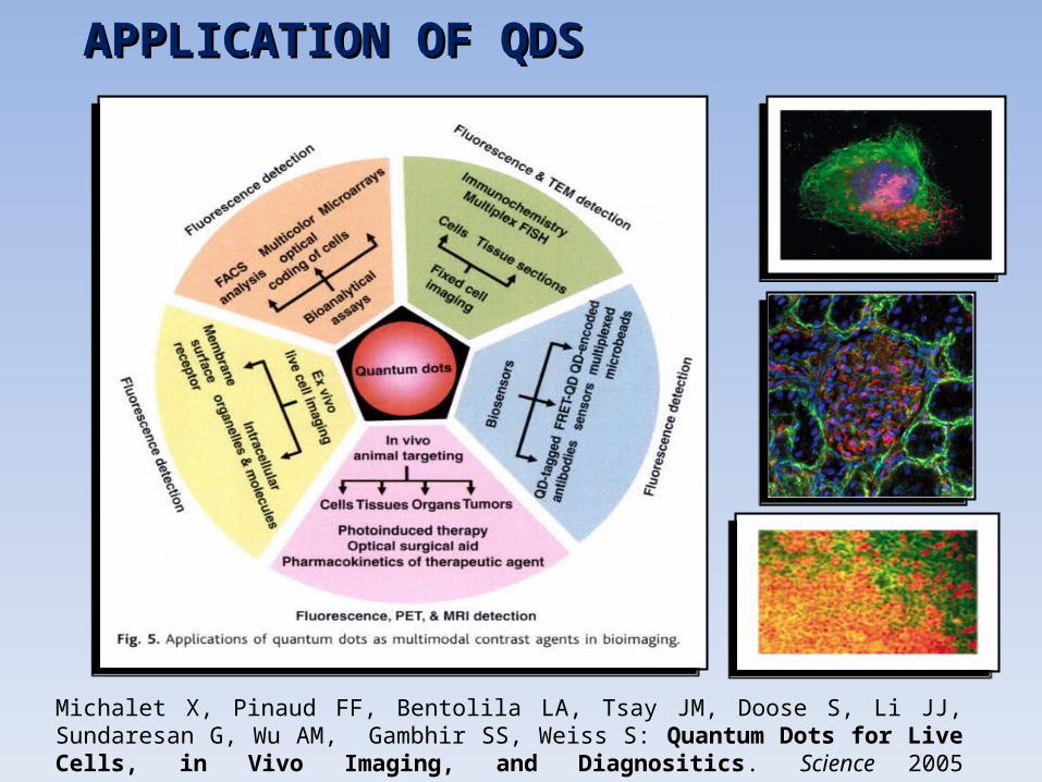
APPLICATION OF QDSAPPLICATION OF QDS
Michalet X, Pinaud FF, Bentolila LA, Tsay JM, Doose S, Li JJ, Sundaresan G, Wu AM, Gambhir SS, Weiss S: Quantum Dots for Live Cells, in Vivo Imaging, and Diagnositics. Science 2005 307(5709):538-544.

QDs is a perspective tools for medical QDs is a perspective tools for medical usage…usage…
TOXIC or NON TOXIC?TOXIC or NON TOXIC?
Distribution Localization and
accumulation in vivo.
Distribution Localization and
accumulation in vivo.
per os Intravenously (i.v.)

TECHNOLOGYTECHNOLOGY
Type of administration
•I.V.• Per os
Localization study
In vivo imaging
Local fluorescence spectroscopy
Confocal fluorescent microscopy
Pathomorphogy
Morphology
Clinical biochemistry
Toxical effects

QDs MPA• λ em 611nm and 630 nm• d ~ 8 – 11нм•QY -10-20%
QDs PolyT• λ em 626 нм• d ~ 15 – 16 нм•QY 10-30%
QDs PolyT-APS• λ em 678 нм• d ~ 36 нм•QY 5-20%
QY

FLUORESCENT EQUPMENT
iBox UVP Laser spectrometer with optical zond

Monitoring of fluorescence in vivoin digestive tract of mice
Excitation 502-547 nm, emission filter 570-640 nm exposition25с.
per osper os

QDsStomach after 2 h
Intenstines after 2 h
Intenstines after 4 h
Intenstines after 6 h
MPA + - - -
PolyT + + ± -
PolyT-APS + + + +No fluorescence (-), weak (±), high (+).
Local fluorescense spectroscopyLocal fluorescense spectroscopy
Fluorescence of excrements after 24 h of QDs adminestering per os
QDs MPA
Black line- excrements of control mice Red line- excrements of after QDs administering
QDs PolyT-APSQDs PolyT

I.V.I.V. QDs MPA
LFS

QDs Poly T
LFS
I.V.I.V.

i.V.i.V. QDs PolyT-APS
LFS

Confocal microscopyConfocal microscopyNB: weak+, medium ++, high +++, no -. (*)in blood vessel only
а – reflecte lights, б – fluorescence, в – peudo color imaging, г – fluorescence spectra
Lungs LiverLiverLungs

а – local haemorragia;
б – stenosis of arteria.
Blood Urea creatinine,
Morphology
Toxicity of QDs MPAToxicity of QDs MPA
No change
alkaline phosphatase alanine- and aspartate
transaminases
Hihg spleen weight (twice)
Pathomorphologyа б

I.V.I.V.QDs MPA toxicity: also depends on initial
solution

1. Distribution and accumalation depend on the modification of coating
2. Qds are dissolving in digestive tract: PolyT-APS are more stable comparing to QDs МPА and PolyT after per os administering
3. The main targeted organs for QDsМPА are the lungs, for PolyТ are the atria, for PolyT-APS are liver after I.V . Excretion of in the urine and faeces was not detected.
4. QDs of small size МPA stay in organism for a long period.They could lead to long-term pathomorphological changes in lungs on the 22-th day after administration.
5. The the severity of the toxic effect of QDs MPA depends on the solvent
CONCLUSIONCONCLUSION

Локальная флуоресцентная спектроскопия Локальная флуоресцентная спектроскопия (ЛФС)(ЛФС)