dr rick robinson, dpm director of podiatric services ... shoes and activity history x-rays? 16...
TRANSCRIPT

Dr Rick Robinson, DPM Director of Podiatric Services USPHS Hospital at Whiteriver, AZ 2015 IHS National Combined Council Winter Meeting Friday, January 30, 2015
1

“We are all athletes, the difference is that some of us are in training, and some are not.”
George Sheehan, MD
2

Overview of heel pain Functional errors Contributing factors to injuries Differential diagnosis of heel pain Evaluation & treatment
3
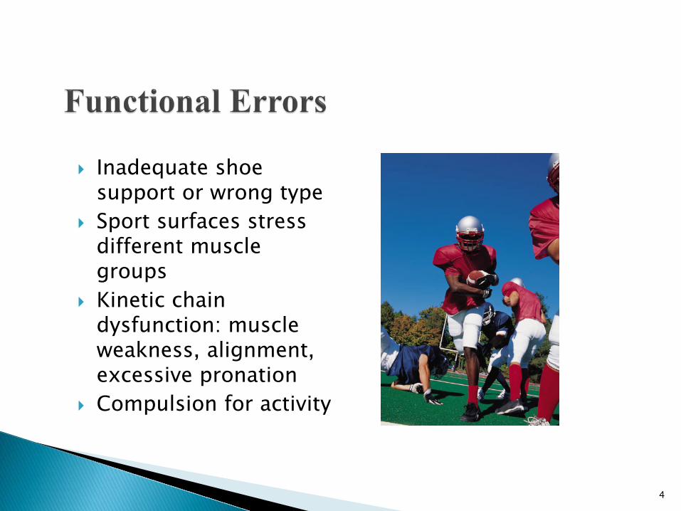
Inadequate shoe support or wrong type
Sport surfaces stress different muscle groups
Kinetic chain dysfunction: muscleweakness, alignment,excessive pronation
Compulsion for activity
4
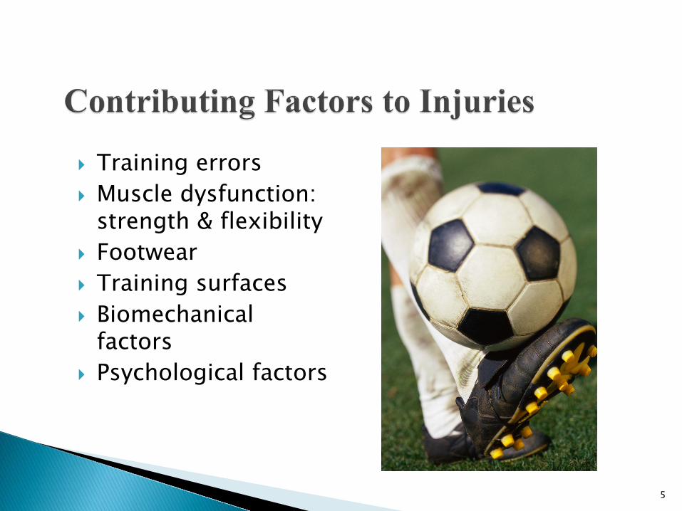
Training errors Muscle dysfunction:
strength & flexibility Footwear Training surfaces Biomechanical
factors Psychological factors
5
Dorsiflexion of hallux produces an archraising effect on the standing foot
Dorsiflexion of the hallux winds the plantar fascia distally and then superiorly around thefirst metatarsal head
Produces raising of arch as it shortens thedistance from the origin of plantar fascia anddistal first metatarsal head
Calcaneal dorsiflexion and first metatarsal plantarflexion raise the medial longitudinal arch
6
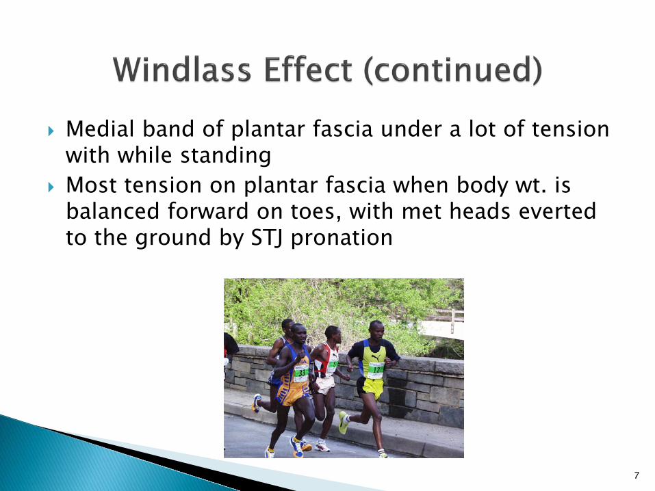
Medial band of plantar fascia under a lot of tensionwith while standing
Most tension on plantar fascia when body wt. isbalanced forward on toes, with met heads everted to the ground by STJ pronation
7
Holds the medial longitudinal arch in a higherarched position (prevents arch collapse)
Assists in resupination of STJ duringpropulsion phase
Assists deep posterior compartment musclesby helping limit STJ pronation during gait
Assists plantar intrinsic muscles to maintainarch height
8
Prevents excessive interosseous compressionforces on dorsal joint surfaces of bones ofmedial and lateral longitudinal arch
Helps maintain purchase of digits duringstanding and prevents floating toes
Acts to store energy with its inherent elasticity within arch for activity
Kirby, K. Foot and Lower ExtremityBiomechanics (1997)
9
1)Pain and inflammation: NSAIDs, cortisone injection, physical therapy, ice, modifiedactivity/cross-training
2)Cause: overuse vs. biomechanical: taping,shoe recommendations/modifications, training/conditioning, stretching, and orthotics
**must address cause, especially when itbecomes chronic**
10
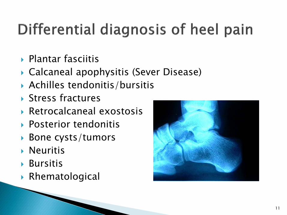
Plantar fasciitis Calcaneal apophysitis (Sever Disease) Achilles tendonitis/bursitis Stress fractures Retrocalcaneal exostosis Posterior tendonitis Bone cysts/tumors Neuritis Bursitis Rhematological
11

Overuse/trauma: calcaneal apophysitis, fracture,contusion/ strain
Developmental: coalition Inflammatory: tendonitis, bursitis, os trigonum Infectious Rheumatologic: rheumatoid arthritis, Reiter synd. Tumorous: benign- bone cyst, osteochondroma Neurologic: tarsal tunnel syndrome
12
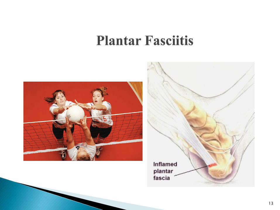
13

Any motion of the foot that puts the fascia inexcessive stretch (increased pronation) or acontracture (cavus or supinated foot)
Traction enthesitis on calcaneus Commonly near the origin of the plantar
fascia Ankle joint equinus that produces hyper-
mobility and increased pull on fascia
14

Pain at medial heel near origin of fascia May also be into longitudinal arch Hallux dorsiflexion can increase pain Rule out calcaneal stress fracture or nerve
entrapment Pes cavus and pes planus foot types Muscle tightness of calf and fascia
15

Post static dyskinesia: cycle Tenderness on palpation of band, commonly near
the origin at the medial tubercle of calcaneus May have edema and erythema Increased pain with ankle dorsiflexion from stretch
on fascia Antalgic gait/ toe walking History is very important Evaluate shoes and activity history X-rays?
16

Evaluate training intensity, surfaces, terrain, and demands of sport or activity
Biomechanical evaluation Shoe recommendations: stability, motion control,
or cushion Cross training is critical for rehabilitation Incorporate stretching
17

18
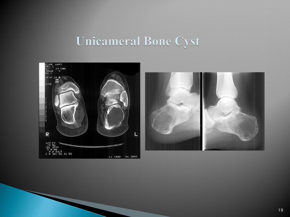
19

Control inflammation Stretching and
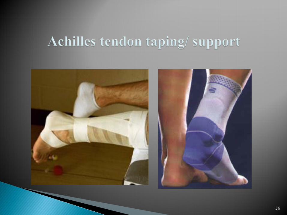
massage Taping/ functional
orthotics Shoe
recommendations: anti-pronation
Night splint Cortisone injections Cross training ECSWT Surgery
20
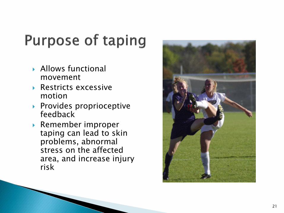
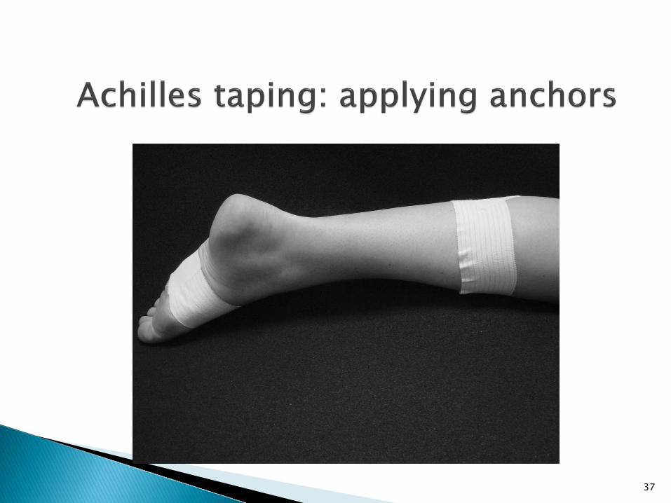
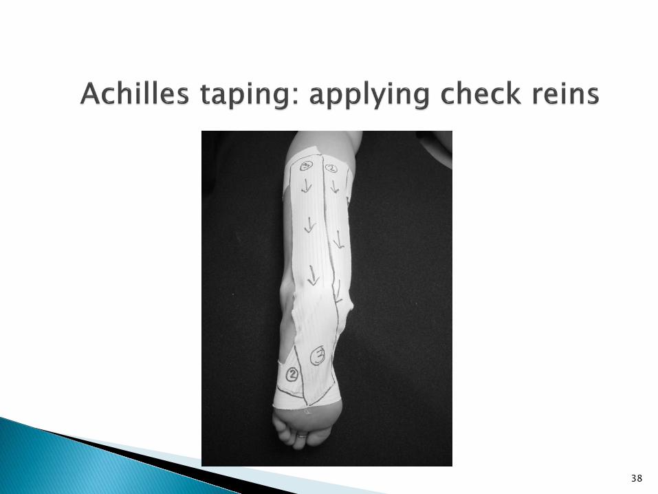
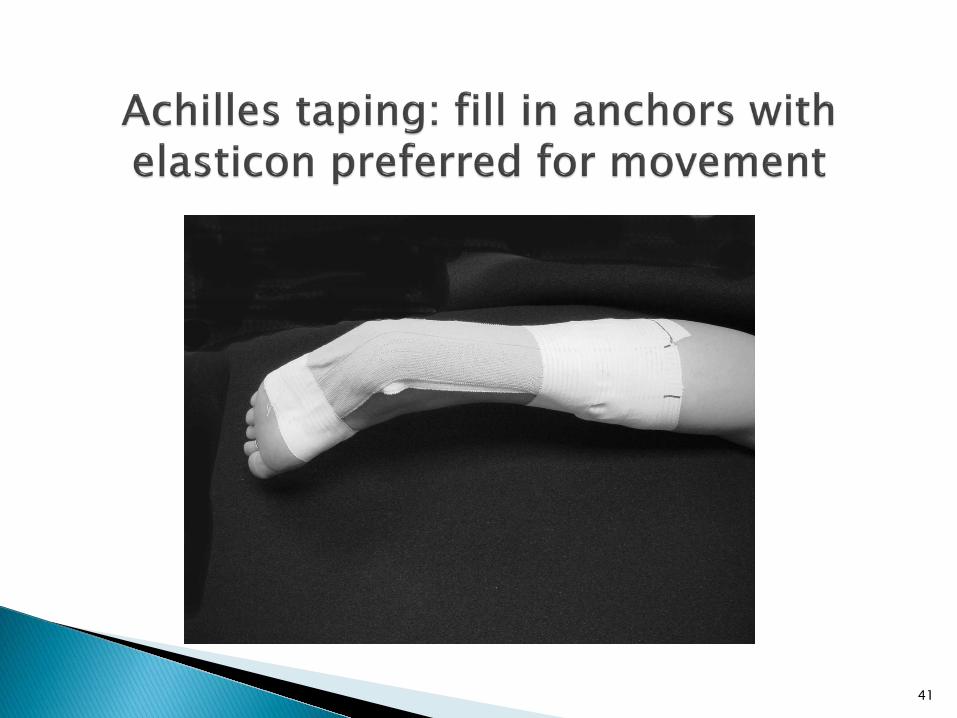
Allows functional movement
Restricts excessive motion
Provides proprioceptivefeedback
Remember improper taping can lead to skinproblems, abnormalstress on the affected area, and increase injury risk
21

22

23
24
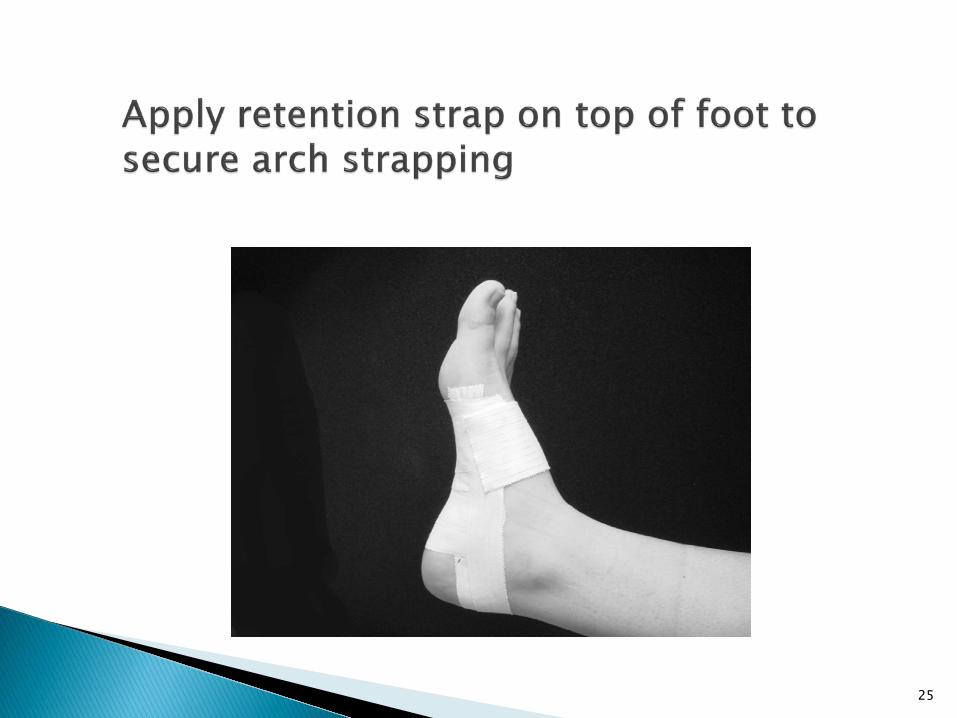
25
26
27

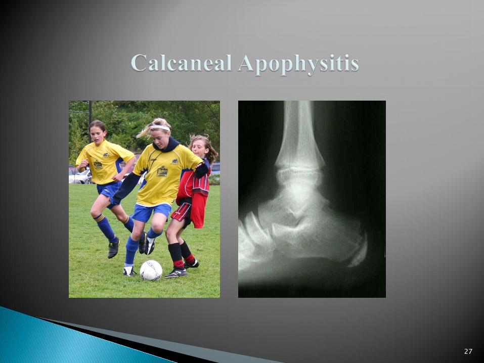
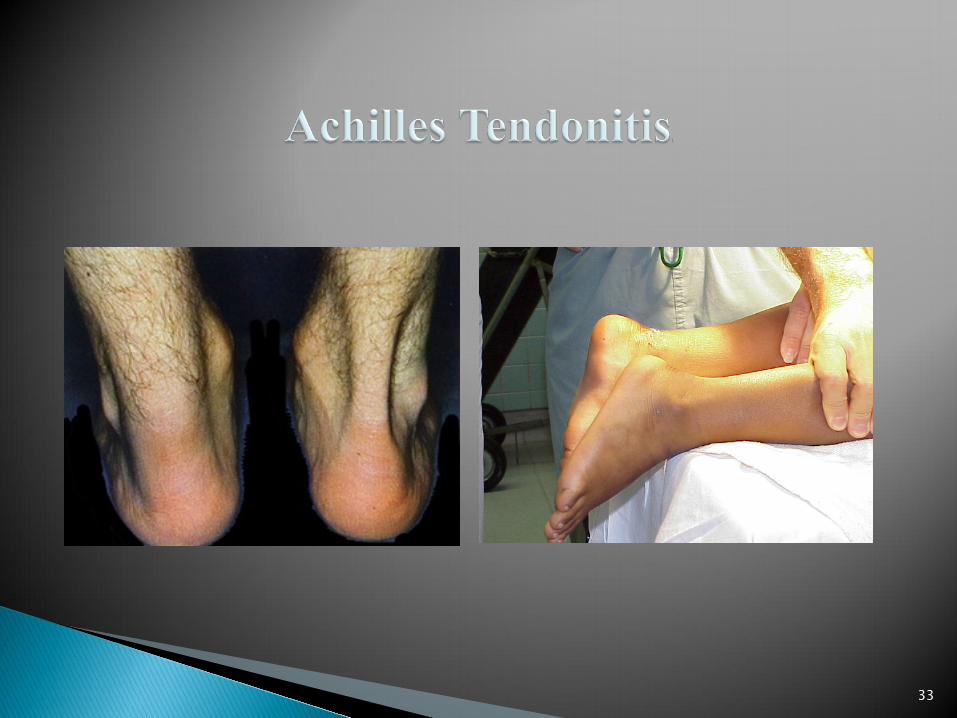
Repetitive micro trauma from pull ofAchilles on unossified calcaneal apophysis
Frequently occurs before or duringgrowth spurt and beginning of newsport
Most commonly in boys of 10-12 yrs ofage and girls of 8-10 years of age
Complain of pain in heels with runningand jumping
28

Calcaneal apophysis develops as independent ossification center
Appears around age 7-9 Fusion of apophysis usually
around 15-17 years Higher composition of
fibrocartilage than epiphyses Subjected to strong shear
stresses due vertical orientation and pull from Achilles tendon
29
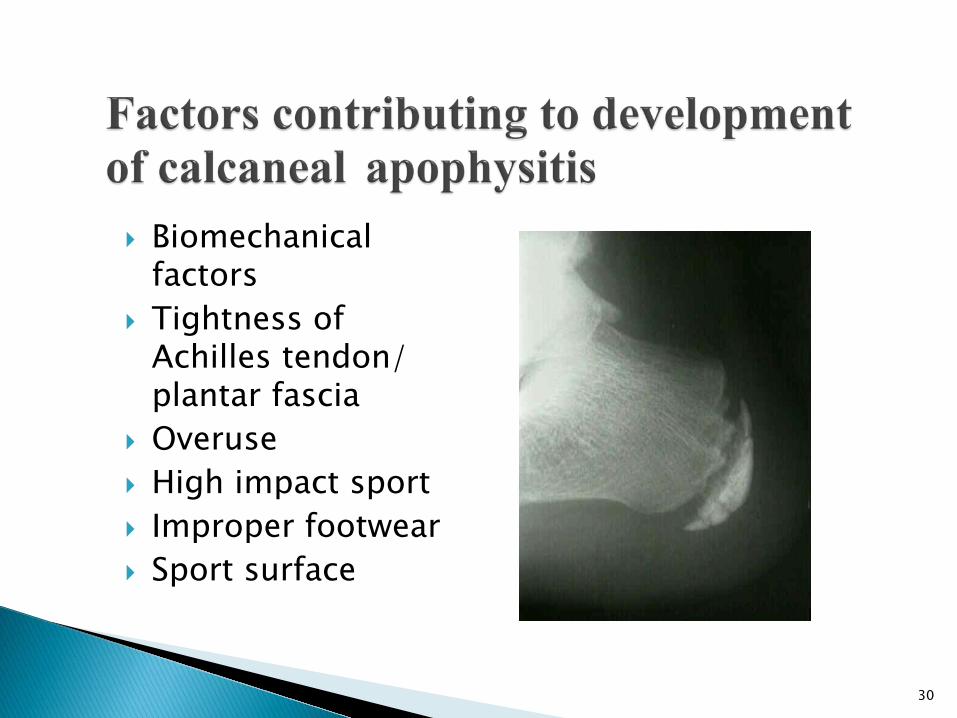
Biomechanical factors
Tightness ofAchilles tendon/ plantar fascia
Overuse High impact sport Improper footwear Sport surface
30
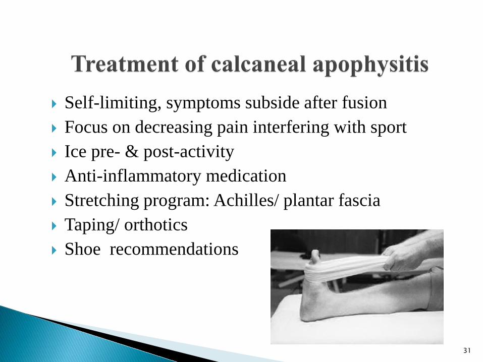
Self-limiting, symptoms subside after fusion Focus on decreasing pain interfering with sport Ice pre- & post-activity Anti-inflammatory medication Stretching program: Achilles/ plantar fascia Taping/ orthotics Shoe recommendations
31

History: muscle imbalance/ tightness;
biomechanical; recent increase in activity;change in shoe
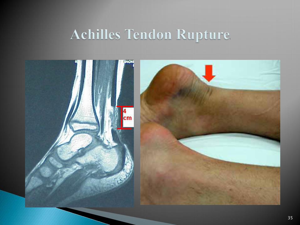
Most commonly at insertion or within 2-6 cm Control inflammation; correct training errors;
stretching program; heel lifts; shoerecommendations; possible orthotics
32
33

34
35
36
37
38

39

40
41
42

Repetitive rapid eversion of calcaneus at heel strike against the heel counter of the shoe
Increased activity and stiffer heel counterscan exacerbate the problem
Treatment: orthotics, shoe modifications, possible surgery
43
Fatigue or insufficiency fractures Fatigue fractures: occur in normal bone from
application of abnormal stress or torque;repeated loads causes osteoclastic activityleading to bone weakening
Insufficiency fractures: diseased bone-osteoporosis, arthritic conditions, metabolicconditions, chronic diseases
Bilateral incidence is not uncommon
44

45
Similar to plantar fasciitis Diffuse heel pain with wt. bearing,
relieved by rest Increased pain with prolonged activity May have post-static dyskinesia Usually no history of trauma New activity or exercise routine started Positive squeeze test as well as on plantar
heel
46
Pes cavus Calcaneal gait Limb length descrepancy Antagonistic pull of Achilles tendon with
the plantar fascia, i.e. concentric andeccentric contraction of gastroc duringactivity
47
lateral x-ray; also on axial or oblique Usually not visualized until 2-3 weeks after
onset of symptoms
Calcaneal stress fractures best seen on
Change of approximately 50% in bonedensity needed for delineation of trabecular lesions with visual sclerosis
Bone scintigraphy is gold standard, withthree-phase technetium bone scan, typically in the posterior half of the calcaneus
MRI is highly sensitive and specific
48
Phase 1: Modified rest of about 6-8 weeks in first phase with protectedweight bearing in walking boot
Phase 2 : Gradual increase to activitywhen pain free
Modify activity and addressbiomechanical factors
49

Do not stretch an injured tendon; should be done during the rehabilitation phase
Gradually increase stretching astolerated once heel pain subsides
Stretch both Gastroc-soleus and plantarfascia muscle groups
50
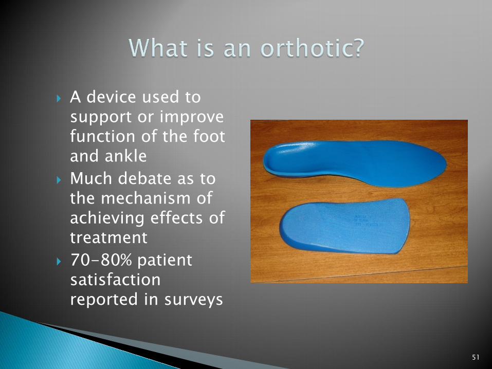
A device used to support or improvefunction of the foot and ankle
Much debate as to the mechanism of achieving effects of treatment
70-80% patientsatisfaction reported in surveys
51
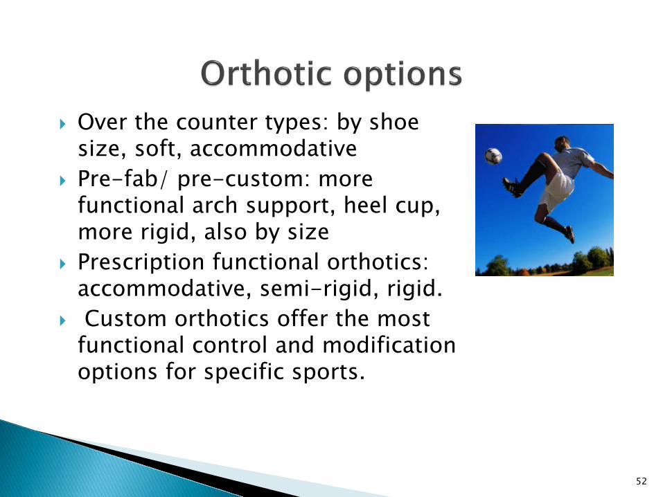
Over the counter types: by shoe size, soft, accommodative
Pre-fab/ pre-custom: more functional arch support, heel cup, more rigid, also by size
Prescription functional orthotics: accommodative, semi-rigid, rigid.
Custom orthotics offer the most functional control and modification options for specific sports.
52

functional accommodative
53
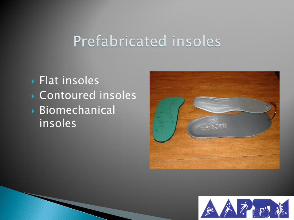
Flat insoles Contoured insoles Biomechanical
insoles
54
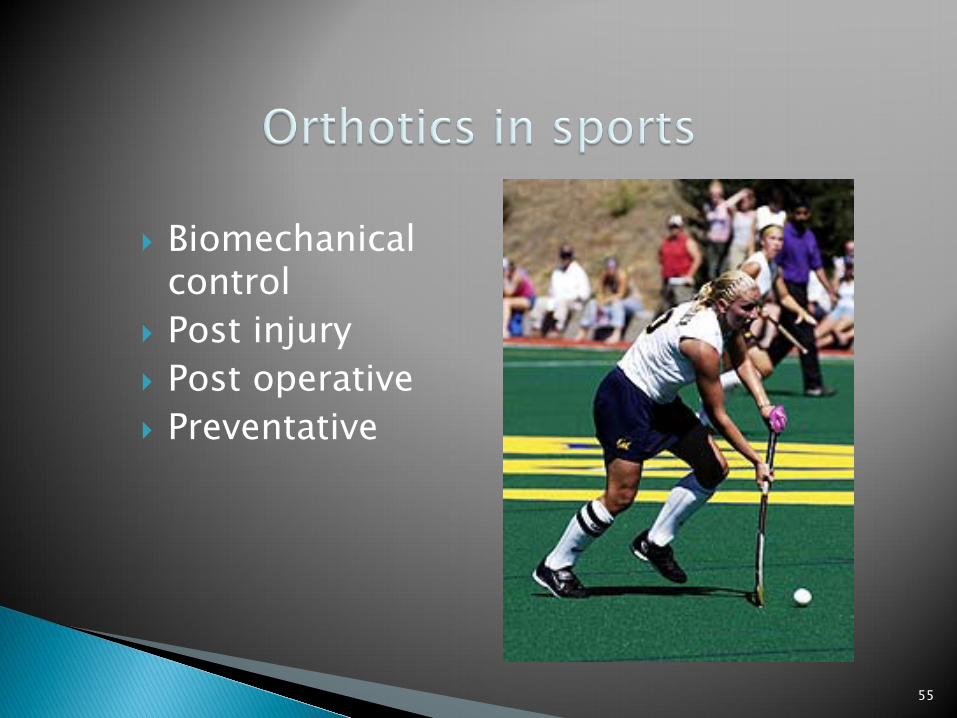
Biomechanical control
Post injury Post operative Preventative
55
56
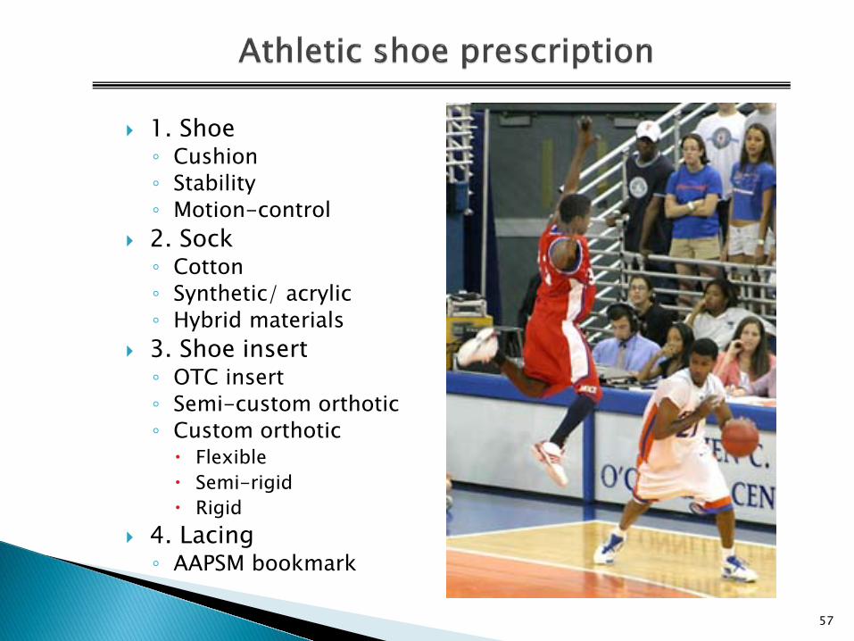
1. Shoe ◦ Cushion ◦ Stability ◦ Motion-control
2. Sock ◦ Cotton ◦ Synthetic/ acrylic ◦ Hybrid materials
3. Shoe insert ◦ OTC insert ◦ Semi-custom orthotic ◦ Custom orthotic Flexible Semi-rigid Rigid
4. Lacing ◦ AAPSM bookmark
57
58
Limitation of ankle joint dorsiflexion withSTJ in neutral position (lack of 10 degrees)
Ankle joint dorsiflexion should be greater than 15 degrees with the knee flexed
Can be present while knee flexed,extended, or in both positions
59
At birth, dorsiflexion unrestricted, about 75 degrees
dorisflexion decreases to 20-25 degrees by age 3
15 degrees by age 10 10 degrees by age 15 Ankle joint dorsiflexion should always be more
than 15 degrees with knee flexed with normal development
60

Congential gastrocnemius equinus Congenital soleus equinus Congential gastroc-soleus equinus Spastic equinus
61

Most common type Limited ankle dorsiflexion with knee extended Flexion of knee demonstrates normal ROM
62

Limited ankle joint dorsiflexion whenknee in flexed position
May also have knee extended and limited ankle joint dorsiflexion
Rare type
63

Limitation of ankle joint dorsiflexion with kneein both the extended and flexed position
Both gastroc and soleus units are tight Usually more dorsiflexion available with knee
flexed but less than 10 degrees May see bony block of talotibial articulation in
older child, has an abrupt feel at end point ofROM
Radiographic changes with bony deformity
64

Associated with spastic disorders like cerebral palsy
Limited dorsiflexion of ankle in both knee positions
May have increased Achilles reflexes andankle clonus
65

Patient supine position Foot maximally dorsiflexed to leg with STJ
maintained in neutral position If STJ pronated, MPJ unlocks and allows
excessive dorsiflexion of FF to RF Slightly supinate the RF to reduce chance
of pronating STJ
66

Normal heel off STJ and MTJ compensating STJ pronated at heel contact Midstance pronation with forefoot abducted
on the rear foot Foot lifts off in two segments: rear foot lifts
early, then forefoot lifts Toe off is apropulsive Characteristics: pronated foot with flattened
medial arch, unlocked to subluxed STJ, forefoot supinatus, prone to HAV
67

Early heel off Inadequate compensation at STJ and MTJ Heel contacts and then comes off early
in midstance Resupination occurs late in propulsion Characteristics: Mildly pronated foot
with normal arch structure
68

No heel contact noted Forefoot contact only “Toe walking” Characteristics: Normal foot type, foot
plantarflexed to leg
69
Posterior muscle contracture: clonic spasm, tonic spasm, accommodative shortening
Congenital short gastroc muscle or triceps surae
Osseous ankle block Tight posterior ankle capsule
70

Abducted gait Knee flexion STJ pronation, with unlocking of MTJ
leading to dorsiflexion of forefoot andrear foot
Genu recurvatum: subluxation of knee posteriorly
Decreased stride
71

72