dr. rezzan khan shifa international hospital
TRANSCRIPT
Dr. Rezzan Khan Consultant Nutritionist
Shifa International Hospital


Concept of Fluid & Electrolyte Balance Body fluid and electrolyte homeostasis Differentiate between hypovolemic, euvolemic, and
hypervolemic hyponatremia Recommend appropriate changes in nutrition
support formulations for treatment of electrolyte abnormalities

Fluid balance ensures that the body stays hydrated; important for normal functioning of the body and
optimal health. Maintaining fluid balance is vital for physical and
mental performance. Water is one of the most essential nutrients that
the body requires.

Loss of 10% body fluid = 8% weight loss SERIOUS
Loss of 20% body fluid = 15% weight loss FATAL
Fluid gained each day should = fluid lost each day (2 -3L/day average)
What is the minimum output per hour necessary to maintain renal function?
Why Fluid balance is important
30 ml/hr

Age ◦ Infants ◦ Older adults
Prior medical history ◦ Acute illness ◦ Chronic illness ◦ Environmental factors ◦ Diet ◦ Lifestyle ◦ Medications

Fetus: 90% water Premie: 80% water Term: 70-75% water Young children: 65-70% Adolescents: 60% water Lean individuals: greater percentage of body weight
is water Fat individuals: smaller percentage of body weight is
water

Method Equation Body Surface Area (BSA) Method 1,500 mL/m2 x BSA = mL/day Recommended Dietary Allowance/Adolph Method
1 mL/kcal of intake = mL/day
Fluid Balance Method Urine output + 500 mL/day Weight Method 25-35 mL/kg/day Age-adjusted Weight Method Average healthy adult: 30-35
mL/kg/day Adult 55-65 years old: 30 mL/kg/day Adult > 65 years old: 25 mL/kg/day
Obese Adult Patient Method [(kg body weight–20) x 15] + 1,500 mL/day
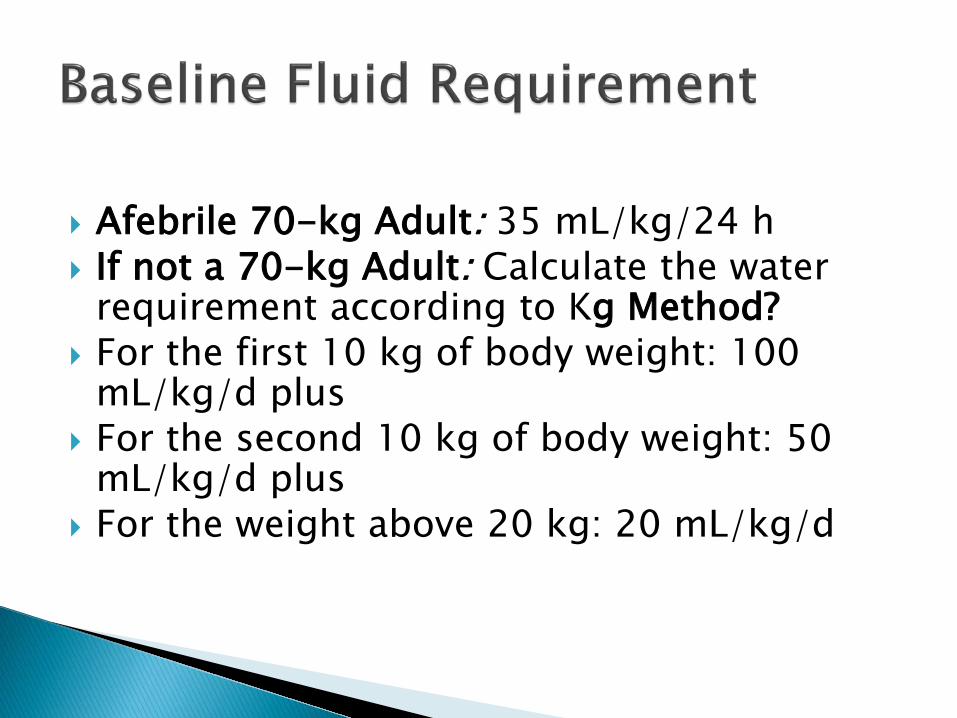
Afebrile 70-kg Adult: 35 mL/kg/24 h If not a 70-kg Adult: Calculate the water
requirement according to Kg Method? For the first 10 kg of body weight: 100
mL/kg/d plus For the second 10 kg of body weight: 50
mL/kg/d plus For the weight above 20 kg: 20 mL/kg/d

State of equilibrium in body Naturally maintained by adaptive responses Body fluids and electrolytes are maintained
within narrow limits

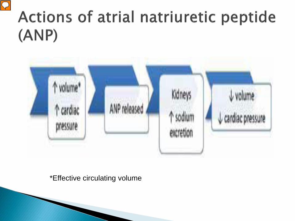
*Effective circulating volume

*Effective circulating volume
*Effective circulating volume


The intravascular volume of an average 70 kg man is approximately 5 L of which 2 L is red cell volume and 3 L plasma volume. The intravascular, extracellular fluid compartment equilibrates with the extracellular, extravascular fluid compartment (ECF ∼ 11 L), with a reduction in one compartment leading to a reduction of the other. Plasma to interstitial fluid shift results in edema Water deficit (increased ECF) Associated with symptoms that result from cell shrinkage as water is pulled into vascular system Water excess (decreased ECF) Develops from gain or retention of excess water

Plasma to interstitial fluid shift results in edema Water deficit (increased ECF) Associated with symptoms that result from cell shrinkage as water is pulled into vascular system Water excess (decreased ECF) Develops from gain or retention of excess water

First spacing ◦ Normal distribution of fluid in ICF and ECF
Second spacing Abnormal accumulation of interstitial fluid
(edema) Third spacing ◦ Fluid accumulation in part of body where it is not
easily exchanged with ECF; fluid trapped and unavailable for functional use (ascites)

.

Treating an ECF requires aggressive management of sepsis, fluid, and renal function; nutritional, electrolyte, and metabolic support; management of fistula output; assessment of fistula anatomy; and when appropriate, corrective surgery.

Hypovolemia Hypervolemia Hypoproteinemia
Definition Extracellular fluid deficit Extracellular fluid excess Loss of oncotic pressure leads to hypovolemia
Causes Congestive heart failure Decreased protein intake Hemorrhage Increased protein loss Overdiuresis Renal failure Liver/kidney disease Vomiting/diarrhea Liver disease Burns Third-spacing Overzealous IV fluids Infection (ascites, burns) Sodium overload Hemorrhage
Clinical Dry mucous membranes findings Sudden weight loss Oliguria Tachycardia Orthostatic hypotension

Type Description and Causes Isotonic Equal loss of sodium and water Gastrointestinal illness Hypertonic Most common cause Water loss exceeds sodium loss Fever Limited fluid intake Hypotonic Sodium loss exceeds water loss Diuretic use
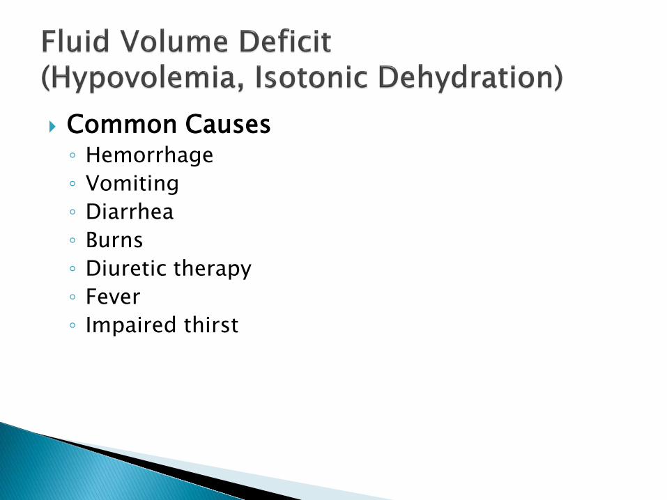
Common Causes ◦ Hemorrhage ◦ Vomiting ◦ Diarrhea ◦ Burns ◦ Diuretic therapy ◦ Fever ◦ Impaired thirst

Signs/Symptoms ◦ Weight loss ◦ Thirst ◦ Orthostatic changes in pulse rate and bp ◦ Weak, rapid pulse ◦ Decreased urine output ◦ Dry mucous membranes ◦ Poor skin turgor

Encourage patient fluid intake of 1,000–3,000 ml daily (e.g., filling a pitcher each day and making sure it is empty at the end of the day)
Monitor patient lab values for changes. –– Increased BUN/creatinine –– Increased serum sodium –– Increased serum osmolarity –– Increased hematocrit. • Monitor patient urine output. • Monitor patient for constipation or diarrhea. • Weigh patient daily. • Teach patient to drink despite not feeling thirsty, particularly if
taking diuretics. • Advise patient to avoid alcoholic, carbonated, and caffeinated
beverages, which can increase diuresis.

Fluid Management ◦ Diet therapy – Mild to moderate dehydration.
Correct with oral fluid replacement. ◦ Oral rehydration therapy – Solutions containing
glucose and electrolytes. E.g., Pedialyte, Rehydralyte. ◦ IV therapy – Type of fluid ordered depends on the
type of dehydration and the clients cardiovascular status.


Common Causes: ◦ Congestive Heart Failure ◦ Early renal failure ◦ IV therapy ◦ Excessive sodium ingestion ◦ SIADH ◦ Corticosteroid
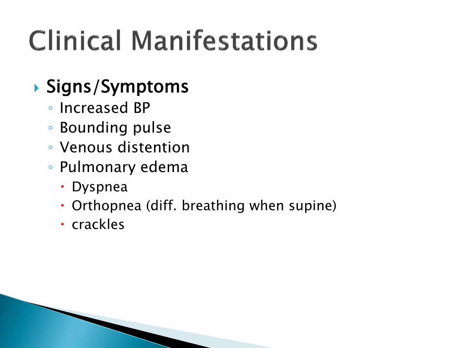
Signs/Symptoms ◦ Increased BP ◦ Bounding pulse ◦ Venous distention ◦ Pulmonary edema Dyspnea Orthopnea (diff. breathing when supine) crackles

Drug therapy ◦ Diuretics may be ordered if renal failure is not the
cause. Restriction of sodium and saline intake I/O Weight
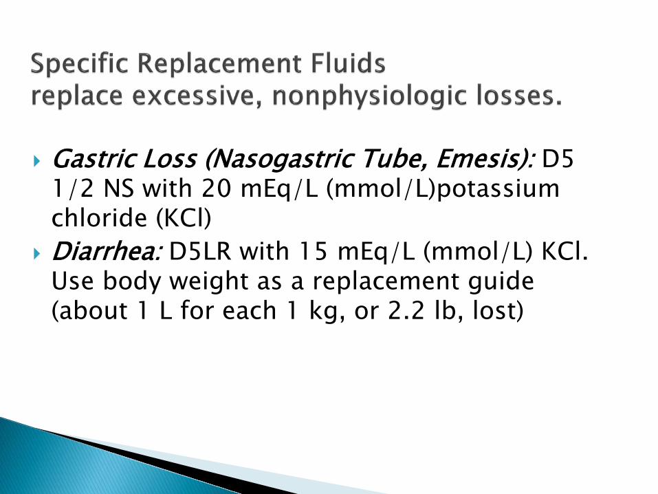
Gastric Loss (Nasogastric Tube, Emesis): D5 1/2 NS with 20 mEq/L (mmol/L)potassium chloride (KCl)
Diarrhea: D5LR with 15 mEq/L (mmol/L) KCl. Use body weight as a replacement guide (about 1 L for each 1 kg, or 2.2 lb, lost)

Normal Saline (0.9%) (Na 154 meq/L, Cl 154 meq/L) Lactate Ringer's (Hartman's solution) (Na 130 meq/L, Cl 109 meq/L, K 4 meq/L, Lactate 28 meq/L, Ca 3
meq/L) Normosol-R (Na 140 meq/L, Cl 90 meq/L, K 5 meq/L, Mg 3
meq/L) Plasmalyte (Na 140 meq/L, Cl 98 meq/L, K 5 meq/L, Mg 3 meq/L)


The goal is to find and correct the underlying cause.
1. Determine presence and severity of signs and symptoms.
2. Determine speed of onset: acute onset necessitates rapid correction of sodium; chronic onset necessitates slow correction of sodium.
3. Determine osmolality (tonicity): rule out isotonic and hypertonic osmolality.
4. Establish extracellular fluid volume status.
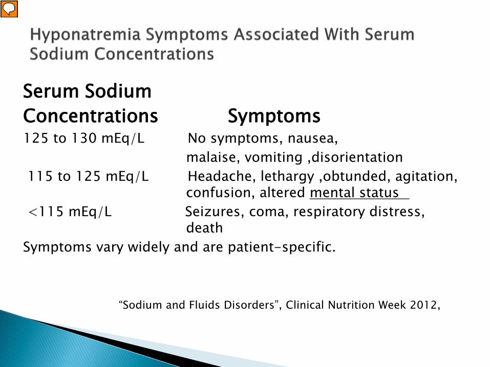
Serum Sodium Concentrations Symptoms 125 to 130 mEq/L No symptoms, nausea, malaise, vomiting ,disorientation 115 to 125 mEq/L Headache, lethargy ,obtunded, agitation,
confusion, altered mental status <115 mEq/L Seizures, coma, respiratory distress,
death Symptoms vary widely and are patient-specific. “Sodium and Fluids Disorders”, Clinical Nutrition Week 2012,

Initial Data: • 49-year-old female • Weight: 50 kg • Plasma sodium day 1: 123 mEq/L • Plasma sodium day 2: 126 mEq/L • Day 1 IV fluids: 1 L normal saline (sodium content is 154 mEq/L) (3) • Day 2 IV fluids: 1.5 L normal saline Step 1: Calculate total body water Total Body Water (TBW) for men: 0.6 L/kg x weight in kg Total Body Water (TBW) for women: 0.5 L/kg x weight in kg (3) Calculation for DD: 0.5 x 50 = 25 L Step 2: Calculate sodium deficit Sodium deficit : TBW x ([desired plasma sodium – current plasma sodium])*(3) Calculation for DD: 25 x (133 – 123) = 25 x 10 = 250 mEq Step 3: Calculate how much 1 L normal saline** initially increases plasma
sodium (7): Increase in plasma sodium = (sodium in NS – plasma sodium) ÷ (TBW + 1) Calculation for DD: (154 – 123) ÷ (25 +1) = 31 ÷ 26 = 1.19mEq/L

Hypervolemia, or fluid overload, is the medical condition where there is too much fluid in the blood.
The opposite condition is hypovolemia, which is too little fluid volume in the blood.
occurs when the body retains more water than it needs.

disruption in the salt/water balance in the body
- short term illness, injury or surgery - Intravenous Fluids - Injury/Illness/Surgery - can also be due to long-term health
conditions. - Kidney Disease - Liver Disease - Heart Disease

The symptoms of hypervolemia are consistent with excess water in the body
Moist Cough Frothy Sputum Increased or Slowed Heart Rate Changes in Blood Pressure Swollen abdomen Shortness of Breath

symptoms of hypervolemia


Considerations When Assessing Electrolyte Imbalances • Electrolyte content of nutrition products (i.e., parenteral nutrition, enteral nutrition) • Addition or removal of medications (i.e., spironolactone, furosemide, insulin) • IV fluids and piggybacks (i.e., normal saline with potassium) • Underlying disease states (i.e., acute renal injury acid-base disorders)

Pathophysiology – ◦ Decrease in K+ causes decreased excitability of
cells, therefore cells are less responsive to normal stimuli

Contributing factors: ◦ Diuretics ◦ Shift into cells ◦ Digitalis ◦ Water intoxication ◦ Corticosteroids ◦ Diarrhea ◦ Vomiting


Interventions ◦ Assess and identify those at risk ◦ Encourage potassium-rich foods ◦ K+ replacement (IV or PO) ◦ Monitor lab values ◦ D/c potassium-wasting diuretics ◦ Treat underlying cause

Pathophysiology – An inc. in K+ causes increased excitability of cells.

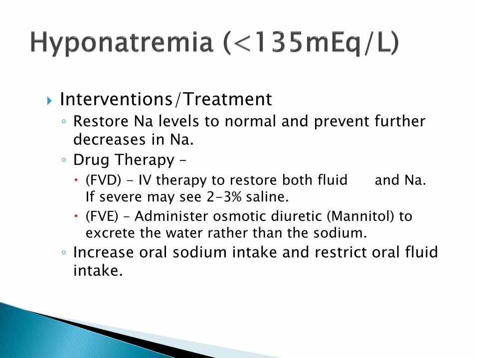
Interventions/Treatment ◦ Restore Na levels to normal and prevent further
decreases in Na. ◦ Drug Therapy – (FVD) - IV therapy to restore both fluid and Na.
If severe may see 2-3% saline. (FVE) – Administer osmotic diuretic (Mannitol) to
excrete the water rather than the sodium. ◦ Increase oral sodium intake and restrict oral fluid
intake.

Contributing Factors ◦ Hyperaldosteronism ◦ Renal failure ◦ Corticosteroids ◦ Increase in oral Na intake ◦ Na containing IV fluids ◦ Decreased urine output with increased urine
concentration

Contributing factors (cont’d): ◦ Diarrhea ◦ Dehydration ◦ Fever ◦ Hyperventilation

Assessment findings: ◦ Neuro - Spontaneous muscle twitches. Irregular
contractions. Skeletal muscle wkness. Diminished deep tendon reflexes ◦ Resp. – Pulmonary edema ◦ CV – Diminished CO. HR and BP depend on
vascular volume.

GU – Dec. urine output. Inc. specific gravity Skin – Dry, flaky skin. Edema r/t fluid volume
changes.

Interventions/Treatment ◦ Drug therapy (FVD) .45% NSS. If caused by both Na and fluid loss,
will administer NaCL. If inadequate renal excretion of sodium, will administer diuretics.
◦ Diet therapy Mild – Ensure water intake
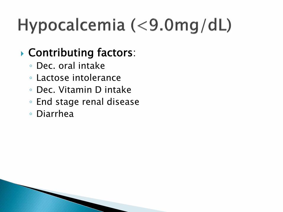
Contributing factors: ◦ Dec. oral intake ◦ Lactose intolerance ◦ Dec. Vitamin D intake ◦ End stage renal disease ◦ Diarrhea

Assessment findings: ◦ Neuro –Irritable muscle twitches. ◦ Resp. – Resp. failure , muscle tetany. ◦ CV – Decreased HR., decreased BP,
diminished peripheral pulses ◦ GI – Increased motility. Increased BS. Diarrhea

Contributing factors: ◦ Excessive calcium intake ◦ Excessive vitamin D intake ◦ Renal failure ◦ Hyperparathyroidism ◦ Malignancy ◦ Hyperthyroidism

Assessment findings: ◦ Neuro – Disorientation, lethargy, coma, profound
muscle weakness ◦ Resp. – Ineffective resp. movement ◦ CV – Increased HR, Increased BP. , Bounding
peripheral pulses, Positive Homan’s sign. Late Phase – Bradycardia, Cardiac arrest ◦ GI – Dec. motility. Dec. BS. Constipation ◦ GU – Inc. urine output. Formation of renal calculi
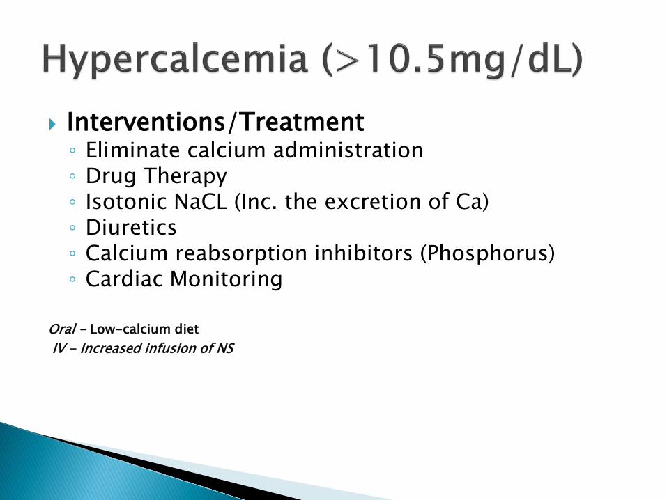
Interventions/Treatment ◦ Eliminate calcium administration ◦ Drug Therapy ◦ Isotonic NaCL (Inc. the excretion of Ca) ◦ Diuretics ◦ Calcium reabsorption inhibitors (Phosphorus) ◦ Cardiac Monitoring
Oral - Low-calcium diet IV - Increased infusion of NS

Contributing Factors: ◦ Malnutrition ◦ Starvation ◦ Hypercalcemia ◦ Renal failure ◦ Uncontrolled DM

Assessment findings Neuro – Irritability, confusion CV – Decreased contractility Resp. – Shallow respirations Musculoskeletal – destruction of muscle cells Hematologic – Increased bleeding Decreased platelet aggregation

Interventions ◦ Treat underlying cause ◦ Oral replacement with vit. D ◦ IV phosphorus (Severe) ◦ Diet therapy Foods high in oral phosphate

Causes few direct problems with body function. Care is directed to hypocalcemia.
Rarely occurs

Contributing factors: ◦ Malnutrition ◦ Starvation ◦ Diuretics ◦ Aminoglcoside antibiotics ◦ Hyperglycemia ◦ Insulin administration

Assessment findings: *Neuro - Hyperreflexia. Seizures *CV – ECG changes. Dysrhythmias. HTN *Resp. – Shallow resp. *GI – Decreased motility. Anorexia.
Nausea
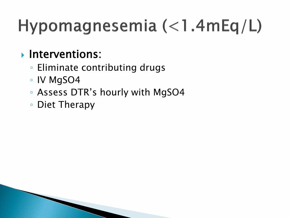
Interventions: ◦ Eliminate contributing drugs ◦ IV MgSO4 ◦ Assess DTR’s hourly with MgSO4 ◦ Diet Therapy

Contributing factors: ◦ Malnutrition ◦ Starvation ◦ Diuretics ◦ Aminoglcoside antibiotics ◦ Hyperglycemia ◦ Insulin administration

Assessment findings: *Neuro - Positive Trousseau’s sign.
Positive Chvostek’s sign. Hyperreflexia. Seizures
*CV – ECG changes. Dysrhythmias. HTN *Resp. – Shallow resp. *GI – Dec. motility. Anorexia. Nausea

Electrolyte Normal Range Equations Corrected magnesium (Mg2+) 1.7 to 2.5 mg/dL Serum Mg2+ x 0.005(40 –
serum albumin mg/dL)
Corrected calcium 8.0 to 11.0 mg/dL ([4 – serum albumin (g/dL)] x 0.8) + measured calcium (mg/dL)
Total body water (TBW) n/a (men) = 0.6 L/kg x weight in kg(women) = 0.5 L/kg x weight in kg
Sodium deficit (mEq) n/a TBW x (140 – measured serum sodium concentration mEq/L)
Water deficit (in L) n/a TBW x ([serum sodium concentration mEq/L/140] – 1)
Serum osmolality (sOsm) 280 to 300 mOsm/L (2 x serum sodium in mEq/L) + (serum glucose in mg/dL/18) + (BUN in mg/dL/2.8)
HCO3- deficit (mmol) n/a 0.5 x ([HCO3-] normal – [HCO3-] measured

Electrolyte Elevation Depletion Calcium Present in concentrations >11
mg/dL; lethargy, anorexia, nausea, vomiting, polyuria, confusion, coma
Hyperactive reflexes, muscle cramps numbness with tingling of fingers, tetany, convulsion
Phosphorus Anorexia, nausea, vomiting, hyperactive reflexes, tetany, tachycardia, muscle weakness
Confusion, seizures, coma, chest pain, difficulty speaking or breathing, weakness, joint stiffness
Magnesium Nausea, vomiting, diaphoresis altered mental status, coma, muscle weakness
Weakness, lethargy, muscle cramps, mood changes, confusion, vomiting
Sodium Increased thirst, fatigue, restlessness, muscle irritability, seizures, coma and death
Nausea, vomiting, headache, muscle cramps, disorientation, weakness, lethargy, confusion, dizziness, seizure, coma and death
Potassium Muscle cramping, weakness, electrocardiographic changes, arrhythmia
Constipation, lethargy, weakness, leg cramps

Electrolyte Elevation Depletion Calcium Oral - Low-calcium diet
IV - Increased infusion of NS
Oral- 1,000 to 1,500 mg/day IV(tetany present) 10 to 20 mL of 10% calcium gluconate over ≥4 hrs
Phosphorus Oral- .Low-phosphorus diet . Phosphate binders IV • Assess the need for volume repletion
Oral- • Increased dietary intake • Oral supplementation (i.e., Na3PO4) IV Moderate) • 0.32 to 0.64 mmol/kg (maximum, 30 mmol)Na3PO4 slowly over 6 hrs IV (Severe) • 1 mmol/kg (maximum, 80 mmol) Na3PO4 slowly over 8 to 12 hrs
Magnesium Oral Oral

Substrate mmol/L Sodium 90 Potassium 20 Bicarbonate 30 Chloride 80 Glucose 111

Ingredients 1 L (33 oz) water 3⁄4 tsp table salt (sodium chloride) 1 tsp baking power (or 2 tsp baking soda) 4 tbsp sugar (sucrose) 1⁄2 tsp 20% potassium chloride* Sugar-free Kool-AidR or Crystal LightR to taste Directions Pour all ingredients into a blender or pitcher.
Mix well.


Primary Compensating Acid-Base Disorder pH Change Reaction Metabolic acidosis ↓ ↓HCO3 ↓ PCO2 Metabolic alkalosis ↑ ↑HCO3 ↑ PCO2 Respiratory acidosis ↓ ↑PCO2 ↑ HCO3 Respiratory alkalosis ↑ ↓PCO2 ↓ HCO3
•Acidosis: process that lowers the extracellular fluid pH (reduction in HCO3 or elevation in pCO2)
•Alkalosis: process that raises extracellular pH (elevation in HCO3 or fall in pCO2)
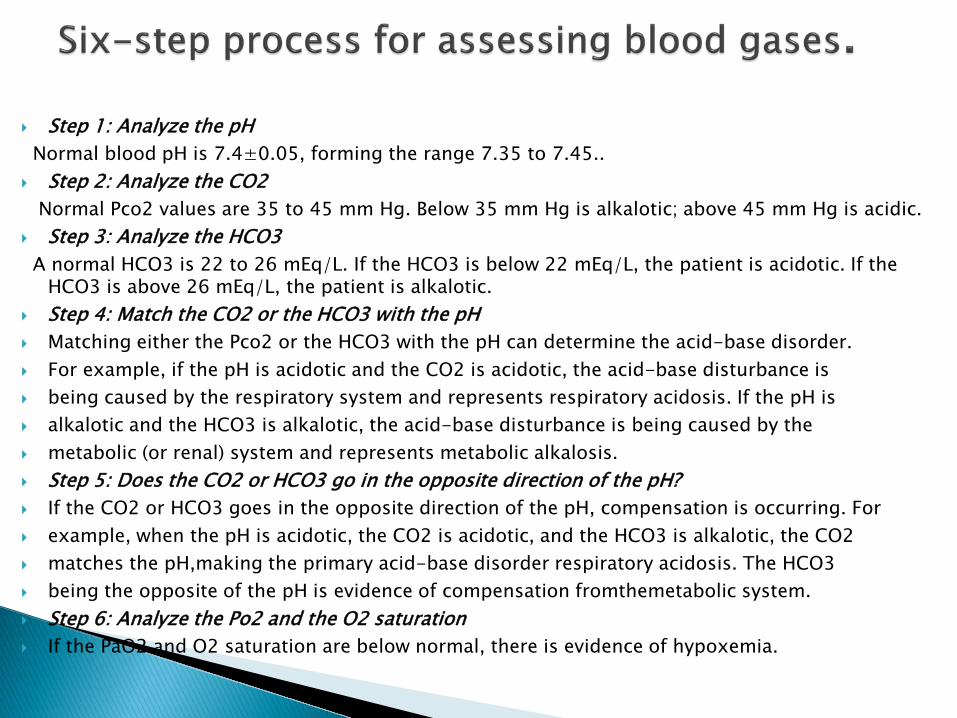
Step 1: Analyze the pH Normal blood pH is 7.4±0.05, forming the range 7.35 to 7.45.. Step 2: Analyze the CO2 Normal Pco2 values are 35 to 45 mm Hg. Below 35 mm Hg is alkalotic; above 45 mm Hg is acidic. Step 3: Analyze the HCO3 A normal HCO3 is 22 to 26 mEq/L. If the HCO3 is below 22 mEq/L, the patient is acidotic. If the
HCO3 is above 26 mEq/L, the patient is alkalotic. Step 4: Match the CO2 or the HCO3 with the pH Matching either the Pco2 or the HCO3 with the pH can determine the acid-base disorder. For example, if the pH is acidotic and the CO2 is acidotic, the acid-base disturbance is being caused by the respiratory system and represents respiratory acidosis. If the pH is alkalotic and the HCO3 is alkalotic, the acid-base disturbance is being caused by the metabolic (or renal) system and represents metabolic alkalosis. Step 5: Does the CO2 or HCO3 go in the opposite direction of the pH? If the CO2 or HCO3 goes in the opposite direction of the pH, compensation is occurring. For example, when the pH is acidotic, the CO2 is acidotic, and the HCO3 is alkalotic, the CO2 matches the pH,making the primary acid-base disorder respiratory acidosis. The HCO3 being the opposite of the pH is evidence of compensation fromthemetabolic system. Step 6: Analyze the Po2 and the O2 saturation If the PaO2 and O2 saturation are below normal, there is evidence of hypoxemia.

Understanding the relationship between electrolytes and their movement within TBW during altered hydration status, electrolyte imbalances, or acid-base imbalance is key to developing a thorough nutrition plan care.
With this knowledge , electrolyte abnormalities can be prevented or treated safely
A diet rich in highly alkaline fruits and vegetables helps reduce a marker of metabolic acidosis and preserve kidney function in patients with chronic kidney disease. ... ACE inhibitors were given to patients to control blood pressure
If your alkalosis is caused by a loss of chemicals such as chloride or potassium, you’ll be prescribed medications or supplements to replace these chemicals.
Some cases of alkalosis result from an electrolyte imbalance, which may be corrected by drinking plenty of fluids or drinks that contain electrolytes. If you have an advanced case of electrolyte imbalance, it will need to be treated in the hospital.