dr. michal ben-shachargondabrain.ls.biu.ac.il/neuroling/courses/218/slides/218..."copyright ©...
TRANSCRIPT

Brain anatomy tutorial
Dr. Michal Ben-Shachar


The human brain
http://www.brainmuseum.org/
Left
hemisphere
Right
hemisphere

Zoom out

Zoom in

Types of Brain Tissue
• Gray Matter: Cell bodies (+ short
connections)
• White Matter: Axons which form
pathways for conducting information
between brain cells.Gray matter
White matter

Gray matter, White matter
Gray matter (stained purple): folded sheet containing
cell bodies, dendrites, local axons collaterals.
White matter: axons, long range connections.

Cerebrospinal Fluid (CSF)
• Located between the menings and in the ventricles of the brain
• Functions– mechanical buffer
– fluid for metabolic functions
"Copyright © 2005 by Thompson Delmar Learning, a division of Thomson Learning, Inc. ALL RIGHTS
RESERVED"

The hemispheres: Two brains for
the price of one?
Fiber tracts
(White matter)

The hemispheres: Two brains for
the price of one?
The Virtual Hospital
• The hemispheres
are connected
through the corpus
callosum
Cortex
(Gray matter)Fiber tracts
(White matter)
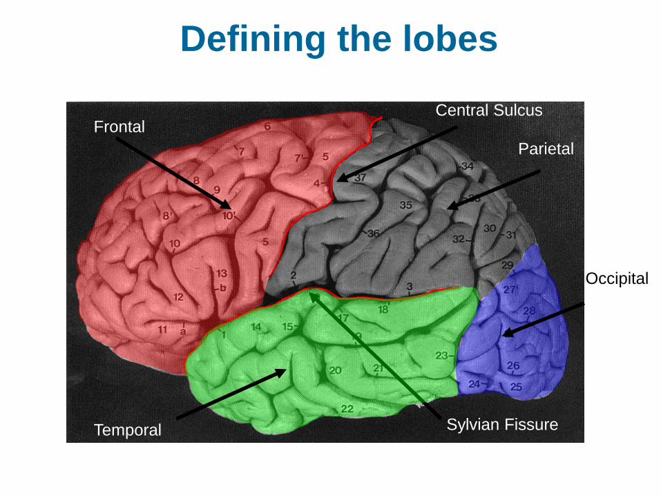
Defining the lobes
Frontal
Parietal
Occipital
Temporal Sylvian Fissure
Central Sulcus
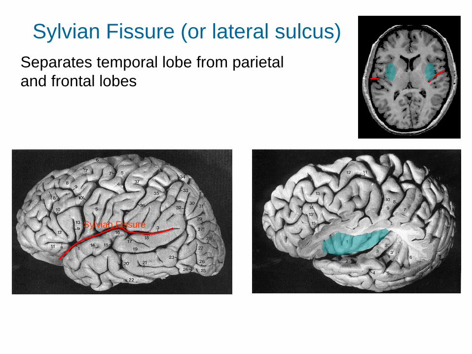
Separates temporal lobe from parietal
and frontal lobes
Sylvian Fissure
Sylvian Fissure (or lateral sulcus)

Gross functional map

Find the differences
Nichols and Newsome, 1999

Localization is not the final goal
“The recent avalanche of PET and fMRI papers has produced many
insights concerning localization of mental functions (Fig. 2b). Although they
are substantially more informative than previous findings based on brain
damage, distillations of data like the one in Fig. 2b are disconcertingly
similar to the one-function-one-brain-area maps of the phrenologists.
Will we ‘understand’ the brain when the map in Fig. 2b is completely filled
with blobs? Obviously not; localization data provide little insight into the
exact nature of the signals encoded in a given structure, the computations
being performed and the interactions between different structures.”
Nichols and Newsome, 1999

Middle Cerebral Artery

MCA stroke
Radiopedia.org; contributed by Dr. Frank Gaillard, July 2010

Gyri of the cerebral cortex
• Much of cortex referred to by combination of coordinate+lobe+gyrus: Superior Temporal Gyrus(STG), Middle Temporal Gyrus(MTG), Inferior Frontal Gyrus (IFG)
Duvernoy 1991

Sulci of the cerebral cortex
• Major sulci: Central Sulcus, Sylvian fissure
• Other important sulci: Superior Temporal Sulcus (STS), Intraparietal sulcus (IPS)
Duvernoy 1991

Brain slices
Sagittal Axial Coronal

Sagittal and Midsagittal
• A Sagittal slice down the
midline is called the
‘midsagittal’ view.
midsagittal sagittal

Describing cortical locations:
Brodman’s areas
Cytoarchitectonically defined brain regions – i.e., areas with the same
physiological characteristics are grouped under a given number.

Methods for structural brain imaging
• CT – Computerized Tomography
• MRI – Magnetic Resonance Imaging

CT
• X-ray based method
• Involves collecting
multiple x-ray images
• Cheaper than MRI
therefore commonly
used in clinical setup

Brain CT

MRI
• Relies on a fixed strong magnetic field
• Applies radio frequency in order to excite
hydrogen atoms, and measures they
energy they release when they return to
low energy state
• No ionizing radiation
• Can’t be used with
cardiac pacemakers,
cochlear implants etc.

Brain MRI

Basic principles of MRI
http://sites.sinauer.com/neuroscience5e/animations01.01.html
For more in depth videos:
https://sites.google.com/site/mritutorial/mri-physics-tutorials

MR images can be
sensitized to many different
tissues
• Blood vessels:
• Gray-white
matter contrast:
• White matter
structure:
• Blood
oxygenation:

Find the differences – MRI or
CT?
Jeffries and Lambon-Ralph, 2006

Methods for measuring brain
function “In Vivo”• ERP – Event related potentials, measures
electrical signals from the scalp
• MEG – Magnetic encephalography, similar
to ERP but measures magnetic signals
• TMS – Transcortical Magnetic Stimulation
• PET – Positron Emission Tomography
• NIRS – Near Infrared Spectroscopy
• fMRI – functional Magnetic Resonance
Imaging, measures blood oxygenation

EEG

ERP

MEG
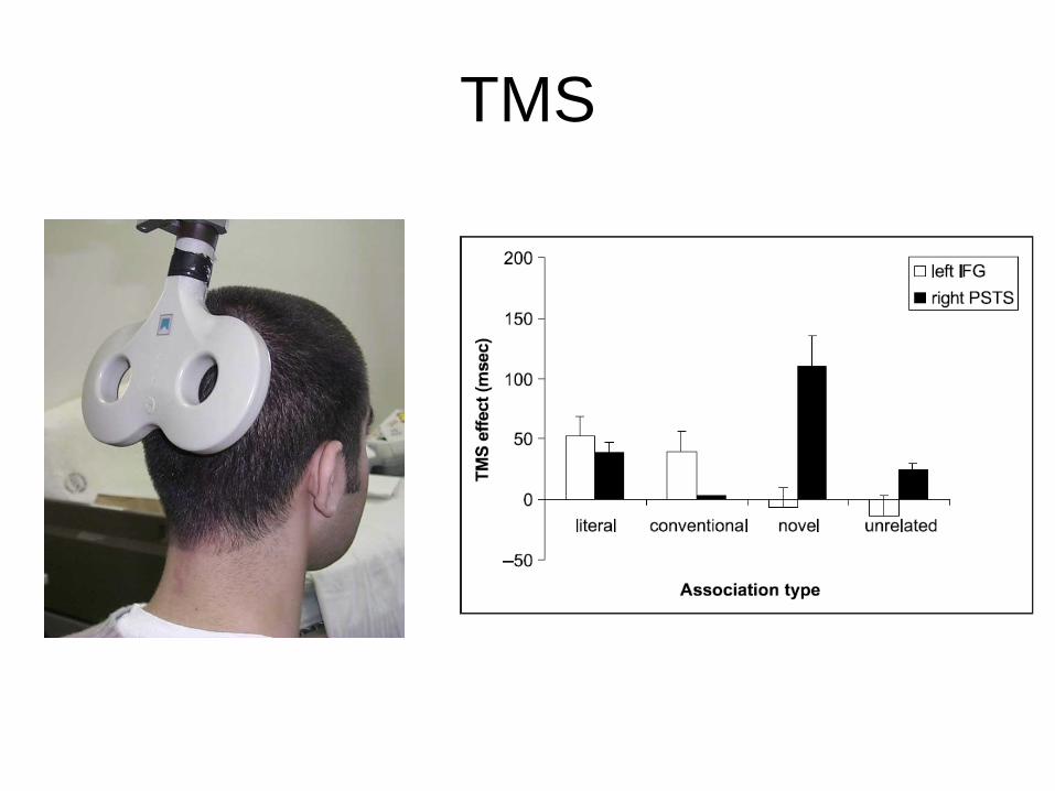
TMS

NIRS

fMRI

Imaging based box models of word
recognition
Price 2000

Updated models: Hickok and
Poeppel 2004 and on
