Section Editor: Ralf Paus
What are melanocytes really doing all day long…?
P. M. Plonka, T. Passeron, M. Brenner, D. J. Tobin, S. Shibahara, A. Thomas, A. Slominski, A. L.
Kadekaro, D. Hershkovitz, E. Peters, J. J. Nordlund, Z. Abdel-Malek, K. Takeda, R. Paus, J. P. Ortonne,
V. J. Hearing and K. U. Schallreuter
Abstract: Everyone knows and seems to agree that melanocytes
are there to generate melanin – an intriguing, but underestimated
multipurpose molecule that is capable of doing far more than
providing pigment and UV protection to skin (1). What about the
cell that generates melanin, then? Is this dendritic, neural crest-
derived cell still serving useful (or even important) functions
when no-one looks at the pigmentation of our skin and its
appendages and when there is essentially no UV exposure? In
other words, what do epidermal and hair follicle melanocytes do
in their spare time – at night, under your bedcover? How much
of the full portfolio of physiological melanocyte functions in
mammalian skin has really been elucidated already? Does the
presence or absence of melanoctyes matter for normal epidermal
and ⁄ or hair follicle functions (beyond pigmentation and UV
protection), and for skin immune responses? Do melanocytes even
deserve as much credit for UV protection as conventional wisdom
attributes to them? In which interactions do these promiscuous
cells engage with their immediate epithelial environment and who
is controlling whom? What lessons might be distilled from
looking at lower vertebrate melanophores and at extracutaneous
melanocytes in the endeavour to reveal the ‘secret identity’ of
melanocytes? The current Controversies feature explores these far
too infrequently posed, biologically and clinically important
questions. Complementing a companion viewpoint essay on
malignant melanocytes (2), this critical re-examination of
melanocyte biology provides a cornucopia of old, but under-
appreciated concepts and novel ideas on the slowly emerging
complexity of physiological melanocyte functions, and delineates
important, thought-provoking questions that remain to be
definitively answered by future research.
Please cite this paper as: Plonka et al. What are melanocytes really doing all day long…? Experimental Dermatology 2009; 18: 799–819.
Praeludium pigmentosum
For those uninformed, the skin is an inert plastic wrap nat-
ure provides to keep us in and everything else out. How
mistaken they are! The skin, in particular the epidermis, is
one of the most active of all tissues ⁄ organs.
Nature wisely placed the capillary circulation in the der-
mis. The epidermis has no vascular circulation thereby
minimizing the probability that toxic chemicals, bacteria or
fungi that penetrate through the stratum corneum can dif-
fuse into the blood stream. That does not leave the epider-
mis defenseless. The epidermis has proteins called defensins
that have anti-microbial properties. There are Toll-like
receptors that recognize invading organisms and incite a
host response. Even more interesting, it is well known that
keratinocytes are avidly phagocytic. They have the capacity
to phagocytize the wandering, invasive fungi or bacteria
and digest them. It is both interesting and important that
a-MSH stimulates the ingestion of candida by keratino-
cytes. a-MSH has a wide array of activities, only one of
which is to stimulate the synthesis of melanin. There are
receptors for a-MSH on Langerhans cells and keratinocytes
as well as melanocytes. It has the ability to suppress
inflammation and alter keratinocyte proliferation. That
seems like an odd property for a molecule that enhances
pigmentation until it is recalled that melanocytes are also
phagocytic cells and are part of the inflammatory response.
Responding virtually to all inflammatory events in the epi-
dermis, the pigment system participates usually by produc-
ing more melanin (postinflammatory hyperpigmentation)
or occasionally by suppressing melanization (i.e. postin-
flammatory hypopigmentation such as pityriasis alba). One
of the most common and desirable forms of postinflamma-
tory hyperpigmentation is tanning following exposure to
sunlight. It is mediated in part by a-MSH. Sunlight dam-
ages the epidermis and sunburn is the inflammatory
response to radiation injury. Production of melanin stimu-
lated by a-MSH is accompanied by all sorts of useful
results such as darker skin that is more resistant to subse-
quent sunburn. a-MSH also stimulates repair of damaged
DNA. It also downregulates the immune response of the
epidermis, maybe preventing more cases of autoimmune
disorders such as lupus erythematosus.
That keratinocytes and melanocytes are involved in host
defenses and inflammation seems incongruous. After all
why should they usurp the function of Langerhans cells?
DOI:10.1111/j.1600-0625.2009.00912.x
www.blackwellpublishing.com/EXDControversies in Experimental Dermatology
ª 2009 John Wiley & Sons A/S, Experimental Dermatology, 18, 799–819 799
But then it is clear that Langerhans cells also affect melan-
ization as well as keratinization. Langerhans cells make a
variety of cytokines that affect keratinocytes and melano-
cytes just as melanocytes make a-MSH and other cytokines
such as the interleukins that affect Langerhans cells and
keratinocytes. In a recent review, the numerous ways that
these cells interact and the response of all of them to
common cytokines has been reviewed (3).
Histological examination of the epidermis shows basal
keratinocytes as proliferating cells. Melanocytes are not
only dendritic but also confined to the basilar layer. Lan-
gerhans cells are mid-epidermal. Most of the epidermal
cells are keratinocytes, about 90% or more. How can these
cells seemingly interact? It is a common misperception that
dendrites of melanocytes and Langerhans cells are fixed in
a given position. The opposite is true. Dendrites are
extended, contracted and moved between different pairs of
keratinocytes continuously. Each melanocyte using just
three or four dendrites must copulate with at least 30 basi-
lar keratinocytes and probably others in the suprabasilar
layers. And it must be in contact with distantly located
Langerhans cells. Of course Langerhans cells must provide
protection for even larger numbers of keratinocytes and
melanocytes lying beneath them. They have just a few den-
drites, but must form a network to trap any invading
organisms or microorganisms coming from above. They
move their dendrites to form a moving network.
The melanocytes inject melanosomes into the cytoplasm
of keratinocytes. Coitus with all of the surrounding cells
requires melanocytes to move their dendrites around in
search of unprotected keratinocytes lacking melanin.
Although both Langerhans cells and melanocytes are den-
dritic cells, they must hold hands so to speak by touching
each others dendrites to communicate. In the absence of a
vascular circulation, melanocytes and Langerhans cells have
dendrites that move constantly to service each other and
keratinocytes. They form one type of circulation within the
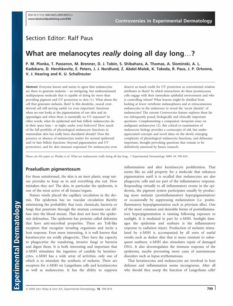
epidermis. It is the promiscuity of melanocytes and Langer-
hans cells to form a menage a’ trios or as shown below a
troika with keratinocytes that makes the epidermis the
unique, highly active tissue that alerts the entire body to
the environment in which we live.
James J. Nordlund
Wright State School of Medicine, Dayton, OH, USA;
E-mail: [email protected]
References
1 Wood et al. Exp Dermatol 1999: 8: 153–164.2 Herlyn. Exp Dermatol 2009; Epub. ahead of print.3 Nordlund JJ. Dermatol Clin 2007: 25: 271–281.
Viewpoint #1
As long as we keratinocytes can remember, melanocytes
have lived inconspicuously among us at the basal mem-
brane and have been primarily known as meek and obedi-
ent pigment producers in the epidermis. However, recent
investigations are suggesting that these fellows have several
identities, work undercover in many other places in the
human body and have functions we can only speculate
about (as summarized in Table 1). Let us critically re-con-
sider, then, the well-known properties and the more
obscure abilities of our ancient, pigment-producing friends.
The keratinocytes’ lamentFirst of all, how does one recognize a melanocyte if it is
encountered somewhere outside the epidermis? They are
usually identified by their expression of melanocyte-specific
proteins, e.g. tyrosinase (TYR), TYRP1, DCT, Pmel17 ⁄gp100, MART-1 and ⁄ or MITF (1). However, melanocyte
precursors (known as melanoblasts) are more difficult to
identify as they do not produce melanin and therefore do
not usually express those markers, although occasionally
DCT and ⁄ or KIT are detectable as specific markers. There-
fore, whatever functions melanoblasts may have in skin,
these are as easily missed as these cells are difficult to detect.
The favourite habitat of melanocytes is the epidermis,
but large numbers of them can be found in hair follicles
and in the eyes where they manufacture melanin for hair
and eye pigmentation respectively and in other less
well-known locations as discussed below. The fact that we
keratinocytes control melanocytes in the skin via an
armamentarium of growth factors (2) has led to the
impression that keratinocytes and melanocytes live in a sort
of master–slave relationship. However, the dependency is
not unilateral at all; melanocytes transport melanin in
membrane-bound organelles (termed melanosomes) via
their elongated dendrites and then transfer them to us (3),
whereupon we arrange them to form a critical protective
barrier (known as supranuclear ‘caps’) to shield our DNA
from UV radiation (4).
Melanocyte
Cutaneous troika
Keratinocyte
Langerhans
Plonka et al.
800 ª 2009 John Wiley & Sons A/S, Experimental Dermatology, 18, 799–819
Melanocytes (and melanin) also function early during
human development; they play critical roles during embry-
onic development as can be seen in individuals with oculo-
cutaneous albinism type 1 (OCA1). OCA1 results from the
dysfunction of TYR, which leads not only to impaired pig-
mentation of skin, hair and eyes (5) but also to misrouting
of the optic nerves at the chiasm (6). Melanocytes express
the melanocortin 1 receptor (MC1R) that regulates the
quality and quantity of their melanin production. MC1R is
controlled by the agonists melanocyte-stimulating hormone
(MSH) and adrenocorticotropic hormone (ACTH) (7),
which stimulate the melanogenic cascade and thus the syn-
thesis of eumelanin, as well as by the antagonist agouti sig-
nalling protein (ASP) (8). It is known that ASP elicits the
production of pheomelanin, but it was shown only recently
that ASP also modulates the expression of genes involved
in morphogenesis (especially in nervous system develop-
ment) (9).
Besides their existence in the skin, melanin and melano-
cytes have been reported to appear in the stria vascularis of
the cochlea (10), in the leptomeninges (11), substantia
nigra and locus coerulus (12) of the brain, in the heart
(13,14) and there is evidence that they operate even in such
inhospitable territories as adipose tissue (15).
Note in passing that there are two distinct types of mela-
nocytes: differentiated melanocytes that originate from the
neural crest and can be found at various locations in the
body, and a second type, the retinal pigment epithelium
(RPE), specifically present only as a single layer of cells
lying behind the retina that develop in situ from the optic
cup of the brain (16). The RPE plays a critical role in the
active phagocytosis and turnover of the rod outer segments
of the retina as well as in the uptake, processing and trans-
port of retinoids and consequently has an important func-
tion in vision (17).
Melanocytes are also present as intermediate cells in the
stria vascularis of the cochlea. Strial intermediate cells are
required for the generation of endolymph-mediated action
potentials that are necessary for normal hearing (10). Hear-
ing impairment can be associated with inherited pigmen-
tary disorders, e.g. Waardenburg syndrome (18), and
studies have shown that the extent of induced temporary
hearing loss is inversely related to skin pigment type (19).
Melanin granules produced by melanocytes in the inner ear
even play important roles in balance (20).
Extracutaneous melanocytes located in the brain may
have several neuroendocrine functions. Human melano-
cytes express lipocalin-type prostaglandin D synthase,
which generates prostaglandin D2 (PGD2) (21). Besides, in
melanocytes, b-endorphin, an endogenous opioid, is gener-
ated from proopiomelanocortin together with MSH and
ACTH. Does this suggest that melanocytes regulate sleep?
PGD2 is a potent sleep-inducing substance (22) and opioid
receptors are located in the nuclei that are active in sleep
regulation (20). Also, there are indications that a certain
melanocyte-derived factor might be involved in controlling
the central chemosensor that generates the respiratory
rhythm (20). Pigment in the brain, termed neuromelanin,
consists of a large, complex, eumelanin-covered pheomela-
nin core which may also contain aliphatics and peptides
(23). Neuromelanin is primarily localized in dopaminergic
neurons of the substantia nigra and in the locus coerulus
and accumulates in the human substantia nigra with age
(12). A selective loss of dopaminergic neurons containing
neuromelanin is associated with Parkinson’s disease (12).
Various studies support the concept that neuromelanins
have a protective function by binding ⁄ removal of reactive
oxygen species (ROS) and metals that would otherwise be
highly toxic to neurons (12,24,25). A recent study showed
that virtually all brain tissues contain significant amounts
of neuromelanin, which are thought to play important
roles in reducing toxicity in those tissues (26).
It has also been brought to our attention that melano-
cytes are located in the valves and septa of the heart
(13,14). Mice presenting with hyper (hypo-) pigmented
skin show increased (or decreased) heart pigmentation
(14). Cardiac melanocytes may originate from the same
precursor population as skin melanocytes as they depend
on the same the signalling molecules known to be required
for proper skin melanocyte development (13), but their
function in this location so far is obscure. It may well turn
out that the production of melanin is not always of benefit,
either in the heart or in other tissues, such as the lungs,
where in a rare disease known as lymphangioleiomyomato-
sis (27), muscle cells revert towards their developmental
origins and express some melanocyte markers, such as
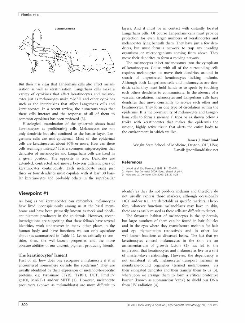
Table 1. Locations and functions of melanocytes (and melanocyte
imitators)
Location Function(s) References
Skin – epidermis Constitutive skin pigmentation, and responsesto and protection against the environment(primarily UV)
2–4
Skin – hair follicles Hair pigmentation, removal of toxicbyproducts
3
Skin – hairbulge region
Melanocyte stem-cell reservoir for skin
Eye – choroid Constitutive eye pigmentation,protection against UV
5,6
Eye – RPE Vision, metabolism of rod outersegments and retinoids
16,17
Ear – cochlea Hearing 10,18,19Inner ear – Balance 20Brain – all tissues Neuroendocrine and detoxification 11,12,21–26Heart Anti-inflammation, reduction ⁄ binding of ROS 13,14Adipose tissue Reduction ⁄ binding of ROS, cellular survival 15Lung Unwanted? No known function,
lethal consequences27
Controversies in Experimental Dermatology
ª 2009 John Wiley & Sons A/S, Experimental Dermatology, 18, 799–819 801
tyrosinase, Pmel17, etc. The resulting production and
accumulation of melanin in lung tissues is eventually lethal.
Recently, we have learnt that melanin biosynthesis also
takes place in the visceral adipose tissue of morbidly obese
humans (15). Hypothetically, the ectopic synthesis of mela-
nin in the cytosol of obese adipocytes may serve as a com-
pensatory mechanism to act as an anti-inflammatory factor
and to reduce oxidative damage. During increases in cellu-
lar fat deposition, adipocytes become more exposed to
endogenous apoptotic signals, especially ROS, which could
be counteracted by ectopically produced melanin. In
addition, adipocytic melanin may suppress the secretion of
proinflammatory molecules (15).
In conclusion, we think it unfair that melanocytes reap
all the glory for their role in pigmenting the skin and pro-
viding it critical protection against UV damage, when in
fact it is we as keratinocytes that form the bulk population
of that tissue deserve all the credit. It adds insult to injury
that melanocytes are now beginning to take more and
more glory for their roles in other tissues of the body.
Where will it all end?
Michaela Brenner1, Vincent J. Hearing2
1Department of Dermatology, Ludwig-Maximilians-Univer-
sity of Munich, Munich, Germany;2Laboratory of Cell Biology, National Cancer Institute,
National Institutes of Health, Bethesda, MD, USA;
E-mail: [email protected]
References
1 Passeron T et al. Exp Dermatol 2007: 16: 162–170.2 Yamaguchi Y et al. J Biol Chem 2007: 282: 27557–27561.3 Tolleson W H. J Environ Sci Health 2005: 23: 105–161.4 Montagna W, Carlisle K. J Am Acad Dermatol 1991: 24: 929–937.5 Spritz R A. Hum Mol Genet 1994: 3: 1469–1475.6 King R A et al. In: Scriver C R et al., eds. The Metabolic and Molecular Bases
of Inherited Disease. New York: McGraw-Hill, 2001: 5587–5627.7 Millington G W. Clin Exp Dermatol 2006: 31: 407–412.8 Suzuki I A et al. J Invest Dermatol 1997: 108: 838–842.9 Le Pape E et al. Proc Natl Acad Sci U S A 2009: 106: 1802–1807.
10 Tachibana M. Pigment Cell Res 1999: 12: 344–354.11 Goldgeier M H et al. J Invest Dermatol 1984: 82: 235–238.12 Zecca L et al. J Neural Transm 2002: 109: 663–672.13 Brito F C, Kos L. Pigment Cell Melanoma Res 2008: 21: 464–470.14 Yajima I, Larue L. Pigment Cell Melanoma Res 2008: 21: 471–476.15 Randhawa M et al. FASEB J 2009: 23: 835–843.16 Bharti K et al. Pigment Cell Res 2006: 19: 380–394.17 Bok D. J Cell Sci Suppl 1993: 17: 189–195.18 Tassabehji M et al. Nat Genet 1994: 8: 251–255.19 Barrenas M L, Lindgren F. Scand Audiol 1990: 19: 97–102.20 Takeda K et al. Tohoku J Exp Med 2007: 211: 201–221.21 Takeda K et al. Biochem Biophys Res Comm 2006: 339: 1098–1106.22 Urade Y, Hayaishi O. Biochim Biophys Acta 2000: 1482: 259–271.23 Bush W D et al. Proc Natl Acad Sci U S A 2006: 103: 14785–14789.24 Sulzer D et al. Proc Natl Acad Sci U S A 2000: 97: 11869–11874.25 Zucca F A et al. Pigment Cell Res 2004: 17: 610–617.26 Zecca L et al. Proc Natl Acad Sci U S A 2008: 105: 17567–17572.27 Ferrans V J et al. J Nippon Med Sch 2000: 67: 311–329.
Viewpoint #2
‘But now here’s the third command: Machine, do Nothing!’
The machine sat still. Klapaucius rubbed his hands in
triumph, but Trurl said:
‘Well, what did you expect? You asked it to do nothing,
and it’s doing nothing’.
‘Correction: I asked it to do Nothing, but it’s doing
nothing’.
‘Nothing is nothing!’
‘Come, come. It was supposed to do Nothing, but it hasn’t
done anything and therefore I’ve won’.
S. Lem (1965) ‘How the World Was Saved’ (1)
While our task here is to dissect non-pigmentary func-
tions of the melanocyte, the problem of melanin produc-
tion and transfer is inseparable from this. Let us begin
therefore with a simple question: Are melanocytes doing
anything at all, besides producing melanin?
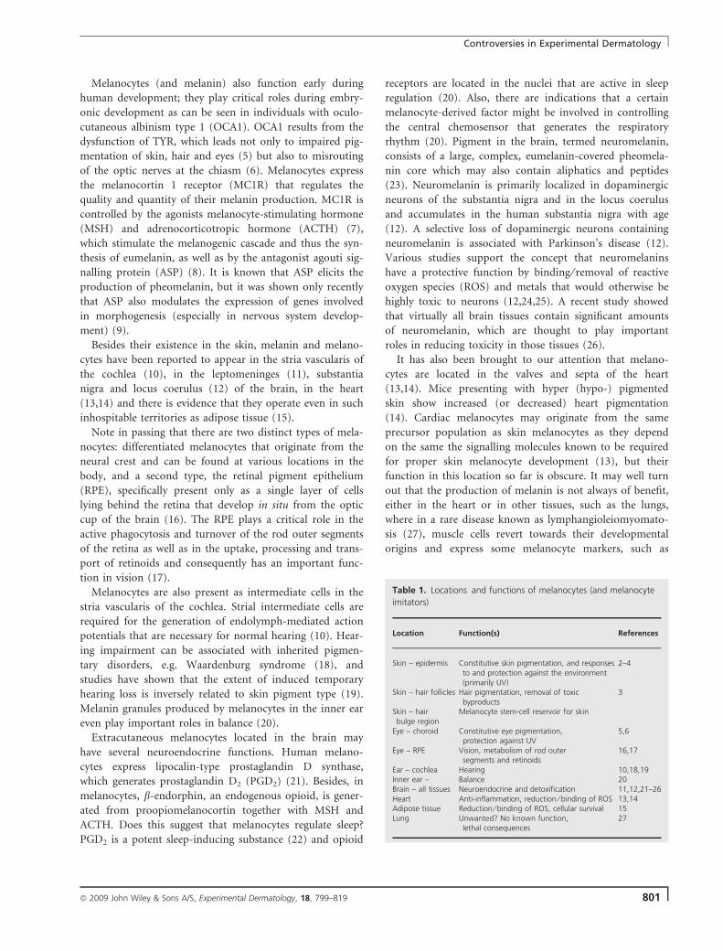
Figure 1. Schematic summarizing the different locations in the body
for melanocytes and potential functions in those locations.
Plonka et al.
802 ª 2009 John Wiley & Sons A/S, Experimental Dermatology, 18, 799–819
The outlines of an answer to this question are hidden in
a few, rarely considered facts:
1. Melanin is not indispensable to dark-colour pigmenta-
tion: e.g. a simple model eukaryote, the myxomycete
Metatrichia vesparium, reveals dark pigmentation of
the plasmodium because of high concentration of man-
ganese (II), but not melanin (2).
2. Cells are not necessary to produce melanin. The capa-
city to produce melanin evolved from the more ancient
phenomenon of extracellular autooxidation of phenolic
compounds (3,4). Recently, we have shown (3) that if
cytoplasm of another myxomycete Fuligo septica turns black
in the process of sclerotization, the pigmented sclerotium is
not able to reproduce viable plasmodium. Moreover,
among several hundreds of myxomycetes, only one, Physa-
rum nudum, has been so far reported to produce melanin
in the living plasmodium in response to light (5). These
experiments carried out on simple, aerobic eukaryotes
strongly suggest that the first thing which evolutionarily
ancient cells must have achieved in the oxidizing atmo-
sphere was clearly how to INHIBIT melanogenesis, i.e.
HOW NOT TO PRODUCE melanin. Later on, their
descendants ‘learned’ (in the evolutionary sense of this
term) how to control extra- and intracellular melanin
production, deposition and how to take advantage of the
ability to synthesize melanin (3,4).
3. Melanocytes are not necessary to produce melanin.
There are numerous coloured organisms producing mela-
nin in other cells (6). Melanocytes appeared earliest in
Deuterostomia (7,8) because of their unique embryogenesis
of the neural system – developing as a tube at the back side
of the animal. In this way, the embryonal neural system
generates a by-product – the neural crest, the origin of a
huge variety of cells, including neurons and skin melano-
cytes (6–8). Melanocytes must have evolved in advanced
Chordata, probably in early vertebrates, in which the pres-
ence of the neural crest is, for many investigators, the most
important feature, if not the only interesting feature really
unique for vertebrates (8). Protostomia are only in the
possession of cells analogical to melanocytes (6). Many of
them produce melanin in or outside the cells, let alone
plants and lower organisms – fungi and prokaryotes (4,6).
4. Moreover, even in the presence of melanocytes, mela-
nin and melanosomes can be generated ab ovo in other
types of cells (9–11).
5. Epithelial cells can produce melanin. Interestingly and
importantly, in the cuttlefish ink sack, an exocrine gland of
the holocrine type, the cells responsible for melanin (‘ink’)
production are in fact specialized epidermal cells (12,13).
In Deuterostomia, epidermal cells can theoretically produce
melanin by themselves, as well. In fact, one is even tempted
to shift the general question covered by this current Con-
troversies to a different, but related enquiry: Why is it that
melanocytes do, and keratinocytes DO NOT produce mela-
nin in mammalian epidermis or hair follicles? Ah, now you
see…! The likely answer directly leads to the conclusion
that melanocytes must be doing something else, from
which the ability to produce melanin has evolved or has
become a mere by-product.
6. Among many disorders of pigmentation, including
total lack of melanin synthesis, the more the impaired
function is basic for melanocyte biology (14–17). Among the
examples of total lack of melanocytes, all of which are
associated with a severe impairment of neural crest
embryogenesis (e.g. Waardenburg syndrome), many are
lethal or sub-lethal (18,19).
7. There are plenty of melanocytes which do not normally
engage in melanogenesis, e.g. the amelanotic melanocytes in
the outer root sheath of – heavily pigmented! – anagen VI
hair follicles (15).
Therefore, when looking for potential evolutionary advan-
tages of possessing melanocytes, it seems that the generation
of melanin itself is not necessarily producing the decisive
selection advantage, but the presence of melanocytes them-
selves. Even if melanogenesis is the primary function of pres-
ent-day mammalian melanocytes, it is certainly not the only
one, and perhaps not even the most important one.
In fact, it is reasonable to assume that melanocytes have
evolved gradually, within an evolutionary continuum of
already coloured animals and have only gradually devel-
oped the capability to control melanin production, as a
secondary specialization. This probably happened in the
earliest vertebrates – Agnatha (today represented by the
lampreys and the hagfish) (8), while typical higher verte-
brate skin with all its adnexa and melanocytes submersed
in the basal layers of the epidermis may have evolved as
late as in Therapsida (i.e. the reptilian evolutionary line
leading to the mammals) (20,21). Some related cells appar-
ently opted for differential evolutionary paths, evolving into
other cells responsible for colouration – namely xantho-
phores, iridophores and erythrophores (8).
What did the ancient melanocytes do all day long, then?
Or, slightly re-phrased: What did neural crest cells do day
and night, before they became melanocytes?
The answer is connected with three cellular properties
which, on first thought, do not seem to be connected with
pigmentation at all, namely the ability to wander around in
the organism (migration) and to then settle down in the
skin epithelium (intraepithelial residence) (7,8), as well as
the ability to create synapses with other cells (22). During
embryogenesis, melanocytes cross the epidermal basement
membrane and settle down in the basal layer of the epider-
mis. The tendency to cross the basal lamina seems pre-
served in mature amphibian melanophores – cells closely
related to melanocytes – which, however, do it the other
way round (23). An even more important property of
Controversies in Experimental Dermatology
ª 2009 John Wiley & Sons A/S, Experimental Dermatology, 18, 799–819 803
melanocytes, which may betray their possible primary
functions, is a striking phenomenon of intercellular
interaction, namely the transfer of melanin-containing
melanosomes to keratinocytes (22). Melanocytes can also
settle down somewhere in the upper layer of the dermis
(e.g. in certain nevi) or in subcutaneously located epithe-
lium (i.e. in the hair follicle pigmentary unit) where mela-
nin production occurs in apparent communication with
dermal fibroblasts or the specialized fibroblasts of the
follicular dermal papilla (24–26). Such pigment cell-
fibroblast interactions are already prominent in amphibian
melanophores, which transfer melanin to dermal fibroblasts
(27).
Melanin is transferred within melanosomes to target
keratinocytes by a mechanism that resembles exocytosis
(22,28). Melanosomes, again, seem to be related to other
secretory granules as they are produced, e.g. by neurons
and immunocytes, while the special connections between
melanocytes and keratinocytes (‘pigmentation synapses’)
resemble other types of synapses: neuronal, immunological
or phagocytic (22). Next, some neurotransmitters and neu-
romodulators (dopamine) can autooxidate to neuromela-
nin, a by-product of their oxidative catabolism in locus
coeruleus and substantia nigra (15,29). [In line with this
‘synaptic’ line of argumentation, it deserves at least a
footnote that pigmented hair emerged during evolution as
sensory organs (tactile mechanoreceptors) (20,21).]
Finally, the evolutionary ancestry of POMC-coding gene,
whose activity eventually leads to the production of pig-
ment-stimulatory neuropeptides (melanocortins), is at least
as ancient as the evolution of the neural crest and much
older than the evolutionary history of typical melanocytes
(30). Taking into consideration the huge variability of
physiological processes that are regulated by POMC-coded
peptides [of which the regulation of melanogenesis is only
one function among multiple others (15,30,31)], one is
almost compelled to conclude that the ancient melanocytes
must have possessed some regulatory functions. One won-
ders, did the ancestral melanocytes initially exocytose those
secretory granules in which inhibition of autooxidation of
neurotransmitters had gone wrong, thus accidentally
creating melanin…?
While the record of endo ⁄ paracrine actions of melano-
cytes is long and well-documented (15,32) (see also the
other essays in this feature), an old, apparently long-forgot-
ten hypothesis on melanin as a ‘solid hormone’ deserves to
be recalled here (33,34). This hypothesis is closely related
to another one, namely that on extraepithelial melanin
transfer, in particular in hyperpigmentation phenotypes
(33), and in the context of the transfer of melanin-contain-
ing apoptotic bodies in catagen to Langerhans cells (35)
and further – to lymph nodes and visceral organs (35,36).
This phenomenon may be related to the ability of melanins
to adsorb various organic and inorganic compounds in
their structure, acting e.g. as cationite (37). Consequently,
melanins can serve as long-distance vehicles for such
substances, working in the systemic dimension, i.e. in an
endocrine manner.
Mechanisms and interactions connected with such a mel-
anocytic export to defined target cells or just to the mela-
nocyte environment may turn out to be quite complicated.
One intriguing example, which has not been fully explored
so far, but which may well be important for future
research, shall suffice here. Nitric oxide synthase-1 (NOS-1)
was suggested to participate in potentiation of melano-
genesis due to UVB by keratinocytes (38,39) which
responded to UVB by increase in NOS-1 activity, and by
production of NO. This parahormone activated melanocy-
tic guanyl cyclase and, subsequently, the protein-kinase G
(PKG) pathway of melanogenesis control (15). Melanocytes
were claimed to react to UVB by a constitutive NOS activa-
tion, too (38,39). As NOS-1 localizes in synaptic granules
thanks to its PDZ-domain (where PDZ stands for PSD-95
discs large ⁄ ZO-1 homology domain, and where PSD-95
stands for post synaptic density protein 95, and ZO-1 for
zonula occludens scaffolding protein 1) (40), it may well be
that the primary signal is perceived by melanocytes, where
it stimulates NOS-1 gene transcription, with the gene prod-
uct being co-transported along with melanosomes to the
more superficially located keratinocytes. From its new in-
traepithelial position, in turn, NOS-1 may be envisioned to
then increase melanogenesis in melanocytes via NO. More-
over, it has recently been suggested that NOS-3 protein
(i.e. the endothelial NOS) can act in co-operation with oes-
trogen receptors as regulators of transcription of human
telomerase in endothelium (41). Perhaps, given its numer-
ous alternate splicing variants (40), NOS-1 may act in a
similar manner? Maybe this is even true for NOS-3, as ele-
vated mRNA levels of the latter have also been found in
melanocytes (42)?
Be this as it may, these observations suggest that melano-
cytes are able to perceive UV light. What could be the
corresponding photoreceptor(s)? Well, for starters, there is
NOS itself: one molecule of NOS contains many prosthetic
groups which may serve as primary photoreceptors, like
FAD, FMN, BH4 or haeme (40). Moreover, numerous
other substances can act as primary photoreceptors in
melanocytes, including rhodopsin (43) and oligomers of
phenolic compounds, i.e. melanin precursors absorbing
light in the visible range (32).
Are there any other stimuli that can be perceived by
melanocytes? Of course, there are numerous growth factors,
hormones and neurotransmitters, for which melanocytes
express cognate receptors and the stimulation or antagon-
ization of which profoundly impacts on melanocyte sur-
vival, proliferation, migration and differentiated functions
Plonka et al.
804 ª 2009 John Wiley & Sons A/S, Experimental Dermatology, 18, 799–819
(14–17,26,31,32). While most of these extracellular signals
sensed by melanocytes within their respective microenvi-
ronment participate in regulation of melanogenesis, in
some cases, these stimuli induce reactions that are typical
of receptors in sensory organs. Besides their well-appreci-
ated presence in the eye, the presence of ancestral melano-
cytes in mechanoreceptors of primitive vibrissae of
therapsids deserves to be recalled here. Another important
property of melanocytes, which is particularly pronounced
in their close relatives, amphibian and fish melanophores,
is the perception of chemical – not unlike the perception
of olfactory stimuli [these ‘olfactory’ properties of melano-
cytes are important for sudden movements of melanosomes
within the melanophores, and the neurogenic change of
pigmentation (44,45), vital survival tools in a predator-rich
environment].
All these phenomena and considerations invite one to
synthesize the vision that the melanocyte is a cell that is
able to receive signals from its biological, physical and
chemical environment, that can adjust its inner state
according to the signals received, and that can generate
physical and biochemical signals to both its immediate
tissue milieu, and to distant environments.
This concept of melanocyte biology resembles the behav-
iour of a classical Turing machine – the theoretical model
of calculability (46). There are, indeed, aspects of melano-
cytic functions where the state of the cell changes rather in
the digital than the analogue way and where the cells
respond in the digital manner to stimuli which change in
the analogue way. This concerns e.g. the phaeo ⁄ eumelano-
genesis switch (47,48), and the pH switch necessary to ini-
tiate melanogenesis in melanosomes (31). Although these
phenomena concern pigmentation, they can serve to show
the analogue-to-digital converter properties of the melano-
cyte. This approach to cell biology has been developed
by many research groups, which have tried to model intra-
cellular processes employing the methodology of the theory
of information (49–53) and which have suggested how
even Turing-uncomputable formalisms and functions may
be computed (52). The thermodynamic aspect of the cellu-
lar computation seems of a much greater importance than
of artificial computers in which the source of energy used
for computation is usually negligible (50). Thus, the mela-
nocyte with its inner system of membranes, with its com-
plex pathways involving coated particles and organelles, its
premelanosome genesis, its trafficking of melanogenesis-
related proteins, its capacity to induce qualitative switches
in melanogenesis, and, finally, with its transfer of melanin
to other target cells is perfectly suited to serve as a model
of membrane automata (51–53).
These examples invite one to create mathematical or
computational models of a living cell that reflect or mimic
more or less what really happens in the cell (49–53). In
fact, as nicely exemplified by a macroscopic unicellular,
multinuclear myxomycete, Physarum polycephalum, avail-
able evidence suggests that cells engage in the act of com-
puting. For example, they can calculate graphs (54) or
perform complex logical Turing-uncomputable calculations
inaccessible for classical, deterministic computers (55).
Moreover, cells can even reveal such advanced computing
characteristics like ‘emerging computing’ or ‘robustness’
(54,55).
The pathways of macroscopically visible pigmentation of
animals can be satisfactorily described in the terms of
Turing-Child models (56–60) (created by the Turing of the
Turing machine), which takes advantage of the system of
melanocytes wandering around the organism, initiated by
the asymmetrical distribution of some morphogens in the
zygote (60). If a single melanocyte can be considered as a
cognitive system able to perform computation, the system
of melanocytes must be a super-system of computing,
performing its own unique abilities. It may e.g. optimize
production of melanin in hair follicles: depending on the
number of hair follicles per skin area unit, it
generates more or less hair shaft pigment so that the total
amount of melanin produced by a skin area remains
constant (61).
The system of potential or actual melanocytes is able to
perceive the morphogen asymmetry, and react accordingly,
starting from the input contained in the zygote and ending
up in the adult organism of a complicated spatial and
temporal pattern of pigmentation (seasonally or sexual
activity-dependent changes of colouration). No wonder
that the temporal aspect is strongly based on the phenome-
non of photoperiodicity: particular sub-populations of skin
melanocytes vary not only in their phenotypic ability to
produce melanin and dendrites (15) but also in the profile
of expression of various NOS synthases (62). Some recent
studies on the expression of hair-type alpha keratins in rep-
tiles suggest that the evolution of hair follicle may have
been longer and concerned not only Therapsida, but more
ancient groups of reptiles (63). This leads to the hypothesis
that follicular and epidermal melanocytes are more distant
relatives than often assumed or even that the epidermal
melanocytes originate from the follicular ones [as hair
follicle may have evolved earlier than a dry and scales-free
skin of mammals (63)]. This may be further supported by
the fact that hair follicle melanocytes can re-populate
depigmented epidermis in patients (e.g. in vitiligo) as well
as in transgenic mice constitutively expressing stem cell
factor (SCF) in their epithelial keratinocytes (64), but not
vice versa and that the hair follicle appears to be the major
seat of melanocyte stem cells (64). In fact, one wonders
whether, during vertebrate evolution, the process of
photoperiodic regulation has actually been transferred
along with the responsible cells from the nervous system
Controversies in Experimental Dermatology
ª 2009 John Wiley & Sons A/S, Experimental Dermatology, 18, 799–819 805
into the skin, i.e. closer to the surface of the organism,
where it then produced secondary functions like photopro-
tection via skin ⁄ hair melanogenesis, or a remote control of
selected functions of visceral organs.
Conclusion
Even if melanocytes are not able to think, at least they are
extensively computing. So, my answer to the overarching
question of these Controversies is: computing is what they
do all day and even all night long. As long as what exactly
they compute remains unknown, let us assume that mela-
nocytes count the card games that Nature plays with the
ever-inquisitive pigment cell researcher…‘–What exactly do you guys do here?
–We’re counting cards.
–You’re counting cards?
–We’re counting cards.
We’re counting cards.
–What else do you do?
–We’re counting cards.’
Barry Morrow (1988) ‘Rainman’ – the screenplay (65)
Przemyslaw M. Plonka
Department of Biophysics, Faculty of Biochemistry,
Biophysics and Biotechnology, Jagiellonian University,
PL-30-387 Krakow, Poland;
E-mail: [email protected];
References
1 Lem S. How the World was Saved. Krakow: Wydawnictwo Literackie, 1965(Engl. transl, by Kandel M., San Diego, CA: Harcourt, Brace & Co., 1985). In:Lem B, Lem T. Stanislaw Lem. Available at http://english.lem.pl/. June 06, 2009.
2 Rakoczy L, Plonka P M. Ochr Srod Zas Nat 1997: 18: 299–308. (in Polish).3 Krzywda A et al. Cell Mol Biol Lett 2008: 13: 130–143.4 Plonka P M, Grabacka M. Acta Biochim Pol 2006: 53: 429–443.5 Plonka P M, Rakoczy L. Curr Top Biophys 1997: 21(1): 83–86.6 Sembrat K. Comparative Histology of Animals, Vol. II. Warsaw: Polish Science
Publishers, 1981 (in Polish).7 Anderson D J. Neuron 1989: 3: 1–12.8 Donoghue P C J et al. BioEssays 2008: 30: 530–541.9 van der Heijden A et al. Labor Anim 1995: 29: 459–463.
10 Stanka P et al. Pigment Cell Res 1988: 1: 358–360.11 Weissman I. Nature 1967: 215: 315.12 Schraermeyer U. Pigment Cell Res 1991: 7: 52–60.
13 Palumbo A. Pigment Cell Res 2003: 16: 517–522.14 Ortonne J P, Ballotti R. J Dermatol Treat 2000: 11: S15–S26.15 Slominski A et al. Physiol Rev 2004: 84: 1155–1228.16 Tomita Y, Suzuki T. Am J Med Genet C Semin Med Genet 2004: 131C: 75–81.17 Grimes P et al. J Am Acad Dermatol 2006: 54: S255–S261.18 Druckenbrod N R et al. Genesis 2008: 46: 396–400.19 Tachibana M et al. Pigment Cell Res 2003: 16: 448–454.20 Maderson P F A. Am Zool 1972: 12: 159–171.21 Maderson P F A. Exp Dermatol 2003: 12: 233–236.22 Van Den Bossche K et al. Traffic 2006: 7: 769–778.23 Yasutomi M. Pigment Cell Res 1987: 1: 181–187.24 Kam E, Hodgins M. Development 1992: 114: 389–393.25 Rendl M et al. PLoS Biol 2005: 3: e331.26 Schneider M R et al. Curr Biol 2009: 19: R132–R142.27 Aspengren S et al. Pigment Cell Res 2006: 19: 136–145.28 Izumi T et al. Cell Struct Funct 2003: 28: 465–474.29 Swartz H M et al. Free radicals and medicine. In: Eaton S S, Eaton G R, Ber-
liner L J, eds. Biomedical ESR. Biological Magnetic Resonance Series, Vol. 23.The Netherlands, New York, Boston: Kluwer Academic Publishers, 2005: 25–74.
30 Kaelin C B et al. Int J Obesity 2008: 32: S19–S27.31 Schallreuter K U, Kothari S, Chavan B, Spencer J D. Exp Dermatol 2008: 17:
395–404.32 Slominski A et al. J Theor Biol 1993: 164: 103–120.33 Wassermann H P. Nature 1967: 213: 282–283.34 Lukiewicz S. Folia Histochem Cytochem (Krakow) 1972: 10: 93–108.35 Tobin D J. Br J Dermatol 1998: 168: 795–798.36 Plonka P M et al. Acta Biochim Pol 2005: 52: 433–441.37 Sarna T, Plonka P M. Biophysical studies of melanin: paramagnetic, ion-
exchange and redox properties of melanin pigments and their photoreactivity.In: Eaton S S, Eaton G R, Berliner L J, eds. Biomedical ESR. Biological MagneticResonance Series, Vol. 23. The Netherlands, New York, Boston: Kluwer Aca-demic Publishers, 2005: 125–146.
38 Romero-Graillet C et al. J Biol Chem 1996: 271: 28052–28056.39 Romero-Graillet C et al. J Clin Invest 1997: 99: 635–642.40 Alderton W K et al. Biochem J 2001: 357: 593–615.41 Farsetti A et al. J Appl Physiol 2009: 106: 333–337.42 Jackson M et al. Arch Dermatol Res 1998: 290: 350–352.43 Miyashita Y et al. J Investig Dermatol Symp Proc 2001: 6: 54–57.44 Lerner M R et al. Proc Natl Acad Sci U S A 1988: 85: 261–264.45 Zubare-Samuelov M et al. Am J Physiol – Cell Physiol 2003: 285: 1255–1262.46 Turing A M. Proc Lond Math Soc 1937: 42: 230–265.47 Oyehaug L et al. J Theor Biol 2002: 215: 449–468.48 Oyehaug L et al. Math Biosci 2003: 185: 123–152.49 Adleman L M. Science 1994: 226: 1021–1024.50 Ji S, Ciobanu G. Biosystems 2003: 70: 165–181.51 Kefalas P et al. Biosystems 2003: 70: 135–148.52 Calude C S, Paun G. BioSystems 2004: 77: 175–194.53 Fisher J, Henzinger T A. Nat Biotechnol 2007: 25: 1239–1249.54 Toshiyuki Nakagaki T et al. Biophys Chem 2004: 107: 1–5.55 Tsuda S et al. BioSystems 2004: 73: 45–55.56 Child C M. Patterns and Problems of Development. Chicago, IL: University of
Chicago Press, 1941.57 Turing A M. Philos Trans R Soc Lond B Biol Sci 1952: 237: 37–72.58 Shoji H et al. J Theor Biol 2003: 224: 339–350.59 Liu R T et al. Phys Rev E Stat Nonlin Soft Matter Phys 2006: 74(1 Pt 1):
011914.60 Schiffmann Y. Progr Biophys Mol Biol 2007: 95: 50–59.61 Plonka P M et al. J Dermatol Sci 2008: 49: 227–240.62 Sowden H M et al. Br J Dermatol 2005: 153: 301–309.63 Eckhart L et al. Proc Natl Acad Sci U S A 2008: 105: 18419–18423.64 Nishimura E K et al. Nature 2002: 416: 854–860.65 Morrow B, Bass R. Rainman. The screenplay. (Levinson B Direction). Century
City, CA: United Artists Corporation, 1988.
Viewpoint #3
Epidermal melanocytes are surrounded by keratinocytes in
approximately a 1:30 ratio, forming the so-called epidermal
melanin unit (1). Skin colour per se and the mechanisms
behind pigmentation have been the targets of active
research over many decades. Indeed, the discovery of the
pigment-producing cell in the human epidermis and in the
eye by Henle dates back to 1837 (2). One area of particular
interest is the mechanism for melanin transfer to keratino-
cytes, which, in part, involves cytophagocytosis and
filopodial transport (3). Generations of researchers have
turned their interest towards this fascinating mechanism to
find a clearer understanding of this intricate process.
Despite all efforts, many controversies and open questions
remain (4).
Plonka et al.
806 ª 2009 John Wiley & Sons A/S, Experimental Dermatology, 18, 799–819
The pigment-producing melanocytes provide important
physiological functions in the skin (epidermis, hair follicle,
sebaceous gland), eyes (uveal tract with choroid, ciliary
body and iris), inner ear (stria vascularis and modulus of
the cochlea, dark cells of the vestibular organ) and in the
leptomeninges of the entire brain (5,6). Melanocytes have
even recently been detected in the heart (7). While those
melanocytes are neural crest-derived, the retinal pigment-
producing epithelium originates from the ectoderm via the
optic cup (8).
However, for a long-time, other functions besides the pro-
duction of melanin have been proposed, especially for those
melanocytes in extracutaneous locations and some of these
functions have been more substantiated than others. This
view point will focus on other activities of this fascinating
mostly senescent cell beyond its role as melanin maker.
Melanocytes are potential accessoryimmuno-competent and secretory cellsThe dendritic and large surface area of melanocytes, cou-
pled with their strategic location in the superficial layers of
the skin, suggest a potential role as sentinel and perhaps
even antigen-presenting cells. Melanocytes express and react
to a panoply of cytokines and growth factors and hence
they can be considered immuno-competent and immuno-
modulatory (9). Moreover, IFN-c can induce the expres-
sion of major histocompatibility complex class II (MHC II)
antigens in melanocytes and indeed appears to be unique
in its ability to induce MHC II expression in these cells
(10). In vitro expression of intercellular adhesion molecule-
1 (ICAM-1) and vascular CAM-1 is upregulated by IFN-c.
ICAM is an important regulator of immune cell–target
interactions. Inflammatory dermatoses are commonly
associated with striking changes in melanocyte function
including increase of eicosanoids (9). The production of
various cytokines including IL-1, IL-3, IL-6, IL-8, TNF-a,
TGF-b and GM-CSF by melanocytes has been documented
under in vitro conditions.
Normal melanocytes can process antigen, including the
mycobacterial protein HSP65 to CD4+ T cells in an anti-
gen-specific MHC II-restricted manner (11). The same
group showed that melanocytes have an antigen presenting
and processing capacity in association with elicitation of a
T-cell proliferative response (11). More recognition of this
melanocyte function was recently provided, where it was
demonstrated that upon stimulation by IFN-c the cell
surface expression of CD40 is upregulated together with
T-lymphocyte proliferation and IL-12 secretion. These find-
ings strongly support a role for cutaneous melanocytes as
active players in the immune response of the skin immune
system. Moreover, melanocyte interaction with Langerhans
cells – the main skin antigen-presenting cell type – has
been a focus of attention (12,13). This is a particularly
interesting finding given the observation that vitiligo is
associated with loss of contact hypersensitivity, i.e. loss of
cutaneous immune reactivity to contact allergens (14).
Phagocytosis has been reported for human melanocytes
(11,15). This observation represents the first recognition
that melanosomes can perform phagosome lysosome-like
fusion. Notably, inner ear melanocytes support a secretory
role for these cells because they are responsible for regula-
tion of ionic and electrical gradients (6). Melanin granules
themselves serve also as calcium scavengers (6).
Are melanosomes chaperones?While melanosomes are best known as factories for the
melanins, recent proteomic assessments have revealed a
huge range of proteins expressed by melanosomes at differ-
ent stages of their maturation (16). For example, these
organelles contain albumin amongst others (17). Why albu-
min? This protein is an excellent buffer. Does melanosomal
albumin exert a regulatory role in control of the pH in this
organelle? Albumin binds many endogenous substances
including calcium via an EF-hands binding site, bilirubin,
long-chain fatty acids, eicosanoids (PGE1) and it also binds
the rare amino acid l-tryptophan to name a few. Albumin
binds many drugs (18). However, because of 17 disulphide
bridges and 1 Cys residue in position 34, albumin offers
great reactive oxygen species (ROS) scavenging capacity.
Given that catalase levels are very low (but inducible) in
melanocytes, it could be that melanosomal albumin con-
tributes to the intracellular redox balance. As melanosomes
are transferred to the surrounding keratinocytes, it would
be interesting to know whether calcium release from this
organelle contributes to the numerous calcium-dependent
processes in keratinocytes. The latter function suggests a
chaperone-like function for albumin.
Melanosomes also contain (6R) and (7R, S) – 5,6,7,8 tet-
rahydrobiopterin (6BH4, 7BH4) (19,20). The presence of
7BH4 appears to be unique because until recently, this
pterin was regarded as a non-physiological pterin, which is
only present in certain diseases when 4a-pterin carbi-
nolamine dehydratase (EC 4.2.1.96) activity is perturbed
(21–23). Under certain conditions, it can also function
as a weak co-factor for phenylalanine hydroxylase (EC
1.14.16.1, PAH) (24). However, in vitiligo, this pterin leads
to inhibition of PAH because of high concentrations of this
unusual pterin (22).
So what is the function of 7BH4 in melanosomes besides
regulation of tyrosinase via b-MSH? (19). Given that a high
affinity MCR-4 is also present on melanosomes and that b-
MSH is the best ligand for this receptor (contributing in
turn to the cAMP pool in this organelle), the question
arises: what else could be the purpose of 7BH4 in melano-
somes? (25). It is tempting to speculate that this pterin
might control the high levels of b-MSH in keratinocytes by
Controversies in Experimental Dermatology
ª 2009 John Wiley & Sons A/S, Experimental Dermatology, 18, 799–819 807
binding this melanocortin via 1:1 stoichiometry (26).
Again, melanosomes would release this pterin in keratino-
cytes after transfer of the organelle as a chaperone.
A third example for possible chaperone function is the
presence of the neurotransmitter acetylcholine (Ach) in mel-
anosomes together with its vesicular transporter (VAChT)
(27,28). Ach stimulates dendricity of melanocytes. It could
be that after the melanin transfer to keratinocytes, this neu-
rotransmitter is released, consequently contributing to extra-
cellular, inter- or intracellular signalling.
L-PDGS in melanocytesLipocalin-type prostaglandin D synthase (EC 5.3.99.2,
L-PGDS) is expressed in melanocytes, but not in other
skin cells (29).
Lipocalin-type prostaglandin D synthase produces pros-
taglandin D2 and functions as an intercellular chaperone
for lipophilic ligands including thyroid hormones and reti-
noic acid as well as bilirubin and biliverdin (30). The pres-
ence of L-PDGS was also shown in the retinal pigment
epithelium (29,31). Interestingly, MITF-M regulates tran-
scription of L-PDGS in melanocytes (32). As PGD2 is a
potent sleep-inducing substance, it has been suggested that
melanocytes could be involved in the regulation of sleep ⁄awake behaviour (32).
Endocrine/sensory function of the melanocyteMelanocytes occupy a uniquely pivotal position in the hypo-
thalamus-pituitary-adrenal axis equivalent in skin and its
appendages. The cutaneous pigmentary system can operate
as an important stress response element by involving cortico-
trophin-releasing factor (CRF), catecholamines, Ach, POMC
derived peptides and steroid hormones (33–35). Feedback
regulation between melanocytes and other skin cells, as well
as the entire system in general can be anticipated. Indeed,
the POMC-derived peptide-mediated functionality in the
skin appears to be organized into symmetrical functional
pigmentary units leading to the concept of ‘self-similarity’ of
melanocortin systems based on their expression both at the
local (skin) and systemic (CNS) levels, where the only appar-
ent difference appears to be one of scale (36).
Melanocytes and antimicrobial ⁄⁄ antifungalproperties?The potent antimicrobial ⁄ antifungal properties of the
reactive quinone intermediates generated during melanin
biosynthesis may have per se and ⁄ or as H2O2 generator
selective advantage given the constant exposure of the skin
and hair follicle to micro-organisms (37). In this context, it
is noteworthy that fungal dermatitis is more prevalent
among individuals with fair skin than in those with dark
skin (37). Moreover, melanin in the eye can bind bacterial-
derived toxins including botulinum A. Here, it is notable
that pigmented animals can tolerate this toxin better than
albino animals (38).
Concluding remarks
The above selected capabilities indicate that melanocytes
are indeed doing something else besides melanin produc-
tion. The transfer and release of potent neurotransmitters
as well as other important regulators place this neuroendo-
crine-derived dendritic cell in a central position of the skin,
7BH7BH44
VAChT/ Ach
AlbuminAlbuminMHC II
IFN γ
ICAM-IVCAM-I
IL-1, IL-3, IL-6, IL-8, TNFa, TGF-b
CD40
, GM-CSF
T-cell IL-12
PGD2
LL--PDGSPDGS
PhagocytosisAntigen presentation
POMC-peptides norepinephrine
steroid hormone, CRF
Langerhanscell
MelaninsMelanins
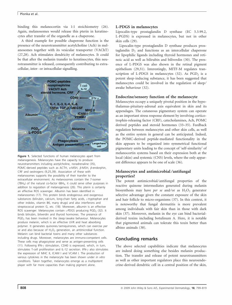
Figure 1. Selected functions of human melanocytes apart from
melanogenesis. Melanocytes have the capacity to produce
neurotransmitters including acetylcholine, noradrenaline (35),
POMC-derived peptides such as ACTH, a-MSH, b-MSH, b-endorphin,
CRF and oestrogens (9,25,39). Association of these with
melanosomes supports the possibility of their transfer to the
extracellular environment. As melanosomes contain the 7-isomer
(7BH4) of the natural co-factor 6BH4, it could serve other purposes in
addition to regulation of melanogenesis (26). This pterin is certainly
an effective ROS scavenger. Albumin has been identified in
melanosomes (17). This protein binds endogenous and exogenous
substances (bilirubin, calcium, long-chain fatty acids, L-tryptophan and
other indoles, vitamin B6, many drugs) and also interferons and
streptococcal protein G, etc. (18). Moreover, albumin is an effective
ROS scavenger. Melanocytes contain L-PDGS producing PGD2 (32). It
binds bilirubin, biliverdin and thyroid hormones. The presence of
PGD2 has been invoked in the sleep ⁄ awake behaviour. Melanocytes
produce melanin, which is an effective UVR and heat absorbing
polymer. It generates quinones ⁄ semiquinones, which can exercise per
se and also because of H2O2 generation, an antimicrobial function.
Melanin can bind bacterial toxins and many other substances
including drugs. Moreover, melanocytes are immuno-competent cells.
These cells may phagocytose and serve as antigen-presenting cells
(11). Following IFN-c stimulation, CD40 is expressed, which, in turn,
stimulates T-cell proliferation and IL-12 secretion. IFN-c also stimulates
the expression of MHC II, ICAM I and VCAM I. The production of
various cytokines in the melanocyte has been shown under in vitro
conditions. Taken together, melanocytes emerge as a multipotent
player with far more capacities than making pigment alone.
Plonka et al.
808 ª 2009 John Wiley & Sons A/S, Experimental Dermatology, 18, 799–819
calling for a tight feedback on neuronal and non-neuronal
interaction ⁄ signalling in the local environment of the skin
and also at distant extracutaneous end points (Fig. 1).
Acknowledgements
The authors would like to acknowledge the support of Stiefel International,
Deutsche Vitiligo Verein Hamburg, Germany and private donations to
KUS and The Welcome Trust, NIH, Procter & Gamble, LVMH Recherche
and Unilever to DJT in funding pigmentation research.
Karin U Schallreuter1,2,3, Desmond J. Tobin3
1Clinical and Experimental Dermatology, School
of Life Sciences, University of Bradford, UK;2Institute for Pigmentary Disorders in association with
EM Arndt University of Greifswald Germany
and University of Bradford, Bradford, UK;3Centre for Skin Sciences, School of Life Sciences,
University of Bradford, Bradford, UK;
E-mail: [email protected]
References
1 Fitzpatrick T B, Breathnach A S. Dermatol Wochenschr 1963: 147: 481–489.2 Westerhof W. Pigment Cell Res 2006: 19: 183–193.3 Singh S K et al. Exp Dermatol 2008: 17: 418–426.4 Schallreuter K U. Dermatol Clin 2007: 25: 283–291, vii.
5 Goldgeier M H et al. J Invest Dermatol 1984: 82: 235–238.6 Gottesberge A M M z. Pigment Cell Res 1988: 1: 238–249.7 Yajima I, Larue L. Pigment Cell Melanoma Res 2008: 21: 471–476.8 Bharti K et al. Pigment Cell Res 2006: 19: 380–394.9 Slominski A et al. Physiol Rev 2004: 84: 1155–1228.
10 Aubock J et al. Arch Dermatol Res 1985: 277: 270–275.11 Le Poole I C et al. J Immunol 1993: 151: 7284–7292.12 Amornsiripanitch S et al. J Immunol 1988: 140: 3438–3445.13 Rheins L A, Nordlund J J. J Immunol 1986: 136: 867–876.14 Nordlund J J et al., eds. The Pigmentary System. Physiology and Pathophysiol-
ogy. New York: Oxford University Press, 1998.15 Le Poole I C et al. J Invest Dermatol 1993: 100: 816–822.16 Chi A et al. J Proteome Res 2006: 5: 3135–3144.17 Hasse S et al. Exp Dermatol 2005: 14: 182–187.18 Peters T J. All about albumin. In: ed. San Diego, CA: Academic Press, 1996,
pp. 17–19.19 Spencer J D et al. J Endocrinol 2005: 187: 293–302.20 Marles L K et al. Exp Dermatol 2003: 12: 61–70.21 Curtius H C et al. Biochem Biophys Res Commun 1990: 172: 1060–1166.22 Schallreuter K U et al. Science 1994: 263: 1444–1446.23 Pey A L et al. Hum Mutat 2004: 24: 388–399.24 Pey A L et al. FASEB J 2006: 20: 2130–2132.25 Spencer J D, Schallreuter K U. Endocrinology 2008: 150: 1235–1258.26 Schallreuter K U, Elwary S. Life Sci 2007: 80: 2221–2226.27 Schallreuter K U et al. Biochem Biophys Res Commun 2007: 355: 1069–1074.28 Elwary S M et al. J Invest Dermatol 2006: 126: 1879–1884.29 Takeda K et al. Biochem Biophys Res Commun 2006: 339: 1098–1106.30 Beuckmann C T et al. Biochemistry 1999: 38: 8006–8013.31 Beuckmann C T et al. J Neurosci 1996: 16: 6119–6124.32 Takeda K et al. J Biochem 2007: 141: 327–333.33 Slominski et al. Physiol Rev 2000: 80: 979–1020.34 Slominski A, Wortsman J. Endocr Rev 2000: 21: 457–487.35 Grando S A et al. J Invest Dermatol 2006: 126: 1948–1965.36 Peters A. Endocrinology 2005: 146: 529–531.37 Mackintosh J A. J Theor Biol 2001: 211: 101–113.38 Ishikawa H et al. Jpn J Ophthalmol 2000: 44: 106–109.39 Spencer J D et al. J Invest Dermatol 2007: 127: 411–420.
Viewpoint #4
In 1969, Fitzpatrick and Breathnach formulated the concept
of the epidermal melanin unit. They proposed that a mela-
nocyte and 36 keratinocytes work together to produce skin
colour (1). Later, Nordlund proposed that the epidermal
melanin unit needs to be expanded to be called the ‘KLM
unit’ (K = keratinocyte; L = Langerhans cell; M = melano-
cyte) to emphasize the many ways all three major epider-
mal cells interact (2). It is now clearly established that
melanocytes exert more complex functions than produc-
tion, transport and transfer of melanin to the surrounding
keratinocytes. In 1993, Slominski et al. (3) proposed the
hypothesis that melanocytes can be considered as ‘sensory’
and regulatory cells in human epidermis. They proposed
that melanocytes act as intra-epithelial ‘stress-sensors’, alter
keratinocyte functions, have immunomodulatory functions,
serve as neuroendocrine cells and amplify and transform
signals received from neighbouring cells into chemical
messengers within an organized regulatory network for the
maintenance of epidermal homeostasis. Further research
demonstrated that there is not only an epidermal melanin
unit but also a real epidermal–dermal unit that could be
called the skin melanin unit. Indeed, in addition to the
three ‘major’ epidermal cells, data now strongly suggest
that the fibroblasts, the cutaneous axon terminal and the
endothelial cells have key interactions with melanocytes.
Alone a melanocyte is nothingMelanocytes are very difficult to grow in culture. Indeed,
such cultures not only need nutrients but also require sev-
eral growth factors for their proliferation and their survival
(4,5). Such fundamental considerations shed light on the
fact that in the skin, melanocytes are surrounded by many
kinds of cells that constantly produce cytokines, hormones
and growth factors that not only modulate the melanin
production but also control the proliferation and the
survival of melanocytes.
Human melanocytes in vitro respond directly to UV-light
with increased melanogenesis (6). One of the main
pathways involved in this direct UV response appears to
activate a transcriptional factor called USF1 through the
stress response kinase p38 (7). However, striking differences
are observed in the melanogenic response of normal
human melanocytes to UV-irradiation depending on cul-
ture conditions and the presence of keratinocytes. Exposure
of melanocytes monoculture with strong UVB doses leads
to slight melanogenic effects, whereas low UVB doses expo-
sure of melanocytes co-cultured with keratinocytes triggers
an increase in melanin synthesis. This experiment demon-
Controversies in Experimental Dermatology
ª 2009 John Wiley & Sons A/S, Experimental Dermatology, 18, 799–819 809
strates that keratinocytes play an important role in mediat-
ing UVB-induced pigmentation. These experiments demon-
strate that melanocytes alone have a limited power on skin
pigmentation and require the effective modulators of mela-
nin synthesis produced by their neighbouring keratinocytes
(8).
The key role of the keratinocyte partnerThe epidermal melanin unit is a functional unit composed
of one melanocyte and several neighbouring keratinocytes.
The pigment donors are melanocytes, which synthesize
melanin and the pigment recipients are epidermal keratino-
cytes. Until recently, we did not know how the pigmentary
units are working and what determines where melanin is
placed. Are keratinocytes acquiring pigment if a melanocyte
offers it or do they act proactively by recruiting melano-
cytes and inducing the transfer of pigments?
Several mechanisms of melanin transfer have been pro-
posed. According to the phagocytosis theory, the keratino-
cyte receptor protease-activated receptor-2 controls
melanosome ingestion and phagocytosis by keratinocytes
and exerts a regulatory role in skin pigmentation (9). It
was recently shown that Foxn1, a transcription factor first
indentifies pigment recipient cells and then recruits pig-
ment donors thereby generating pigmentary units. Foxn1
stimulated epithelial cells (keratinocytes) to emit signals,
one of which is FgF2. As a result of these signals, melano-
cytes recognize the Foxn1-positive cells as targets, connect
to these targets via dendrites and transfer pigment (10).
Melanogenic paracrine and autocrine cytokines have
been discovered in vitro between melanocytes and other
types of skin cells. Alpha-melanocyte-stimulating hormone
(aMSH), adrenocorticotrophic hormone, basic fibroblast
growth factor (bFGF), nerve growth factor (NGF), endoth-
elins (ET), granulocyte-macrophage colony-stimulating fac-
tor (GMCSF), stem-cell factor (SCF), leukaemia inhibitory
factor and hepatocyte growth factor (HGF) have been
reported to be the keratinocyte-derived factors and to regu-
late the proliferation and ⁄ or differentiation of mammalian
epidermal melanocytes (11). In fact, numerous factors may
be produced in and released from keratinocytes and be
involved in regulating the proliferation and differentiation
of mammalian epidermal melanocytes through receptor-
mediated signalling pathways. ET-1, GMCSF, SCF, HGF
and aMSH produced by keratinocytes and their corre-
sponding receptors on melanocytes respectively ETB recep-
tor, GMCSF receptor, c-kit, Cmet and MC1R appear to
play a key role in the interactions between keratinocytes
and melanocytes (12). Beta-endorphin is an opioid peptide
cleaved from the precursor pro-hormone, pro-opiomelano-
cortin (POMC). Human epidermal melanocytes express a
fully functioning beta-endorphin ⁄ l-opiate receptor system.
Beta-endorphin has potent melanogenic, mitogenic and
dendritogenic effects in cultured epidermal melanocytes
(13). These observations suggest that the beta-endor-
phin ⁄ l-opiate receptor system also participates in the
regulation of skin pigmentation.
Thus, this complex network of cytokines, hormones and
peptides finely regulates the melanocyte proliferation and
the production of melanin. Interestingly, the keratinocytes
also appear to control the pheo ⁄ eumelanin ratio in cul-
tured normal human melanocytes. Indeed, in melanocytes
monocultures, there is a very limited eumelanin production
and a very high pheomelanin synthesis leading to a very
high pheo ⁄ eumelanin ratio. In melanocytes–keratinocytes
co-cultures, there is an induction of eumelanin synthesis
accompanied by an important reduction in pheomelanin
formation. In this situation, the pheo ⁄ eumelanin ratio
dropped from 1043 (melanocyte monocultures) to about
25 in the presence of keratinocytes–melanocytes co-cul-
tured (14).
Is the fibroblast ‘the third man’?It is commonly accepted that melanocyte and keratinocyte
make the necessary couple to product pigmentation within
the skin. However, there are growing lines of evidence that
a third guy, the fibroblast, may also play a critical role for
the regulation of the pigmentation. In 1994, Hori et al.,
already suggest that fibroblasts might have a role on mela-
nocyte proliferation through the secretion of basic bFGF
(15). An increased tyrosinase activity was also reported in
melanocytes co-cultured with fibroblasts (16). Human
reconstructed skin models with melanocytes, keratinocytes
and fibroblasts also showed the importance of the fibro-
blasts for the melanin production (17). On the other hand,
it has been demonstrated in reconstructed skin models that
fibroblasts could reduce the pigmentation of the skin (18).
However, until recently, the mechanisms of action of fibro-
blasts on pigmentation remained obscure. One clue came
from the observation that pigmented non-palmoplantar
epidermis became hypopigmented when they were grafted
onto palmoplantar wounds (19). It was then demonstrated
that fibroblasts derived from palmoplantar skin expressed
high levels of dickkopf 1 (DKK1; an inhibitor of the canon-
ical Wnt signalling pathway), whereas non-palmoplantar
fibroblasts expressed higher levels of DKK3. The DKK1
secreted by fibroblasts decreased melanocyte function,
through beta-catenin-mediated regulation of microphthal-
mia-associated transcription factor activity, which in turn
modulates the growth and differentiation of melanocytes
(20). Thus, DKK1 modulate the melanocyte production of
melanin through the Wnt pathway but concomitantly acts
on keratinocyte to increase the thickness of the skin and
decrease the transfer of melanosomes (21,22). Those results
explain, at least in part, the thickness of the skin and the
relative hypopigmentation observed in palms and soles.
Plonka et al.
810 ª 2009 John Wiley & Sons A/S, Experimental Dermatology, 18, 799–819
Moreover, they clearly show the strong impact on melano-
cyte function of the factors secreted by fibroblasts.
Other lines of evidence suggest the critical role of fibro-
blasts in regulating skin pigmentation. In vitiligo, it is com-
monly accepted that extremities are highly difficult to
repigment. The most frequently accepted explanation is the
lack of hair follicles. However, the recent data on DKK1
and the success of some full skin grafts as compared to epi-
dermal grafts also suggest that dermis’ factors play a role in
the treatment failures. Recently, a late redepigmentation
occurring 7 years after a success of an epidermal graft for
treating a nevus depigmentosus was reported (23). This
clinical observation additionally suggests that dermal fac-
tors might have induced the recurrence of this hypopig-
mentation. Thus, the study of achromic nevi could help us
better understand the dermal influence on pigmentation
processes.
The increasing role of the vascularizationInteractions between melanocytes have not yet been studied
in depth. However, a recent report suggests that increased
vascularity is one of the major findings in melasma (24).
Vascular endothelial growth factor (VEGF) may be a major
angiogenic factor for altered vessels in melasma. Interest-
ingly, normal human melanocytes in vitro express VEGF
receptor-1, VEGFR-2 and neuropilin (25). Several data
suggest that some of these receptors are functional, and
that VEGF may play a role in melanocyte behaviour in
skin. Thus, the vascularization of the skin might play a role
in the pigmentation processes. This field of research clearly
needs to be further studied, but may provide new thera-
peutic options for pigmentary disorders such as chemical
agents with anti-angiogenic properties or physical
treatments such as intense pulsed light or lasers that could
target the vessels.
Melanocytes and the skin neural systemIt has been shown that cutaneous axon terminals and epi-
dermal melanocytes make contact via chemical synapses in
human skin (26). The control of melanocytes function by
the cutaneous nervous system has been poorly understood.
However, NGF is known to contribute to dendrite forma-
tion of melanocytes (27). CGRP, a neuropeptide known to
be present in intra-epidermal nerve fibres and to induce
melanocyte proliferation, upregulates melanogenesis includ-
ing melanosome maturation, tyrosinase activity and mela-
nin content in melanocytes, via secondary signals from the
surrounding CGRP-stimulated keratinocytes. Some CGRP-
induced mediators derived from keratinocytes also increase
melanocyte dendricity (28).
Human melanocytes are equipped with voltage-depen-
dent Na+ channels a delayed rectifying K+ current and a
K+ current in neurones. Whether Na+ channels in melano-
cytes might be of importance in the functional co-opera-
tion between melanocytes and keratinocytes remains to be
studied (29). The transient receptor potential (TRP) genes
encode for ion-channel proteins that are present on mela-
nocytes. Nevertheless, melanocytes are not considered as
excitable cells even if synaptic-like structures and excitable
cell-specific ion-channels are present.
Melanocyte: a member of the skin immune systemand key-player in an epidermal stress–responsesystem?The phagocytic capacity of normal human skin melano-
cytes has been demonstrated by a Dutch group (30). Fur-
thermore, the same group observed that melanocytes can
indeed process microorganisms and present antigen in the
context of MHC class II molecules to antigen specific T-cell
clones. They concluded that melanocytes may be capable of
antigen processing and presentation, suggesting their active
participation in the skin immune system.
Alpha-melanocyte-stimulating hormone has been identi-
fied as a potent anti-inflammatory peptide (by inhibiting
the NF-jB activation) effective in a number of cell types
including melanocytes and melanoma cells. aMSH is acting
via a local paracrine ⁄ autocrine mechanism to control the
expression of molecules involved in inflammation by atten-
uation of the pro-inflammatory cytokine pathway (31).
Melanocytes express POMC, corticotrophin-releasing
hormone (CRH) and CRH-receptor-1 (CRH-R1) and can
produce corticosterone. Normal epidermal melanocytes
stimulated by CRH trigger a functional cascade structure
hierarchically and arranged along the same algorithm as in
the hypothalamic-pituitary-adrenal axis (32). Interestingly,
CRH was demonstrated to inhibit NF-jB in melanocytes
through the POMC production and thus may serve as a
feedback mechanism to self-restrict inflammatory response
in the skin (33).
A plea in favour of an epidermal stem-cellreservoir of melanocytesDuring embryogenesis, some cells from the neural crest
differentiate under the stimulation of several factors into
mature melanocytes that colonize the epidermis and the
hair follicles. It has been now clearly demonstrated that the
lower permanent portion of the hair follicle provides
shelter for melanocyte stem-cells that are self-renewing.
After stimulation with several factors including the SCF,
these cells can migrate in the basal layer of the epidermis
and differentiate into mature melanocyte (34). The perifol-
licular repigmentation of vitiligo patients after photothera-
py provides a good clinical example of this recolonization
of the epidermis from the hair follicle (35–37).
However, there are growing lines of evidence that the
epidermis is also a reservoir of melanocytes. Again, vitiligo
Controversies in Experimental Dermatology
ª 2009 John Wiley & Sons A/S, Experimental Dermatology, 18, 799–819 811
provides very interesting clues that are worth following.
Mucosal areas (such as lips) and palms and soles depig-
mented lesions have been reported to be repigmented after
phototherapy (38) (Passeron T & Ortonne JP, unpublished
observation). In those cases, the repigmentation did not
come from the melanocytes of the borders of the lesions,
but was observed in the centre of the depigmented glabrous
skin. One indirect clue is the fact that melanocytes can be
grown from depigmented vitiligo skin samples that are told
to be devoid of melanocytes (39). However, without spe-
cific markers of melanocyte progenitors, it remains very
difficult to have a direct proof of the existence of an epi-
dermal reservoir of melanocytes. If such an epidermal res-
ervoir exists, it would be of interest from a physiological
point of view, but it could also raise new therapeutic
options for acquired hypopigmentary disorders including
vitiligo. Indeed, repigmentation from areas devoid of
follicle is much more difficult to obtain than the follicular
repigmentation that can be observed after phototherapy. If
such epidermal reservoir exists, it might respond to factors
other than the melanocyte progenitors from the follicle
niche. Thus, there is a reasonable hope that the determina-
tion of those factors could lead us to new treatment
options.
Conclusion
Melanocytes are amazing cells that should not be consid-
ered anymore as only ‘melanin factories’. They are active
partners of the skin immune system, they participate in the
stress response and have immunomodulatory properties.
Their presence, not only in the skin and the hair follicle
but also in the central nervous system and in some visceral
organs, emphasizes the alternative functions of the melano-
cytes that still remain poorly understood (40). The melano-
cytes are in the centre of a very complex and tight
regulation that involves mostly all the cell types of the skin.
Indeed, keratinocytes, fibroblasts, Langerhans cells and also
vascularization and nerve connections actively interact with
the melanocytes and influence their functioning. The cru-
cial role of such interactions that are now better under-
stood should stress the importance of studying melanocytes
under physiological conditions. Indeed, melanocyte mono-
cultures are still required for some experiments, but the
results should be taken with care. The use of reconstructed
skin models or studies on skin biopsies by using immuno-
histochemistry, in situ hybridization or transcriptional anal-
ysis with arrays using full skin biopsy or laser dissection of
specific cells, are part of the tools that should help us fur-
ther understand those complex regulations. Finally, such
tight interactions have to be taken into consideration for
the treatment of pigmentary disorders as most of them
now only focus their action on melanocytes. Targeting not
only the melanocytes but also the surrounding factors
might help get better and extended results.
Thierry Passeron, Jean-Paul Ortonne
Department of Dermatology, University Hospital of Nice,
Archet 2 hospital, 06200 Nice, France;
E-mail: [email protected]
References
1 Fitzpatrick T B, Breathnach A S. Dermatolog Wochenschrift 1963: 147: 481–489.
2 Nordlund J J. Dermatol Clin 2007: 25: 271–281, vii..3 Slominski A et al. J Theor Biol 1993: 164: 103–120.4 Andreassi L et al. Human Melanocyte Cultures from Normal Skin and Pig-
mented Lesions Biological, Molecular and Clinical Aspects of Pigmentation.Tokyo: University of Tokyo Press, 1985: 563–568.
5 Halaban R. Pigment Cell Res 1988: 1(Suppl. 1): 18–26.6 Friedmann P S, Gilchrest B A. J Cell Physiol 1987: 133: 88–94.7 Galibert M D et al. EMBO J 2001: 20: 5022–5031.8 Duval C et al. Pigment cell research ⁄ sponsored by the European Society for
Pigment Cell Research and the International Pigment Cell Society 2001: 14:348–355.
9 Paine C et al. J Invest Dermatol 2001: 116: 587–595.10 Weiner L et al. Cell 2007: 130: 932–942.11 Hirobe T. Pigment Cell Res 2005: 18: 2–12.12 Imokawa G. Pigment cell research ⁄ sponsored by the European Society for Pig-
ment Cell Research and the International Pigment Cell Society 2004: 17: 96–110.
13 Kauser S et al. J Invest Dermatol 2003: 120: 1073–1080.14 Duval C et al. Pigment cell research ⁄ sponsored by the European Society for
Pigment Cell Research and the International Pigment Cell Society 2002: 15:440–446.
15 Imayama S et al. Pigment cell research ⁄ sponsored by the European Society forPigment Cell Research and the International Pigment Cell Society 1994: 7:170–174.
16 Buffey J A et al. Br J Dermatol 1994: 131: 836–842.17 Archambault M et al. J Invest Dermatol 1995: 104: 859–867.18 Hedley S J et al. Pigment cell research ⁄ sponsored by the European Society for
Pigment Cell Research and the International Pigment Cell Society 2002: 15:49–56.
19 Yamaguchi Y et al. J Dermatol 2001: 28: 521–534.20 Yamaguchi Y et al. J Cell Biol 2004: 165: 275–285.21 Yamaguchi Y et al. J Invest Dermatol 2007: 127: 1217–1225.22 Yamaguchi Y et al. FASEB J 2008: 22: 1009–1020.23 Kim do Y et al. J Am Acad Dermatol 2008: 58: 527–529.24 Kim E H et al. J Dermatol Sci 2007: 46: 111–116.25 Kim E J et al. Exp Dermatol 2005: 14: 625–633.26 Hara M et al. J Exp Med 1996: 184: 1385–1395.27 Yaar M, Gilchrest B A. J Invest Dermatol 1991: 97: 611.28 Toyoda M et al. J Investig Dermatol Symp Proc 1999: 4: 116–125.29 Ekmehag B et al. Pigment cell research ⁄ sponsored by the European Society
for Pigment Cell Research and the International Pigment Cell Society 1994: 7:333–338.
30 Le Poole I C et al. Exp Cell Res 1993: 205: 388–395.31 Haycock J W et al. J Invest Dermatol 1999: 113: 560–566.32 Slominski A et al. Am J Physiol Endocrine Metab 2005: 288: E701–E706.33 Slominski A et al. J Cell Physiol 2006: 206: 780–791.34 Nishimura E K et al. Nature 2002: 416: 854–860.35 Ortonne J P et al. J Invest Dermatol 1980: 74: 40–42.36 Ortonne J P et al. Brit J Dermatol 1979: 101: 1–12.37 Ostovari N et al. Lasers Surg Med 2004: 35: 152–156.38 Davids L M et al. Clin Exp Dermatol 2009: 34: 246–248.39 Tobin D J et al. J Pathol 2000: 191: 407–416.40 Yajima I, Larue L. Pigment cell Melanoma Res 2008: 21: 471–476.
Plonka et al.
812 ª 2009 John Wiley & Sons A/S, Experimental Dermatology, 18, 799–819
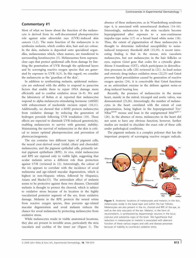
Commentary #1
Most of what we know about the function of the melano-
cyte is derived from its well-documented photoprotective
role against solar ultraviolet rays (UVR)-induced skin
cancers (1,2). The main function of the melanocyte is to
synthesize melanin, which confers skin, hair and eye colour.
In the skin, melanin is deposited onto specialized organ-
elles, melanosomes which, when mature, are transferred to
surrounding keratinocytes (3). Melanosomes form supranu-
clear caps that protect epidermal cells from damage by lim-
iting the penetration of UVR through the epidermal layers
and by scavenging reactive oxygen species that are gener-
ated by exposure to UVR (4,5). In this regard, we consider
the melanocyte as the ‘guardian of the skin’.
In addition to synthesizing melanin, epidermal melano-
cyte are endowed with the ability to respond to paracrine
factors that enable them to repair DNA damage more
efficiently and to combat oxidative stress (6–9). We and
the laboratory of Bohm et al. reported that melanocytes
respond to alpha-melanocyte-stimulating hormone (aMSH)
with enhancement of nucleotide excision repair (10,11).
Additionally, we showed that a-MSH protects melanocytes
from oxidative damage by reducing the generation of
hydrogen peroxide following UVR irradiation (10). These
effects are expected to diminish UVR-induced genotoxicity,
enabling melanocytes to survive with genomic stability.
Maintaining the survival of melanocytes in the skin is criti-
cal to insure optimal photoprotection and prevention of
photocarcinogenesis.
The eye contains two different types of pigment cells,
the neural crest-derived uveal (iridal, ciliary and choroidal)
melanocytes, and the pigment epithelial cells, primarily ret-
inal pigment epithelium (RPE). In vivo, iridal melanocytes
and RPE are exposed only to visible light, suggesting that
ocular melanin serves a different role than protection
against UVR (reviewed in 12). Interestingly, the colour of
the iris appears to correlate with the incidence of uveal
melanoma and age-related macular degeneration, which is
highest in non-Hispanic whites, followed by Hispanics,
Asians and blacks(13). The antioxidant effect of melanin
seems to be protective against these two diseases. Choroidal
melanin is thought to protect the choroid, which is subject
to oxidative stress because of its location in the highly
vascularized posterior segment of the eye, from oxidative
damage. Melanin in the RPE protects the neural retina
from reactive oxygen species, thus prevents age-related
macular degeneration and uveal melanin reduces the
chance for uveal melanoma by protecting melanocytes from
oxidative stress.
While melanocytes reside in visible anatomical locations,
they also are present in invisible areas, particularly the stria
vascularis and cochlea of the inner ear (Figure 1). The
absence of these melanocytes, as in Waardenburg syndrome
type 4, is associated with sensorineural deafness (14–16).
Interestingly, melanocytes in the stria vacularis become
hyperpigmented after exposure to a non-continuous
impulse-type noise (17) or a broad band white noise (18).
Thus, the extent of pigmentation of these melanocytes is
thought to determine individual susceptibility to noise-
induced temporary threshold shift (15,19). A recent inter-
esting finding is that in the mouse, stria vascularis
melanocytes, but not melanocytes in the hair follicles or
eyes, express Gsta4 gene that codes for a cytosolic gluta-
thione S-transferase (GST), which participates in detoxifica-
tion processes in cells (20) reviewed in (21). As loud noises
and ototoxic drug-induce oxidative stress (22,23) and Gsta4
prevents lipid peroxidation caused by generation of reactive
oxygen species (24), it is conceivable that Gsta4 functions
as an antioxidant enzyme in the defense against noise-or
drug-induced hearing loss.
Recently, the presence of melanocytes in the mouse
heart, mainly in the mitral, tricuspid and aortic valves, was
demonstrated (25,26). Interestingly, the number of melano-
cytes in the heart correlated with the extent of coat
pigmentation, being highest in the hyperpigmented Tyr ⁄NRasQ61K mouse and least in the unpigmented Mitfvga9 ⁄ +
(26). In the absence of stress, melanocytes in the heart did
not seem to have any obvious function; however, further
studies are needed to elucidate the exact role of these cells
under pathological conditions.
The pigment melanin is a complex polymer that has the
important property of scavenging reactive oxygen radicals.
Figure 1. Anatomic locations of melanocytes and melanin. In the skin,
melanocytes reside in the basal layer and within the hair follicles.
Melanocytes are also present in the iris, choroid and RPE of the eye, as
well as the stria vascularis of the ear. Melanin, in the form of
neuromelanin, is synthesized by dopaminergic neurons in the locus
coerulus and substantia nigra of the brain. We hypothesize that
reduction in melanocytes or melanin is associated with aberrant
function of these various organs and cells and disease processes
because of inability to counteract oxidative stress.
Controversies in Experimental Dermatology
ª 2009 John Wiley & Sons A/S, Experimental Dermatology, 18, 799–819 813
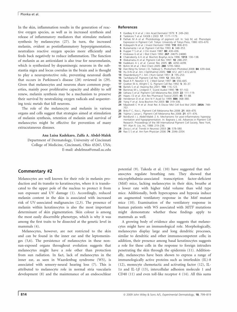
In the skin, inflammation results in the generation of reac-
tive oxygen species, as well as in increased synthesis and
release of inflammatory mediators that stimulate melanin
synthesis by melanocytes (27). In turn, the increased
melanin, evident as postinflammatory hyperpigmentation,
neutralizes reactive oxygen species more efficiently and
feeds back negatively to reduce inflammation. The function
of melanin as an antioxidant is also true for neuromelanin,
which is synthesized by dopaminergic neurons in the sub-
stantia nigra and locus coerulus in the brain and is thought
to play a neuroprotective role, preventing neuronal death
that occurs in Parkinson’s disease (28) reviewed in (29).
Given that melanocytes and neurons share common prop-
erties, mainly poor proliferative capacity and ability to self
renew, melanin synthesis may be a mechanism to preserve
their survival by neutralizing oxygen radicals and sequester-
ing toxic metals that kill neurons.
The role of the melanocyte and melanin in various
organs and cells suggest that strategies aimed at stimulation
of melanin synthesis, retention of melanin and survival of
melanocytes might be effective for prevention of many
extracutaneous diseases.
Ana Luisa Kadekaro, Zalfa A. Abdel-Malek
Department of Dermatology, University of Cincinnati
College of Medicine, Cincinnati, Ohio 45267, USA;
E-mail: [email protected]
References
1 Kaidbey K H et al. J Am Acad Dermatol 1979: 1: 249–260.2 Tadokoro T et al. FASEB J 2003: 17: 1177–1179.3 Pathak M A et al. Photobiology of pigment cell. In: Seiji M, ed. Phenotypic
Expression in Pigment Cell. Tokyo: University of Tokyo Press, 1980: 655–670.4 Kobayashi N et al. J Invest Dermatol 1998: 110: 806–810.5 Bustamante J et al. Pigment Cell Res 1993: 6: 348–353.6 Kupper T S et al. J Clin Invest 1987: 80: 430–436.7 Imokawa G et al. J Biol Chem 1992: 267: 24675–24680.8 Chakraborty A K et al. Biochim Biophys Acta 1996: 1313: 130–138.9 Wakamatsu K et al. Pigment Cell Res 1997: 10: 288–297.
10 Kadekaro A L et al. Cancer Res 2005: 65: 4292–4299.11 Bohm M et al. J Biol Chem 2005: 280: 5795–5802.12 Dan-Ning Hu Simon J D, Tadeusz S. Photochem Photobiol 2008: 84: 639–644.13 Hu D-N et al. Am J Ophthalmol 2005: 140: 612. e611–612.e618.14 Waardenburg P J. Am J Hum Genet 1951: 3: 195–253.15 Tachibana M. Pigment Cell Res 1999: 12: 344–354.16 Read A P, Newton V E. J Med Genet 1997: 34: 656–665.17 Gratton M A, Wright C G. Pigment Cell Res 1992: 5: 30–37.18 Bartels S et al. Hearing Res 2001: 154: 116–123.19 Barrenas M L, Lindgren F. Scand Audiol 1990: 19: 97–102.20 Uehara S et al. Pigment Cell Melanoma Res 2009: 22: 111–119.21 Hayes J D et al. Ann Rev Pharmacol Toxicol 2005: 45: 51–88.22 Henderson D et al. Ann N Y Acad Sci 1999: 884: 368–380.23 Yang Y et al. Acta Biochim Pol 2003: 50: 319–336.24 Mjaatvedt C H et al. Anat Rec A Discov Mol Cell Evol Biol 2005: 285A: 748–
757.25 Brito F C, Kos L. Pigment Cell Melanoma Res 2008: 21: 464–470.26 Yajima I, Larue L. Pigment Cell Melanoma Res 2008: 21: 471–476.27 Nordlund J J, Abdel-Malek Z A. Mechanisms for post-inflammatory hyperpig-
mentation and hypopigmentation. In: Bagnara J, ed. Advances in Pigment CellResearch. Proceedings of the XIII International Pigment Cell Society. New York,NY: Alan R. Liss, Inc, 1988: 219–236.
28 Zecca L et al. Trends in Neurosci 2003: 26: 578–580.29 Rao S S et al. Am Fam Physician 2006: 74: 2046–2054.
Commentary #2
Melanocytes are well known for their role in melanin pro-
duction and its transfer to keratinocytes, where it is translo-
cated to the upper pole of the nucleus to protect it from
sun exposure and UV damage (1). Accordingly, reduced
melanin content in the skin is associated with increased
risk of UV-associated malignancies (2,3). The presence of
melanin within keratinocytes is also the most important
determinant of skin pigmentation. Skin colour is among
the most easily discernible phenotype, which is why it was
among the first traits to be dissected at the genetic level in
mammals (4).
Melanocytes, however, are not restricted to the skin
and can be found in the inner ear and the leptomenin-
ges (5,6). The persistence of melanocytes in these non-
sun-exposed organs throughout evolution suggests that
melanocytes might have a role other than protection
from sun radiation. In fact, lack of melanocytes in the
inner ear, as seen in Waardenbug syndrome (WS), is
associated with sensory-neural hearing loss (7). This is
attributed to melanocyte role in normal stria vascularis
development (8) and the maintenance of an endocochlear
potential (9). Takeda et al. (10) have suggested that mel-
anocytes regulate breathing rate. They showed that
microphthalmia-associated transcription factor-deficient
(Mitf) mice, lacking melanocytes in their skin, breathe at
a lower rate with higher tidal volume than wild type
mice. Additionally, both hypercapnea and hypoxia induce
an augmented ventilatory response in the Mitf mutant
mice (10). Examination of the ventilatory response in
human patients with WS associated with MITF mutations
might demonstrate whether these findings apply to
mammals as well.
A growing body of evidence also suggests that melano-
cytes might have an immunological role. Morphologically,
melanocytes display large and long dendritic processes,
similar to dendritic and other immunocompetent cells; in
addition, their presence among basal keratinocytes suggests
a role for these cells in the response to foreign intruders
penetrating the skin through the epidermis (11). Addition-
ally, melanocytes have been shown to express a range of
immunologically active proteins such as interleukin (IL)-8
(12), monocyte chemotactic and activating factor (12), IL-
1a and IL-1b (13), intercellular adhesion molecule 1 and
CD40 (11) and even toll-like receptor 4 (14). All this sums
Plonka et al.
814 ª 2009 John Wiley & Sons A/S, Experimental Dermatology, 18, 799–819
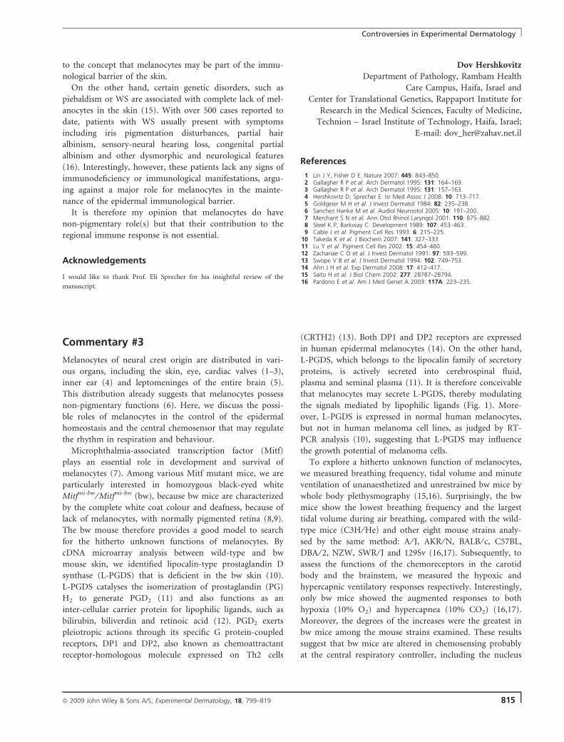
to the concept that melanocytes may be part of the immu-
nological barrier of the skin.
On the other hand, certain genetic disorders, such as
piebaldism or WS are associated with complete lack of mel-
anocytes in the skin (15). With over 500 cases reported to
date, patients with WS usually present with symptoms
including iris pigmentation disturbances, partial hair
albinism, sensory-neural hearing loss, congenital partial
albinism and other dysmorphic and neurological features
(16). Interestingly, however, these patients lack any signs of
immunodeficiency or immunological manifestations, argu-
ing against a major role for melanocytes in the mainte-
nance of the epidermal immunological barrier.
It is therefore my opinion that melanocytes do have
non-pigmentary role(s) but that their contribution to the
regional immune response is not essential.
Acknowledgements
I would like to thank Prof. Eli Sprecher for his insightful review of the
manuscript.
Dov Hershkovitz
Department of Pathology, Rambam Health
Care Campus, Haifa, Israel and
Center for Translational Genetics, Rappaport Institute for
Research in the Medical Sciences, Faculty of Medicine,
Technion – Israel Institute of Technology, Haifa, Israel;
E-mail: [email protected]
References
1 Lin J Y, Fisher D E. Nature 2007: 445: 843–850.2 Gallagher R P et al. Arch Dermatol 1995: 131: 164–169.3 Gallagher R P et al. Arch Dermatol 1995: 131: 157–163.4 Hershkovitz D, Sprecher E. Isr Med Assoc J 2008: 10: 713–717.5 Goldgeier M H et al. J Invest Dermatol 1984: 82: 235–238.6 Sanchez Hanke M et al. Audiol Neurootol 2005: 10: 191–200.7 Merchant S N et al. Ann Otol Rhinol Laryngol 2001: 110: 875–882.8 Steel K P, Barkway C. Development 1989: 107: 453–463.9 Cable J et al. Pigment Cell Res 1993: 6: 215–225.
10 Takeda K et al. J Biochem 2007: 141: 327–333.11 Lu Y et al. Pigment Cell Res 2002: 15: 454–460.12 Zachariae C O et al. J Invest Dermatol 1991: 97: 593–599.13 Swope V B et al. J Invest Dermatol 1994: 102: 749–753.14 Ahn J H et al. Exp Dermatol 2008: 17: 412–417.15 Saito H et al. J Biol Chem 2002: 277: 28787–28794.16 Pardono E et al. Am J Med Genet A 2003: 117A: 223–235.
Commentary #3
Melanocytes of neural crest origin are distributed in vari-
ous organs, including the skin, eye, cardiac valves (1–3),
inner ear (4) and leptomeninges of the entire brain (5).
This distribution already suggests that melanocytes possess
non-pigmentary functions (6). Here, we discuss the possi-
ble roles of melanocytes in the control of the epidermal
homeostasis and the central chemosensor that may regulate
the rhythm in respiration and behaviour.
Microphthalmia-associated transcription factor (Mitf)
plays an essential role in development and survival of
melanocytes (7). Among various Mitf mutant mice, we are
particularly interested in homozygous black-eyed white
Mitfmi-bw ⁄ Mitfmi-bw (bw), because bw mice are characterized
by the complete white coat colour and deafness, because of
lack of melanocytes, with normally pigmented retina (8,9).
The bw mouse therefore provides a good model to search
for the hitherto unknown functions of melanocytes. By
cDNA microarray analysis between wild-type and bw
mouse skin, we identified lipocalin-type prostaglandin D
synthase (L-PGDS) that is deficient in the bw skin (10).
L-PGDS catalyses the isomerization of prostaglandin (PG)
H2 to generate PGD2 (11) and also functions as an
inter-cellular carrier protein for lipophilic ligands, such as
bilirubin, biliverdin and retinoic acid (12). PGD2 exerts
pleiotropic actions through its specific G protein-coupled
receptors, DP1 and DP2, also known as chemoattractant
receptor-homologous molecule expressed on Th2 cells
(CRTH2) (13). Both DP1 and DP2 receptors are expressed
in human epidermal melanocytes (14). On the other hand,
L-PGDS, which belongs to the lipocalin family of secretory
proteins, is actively secreted into cerebrospinal fluid,
plasma and seminal plasma (11). It is therefore conceivable
that melanocytes may secrete L-PGDS, thereby modulating
the signals mediated by lipophilic ligands (Fig. 1). More-
over, L-PGDS is expressed in normal human melanocytes,
but not in human melanoma cell lines, as judged by RT-
PCR analysis (10), suggesting that L-PGDS may influence
the growth potential of melanoma cells.
To explore a hitherto unknown function of melanocytes,
we measured breathing frequency, tidal volume and minute
ventilation of unanaesthetized and unrestrained bw mice by
whole body plethysmography (15,16). Surprisingly, the bw
mice show the lowest breathing frequency and the largest
tidal volume during air breathing, compared with the wild-
type mice (C3H ⁄ He) and other eight mouse strains analy-
sed by the same method: A ⁄ J, AKR ⁄ N, BALB ⁄ c, C57BL,
DBA ⁄ 2, NZW, SWR ⁄ J and 129Sv (16,17). Subsequently, to
assess the functions of the chemoreceptors in the carotid
body and the brainstem, we measured the hypoxic and
hypercapnic ventilatory responses respectively. Interestingly,
only bw mice showed the augmented responses to both
hypoxia (10% O2) and hypercapnea (10% CO2) (16,17).
Moreover, the degrees of the increases were the greatest in
bw mice among the mouse strains examined. These results
suggest that bw mice are altered in chemosensing probably
at the central respiratory controller, including the nucleus
Controversies in Experimental Dermatology
ª 2009 John Wiley & Sons A/S, Experimental Dermatology, 18, 799–819 815
tractus solitarius (Fig. 1). To the best of our knowledge,
bw mice represent the first example with the augmented
central chemosensitivity. In this connection, the authors
have noticed the abnormality in the behaviour of bw mice
(Takeda K, Yokoyama S, Han F, Nakai K, Sato H, Yama-
moto H, Shibahara S, unpublished observation).
In conclusion, we suggest the possibility that melanocytes
may be involved in the complex regulatory network for
epidermal homeostasis as well as for the central chemosen-
sing that affects respiration and behaviour.
Shigeki Shibahara, Kazuhisa Takeda
Department of Molecular Biology and Applied Physiology,
Tohoku University School of Medicine, Aoba-ku, Sendai,
Miyagi 980-8575, Japan;
E-mail: [email protected]
References
1 Mjaatvedt C H et al. Anat Rec A Discov Mol Cell Evol Biol 2005: 285: 748–757.
2 Brito F C, Kos L. Pigment Cell Melanoma Res 2008: 21: 464–470.3 Yajima I, Larue L. Pigment Cell Melanoma Res 2008: 21: 471–476.4 Uehara S et al. Pigment Cell Melanoma Res 2009: 22: 111–119.5 Goldgeier M H et al. J Invest Dermatol 1984: 82: 235–238.6 Takeda K et al. Tohoku J Exp Med 2007: 211: 201–221.7 Hodgkinson C A et al. Cell 1993: 74: 395–404.8 Motohashi H et al. Hear Res 1994: 80: 10–20.9 Yajima I et al. Hum Mol Genet 1999: 8: 1431–1441.
10 Takeda K et al. Biochem Biophys Res Commun 2006: 339: 1098–1106.11 Urade Y, Hayaishi O. Biochim Biophys Acta 2000: 1482: 259–271.12 Beuckmann C T et al. Biochemistry 1999: 38: 8006–8013.13 Pettipher R et al. Nat Rev Drug Discov 2007: 6: 313–325.14 Satarug S et al. Biochem Biophys Res Commun 2008: 377: 878–883.15 Adachi T et al. Biochem Biophys Res Commun 2004: 320: 514–522.16 Adachi T et al. Tohoku J Exp Med 2006: 209: 125–134.17 Takeda K et al. J Biochem (Tokyo) 2007: 141: 327–333.
Stratum corneum
Melanocyte: melanosomes
Keratinocyte
L-PGDSPGD2y
L D d
2
BilirubinRetinoic acid
Motor cortexSensory cortex
L-PGDSPGD2BilirubinRetinoic acid
- opa anrelated metabolites
Limbic system
CO
Respiratory controller
NTS O2 sensor
CO2 sensor
Figure 1. Proposed non-pigmentary functions of the melanocyte.
Melanin is produced within melanosomes (closed symbols), which are
transported to keratinocytes (nuclei, shown as open ovals). Melanocytes
secrete PGD2 and L-PGDS both of which may regulate the epidermal
homeostasis. L-PGDS also functions as a transporter for bilirubin and
retinoic acid. Melanocytes, located on the leptomeninges, may
influence the function of the nucleus tractus solitarius (NTS) in the
medulla, which is an essential centre for augmentation of ventilation
during hypoxia and hypercapnia (long oval). Also shown near NTS is
central O2 sensor (small oval). Hypercapnea stimulates ventilation
through the central chemoreceptor (CO2 sensor) in the ventrolaletral
medullary area (three circles). Broken lines with arrowhead(s) indicate
the direction of neural signalling. The presented cartoon is the modified
version of the figures published in the reference (6).
Commentary #4
As one who studies the early development and differentia-
tion of melanocytes, I cannot directly answer this question,
but I can tell you where melanocytes come from. By the
end, perhaps you will be surprised to learn what other
functions they evolved to perform. In vertebrates, melano-
cytes differentiate from the neural crest, a population of
cells that emigrate from the neural tube early during devel-
opment (1). These cells migrate extensively throughout the
embryo, where they differentiate into numerous cell types.
Some of the neural crest derivatives are melanocytes, neu-
rons and glia of the peripheral nervous system, bone, teeth
and connective tissue in the head, smooth muscle in some
arteries and the heart and chromaffin cells of the adrenal
medulla (1). The neural crest is a cell type exclusive to ver-
tebrates and its evolution has been a subject of considerable
study in recent years. Some theorize that the neural crest
evolved as cells from the border of the neural plate and
ectoderm and developed the ability to migrate and adopt a
neural fate (2–5). From there, these migratory neural cells
evolved to differentiate into the other neural crest deriva-
tives found in modern vertebrates. However, recent evi-
dence from more basal chordates, such as tunicates and
amphioxus, suggests another possibility – that neural crest
evolved from pigment cells as they developed the ability to
differentiate into other cell types.
To understand this hypothesis requires that we look at
pigment cells and gene expression in other chordates. Ascidi-
ans are tunicates, and like vertebrates, have a notochord and
dorsal hollow neural tube at some time during development.
Although tunicate embryos do not have neural crest cells,
neural crest-like cells have recently been identified in 12 spe-
cies of ascidians (6,7). These cells emigrate from the neural
tube, migrate throughout the embryo and are positive for the
neural crest markers HNK-1 and Zic (6–8). In addition, and
Plonka et al.
816 ª 2009 John Wiley & Sons A/S, Experimental Dermatology, 18, 799–819
of most interest in this context, these cells express tyrosinase
and differentiate into pigment cells (6,7).
Jeffery et al. recently analysed expression of 16 genes
known to be important for neural crest induction and
specification. They discovered that seven of those genes are
expressed in these neural crest-like cells in the ascidian
Ciona intestinalis. One of the genes not expressed, which is
key to the discussion here, is Cs-FoxD, the Ciona homo-
logue of FOXD3, a forkhead-box transcriptional repressor
found in neural crest cells in vertebrates. FOXD3 represses
melanoblast specification in the neural crest and directs
neural crest cells to non-melanogenic fates (9–12). It does
so, at least in part, by repressing expression of MITF (13–
15), a transcription factor that regulates genes necessary for
melanin production and is required at the earliest stages of
melanoblast specification (9,12). Loss of FOXD3 in chick
neural crest cells results in a dramatic increase in the num-
ber of melanocytes and, likewise, misexpression of FOXD3
represses expression of MITF and directs neural crest cells
to other fates (10,12). In the non-vertebrate chordates
amphioxus and Ciona, the FOXD3 homologues, Amphi-
FoxD, and Cs-FoxD respectively are expressed in the noto-
chord and other tissues around the neural plate, but not in
the neural tube or neural plate (16,17).
Bringing these several pieces of evidence together
suggests an evolutionary pathway for development of the
neural crest. According to this model, in prevertebrate
chordates, melanocyte precursors originated in the neural
tube, as they do in tunicates. A FoxD transcription factor
with expression domains in the notochord and other tis-
sues surrounding the neural plate ⁄ tube was co-opted in
these pigment cell precursors (likely after a genetic duplica-
tion event). This expression of FoxD repressed melanogene-
sis and these cells were then able to differentiate into other
cell types. This model is not without precedent; a similar
gene co-option event likely led to the expression of Id and
other transcription factors in the neural plate early in the
evolution of the vertebrates, leading to the development of
bona fide neural crest (18,19). So, what are melanocytes
really doing all day long? Well, evolutionarily modified
melanocytes may be doing tasks as wide ranging as carrying
neural impulses to and from the central nervous
system, secreting hormones, maintaining blood pressure
and chewing food.
Aaron Thomas
Department of Molecular and Cellular Biology, University
of California at Davis, Davis, CA, USA;
E-mail: [email protected]
References
1 Le Douarin N M, Kalcheim C. The Neural Crest. Cambridge: Cambridge Uni-versity Press, 1999.
2 Baker C V. Curr Opin Genet Dev 2008: 18: 536–543.3 Holland L Z, Holland N D. J Anat 2001: 199: 85–98.4 Schlosser G. BioEssays 2008: 30: 659–672.5 Holland N D et al. Development 1996: 122: 2911–2920.6 Jeffery W R. J Exp Zool B Mol Dev Evol 2006: 306: 470–480.7 Jeffery W R et al. Nature 2004: 431: 696–699.8 Jeffery W R. Semin Cell Dev Biol 2007: 18: 481–491.9 Ignatius M S et al. Dev Biol 2008: 313: 568–583.
10 Kos R et al. Development 2001: 128: 1467–1479.11 Lister J A et al. Dev Biol 2006: 290: 92–104.12 Thomas A J, Erickson C A. Development 2009: 136: 1849–1858.13 Levy C et al. Trends Mol Med 2006: 12: 406–414.14 Goding C R. Genes Dev 2000: 14: 1712–1728.15 Steingrimsson E et al. Annu Rev Genet 2004: 38: 365–411.16 Imai K S et al. Development 2002: 129: 3441–3453.17 Yu J K et al. Dev Dyn 2002: 225: 289–297.18 Meulemans D, Bronner-Fraser M. J Exp Zool B Mol Dev Evol 2005: 304: 298–
303.19 Meulemans D et al. Dev Biol 2003: 264: 430–442.
Commentary #5
The fundamental role of melanocytes in the protection
against harmful effects of solar radiation, and as instru-
ments of visual communication or camouflage via produc-
tion and transfer of melanin pigment to epidermal and
hair follicle keratinocytes is well-documented and univer-
sally accepted (1). However, the important role of the
melanogenic apparatus and pathway in regulation and ⁄ or
defining additional functions of the skin and its adnexal
structures, which we had proposed (2–4), have largely been
overlooked by pigment and skin cell researchers alike.
Therefore, we take the opportunity of this commentary to
re-emphasize the bioregulatory, metabolic and homeostatic
functions of melanogenically active melanocytes, which are
distinct from their extensive neuroendocrine activity (5–7).
We had proposed that melanocytes regulate epidermal
functions by metabolically active melanosomes after trans-
fer to adjacent keratinocytes with subsequent field effects to
the respective epithelial skin compartments through altera-
tions in the flow of ions, nutrients and electric current
potential (3). This underappreciated, underinvestigated
homeostatic and bioregulatory activity of intraepithelial
melanocytes in the epidermis, hair follicle and possibly the
nail apparatus, is based in part on the intriguing physico-
chemical properties of melanin. These include melanin’s
ability to reversibly bind Ca2+, diverse ions and drugs and
to serve as free radical scavenger (3,8,9). In addition, the
active process of melanogenesis that continuously generates
reactive oxygen species (ROS) and quinone ⁄ semiquinone
intermediates (8–11) forms another basis of the regulatory
impact of melanocytes on epithelial cell function.
Controversies in Experimental Dermatology
ª 2009 John Wiley & Sons A/S, Experimental Dermatology, 18, 799–819 817
For example, both melanin and melanogenesis can
change the oxido-reduction potential of the cell, the intra-
cellular ROS levels, can consume oxygen and reversibly reg-
ulate NAD/NADH and NADP/NADPH ratios (which has
powerful metabolic consequences) (reviewed in 6,8–12).
These considerations had made it an obvious hypothesis to
propose that melanocytes, depending on their melanogenic
activity, regulate differentiation and proliferation as well as
other functional activities of keratinocytes or macrophages,
once melanosomes are injected into or are phagocytized by
these cells (2,3). As then, experimental data have accumu-
lated that support our concept directly (13) or indirectly
(8,14–16). For instance, active melanogenesis has the
potential to stimulate NFjB and AP1 or hypoxia inducible
1a signalling pathways (8,16,17), change cellular metabo-
lism (15), regulate intracellular calcium concentration (13),
inhibit immune responses (18) and change cellular respon-
siveness to chemo- or radiotherapy (19).
Under physiological conditions, the entire process of
melanin synthesis has to be tightly regulated starting from
the organelle and ending on the organ levels. Such role is
played by melanosomal proteins acting as active transporters
or ion channels including product of P locus (20), metal (21)
or SLC24A5 (22), SLC45A2 (23) transporters or as vacuolar
proton ATPases, Na,K-ATPase or ABC transporters (24).
Thus, there are many potential mechanisms by which
melanosome could regulate cellular and by continuation
epidermal homeostasis as proposed previously (3).
In this context, we had also proposed that intermediates
of melanogenesis, in particular L-DOPA and products of
its intramelanosomal metabolism, can act as intracellular
second messengers and as hormone-like bioregulators for
intercellular communication (3,25–27). This was based on
the capability of L-DOPA (or its metabolites) to induce the
synthesis and assembly of the melanogenic apparatus (28),
its stimulatory effect on aerobic glycolysis and glucose
metabolism (29,30), the inhibition of glycoprotein phos-
phorylation of L-DOPA (31) and its anti-proliferative
effects on lymphocytes (32). This concept, namely, that the
melanogenesis-associated production of L-DOPA and L-
DOPA metabolites can impact on neighbouring cells, has
most recently been supported by our demonstration that
L-DOPA not only inhibits lymphocytes DNA incorporation
but also abolishes proinflammatory cytokines production
(18). In addition, a recent report has suggested that L-
DOPA can act as an endogenous ligand for OA1 (33), con-
firming our original proposition for its hormone-like role
and the existence of putative ‘DOPA receptors’ (26–28,34).
Thus, the regulation of epidermal functions by melanocyte-
derived L-DOPA or its metabolites should be further
explored because of its clinical implications (4,18).
The time has come to appreciate, at long last, that –
beyond their neuroendocrine and other secretory functions
(2,3,5,6,35) – melanocytes are integral components of epi-
thelial cell biology and regulators of keratinocyte function
– just as, vice versa, melanocytes are controlled in multiple
ways by the keratinocyte environment they live in (6,36–
38). Whether or not, to which extent and under generation
of which by-products a melanocyte engages in melanogene-
sis deeply affect keratinocyte function. One short answer to
the central question posed in this Controversies feature,
then, is: ‘All day long’ intraepidermal and intrafollicular
melanocytes actively regulate the functions of those kerati-
nocyte into which they inject melanosomes, above and
beyond protecting their DNA from UV-induced damage.
Andrzej Slominski1, Ralf Paus2,3
1Department of Pathology and Laboratory Medicine,
University of Tennessee HSC, Memphis, TN, USA;2Department of Dermatology, University
of Luebeck, Luebeck, Germany;3School of Translational Medicine, University
of Manchester, Manchester, UK;
E-mail: [email protected]
References
1 Nordlund J J et al. The Pigmentary System: Physiology and Pathophysiology,2nd edn. Malden: Blackwell Publishing, 2006.
2 Slominski A, Paus R. J Invest Dermatol 1993: 101: 90S–97S.3 Slominski A et al. J Theor Biol 1993: 164: 103–120.4 Slominski A et al. Anticancer Res 1998: 18: 3709–3715.5 Slominski A, Wortsman J. Endocrine Rev 2000: 21: 457–487.6 Slominski A et al. Physiol Rev 2004: 84: 1155–1228.7 Slominski A. Dermatology 2005: 211: 199–208.8 Meyskens F L Jr et al. Recent Results Cancer Res 2007: 174: 191–195.9 Wood J M et al. Exp Dermatol 1999: 8: 153–164.
10 Hu D N et al. Photochem Photobiol 2008: 84: 639–644.11 Plonka P M, Grabacka M. Acta Biochim Pol 2006: 53: 429–443.12 Gidanian S et al. Photochem Photobiol 2008: 84: 556–564.13 Hoogduijn M J et al. Exp Cell Res 2004: 294: 60–67.14 Liu F et al. J Invest Dermatol 2009: 129: 422–431.15 Li W et al. Anal Biochem 2009: 386: 282–284.16 Fruehauf J P, Meyskens F L Jr. Clin Cancer Res 2007: 13: 789–794.17 Meyskens F L Jr et al. Clin Cancer Res 1999: 5: 1197–1202.18 Slominski A et al. Int J Cancer 2009: 124: 1470–1477.19 Brozyna A A et al. Int J Cancer 2008: 123: 1448–1456.20 Puri N et al. J Invest Dermatol 2000: 115: 607–613.21 Setty S R et al. Nature 2008: 454: 1142–1146.22 Lamason R L et al. Science 2005: 310: 1782–1786.23 Norton H L et al. Mol Biol Evol 2007: 24: 710–722.24 Chi A et al. J Proteome Res 2006: 5: 3135–3144.25 Slominski A, Paus R. J Theor Biol 1990: 143: 123–138.26 Slominski A, Paus R. Mol Cell Endocrinol 1994: 99: C7–C11.27 Slominski A et al. Pigment Cell Res 1989: 2: 109–116.28 Slominski A et al. J Cell Sci 1988: 89: 287–296.29 Scislowski P W et al. Int J Biochem 1984: 16: 327–331.30 Scislowski P W et al. Neoplasma 1985: 32: 593–598.31 Slominski A, Friedrich T. Pigment Cell Res 1992: 5: 396–399.32 Slominski A, Goodman-Snitkoff G G. Anticancer Res 1992: 12: 753–756.33 Lopez V M et al. PLoS Biol 2008: 6: e236.34 Slominski A, Pruski D. Biochim Biophys Acta 1992: 1139: 324–328.35 Tobin D J. Chem Soc Rev 2006: 35: 52–67.36 Nordlund J J. Dermatol Clin 2007: 25: 271–281.37 Wood J M, Schallreuter K U. Exp Dermatol 2008: 17: 569–578.38 Schallreuter K U et al. Exp Dermatol 2008: 17: 395–404.
Plonka et al.
818 ª 2009 John Wiley & Sons A/S, Experimental Dermatology, 18, 799–819
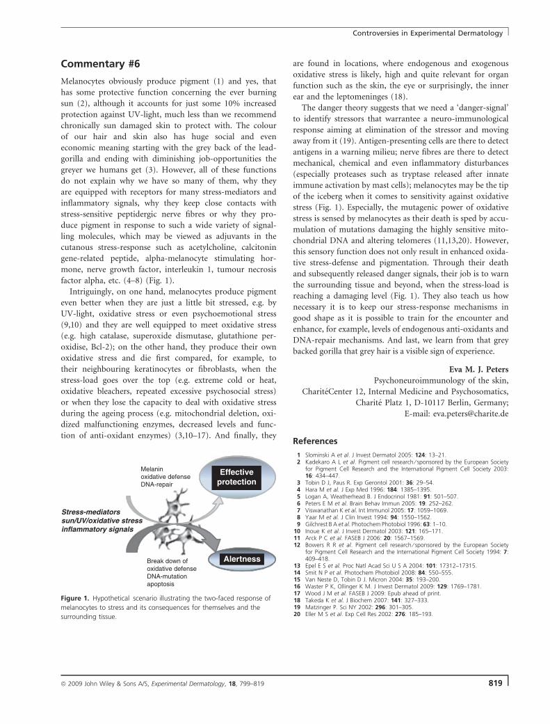
Commentary #6
Melanocytes obviously produce pigment (1) and yes, that
has some protective function concerning the ever burning
sun (2), although it accounts for just some 10% increased
protection against UV-light, much less than we recommend
chronically sun damaged skin to protect with. The colour
of our hair and skin also has huge social and even
economic meaning starting with the grey back of the lead-
gorilla and ending with diminishing job-opportunities the
greyer we humans get (3). However, all of these functions
do not explain why we have so many of them, why they
are equipped with receptors for many stress-mediators and
inflammatory signals, why they keep close contacts with
stress-sensitive peptidergic nerve fibres or why they pro-
duce pigment in response to such a wide variety of signal-
ling molecules, which may be viewed as adjuvants in the
cutanous stress-response such as acetylcholine, calcitonin
gene-related peptide, alpha-melanocyte stimulating hor-
mone, nerve growth factor, interleukin 1, tumour necrosis
factor alpha, etc. (4–8) (Fig. 1).
Intriguingly, on one hand, melanocytes produce pigment
even better when they are just a little bit stressed, e.g. by
UV-light, oxidative stress or even psychoemotional stress
(9,10) and they are well equipped to meet oxidative stress
(e.g. high catalase, superoxide dismutase, glutathione per-
oxidise, Bcl-2); on the other hand, they produce their own
oxidative stress and die first compared, for example, to
their neighbouring keratinocytes or fibroblasts, when the
stress-load goes over the top (e.g. extreme cold or heat,
oxidative bleachers, repeated excessive psychosocial stress)
or when they lose the capacity to deal with oxidative stress
during the ageing process (e.g. mitochondrial deletion, oxi-
dized malfunctioning enzymes, decreased levels and func-
tion of anti-oxidant enzymes) (3,10–17). And finally, they
are found in locations, where endogenous and exogenous
oxidative stress is likely, high and quite relevant for organ
function such as the skin, the eye or surprisingly, the inner
ear and the leptomeninges (18).
The danger theory suggests that we need a ‘danger-signal’
to identify stressors that warrantee a neuro-immunological
response aiming at elimination of the stressor and moving
away from it (19). Antigen-presenting cells are there to detect
antigens in a warning milieu; nerve fibres are there to detect
mechanical, chemical and even inflammatory disturbances
(especially proteases such as tryptase released after innate
immune activation by mast cells); melanocytes may be the tip
of the iceberg when it comes to sensitivity against oxidative
stress (Fig. 1). Especially, the mutagenic power of oxidative
stress is sensed by melanocytes as their death is sped by accu-
mulation of mutations damaging the highly sensitive mito-
chondrial DNA and altering telomeres (11,13,20). However,
this sensory function does not only result in enhanced oxida-
tive stress-defense and pigmentation. Through their death
and subsequently released danger signals, their job is to warn
the surrounding tissue and beyond, when the stress-load is
reaching a damaging level (Fig. 1). They also teach us how
necessary it is to keep our stress-response mechanisms in
good shape as it is possible to train for the encounter and
enhance, for example, levels of endogenous anti-oxidants and
DNA-repair mechanisms. And last, we learn from that grey
backed gorilla that grey hair is a visible sign of experience.
Eva M. J. Peters
Psychoneuroimmunology of the skin,
ChariteCenter 12, Internal Medicine and Psychosomatics,
Charite Platz 1, D-10117 Berlin, Germany;
E-mail: [email protected]
References
1 Slominski A et al. J Invest Dermatol 2005: 124: 13–21.2 Kadekaro A L et al. Pigment cell research ⁄ sponsored by the European Society
for Pigment Cell Research and the International Pigment Cell Society 2003:16: 434–447.
3 Tobin D J, Paus R. Exp Gerontol 2001: 36: 29–54.4 Hara M et al. J Exp Med 1996: 184: 1385–1395.5 Logan A, Weatherhead B. J Endocrinol 1981: 91: 501–507.6 Peters E M et al. Brain Behav Immun 2005: 19: 252–262.7 Viswanathan K et al. Int Immunol 2005: 17: 1059–1069.8 Yaar M et al. J Clin Invest 1994: 94: 1550–1562.9 Gilchrest B A et al. Photochem Photobiol 1996: 63: 1–10.
10 Inoue K et al. J Invest Dermatol 2003: 121: 165–171.11 Arck P C et al. FASEB J 2006: 20: 1567–1569.12 Bowers R R et al. Pigment cell research ⁄ sponsored by the European Society
for Pigment Cell Research and the International Pigment Cell Society 1994: 7:409–418.
13 Epel E S et al. Proc Natl Acad Sci U S A 2004: 101: 17312–17315.14 Smit N P et al. Photochem Photobiol 2008: 84: 550–555.15 Van Neste D, Tobin D J. Micron 2004: 35: 193–200.16 Waster P K, Ollinger K M. J Invest Dermatol 2009: 129: 1769–1781.17 Wood J M et al. FASEB J 2009: Epub ahead of print.18 Takeda K et al. J Biochem 2007: 141: 327–333.19 Matzinger P. Sci NY 2002: 296: 301–305.20 Eller M S et al. Exp Cell Res 2002: 276: 185–193.
Stress-mediators sun/UV/oxidative stress inflammatory signals
Break down of oxidative defense DNA-mutation apoptosis
Melanin oxidative defense DNA-repair
Effective protection
Alertness
Figure 1. Hypothetical scenario illustrating the two-faced response of
melanocytes to stress and its consequences for themselves and the
surrounding tissue.
Controversies in Experimental Dermatology
ª 2009 John Wiley & Sons A/S, Experimental Dermatology, 18, 799–819 819


