
1
Vibrio vulnificus integration into marine aggregates and subsequent uptake by 1
the oyster, Crassostrea virginica 2
Brett Froelicha, Mesrop Ayrapetyan, and James D. Oliverb 3
The University of North Carolina at Charlotte 4
5
6
7
Running Title: Vibrio vulnificus in marine aggregates 8
Keywords: marine aggregates/oysters/Vibrio 9
Subject Category: Microbial ecology 10
11
12
aCurrent address: The University of North Carolina at Chapel Hill Institute of Marine Sciences, Morehead 13
City, N.C. 14
bCorresponding author: 9201 University City Blvd., Charlotte, NC 28223, (704) 867-8516, 15
17
18
Copyright © 2012, American Society for Microbiology. All Rights Reserved.Appl. Environ. Microbiol. doi:10.1128/AEM.03095-12 AEM Accepts, published online ahead of print on 21 December 2012
on March 2, 2019 by guest
http://aem.asm
.org/D
ownloaded from

2
Abstract 19
Marine aggregates are naturally forming conglomerations of larvacean houses, 20
phytoplankton, microbes, and inorganics adhered together by exocellular polymers. In this 21
study we show in vitro that the bacterial pathogen, Vibrio vulnificus, can be concentrated into 22
laboratory-generated aggregates from surrounding water. We further show that 23
environmental (E-genotype) strains exhibit significantly more integration into these aggregates 24
than clinical (C-genotype) strains. Experiments where marine aggregates, with attached V. 25
vulnificus cells, were fed to oysters (Crassostrea virginica) resulted in greater uptake of both C- 26
and E-types, than non-aggregated controls. When C- and E-genotype strains were co-cultured 27
in competitive experiments, the aggregated E-genotype strains exhibited significantly greater 28
uptake by oyster than the C-genotypes strains. 29
Introduction 30
Vibrio vulnificus is a gram negative, halophilic bacterium capable of causing 31
gastroenteritis, wound infections, and fatal septicemia in humans (1, 2, 3). It is routinely found 32
in waters of estuarine environments as part of the normal microflora, as well as in oysters and 33
other shellfish inhabiting those estuaries (3). V. vulnificus infection is the leading cause of 34
seafood-borne deaths in the United States, usually resulting from the consumption of raw or 35
undercooked oysters (3). Infections caused by ingestion commonly result in primary 36
septicemia, almost always require hospitalization, and have a fatality rate greater than 50% (3, 37
4). Wound infections usually result from exposure of open wounds to sea water containing 38
the bacterium, and can progress to fatal necrotizing fasciitis (5, 6). 39
on March 2, 2019 by guest
http://aem.asm
.org/D
ownloaded from
3
V. vulnificus exhibits high genotypic and phenotypic variation (3) and is divided into two 40
genotypes, a difference originally discovered by RAPD-PCR analysis of strains from both clinical 41
and environmental sources (7). In this classification system a gene designated as vcg (virulence 42
correlated gene) was found to have two variations (8). One allele (vcgC) correlates highly with 43
strains obtained from clinical sources, designated the C-genotype, while the other (vcgE) is 44
correlated with environmentally isolated strains and is designated the E-genotype (7, 8). 45
Over 95% of infections resulting in septicemia caused by V. vulnificus involve the 46
consumption of raw oysters, with the remainder arising from ingestion of steamed oysters and 47
clams (3). While millions of people in the US eat raw oysters (9), if a consumer is afflicted with 48
a predisposing condition, such as liver impairment or immune system dysfunction (10), the risk 49
for infection increases 80 fold (11). Even considering these two facts, it is surprising that there 50
are only ca. 40 primary septicemia cases reported per year (10). Usually, oysters predominately 51
contain the E-genotype strains of V. vulnificus, which is a likely a factor in the low number of 52
infections (12, 13, 14, 15, 16, 17, 18) 53
A study comparing the population dynamics of the V. vulnificus genotypes revealed an 54
interesting phenomenon. While Warner and Oliver (13) found a nearly even ratio of C-type 55
strains to E-type strains in North Carolina and Florida seawater samples, strains isolated from 56
oysters in those same waters were predominately (>84%) E-type (13, 19). Presently, the reason 57
for this incongruity has not been determined. It is especially odd considering that oysters are 58
filter feeders, pumping water through their gills and straining food particles from the flow (20). 59
Incredibly, C. virginica is able to pump water at a rate of 10 L h-1g-1 dry tissue weight (20). With 60
on March 2, 2019 by guest
http://aem.asm
.org/D
ownloaded from

4
such a rapid rate of water clearance, one would naturally expect the oyster’s internal 61
composition of V. vulnificus to mimic that of the surrounding water. However, in experiments 62
where marked V. vulnificus strains of the C- or E-genotype were added to oysters and their 63
entry into and exit out of the shellfish was followed, no difference between uptake or 64
depuration rates of the two types was observed (14, 21). 65
An oyster is able to select the particles of food it eats based on size. The gills act as a 66
sieve, catching particles of optimum size and moving them toward the mouth, while particles 67
that are too large are stopped and passed from the oyster as pseudo-feces. Particles that are 68
smaller than the optimum size pass through the gills without capture. These too are excreted 69
from the oyster, undigested. For C. virginica, the optimum particle size is 5-7µm in diameter, 70
with particles of this size being retained with 90% efficiency (22). Particle retention rates drop 71
to 50% when the diameter is only 1.8µm, and when particles are the size of a single V. vulnificus 72
bacterium (ca. 1µm), oysters only retain ca. 16% of what is passed through the gills (22). This 73
size selection would likely limit the effectiveness of bacterial uptake experiments where 74
bacterial cells are simply added to oyster tanks. 75
Marine aggregates, also known as marine snow, are a natural part of marine waters. 76
These particles consist of fecal pellets, larvacean houses, phytoplankton, microbes, and 77
inorganics brought together by shear forces and Brownian movement. These particles are 78
conglomerated by exocellular polymers and physical/chemical forces (23) and once achieving a 79
critical size, sink to the ocean and estuary floor. Visible aggregates are termed “marine snow” 80
after the “long snowfall” of sedimentary material described by Rachel Carson (23, 24, 25). 81
on March 2, 2019 by guest
http://aem.asm
.org/D
ownloaded from
5
The purpose of this study was to compare the integration of C- and E-genotype V. 82
vulnificus cells into marine aggregates. These particles, with added V. vulnificus, were then fed 83
to oysters to measure uptake and depuration rates of this pathogen. We hypothesized that 84
differences in the ability of V. vulnificus strains to incorporate into marine aggregates could play 85
a role in the population disparity we have observed within oysters. 86
Materials and methods 87
Bacterial strains and growth conditions 88
V. vulnificus CVD713 is a C-genotype strain possessing a stable (for at least 10 days) 89
TnphoA transposon that confers kanamycin resistance and alkaline phosphatase activity (26, 90
27, 28). This strain forms blue colonies when grown on Tn Agar, consisting of Luria Agar with 91
the addition of 0.2 g L-1 kanamycin, 2 g L-1 glucose, and 0.04 g L-1 5-bromo-4-chloro-3-indolyl 92
phosphate (BCIP). Tn Agar selects for this TnphoA-possessing strain via its kanamycin 93
resistance and is differential by means of the BCIP breakdown (28). V. vulnificus strain JDO-2 is 94
a C-type strain derived from the parent strain C7184. This strain is resistant to chloramphenicol 95
and grows on heart infusion agar in the presence of 2 mg/ml of the antibiotic. V. vulnificus 96
strains pGTR-JY1305 and pGTR-Env1 are E-type strains that contain the stable pGTR plasmid 97
which confers kanamycin resistance when the strain is grown on Luria Agar containing 10 g L-1 98
L-arabinose and 0.3 g L-1 kanamycin (29). These genetically marked strains were used in 99
aggregation and oyster uptake experiments to allow differentiation of the added strains from 100
naturally occurring Vibrio spp. and other bacteria found in oysters and seawater. 101
on March 2, 2019 by guest
http://aem.asm
.org/D
ownloaded from

6
In competition experiments, wherein both kanamycin resistant C- and E-genotype V. 102
vulnificus strains were used simultaneously, a selective mannitol detection medium consisting 103
of 16 g of BBLTM Phenol Red broth base (BD, New Jersey) and 1.0% D-mannitol per liter of 104
deionized water, autoclaved for 5 minutes at 121°C and then supplemented with 0.3 g L-1 105
kanamycin and 10 g L-1 L-arabinose was employed. Distinct yellow colonies are formed by 106
pGTR-JY1305 and pGTR-Env1 (E-genotypes) and red colonies are formed by CVD713 and JDO-2 107
(C-genotype), respectively (12). 108
Oyster maintenance 109
Oysters (Crassostrea virginica) from the North Carolina coast were collected by hand in 110
the intertidal zone, rinsed, and placed into holding aquaria to acclimate to laboratory 111
conditions. The tanks contained a 1:1 mixture of artificial seawater (ASW, Instant Ocean, 112
Aquarium Systems, Mentor, OH) and natural seawater (NSW, collected from North Carolina 113
coast) which had been passed through a 0.45μm filter (Millipore, Bedford, MA) and finally 114
adjusted to 20‰ salinity with deionized water. Tank water was kept at 23°C, and oysters were 115
fed an algal mixture of Skeletonema, and Isochrysis species daily. The algal cultures were grown 116
at room temperature in vented, 1 liter flasks containing F/2 medium and were provided with 117
constant fluorescent light for one to two weeks until a dense population was achieved (30, 31). 118
Serial transfer of algal cultures was used to generate new cultures. 119
Marine aggregates 120
Laboratory-created aggregates (marine snow) were generated using the method 121
described by Shanks and Edmondson with the modifications suggested by Ward and Kach (32, 122
on March 2, 2019 by guest
http://aem.asm
.org/D
ownloaded from

7
33). Briefly, cells were added to seawater diluted to 15‰ salinity with deionized water in 250 123
ml roller bottles, 10 µg l-1 hyaluronic acid was added, and the bottles were placed on a roller 124
table at 15 RPM for 24 hours. Static bottles were placed next to the roller table to serve as non-125
aggregated controls. 126
Bacterial incorporation into aggregates was measured by allowing the aggregates to 127
settle for 20 minutes and removing a 750μl sample including the aggregates (or non-aggregated 128
particulate matter in the static controls), disrupting the aggregates via vortexing to 129
disaggregate the bacterial cells for more accurate enumeration, and subsequent plating onto 130
media specific for the added V. vulnificus strain. Control bottles not rolled were inverted three 131
times and allowed to settle for 20 minutes prior to sampling. Each experiment involved four 132
static control bottles and four rolled bottles per bacterial strain, and each experiment was 133
performed in triplicate. For uptake experiments, roller bottles containing aggregates, and 134
control bottles without aggregates, were inverted three times before being gently poured into 135
oyster aquaria. 136
Oyster uptake and depuration 137
For each experiment, oysters were fed 24 h prior to being removed from maintenance 138
tanks and placed in experimental tanks with 20‰ salinity ASW at 23°C. Twenty-five oysters 139
were placed into each tank and five oysters were sampled at each time point. Five oysters 140
were removed from the tanks and sampled to establish a background population count of V. 141
vulnificus (sampling methods described below). V. vulnificus cells grown to a concentration of 142
108-9 CFU per ml were added to the experimental tanks, to a final concentration of 7.5x10⁴ 143
CFU/ml of tank water. Oysters were incubated in the V. vulnificus-supplemented water for 24h. 144
on March 2, 2019 by guest
http://aem.asm
.org/D
ownloaded from

8
After the initial 24h exposure, and every 48 hours thereafter, the oysters were removed from 145
the tanks and the aquaria cleaned, sanitized, and refilled with fresh ASW (20‰ salinity). The 146
oysters were then placed back into the clean tanks before selecting five oysters that were 147
removed for sampling, allowing the determination of bacterial uptake and depuration rates. All 148
studies were conducted in triplicate. 149
C- and E-genotype competition experiments 150
To allow competition between the C- and E-genotype V. vulnificus cells, strain pGTR-151
JY1305 and strain CVD713 or strain pGTR-Env1 and strain JDO-2 were grown to a concentration 152
of 108-9 and then combined in a 1:1 ratio. The mixture of cells was washed twice with PBS 153
added to the roller bottles as described above. Oysters in competition uptake experiments 154
were placed into individual 1L aquaria with the oyster resting on a raised metal platform. These 155
individual oyster aquaria contained a stir bar to maintain water flow and prevent the settling of 156
aggregates. 157
Oyster dissection and homogenization 158
Oysters, once removed from experimental tanks, were rinsed with ethanol and patted 159
dry with paper towels. The oysters were shucked with a flame-sterilized oyster knife, and the 160
meat washed with sterile ASW of 20‰ salinity and placed into a sterile test tube. 161
The oyster meat was homogenized in 20‰ ASW at a 1:1 w:v ratio (minimum 5 ml ASW) 162
using sterile blender cups (Waring, Torrington, CT) and a blending pattern of 3 bursts of 15s 163
each, with a 5s pause between the bursts. 164
Sampling methods for marked strains 165
on March 2, 2019 by guest
http://aem.asm
.org/D
ownloaded from
9
After homogenization, samples were serially diluted in sterile phosphate-buffered saline 166
(PBS) and plated onto the appropriate medium for the specific detection of the inoculated 167
strains of V. vulnificus, as previously described. Total colony forming units (CFU) per gram of 168
oyster tissue were calculated. 169
Statistics 170
Data were compared using a two-way analysis of variance followed by post-hoc tests 171
with Bonferroni corrections for multiple comparisons (34). Data were analyzed using SigmaStat 172
statistical software (Version 2.0, Access Softek Inc). 173
Results and Discussion 174
Using the method modified from Ward and Kach (32), we formed aggregates of particles 175
suspended in natural sea water by mixing the water using roller bottles. By adding C- or E- 176
strains of V. vulnificus to these aggregates as they formed, we were able to measure the 177
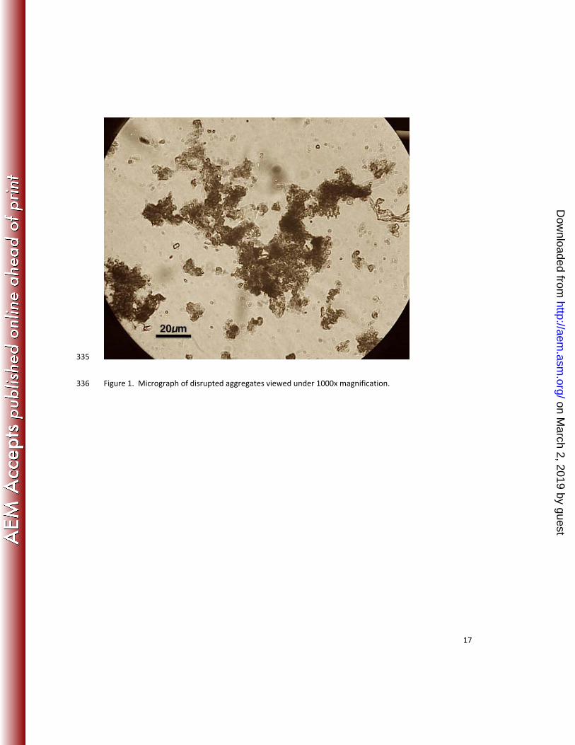
incorporation of the bacteria into these particulate conglomerations. Macroscopic aggregates 178
were always observed in the bottles placed onto the roller table, whereas the static control 179
bottles never formed such aggregates. Aggregates ranged in size from centimeter scale to 180
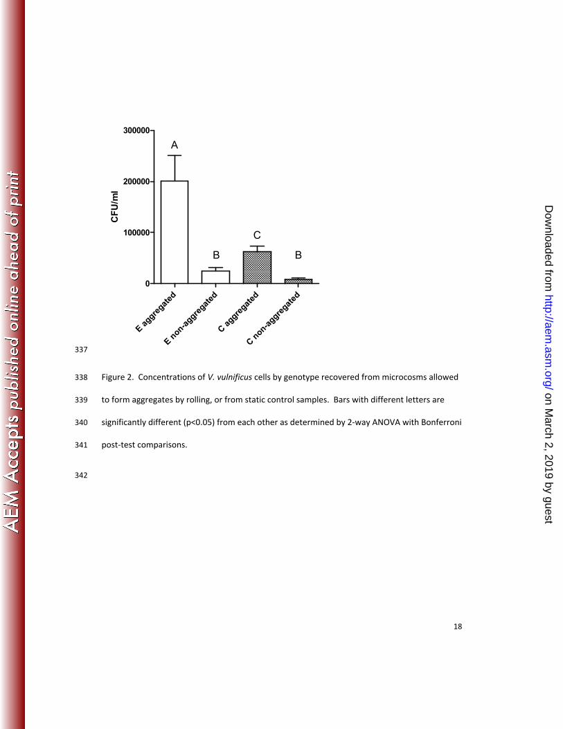
micrometer scale (Figure 1). The concentration of V. vulnificus cells was significantly greater in 181
the samples with aggregated marine snow than in the samples where aggregates did not form 182
(p<0.001), regardless of genotype (Figure 2). In the environment, V. vulnificus and other vibrios 183
have also been reported to be in higher concentrations in natural marine aggregates compared 184
to the surrounding water (35, 36). Marine aggregates are believed to increase bacterial survival 185
when moving between hosts (35). Furthermore, direct access to organic substrates and 186
on March 2, 2019 by guest
http://aem.asm
.org/D
ownloaded from

10
protection from chemical or physical stress can be gained by association with aggregates (36, 187
37). Thus, it is beneficial for the cells to concentrate in marine snow in a short time. 188
E-type cells showed significantly more integration into marine aggregates than C-type 189
cells (p<0.001) while the non-aggregated V. vulnificus concentrations between the two 190
genotypes was not statically different (p>0.05, Figure 2). The cause of the increased affinity for 191
the E-genotype strain for marine snow is not known, but because aggregate affinity and 192
assimilation involves cell-cell interactions, motility, chemokenetics, and exoenzyme production, 193
we assume that one of these or other factors are different between the two genotypes (35, 37, 194
38, 39). Recently, sequencing data revealed genomic differences between C- and E-genotypes 195
strains showing that E-type strains have genes linked to attachment proteins (40). A PKD 196
domain present in the E-types, and absent from the C-types, could allow for more effective 197
binding and hydrolysis of chitin, a component of marine aggregates (40, 41, 42). 198
For decades, experiments looking at the uptake of V. vulnificus by oysters have been 199
conducted by simply adding planktonic bacteria to tanks containing the oysters or, occasionally, 200
by adding the bacteria to algae and feeding the algae to oysters. Because oysters sort particles 201
based on size, the efficiency of planktonic bacterial uptake could be so low that results do not 202
mimic what would be expected to occur in situ. Thus the planktonic model, while useful, is 203
likely to be highly inefficient. Aggregates, with integrated V. vulnificus cells, were added to 204
oyster tanks, and the uptake and depuration of the cells was recorded (Figure 3). Oysters 205
sampled after one day of incubation with aggregated V. vulnificus treatments were found to 206
have significantly more E-genotype cells (Figure 3A) or more C-genotypes cells (Figure 3B) than 207
on March 2, 2019 by guest
http://aem.asm
.org/D
ownloaded from
11
the non-aggregated control treatments (p=0.025 and p=0.002, respectively). The aggregated C-208
type cell numbers were also significantly higher than control at 3 days after incubation (p=0.03) 209
but by day 6 were undetectable (Figure 3B). E-type cell numbers were not significantly 210
different in aggregated samples and controls (p>0.05, Figure 3A), and were very low in both. 211
When C- and E-genotype cells were added to aggregate bottles in competition, we 212
observed significantly greater (p<0.001) incorporation of the E-type strain, compared to the C-213
type strain, into the newly formed marine snow (Figure 4). Furthermore, when comparing the 214
uptake data presented in Figure 2 vs. Figure 4, it appears that the presence of the E-type strains 215
caused the C-type strain to incorporate into the snow less than if cultured individually. 216
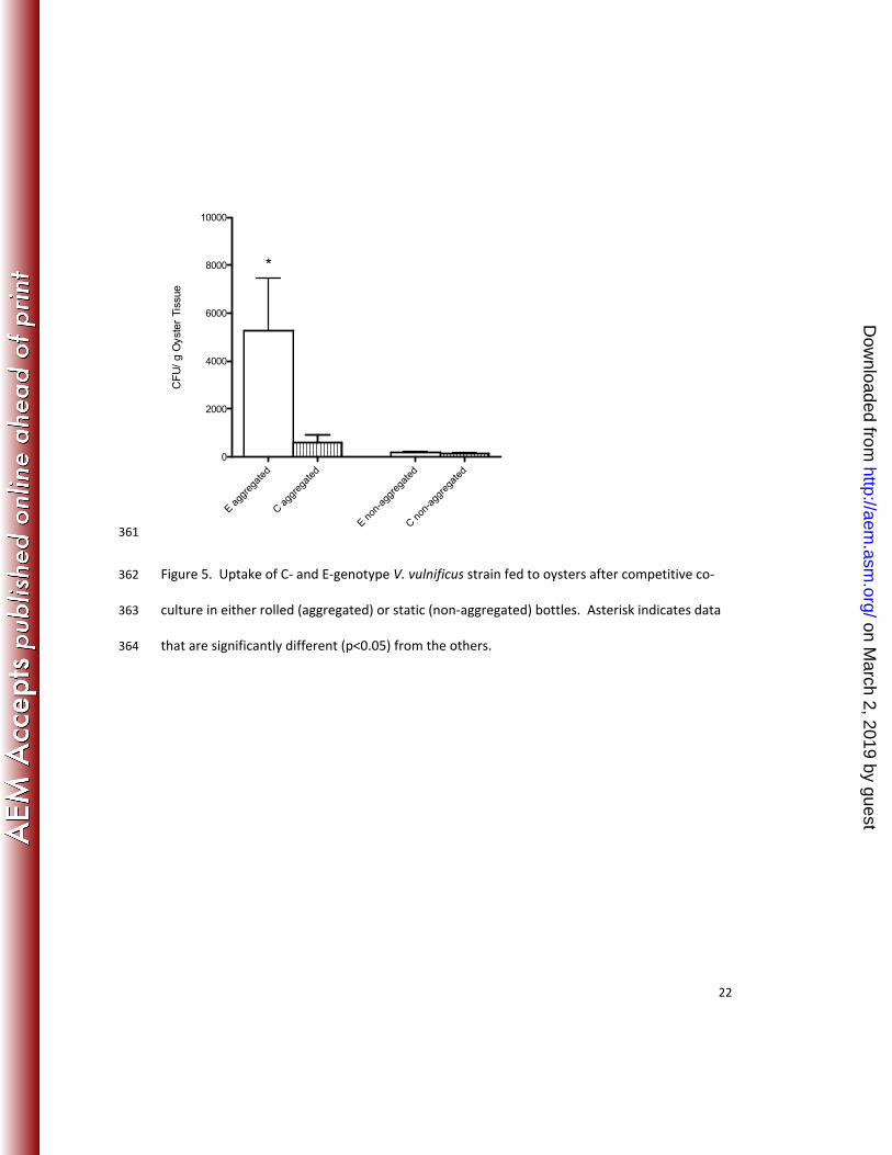
Aggregates generated with C- and E-type strains in competitive co-culture were subsequently 217
fed to oysters (Figure 5); we found that oysters exhibited significantly greater (p=0.029) uptake 218
of the E-type cells when compared to the C-type cells that were added at the same time. Non-219
aggregated controls showed no significant (p>0.05) differences in uptake between the two 220
genotypes in these competitive studies. Thus, it appears that in a mixed-genotype 221
environment, such as would be found in an estuarine system, E-genotype cells outcompete the 222
C-type cells for integration into marine aggregates. Moreover, while both genotypes of V. 223
vulnificus individually exhibit greater uptake by oysters when they are associated with these 224
aggregates, the E-types benefit from their advantage and can display increased uptake into 225
oysters compared to the C-genotype counterparts. This finding may explain why oysters are 226
consistently found to contain disproportionate ratios of E-genotype cells even when they are 227
not dominant in the surrounding water column. 228
on March 2, 2019 by guest
http://aem.asm
.org/D
ownloaded from

12
229
Acknowledgements 230
We would like to thank Melissa Jones for the creation of V. vulnificus strain pGTR JY1305 231
and pGTR-Env1. We would also like to thank Tiffany Williams for assistance with aquaria 232
maintenance and oyster collection. This material is based upon work supported by the 233
Cooperative State Research, Education, and Extension Service, U.S. Department of Agriculture, 234
under Award Nos. 2007-35201-18381. Any opinions, findings, conclusions, or 235
recommendations expressed in this publication are those of the authors and do not necessarily 236
reflect the views of the U.S. Department of Agriculture. 237
238
References 239
1. Johnston, J. M., S. F. Becker, and L. M. McFarland. 1985. Gastroenteritis in patients with stool 240
isolates of Vibrio vulnificus. Am J Med 80:336-338. 241
2. Oliver, J. D. 1989. Vibrio vulnificus, p. 569-599. In M. P. Doyle (ed.), Foodborne bacterial pathogens. . 242
Marcel Dekker, Inc., New York, NY. 243
3. Oliver, J. D. 2006. Vibrio vulnificus, p. 349-366. In F. L. Thompson, B. Austin, and J. Swings (ed.), The 244
Biology of Vibrios. American Society of Microbiology, Washington, DC. 245
4. Mead, P. S., L. Slusker, V. Dietz, L. F. McCaig, J. S. Bresee, C. Shapiro, P. M. Griffin, and R. B. V. 246
Tauxe. 1999. Food-related illness and death in the United States. Emerg Infect Dis 5:607-625. 247
5. Jones, M. K., and J. D. Oliver. 2009. Vibrio vulnificus: Disease and pathogenesis. Infect Immun 248
77:1723-1733. 249
on March 2, 2019 by guest
http://aem.asm
.org/D
ownloaded from

13
6. Oliver, J. D. 2005. Wound infections caused by Vibrio vulnificus and other marine bacteria. Epidemiol 250
Infect 133:383-391. 251
7. Warner, J. M., and J. D. Oliver. 1999. Randomly amplified polymorphic DNA analysis of clinical and 252
environmental isolates of Vibrio vulnificus and other Vibrio species. Appl Environ Microbiol 65:526-534. 253
8. Rosche, T., Y. Yano, and J. D. Oliver. 2005. A rapid and simple PCR analysis indicates there are two 254
subgroups of Vibrio vulnificus which correlate with clinical and environmental isolation. Microbiol 255
Immunol 49:381-389. 256
9. U.S. Food and Drug Administration. 2009. Vibrio vulnificus Heath Education Kit. In U.S. Department 257
of Health and Human Services, Silver Spring, MD. 258
10. Oliver, J. D. 2005. Vibrio vulnificus, p. 253-276. In S. Belkin and R. R. Colwell (ed.), Oceans and 259
Health: Pathogens in the marine environment. Springer, New York. 260
11. National Center for Emerging and Zoonotic Infectious Diseases. 2009. Vibrio vulnificus. In Centers 261
for Disease Control and Prevention, Atlanta, GA. 262
12. Froelich, B., and J. Oliver. 2011. Orientation of mannitol related genes can further differentiate 263
strains of Vibrio vulnificus possessing the vcgC allele. Adv Stud Biol 3:151-160. 264
13. Warner, E. B., and J. D. Oliver. 2007. Population Structures of Two Genotypes of Vibrio vulnificus in 265
Oysters (Crassostrea virginica) and Seawater. Appl Environ Microbiol 74:80-85. 266
14. Froelich, B., and J. D. Oliver. 2012. The interactions of Vibrio vulnificus with the oyster, Crassostrea 267
virginica. Micro Ecol (In press). 268
15. Han, F., S. Pu, A. Hou, and B. Ge. 2009. Characterization of clinical and environmental types of 269
Vibrio vulnificus isolates from Louisiana oysters. Foodborne Pathog Dis 6:1251-1258. 270
16. Staley, C., M. K. Jones, A. C. Wright, and V. J. Harwood. 2011. Genetic and quantitative assessment 271
of Vibrio vulnificus populations in oyster (Crassostrea virginica) tissues. Environ Microbiol Rep 3:543-272
549. 273
on March 2, 2019 by guest
http://aem.asm
.org/D
ownloaded from

14
17. Nilsson, W. B., R. N. Paranjype, A. DePaola, and M. S. Strom. 2003. Sequence polymorphism of the 274
16S rRNA gene of Vibrio vulnificus is a possible indicator of strain virulence. J Clin Microbiol 41:442-446. 275
18. Vickery, M., W. Nilsson, M. Strom, J. Nordstrom, and A. DePaola. 2007. A real-time PCR assay for 276
the rapid determination of 16 S rRNA genotype in Vibrio vulnificus. J Microbiol Methods 68:376 - 384. 277
19. Krantz, J. A., J. L. Nordstrom, J. C. Bowers, and A. Depaola. 2008. An evaluation of CPC+, a new 278
medium for isolation and enumeration of Vibrio vulnificus from U.S. market oysters, 108th Gen. Meet. 279
Am. Soc. Micriobiol., Boston, MA. 280
20. Newell, R. I. E., and C. J. Langdon. 1996. Mechanisms and physiology of larval and adult feeding, p. 281
185-229. In V. S. Kennedy, R. I. E. Newell, and A. F. Eble (ed.), The eastern oyster: Crassostrea virginica. 282
Maryland Sea Grant College, College Park. 283
21. Froelich, B., A. Ringwood, I. Sokolova, and J. Oliver. 2010. Uptake and depuration of the C-and E-284
genotypes of Vibrio vulnificus by the Eastern Oyster (Crassostrea virginica). Environ Microbiol Rep 2:112-285
115. 286
22. Ward, J. E., and S. E. Shumway. 2004. Separating the grain from the chaff: particle selection in 287
suspension- and deposit-feeeing bivalves. J Exp Mar Biol Ecol 300:83-130. 288
23. Alldredge, A. L., and M. W. Silver. 1988. Characteristics, dynamics and significance of marine snow. 289
Prog Oceanog 20:41-82. 290
24. Suzuki, N., and K. Kato. 1953. Studies on suspended materials. Marine snow in the sea. I. Sources 291
of marine snow. Bulletin of the Faculty of Fisheries of Hokkaido University 4. 292
25. Carson, R. 1951. The Sea Around Us. Oxford University Press. 293
26. Morris, J. G., A. C. Wright, D. M. Roberts, P. K. Wood, L. M. Simpson, and J. D. Oliver. 1987. 294
Identification of environmental Vibrio vulnificus isolates with a DNA probe for the cytotoxin-hemolysin 295
gene. Appl Environ Microbiol 53:193-195. 296
on March 2, 2019 by guest
http://aem.asm
.org/D
ownloaded from

15
27. Murphy, S. K., and J. D. Oliver. 1992. Effects of temperature abuse on survival of Vibrio vulnificus in 297
oysters. Appl Environ Microbiol 58:2771-2775. 298
28. Wright, A. C., L. M. Simpson, J. D. Oliver, and J. G. Morris. 1990. Phenotypic evaluation of acapsular 299
transposon mutants of Vibrio vulnificus. Infect Immun 58:1769-1773. 300
29. Ochi, K., D. Zhang, S. Kawamoto, and A. Hesketh. 1997. Molecular and functional analysis of the 301
ribosomal L11 and S12 protein genes (rplK and rpsL) of Streptomyces coelicolor A3 (2). Mol Gen Genet 302
256:488-498. 303
30. Miescier, J. J., D. A. Hunt, J. Redman, A. Salinger, and J. P. Lucas. 1992. Molluscan shellfish: oysters, 304
mussels, and clams, p. 901-907. In D. F. Splittstoesser (ed.), Compendium of methods for microbiological 305
examination of food, vol. 3rd edition. American Public Health Association, Inc., Washington, DC. 306
31. James, D. E. 1978. Culturing Algae. Carolina Biological Supply Company. 307
32. Ward, J. E., and D. J. Kach. 2009. Marine aggregates facilitate ingestion of nanoparticles by 308
suspension-feeding bivalves. Mar Environ Res 68:137-142. 309
33. Shanks, A. L., and E. W. Edmondson. 1989. Laboratory-made artifical marine snow: a biological 310
model of the real thing. Mar Biol 101. 311
34. Sokal, R. R., and F. J. Rohlf. 1995. Biometry: the principles and practice of statistics in biological 312
research. WH Freeman, San Francisco, CA. 313
35. Lyons, M. M., Y.-T. Lau, W. E. Carden, J. E. Ward, S. B. Roberts, R. Smolowitz, J. Vallino, and B. 314
Allam. 2007. Characteristics of Marine Aggregates in Shallow-water Ecosystems: Implications for Disease 315
Ecology. EcoHealth 4:406-420. 316
36. Lyons, M. M., J. E. Ward, H. Gaff, R. E. Hicks, J. M. Drake, and F. C. Dobbs. 2010. Theory of island 317
biogeography on a microscopic scale: organic aggregates as islands for aquatic pathogens. Aquat Microb 318
Ecol 60:1-13. 319
on March 2, 2019 by guest
http://aem.asm
.org/D
ownloaded from

16
37. Tang, K. W., C. Dziallas, and H. P. Grossart. 2011. Zooplankton and aggregates as refuge for aquatic 320
bacteria: protection from UV, heat and ozone stresses used for water treatment. Environ Microbiol 321
13:378-90. 322
38. Kiorboe, T., and G. A. Jackson. 2001. Marine Snow, Organic Solute Plumes, and Optimal 323
Chemosensory Behavior of Bacteria. Limnol Oceanogr 46:1309-1318. 324
39. DeLong, E. F., D. G. Franks, and A. L. Alldredge. 1993. Phylogenetic Diversity of Aggregate-Attached 325
vs. Free-Living Marine Bacterial Assemblages. Limnol Oceanogr 38:924-934. 326
40. Morrison, S. S., T. Williams, A. Cain, B. Froelich, C. Taylor, C. Baker-Austin, D. Verner-Jeffreys, R. 327
Hartnell, J. D. Oliver, and C. J. Gibas. 2012. Pyrosequencing-Based Comparative Genome Analysis of 328
Vibrio vulnificus Environmental Isolates. PLoS ONE 7. 329
41. Smith, D. C., M. Simon, A. L. Alldredge, and F. Azam. 1992. Intense hydrolytic enzyme activity on 330
marine aggregates and implications for rapid particle dissolution. Nature 359:139-142. 331
42. Yatsunami, R. 2004. Enzymatic Syntheses of Novel Oligosaccharides Using Haloarchaeal 332
Glycosidases. NISR Research Grant. 333
334
on March 2, 2019 by guest
http://aem.asm
.org/D
ownloaded from
17
335
Figure 1. Micrograph of disrupted aggregates viewed under 1000x magnification. 336
on March 2, 2019 by guest
http://aem.asm
.org/D
ownloaded from
18
E aggre
gated
E non-a
ggregat
ed
C aggre
gated
C non-a
ggregat
ed
0
100000
200000
300000
A
B
C
B
CF
U/m
l
337
Figure 2. Concentrations of V. vulnificus cells by genotype recovered from microcosms allowed 338
to form aggregates by rolling, or from static control samples. Bars with different letters are 339
significantly different (p<0.05) from each other as determined by 2-way ANOVA with Bonferroni 340
post-test comparisons. 341
342
on March 2, 2019 by guest
http://aem.asm
.org/D
ownloaded from

19
0 2 4 60
1
2
3 *
Time in Days
Lo
g c
fu/g
oys
ter
tiss
ue
343
Figure 3A. Uptake and depuration of aggregated (circles) or non-aggregated (squares) E-344
genotype cells in oysters. Asterisk indicates time point at which experimental group was 345
significantly different (p<0.05) from control. Cells were added immediately after recording the 346
zero time point. 347
348
349
on March 2, 2019 by guest
http://aem.asm
.org/D
ownloaded from

20
0 2 4 60
1
2
3 *
*
Time in Days
Lo
g c
fu/g
oys
ter
tiss
ue
350
Figure 3B. Uptake and depuration of aggregated (circles) or non-aggregated (squares) C-351
genotype cells in oysters. Asterisks indicate time points at which experimental group was 352
significantly different (p<0.05) from control. Cells were added immediately after recording the 353
zero time point. 354
355
356
on March 2, 2019 by guest
http://aem.asm
.org/D
ownloaded from

21
E- agg
rega
ted
C agg
rega
ted
0200400600800
1000
80000
100000
120000
140000
*
CF
U/m
l
357
Figure 4. The incorporation of E- and C-type V. vulnificus into marine aggregates when 358
incubated in competitive co-culture. Asterisk indicates significantly different (p<0.05) values. 359
360 on March 2, 2019 by guest
http://aem.asm
.org/D
ownloaded from
22
E agg
rega
ted
C agg
rega
ted
E non
-agg
rega
ted
C non
-agg
rega
ted
0
2000
4000
6000
8000
10000
*
CF
U/
g O
yste
r Ti
ssue
361
Figure 5. Uptake of C- and E-genotype V. vulnificus strain fed to oysters after competitive co-362
culture in either rolled (aggregated) or static (non-aggregated) bottles. Asterisk indicates data 363
that are significantly different (p<0.05) from the others. 364
on March 2, 2019 by guest
http://aem.asm
.org/D
ownloaded from


