
UvA-DARE is a service provided by the library of the University of Amsterdam (http://dare.uva.nl)
UvA-DARE (Digital Academic Repository)
Morphology and mechanical properties of the cancellous bone in the mandibularcondyleGiesen, E.B.W.
Link to publication
Citation for published version (APA):Giesen, E. B. W. (2002). Morphology and mechanical properties of the cancellous bone in the mandibularcondyle
General rightsIt is not permitted to download or to forward/distribute the text or part of it without the consent of the author(s) and/or copyright holder(s),other than for strictly personal, individual use, unless the work is under an open content license (like Creative Commons).
Disclaimer/Complaints regulationsIf you believe that digital publication of certain material infringes any of your rights or (privacy) interests, please let the Library know, statingyour reasons. In case of a legitimate complaint, the Library will make the material inaccessible and/or remove it from the website. Please Askthe Library: http://uba.uva.nl/en/contact, or a letter to: Library of the University of Amsterdam, Secretariat, Singel 425, 1012 WP Amsterdam,The Netherlands. You will be contacted as soon as possible.
Download date: 31 Aug 2018

Morphologyy and M off the Cancellous^ inn the Mandibul
Eric c sen n

Morphologyy and Mechanical Properties of the
'MMJVÊu 'MMJVÊu

c_ _

Morphologyy and Mechanical Properties of the Cancellouss Bone in the Mandibula r Condyle
ACADEMISCHH PROEFSCHRIFT
terr verkrijging van de graad van doctor
aann de Universiteit van Amsterdam
opp gezag van de Rector Magnificus
prof.mr.. P.F. van der Heijden
tenn overstaan van een door het college voor promoties ingestelde
commissie,, in het openbaar te verdedigen in de Aula der Universiteit
opp vrijdag 5 juli 2002, te 10.00 uur
doorr Eric Bernardus Wilhelmus Giesen
geborenn te Doetinchem

P r o m o t i e c o m m i s s ie e
Promotor: Promotor:
prof.dr.. T .M.G.J. van Eijden
OverigeOverige leden:
prof.dr.. W. Beertsen
prof.dr.. A.J. Feilzer
prof.dr. i r.. R. Huiskes
dr.. J. Klein-Nulend
prof.dr.. P.F. van der Stelt
prof.dr.. W.A. Weijs
Faculteitt Tandheelkunde
Thee PhD-program was attended at the Inter-university Research School of
Dentistryy (ÏOT). The study described in this thesis was performed at the
Functionall Anatomy section of the Academic Centre for Dentistry
Amsterdam. .
4 4

Contents s
Chapterr 1 Introduction 7
Chapterr 2 The Three-Dimensional Cancellous Bone Architecture of the 19
Humann Mandibular Condyle
Chapterr 3 Prediction of Strains in the Human Mandibular Condyle 35
Chapterr 4 Mechanical Properties of Cancellous Bone in the Human 47
Mandibularr Condyle are Anisotropic
Chapterr 5 Morphology of the Cancellous Bone of the Mandibular Condyle 61
andd its Predictive Value for the Mechanical Properties
Chapterr 6 Reduced Mechanical Load Decreases the Density, Stiffness, and 75
Strengthh of Cancellous Bone of the Mandibular Condyle
Chapterr 7 Changed Morphology and Mechanical Properties of Cancellous 85
Bonee in the Mandibular Condyle of Edentate People
Chapterr 8 Summary and Conclusions 95
Samenvattingg en Conclusies 100
Dankwoordd 103
Curriculumm vitae 104

ThisThis thesis is based on thejolloiving publications:
Giesen EBW, Van Eijden TMGJ (2000). The three-dimensional cancellous
bonee architecture of the human mandibular condyle. J Dent Res 79:957-963.
(Chapterr 2)
Van Ruijven LJ, Giesen EBW, Van Eijden TMGJ (2002). Prediction of strains
inn the human mandibular condyle. Submitted. (Chapter 3)
Giesen EBW, Ding M, Dalstra M, Van Eijden TMGJ (2001). Mechanical
propertiess of cancellous bone in the human mandibular condyle are
anisotropic.. J Biomech 34:799-803. (Chapter4)
Giesen EBW, Ding M, Dalstra M, Van Eijden TMGJ (2002). Morphology of
thee cancellous bone of the mandibular condyle and its predictive value for the
mechanicall properties. Submitted. (Chapter 5)
Giesen EBW, Ding M, Dalstra M, Van Eijden TMGJ (2002). Reduced
mechanicall load decreases the density, stiffness, and strength of cancellous
bonee of the mandibular condyle. Submitted. (Chapter 6)
Giesen EBW, Ding M, Dalstra M, Van Eijden TMGJ (2002). Changed
morphologvv and mechanical properties of cancellous bone in the mandibular
condylee of edentate people. To be submitted. (Chapter 7)
6 6

te&ï te&ï
Introductio n n

ChapterChapter I
Introduct io n n
Thee principal function of the bony skeleton is to provide structural support for the
body.. It is the basis of posture, opposes muscular contraction resulting in mot ion,
withstandss forces, and protects the internal organs. The skeleton also serves as the
body'ss mineral reservoir. Generally, two basic types of bone can be distinguished in
thee body: cortical bone and cancellous (or trabecular) bone. Cortical bone is
compactt and forms the shaft of long bones and the shell of, for instance, the
vertebrall bodies and the bones of the skull. Cancellous bone has an open structure
formedd by trabeculae with bone marrow to fil l the spaces. Cancellous bone is found
underr articulating surfaces at the end of long bones and within flat and irregular
bones.. Although the material propert ies of cortical and cancellous bone tissue have
thee same order of magnitude (Rho et al, 1993; Rho et ah, 1999), due to differences
inn the amount and spatial distribution of the bone material, the apparent mechanical
propert iess of cortical and cancellous bone are different. For instance, the stiffness of
cancellouss bone is 10 to 20 times less than that of cortical bone. The differences in
mechanicall propert ies presume that the types of bone have different functions. The
lesss stiff cancellous bone is likely to absorb forces and guide and transfer these to the
sufferr cortical bone, which supplies the support.
Cancellouss bone is built from interconnected trabeculae (Fig. 1). The
trabeculaee arc shaped like plates and rods and together they form a structure that can
resistt loads. The structure is anisotropic, i.e., the trabeculae are typically or iented,
Fig.. 1 An example of a cancellous bone st ructure. Plate-like trabeculae are
interconnectedd by rods. The specimen ( length ~ -> mm) was scanned in a
micro-computedd tomography system. The open spaces were originally filled
wit hh mar row, which was virtually removed in processing the three-
dimensionall reconstruct ion.
8 8

Introduction Introduction
suchh that it has different mechanical properties in different directions. The hone is
alsoo inhomogeneous, i.e., large differences exist in the spatial arrangement of
trabeculaee for different anatomical sites. Even within an anatomical site, for example
aa vertebral body, the architecture of the trabecular structure varies locally (Smit et
al.,al., 1997). The architecture of the trabecular structure is determinant for the bone's
reactionn on compression, tension and shear. Its mechanical behavior depends on
severall architectural characteristics: the mass of the bone, the quality ol the
trabeculae,, and the three-dimensional orientation and structure of the trabeculae.
Consequently,, this behavior is likely to vary considerably throughout the skeleton.
Sincee the nineteenth century, a close relationship between the function of bone and
itss structure has been established (Wolff, 1892). It was found that trabeculae, for
instancee in the bone of the upper leg, have an orientation that is closely in line with
thee stress patterns within the bone. Thus, the trabeculae arc oriented such that they
cann optimally withstand the forces during functioning. Bone is also sensitive to the
amountt of mechanical loading. For instance, patients who have to keep bed rest and
astronautss who lack gravity lose bone weight, whereas athletes gain bone weight.
Thiss process of adaptation is called adaptive bone remodeling. It is autonomically
regulatedd in the bone itself. Recently, computer models were able to simulate the
adaptivee remodeling process and to predict the new bone architecture as a result of
changedd mechanical loading (Mullender at a/., 1998; Huiskes, 2000; Adachi et al.,
2001). .
Thee adaptation has the consequence that the mechanical properties of the
adaptedd bone fit the forces in the mechanical environment. Adaptation thus links
usa^c,, trabecular architecture and mechanical properties. It implies that in a
stationaryy situation bonv architecture reflects the mechanical loading of the bone
(Odgaardd et al., 1997; Herring and Liu, 2001). This enables to estimate the
mechanicall environment by studying the morphology of the bone. These
mechanismss do probably also apply to the human mandible (or lower jaw).
Thiss thesis addresses the struct ure-function relationships of the cancellous bone in
thee human mandible. During mastication and biting, the mandible is subjected to
forcess produced by the muscles of mastication and by reaction forces applied to the
jaww joints and the teeth. As a result of this loading, deformations and tensions are
producedd in the mandible. The range and distribution of these deformations and
tensionss depend on the nature of the loading and on the material properties and
amountt and distribution of bone. An understanding of the structure-function
9 9

ChapterChapter 1
relationshipss of the mandibular bone is important for several reasons. It may give us
insightt into the way this structure is optimized to resist loading. In addition, clinical
situations,, e.g., tooth loss, orthodontic treatment, dental implants, or reconstructive
surgery,, will alter the loading of the mandible. This, in turn, might affect the
architecturee of the mandibular bone.
Inn the mandible, cancellous bone is found in various areas, for instance in the
alveolarr process to support teeth, in-between the cortical sheets of the ramus and
mandibularr body, and in the condyle (or mandibular head). This thesis will focus on
thee cancellous bone in the mandibular condyle (Fig. 2).
Mo rpho logy y
Basedd on frontal, transversal, and sagittal sections, an inhomogeneous bone
distributionn has been reported in the human mandibular condyle (Hongo et a/.,
1989).. The density of the bone and the amount of trabcculae are larger in the upper
andd anterior regions. The trabeculae are oriented in the anteroposterior direction,
whichh has been functionally related to the direction of one of the masticatory
Fig.. 2 Ot a lower jaw the right condyle (or head) is enlarged. The condyle is approximatelyy 2 cm wide. The thin cortical shell is removed that allows us to lookk at. the cancellous bone inside the condyle.
10 0

Introduction Introduction
muscles,, the lateral pterygoid muscle, which attaches closely to the condyle. In the
pigg mandibular condyle, however, the latter was not confirmed (Teng and Herring,
1995).. Due to the two-dimensional nature of these studies, the findings were limited
too the characteristics occurring within the analyzed planes. While the loading of the
condylee is not limited to a plane only, this approach is not sufficient to establish the
relationshipp between architecture and loading. In order to assess this relationship
adequately,, a three-dimensional analysis of the condylar bone architecture is a
prerequisitee (for review: Odgaard, 1997).
Statee of the art micro-computed tomography (micro-CT) systems enable us to
studyy the cancellous bone in its three dimensions (Rüegsegger et al, 1996). Micro-
CTT assesses the complete three-dimensional bone structure at the micro level
(resolution:: 8 - 50 pm), in a non-destructive fashion. Depending on the system and
thee required resolution a complete mandibular condyle can be imaged at once.
Parameterss describing, for instance, the bone volume fraction, the thickness and
numberr of trabcculae, the space between trabeculae and the predominant orientation
off the trabeculae can be computed with standard system software (for review:
Odgaard,, 1997).
Inn this thesis, the micro-CT technique was applied to determine the three-
dimensionall architecture of the cancellous bone of the human mandibular condyle
(Chapterr 2). More specifically, an inhomogeneous and anisotropic distribution of
cancellouss bone was expected. It was applied to elucidate a possible relationship
betweenn the cancellous bone structure of the condyle and its function.
Duringg masticatory functioning the mandibular condyle translates and rotates along
thee articular surface of the temporal bone of the skull. The shape of the articular
surfacee and that of the condyle do not match, which makes the joint incongruent.
Duringg movement the condyle is subjected to complex loading patterns. The
temporomandibularr joint disc, which is situated between the skull and mandible,
playss an important role in the distribution of the loads (Beek, 2001). These loads are
localizedd in an area on the condyle, which is shaped as a mediolaterally oriented
band.. During function the loaded area moves anteroposteriorly. The joint forces are
absorbedd by the cancellous bone and are transferred toward the mandibular collum.
Duringg loading the condyle, and herewith its cancellous bone, deforms, giving rise
too a pattern of internal tensions. The nature of these deformations and tensions,
however,, is unknown. As thev cannot be measured directly, a model approach is
required. .
11 1

ChapterChapter J
Too estimate the deformations and tensions in the mandibular condyle a three-
dimensionall finite element model of the mandibular condyle was developed (Chapter
3).. Different static anteroposterior load cases were applied and the magnitude and
orientationn of the local deformations in the condyle were calculated. Furthermore,
thee deformation of the whole condyle due to the loading was analyzed. By comparing
thee direction of the deformations with the orientation of the trabecular bone, it
couldd be verified if the architecture of the bone of the condvie reflects its mechanical
loading. .
Mechan icall p roper t i es
Thee mechanical properties of cancellous bone are of interest for the bone's load
bearingg capacities. Different mechanical properties can be distinguished. It is
noteworthyy to distinguish between the mechanical behavior at the level of the
structuree of the bone, the apparent properties, and that at the level of the individual
trabeculae,, the tissue properties. The apparent properties are referred to in this
thesis.. The following are used: stiffness, strength, failure energy and the ultimate
strain.. The stiffness is a measure of the ability of bone to resist deformation in the
directionn of the applied load. The strength or ultimate stress is the amount of stress
thee bone can maximally sustain. Any surplus of stress wil l break the bone. The strain
att that point is called the ultimate strain. The energy that is needed to break the bone
iss called the failure energy.
Mechanicall properties can be determined by mechanical testing (Carter and
Hayes,, 1977; lor review; Kcaveny et a/., 2001). Generally, a specimen is deformed
att a constant speed in a materials testing machine, and simultaneously the force is
recorded.. From the force-deformation recordings, the above-described mechanical
propertiess can be determined. This method has the disadvantage that the behavior at
thee interlace between the specimen and the machine may induce substantial errors
(Keavenyy eta!., 1997).
Mechanicall properties of cancellous bone strongly depend on the density of
thee bone (Carter and Hayes, 1977). Different density parameters can be
distinguished.. Density at the trabecular level is called the tissue density. The density
onn the structure level is called the bulk density or apparent density.
Onlyy few studies are available on mechanical properties of the bone of the
mandible.. The only study in which the cancellous bone of the mandibular condvie
wass involved concerned the pig (Teng and Herring, 1996). In the human mandible,
determinationn of mechanical properties has been limited to the cortical bone (e.g.,
\2 \2

Introduction Introduction
Arendtss and Sigolotto, 1989; Dechow et a/., 1993; Zioupos and Currey, 1998; for
review:: Van Eijden, 2000), or to the cancellous bone of its body (Misch et a}.,
1999).. The mechanical properties of the cancellous bone of the human mandibular
condylee were determined in the study described in Chapter 4, The mechanical
propertiess were related to the density of the bone. As it was expected that due to the
anisotropicc nature of the bone, the stiffness and strength would differ between
differentt loading directions, these properties were determined in different
directions. .
Relationn between 3-D morphology and mechanical propert ies
Thee mechanical properties of cancellous bone depend not only on bone density, but
alsoo on the three-dimensional morphology of bone tissue (Hodgskinson and Currey,
1990;; Uchiyama et al., 1999; Ulrich et al., 1999; Nafei et al., 2000a,b; Borah et a/.,
2000;; Ikcda et al., 2001). By establishing the relationship between morphology and
mechanicall properties, predictions can be made of the mechanical quality of the bone
fromm its architecture. This may relate to healthy bone, but possibly also to situations
likee osteoporosis and other pathologic circumstances like the edentulous jaw.
Generally,, the aim in relating mechanical properties and morphology is to
ascribee the variation in mechanical properties to the variation in morphology. In
orderr to assess the relationship between morphological parameters and mechanical
properties,, mostly these parameters arc entered into regression analyses one by one.
Onee of the problems is that various morphological variables are mutually dependent.
Forr example, an increase in bone mass can be achieved by an increase of the amount
off the trabeculae, a thickening of the trabeculae or, alternatively, by a transition
fromm rod-like trabeculae to more plate-like trabeculae, or a combination of these.
Thuss if some parameters represent the same kind of property it is likely they can
explainn the same amount of variance in mechanical properties individually, but
togetherr thev wil l not add more. Therefore, it can be expected that the additional
explanatoryy value of a large number of variables is rather limited.
Onee of the objectives of the studv described in Chapter 5 was to determine
whichh of the morphological variables of cancellous bone could be considered as
covariates.. For this purpose, a principal components analysis was used. The other
objectivee of this chapter was to investigate the predictive value of the micro-
structurall parameters for the mechanical properties of the bone in the mandibular
condyle.. The effect of trabecular orientation on the mechanical properties was
emphasized.. Usually, in the determination of mechanical properties of cancellous
13 3

ChapterChapter 1
bone,, specimens are taken relative to an anatomical axis or a presumed physiological
loadingg direction. It is then assumed that the main trabecular orientation coincides
thatt direction. The orientation itself, however, is seldom measured. If the principal
trabecularr orientation misaligns the loading direction with for example 10 degrees,
errorss of 9.5% in the estimation of the clastic modulus can be expected (Turner and
Cowin,, 1988).
Adaptatio n n
Thee continuous adaptation of bone to its mechanical environment has consequences
whenn the loading on the bone changes. In the masticatory system the amount of
mechanicall load is changed as people grow older or lose their teeth. The masticatorv
functionn decreases (Boretti et a!., 1995), which coincides with an atrophy of the
masticatoryy muscles (Newton et al, 1993) and a reduction in bite force (Helkimo et
aL,aL, 1977). Tlii s leads to a reduction in the forces acting on the mandible. It can,
therefore,, be expected that the structure and mechanical properties of the
mandibularr bone change with a decreased masticatory function.
Inn Chapter 6 the density and mechanical properties of condylar cancellous
bonee ol dentate and edentate subjects were compared. The loss of teeth in the
edentatee group was used as a model of reduced mechanical loading. It was
hypothesizedd that the density, stiffness, and strength of the cancellous bone would be
lowerr in the edentate group than in the dentate group. Furthermore, it was
investigatedd whether the mechanical anisotropv differed between the two groups.
Forr the mandibular condyle, it is not known bv which structural changes a
decreasee in bone density is accompanied. In the human tibia, for example, such a
decreasee is accompanied by a change of the bone structure type toward more rod-
likee trabeculae (Ding and Hvid, 2000) and by an increase in the anisotropv (Ding et
aL,aL, 2002). Also in patients with hip fractures, a lower bone density is accompanied
byy a higher degree of anisotropy (Ciarelli et a/., 2000). In Chapter 7 it was,
therefore,, questioned if the morphology of the cancellous bone of the mandibular
condylee of edentate subjects changes in a similar fashion. Further, it was investigated
iff a difference exists in the relationship between the morphology and mechanical
propertiess of the bone of dentate and edentate subjects.
14 4

introduction introduction
S u m m a ryy of o b j e c t i v es for t h e thes is
Describe the three-dimensional architecture of the cancellous bone of the
mandibularr condyle (Chapter 2).
Determine the deformations occurring in the mandibular condyle due to static
loadss and compare the direction of these deformations with the orientation of
thee trabecular bone (Chapter 3).
Determine the mechanical propert ies of the cancellous bone of the mandibular
condylee and relate these to the density of the bone (Chapter 4).
Determine morphological covariates of condylar bone and determine the
relationshipp between groups of morphological measures and the mechanical
propert iess (Chapter 5).
Determine the effect of reduced mechanical load on the cancellous bone of the
mandibularr condyle on both mechanical propert ies and morphology (Chapter
66 and 7).
R e f e r e n c es s
Adachii T, Tsubota K, Tomita Y, Hollister SJ (2001). Trabecular surface remodeling
simulationn for cancellous bone using; microstructural voxel finite element models. J
BiomechEngBiomechEng 123:403-9. Arendtss FJ, Sigolotto C (1998). Standardabmcssungen, Elasti/itatskennwertc und
Fcstigkeitsverhaltcnn des Human-Unterkiefcrs, cin Beitrag zur Darstellung der Biomechanikk dcr Untcrkiefer - Teil I. Biomed Tech 34:248-255.
Beekk M (2001). Biomechanica! modeling of the human jaw joint. PhD thesis, University of
Amsterdam,, Netherlands.
Borahh B, Dufresne TE, Cockman MD, Gross GJ, Sod EW, Myers WR, Combs KS, Higgins
RE,, Pierce SA, Stevens ML (2000). Evaluation of changes in trabecular bone architecture
andd mechanical properties of minipig vertebrae by three-dimensional magnetic resonance
microimagingg and finite element modeling. J Bone Miner Res 15:1 786-1797.
Borettii G, Bickel M, Gecrin AH (1995). A review of masticatory abilitv and efficiency. J
ProsthetDemProsthetDem 74:400-403."
Carterr DR, Haves WC (1977). The compressive behavior of bone as a two-phase porous
structure,, J Bone Joint Surg Am 59-A:954-962.
Ciarcllii TE, Fvhrie DP, Schafiler MB, Goldstein SA (2000). Variations in three-dimensional
cancellouss bone architecture of the proximal femur in female hip fractures and in
controls,, j Bone Miner Res 1 5:32-40,
Dechoww PC, Nail GA, SchwarU-Dabney CL, Ashman RB (1993). Elastic properties of
humann supraorbital and mandibular bone. Am J Phys Anthropol 90:291-306.
Dint;; M, Hvid 1 (2000). Quantification of age-related changes in the structure model type
andd trabecular thickness of human tibial cancellous bone. Bone 26:291-295.
15 5

ChapterChapter /
Dingg M, Odgaard A, Lindc F, Hvid I (2002). Age-related variations in the microstructure of
humann tibial cancellous bone.y Orthop Res (in press).
Helkimoo E, Carlsson GE, Hclkimo M (1977). Bite force and state of dentition. Acta Odontoi
ScandScand 35:297-303.
Herringg SW, Liu ZJ (2001). Loading of the temporomandibular joint: anatomical and in
vivoo evidence from the bones. Cells Tissues Organs 169:193-200.
Hodgskinsonn R, Currey JD (1990). Effects of structural variation in Young's modulus of
non-humann cancellous bone. Proc hst Mech Eng [H] 204:43-52.
Hongoo T, Yotsuya H, Shibuya K, Kavvase M, Ide Y (1989). Quantitative and morphological
studiess on the trabecular bones in the condyloid processes of the Japanese mandibles.
Comparisonn between dentulous and edentulous specimens. Bull Tokyo Dent Coll 30:67-
76. .
Huiskess R (2000). If bone is the answer, then what is the question? j Anat 197(Pt 2):I45-
156. .
Ikcdaa S, Tsurukami H, Ito M, Sakai A, Sakata T, Nishida S, Takeda S, Shiraishi A, Nakamura
TT (2001). Effect of trabecular bone contour on ultimate strength oflumbar vertebra after
bilaterall ovariectomv in rats. Bone 28:625-633.
Keavenyy TM, Morgan EF, Niebur GL, Ych OC (2001). Biomechanics of trabecular bone.
AnnAnn Rev Biomed Eng 3:307-333.
Keavenyy TM, Pinilla TP, Crawford RP, Kopperdahl DL, Lou A (1997). Systematic and
randomm errors in compression testing of trabecular bone. J Orthop Res 1 5:101-1 10.
Mischh CE, Qu Z, Bidez MW (1999). Mechanical properties of trabecular bone in the human
mandible:: Implications for dental implant treatment planning and surgical placement. J
OralOral Maxillofacial Surg 57:700-706.
Mullenderr M, Van Rietbergen B, Riiegsegger P, Huiskes R (1998). Effect of mechanical set
pointt of bone cells on mechanical control of trabecular bone architecture. Bone 22:125-
131. .
Nafeii A, Danielsen CC, Lindc F, Hvid I (2000a). Properties of growing trabecular ovine
bone.. Part 1: Mechanical and physical properties. J Bone Joint Surg Br 82-B:910-920.
Nafeii A, Kabel J, Odgaard A, Linde F, Hvid I (2000b). Properties of growing trabecular
ovinee bone. Part 2: Architectural and mechanical properties. J Bone Joint Surg Br 82-
B:921-927. .
Newtonn JP, Yemm R, Abel RW, Mcnhinick S (1993). Changes in human jaw muscles with
agee and dental state. Cerodontology 10:16-22.
Odgaardd A (1997). Three-dimensional methods for quantification of cancellous bone
architecture.. Bone 20:31 5-328.
Odgaardd A, Kabel J, Van Rietbergen B, Dalstra M, Huiskes R (1997). Fabric and elastic
principall directions of cancellous bone are closely related. J Biomech 30:487-495.
Rhoo JY, Ashman RB, Turner CH (1993). Young's modulus of trabecular and cortical bone
materiall ultrasonic and microtensile measurements. / Biomech 26:111 -119.
Rhoo JY, Roy II ME, Tsui TY, Pharr GM (1999). Elastic properties of micro structural
componentss of human bone tissue as measured bv nanoindentation. J Biomed Mater Res
45:48-54. .
16 6

Introduction Introduction
Rüegscggcrr P, Koller B, Muller R (1996). A microtomographic svstem for the nondestructivee evaluation of bone architecture. Calcif Tissue Int 58:24-29.
Smitt TH, Odgaard A, Schneider E (1997). Structure and function of vertebral trabecular bone.. Spin* 22:2823-2833.
Tcngg S, Herring SW (1995). A stcreological study of trabecular architecture in the mandibularr condyle of the pig. Arch Oral Biol 40:299-310.
Tengg S, Herring SW (1996). Anatomical and directional variation in the mechanical propertiess of the mandibular condyle in pigs.y Dem Res 75:1842-1850.
Turnerr CH, Cowin SC (1988). Errors induced by off-axis measurement of the elastic-propertiess of bone, j Biomech Eng 110:21 3-215.
Uchiyamaa T, Tanizawa T, Muramatsu H, Endo N, Takahashi HE, Hara T (1999). Three-dimensionall microstructural analysis of human trabecular bone in relation to its mechanicall properties. Bone 25:487-491.
Ulrichh D, Van Rietbergen B, Laib A, Rüegscggcr P (1999). The ability of three-dimensional structurall indices to reflect mechanical aspects of trabecular bone. Bone 25:55-60.
Vann Eijden TMGJ (2000). Biomechanics of the mandible. Crit Rei Oral Biol.Med 11:123-1 36.
Wolfff J (1892). Das Gcsctz der Transformation der Knochcn, (Translated as: The law of bonee remodelling. Maquet P, Furlong R. Berlin, Springer Verlag, 1986).
Ziouposs P, Currey JD (1998). Changes in the stiffness, strength, and toughness of human corticall bone with age. Bone 22:57-66.
17 7

L L

Chapterr 2
Thee Three-Dimensional Cancellous Bone
Architecturee of the Human
Mandibularr Condyle
Abstract t Inn the present studv the hypothesis was tested that the cancellous bone
off the mandibular condyle is inhom o^eneous and anisotropic. For this
purpose,, eleven mandibular condyles of embalmed human cadavers
weree scanned in a micro-CT svstem. Within each condyle nine
volumess oi interest were selected from diliferent mediolateral and
superoinferiorr regions. Several bone parameters were calculated to
describee the morphology. It appeared that the cancellous bone of the
condylee could be approximated by parallel plates. These plates were
almostt vertically oriented at an angle of 17° relative to the sagittal
plane,, i.e., perpendicular to the condylar axis. In die superior regions
oll the condyle the cancellous bone had the largest bone volume
fractionn (19%), associated with the thickest trabeculae (0.1 I mm) and
thee highest trabecular number (1.72 mm ) , The lowest bone volume
tractionn {15%) was found more inferiorlv. The degree of anisotropv
increasedd from superior to inferior across the condvle. No mediolateral
diflerencess in bone morphology were found, but superiorly central
regionss contained more bone than peripheral regions. The plate-like
trabeculaee could indicate that the condvle is optimally adapted to
sustainn loads from all directions in a plane perpendicular to the
condylarr axis. The high bone mass and lower anisotropv in the superior
regionss could enable the condvle to sustain multiple load directions.
Towardss the coltum the trabeculae are more aligned. This could point
too stresses acting predominantly in one direction.

ChapterChapter 2
Introduct io n n
Duringg masticatory function the mandibular condyle translates and rotates along the
saddle-likee shaped articular surface of the temporal bone. During these movements
thee condyle is subject to complex loading patterns, jo int forces are absorbed bv the
cancellouss bone within the condyle and are transferred towards the mandibular
col lum.. As a consequence, stresses and strains are produced. According to Wolff s
laww (1892), the trabecular bone structure is supposed to be optimally adapted to
sustainn the loads by aligning the direction of the trabeculae to the direction of the
stresss trajectories. Joint forces arc assumed to act perpendicular to the articular
surfacee of the condyle and therefore the range of joint force directions is primarily
determinedd by the shape of the condyle. While the condyle is more or less ovoid
shaped,, joint forces may act predominantly towards its mediolaterallv directed axis.
Hence,, a radially oriented stress pattern and thus a concomitant anisotropic
cancellouss bone architecture can be expected in the condyle. Fur thermore, because
off the condyle's shape the distribution of condylar loading is probably not
homogeneous,, which could imply that the distribution of the cancellous bone is
inhomo^eneous.. This anisotropy and inhomo^eneity then also apply for the
mechanicall propert ies of the cancellous bone, like stiffness and strength, while they
aree largely determined by its structure. Bone volume fraction and anisotropy arc of
speciall interest, as 9 0% of the variance in mechanical propert ies can be explained by
thesee parameters (Odgaard et a!., 1997; Van Rietbergen et ai, 1998; Zyssct et a!.,
1998). .
AA few studies hint to a relationship between condylar loading and condylar
bonee structure. In edentulous people the trabecular bone of the condyle was less
densee and the trabeculae were thinner than in dentate people (Hongo et a!., 1989b;
Kawashimaa et al., 1997). In addition, the direction of the trabeculae was found to be
relatedd to the direction of the lateral pterygoid muscle (Hon^o et a!., 1989b). In the
pi£jj mandibular condyle, however, the latter relationship was not confirmed (Ten
andd Herring, 1995). The results of these studies were based on condylar sections in
threee perpendicular planes. Therefore, thev were limited to the aspects occurring
withinn these planes only and the results are dependent on the choice of sectional
planes.. This is less adequate for describing cancellous bone architecture because of its
anisotropicc nature (Muller et a!., 1998), therefore, three-dimensional analyses are
neededd (for review: Odgaard, 1997).
Thee purpose of the present study was to determine architectural parameters
fromm a three-dimensional analysis of the cancellous bone architecture of the human
20 0

3D3D cancellous bone architecture
mandibularr condyle, using micro-computed tomography. The hypothesis was tested
thatt the distribution of cancellous bone of the mandibular condyle is inhomogeneous
andd anisotropic. The possible inhomogeneity and anisotropv observed might
elucidatee a possible relationship between cancellous bone structure and mechanical
function. .
Materia ll and Methods
SpecimenSpecimen preparation
Sevenn left and four right condyles of eleven cadavers were used (three male, eight
female,, mean age + SD: 72.6 +11 .2 years, range: 56 - 89 years). The use of human
specimenss conforms to a written protocol that was reviewed and approved by the
Departmentt of Anatomy and Embryology of the Academic Medical Center of the
Universityy of Amsterdam. The cadavers were fixed with a mixture of formalin,
glycerol,, alcohol, and phenol. The mouth was closed in all cadavers. The number of
teethh in the upper jaw was 9.6 i : 3.5, in the lower jaw 11.9 2.8.
First,, four small reference holes were drilled in the mandible. An
electromagneticc tracking device (3space digitizer, Polhemus, Colchester, VT, USA)
wass used to obtain the three-dimensional position of these reference points relative
too a skull-related Cartesian coordinate system (for an extensive description, sec Van
Eijdenn et a/., 1995). The origin was centered between the two condyles, the x- and
y-axess were parallel to the Frankfort Horizontal plane, and the z-axis was
perpendicularr to that plane. Then, the mandible was removed and four reference
pointss were drilled in the cortex of the condyle. By using the reference points of the
mandible,, the three-dimensional positions of the reference points of the condyle
weree determined relative to the skull-related coordinate system. Finally, the condyle
wass separated from the mandible at the collum by an electric cyclic saw.
Micro-CT Micro-CT
Too obtain the three-dimensional bone structure we used a micro-computed
tomographh (uCT 20, Scanco Medical AG, Zurich, Switzerland). The micro-CT
systemm is based on an X-ray tube, which produces a fan beam that is detected by a
CCD-array.. The system has been described in detail by Rüegsegger et al. (1996).
Thee condyles were placed in a cylindrical sample holder with a diameter of 17.4
mm. .
Slicess were scanned with a size of 512 x 512 pixels. The pixel size was 34 x 34
urn"" and the intcrslice distance was 34 urn, resulting in a voxel size of 34 x 34 x 34
21 1
Chapte e
umm . The number of slices depended on the size of the condyle and ranged from 526
too 770. The mean scanning time was about 1 3 hours. By combining the slices the
three-dimensionall structure was reconstructed. 1 he reference holes ol the condyle
couldd be detected relative to the coordinate system ol the reconstruction, which
enabledd the reorientation of the reconstructed condyle to the skull-related
coordinatee system.
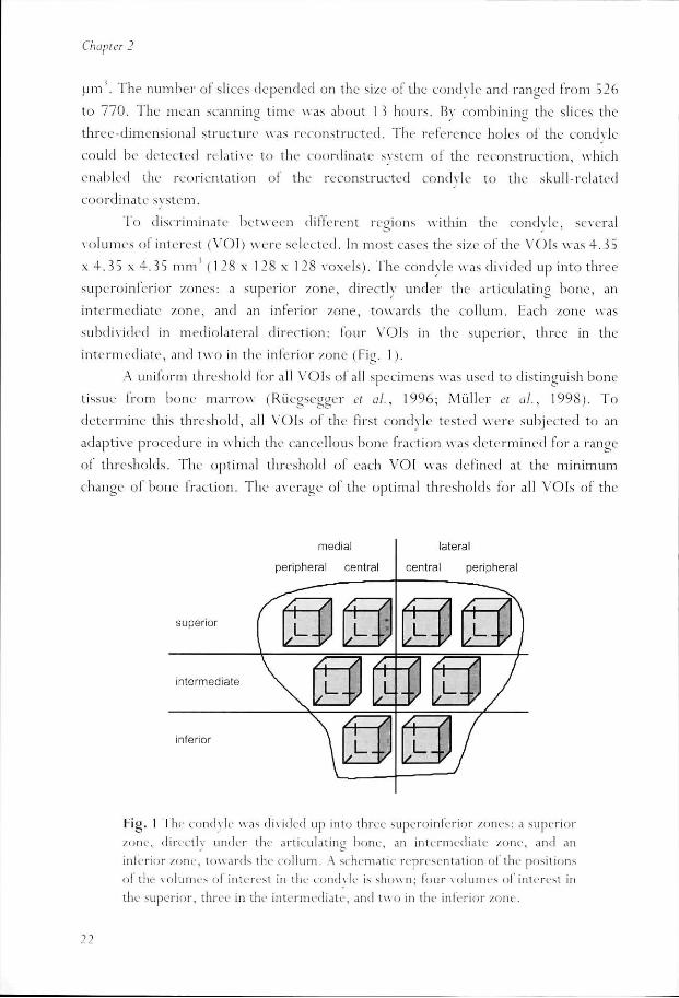
Too discriminate between different regions within the condyle, several
volumess ol interest (VOI) were selected. In most cases the size of the VOIs was 4.35
xx 4. 35 x. 4.35 mm (128 x 1 28 x 1 28 voxels). The condyle was divided up into three
superoinleriorr /.ones: a superior /one, directly under the articulating bone, an
intermediatee /one, and an inferior /one, towards the col lum. Each /one was
subdividedd in mediolateral direction: lour VOIs in the superior, three in the
intermediate,, and two in the inferior /one (Fig. 1).
AA uniform threshold for all VOIs ol all specimens was used to distinguish bone
tissuee from bone marrow (Rüegsegger et ah, 1996; Muller et al., 1998). To
determinee this threshold, all VOIs of the first condyle tested were subjected to an
adaptivee procedure in which the cancellous bone fraction was determined for a ran e
off thresholds. The optimal threshold of each VOI was defined at the minimum
changee ol bone traction. The average of the optimal thresholds for all VOIs of the
superior r
inferior r
medial l
)eripherall central
lateral l
centrall peripheral
FijJ.. 1 The condyle was divided up into three superoinlerior /ones: a superior
zone,, directly under the articulating bone, An intermediate zone, ami an
inferiorr zone, towards the collum. A schematic representation of the positions
oll the volumes ol interest in the condyle is shown; four volumes ol interest in
thee superior, three in the intermediate, and two in the inferior zone.
11 11

3D3D cancellous bone architecture
firstt specimen (19 .6% of the maximum grav level) was applied to all specimens.
BoneBone architecture
Thee bone architecture was described by the morphology and orientation of the
trabecularr structure. The former was described using the parallel plate model of
Parfittt et al. (1983) by determining the bone volume (BV), total volume (TV) and
trabecularr number (Tb.N). The bone volume and total volume were determined by
countingg voxels. The trabecular number was determined by the mean intercept
lengthh (MIL ) method. This method determines the mean distance between two
bone-marroww intersections in a linear grid of parallel lines, as a function of the gr id 's
three-dimensionall orientation in the VOI (Whi tehouse, 1974). The trabecular
numberr was defined by: Tb.N = MIL" 1, the trabecular thickness (Tb.Th) by: Tb.Th
== ( B V / T V ) / T b . N , trabecular separation (Tb.Sp) by: Tb.Sp = ( l - B V / T V ) / T b . N ,
andd bone surface density (BS/BV) by: BS/BV = 2"Tb .N/ (BV/TV) (nomenclature
accordingg to Parfitt et a/., 1987).
Thee orientation of the trabecular structure was described by the principal
directionn of the trabeculae and the degree of anisotropy. For this purpose, a mean
interceptt length tensor was calculated by fitting an ellipsoid to the mean intercept
lengthh measurements (Harrigan and Mann, 1984). The eigenvector of the mean
interceptt length tensor associated with the major axis of the ellipsoid gives the
predominantt principal direction of the bone material. The eigenvalues of the mean
interceptt length tensor, H I , H2, and H 3, are associated with the length of the axes
off the ellipsoid and express the prevalence of the material distribution in the
correspondingg principal directions. The eigenvalues were sorted such that
H 1 > H 2 > H 3.. To describe the dominance of trabecular orientation the degree of
anisotropyy (DA) was used. This was defined by the ratios between the eigenvalues,
thee first DA = H 1 / H 3, the second DA = H 1 / H 2. If all three eigenvalues are equal,
thuss the first DA = 1, the bone architecture is considered to be isotropic. If the
secondd DA = 1 the structure is considered to be oblately transversely isotropic. Al l
bonee architectural parameters were calculated using the software package at the
micro-CTT system (Riiegseggcr et al., 1996).
StatisticalStatistical analysis
Univariatee analyses for repeated measurements were used to test the regional
differences.. To analyze differences between the medial and lateral hall of the condyle
thee VOIs in each zone were divided into a medial and lateral group. Using this
23 3
ChapterChapter 2
methodd not only zone and mcdiolateral specific differences could be tested, but also
thee interaction between these. In the superior zone differences between central and
peripherall volumes were also tested. All tests were conducted using the General
Linearr Model for repeated measures with the SPSS 8.0 software (SPSS Inc.).
Results s
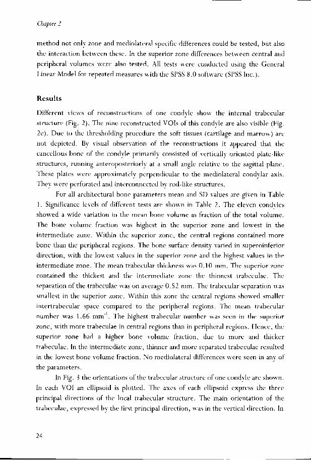
Differentt views of reconstructions of one condvle show the internal trabecular
structuree (Fig. 2). The nine reconstructed VOIs of this condyle are also visible (Fig.
2e).. Due to the thresholding procedure the soft tissues (cartilage and marrow) arc
nott depicted. By visual observation of the reconstructions it appeared that the
cancellouss bone of the condvle primarily consisted of vertically oriented plate-like
structures,, running anteroposteriorly at a small angle relative to the sagittal plane.
Thesee plates were approximately perpendicular to the mediolateral condvlar axis.
Theyy were perforated and interconnected by rod-like structures.
Forr all architectural bone parameters mean and SD values arc (riven in Table
1.. Significance levels of different tests are shown in Table 2. The eleven condyles
showedd a wide variation in the mean bone volume as fraction of the total volume.
Thee bone volume fraction was highest in the superior zone and lowest in die
intermediatee zone. Within the superior zone, the central regions contained more
bonee than the peripheral regions. The bone surface density varied in superoinferior
direction,, with the lowest values in the superior zone and the highest values in the
intermediatee zone. The mean trabecular thickness was 0.10 mm. The superior zone
containedd the thickest and the intermediate zone the thinnest trabeculae. The
separationn of the trabeculae was on average 0.52 mm. The trabecular separation was
smallestt in the superior zone. Within this zone the central regions showed smaller
intertrabecularr space compared to the peripheral regions. The mean trabecular
numberr was 1.66 mm . The highest trabecular number was seen in the superior
zone,, with more trabeculae in central regions than in peripheral regions. Hence, the
superiorr zone had a higher bone volume fraction, due to more and thicker
trabeculae.. In the intermediate zone, thinner and more separated trabeculae resulted
inn the lowest bone volume fraction. No mediolateral differences were seen in any of
thee parameters.
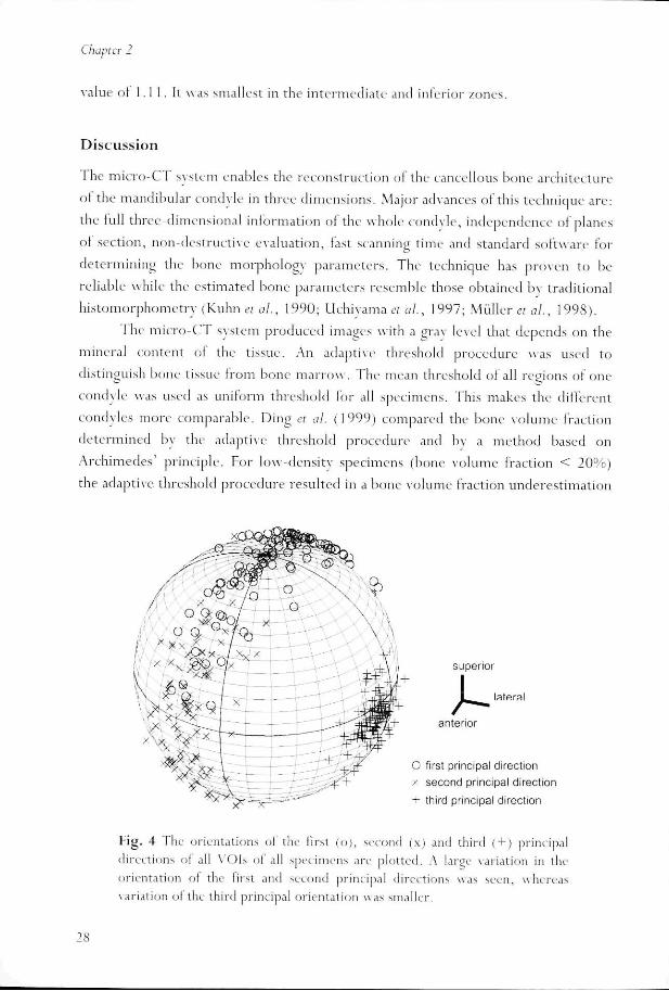
Inn Fijr. 3 the orientations of the trabecular structure of one condvle are shown.
Inn each VOI an ellipsoid is plotted. The axes of each ellipsoid express the three
principall directions of the local trabecular structure. The main orientation of the
trabeculae,, expressed by the first principal direction, was in the vertical direction. In
24 4

3D3D cancellous bone architectun
55 mm
Fig.. 2 a) Three-dimensional reconstruct ion oi a left mandibular condyle. O ne
off the lour condyle related reference points can be clearly seen in the anter ior
rimm of the col lum. Of this condyle the b) superior, c) anter ior, and d) lateral
halff is removed to show the internal trabecular s t ructure, e) Reconstruct ion
off nine volumes of interest of the same condyle. The size of the VOIs v\ as
4.355 x 4 .35 x 4.35 m m' and had small overlap. For visualization purposes the
VOI ss are separated. The vertically or iented plate-like s t ructure is visible.
25 5

ChapterChapter 2
'"" o CO O
OO T
C73 3 O O
T3 3
W W
Li. .
Q. . O O
n n to o c c lU U
cc c - i i o o (i) )
X I I
fc fc F F CU U
££ E
E E too E
,__ E rara E
xii ju
h-- o
CU U u u ra ra
t t
^ ^ t/> >
c c O O
Cü ü
E E (--
er r Q) )
T l l
EE -S
oo £ CQQ *~
ID D O O O O
CM M
O O O O
q q
TT T
o o o o O) )
q q
o o o o CN N
in n o o o o co o
m m o o o o
CM M
O O O O
--
CN N O O O O
--
T-- |S. ^f
dd ö o
coo m m
^^ T - O) oii iri cd
i -- o oo o
CD D CM M
O O
CM M |s--
co o co o o o
q q
CD D CM M
O O
O O
q q
m m CN N
o o CO O
q q
CN N
o o
» »
co o o o CD D
q q
co o CM M
o o CD D
q q
CN N
o o o o
CN N
o o o o
CN N
o o o o
CN N
o o o o
CM M O O O O
CN N O O O O
CN N O O O O
OO i - i -
000 CN
f ^ q ^ *" q oóó ri d ai co T -- CN CN -«- - -
CNN T - T -
COO CO
CM M
to o O O
CN N
in n o o
o o a> > CL CL =5 5
03 3 CC C
T3 3 Cl) )
E E Q) )
_ _ O O
Q. .
0) ) C l l
O O
o o o. .
£; ; c c o o o o
o o CU U Q. .
cc c te e
CU U
o o o o "o o o o ra ra CU U
> >
CDD >>
aa ^ oo <£
SS '<= oo ra
££
o o »» Q ££ o CDD . t CUU O -aa </)
=»» SU oo xt CUU e r ss ^
ee & »» c a>> n cc "a o o CO O
öö •£ >> o coo ra cc £: o o m m
oo *-
o o CJ J o o
CD D
m m CN N
<y> >
o o i n n o o o o o o
i -- o OO G> oo -sr oo T-
o o a a
o o Cl) ) Q. .
Cl) ) CJ J
m m CU U
n n 03 3
(1) )
ra a o o u u
COO CL
26 6

3D3D cancellous bone architecture
Fig.. 4 the orientations of all principal directions of all VOIs ol all specimens are
plottedd on the surface of a sphere. The variation in orientation ol the first and second
principall directions was large within the condyles. The third principal orientation
hadd a smaller variation. The narrow band formed by the two main orientations
representss the plane of the trabecular plates and indicates that the plates of different
VOIss run more or less parallel. The plane through the band was almost vertical and
hadd an angle of 17° with respect to the sagittal plane. Within a zone or site the
variationn of the first two principal directions was smaller, i.e., the band became
smallerr and data points were more grouped. The directions of the principal axes in
thee peripheral regions in the superior zone deviated from the vertical axis. They
weree pointing upwards to the periphery of the condyle. In the center ol the condyle
andd more inferiorly the principal directions were more vertical and parallel. In
superoinferiorr direction the first principal direction followed die curve of the
condylee (Fig. 5).
Thee first degree of anisotropv had a mean value ol 1.51 ("fables 1 and 2). The
highestt first degree of anisotropv was found in the inferior zone. The superior and
intermediatee zones had an equal first degree of anisotropv. No peripheral/central or
mediolaterall differences were found. The second degree of anisotropv had a mean
firstt principal direction
secondd principal direction
thirdd principal direction anterior r lateral l
Fig.. 3 The nine ellipsoids express the orientation ol trabecular structure ol
differentt VOIs in one mandibular condyle (the same as in Fig. 2). The main
orientationn of the trabecular structure, first principal direction, is primarily in
thee vertical direction.
21 21
ChapterChapter 2
valuee oi 1.1 1. It was smallest in the intermediate and inferior /ones .
Discussion n
II he micro-CI system enables the reconstruction of the cancellous bone architecture
oll the mandibular condyle in three dimensions. Major advances of this technique are:
thee full three-dimensional information of the whole condyle, independence of planes
off section, non-destructive evaluation, last scanning time and standard software for
determiningg the bone morphology parameters. The technique has proven to be
reliablee while the estimated bone parameters resemble those obtained by traditional
his tomorphometryy (Kuhn et al., 1990; Uchiyama et al., 1997; Muller et ai, 1998).
Thee micro-CT system produced images with a gray level that depends on the
minerall content ol the tissue. An adaptive threshold procedure was used to
distinguishh bone tissue from bone marrow . The mean threshold of all regions of one
condylee was used as uniform threshold for all specimens. This makes the different
condyless more comparable. Ding et al. (1999) compared the bone volume fraction
determinedd by the adaptive threshold procedure and by a method based on
Archimedes'' principle. For low-density specimens (bone volume fraction < 20%)
thee adaptive threshold procedure resulted in a bone volume fraction underest imation
Fig.. 4 I he orientations of the first (o), second (x) and third (+) principal
directionss of all VOIs ol all specimens are plotted. A large variation in the
orientationn ol the first and second principal directions was seen, whereas
variationn ol the third principal orientation was smaller.

3D3D cancellous bone architecture
off 1 2%. The choice of the threshold influences the detected bone volume and thus it
alsoo influences secondary determined hone parameters. Rüegsegger ct al. (1996)
foundd a change in hone volume traction of 59o with a 10% change of threshold. In
thee present study an analysis of changes in threshold in all conchies revealed a hone
volumee fraction change ol 7 . 9% by variation of the threshold by 50/o (data not
shown) .. The lower contrast might he the result ot the embalming procedure and the
presencee of marrow in the condyles.
Thee specimens used were relativelv old. In the process of aging a change ol
trabecularr structure from plate-like to more or less rod-like is seen and in advanced
casess the remaining bone may consist entirely of rods (Feldkamp ct al., 1989). The
parallell plate model of Parfïtt ct al. (198 3) may then become less reliable. W e found
thatt the cancellous bone in the human mandibular condyle consisted of plates rather
thann rods, which validated the use of the plate model. As in trabecular bone apparent
densitvv loss is correlated with aging (e.g., Ding ct al., 1997), we would expect to
findd a low bone volume traction. However , in the human mandibular condyle, this
agee related change was not found (Hongo ct al., 1989a).
Thee size ol the volumes of interest (sides ot 4 . 3 D mm) prevented us from
comparingg anteroposterior regions. Smaller VOIs, however, could have introduced
edgee artifacts influencing the measurements . Therefore, we could not confirm the
resultss of histological studies, in which the anterior regions showed higher bone
densitvv (Hongo et al., 1989a) and thicker trabeculae (Teng and Herring, 1995) than
superior r
intermediate e
inferior r
Fig.. 5 Means of the principal directions lor all regions of all condyles in lateral
vieww . The first principal direction follows the curve ol the condyle.
29 9

ChapterChapter 2
posteriorr regions.
Thee mean values for bone volume fraction (17%) and trabecular thickness
(0.100 mm) of the present studv were relatively low compared to the ranges reported
inn other studies involving the human mandibular condyle (bone volume fraction: 18-
30%,, trabecular thickness: 0.27-0.34 mm; Hongo et al., 1989a; Kawashima et al.,
1997)) and pig mandibular condyle (trabecular thickness: 0.23-0.27 mm; Teng and
Herring,, 1995), but correspond well with the ranges reported for various trunk and
limbb bones (bone volume fraction: 16-21%; Ding et al., 1997; Zysset et al., 1998;
Houu et al., 1998, trabecular thickness: 0.10-0.14 mm; Goulet et al., 1994; Muller et
al.,al., 1998; Kinnev and Ladd, 1998). The relatively high values of Hongo et al.
(1989a)) and Kawashima et al. (1997) mav be explained bv their technique, i.e., soft
X-rayy images of 400-500 urn sections. Using this technique, these authors also found
relativee high bone volume fractions for the vertebral body, compared to the values
reportedd elsewhere (Kinney and Ladd, 1998; Hou ex al, 1998).
Thee trabecular separation in the human mandibular condyle (0.52 mm) was
relativelyy large compared to that in the pig mandibular condyle (0.23-0.27 mm;
Tengg and Herring, 1995), but relatively small compared to the human vertebral
bodyy (1.47 mm; Kinnev and Ladd, 1998) and various limb bones (0.64-0.77 mm;
Goulett et al., 1994; Muller « al., 1998; Kinnev and Ladd, 1998). We also found that
thee trabecular number (1.66 mm ' ) was relative small compared to the pig
mandibularr condyle (2.4-2.9 mm '; Teng and Herring, 1995), but relatively large
comparedd to the vertebral body (1.18 mm '; Hou et al., 1998) and limb bones (1.39
mmm ; Goulet et al., 1994). Because of the relative thick trabeculae in the pit*
mandibularr condyle it was concluded that the condyle is a highly stressed structure
(Tengg and Herring, 1995). We found the trabecular structure comparable to that of
otherr anatomical sites. The relatively high values in the pig mandibular condyle
mightt be explained by a species effect.
Thee results of the present study confirm our hvpothesis that the distribution of
cancellouss bone in the human mandibular condyle is inhomogeneous and anisotropic.
Wee found highest the bone volume fraction in the central superior regions,
associatedd with the thickest, the least separated and the most numerous trabeculae.
Higherr bone volume fraction and thicker trabeculae in these regions were also found
byy Hongo et al. (1989b). In the pig mandibular condyle trabeculae were less
separatedd and more numerous in the superior regions, but thinner trabeculae were
foundd compared to the inferior regions (Teng and Herring, 1995).
Underneathh the articulating surface the cortical shell is verv thin. It is
generallyy believed that the cortical shells of most cancellous bone regions contribute
30 0

3D3D cancellous bone architecture
littlee to the stiffness and strength of the structure (Odgaard, 1997). The cancellous
bonee beneath the cortical shell will deform easily during loading, and, therefore,
peakk forces that could destroy the cartilage layer can be absorbed. The stresses are
distributedd and transferred to the thicker distal cortex. In the present study the
highestt bone volume fraction was found in the superior regions, adjacent to the
articularr surface. This is comparable to the vertebral body, where the highest bone
volumee fraction was found under the endplatc and near the cortex. It has been
suggestedd that local higher trabecular bone densities are related to multiple load
conditions,, rather than to higher stresses (Smit et aiy 1997). Photo-elastic stress
studiess also indicated that the condyle is designed to receive less heavy but varied
forcess rather than heavy, cyclical unidirectional forces (Standlec et al., 1981). We
alsoo found a higher trabecular number in the superior regions. This indicates a finer
bonee structure that mav be in favor for a multiple loading situation (Smit et al.,
1997). .
Thee distribution of bone in different orthogonal directions was expressed bv
thee degree of anisotropy. The first degree of anisotropy was 1.51. This implies that
thee trabecular structure has a pronounced orientation and that the trabeculae are not
randomlyy distributed. For the pig mandibular condyle values of 1.43 to 2.60 were
foundd for three orthogonal planes (Teng and Herring, 1995). In three-dimensional
analysess values of the first degree of anisotropy were 1.61 for various limb bones
(Goulett et al, 1994) and 1.59 for the vertebral body (Hou et al, 1998). The second
degreee of anisotropy was 1.11; this means that the trabecular structure of the
condylee tends to be oblately transversely isotropic. If it were equal to unity the
structuree would consist of perfect parallel plates. The slightly larger number could
indicatee that the plates have oval shaped perforations and arc not truly parallel. The
degreee of anisotropy was highest in the inferior regions and smallest in the superior
regions.. This suggests that the trabeculae in the inferior regions arc more oriented
intoo one direction, in contrast to the superior regions where the trabecular plates
weree more densely interconnected.
Thee plane of the two main orientations (Fig. 4) represents the plane of the
parallell plates. It was almost vertical and had an angle of 17° relative to the
anteroposteriorr axis, Hongo et al. (1989b) related the orientation of the trabeculae
too the direction of the lateral pterygoid muscle, which was 30.4° to 42° in the
transversall plane relative to the anteroposterior axis. However, in another studv
correspondingg angles of 41.7° for the superior head and of 47.4° for the inferior
headd were found (Van Eijden et al., 1995). Teng and Herring (1995) found the
trabeculaee to be in the growth direction of the condyle in immature pigs. We
31 1

ChapterChapter 2
suggest,, however, that the plate orientation is likely to coincide with the plane
perpendicularr to the condylar axis. Such a trabecular structure would be optimally
adaptedd to sustain loads in directions that presumably coincide with the majority of
jointt forces applied to the condyle. The trabeculae in the peripheral superior regions
weree directed from the periphery towards the center of the condyle. In the central
regionss the trabeculae were more ycrticallv oriented. Thus the ovoid shaped
articularr surface is supported by trabeculae oriented perpendicular to this surface.
Becausee the cancellous bone of the condyle is anisotropic there is an obvious
dependencee of the material propert ies . From several studies it is known that the
anisotropyy of the trabecular structure and the anisotropy of mechanical properties
aree closely related (Van Rietbergen et al, 1998; Od^aard et al, 1997; Goulet et al,
1994;; Turner et al, 1990; Zvsset et ai, 1998). The three-dimensional condylar
reconstructionss of the present study can be used to construct large-scale finite
e lementt models, which allow for calculation of mechanical propert ies and for
simulationn ot different loading conditions. Fur thermore, these models offer the
possibilityy to simulate the effects of adaptational and remodeling processes on the
mechanicall propert ies (Van Rietbergen et al, 1996; Mullender et al, 1998).
Inn conclusion, the plate-like trabeculae could indicate that the condylar
s t ructuree is optimally adapted to sustain the range of joint loads that act on the
articularr surface. The high bone mass and lower anisotropy in the superior regions
couldd enable the condyle to sustain loads acting in diverse directions. Towards the
collumm the trabeculae are more aligned. This could point to stresses acting in
primarilyy one direction.
References s
Dingg M, Dalstra M, Danielscn CC, Kabel J, Hvid 1, Linde F (1997). Age variations in the
propertiess ot human tibial trabecular bone, ƒ Bone joint Surg Br 79-B:995-1002.
Dingg M, Odgaard A, Hvid I (1999). Accuracy of cancellous bone volume fraction measured
byy micro-CT scanning. / Biomech 32:323-326.
Feldkampp LA, Goldstein SA, Parlltt AM, jesion G, Kleerekoper M (1989). The direct
examinationn of three-dimensional bone architecture in vitro by computed tomography. J
BoneBone Miner Rei 4 : 3-1 1.
Goulctt RW, Goldstein SA, Ciarclli MJ, Kuhn JL, Brown MB, Feldkamp LA (1994). The
relationshipp between the structural and orthogonal compressive properties of trabecular
bone.bone. J Biomech 27:375-389.
Flarriuann TP, Mann RW (1984). Characterization of microstructural anisotropy in
orthotropicc materials using a second rank tensor, j Mater Sci 1 9:761 -767.
32 2

3D3D cancellous bone architecture
HongoT,, Orihara K, Onoda Y, Nakajima K, lde Y (1989a). Quantitative and morphological
studiess of the trabecular bones in the condyloid processes of the Japanese mandibles;
changess due to aging. Bull Tokyo Dent Coll 30:165-174.
Hongoo T, Yotsuya H, Shibuva K, Kawase M, lde Y (1989b). Quantitative and morphological
studiess on the trabecular bones in the condyloid processes of the Japanese mandibles.
Comparisonn between dentulous and edentulous specimens. Bull Tokyo Dent Col! 30:67-
76. . Houu FJ, Lang SM, Hoshaw SJ, Rcimann DAFDP (1998). Human vertebral body apparent
andd hard tissue stiffness, ] Biomech 31:1009-101 5. Kawashimaa T, Abe S, Okada M, Kawada E, Saitoh C, lde Y (1997). Internal structure of the
temporomandibularr joint and the circumferential bone: comparison between dentulous
andd edentulous specimens. Bull Tokyo Dent Coll 38:87-93. Kinnevv JH, Ladd AJC (1998). The relationship between three-dimensional connectivity and
thee clastic properties of trabecular bone. J Bone Miner Res 1 3:839-845. Kuhnn JL, Goldstein SA, Fcldkamp LA, Goulet RW, Jesion G (1990). Evaluation of a
microcomputedd tomography system to study trabecular bone structure, j Orthop Res
8:833-842. . Muilenderr M, Van Rietbergen B, Rüegscggcr P, Huiskes R (1998). Effect of mechanical set
pointt of bone cells on mechanical control of trabecular bone architecture. Bone 22:125-
131. . Mullerr R, Van Campenhout H, Van Damme B, Van Der Perre G, Dequeker J, Hildebrand
T,, Rüegscggcr P (1998). Morphometric analysis of human bone biopsies: a quantitative
structurall comparison of histological sections and micro-computed tomography. Bone
23:59-66. . Odgaardd A, Kabel J, Van Rietbergen B, Dalstra M, Huiskes R (1997). Fabric and clastic
principall directions of cancellous bone arc closely related./ Biomech 30:487-495. Odgaardd A (1997). Three-dimensional methods for quantification of cancellous bone
architecture.. Bone 20:31 5-328. Parfittt AM, Mathews CHE, Villanueva AR, Klccrekoper M, Frame B, Rao DS (1983).
Relationshipss between surface, volume, and thickness of iliac trabecular bone in aging andd in osteoporosis. Implications for the microanatomic and cellular mechanisms of bone
loss,, j Clin Invest 72:1 396-1409. Parfittt AM, Drczner MK, Gloricux FH, Kanis JA, Malluche H, Meunier PJ, Cut SM, Recker
RRR (1987). Bone histomorphomctry: standardization of nomenclature, symbols, and
units.. J Bone Miner Res 2:595-610. Rüc^seggerr P, Roller B, Muller R (1996). A microtomographic system for the
nondestructivee evaluation of bone architecture. Calcij Tissue Int 58:24-29, Smitt TH, Odgaard A, Schneider E (1997). Structure and function of vertebral trabecular
bone.. Spine 22:2823-28 33. Standlecc JP, Caputo AA, Ralph JP (1981). The condyle as a stress-distributing component of
thee temporomandibular joint . / Oral Rehabii 8:391-400. Tengg S, Herring SW (1995). A stercological study of trabecular architecture in the
mandibularr condyle of the pig. Arch Oral Biol 40:299-3 10.
33 3

ChapterChapter 2
Turnerr CH, Cowin SC, Rho JY, Ashman RB, Rice JC (1990). The fabric dependence of the orthotopicc elastic constants of cancellous bone. J Bwmcch 23: S49-56I.
Uchiyamaa T, Tanizawa T, Muramatsu H, Endo N, Takahashi HE, Hara T (1997). A morphometryy comparison of trabecular structure of human ilium between microcomputcdd tomography and conventional histomorphometrv. Caltif Tissue Int
61:493-498. .
Vann Eijden TMGJ, Koolstra JH, Brugman P (1995). Architecture of the human pterygoid muscles,, j Dent Res 74:1489-1495."
Vann Rietbergen B (1996). Mechanical behavior and adaptation of trabecular bone in relation too bone morphology. PhD thesis, University of Nijmegen, Netherlands.
Vann Rietbergen B, Odgaard A, Kabel J, Huiskes R (1998). Relationships between bone morphologyy and bone elastic properties can be accurately quantified usinjr high-resolutionn computer reconstructions, j Orthop Res 16:2 3-28.
Whitehousee WJ (1974). The quantitative morphology of anisotropic trabecular bone, j
MicroscMicrosc 101:153-168.
Wolff'JJ (1892). Das Gcsctz der Transformation der Knochen. (Translated as: The law of
bonee remodelling. Maquet P, Furlong R. Berlin, Springer Verlag, 1 986).
Zyssett PK, Goulet RW, Hollister Sj (1998). A global relationship between trabecular bone
morphometryy and homogenized elastic properties. / Biomech Eng 1 20:640-646.
34 4

Chapterr 3
Predictionn of Strains in the Human Mandibula rr Condyle
Abstract t Thee structure of the cancellous hone in the human mandibular condyle
iss characterized by plate-like trabecular oriented in the sagittal plane.
Wee tested the hypothesis whether this structure reflects the mechanical
loadingg of the condyle. For this purpose a finite element model of the
condylee was developed to analyze the occurring strains during static
compressivee loading. The principal strains in the trabecular bone were
primarilyy oriented in the sagittal plane. The first component was
compressivee and oriented superoinferiorly, the second component was
negligiblyy small and oriented mediolateraliv, the third component was
tensilee and oriented anteroposterior^ The tensile strain was almost
equall to the compressive strain. This relatively large tensile strain was
duee to the anteroposterior bulging of the cortex, which caused a tensile
stresss in the cancellous bone. It suggests that the cancellous bone also
hass to resist significant tensile forces. The orientation of the principal
strainss within the sagittal plane changed with the direction of the
appliedd load. It was concluded that the platedikc trabecular structure
off the mandibular condyle is optimal to resist compressiye and tensile
strains. .

ChapterChapter 3
I n t r o d u c t i o n n
Accordingg to the law of bone remodeling (Wolff, 1892) the trabecular structure of
bonee is optimized to offer maximum resistance to stresses and strains with a
minimumm amount of bone mass. By using finite element models , the correspondence
betweenn trabecular s t ructure, bone density, and mechanical loading; has been
demonstratedd in several studies (e.g., Smit et al., 1997; Muilender et al., 1998;
HuiskesefuA,, 2000) .
Recently,, the three-dimensional structure of the cancellous bone of the human
mandibularr condyle was analyzed by Giesen and Van Eijden (2000) . Thev found that
thee cancellous bone mainly consists of parallel plates perpendicular to the medio-
laterall condylar axis. This means that the trabecular structure can withstand larger
stressess in sagittal planes than in the mcdiolatcral direction, as indeed has been
demonstratedd by mechanical tests (Giesen et tiA, 2001). This suggests that the
orientationn of the plate-like structure is related to the orientation of the stresses, i.e.,
thatt the condyle is optimally adapted to sustain stresses and strains occurring; in vivo.
Untill now, however , a detailed analysis of the stresses a n d / o r strains in the human
mandibularr condyle has never been performed. The available finite e lement models
(e.g.,(e.g., Korioth et al., 1992; Beck et al., 2000) do not provide enough details to study
thee distribution of strains in the bone of the condyle.
Inn the present study, we determined the strains occurring in the mandibular
condylee due to static loads to verify if the structure of the cancellous bone is
optimizedd to sustain these loads. For this purpose, we developed a three-dimensional
finitefinite element model of the mandibular condyle. The strain matr ix, the principal
strains,, and the total principal strain in the condyle were calculated for three
differentt load cases. Fur thermore , the deformation of the condyle due to the loading
wass analyzed.
Mater ia ll a nd M e t h o d s
FiniteFinite Element Model
Thee right condyle of a dried mandible was separated from the rest at the collum by
ann electrical saw. The condyle was scanned with a micro-CT scanner (uCT 20,
Scancoo Medical AG, Zurich, Switzerland). The scan had a pixel size of 34 x 34 urn '
andd the interslice distance was 34 urn. Bone tissue was identified from background
noisee with a uniform threshold (Giesen and Van Eijden, 2000). Using a bitmap
editorr the trabecular structure was removed from all CT slices and artifacts in the
36 6

PredictionPrediction of strains
cortexx were repaired. Next, all the cortical surface points (pixels) were extracted. A
pointt belonged to the surface if it contained bone and at least one of the six
neighboringg points was empty. The surface points were divided into three sets: the
outerr surface, the inner surface and the saw plane. After filtering, both the inner and
outerr surface sets contained approximately 400,000 points.
Throughh the points of the saw plane a plane was fitted. To model the inner
andd outer cortical surfaces, 30,000 points were selected randomly from both
surfaces.. A method developed by Hoppe (1994) and Schweitzer (1996) was used to
fitfit surfaces through these data points.
Inn order to make a volume mesh, a closed surface mesh had to be made first.
Triangularr meshes were generated from the subdivision models of the inner and the
outerr surface of the cortex. The boundary vertices of these meshes were projected
rectangularlyy on the saw plane. The meshes and the projections of the vertices were
importedd in an automated mesher (Mentat 3.2, MSC Software). First, the spaces
betweenn the projected boundaries and the surface boundaries were closed with a
triantnilarr mesh. Next, the area within the projection of the inner boundary as well
ass the area between the projected boundaries was closed with planar triangular
meshes.. Finally, tetrahedron meshes were created for the cancellous and the cortical
bonee volumes (44,000 and 13,000 tetrahedrons, respectively). In Fig. 1 a frontal
vieww of the condyle obtained from the unedited CT scan and two views ol the model
off the cortical surface are shown. The model matched the CT scan very well, as the
meann distance between the outer surface points and the surface model was 14 um.
Thee triangular mesh of the outer surface contained only 5,632 vertices,
Thee cancellous and cortical bone volumes were modeled as an isotropic
homogeneouss material. For cortical bone an elastic (E-) modulus of 19.0 GPa (Rho
etet ai, 1993) was used. Based on a mean condylar cancellous bone volume fraction ot
17%% (Giesen and Van Eijden, 2000), the estimated E-modulus for cancellous bone
wass 1.11 GPa (Rho ct al, 1993). Poisson ratios of 0.32 and 0.20 (Carter and Hayes,
1977;; Kabel et al., 1999) were used for the cortical and the cancellous bone,
respective!! v.
Simulations Simulations
Too mark the boundary of the articular surface on the finite element model, the
articularr surface and its rim were measured with an electromagnetic tracking device
(Spacee Tracker, Polhemus 3Space). A three-dimensional polynomial function was
fittedd through the surface points (Van Ruijven et al, 1999,2000). This polynomial
37 7

(( hapter 3
functionn was lil to the outer surface of the finite element model , after which the
pointss ol the articular rim were projected on the outer surface of the model (Fig. 1).
Thee articular area was equally divided into anterior, apical and posterior parts,
whichh were loaded separately with a constant pressure. Since the parts had a surface
cancellouss bone volume corticall bone volume
Fig.. 1 The original CT-scan (A) and the finite element model (B+C). The thickk line marks the border of the articular area.
5«S S

PredictionPrediction of strains
areaa of 7 - 1 0 ' m , and the total estimated load during static clenching is
approximatelyy 300 N (Koolstra et al., 1988), a pressure of 4 .28 MPa was used. The
nodess belonging to the saw plane were fixed.
Thee finite element problem was solved with MARC (MSC Software) on a
Beowulff Linux-PC cluster. Principal strains as well as the total principal strain were
calculatedd for every cancellous and cortical bone element . The total principal strain
off an element is equal to the change in volume of that element . Three-dimensional
plotss were made with The Visualization Toolkit v3.1 (Kitware) on a Windows / In te l
computer . .
Resu l ts s
Forr each of the load cases, the deformation of the condyle in the midsagittal cross-
sectionn is shown in bio. 2. In all simulations the condyle was compressed along the
meann loading direction and extended perpendicular to this direction. The amount of
compressionn and extension were estimated to be approximately 0 .2%. Frontal cross-
sectionss did not reveal a deformation in the mediolateral direction. For all load cases
together ,, 9 5 % of the elements of the cortical bone had a strain less than 1,600
Fi<*.. 2 Deformations of the midsagittal cross-section ol the condyle due to the
threee loads. The grav area indicates the unloaded shape. The lines depict die
deformedd shapes. For reasons of clarity, these deformations were amplified
fiftyy times. At the bottom the deformation was zero, because the saw plane
waswas fixed during the simulation. The thick lines mark the region where the
loadss were applied.
39 9

ChapterChapter 3
Tablee 1 Mean (SD) of the principal strains in the trabecular bone [ustrain]. Components are sortedd by size.
Firstt Principal Strain Second principal strain Third principal strain
Antehorloadd -2722(1127) 54(210) 2455(1082)
Apicall load -1362(501) -26(186) 1088(394)
Posteriorr load -3301 (1720) 109 (204) 2762 (1241)
ustrain,, and 95% of the elements of the cancellous bone had a strain less than 4,600
ustrain. .
Thee mean magnitudes of the three principal strains in the cancellous bone
elementss are given in Table t. The first principal strain was always compressive. The
thirdd principal strain was always tensile and onlv 10-20% smaller in absolute value
thann the first. The second principal strain alternated between compressive and
tensile.. Its mean magnitude as well as its standard deviation, however, was negligible
smalll compared to the other principal strains.
Thee total principal strains and the magnitudes and directions of the first and
thirdd principal strain at different positions in the condyle arc shown in Fig. 3. The
totall principal strain was dependent on the load case. It was minimal at the apical
loadd case, and maximal at the posterior load case. Furthermore, the total principal
strainn changed across the condyle. In every cross-section the largest total principal
strainss were found cranially in the region just below the loaded area. Mediolateral
differencess were relatively small.
Withinn the cancellous bone, the directions of the first and third principal
strainss coincided with the sagittal planes. Compression occurred in the supero-
inferiorr direction (direction of the load), extension occurred in the anteroposterior
directionn (rectangular to the direction of the load). The orientation and magnitudes
off the principal strains in the sagittal plane depended on the load case. The angle
betweenn the anterior and the posterior load was 80°. The apical load case resulted in
thee smallest principal strains. Within one load case, the orientation was
approximatelyy constant throughout the condyle. The magnitudes of the principal
strainss varied throughout the condyle. They were maximal in the cranial region just
beloww the area where the load was applied. In the mediolateral direction the
differencess were small.
40 0

PredictionPrediction of strains
-2000p p
2000M M
anterior r
apical l
posterior r
Fig.. 3 Total principal strain and the principal strains for the three different
loadd cases. The total principal strain is indicated with colors. The color bar
givess the total principal strain in ustrain. The orientations and magnitudes of
thee principal strains in the sagittal plain are indicated with the black lines. The
strainss of onlv a random selection of the elements are shown.
4-1 1

ChapterChapter 3
Discussion n
Oftenn the principal orientations and density of the cancellous bone are compared
withh stress related parameters (stress trajectories: Wolff, 1 892; principal stresses:
Carterr et a!., 1989; strain energy density: Huiskes et al., 19S7; Wcinans et al.,
1992).. In recent studies, strains or strain derived parameters have become more
popularr (energy equivalent strain: Mildc and Carter, 1995; strain gradients: Turner
etet al., 1997; strain rate: Mosley and Lanvon, 199S; equivalent strain: Smit and
Burger,, 2000). This is more in agreement with studies, showing that bone cells are
sensitivee for (pulsating) fluid streams (Klcin-Nulend et al., 1995; Bakker er al., 2001)
inn the lacunae and canaliculi. These are caused by strain gradients and strain rates
(Judexx et al., 1997). In the present study, strains were analyzed, because strain
gradientss as well as strain rates are proportional to the strain.
AA number of assumptions had to be made for the simulations. Since the bone
volumee fraction was known, we assumed that the volume fraction was equal to the
bonee density and used the E-moduli found bv Rho et. al. (1993) for cortical and
cancellouss bone. The cancellous bone was modeled isotropic, because only then a
possiblee correspondence between the orientation of the trabeculae and the principal
strainss can be expected. Experimentally determined E-moduli on specimens from
thee cancellous bone of the condyle (Giesen et al., 2001) were half the value used in
thiss study. For a reliable calculation of the force distributions, however, the ratio of
thee moduli for cortical and cancellous bone is more important than the absolute
value.. Rho et al. (1993) used the same protocol for determining the moduli for both
typess of bone. Literature values of the Poisson ratios of other cortical and cancellous
boness have been used. We do not expect, however, that the results of the present
studyy are very sensitive to the value of the Poisson ratio, because the volume changes
aree small compared to the strains. Finally, there is no experimental data available,
whichh describes the contact area and the distribution of the contact forces during
staticc clenching. In the present study, the strain distributions in the mandibular
discuss calculated by Beek et al. (2000) were used to model the shape of the loaded
areas. .
Despitee these limitations, the magnitudes of the principal strains found in the
presentt study are in agreement with experimentally determined strains. During in
vivovivo measurements strains of up to 2000 ustrain have been found in cortical bone of
thee mandible (Hvlander and Johnson, 1992, 1997). In vivo measurements of
cancellouss bone strains are not available, but in vitro experiments show that
cancellouss bone can be subjected up to 5000 ustrain without being damaged
(Keavenyy et al., 1994; Wachtel and Keavcny, 1997).
42 2

PredictionPrediction of strains
Inn the cancellous bone of the condvle, the tensile strain in the anteroposterior
directionn was almost as large as the compressive strain in the superoinferior
direction.. The Poisson ratio used in the simulations is too small to explain the tensile
strainn as a result of the compression. This means that, besides the superoinferiorly-
orientedd compressive stress, there was also an antcropostcriorlv-oriented tensile
stresss acting on the cancellous bone. Most likely, the anteroposterior bulging of the
cortexx caused the tensile stress. This also implies that the bulging was not caused by
thee cancellous bone, but by other factors (e.g., shape of the cortex, shape of the
loadedd area, etc.). Apparently, the cancellous bone not only serves to resist
compressionn in the superoinferior direction, but also to resist tension in the
anteroposteriorr direction. It should be noted that the fixation of the saw plane
preventss any anteroposterior extension near the saw plane. It is therefore possible
thatt in reality even larger tensile strains occur.
Inn the mediolateral direction the strains were negligible. This is probably
relatedd to the fact that the compressive and tensile strains in the sagittal plane were
almostt equal. Compressive strains in the sagittal plane cause extension in the medio
laterall direction, while tensile strains will result in compression in the mediolateral
direction.. Since the compressive and tensile strains were almost equal in the
simulations,, it could be expected that the mediolateral strain would be negligible.
Thee mean principal strain was smallest for the apical load case. The percentile
variationn of the mean principal strain was also smallest for the apical load case. This
impliess that the apical load is distributed more evenly throughout the condyle than
thee other loads. Probably, not only the mean strain is lower for apical loads, but also
thee ratio maximal strain/mean strain. Therefore, the present results suggest that the
condylee is more optimized to sustain apical loads than anterior and posterior loads.
Thee strains in the cancellous bone were concentrated in sagittal planes, both
tensilee and compressive. The orientation of these strains rotated over an angle of 80°
inn this plane by varying the load case. To withstand these strains, a plate-like
structuree with the plates parallel to the sagittal plane is the optimal structure. Note
thatt such a plate-like structure not only withstands compression in sagittal planes,
butt also tension. This is exactly the structure Giesen and Van Eijden (2000) found in
thee human mandibular condyle. They also found that the bone volume fraction and
thee trabecular thickness are higher in the superior region of the condyle. This also
correspondss with the regions where the highest total principal strains were found.
Therefore,, the results of the present studv suggest that the law of bone remodeling is
alsoo applicable to the trabecular structure of the mandibular condyle.
43 3

ChapterChapter 3
References s
Bakkerr AD, Soejima K, Klein-Nulend J, Burner EH (2001}. The production of nitric oxide
andd prostaglandin E2 by primary bone cells is shear stress dependent, j Biomech 34:671 -
677. .
Beekk M, Koolstra JH, Van Ruijven LJ, Van Eijdcn TMGJ (2000). Three-dimensional finite
elementt analysis of the human temporomandibular joint disc. J Biomech 33:307-316.
Carterr DR, Hayes WC (1977). The behavior of bone as a two-phase porous structure. J Bone
jointjoint Surg Am 59-A:954-962.
Giesenn EBW, Van Eijdcn TMGJ (2000). The three-dimensional cancellous bone architecture
off the human mandibular condyle, j Dent Res 79:957-963.
Giesenn EBW, Ding M, Dalstra M, Van Eijdcn TMGJ (2001). Mechanical properties of
cancellouss bone in the human mandibular condyle are anisotropic, j Biomech 34:799-803.
Hoppee H (1994). Surface reconstruction from unorganized points. PhD thesis, Dept. of
Computerr Science and Engineering, Unie. of Washinghton.
Huiskess R, Weinans H, Grooteiiboer HJ, Dalstra M, Fudala B, Slooff TJ (1987). Adaptive
bone-remodelingg theory applied to prosthetic-design analysis. / Biomech 20:1 1 35-11 50.
Huiskess R, Ruimerman R, Van Lcnthe GH, Janssen JD (2000). Effects oi mechanical forces
onn maintenance and adaptation of lonn in trabecular bone. Mature 405:704-706.
Hylandcrr WL, Johnson KR (1992). Strain gradients in the craniofacial region of primates.
In:: Daviclovitch, Z. (Ed.), The Biological Mechanism of Tooth Movement and
Craniofaciall Adaptation. The Ohio State University College of Dentistry, Columbus, pp.
559-569. .
Hylandcrr WL, Johnson KR (1997). In vivo bone strain patterns in the craniofacial region of
primates.. In: McNeill, C. (Ed.), Science and Practice of Occlusion, Quintessence
Publishingg Co. Inc., Chicago, pp. 165-178.
Judexx S, Gross TS, Bray RC, Zernicke RF (1997). Adaptation of bone to physiological
stimuli. ;; Biomech 30:421-429.
Kabell J, Van Rietbergen B, Odgaard A, Huiskes R (1999). Constitutive relationships of
fabric,, density, and elastic properties in cancellous bone architecture. Bone 2 5:48 1 -486.
Kcavcnyy TM, Guo XE, Wachtel EF, McMahon TA, Hayes WC (1994). Trabecular bone
exhibitss full}' linear elastic behavior and yields at low strains. / Biomech 21:1 1 27-1 1 36.
Klein-Nulendd J, Semeins CM, Ajubi NE, Frangos JA, Nijwcide PJ, Burger EH (1995).
Pulsatingg fluid flow increases nitric oxide (NO) synthesis by ostcoevtes but not periosteal
fibroblastss correlation with prostaglandin upregulation. Biochem Biophys Res Commun
217:: 640-648.
Koolstraa JH, Van Eijdcn TMGJ, Weijs WA, Naeije M (1988). A three-dimensional
mathematicall model of the human masticatory system predicting maximum possible bite
forces.. J Bwmech 21:563-576.
Koriothh TWP, Romillv DP, Hannam AG (1992). Three-dimensional finite element stress
analysiss of the dentate human mandible. Am j Phvs Anthropol S8:69-96,
Mikicc B, Carter DR (1995). Bone strain «age data and theoretical models of functional
adaptation,, J Biomech 28:465-469.
Moslcvv JR, Lanvon LE (1998). Strain rate as a controlling influence on adaptive modeling in
responsee to dynamic loading ol the ulna in growing male rats. Bone 2 3:313-318.
44 4

PredictionPrediction of strains
Mullcndcrr M, Van Rietbergen B, Rücgscggcr P, Huiskes R (1998). Effect of mechanical set
pointt of bone cells on mechanical control of trabecular bone architecture. Bone 22:125-131. .
Rhoo JY, Ashman RB, Turner CH (1993). Young's modulus of trabecular and cortical bone
materiall - ultrasonic and microtensile measurements. J Biomech 26:111-1 19. Schweitzerr JE (1996). Analysis and application of subdivision surfaces. PhD thesis, Dept. of
Computerr Science and Engineering, Univ. of Washinghton. Smitt TH, Odgaard A, Schneider E (1997). Structure and function of' vertebral trabecular
bone.. Spine 22:282 3-2833. Smitt TH, Burger EH (2000). Is BMU-coupling a strain-regulated phenomenon? A finite
clementt analysis. J Bone Miner Res 1 5:301-307. Turnerr CH, Anne V, Pidaparti RMV (1997). A uniform strain criterium for trabecular bone
adaptation:: do continuum-level strain gradients drive adaptation?/ Biomech 30:555-563. Vann Ruijvcn LJ, Beck M, Van Eijdcn TMGJ (1999), Fitting parametrized polynomials with
scatteredd surface data.7 Biomech 32:71 5-720. Vann Ruijven LJ, Beck M, Donker E, Van Eijden TMGJ (2000). The accuracy of joint surface
modelss constructed from data obtained with an electromagnetic tracking device. J
BwmechBwmech 33:1023-1028. Wachtell EF, Keaveny TM (1997). Dependence of trabecular damage on mechanical strain, j
OrthopResOrthopRes 15:78 {-787. Weinanss H, Huiskes R, Grootenboer HJ (1992). The behaviour of adaptive bone
remodelingg simulation models . / Biomech 25:1425-1441. Wolfff J (1892). Das Gesetz der Transformation der Knochen. (Translated as: The law of
bonee remodelling. Maquet P, Furlong R. Berlin, Springer Verlag, 1986).
45 5

-J J

Chapterr 4
Mechanicall Properties of Cancellous Bone inn the Human Mandibular Condyle
aree Anisotropic
Abstract t Thee cancellous bone of the mandibular condyle consists of' plate-like
trabeculaee primarily oriented vertically and anteroposterior^ The
objectivee of (he present study was 1) to test the hypothesis that the
elasticc and failure properties depend on the loading direction, and 2) to
relatee these properties to bone density parameters. Uniaxial
compressionn tests were performed on cylindrical specimens (n=47)
obtainedd from the mandibular condyles of 24 embalmed cadavers. Two
loadingg directions were examined, i.e., a direction coinciding with the
predominantt orientation of the plate-like trabeculae (axial loading) and
aa direction perpendicular to the plate-like trabeculae (transverse
loading).. Archimedes' principle was applied to determine bone density
parameters.. The cancellous bone appeared to be highly anisotropic. It
waswas in axial loading 3.4 times stiffer and 2,8 times stronger than in
transversee loading. High coefficients of correlation were found among
thee various mechanical properties and between them and the apparent
densityy and bone volume fraction. For each loading direction about
60°oo of the variance in the mechanical properties could be explained
Iromm apparent density. When the results of both loading directions
weree combined, only 25% of the variance could be explained. The
anisotropicc mechanical properties can possiblv be considered as a
mechanicall adaptation of the bone to the loading of the condyle in vivo.

ChapterChapter 4
Introductio n n
Mechanicall properties of cancellous hone strongly depend on its apparent density
(Carterr and Haves, 1977) and structure (Hodgskinson and Currcy, 1990; Uchivama
etet al, 1999; Ulrich et al, 1999). Over 90% of the variance in the mechanical
propertiess can be explained by these two aspects (Odgaard et al., 1997; Van
Rietbergenn et a/., 1998; Zyssct et al., 1998). The stiffness and the strength of the
bonee are proportional to apparent density. The bone is stillest and strongest in the
directionn in which the trabeculae arc aligned. Previous studies revealed that in the
mandibularr condyle the cancellous bone is anisotropic (Hongo et al., 1989b; Giescn
andd Van Eijdcn, 2000). It consists of parallel plate-like trabeculae primarily oriented
inn the vertical direction, perpendicular to the mediolateral condylar axis. In the
horizontall direction the plate-like trabeculae are interconnected with rods. From this
itt can be expected that the bone is stiffer and stronger in vertical and anteroposterior
directionss compared to the mediolateral direction. In addition, a wide range in bone
volumee fraction was found between condyles of different individuals (Giesen and
Vann Eijden, 2000). Hence, a wide range in apparent density, and as a consequence, a
widee range in mechanical properties can be expected.
Thuss far, to our knowledge no studies are available on mechanical properties
off the cancellous bone of the human mandibular condyle. Adequate estimates of the
mechanicall properties can be used, e.g., in finite element modeling of the mandibular
condylee and tissue engineering of mandibular implants. The only study in which the
cancellouss bone of the mandibular condyle was involved concerned the pig (Teng
andd Herring, 1996). Previously, determination of mechanical properties of the
humann mandible was limited to the cortical bone (e.g., Arendts and Sigolotto, 1989;
Dechoww et ai, 1993; Zioupos and Currey, 1998; for review: Van Eijdcn, 2000), or
too the cancellous bone of its body (Misch et al., 1999), The purpose of this study was
too determine the mechanical properties of the cancellous bone of the mandibular
condylee in different directions and to relate these to the density of the bone. The
hypothesess were that, due to the vertical orientation of the plate-like trabeculae in
thee sagittal plane, the stiffness and strength would be higher for loading in the
verticall direction (axial loading) than for loading in the mediolateral direction
(transversee loading).
48 8

MechanicalMechanical properties are anisotropic
Methods s
SpecimenSpecimen preparation
Cylindricall cancellous bone specimens were obtained trom the mandibular condyles
oll 24 embalmed human cadavers (19 female, 5 male, mean age SD: 74.8 1 1.7
yrs).. The embalming fluid was a mixture of water, alcohol, glycerin, and formol.
11 he cadavers were selected according to the slate of dentit ion; the mean number of
teethh in the upper jaw was 8. 5 and in lower jaw 10.7.
Too produce cylindrical bone specimens a custom-made hollow drill was used.
Thee inner diameter of its shaft was in the middle larger than at the drilling; site to
preventt the specimens to get jammed. Fur thermore, an opening in the shaft of the
drilll was constructed to remove the specimen easily. Two different groups of
specimenss were fabricated, drilled in two directions, i.e., superoinferiorly (axial
group,, n=24) and mediolaterally (transverse group, n=23) (Fig. 1). The resulting
specimenss had a diameter of 3.57 0.08 (mean SD) mm. The position within the
condylee lor the axial specimen, medial or lateral, was chosen by randomization.
Mostly,, both specimens originated from one condyle. In a few cases (1-1=3) the
I ig .. I The specimens were drilled from the condyle (anterior view) in two directionss 1) superoinlcrior (axial) and 2) mecliolateral (transverse). After the axiall specimen was drilled out, the condyle was sawed off perpendicular lo thee direction ol the second specimen. Finally, the ends of the core were cut-oill perpendicular to the cylindrical axis leaving the actual specimen (lightened). .
49 9

ChapterChapter 4
specimenss came from both condyles of the same individual. The specimens were cut
perpendicularr to their long axes with a Leitz Saw Microtome 1600 (Ernst Leitz
Wetzlarr Gmbh, Wetzlar , Germany). The exact specimen Lengths were measured
usingg a caliper (mean + SD: 4 .88 i 0.04 m m ) . The specimens were stored in the
embalmingg fluid prior to testing.
MechanicalMechanical testing
Too measure the elastic and failure properties, the specimens were subjected to
uniaxiall destructive compressive mechanical tests in a materials testing machine (858
Minii Bionix, MTS Systems Corporation, Minneapolis, MN, USA) using a 1 kN load
cell.. The specimens were placed between two steel loading rods with low friction
usingg low-viscosity mineral oil as a lubricant. A strain gauge extensometer (model
632.11F-20,, MTS) was attached to the loading rods close to the specimen to
moni torr its deformation and calculate the strain. The specimens were
preconditionedd with 5 cycles between a pre-load of 3 N (zero strain) and 0 .6% bone
strainn to reach a viscoelastic steady state (Linde et ah, 1988). Then a load was applied
ithh a istantt strain rate of 0 .2% s until a strain of V ass reached. Four
mechanicall parameters were calculated from the stress-strain curve, i.e., elastie-(E-)
Stresss (MPa
Ultimatee stress (MPa)
0.6%% Ultimate strain
Fig.. 2 Diagram of stress-strain curve. Ultimate stress and ultimate strain were
determinedd at the maximum of the curve. The elastic modulus was
determinedd at 0.6% strain as the first derivative of the fitted 5 -order
polynomial. .
50 0

MechanicalMechanical properties are anisotropic
modulus,, ult imate stress, ultimate strain, and failure energy (Fig. 2). The ultimate
stress,, or strength, was defined as the maximal stress during the test and the ultimate
strainn as the corresponding strain value at that point. The part of the stress-strain
curvee between zero and ultimate strain was fitted with a filth order polynomial
(Dalstraa et a]., 1993). The E-modulus was defined as the tangent of the stress-strain
curvee at a strain of 0 . 6% (Linde et ai, 1988; Ding et al., 1997). The failure energy
wass calculated as the area underneath the stress-strain curve between zero strain and
ultimatee strain.
BaneBane density
Afterr the mechanical testing Archimedes' principle was applied to determine
cancellouss bone density parameters: apparent density, tissue density, and volume
fractionn (Ding et al., 1997). To remove the fatty mar row from the bone samples an
airr jet was used, after which the specimens were soaked in an alcohol/acetone (1 / 1)
mixturee for 5 days. The dry weight (u()) of the defatted specimens and the
submergedd weight (us) were recorded on a balance (Mett ier AG204 DeltaRange,
Mett lerr Instruments AG, Greifensee, Switzerland). The apparent density (p™) was
calculatedd from the dry weight divided by its original volume. The tissue density was
calculatedd as: P l i s s=u,f Pi/(M.rM,)i where pL is the density of the submersion liquid.
Thee volume fraction was derived as papp/p t i s l ] .
StatisticalStatistical analysis
Pairedd t-tests were used to test for differences between loading direction (axial vs.
transverse),, mandibular side (left vs. right), and condylar site (lateral vs. medial).
Thee mechanical parameters and the density parameters were logarithmically
transformedd and Pearson's coefficients of correlation were calculated (Hodgsldnson
andd Currey, 1990; Kabel et al, 1999). Further, to predict the mechanical properties
fromm density or volume fraction power relationships were determined (Carter and
Haves,, 1977; Dalstra et ai, 1993), i.e., E-modulus = B*p \ where p is the
apparentt density and A and B are constants. SPSS 10.0 software (SPSS Inc.) was used
too perform all statistical analyses.
51 1

CC havter 4
Results s
Inn Table 1 the descriptive statistics of the mechanical and the density parameters are
presented.. The mechanical properties of the cancellous hone in the mandibular
condylee appeared tu be highly anisotropic. In axial loading the bone was 3,4 times
stillerr and 2.8 times stronger upon failure than in transverse loading. In axial loading
thee failure energy was about 2 times as high as in transverse loading. The ultimate
strain,, however, was larger in transverse loading than in axial loading. Obviously,
becausee the specimens of both groups were taken from the same individual, the
densityy parameters did not differ between the two groups. There were no
dependenciess of mandibular half (left or right condyle) for any of the parameters.
However,, mechanical properties differed within the condyle, i.e., for the transverse-
loadingg group the lateral site of the condyle had a significantly higher E-modulus,
ultimatee stress and failure energy than the medial site; such a mediolateral difference
wass not found for the axial loading group.
Pearson'ss correlation coefficients of the log-transformed mechanical and
densityy parameters are presented in Table 2. High correlation coefficients were
observedd among the mechanical parameters E-modulus, ultimate stress, and failure
energy,, and between them and the apparent density and volume fraction. Only the
ultimatee strain in the .samples from the transverse group did not correlate to the
otherr parameters. The tissue density showed no correlation with any other
Tablee 1 Average of mechanical and density parameters for the different groups.
E-moduluss [Mpa]
Ultimatee stress [Mpa]
Ultimatee strain [%]
Failuree energy [kJ/m3]
Axial l
Meann (SD)
(n=24)a a
4311 (217)
4.55 (1.9)
1.655 (0.29)
48.899 (21.64)
Transverse e
Meann (SD)
<n=19)a a
1277 (92)
1.66 (1.0)
2.111 (0.46)
23.955 (16.59)
Pairedd T-Test
Tissuee density [g/cm3]
Apparentt density [g/cm3]
Bonee volume fraction [%]
(n=17)a a
2.1466 (0.054)
0.3522 {0.063}
16.44 (3.1)
(n=16)a a
2.1277 (0.045)
0.3366 (0.069)
15.88 (3.3)
33 The number of specimens with valid results is indicated.
*** p<0.01
" ** p<0.001
52 2

Mechanica}Mechanica} properties are anisotropic
parameterr in cither the axial or the transverse group.
AA scatter plot of the E-modulus and the apparent densitv is shown in Fig. 3. In
bothh the axial and transverse loading groups, the E-modulus depended on the
apparentt densitv. The solid line in the figure represents the power relationship E-
moduluss ~B*p , where A and B are constants. The relative difference between the
twoo groups is smaller at higher densities. The power relationships of the mechanical
parameterss E-modulus, ultimate stress, and failure energy with the apparent density
o o
ii c £ >> t3 oo CO cc J= o o
CD D
# 1 1
EE 2
r r to o <r> <r> r r <o <o o o
o o o o
o o
_ty y
22 w EE £ BB «
^ ^ O O
E E LU U
O) ) ü. . re re
M M O) )
.Q Q
c/3 3
3 3 T 3 3 O O
E E
coo T- co en CMM CO QO ^ h-- O co co öö ö ó o
oo o 0 00 CO
inn f2 m (O
o)) cp t : p)) ~ = ÜÜ — o
(0 0 E E
OO CU £
=>> U_ I - <
0) )
t---J J o o
> > r r n n m m
c c o o o o (0 0
I -- "O
coo .b EE 2
coo w
EE £
T-- 1** m
en n o o o o
2 2 o o
CO O
o o
O) ) CD D
O O
O O m m co o o o
PP E £ —
CO O
« « OJ J co o
E E i i
D D
ra ra
<_ _ 0) ) 0} }
_2 2 ro o
ü . .
to o
T3 3 0) )
H H
c c (1) )
C C g) ) co o Q . . a a < <
o o E E D D O O
> > C C O O
m m
m m o o o o V V o. .
+ +
o o o o V V a. .
+ +
* *
53 3

ChapterChapter 4
Tablee 3 Power relationships of the apparent density and volume fraction with the mechanicall parameters.
Axial l
E-modulus s
Ultimatee stress
Failuree energy
Transverse e
E-modulus s
Ultimatee stress
Failuree energy
Apparentt density
42177 p^2 2 5
311 o 1S1
4 7 11 - p a p p2 2 7
2 7 8 66 - p a p p3 2 2
188 papp
4 2 88 papp3 00
r2 2
0.63 3
0.65 5
0.58 8
0.49 9
0.52 2
0.65 5
Bonee volume fraction
195888 BV/TV216
1122 BV/TV1 e2
20466 BV/TV213
168277 - BV/TV287
777 BV/TV223
27355 BV/TV273
r2 2
0.63 3
0.64 4
0.55 5
0.42 2
0.46 6
0.61 1
pappp = apparent density, BV/TV = bone volume fraction
andd volume fraction were calculated (Table 3). When the orientation of the
specimenss was taken into account by specifying bv group it appeared that over 60%
of'thee variance in mechanical properties could be explained from the density. In the
transversee loading group this percentage was not as high. When the data for both
groupss were combined only 2 5% of the variance within the mechanical properties
couldd be explained.
Discussion n
MechanicalMechanical amsotrapv
Too our knowledge, this is the first study in which mechanical properties of the
cancellouss bone in the human mandibular condyle were investigated. A comparison
wass made between loading in a direction coinciding with an orientation parallel to
thee plate-like trabeculae (axial loading;) and a direction perpendicular to these plate-
likee trabeculae (transverse loading). It appeared that the mechanical properties of the
cancellouss bone were highlv anisotropic. In axial loading the bone was 3.4 times
stt iff e r and 2.8 times stronger upon failure than in transverse loading. The failure
energyy was about 2 times higher in transverse loading than in axial loading. The
ultimatee strain was found to be larger in a direction perpendicular to the plate-like
trabeculae,, indicating that in the transverse direction the bone can be deformed over
aa larger distance before it collapses. A direction dependency of ultimate strain was
alsoo found bv Zysset and Curnier (I 996), but not bv Chang ct ai (1999). For the
mandibularr condyle of the pig (leng and Herring, 1996) and tor other bones
34 4

MechanicalMechanical properties are anisotropic propertie propertie ropi ropi
comparablee anisotropy ot mechanical properties relative to direction has been
demonstratedd {e.g., proximal femur: Wi r t / et a/., 2000; calcaneus, femoral head,
iliac-- crest, lumbar vertebral body: Ulrich et oi, 1999; bovine proximal femur:
Changg et ai, 1999). During normal Functioning the mandibular condyle is primarily
loadedd in a vertical direction (Marks et ah, 1997), i.e., along the plate-like
trabeculae.. That the bone is stiller and stronger in this direction can possibly be
consideredd as a mechanical adaptation to this external loading.
Thee values for E-modulus and strength we found were considerably larger
thann the values reported for cancellous bone of the mandibular bodv (E-modulus:
56.00 MPa, compressive strength: 3.9 MPa; Misch et al, 1999). The difference can
probablyy be ascribed to differences in structure and function. For example, in
contrastt to the condyle, a thick layer of cortical bone surrounds the cancellous bone
inn the bodv. Our values of the E-modulus lor the axial group are in the range found
forr other bones (proximal tibia: 635 386 MPa (mean SD), Ding ei ai, 1997;
proximall femur: 441 271 MPa, Lot/ et ah, 1990; acetabulum: 60 48 MPa,
Dalstraa et oi., 1993; vertebral bodv: 316 227 MPa, Hou et ai, 1998).
Att the lateral site of the condyle we found for the transverse loading a
significantlyy higher E-modulus, ultimate stress and failure energy than at the medial
900 0
6000 -
o o E E LUU 300 -I
axial
OO transverse Eaxiaii = 4217 p
r22 = 0.63
transversee = 2786 p
r22 = 0.49
0.15 5 0.255 0.35
apparentt density (g/crrC)
0.45 5
Fig.. 3 Scatter plot of E-modulus versus apparent density, filled circles: axial-
group,, open circles: trans verse-group. The fitted line through each group is
the-- power relationship shown in the figure.

ChapterChapter 4
site.. The lateral site is the site in which in the temporomandibular disc mostly
perforationss are seen (Werner et ai, 1991), which was suggested to be due to the
highestt friction. Maybe, this high friction might have triggered the bone to increase
thee mechanical properties in the non-preferential direction at the lateral site of the
condyle.. Computer simulation studies of the temporomandibular disc show the
highestt compressive loads at the lateral site (Beek et al., 2000). However, in the
verticall direction we did not find any differences in mechanical properties between
thee lateral and medial half of the condvle.
Somee points should be noted. Firstly, in studying mechanical properties of
cylindricall bone specimens a diameter of 7.5 mm and length of 6.5 mm is
recommended,, resulting in the testing of a structure and not of a few trabeculae
(Lindee et a/., 1992). However, we had to limit the diameter to S.6 mm because of
thee relatively small size and curved shape of the human condvle. Larger specimens
wouldd have included cortical bone within the specimen. The small specimen size
couldd have influenced the measurements, e.g., underestimation of the modulus of
elasticityy (Linde et a}., 1992). To prevent buckling during the mechanical tests the
lengthh ot the specimen was predetermined to approximately 5 mm. Due to the
specimenss size the continuum assumption (Harrigan et a/., 1988) is not valid, but at
thiss point no alternative testing methods arc available. Further, the compression tests
betweenn parallel plates generate systematical and random errors (Keaveny et al.,
1997).. The mechanical properties are therefore indicative, but the major findings of
thiss study in terms of anisotropy are not invalidated. Secondlv, the elastic modulus
wass determined at 0.6% strain. The elastic modulus might be underestimated as in
somee specimens the bone already begins to yield at that strain level. However, in
literaturee often 0.6% strain is used to calculate the elastic modulus (Linde et ai,
11 988; Ding et ah, 1997), but it remains arbitrary. Thirdly, the embalming procedure
couldd have changed the mechanical properties, i.e., a slight increase in stiffness
(Linde,, 1994). Fourthly, the specimens were taken from relatively old cadavers.
Age-relatedd decrease of density and E-modulus (Ding et al., 1997) could be
expected.. In the present study (data not shown) and that of Hongo et ai (1989a),
bothh examining the human mandibular condyle, no correlation between these
parameterss and a^e were found.
MechanicalMechanical properties and Jensit v
Thee bone volume fraction appeared to be in the same range as found in a previous
studyy (Gicsen and Van Eijden, 2000). The apparent density, 0.35 g / cm \ was in the
rangee found for other bone (acetabular bone: 0.20-0.35 g / c m \ Dalstra ct ai, 1993;
56 6

MechanicalMechanical properties are anisotropic
proximall tibia: 0.46 0,12 g / cm\ Ding et al, 1997; 0.24 0.09 g / cm \ Carter
andd Hayes, 1977).
Nott only hi^h coefficients of' correlation among mechanical properties were
found,, but also between mechanical properties and the apparent density and bone
volumee fraction. The power relationships of mechanical and density parameters
couldd explain over 60% of the variance in the mechanical parameters within each
^roup.. When the results of both loading directions were combined, only 2 5% ol its
variancee could be explained. Thus, it is of great importance to take the orientation of
thee trabecular structure into account when using density parameters to model bone
mechanicall parameters. The explained variance in mechanical properties would have
beenn higher if we had no variation in trabecular orientation at all. We fabricated our
specimenss approximately in directions relatively parallel or perpendicular to the
predominantt trabecular orientation of the bone, as found in Giesen and Van Eijden
(2000).. However, as already mentioned in that study, the plate-like trabeculae are
nott truly parallel throughout the whole condyle. In the upper regions of the condyle
thee trabeculae are directed outwards and converge to the mandibular neck. This
meanss that the trabeculae in the cylindrical specimen are not perfectly parallel or
perpendicularr to its axis. Comparable r-squared values to explain the variance in
mechanicall properties for different kinds of bones (calcaneus, femoral head, iliac
crest,, and lumbar spine) from density parameters alone were found (36%, Uchiyama
etet al, 1999; 37-67%, Ulrich et al, 1999). Other authors reported the explanation of
thee variance in mechanical properties solely from bone volume fractions up to 94%.
Inn those studies mostly homogenous bone is analyzed (Van Rietbergen et al, 1998),
orr comparison is made relative to a calculated primary principal direction or mean
elasticc properties of the bone of the specimen (Kabel et al., 1999).
Ultimatee strain does not correlate with apparent density (Kopperdahl and
Keavenv,, 1998). This was confirmed in the present study. It implicates that
independentlyy of the amount of tissue the bone can deform to a predetermined strain
beforee it collapses. If the bone is subject to larger loads it will adapt and increase the
apparentt density, allowing the same amount of strain. Ultimate strain did not
correlatee to any of the other parameters investigated.
Inn conclusion, despite the small dimensions of the human mandibular condyle
wee were able to perform mechanical tests to its cancellous bone. We showed that
thee cancellous bone has anisotropic mechanical properties. The present data can
providee input for future finite element modeling of the temporomandibular joint.
57 7

ChapterChapter 4
References s
Arendtss FJ, Sigolotto C {1998). Standardabmessungen, Elastizitatskennwerte und
Festi^keitsverhaltenn des Human-Unterkiefers, ein Beitracr zur Darstellung der
Biomcchanikk der Unterkiefer - Tcil I. BiomeJ Tech 34:2+8-255.
Beekk M, Koolslra JH, Van Ruijvcn LJ, Van Eijdcn TMGj (2000). Three-dimensional finite
elementt analysis ot the human temporomandibular joint disc, y Biomech 33:307-316.
Carterr DR, Hayes WC (1977). The compressive behavior of bone as a two-phase porous
structure.. J Bone Joint Surg Am 59-A:954-962.
Changg VVCW, Christcnsen TM, Pinilla TP, Keavenv TM (1999). Uniaxial vield strains for
bovinee trabecular bone arc isotropic and asymmetric. J Onhop Res 17:582-585.
Dalstraa M, Huiskcs R, Odgaard A, Van Erning L (1993). Mechanical and textural properties
off pelvic trabecular bone. J Biomech 26:523-535.
Dechoww PC, Nail GA, Schwartz-Dabncv CL, Ashman RB (1993). Elastic properties of
humann supraorbital and mandibular bone. Am J Phys Anthropol 90:291 -306.
Dingg M, Dalstra M, Danielscn CC, Kabel j , Hvid 1, Linde F (1997). Age variations in the
propertiess of human tibial trabecular bone. J Bone Joint Surg Br 79-B:995-1 002.
Giesenn EBW, Van Eijdcn TMGJ (2000). The three-dimensional cancellous bone architecture
off the human mandibular condyle. J Dent Res 79:957-963.
Harrigann TP, Jasty M, Mann RW, Harris WH (1988). Limitations of the continuum
assumptionn in cancellous bone. J Biomech 21:269-275.
Hodgskmsonn R, Currey JD (1990). Effects of structural variation in Young's modulus of
non-humann cancellous bone. Proc Inst Mech Eng [Hj 204:43-52.
Hongoo T, Orihara K, Onoda Y, Nakajima K, Idc Y (1989a). Quantitative and morphological
studiess of the trabecular bones in the condyloid processes of the Japanese mandibles,
changess due to aging. Bull Tokyo Dent Coll 30:165-174.
Hongoo T, Yotsuya H, Shibuya K, Nishii T, Idc Y (1989b). Quantitative and morphological
studiess of the bones by mathematical morphology - theory and practice of mathematical
morphologyy -. Bull Tokyo Dent Coll 30:59-65.
Houu FJ, Lang SM, Hoshaw SJ, Reimann DA, Fvhrie DP (1998). Human vertebral body
apparentt and hard tissue stiffness, j Biomech 31:1009-101 5.
Kabell J, Odgaard A, Van Rietbergen B, Huiskes R (1999). Connectivity and the elastic
propertiess of cancellous bone. Bone 24:11 5-120.
Keavenvv TM, Pinilla TP, Crawford RP, Kopperdahl DL, Lou A (1997). Systematic and
randomm errors in compression testing of trabecular bone, J Onhop Res 15:101-110.
Kopperdahll DL, Keavenv TM (1998). Yield strain behavior of trabecular bone, j Biomech
31:601-608. .
Lindee F (1994). Elastic and viscoelastic properties of trabecular bone by a compression
testingg approach. Dan Med Bull 41:1 19-138.
Lindee F, Gothgen CB, Hvid I, Pongsoipetch B, Bentzcn S (1988). Mechanical properties of
trabecularr bone bv a non-destructive compression testing approach. Eng Med 17:23-29.
Lindee F, Hvid I, Madsen F (1992). The effect of specimen geometry on the mechanical
behaviourr ol trabecular bone specimens. J Biomech 25:359-368,
Lotzz JC, Gerhart TX, Haves WC (1990), Mechanical properties of trabecular bone from the
proximall femur: a quantitative CT study, j Comput Assist Tomogr 14:107-1 14.
58 8

Mechanica]Mechanica] properties are anisotropic
Markss L, Ten£ S, Artun J, Herring S (1997). Reaction strains on the condylar neck during
masticationn and maximum muscle stimulation in different condylar positions: an
experimentall study in the miniature pig.7 Dent Res 76:1412-1420. Mischh CE, Qu Z, Bide/ MW (1999). Mechanical properties of trabecular hone in the human
mandible:: Implications for dental implant treatment planning and surgical placement. J
OralOral Maxillofacial Surg 57:700-706. Odgaardd A, Kabel J, Van Rietbergen B, Dalstra M, Huiskcs R (1997). Fabric and clastic
principall directions of cancellous bone arc closely related. J Biomech 30:487-495. Ten££ S, Herring SW (1996). Anatomical and directional variation in the mechanical
propertiess of the mandibular condyle in pigs. J Dent Res 75:1842-1850. Uchivamaa T, Tanizawa T, Muramatsu H, Endo N, Takahashi HE, Hara T (1999). Three-
dimensionall microstructural analysis of human trabecular bone in relation to its
mechanicall properties. Bone 25:487-491. Ulrichh D, Van Rietbergen B, Laib A, Riiegsegger P (1999). The ability of three-dimensional
structurall indices to reflect mechanical aspects of trabecular bone. Bone 25:55-60. Vann Eijden TMGJ (2000). Biomechanics of the mandible. Crit Rev Oral Biol Med 11:1 23-1 36. Vann Rietbergen B, Odgaard A, Kabel J, Huiskcs R (1998). Relationships between bone
morphologyy and bone elastic properties can be accurately quantified using high-resolutionn computer reconstructions. J Orthop Res 16:23-28.
Wernerr J A, Tillmann B, Schleicher A (1991). Functional anatomy of the
temporomandibularr joint. A morphologic study on human autopsy material. Anat Embryol
183:89-95. . Wirtzz DC, Schiffers N, Pandorf T, Radermachcr K, Wcichcrt D, Forst R (2000). Critical
evaluationn of known bone material properties to realize anisotropic FE-simulation of the
proximall femur. J Biomeeh 33:1 325-1 330. Ziouposs P, Currev JD (1998). Changes in the stiffness, strength, and toughness of human
corticall bone with age. Bone 22:57-66. Zyssett PK, Curnier A (1996). A 3D damage model for trabecular hone based on fabric
tensors.. J Biomech 29:1549-1 558. Zvssctt PK, Goulet RW, Hollister SJ (1998). A global relationship between trabecular bone
morphometryy and homogenized elastic properties, j Biomech Eng 120:640-646,
59 9


Chapterr 5
Morpholog yy of the Cancellous Bone of the
Mandibula rr Condyle and it s Predict ive Value
forr the Mechanical Properties
Abstract t Thee purpose of this study was to determine the relationships between
variouss morphological parameters of the cancellous hone ot the
mandibularr condvle and the correlations of these parameters to the
mechanicall properties. Cylindrical specimens (n —24) were obtained in
differentt orientations from embalmed mandibular condyles.
Morphologicall parameters were determined by a method based on
Archimedes'' principle and hv micro-CT scanning and the mechanical
propertiess were obtained by mechanical testing. As it was expected that
severall morphological parameters express more or less the same
architecturall measure, we applied a factor analysis to investigate their
relationshipss and to obtain a set of independent components to describe
thee morphology. This set \\ as entered into linear regression analyses tor
explainingg the variance in mechanical properties. The factor analysis
revealedd four principal components: "amount of bone", "number ot
trabeculae",, "trabecular orientation", and "miscellaneous". They
accountedd for about 90% of the variance in the morphological variables.
Thee factor loadings indicated that a higher amount of bone was
primarilyy associated with more plate-like trabecular, and not with more
orr thicker trabecular. The "trabecular orientation" was most
determinativee (about 50%) in explaining stiffness, strength, and failure
energv.. The "amount of bone" was second most determinative and
increasedd the explained variance to about 72%. These results suggest
thatt trabecular orientation and amount of bone arc important in
explainingg the anisotropic mechanical properties ot the cancellous bone
off the mandibular condvle.

ChapterChapter 5
Introductio n n
Thee mechanical properties of cancellous bone, such as stiffness and strength, depend
onn the materia] properties and the amount and the spatial distribution of bone tissue.
AA large number of parameters have been introduced to characterize the amount and
spatiall distribution of bone tissue (Odgaard, 1997). Examples of parameters
quantifyingg the amount of bone tissue are apparent density and volume fraction.
Exampless of parameters quantifying the spatial distribution arc the thickness and
numberr of trabeculae and their orientation, the connectivity density, the decree of
anisotropyy and the trabecular structure type (structure model index). One of the
problemss in studying the architecture of cancellous bone is that various
morphologicall variables are not mutually independent and that no information is
availablee about the nature of the dependencies. For example, an increase of the bone
volumee fraction can be achieved by an increase of the number and/or thickness of
thee trabeculae or, alternatively, by a transition from rod-like trabeculae to more
plate-likee trabeculae, or a combination of both. One of the objectives of the present
studyy was to determine which of the variables describing the architecture of
cancellouss bone can be considered as covariatcs.
Manyy studies have examined the relative influence of various morphological
variabless of cancellous bone on its mechanical properties (Ulrich et al., 1999; Nafei et
al,al, 2000a,b; Borah et al., 2000; Ikeda et al., 2001). Generally, the aim is to ascribe
variationn in mechanical properties to variation in morphology. In order to model the
relationshipp between morphological parameters and mechanical properties, in most
studiess these parameters are entered into regression analyses one bv one. However,
ass explained above, a number of parameters to describe the bone morphology are
probablyy correlated and, therefore, redundancy in these parameters can be
expected.. If some parameters represent the same kind of property it is likely thev
cann explain the same amount of variance individually, but together thev will not add
more.. Therefore, we expect that the value of a lartre number of variables as an
additionall explanatory variable is rather limited.
Ass cancellous bone is highly anisotropic (Odgaard, 1997; Giesen et a!., 2001)
itt can be expected that the mechanical properties depend on the loading direction
relativee to the trabecular structure. Generally, this property has been excluded by
performingg mechanical tests of cancellous hone along an anatomical axis or a
presumedd physiological loading direction, where the trabecular orientation is
assumedd to be in that direction. However, if the principal trabecular orientation
misalignss the loading direction with for example 10 degrees, errors of 9.5% in the
62 2

MorphologyMorphology and predictive value for mechanica} properties
estimationn of the elastic modulus can be expected (Turner and Cowin, 1988).
Previouss studies have revealed that the trabeculae in the human mandibular condyle
aree plate-like antl oriented roughly in a vertical direction (Giesen and Van Eijden,
2000) .. The mechanical properties are highly anisotropic, i.e., the bone is 3.4 times
stifferr when loaded in the axial direction than loaded in the transverse direction
(Giesenn et al., 2001). Therefore, we assume this bone to be suitable for analyzing the
relativee influence of the trabecular orientation on the mechanical propert ies .
Thee objective of the present study was to investigate the relationships be tween
thee microstructural parameters of the cancellous bone of the mandibular condyle and
too determine their predictive value for the mechanical propert ies. The effect of
trabecularr orientation on the mechanical properties was emphasized.
Mate r ia l ss and M e t h o d s
SpecimenSpecimen preparation
Cylindricall cancellous bone specimens were obtained from the mandibular condyles
off 24 embalmed human cadavers (19 female, 5 male, mean age + SD: 74 .8 11.7
yr.)) (Giesen et al, 2001). The embalming fluid was a mixture of water , alcohol,
glycerin,, and formol. The cadavers were selected according to the state of denti t ion;
thee mean number of teeth in the upper jaw was 8.S and in lower jaw 10.7. To
producee cylindrical bone specimens a custom-made hollow drill was used.
Specimenss were prepared in two directions, i.e., superoinferiorly (axial group,
n=24 )) and mediolaterally (transverse ^ roup , n~2u ) . The specimens had a diameter
off 3.57 0.08 (mean SD) m m and a length of 4 .88 0 .04 m m . To avoid bias
duee to specimens originating from the same condyle, we randomly selected one
specimenn from each condyle (axial, n = 1 3 ; transverse, n = l l ) . The specimens were
storedd in the embalming; fluid prior to testing.
Micra-CT Micra-CT
Too obtain the three-dimensional trabecular microstructure of the intact specimens
thevv were scanned in a micro-computed tomography (micro-CT) system (uCT20 ,
Scancoo Medical AG, Zurich, Switzerland) prior to mechanical testing (Rüegsegger et
al.,al., 1996). During scanning the specimens were placed in fluid to avoid dehydration.
Scanningg was performed at a resolution of 18 urn. A fixed threshold was used to
distinctt bone from non-bone. This threshold had been obtained experimentally by
matchingg the bone volume fraction from the scans with the one that was measured
withh the mediod based on Archimedes ' principle (Ding et al., 1999). From the result
63 3

ChapterChapter D
(Fig.. la), several three-dimensional bone morphology parameters were calculated:
bonee volume fraction, bone surface to volume ratio, trabecular thickness, trabecular
separation,, connectivity density, structure model index, and degree of anisotropy
(Softwaree Revision 3.1, Scanco Medical AG, Zurich). The volume enclosed by the
triangulatedd surface determined the bone volume fraction. Bone surface to volume
ratioo was the ratio of the triangulated surface and the bone volume. Trabecular
thicknesss and trabecular separation were determined using a model independent
methodd (Hildebrand and Rüegsegger, 1997a). Connectivity density is a measure for
thee number of trabcculae per unit volume (C)dgaard and Gundersen, 1993). The
structuree model index quantified the characteristic form of the cancellous bone in
termss of plate-like to rod-like that composed the structure. For an ideal plate and
rodd structure this index is 0 and 3, respectively (Hildebrand and Rüegsegger,
1997b).. The principal directions of the trabecular structure were estimated bv the
meann intercept length (MIL) ellipsoid. The degree of anisotropv was defined as the
ratioo between the MILnuiii and MILmin. The angle between the first principal direction
(MILmiJJ and the axis of the cylindrical specimen was taken to express the orientation
off the trabeculae (Fig. lb). The amount of bone in alignment with the axis of the
specimenn (loading direction) was estimated as the MIL in that direction. It is, thus, a
measuree for the rotation of the MIL ellipsoid relative to the main axis of the
specimen. .
MechanicalMechanical testing
Destructivee mechanical compression tests were carried out with a materials testing
machinee (858 Mini Bionix, MTS Systems Corporation, Minneapolis, Minnesota,
USA)) equipped with a 1 kN load cell. The specimen was placed between two steel
loadingg rods lubricated by low-viscositv mineral oil obtaining low friction, An
extensometerr (model 632.1 1F-20, MTS) was attached to the loading rods close to
thee specimen to monitor its deformation and the load cell registered the applied
force.. The specimen was preconditioned to reach a viscoelastic steady state (Linde et
al.,al., 1988). It was then compressed at a constant strain rate of 0.2% s ' until a strain
off 3% was reached. From the stress-strain curve four mechanical parameters were
calculated,, i.e., elastic-modulus (E-modulus), ultimate stress, ultimate strain, and
failuree energy. The ultimate stress (or strength) was defined as the maximal stress
duringg the test and the ultimate strain as the corresponding strain value at that point.
Thee part of the stress-strain curve below the ultimate strain was fitted to a fifth-
orderr polynomial (Dalstra et ai, 1993). The area underneath this part determined
64 4

MorphologyMorphology Lin J predictive value jar mechanical properties
thee failure energy. The E-modulus (or stiffness) was defined as the maximum of the
slopee ol the stress-strain curve.
BoneBone density
Afterr the micro-CT scanning and mechanical testing Archimedes' principle was
appliedd to determine cancellous hone density parameters: apparent density, tissue
density,, and volume fraction (Dinger al., 1997; Giesen et a/., 2001).
II angle
A A
axiss length
0°° 10'
C C anglee distribution
Tig.. 1 A) 3D reconstruction of a cylindrical specimen. R) The ellipsoid
visualizess the orientation ol the structure in A. The axes of the ellipsoid
representt the three principal directions. "Angle" is defined as the angle
betweenn the first principal direction of the ellipsoid and the axis of the
cylindricall specimen. An angle ol zero degrees aligns to the axis of the
specimen.. The length ol the line within the ellipsoid along the specimen's axis
wass calculated to estimate the amount of bone in the direction of mechanical
testingg (axis length). C) The angle distribution of all specimens is visualized on
thee plot. A wide range of angles was found.
65 5

ChapterChapter J
Tablee 1 Descriptive statistics of bone parameters determined by micro-CT,, Archimedes' principle and mechanical tests.
Meann SD
Micro-CT T
Bonee volume fraction [%]
Bonee surface to volume ratio [mm'1]
Trabecularr thickness [mm]
Trabecularr spacing [mm]
Structuree model index [-]
Connectivityy density [trabec/mm3]
Anisotropyy 1/3 [-]
Anglee [B]
Axiss length [-]
Archimedes s
Apparentt density [g/cm3]
Bonee volume fraction [%]
Tissuee density [g/cm3]
Mechanical l
E-moduluss [MPa]
Ultimatee stress [MPa]
Ultimatee strain [%]
Failuree energy [kJ/m3]
24 4
24 4
24 4
24 4
24 4
24 4
24 4
24 4
24 4
18 8
18 8
18 8
17.3 3
17.85 5
0.128 8
0.641 1
0.82 2
4.28 8
1.97 7
50 0
0.83 3
0.351 1
16.5 5
2.126 6
3.4 4
2.38 8
0.018 8
0.093 3
0.32 2
1.58 8
0.28 8
29 9
0.08 8
0.056 6
2.7 7
0.047 7
23 3
23 3
23 3
23 3
373 3
3.3 3
1.83 3
41.38 8
197 7
2.0 0
0.46 6
23.16 6
aa The number of specimens with valid results in different measurements aree indicated.
StatisticalStatistical i.malvsis
AA factor analysis was performed to detect groups of morphological variables that are
closelvv related, the so-called principal components . Components with eigenvalues
greaterr than one were extracted. Varimax rotation was used to minimize the number
off variables that correlate highly with each factor, which simplified the interpretation
off the factors. The correlation of a variable with a component is called a factor
loading.. The extracted components were then entered in stepwise linear regression
analysess to determine the amount of variance in mechanical characteristics they can
explain.. SPSS 10.1.0 software (SPSS Inc.) was used for all statistical analyses.
Resu l ts s
Inn Table 1 the descriptive statistics of the bone parameters determined by micro-CT,
Archimedes'' principle, and mechanical tests are presented. The mean structure
66 6

MorphologyMorphology and predictive value for mcchanici]} properties
modell index was 0.82, indicating that the trabeculae tended to he plate-like. Note
that,, except for the angle of the first principal direction with the axis of the specimen
andd the connectivity density, the morphological parameters had relatively small
standardd deviations, in contrast to the mechanical parameters. The large standard
deviationn of the angle indicated that the trabecular orientations of the specimens
variedd widely (Fig. 1c) while they were non-normally distrihuted.
Thee factor analysis revealed four principal components with eigenvalues
greaterr than one, explaining 89.5% of the variance of the morphological variables
(Tablee 2). These components were named: "amount of bone", "number of
trabeculae",, "trabecular orientation", and "miscellaneous". The component
"amountt of bone" was marked by high positive loadings on apparent density and
Archimedess and micro-CT based hone volume fractions, and smaller, negative
loadingss on structure model index, trabecular spacing, and bone surface to volume
ratio.. In the component "number of trabeculae" tbe variables connectivity density
Tablee 2 Four components with eigenvalues >1.
Componentt 1
Componentt 2
Componentt 3
Componentt 4
Eigenvalues s
4.34 4
2.72 2
1.95 5
1.73 3
%% of Variance
36.2 2
22.7 7
16.2 2
14.4 4
Cumulativee %
36.2 2
58.8 8
75.1 1
89.5 5
Rotatedd Component Matrix, The rotation method was varimax with Kaiser Normalization. Loadings >0,44 are displayed.
Apparentt density
Bonee volume fraction (Archimedes)
Bonee volume fraction (micro-CT)
Structuree model index
Trabecularr spacing
Connectivityy density
Bonee surface to volume ratio
Trabecularr thickness
Angle e
Axiss length
Anisotropyy 1/3
Tissuee density
Componentt 1
"amountt of bone" "
0.983 3
0.975 5
0.974 4
-0.783 3
-0.679 9
-0.454 4
Componentt 2
"numberr of trabeculae" "
-0.623 3
0.885 5
0.848 8
-0.844 4
Componentt 3
"trabecular r orientation" "
-0.913 -0.913
0.895 5
Componentt 4
"miscellaneous" "
-0.886 -0.886
0.668 8
67 7

ChapterChapter J
Tablee 3 Stepwise linear regression models. The components that were entered in the subsequent stepss are listed.
Modell Adjusted R2 SEE p-value
E-modulus s
"trabecularr orientation"
"trabecularr orientation" and "amount of bone"
Ultimatee stress
"trabecularr orientation"
"trabecularr orientation" and "amount of bone"
Ultimatee strain
Failuree Energy
"trabecular"trabecular orientation"
"trabecularr orientation" and "amount of bone"
"trabecularr orientation", "amount of bone" and
SEE:: standard error of the estimate
NS:: not significant, p-value>0.05
andd bone surface to volume ratio loaded positively, whereas trabecular spacing and
trabecularr thickness loaded negatively. In the component "trabecular orientation"
thee anjrlc of the trabeculac relative to the axis of the cylindrical specimen loaded
negativelyy and the axis length loaded positively, indicating that a small deviation of
thee direction of trabeculac from the cylindrical axis was associated with a long axis
length.. In the last component, "miscellaneous", anisotropy and tissue density were
found.. Thus the factor analysis demonstrated that a higher amount of bone in the
specimenss (component 1) was primarily associated with more plate-like trabeculac
andd not with a larger number of trabeculac and not with thicker trabeculac. For
givenn amounts of hone a larger trabecular number (component 2) was associated
withh thinner and less separated trabeculac; obviously, this was associated with a
largerr hone surface to volume ratio. The connectivity density did vary with the
numberr of trabeculac, but nol with the amount ot bone.
AA stepwise regression analysis was used to determine the amount of variance
inn the mechanical properties that could he explained by the four components
describingg cancellous bone morphology. It was found that the component
"trabecularr orientation" could explain the most (52.6%) of the variation in E-
moduluss (Table 3). The component "amount of bone" increased the amount of
explainedd variance in E-modulus to 71.5%. Similarly, 73.5% of the variation in
ultimatee stress could he explained by the components "trabecular orientation" and
"amountt of bone". The linear model of failure energy was also built from "trabecular
orientation"" and "amount of bone" and was even improved when "number of
"numberr of trabeculae"
0.526 6
0.715 5
0.515 5
0.735 5
0.377 7
0.762 2
0.814 4
133 3
103 3
1.3 3
1.0 0
17.11 1
10.57 7
9.34 4
0.000 0
0.000 0
0.000 0
0.000 0
NS S
0.004 4
0.000 0
0.000 0
68 8

MorphologyMorphology and predictive value for mechanical properties
trabeculae"" was added. No statistically significant model of ultimate strain could he
builtt from one or a combination of the components .
Fromm both a measure for "trabecular orientation" and a measure lor "amount
off hone" a linear model for E-modulus was built. We chose the angle relative to the
specimen'ss axis and bone volume fraction. Together they could explain 7 3 % of the
variancee in the E-modulus. The stiffness decreased with increasing angle and
increasedd with increasing bone volume fraction (Fig. 2).
Discussion n
Inn this stuclv, we examined the relationships between the various morphological
parameterss of the condylar cancellous bone as well as their relationships with its
mechanicall properties. W e used factor analysis to establish which morphological
parameterss can be considered as covariales. With the factor analysis the smallest set
off independent measures is obtained from the total set ol morphological parameters.
Highlyy correlating variables are grouped into single principal components . Bv using
Fig.. 2 E-modulus versus angle and bone volume traction. The data were lit
withh linear regression: E-modulus = 75.42 + 3297 • BV/TV 5.64 • angle,
wheree BV/TV (-) is the bone volume fraction and angle ( ) the angle of the
mainn trabecular direction relative to the direction of testing. R = 0.73.
69 9

ChapterChapter 5
thee factor analysis the associations between the various architectural parameters can
bee unraveled. It can supply a clear and coherent understanding of cancellous bone
architecturee and its changes, such as during, for example, maturation or bone loss
withh ajre.
Thee factor analysis revealed four components: "amount of bone", "number of
trabeculae",, "trabecular orientation", and "miscellaneous". The distribution ol
variabless within the various components showed which morphological variables can
bee considered as covariates. Specimens with a higher amount of bone (component 1)
hadd more plate-like trabeculae, independent of their number and thickness. For a
jjivenn amount of bone, specimens with a higher trabecular number (component 2)
hadd thinner trabeculae and less space between them. Connectivity density did not
varvv with the amount of bone, but with the number of trabeculae.
Inn the component "amount of bone" the parameters expressing hulk density
weree found. These bulk density parameters correlated such that the "number of
trabeculae"" appeared as a separate component. Parameters expressing the trabecular
numberr have previously been reported to correlate highly with bone volume fraction
ass well (Uchiyama et al., 1999). The structure model index turned out to be a part of
thee component "amount of bone", which is not surprising as the structure model
indexx and the bone volume fraction are reported to show high negative coefficients
off correlation (Hildebrand et al., 1999). The association between the bone volume
fractionn and the structure model index is similar to that found in maturing bone, in
whichh with an increase of the bone volume fraction there is a transition from rod-like
trabeculaee to plate-like trabeculae (Tanck, 2001) and a decrease in the bone surface
too volume ratio (Nicholson et al., 2001). The component "trabecular orientation"
wass marked by hi^h loadings for the angle of the trabeculae relative to the specimen's
axiss and for the axis length. The axis length incorporates the direction of the
trabeculaee and the degree of anisotropy. It can be considered as a measure for the
alignmentt of the bone along the axis of the specimen. Although the axis length and
anisotropyy relate mathematically, they did not appear in the same component.
Apparently,, in this new defined measure the angle of the trabeculae was of bigger
influencee than anisotropy. The tissue density and the degree of anisotropy were
foundd to be a part of the component "miscellaneous". We could not clarify the high
loadingss for these parameters in this component rationally, but it suggests a negative
correlationn of tissue density and anisotropy. Taken into account the relatively small
numberr of observations, the last component might be explained by coincidence.
Inn bone research, linear regression analysis is often used to assess relationships
betweenn morphology and mechanical properties (Ulrich et al., 1999; Nafci et al,
70 0

MorphologyMorphology and predictive value for mechanical properties
2000a,h;; Borah et a/., 2000; Nicholson et ai, 2001). By using only a measure for
hulkk density to estimate bone mechanical properties, a range of 36% to 74% is
reportedd to explain the variance in mechanical properties (Ulrich et ai, 1999; Borah
etet a/., 2000; Uchiyama et ai, 1999). In these studies, the unexplained variance was
reducedd when morphological parameters such as trabecular separation, connectivity
density,, and degree of anisotropy were added. However, several combinations of
morphologicall parameters are reported to result in the same amount of explained
variance.. As explained above, these parameters can express the same kind of physical
measuree as revealed by the factor analysis. In the present study, the principal
components,, in which the correlated variables were grouped, acted as new variables
thatt were entered into the linear regression analyses.
Thee regression analysis revealed that the "trabecular orientation" and the
"amountt of bone" were the most important variables to explain the variance in E-
modulii and ultimate stress. The "trabecular orientation" alone could explain about
52%% of the variance and the "amount of bone" was able to increase this even up to
72%.. The relative contribution of both components was nearly equal for explaining
stiffnesss and strength. This is in contrast to a previous study (Borah et a!., 2000) in
whichh it wras suggested that the stiffness depended relatively more on bulk densitv,
whereass strength was relatively more sensitive to architectural changes. A model of
thee failure energy was built from "trabecular orientation", "amount of bone", and
"numberr of trabeculae". No models for the ultimate strain appeared. Previously, it
wass found that the ultimate strain did not correlate with apparent density (Giesen et
a!.,a!., 2001; Keaveny et ai, 2001). Although it has been reported that ultimate strain
didd differ between two directions of testing (Giesen et ah, 2001) it now appeared not
too be linearly dependent on "trabecular orientation". Where others have reported
thatt anisotropy could increase the explained variance in mechanical properties
(Odgaardd et al., 1997; Van Rietbergen et aL, 1998; Zysset et al, 1998), the
componentt "miscellaneous", which was marked by a high loading on anisotropy,
did,, surprisingly, not appear in any of the regression models. This could be explained
byy the dominant influence of the component "trabecular orientation" in the
regressionn models.
Inn the present study, the component "trabecular orientation" appeared to be
mostt important in describing the mechanical properties, whereas in other studies
bonee densitv was found to be the most important (Hodgskinson and Currey, 1990;
Goulett et a/., 1994). In these studies, bone specimens were usually taken relative to
anatomicall or physiologic loading directions, assuming that these directions coincide
withh the main trabecular orientations. As the mechanical properties were examined
71 1

ChapterChapter i
relativee to an anatomical axis or the main orientation of the bone, the relationships
foundd were limited to that direction. In our sample, we had specimens that were
intendedd to be fabricated along the physiological loading direction and alon^ a
directionn perpendicular to this (Giesen et al., 2001). The first group had a mean
an^iee of 25° relative to the axis of the specimen (SD: 14°, 95% confidence interval:
17-33°)) and the latter had a mean angle of 78° (SD: 8°, 95% confidence interval:
73-84°).. Hence, the trabecular orientation appeared to differ significantly from the
angless intended of 0 and 90 The present results demonstrate that, because of the
anisotropicc nature of the mechanical properties of cancellous bone, trabecular
orientationn should be controlled or corrected for.
Thee axis length was calculated to account for the variation in trabecular
orientationn other than the principal direction. With an increasing angle between the
primaryy direction and the specimen's axis, the axis length becomes more important.
Further,, it becomes of more importance in plate-like structures compared to rod
likee structures. The axis length was calculated as a combination of direction of the
cancellouss structure and its anisotropv (Fig. lb). Others tried to assess this problem
byy calculating the percentage of bone in the major load direction (Borah et al., 2000)
or,, similar to our method, by directly assessing the mean intercept length in the axial
directionn of'the specimen (Goulet et al.y 1994).
Wee could explain 71.5% of the variance in E-modulus, while other studies
explainedd 90% (Borah et al., 2000). This discrepancy might be due to errors
introducedd bv the material itself and by our methods to determine the mechanical
andd morphological parameters. An inhomogeneous bone volume fraction
distributionn has been found in the mandibular condyle (Giesen and Van Eijden,
2000)) which can increase the variance in the mechanical tests (Yeh and Keaveny,
1999).. Further, small specimen size and the mechanical testing procedure (Giesen et
al.,al., 2001) could have influenced the consistency in the data. The fixed threshold
usedused to segment the micro-CT data depended on the bone volume fractions
determinedd according to Archimedes' principle (Ding et al,, 1999). However, small
differencess existed between the bone volume fractions determined bv the
Archimedess based method and micro-CT (Table 1). As morphology parameters like
trabecularr thickness and separation depend on volume fraction, errors could have
beenn introduced in these parameters. Recently, however, a correlation coefficient of
0.944 was observed between the measured stiffness and the predicted stiffness in a
finitee element analysis of the same set of specimens (data not shown), which suggests
littlee random error in the determination of the mechanical properties.
72 2

MorphologyMorphology and predictive value for mechanica! properties
Inn conclusion, the present results suggest that factor analysis is a powerful tool
too determine the relationships between the parameters describing the morphology of
cancellouss bone. When mechanical parameters are estimated from morphology, it is
nott necessary to measure all parameters . For the present material, one could suffice
too measure a parameter that expresses the direction of the traheculae and a
parameterr that expresses the amount of bone (Fig. 2). Although the results arc
limitedd to the cancellous bone of the human mandibular condyle, we believe the
describedd methods can be useful for bone studies in general.
References s
Borahh B, Dufresne TE, Cockman MD, Gross GJ, Sod EW, Myers WR, Combs KS, Higgins
RE,, Pierce SA, Stevens ML (2000). Evaluation of changes in trabecular bone architecture
andd mechanical properties of minipig vertebrae bv three-dimensional magnetic resonance
microimagingg and finite element modeling. J Bone Miner Res 15:1786-1797.
Dalstraa M, Huiskcs R, Odgaard A, Van Erning L (1993). Mechanical and textural properties
off pelvic trabecular bone, J Biomech 26:523-535.
Dingg M, Dalstra M, Daniclsen CC, Kabel J, Hvid I, Linde F (1997). Age variations in the
propertiess of human tibial trabecular bone, j hone Joint Surg Br 79-B:99$-1002.
Dingg M, Odgaard A, Hvid I (1999). Accuracy of cancellous bone volume fraction measured
bvv micro-CT scanning, j Biomech 32:323-326.
Giesenn EBW, Ding M, Dalstra M, Van Eijden TMGJ (2001). Mechanical properties of
cancellouss bone in the human mandibular condyle are anisotropic. J Biomech 34:799-803.
Giesenn EBW, Van Eijden TMGJ (2000). The three-dimensional cancellous bone architecture
off the human mandibular condvle. J Dent Res 79:957-963.
Goulett RW, Goldstein SA, Ciarelli MJ, Kuhn JL, Brown MB, Feldkamp LA (1994). The
relationshipp between the structural and orthogonal compressive properties of trabecular
bone,, j Biomech 27:375-389.
Hildcbrandd T, Laib A, Muller R, Dcqueker J, Rüegsegger P (1999). Direct three-
dimensionall morphometric analysis oi human cancellous bone: Microstrucrural data from
spine,, femur, iliac crest, and calcaneus. 7 Bone Miner Res 14:1167-1 174.
Hildcbrandd T, Rüegsegger P (1997a). A new method for the model-independent assessment
off thickness in three-dimensional images. J Microsc 185:67-75.
Hildcbrandd T, Rüegsegger P (1997b). Quantification of bone microarchitecture with the
structuree model index. Comp Meth Biomech Eng 1:15-23.
Hodgskinsonn R, Currey JD (1990). Effects of structural variation in Young's modulus of
non-humann cancellous bone. Proc Inst Mech Eng [Hj 204:43-52,
Ikcdaa S, Tsurukami H, Ito M, Sakai A, Sakata T, Nishida S, Takeda S, Shiraishi A, Nakamura
TT (2001). Effect of trabecular bone contour on ultimate strength of lumbar vertebra after
bilaterall ovariectomy in rats. Bone 28:625-633.
Keavenvv TM, Morgan EF, Niebur GL, Yeh OC (2001). Biomechanics of trabecular bone.
AnnAnn Rev Biomed Eng 3:307-3 33.
73 3

ChapterChapter 5
Lindee F, Gothgen CB, HvicI I, Pongsoipetch B, Bcntzcn S (1988). Mechanical properties of
trabecularr bone by a non-destructive compression testing approach. Eng Med 17:2 3-29.
Nat'eii A, Danielsen CC, Linde F, Flvid I (2000a), Properties of growing trabecular ovine
bone.. Part 1: Mechanical and physical properties. / Bone Joint Surg Br 82-B:910-920.
Nafeii A, Kabel J, Odgaard A, Linde F, Hvid I (2000b). Properties of growing trabecular
ovinee bone. Part 2: Architectural and mechanical properties. / Bone joint Surg Br 82-
B:921-927. .
Nicholsonn PHF, Muller R, Cheng XG, Riicgsegger P, Van der Perrc G, Dequeker J, Boonen
SS (2001). Quantitative Ultrasound and trabecular architecture in the human calcaneus./
BoneBone Miner Res 16:1886-1892.
Odgaardd A (1997). Three-dimensional methods for quantification of cancellous bone
architecture.. Bone 20:31 5-328.
Odgaardd A, Gundersen HJG (1993). Quantification of connectivity in cancellous bone, with
speciall emphasis on 3D reconstruction. Bone 14:173-182.
Odgaardd A, Kabel J, Van Rietbergen B, DaLstra M, Huiskes R (1997). Fabric and elastic
principall directions of cancellous bone are closely related, ƒ Biomech 30:487-495.
Rüegseggerr P, Koller B, Muller R (1996). A microtomographic system for the
nondestructivee evaluation of bone architecture. Calcif Tissue Int 58:24-29.
Tanckk EJM (2001). Mechanical regulation of bone development. PhD thesis, Chapter 7,
Universityy of Nijmegen, Netherlands.
Turnerr CH, Cowin SC (1988). Errors induced bv off-axis measurement of the elastic
propertiess of bone, j Biomech Eng 110:213-215.
Uchiyamaa T, Tanizavva T, Muramatsu H, Endo N, Takahashi HE, Hara T (1999). Three-
dimensionall microstructural analysis of human trabecular bone in relation to its
mechanicall properties. Bone 25:487-491.
Ulrichh D, Van Rietbergen B, Laib A, Rüegsegger P (1999). The ability of three-dimensional
structurall indices to reflect mechanical aspects of trabecular bone. Bone 25:55-60.
Vann Rietbergen B, Odgaard A, Kabel J, Huiskes R (1998). Relationships between bone
morphologyy and bone elastic properties can be accurately quantified using high-
resolutionn computer reconstructions. J Orthop Res 16:23-28.
Yehh OC, Keaveny TM (1999). Biomechanica! effects of intraspecimen variations in
trabecularr architecture: A three-dimensional finite element study. Bone 25:223-228.
Zyssctt PK, Goulet RW, Hollister Sj (1998). A global relationship between trabecular bone
morphometryy and homogenized elastic properties, J Biomech Eng 1 20:640-646.
74 4

Chapterr 6
Reducedd Mechanical Load Decreases the
Density,, Stiffness, and Strength of
Cancellouss Bone of the Mandibular Condyle
Abstract t Wee investigated the influence of decreased mechanical loading on the
densityy and mechanical properties of the cancellous hone ot the human
mandibularr condyle. For that purpose, we compared cylindrical bone
specimenss obtained from dentate and edentate embalmed cadavers.
Withh destructive compressive tests mechanical parameters were
determinedd in the axial and in the transverse directions. Subsequently,
densityy parameters were determined according to a method based on
Archimedes'' principle. The apparent density and volume fraction of
thee bone were about 18% lower in the edentate group; no age-related
effectt on density was found. The decrease of bone in the edentate
groupp was associated with a lower stiffness and strength {about 22%
andd 28%, respectively). The ultimate strain, however, did not differ
betweenn the two groups. Both groups had similar mechanical
anisotropv;; in axial loading the bone was stiller and stronger than in
transversee loading. To conclude, reduced mechanical load had affected
thee density and herewith the mechanical properties of condylar
cancellouss bone, but not its anisotropv.

ChanterChanter 6
Introduction n
Inn general, as people grow older and lose their teeth, their masticatory function
decreasess (Boretti et ah, 1995). This decrease in function coincides with an atrophy
oll the masticatory muscles (Newton et aJ., 1993) and a reduction in bite force
(Helkimoo et al., 1977). This leads to a reduction in the forces acting on the
mandible,, ami, as it is known that hone reacts to its mechanical environment
(Turner ,, 1998; Huiskes, 2000) , consequently to a change in its mechanical
propert ies. .
AA number ol studies have compared the mandibles of dentate and edentate
subjects.. All indicate that the density of cancellous bone is lower in edentate than in
dentatee subjects (mandibular body: Bassi ct a/., 1999, mandibular condyle: Hongo et
ol.,ol., 1989b; Kawashima a ol., 1997). As the mechanical properties of bone are
dependentt on density (Carter and Haves, 1977), it can be expected that the
cancellouss bone in edentate people is less stiff and strong than in dentate people.
Thee cancellous bone in the mandibular condyle has an anisotropic morphology
(Giesenn and Van Lijden, 2000). Its principal orientation coincides with the
physiologicc (superoinlerior or axial) loading direction, in accordance with this
Fig.. 1 Method ol specimen preparation. The specimens were drilled in axial
andd transverse directions. Alter the axial specimen was drilled out, the
condylee was sawed oil perpendicular to the second specimen direction.
Finally,, the ends of the core were cut-oil perpendicular to the cylindrical axis
leavingg the actual specimen (lightened).
76 6

ReducedReduced mechanical load decreases density, stiffness, and strength
anisotropicc morphology, anisotropic mechanical properties have also been
demonstratedd (Giesen et ah, 2001). In the axial direction, the bone appears to be
moree than three times stiffer than in the mediolateral direction. Whether this degree
off anisotropy changes according to alterations in mechanical loading ol the mandible
iss unknown.
Thee objective of the present studv was to compare the density and mechanical
propert iess of condylar cancellous bone in dentate and edentate subjects. The loss of
teethh in the edentate group was used as a model of reduced mechanical loading, and
concomitantlyy reduced density. W e hypothesized that the density, stiffness, and
strengthh of the cancellous bone would be lower in the edentate group than in the
dentatee group. Fur thermore , we investigated the presence of differences in
mechanicall anisotropy between the two groups .
Mate r i a l ss a nd M e t h o d s
SpecimenSpecimen preparation
Cancellouss bone specimens were taken from the mandibular condyles of 49
embalmedd human cadavers. Twenty-five of them were edentate (14 female, 11
male,, mean age + SD: 85.2 + 8.5 yr) . It was not known at what age they had lost
theirr teeth. Twenty-four subjects were dentate (19 female, 5 male, mean age + SD:
74 .88 + 1 1 . 7 yr); their mean number of teeth was 8.5 in the upper jawr and 10.7 in
thee lower jaw. The use of human specimens conforms to a written protocol that was
reviewedd and approved by the Depar tment of Anatomy and Embryology of the
Academicc Medical Center of the University of Amsterdam.
Usingg a custom-made hollow drill, cylindrical specimens were produced in
thee axial direction to coincide with the predominant trabecular orientation (i.e., in a
superoinferiorr direction) and in the transverse direction, perpendicular to this
orientationn (i.e., mediolaterally) (Fig. 1). In most cases, the axial and transverse
specimenss were taken from the same condyle. Their position within the condyle,
i.e.,i.e., either medial or lateral, was randomized. The specimens were cut perpendicular
too their axis with a Leitz Saw Microtome 1600 (Ernst Lcitz Wetzlar Gmbh, Wetzlar ,
Germany) .. The specimens had a diameter of 3.6 + 0.14 mm and a length of 4 .9
0.066 m m . They were stored in the embalming fluid prior to testing.
MechanicalMechanical testing
Destructivee mechanical compression tests were carried out with a materials testing
machinee (858 Mini Bionix, MTS Systems Corporat ion, Minneapolis, Minnesota,
77 7

ChapterChapter 6
USA)) equipped with a I kN load cell. For an extensive description of the method we
referr to Giesen a al. (2001). Briefly, the specimen was placed between two steel
loadingg rods; with low-viscosity mineral oil as a lubricant it was possible to obtain
loww friction. An extensometer (model 632.11F 20, MTS) was attached to the
loadingg rods close to the specimen to moni tor its deformation. The load cell
registeredd the applied force. The specimen was preconditioned to reach a viscoelastic
steadyy state (Linde et ai, 1988). It was then compressed at a constant strain rate of
0 . 2%% s until a strain of Wo was reached. From the stress-strain curve three
mechanicall parameters were calculated, i.e., elastic-modulus (E-modulus), ult imate
stress,, and ultimate strain. The ultimate stress was defined as the maximal stress
duringg the test and the ult imate strain as the corresponding strain value at that point.
Thee part of the stress-strain curve below the ultimate strain was fitted to a fifth-
orderr polynomial (Dalstra ct al., 1993). The E-modulus was defined as the maximum
off the slope of the stress-strain curve.
BoneBone density
Afterr the mechanical testing, Archimedes' principle was applied to de te rmine the
densityy parameters (Ding et ah, 1997). In short, the fatty marrow was removed from
thee bone by an air jet, after which the specimen was soaked in an a lcohol /acetone
( 1 / 1 )) mixture for 5 days. The dry mass (u(1) of the defatted specimens and the
submergedd mass ( u j were recorded on a balance (Mettier AG204 DeltaRange,
Mctt lerr Instruments AG, Greifensee, Switzerland). The apparent density (p.,,,) was
calculatedd from the dry mass of the specimen divided by its original volume. The
tissuee density was calculated as: p,„ s—ivPi / ( u , r u j , where p, is the density of the
submersionn liquid. The volume fraction was derived from p i , , / p r i „ .
StatisticalStatistical analysis
One-tailedd t-tests were used to detect differences between the dentate and edentate
groups.. The parameters in the axial and transverse directions were compared with
pairedd t-tests. Significance level was set at p < 0 . 0 5 . The SPSS 10.0 software (SPSS
Inc.)) was used to perform all statistical analyses.
Resu l ts s
Ann example of a stress-strain recording (axial loading) in a dentate and an edentate
subjectt is depicted in Fig. 2. The cancellous bone of the dentate subject was stiffer
78 8

ReducedReduced mechanical load decreases density, stiffness, and strength
andd stronger than that of the edentate subject. In Fig. 3 the density and mechanical
parameterss are given lor both groups. As there was no systematic difference in the
densityy parameters be tween the axial and transverse specimens, their average values
weree used to compare the dentate and edentate groups. In the edentate group the
apparentt density ami volume fraction, as well as the tissue density were significantly
lesss than in the dentate group (big. 3A-C). Thus, not only did the amount of
mineralizedd tissue change with loss of teeth, so, too, did its composit ion.
Inn axial loading, the E-modulus and ultimate stress differed significantly
betweenn the groups, whereas such a difference was not found in transverse loading
(Fig.. 3D-F). For both loading directions the ultimate strain did not differ
significantlyy between the dentate and the edentate group. Further, the figure shows
thatt in both groups the cancellous bone in the condyle was highly anisotropic. It was
significantlyy stiller (dentate: p < 0 . 0 0 1 ; edentate: p<0 .01 ) and stronger (dentate:
p < 0 . 0 0 l ;; edentate: p < 0 . 0 5 ) in the axial direction than in the transverse direction.
Althoughh the absolute difference between the mechanical parameters obtained from
thee axial and transverse loading experiments was higher in the dentate group than in
thee edentate group, the ratios of these parameters were the same. This indicates that
stresss (MPa)
55 i 1
00 0.5 1 1.5 2 2.5
strainn (%)
Fig.. 2 Stress-strain registrations of axial loading lor a specimen from the
dentatee group (A) and from the edentate group (B) are depicted. The fitted
linee is a filth-order polynomial. The elastic modulus was determined at the
steepestt part of the curve, and is presented by the straight line.
79 9

ChapterChapter 6
thee degree of anisotropy did not differ between the two groups.
Figuree 4 shows the relationship between E-modulus and apparent density.
Regressionn curves were fitted through the scattered data points using power
relationships.. Although the mean values for E-modulus and apparent density differed
betweenn the dentate and edentate group, both groups depended similarly for
stiffnesss on apparent density, i.e., the fitted lines within one loading direction were
comparable. .
11 o exclude a possible confounding effect of age, we also examined the
correlationss between age and mechanical parameters , and age and density
Apparentt density (g/cm3
--
--
11 * *
_J_ _J_ A A
E-moduluss (MPa)
--
--
* * NS S
1 1
D D
axial l
Ultimatee stress (MPa)
--
--
* *
1 1
E E
Tissuee density (g/cm ;
--
--
** * c c
II 1 SD ** p<0.05
*** p<0.01 E HH Dentate
Ultimatee strain (%)
" "
--
NS S
II |
NS S F F
H3H3 Edentate
Fig.. $ liar diagrams of density (A-C) and mechanical parameters (D-F) for the
dentatee and edentate groups. For the density parameters, the values for the
axiall and transverse directions were averaged and their means for dentate and
edentatee groups were compared. The probability \alues of the tests for
differencess between the dentate and edentate group are indicated by symbols.
80 0

ReducedReduced mechanical load decreases density, stiffness, and strength
parameters.. Although there was a significant age difference between the dentate and
thee edentate group, age had neither a predictive value tor mechanical parameters,
norr tor density parameters.
D i s c u s s i on n
AA number ot studies in rats have indicated that the density ol the cancellous bone ol
thee mandibular bodv (Kiliaridis et al., 1996; Bresin ct al., 1999) and mandibular
condylee (Bouvier, 1988) is affected by a decrease ot mechanical loading, i.e., the
densityy ot the bone decreased when the animals were fed a solt diet. As a model ol
reducedd mechanical loading; in the human masticatory system, we used the edentate
mandible.. From the literature it is known that with a loss ot teeth the cross-sectional
areaa of the masticatorv muscles reduces by about 30% (Newton et al., 1993) and that
maximall bite force decreases by 4-6 times (Helkimo et al., 1977; Haraldson et al.,
1979).. Therefore, we hypothesized that the cancellous bone of the mandibular
condylee in edentate subjects has a lower density and is less still and strong than in
dentatee subjects. We indeed found that the density ot the bone ol the edentate group
wass lower (about 18%) than that ot the dentate group. This is in agreement with
900 0
|| 600
3000 -
dentate axial
OO edentate axial
AA dentate transverse
AA edentate transverse
^^ = 0.38
O O
O O A A
O O
A-K-t-^+^A-^* A-K-t-^+^A-^*
A .. A r*= 0 .59 A
. .. . V —---A---- - A ^ = 0 , 4 3
.15 5 .255 .35
apparentt density (g/cm1)
.45 5
Fig.. 4 Scatter plot ot E-modulus vs. apparent density. Power relationships
weree litted to the data, the- r-squared values lor the different regression
curvess are shown. Solid line: dentate axial; dashed-dotted line: edentate axial;
dashedd line: dentate transverse; dotted line: edentate transverse.
81 1

ChapterChapter 6
otherr studies in which the amount of cancellous bone oi the condyle of edentate and
dentatee subjects was compared (22% - 25% decrease, Hongo et al., 1989b;
Kawashimaa et al., 1997). It should be noted that not only the volume fraction and
apparentt density had lower values in the edentate jrroup, but also that the tissue
densityy decreased. Hence both the density and the composition of the cancellous
bonee changed.
Ourr edentate group was older than the dentate group. This might have
influencedd the results, as a decrease of bone mass with aging is well known (e.g.,
tibiall bone: Ding, 2000; vertebral bone: Ebbesen et al., 1999). In the present study,
noo significant relationships were found between age and any of the density and
mechanicall parameters. We, therefore, conclude that the difference in density
betweenn the dentate and edentate tu'oups cannot be ascribed to a difference in aqe.
Thiss is in agreement with the results of Von Wowcrn and Stoltze (1978) and Hongo
etet al. (1989a), but not with those of Yamada et al. (1997).
Ass far as we know, this is the first study in which the mechanical properties of
thee cancellous bone of the condyle of dentate and edentate mandibles were
compared.. We found that the stiffness and strength of the bone of edentate subjects
weree significantly lower. This was not surprising due to the lower density in the
edentatee ^roup and the established relationship between apparent density and
mechanicall parameters (Carter and Hayes, 1977). Although the mean values for E-
moduluss and apparent density differed between the dentate and edentate group,
bothh groups showed similar relationships between stiffness and apparent density (Fig.
4).. This suggests that the mechanical properties of the total bone would have been
equall in both groups if the apparent density had been the same.
Thee mechanical parameters in both groups were anisotropic, i.e., the bone-
wass significantly stifler and stronger in the axial direction, in which the condyle is
normallyy loaded. The decree of mechanical anisotropy was not increased in the
edentatee ^roup. This might indicate that the bone loss associated with reduced
loadingg differs from other osteoporotic bone loss, in which an increase in anisotropy
hass been observed (Ciarelli et al., 2000).
Somee points regarding the method should be noted. Firstly, we used relatively
smalll and embalmed specimens. The small dimensions could have led to an
underestimationn (Linde et al., 1992), while the embalming procedure could have led
too an oyerestimation of the modulus of elasticity (Linde, 1994). The compression
testss were performed between parallel plates, which may have generated systematic
andd random errors (Keaveny et a/., 1997). The mechanical properties are therefore
indicative.. However, the major findings of this study, that is the differences between
82 2

ReducedReduced mechanical load decreases density* stiffness, and strength
dentatee and edentate subjects, and anisotropv, are not invalidated. Secondly, the
methodd of determining the E-modulus in the present studv was different from a
previouss study (Giesen et ah, 2001) , where the tangent of the curve at 0 . 6% strain
definedd the E-modulus. It appeared that some specimens in the edentate group had
alreadyy yielded at 0 .6% strain. Therefore, we used the maximum slope of the stress-
strainn curve (Ciarelli et al., 2000). Thirdlv, some of the specimens could have been
damagedd before the actual test was performed, as we preconditioned them to a strain
off 0 . 6%. The edentate group was more susceptible to failure during preconditioning
thann the dentate group. Therefore, the values for ultimate strain in the edentate
groupp might have been overestimated.
R e f e r e n c es s
Bassii F, Procchio M, Fava C, Schierano G, Prcti G (1999). Bone density in human dentate
andd edentulous mandibles using computed tomography. CJin Oral Implan Res 10:356-361.
Borcttii G, Bickel M, Geerin^ AH (1995). A review of masticatory ability and efficiency, j
ProxhetProxhet Dent 74:400-403.
Bouvierr M (1988). Effect of age on the abilitv of the rat temporomandibular joint to respond
too changing functional demands. J Dent Res 67:1206-1 2.
Bresinn A, Kiliaridis S, Strid KG (1999). Effect of masticatory function on the internal bone
structuree in the mandible of the growing rat. Eur] Oral Sci 107:35-44.
Carterr DR., Hayes WC (1977). The compressive behavior of bone as a two-phase porous
structure,, ƒ Bone Joint Surg Am 59-A:954-962.
Ciarellii TE, Fyhrie DP, Schaffier MB, Goldstein SA (2000). Variations in three-dimensional
cancellouss bone architecture of the proximal femur in female hip fractures and in
controls.. J Bone Miner Res 1 5:32-40.
Dalstraa M, Huiskes R, Odgaard A, van Erning L (1993). Mechanical and textural properties
off pelvic trabecular hone . / Biomech 26:523-535.
Dingg M (2000). Age variations in the properties of human tibial trabecular bone and
cartilage.. Acta Orthop Scand 71(Suppl 292): 1-45.
Dingg M, Dalstra M, Danielsen CC, Kabel J, Hvid I, Linde F (1997). Age variations in the
propertiess of human tibial trabecular bone . / Bone joint Surg Br 79-B:995-1002.
Ebbesenn EN, Thomsen JS, Beck-Nielsen H, Ncppcr-Rasmusscn HJ, Mosekilde L (1999).
Age-- and gcndcr-rclatcd differences in vertebral bone mass, density, and strength. / Bone
MinerMiner Res 14:1394-1403.
Giesenn EBW, Din^ M, Dalstra M, Van Eijden TMGJ (2001). Mechanical properties of
cancellouss bone in the human mandibular condyle are anisotropic./Biomech 34:799-803.
Giesenn EBW, Van Eijden TMGJ (2000). The three-dimensional cancellous bone architecture
off the human mandibular condyle, j Dent Res 79:957-963.
Haraldsonn T, Karlsson U, Carlsson GE (1979). Bite force and oral function in complete
denturee wearers. J Oral Rehahil 6:41-48.
83 3

ChapterChapter 6
Hclkimoo E, Carlsson GE, Helkimo M (1977). Bite force and state of dentition. Acta Odontol
ScandScand 35:297-303.
Hongoo T, Orihara K, Onoda Y, Nakajima K, Ide Y (1 989a). Quantitative and morphological
studiess of the trabecular bones in the condyloid processes of the Japanese mandibles;
changess due to aging. Bull Tokyo Dent Coll 30:1 65-174.
Hongoo T, Yotsuya H, Shibuya K, Kawase M, Ide Y (1989b). Quantitative and morphological
studiess on the trabecular bones in the condyloid processes of the Japanese mandibles.
Comparisonn between dentulous and edentulous specimens. Bull Tokyo Dent CoU 30:67-
76. .
Huiskess R (2000). If bone is the answer, then what is the question? / Anat 197(Pt 2): 14-5-
156. .
Kawashimaa T, Abe S, Okada M, Kawada E, Saitoh C, Ide Y (1997). Internal structure of the
temporomandibularr joint and the circumferential bone: comparison between dentulous
andd edentulous specimens. Bull Tokyo Dent Coll 38:87-93.
Keavenyy TM, Pinilla TP, Crawford RP, Kopperdahl DL, Lou A (1997). Systematic and
randomm errors in compression testing of trabecular bone, j Onhopaed Res 15:101-110.
Kiliaridiss S, Bresin A, Holm J, Strid KG (1996). Effects of masticatory muscle function on
bonee mass in the mandible of the growing rat. Acta Anat 1 55:200-205.
Lindee F (1994). Elastic and viscoclastic properties of trabecular bone by a compression
testingg approach. Dan Med Bui! A]: 119-138.
Lindee F, Gothgen CB, Hvid I, Pongsoipetch B, Bcntzen S (1988). Mechanical properties of
trabecularr bone by a non-destructive compression testing approach, Enq Med 17:2 3-29.
Lindee F, FIvid I, Madscn F (1992). The effect of specimen geometry on die mechanical
behaviourr of trabecular bone specimens. J Biomcch 25:359-68.
Newtonn JP, Yemm R, Abel RW, Mcnhinick S (1993). Changes in human jaw muscles with
agee and dental state. Gerodontolog\- 10:16-22.
Turnerr CH (1998). Three rules for bone adaptation to mechanical stimuli. Bone 23:399-407.
Vonn Wowern N, Stoltze K (1978). Sex and age differences in bone morphology of
mandibles.. Scand J Dent Res 86:478-485.
Yamadaa M, Ito M, Hayashi K, Sato H, Nakamura T (1997). Mandibular condyle bone
minerall density measurement bv quantitative computed tomography: a gender-related
differencee in correlation to spinal bone mineral density. Bone 21:441-445.
84 4

Chapterr 7
Changedd Morphology and Mechanical
Propertiess of Cancellous Bone in the
Mandibularr Condyle of Edentate People
Abstract t Ass edentate subjects have a reduced masticatory function, it can be
expectedd that the morphology of the cancellous bone of their
mandibularr condyles has changed according to the altered mechanical
environment.. In the present study, the morphology of cylindrical
cancellouss bone specimens of the mandibular condyle of edentate
subjectss <n=25) was compared with that of dentate subjects (n—24) by-
meanss of micro-computed tomography and bv applying Archimedes1
principle.. The stiffness and strength were determined bv destructive
mechanicall testing. Compared to that of dentate subjects, it appeared
thatt in edentate subjects the bone was less dense and the trabecular
structuree was less plate-like. The regression models of stiffness and
strengthh built from bone volume fraction and the trabecular orientation
relativee to the axis of the specimen were similar for the dentate and
edentatee subjects. This indicates that under reduced mechanical load,
thee fundamental relationship between bone morphology and
mechanicall properties did not change.

ChapterChapter 7
Introductio n n
AA decreased masticatory function has been reported in relation with aging and loss of
teethh (Boretti et al., 1995). The decrease in function is associated with an atrophy of
masticatoryy muscles (Newton et al., 1993) and a reduction of bite force (Helldmo et
al,al, 1977). This implies a reduction of forces acting on the mandibular condyle. As
bonee reacts to its mechanical environment (e.g., Turner, 1998; Huiskes, 2000), it is
likelyy that the morphology of the condylar bone changes accordingly.
Inn previous studies, it has been shown that the cancellous bone of the
mandibularr condyle is adaptive, i.e., in edentate subjects the apparent density and
bonee volume fraction were found to be lower than in dentate subjects (Hon£*o et al.,
1989b;; Kawashima et al., 1997; Giescn et al., 2002b). The mechanical consequence
iss a reduction (20-30%) in stiffness and strength (Giescn et al., 2002b). Thus far,
however,, there is no information available on the nature of the morphological bone
changess in edentate subjects. It has, for example, been demonstrated that the
apparentt density of the cancellous bone in the human tibia decreases during aging
(Dingg ct al., 1997) and that this decrease is accompanied by a change in bone
structuree type from plate-like toward more rod-like (Din^ and Hvid, 2000).
Furthermore,, the anisotropy of the bone and the bone surface to volume ratio has
beenn demonstrated to increase with age (Ding et al., 2002). In patients with hip
fractures,, a lower bone density has been found, accompanied by a higher degree of
anisotropvv (Ciarclli et al., 2000).
Forr the mandibular condyle ol dentate subjects a close relationship between
thee bone density and the type of trabecular structure has been shown (Giesen et a!.,
2002a),, i.e., a higher apparent density is associated with more plate-like trabeculae,
whereass a lower apparent density is associated with more rod-like trabeculae. It can
bee questioned whether the cancellous bone of edentate subjects exhibits the same
kindd of relationships. If the bone adapts similarly in dentate and edentate subjects we
wouldd expect that in edentate subjects the changes in density are similarly
accompaniedd by changes in the trabecular structure type and not by changes in, for
instance,, trabecular thickness or connectivity density.
Inn the present study, we investigated whether the stiffness of the bone of
dentatee and edentate subjects depended differently on the amount of bone and
trabecularr orientation. These measures were applied according to Giescn ct al.
(2002a)) and were entered into linear recession analyses in order to explain the
variancee in mechanical properties. It was hypothesized that, if only the amount of
bonee related morphological parameters changed in the edentate, the regression
86 6

ChangedChanged morphology in cdcntarc people
modelss to describe these mechanical properties are more or less the same for
edentatee and dentate subjects.
Materia ll and Methods
Cylindricall cancellous bone specimens were taken from the mandibular condyles of
499 embalmed human cadavers. Twenty-five of them were edentate (14 female, 11
male,, mean a^e + SD: 85.2 + 8.5 yr); it was not known at what age they had lost
theirr teeth. Twenty-four subjects were dentate or partially dentate (19 female, 5
male,, mean age i SD: 74.8 + 1 1 . 7 yr); their mean number of teeth was 8.5 in the
upperr jaw and 10.7 in the lower jaw. The use of human specimens conforms to a
writtenn protocol that was reviewed and approved by the Department of Anatomy
andd Embryology of the Academic Medical Center of the University of Amsterdam.
Thee specimens were taken from different directions and locations within the condyle
(Giesenn et al., 2001). From each condyle only one specimen was selected. The
specimenss had a diameter of 3.65 + 0.14 (mean i SD) mm and a length of 4.87 +
0.077 mm. The specimens were stored in the embalming fluid prior to testing.
Too obtain their three-dimensional trabecular microstructure the specimens
weree scanned in a micro-computed tomography (micro-CT) system (uCT20, Scanco
Medicall AG, Zurich, Switzerland). The specimens were placed in embalming fluid
too avoid dehydration during scanning. The scanning was performed at a resolution of
188 urn. To distinct bone from non-bone, a fixed threshold was used. This threshold
hadd been obtained experimentally by matching the bone volume fraction from the
scanss with the one that was measured according to a method based on Archimedes'
principlee (Ding et ah, 1999). Several bone morphology parameters were calculated
(Softwaree Revision 3.1, Scanco Medical AG): bone volume fraction, bone surface to
volumee ratio, trabecular thickness, trabecular separation, connectivity density,
structuree model index, and degree of anisotropy. The bone yolume fraction is the
ratioo of the bone volume and the specimen's volume. Bone surface to volume ratio
wass the ratio of the bone surface and the bone volume. Trabecular thickness and
trabecularr separation were determined using a model independent method
(Hildebrandd and Rüegsegger, 1997a). Connectivity density is a measure for the
numberr of trabeculae per unit volume (Odgaard ana" Gundcrscn, 1993). The
structuree model index quantifies the characteristic form of the cancellous bone in
termss of plate-like to rod-like that composes the structure. For an ideal plate and rod
structuree this index is 0 and 3, respectively (Hildebrand and Rüegsegger, 1997b).
Thee principal directions of the trabecular structure were estimated by the mean
87 7

(.. hapt er i
interceptt length (MIL) ellipsoid. The degree of anisotropy was defined as the ratio
be tweenn the MILmax and MILmin. Further, the orientation of the trabeeulae was
expressedd by the angle between the first principal direction (MILmaLV) and the axis ol
thee cylindrical specimen (tor more detail: Giesen ci ah, 2002a).
Stiffnesss and strength were determined by destructive mechanical
compressionn lests, carried out with a materials testing machine (858 Mini Bionix,
MTSS Systems Corporation, Minneapolis, Minnesota, LISA) equipped with a 1 kN
loadd cell. The specimens were compressed at a constant strain rale ot 0 . 2% s ' until a
strainn of 3% was reached. The E-modulus (or stillness) is defined as the maximum ol
thee slope of the stress-strain curve. The ultimate stress (or strength) is defined as the
maximall stress during the test. For more details about the mechanical tests we refer
too Giesen el al. (2001).
Alterr the micro-CT scanning and mechanical testing Archimedes' principle
wass applied to determine cancellous bone density parameters (Ding et al., 1997).
Thee marrow was removed from the specimens. Apparent density was defined as the
ratioo ol the mass ol specimen and its total volume. Bone volume fraction is the space
occupiedd by mineral tissue relative to the specimen's volume. Tissue density was
takenn as the ratio between the mass of specimen and the space occupied by mineral
tissue. .
Student'ss l-tests were applied to detect differences between dentate and
edentatee subjects. A p-value of less than 0.05 was considered statistically significant.
edentatee dentate
Fig.. 1 Examples ol three-dimensional reconstructions ol samples originating
fromm the dentate and edentate groups. The structure changed from plate-like
(dentate,, SMI=0.39) toward more rod-like (edentate, SMI=1.26).
88 8

ChangedChanged morphology in edentate people
Too determine whether bone of dentate and edentate subjects had different
relationshipss between bone morphology (bone volume fraction and the an^Ie of the
principall trabecular orientation) and mechanical properties (E-modu!us and ultimate
stress)) linear regression analvses were conducted. SPSS 10.1.0 software (SPSS Inc.)
wass used for all statistical analvses.
Results s
Typically,, the samples originating from the edentate group had a less massive
appearancee than those obtained from the dentate £roup (Fig. 1). The descriptive
statisticss and the probability values for different statistical tests for the various
parameterss are summarized in Table 1. The apparent density, bone volume fraction,
andd tissue density were lower in the edentate group than in the dentate group. As
thee structure model index was lower in the edentate group, the reduction of the
amountt of bone in this group was associated with a transition to more rod-like
trabecularr No gender differences or relationships between the number of teeth and
thee morphological parameters were found.
Thee E-modulus and the ultimate stress depended on the bone volume fraction
Tablee 1 Descriptive statistics of bone parameters of the edentate and dentate group determined by micro-CTT and a method based on Archimedes' principle. One-tailed probability values of the differencess between both groups are indicated (Student's t-test).
Micro-CT T
Bonee volume fraction [%]
Bonee surfdace to volume ratio [mm1]
Trabecularr thickness [mm]
Trabecularr spacing [mm]
Structuree model index [-]
Connectivityy density [trabec/mm3]
Anisotropyy 1/3 [-]
Anglee ["]
Archimedes' '
Apparentt density [g/cm3]
Bonee volume fraction [%]
Tissuee density [g/cm3]
n3 3
25 5
25 5
25 5
25 5
25 5
25 5
25 5
25 5
21 1
21 1
21 1
edentate e
Mean n
14.7 7
19.22 2
0.124 4
0.679 9
1.21 1
4.03 3
1.98 8
45 5
0.30 0
14.4 4
2.08 8
SD D
4.8 8
2.98 8
0.022 2
0.110 0
0.38 8
1.86 6
0.20 0
30 0
0.07 7
3.2 2
0.07 7
n3 3
24 4
24 4
24 4
24 4
24 4
24 4
24 4
24 4
18 8
18 8
18 8
dentate e
Mean n
17.3 3
17.85 5
0.128 8
0.641 1
0.82 2
4.28 8
1.97 7
50 0
0.35 5
16.5 5
2.13 3
SD D
3.4 4
2.38 8
0.018 8
0.093 3
0.32 2
1.58 8
0.28 8
29 9
0.06 6
2.7 7
0.05 5
p-vaiue e
0.016 6
0.042 2
0.250 0
0.102 2
0.000 0
0.306 6
0.409 9
0.305 5
0.006 6
0.017 7
0.013 3 33 The number of specimens with valid test results are indicated.
89 9

ChapterChapter 1
Tablee 2 Regression models of mechanical properties with bone volume fraction andd angle.
constant t
BV/TV V
Angle e
constant t
BV/TV V
Angle e
E-moduluss [MPa]
Edentatee (R2 = 0.81)
coefficients s
66.02 2
3195 5
-5.45 5
{95%% CI)
(-166;; 298)
{1748;4643) )
(-7.06;; -3.85)
Dentatee {R2 = 0.73)
coefficients s
75.42 2
3297 7
-5.64 4
Ultimatee stress [MPa]
Edentatee {R2 = 0.83)
coefficients s
-0.66 6
35.38 8
-0.046 -0.046
(95%% CI)
(-2.69;; 1.36)
{22.76;48.01) )
(-0.060;; -0.032)
(95%% CI)
(-168;; 319)
(1841;4751) )
(-7.39;; -3.90}
Dentatee (R2 = 0.73}
coefficients s
-0.20 0
35.51 1
-0.054 4
{95%% CI)
(-2.59:2.19) )
(21.24;49.79) )
(-0.071;; -0.037)
BV/TVV [-] is the bone volume fraction, angle ] is the angle of the main trabecular directionn relative to the direction of testing, and 95% CI is the 95% confidence interval. .
andd the trabecular orientation (Fit*. 2). Both were proportional to the bone volume
tractionn and inversely proportional to the an^le of the principal trabecular
orientationn relative to the direction of'testing. On average, E-modulus and ultimate
stresss were 2 5-30% lower in the edentate group than in the dentate group. The
coefficientss of'the regression models to describe E-modulus and ultimate stress from
anjdee and bone volume fraction were verv similar for both groups (Table 2). The
95%% confidence intervals for the coefficients indicated that the regression models for
thee dentate and edentate ^roup did not differ significantly.
Discussion n
Thee present study aimed at characterizing the morphology of the cancellous bone ot
thee mandibular condyle of edentate subjects. As loss of teeth is associated with
decreasedd masticatory function, it was hypothesized that the bone would react to the
changedd mechanical environment. It appeared that the morphology of the cancellous
bonee of the edentate subjects did differ from that of dentate subjects. In edentate
subjectss the bone volume fraction was lower. The decrease oi the amount of bone
wass associated with more rod-like trabeculae and not with a decrease of the number
off trabeculae or with a thinning of the trabeculae. The latter is in contrast to the
resultss reported bv Hon go ct al. {!998b) and Kawashima et al. (1997) who found a
thinningg of the trabeculae in edentate subjects. Although connectivity density and
90 0

ChangedChanged morphology- in edentate people
volumee fraction have been reported to correlate negatively (Kinnev and Ladd,
1998),, we did not find a change in connectivity density, which is, in turn, consistent
withh the non-significant relationships found bv Kabel et al. (1999). The association of
aa lower volume fraction with more rod-like trabeculae is consistent with the findings
torr cancellous bone of the mandibular condyle of dentate subjects (Giesen et ai.,
2002a),, for bone from other anatomic sites (Hildebrand et al., 1999), and for ajn'ng
bonee (Ding and Hvid, 2000).
Thee degree of anisotropv did not differ between the edentate and dentate
subjects,, although in aging bone an increase in the degree of anisotropv has been
reportedd (Ding et al., 2002). Also in patients with hip fractures the degree of
anisotropvv increased with proportionally fewer trabecular elements transverse to the
primaryy loading axis (Ciarelli et ai, 2000). In a previous study we found an
unchangedd mechanical anisotropv in the edentate subjects (Giesen et ai, 2002b).
Thiss is consistent with the unchanged morphological anisotropv in the present study.
Itt indicates that the changes in bone structure occurred equally in all directions.
Thee regression models to describe E-modulus and ultimate stress from the
anglee of the trabeculae relative to the testing direction and the bone volume fraction
weree very similar for both the dentate and edentate groups. As in edentate subjects
thee volume fraction was lower, the cancellous bone of the condyle was not as stiff
andd strong as in dentate subjects. The similar regression models indicate a similar
dependencee of mechanical properties on the trabecular structure. This is in line with
thee changes in morphology, i.e., in edentate subjects the amount of bone was
reducedd and this reduction was associated with a change toward more rod-like
trabeculae.. If the degree of anisotropv had changed, the mechanical properties would
havee depended differently on the trabecular orientation, which was not the case.
Somee remarks have to be made about the material used. Firstly, it was not
knownn at what age the edentate subjects had lost their teeth, and whether thev had
wornn dentures. Therefore, the time-period and the level of reduced mechanical
loadingg were unknown. Further, we do not know if they had lost their teeth as a
resultt of proceeding general osteoporosis, generating an overall lower bone density.
Nevertheless,, as it has been reported that edentate subjects have lower masticatory
functionn (Borctti et ai, 1995) and produce lower bite forces (Helkimo et al., 1977),
wee can safely assume that the mandibles of our edentate group had been subjected to
aa reduced mechanical loading. Secondly, the edentate subjects were significantly
olderr than the dentate subjects. Therefore, an age-related decrease of density and E-
moduluss (Ding et al., 1997) could be expected in the edentate group. However,
previouslyy no age related changes were found in density of the human mandibular
91 1

CC hapter i
condylee (Hongo et al., 1989a). Also in the present study we found no correlations of
aennaa and any of the morphological parameters tor both tin- edentate and the dentate
group.. In addition, in the dentate group, no correlations were found between the
numberr ot absent teeth and any of these parameters.
A A
--
600 0
30C C
B B
edentate e
dentate e
200 .0<c*= 155 ^ ° yO|o>
Tig.. 2 A) F-modulus and B) ultimate stress versus angle and bone volume
fractionn of both the edentate (open symbols) and dentate (closed symbols)
subjects;; angle (°) is the angle of the main trabecular direction relative to the
directionn ol testing (see text).
92 2

ChangedChanged morphology in edentate people
Too conclude, the morphology of the cancellous bone in the mandibular
condylee in edentate subjects was less dense than in dentate subjects and had changed
towardd a more rod-like structure. The regression models of the mechanical
propertiess built from the bone volume fraction and the trabecular orientation were
similarr for the two groups. Thus mechanical properties depended similarly on
morphology. .
R e f e r e n c es s
Borettii G, Bickcl M, Gccring AH (1995). A review of masticatory ability and efficiency, j
ProsthctProsthct Dent 74:400-403.
Ciarellii TE, Fyhrie DP, Schaffler MB, Goldstein SA (2000). Variations in three-dimensional
cancellouss bone architecture of the proximal femur in female hip fractures and in
controls.. J Bone Miner Res 1 5: 32-40,
Dingg M, Dalstra M, Danielscn CC, Kabel J, Hvid I, Linde F (1997). Age variations in the
propertiess of human tibial trabecular hone. / Bone Joint Surg Br 79-B:995-1002.
Dingg M, Hvid I (2000). Quantification of age-related changes in the structure model type
andd trabecular thickness of human tibial cancellous hone. Bone 26:291-295.
Dingg M, Odgaard A, Hvid I (1999). Accuracy of cancellous bone volume fraction measured
byy rnicro-CT scanning. J Biomech 32:323-326.
Dingg M, Odgaard A, Linde F, Hvid I (2002). Age-related variations in the microstructure of
humann tibial cancellous bone. J Orthop Res (in press).
Gicscnn EBW, Ding M, Dalstra M, Van Eijden TMGJ (2001). Mechanical properties of
cancellouss bone in the human mandibular condyle arc anisotropic. J Biomech 34:799-803.
Gicscnn EBW, Ding M, Dalstra M, Van Eijden TMGJ (2002a). Morphology of the cancellous
bonee of the mandibular condyle and its predictive value for the mechanical properties.
Submitted, ,
Gicscnn EBW, Ding M, Dalstra M, Van Eijden TMGJ (2002b). Reduced mechanical load
decreasess the density, stiffness, and strength of cancellous bone of the mandibular
condyle.. Submitted.
Gicscnn EBW, Van Eijden TMGJ (2000). The three-dimensional cancellous bone architecture
off the human mandibular condyle. / Dent Res 79:957-963.
Helkimoo E, Carlsson GE, Helkimo M (1977). Bite force and state of dentition. Acta OJontol
W u ii 35:297-303.
Hildcbrandd T, Laib A, Muller R, Dequeker J, Rüegsegger P (1999). Direct three-
dimensionall morphomctric analysis of human cancellous bone: Microstructural data from
spine,, femur, iliac crest, and calcaneus. / Bone Miner Res 14:11 67-1 1 74.
Hildcbrandd T, Rucijscwer P (1997a). A new method for the model-independent assessment
off thickness in three-dimensional images, j Microsc 185:67-75.
Hildcbrandd T, Rüegsegger P (1997b). Quantification of bone microarchitecture with the
structuree model index. Comp Meth Biomech Eng 1:1 5-23.
93 3

ChapterChapter 1
Hongoo T, Orihara K, Onuda Y, Nakajima K, Ide Y (1989a). Quantitative and morphological
studiess ol the trabecular bones in tbc condyloid processes of die Japanese mandibles,
changess due to aging. Bull Tokyo Dent Coll 30, 165-174.
Hongoo T, Yotsuva H, Shibuva K, Kawase M, Idc Y (1989b). Quantitative and morphological
studiess on the trabecular bones in the condyloid processes of die Japanese mandibles.
Comparisonn between dentulous and edentulous specimens. Bull Tokyo Dent Coll 30:67-
76. .
Huiskess R (2000). If bone is the answer, then what is the question? / Anat 197{Pt 2): 145-
156. .
Kabell J, Odgaard A, van Rietbergen B, Huiskes R (1999). Connectivity and the elastic
propertiess of cancellous bone. Bone 24: i 1 5-120.
Kawashimaa T, Shinichi A, Okada M, Kawada E, Saitoh C, kle Y (1997). Internal structure of
thee temporomandibular joint and the circumferential bone: comparison between
dentulouss and edentulous specimens. Bull Tokyo Dent Coll 38:87-93.
Kinneyy JH, Ladd AJC (1998). The relationship between diree-dimcnsional connectivity and
thee elastic properties of trabecular bone. J Bone Miner Res 1 3:839-845.
Newtonn JP, Ycmm R, Abel RW, Menhinick S (1993). Changes in human jaw muscles with
agee and dental state. Geradontolog)- 10:16-22.
Odgaardd A, Gundersen HJG (1993). Quantification of connectivity in cancellous bone, with
speciall emphasis on 3D reconstruction. Bone 14:173-182.
Turnerr CH (1998). Three rules for bone adaptation to mechanical stimuli. Bone 23:399-407.
94 4

Chapterr 8
Summaryy and Conclusions
Samenvattingg en Conclusies

ChapterChapter S
Summaryy and Conclusions
Cancellouss bone is found at the ends of long bones and in fiat and irregular bones. It
playss an important role in the absorption, transfer, and distribution of loads. The
bonee consists of plates and rods, the trabeculac. The trabeculae vary in, for instance,
thicknesss and orientation. Trabeculae are built and oriented such that they are able to
sustainn the common loads on the bone both in magnitude and direction. When load
patternss change structurally in magnitude or in direction, the bone adapts to the new
situation.. Trabeculae become stronger or weaker and the orientation of the
trabeculaee changes according to the bone's new needs. Thus the mechanical
environmentt affects the bone morphology. It is, therefore, reasonable to assume that
thiss morphology reflects the loading of the bone. Conclusively, if the morphology of
thee bone is studied, the underlying mechanical situation can be estimated.
Thiss thesis addresses the structure-function relationships of the cancellous
bonee in the condyle (head) of the human mandible (lower jaw). An understanding of
thesee relationships is important because it may give us insight into the factors that
affectt the structure of the bone and into the way this structure is optimized to resist
loadingg of the condyle.
Morpholog y y
Soo far, not much was known about the morphology of the condylar cancellous bone.
Therefore,, in Chapter 2 the morphology of the bone was described. The hypothesis
wass tested that the cancellous bone of the mandibular condyle is inhomo^eneous and
anisotropic.. For this purpose, mandibular condyles of embalmed human cadavers
weree scanned in a micro-CT system. Its structure was examined in different
mediolaterall and superoinferior regions. It appeared that the cancellous bone of the
condylee could be approximated by parallel plates. These plates were almost
verticallyy oriented, perpendicular to the mediolateral condylar axis. In the superior
regionss of the condyle the cancellous bone had the largest hone volume fraction,
associatedd with the thickest and largest amount of trabeculac. The plate-like
trabeculaee could indicate that the condyle is optimally adapted to sustain loads from
alll directions in a plane perpendicular to the condylar axis. The high bone mass and
lowerr anisotropy in the superior regions could enable the condyle to sustain multiple
loadd directions.
Too test whether the bone structure reflects the mechanical loading of the
condyle,, a finite element model was developed (Chapter 3). A micro-CT scan was
96 6

SummarySummary and conclusions
usedd to construct cortical and trabecular bone volumes. The volumes were filled
withh tetrahedral elements and isotropic homogeneous material properties were
applied.. The model was loaded with three different static compressive loads. The
deformationn of the whole condyle was examined as well as the local deformations in
thee separate elements.
Thee local deformations in the trabecular bone were primarily oriented in the
sagittall plane. In the vertical direction they were compressive and in the
anteroposteriorr direction they were tensile. The magnitudes of the tensile
deformationss were almost equal to those of the compressive deformations. These
relativelyy large tensile deformations were due to the anteroposterior bulging of the
cortex.. It suggests that the cancellous bone also has to resist significant tensile forces.
Thee orientation of the deformations within the sagittal plane changed with the
directionn of the applied load. It was concluded that the plate-like trabecular structure
off the mandibular condyle is optimal to resist compressive and tensile deformations.
Mechanicall propert ies
Ass the cancellous bone of the mandibular condyle consists of plate-like trabeculae,
whichh are primarily oriented vertically and anteroposteriorly, it was likely that its
mechanicall properties are not equal in each direction. Therefore, in Chapter 4 the
hypothesiss was tested that the stiffness and strength depend on the loading direction.
Alsoo their relationship with bone density parameters was determined. Cylindrical
specimenss were obtained from the mandibular condyle of embalmed cadavers in two
differentt loading directions, i.e., a direction coinciding with the predominant
orientationn of the plate-like trabeculae (axial loading) and a direction perpendicular
too the trabeculae (transverse loading). The mechanical properties were obtained by
uniaxiall compression tests. The bone density parameters were obtained by applying
Archimedes'' principle. The cancellous bone appeared to be highly anisotropic. It was
inn axial loading 3.4 times stiffer and 2.8 times stronger than in transverse loading.
Thee various mechanical properties and apparent density and volume fraction of the
bonee appeared to be interrelated. About 60% of the variance in the mechanical
propertiess could be explained from apparent density. The anisotropic mechanical
propertiess can possibly be considered as a mechanical adaptation of the bone to the
loadingg of the condyle in vivo.
97 7

ChapterChapter S
Relationn be tween 3-D morphology and mechanical propert ies
Inn order to relate the morphology of the bone to its mechanical properties it was
necessaryy to determine the most relevant morphological properties (Chapter 5). It
wass expected that several morphological parameters express more or less the same
architecturall measure. Therefore, a factor analysis was applied to investigate their
relationshipss and to obtain a set of independent components to describe the
morphology.. The factor analysis revealed four principal components, which were
identifiedd as: "amount of bone", "number of trabeculae", "trabecular orientation",
andd "miscellaneous". They accounted for about 90% of the variance in the
morphologicall variables. It was demonstrated that a higher amount of bone was
primarilyy associated with more plate-like trabeculae, and not with, for instance,
moree or thicker trabeculae. The "trabecular orientation" was most determinative
(aboutt 50%) in explaining stiffness, strength, and failure cnergv. The "amount of
bone"" was second most determinative and increased the explained variance in
mechanicall properties to about 72%. These results suggest that trabecular
orientationn and amount of bone arc important in explaining the anisotropic
mechanicall properties of the cancellous bone of the mandibular condvle.
Adaptat io n n
Inn general, as people lose their teeth their masticatorv function decreases. The
decreasee in function coincides with an atrophy of the masticatory muscles and a
reductionn in bite force. This leads to a reduction in the forces acting on the
mandible.. As bone adapts to its mechanical environment a change in its mechanical
propertiess can be expected. Therefore, in Chapter 6, the influence of the loss of
teeth,, and, thus, the influence of decreased mechanical loading, on the density and
mechanicall properties of the bone of the condyle was investigated. For that purpose,
wee compared cylindrical bone specimens obtained from dentate and edentate
embalmedd cadavers. The mechanical parameters of these bone specimens were
determinedd by compressive tests. The apparent density and volume fraction of the
bonee were about 18% lower in the edentate group, while no a^c-related effect on
bonee density was found. The decrease of bone in the edentate group was associated
withh a lower stiffness and strength (about 22% and 28%, respectively). The
mechanicall anisotropy, i.e., the ratio of the mechanical properties in axial and
transversee loading, was similar for both groups. To conclude, the reduced
mechanicall load had affected the density and herewith the mechanical properties of
condylarr cancellous bone, but its anisotropy remained the same.
98 8

SummarySummary and conclusions
Itt could be expected that not only the density of the bone in the condyles had
changedd according to the altered mechanical environment but that the morphology
hadd changed as well. In Chapter 7, the morphology of cancellous bone specimens of
thee mandibular condyle of edentate subjects was compared with that of dentate
subjects.. The bone in edentate subjects was less dense and the trabecular structure
wass less plate-like. The relationship between stiffness or strength on the one hand
andd bone volume fraction and trabecular orientation relative to the axis of the
specimenn on the other hand was similar for the dentate and edentate subjects. This
indicatess that under reduced mechanical load, the fundamental relationship between
bonee morphology and mechanical properties does not change.
99 9

ChapterChapter S
Samenvattingg en Conclusies
Trabcculairr (of sponsachtig) bot bevindt zich aan de uiteinden van pijpbeenderen en
inn platte en onregelmatige botten. Het speelt een belangrijke rol bij de absorptie,
verdelingg en doorgeleiding van krachten. Het bot bestaat uit kleine plaatjes en
balkjes,, de zogenaamde trabekels, die bijvoorbeeld in dikte en aantal variëren. De
trabekelss zijn zodanig gebouwd en georiënteerd dat het bot de meest voorkomende
krachten,, zowel in grootte als in oriëntatie, kan opvangen. Wanneer de belasting van
eenn bot blijvend verandert, zal zijn structuur zich aan de nieuwe situatie gaan
aanpassen.. De trabekels worden dan sterker of slapper en ook hun oriëntatie
verandertt al naar gelang de nieuwe behoefte. De mechanische belasting heeft dus
effectt op de structuur, oftewel morfologie, van het bot. Door de morfologie van het
bott te bestuderen kan dus een indruk worden verkregen van de mechanische situatie.
Ditt proefschrift beschrijft de structuur-functie relatie van het trabeculaire bot
inn het kaakkopje. Het kaakkopje is een deel van de onderkaak en vormt mede het
kaakgewricht.. Kennis over deze relatie vergroot het inzicht in de factoren die
invloedd hebben op de totstandkoming van de structuur van het bot en ^eeft
informatiee over hoe deze structuur is geoptimaliseerd om belastingen in het
kaakgewrichtt op te vangen.
Morfologi e e
Tott nu toe was er nog weinig bekend over de morfologie van het trabeculaire bot
vann het kaakkopje. In hoofdstuk 2 wordt deze morfologie beschreven. De hypothese
wass dat het kaakkopje bestaat uit inhomogcen en anisotroop trabcculair bot. Van
gebalsemdee stoltelijke overschotten werden de kaakkopjes verwijderd en deze
werdenn gescand in een micro-CT scanner. Het bot werd op verschillende plaatsen in
hett kaakkopje vergeleken. De trabeculaire structuur bleek voornamelijk uit verticaal
enn voor-achterwaarts georiënteerde botplaatjes te bestaan. In de bovenste regionen
vann het kaakkopje werd de grootste hoeveelheid bot met de dikste en het grootste
aantall trabekels aangetroffen. De plaatvormige trabekels duiden erop dat het
kaakkopjee voornamelijk krachten kan opvangen in het verticale vlak.
Omm na te gaan of de morfologie van het bot de mechanische belasting van het
kaakkopjee weerspiegelt, werd een eindige elementen model ontwikkeld (hoofdstuk
3).. Met behulp van een micro-CT scan van een kaakkopje werd een model gemaakt
bestaandee uit compact en trabeculair bot. Drie verschillende belastingen op het
gewrichtsoppervlakk werden gesimuleerd. De resulterende vervormingen van het
kaakkopjee als geheel en van de afzonderlijke elementen werden onderzocht.
100 0

SamenvaltmgSamenvaltmg en conclusies
Dee lokale vervormingen in het trabeculaire bot bleken voornamelijk in het
verticalee voor-achterwaartse vlak georiënteerd te zijn. Het bot werd samengedrukt
inn verticale richting terwijl het juist uit elkaar werd getrokken in voor-achterwaartse
richting;; dit laatste werd veroorzaakt door het uitzetten van het compacte bot in
dezee richting. De grootte van de vervormingen was in beide richtingen ongeveer
gelijk.. Naast drukkrachten ondergaat het trabeculaire bot dus ook aanzienlijke
trekkrachten.. De oriëntatie van de vervormingen in het verticale vlak veranderde
mett de richting van tie belasting op het gewrichtsoppervlak. Geconcludeerd kan
wordenn dat de plaatvormige botstructuur in het kaakkopje een optimale structuur is
omm zowel druk- als trekvervormingen te weerstaan.
Mechanischee eigenschappen
Omdatt het trabeculaire bot in het kaakkopje voornamelijk uit plaatvormige trabekels
bestaat,, is het aannemelijk dat het bot in verschillende richtingen andere
mechanischee eigenschappen heeft. In hoofdstuk 4 werden de stijfheid en sterkte van
hett bot in verschillende richtingen onderzocht en werd hun relatie met de
botdichtheidd bepaald. Uit de kaakkopjes van gebalsemde stoffelijke overschotten
werdenn cilindervormige botstukjes geboord in de richting van de plaatvormige
trabekelss (axiaal) en in een richting daar loodrecht op (transversaal). De mechanische
eigenschappenn werden bepaald met behulp van drukproeven. Het trabeculaire bot
bleekk anisotroop te zijn, het was in axiale richting 3,4 keer stijver en 2,8 keer sterker
dann in transversale richting. Er bleek een sterke relatie te bestaan tussen de
mechanischee eigenschappen en de botdichtheidsparameters. Ongeveer 60% van de
variatiee in mechanische eigenschappen kon worden verklaard uit de apparente
dichtheid.. De anisotrope mechanische eigenschappen kunnen waarschijnlijk opgevat
wordenn als een adaptatie van het bot aan de normale belastingen van het kaakkopje.
Relatiee tussen ï -D morfologie en mechanische eigenschappen
Omm de relatie te kunnen bepalen tussen de morfologie van het trabeculaire bot en
zijnn mechanische eigenschappen moet eerst worden vastgesteld welke verschillende
morfologischee parameters ongeveer dezelfde morfologische eigenschap uitdrukken.
Hiervoorr werd een factor analvse toegepast (hoofdstuk 5). De factor analyse
extraheerdee 4 principale componenten die geïdentificeerd werden als: "hoeveelheid
bot",, "aantal trabekels", "trabekel oriëntatie" en "gemengd". Samen konden deze
componentenn 90% van de variatie in de morfologische parameters verklaren. Een
101 1

ChapterChapter 8
toenamee van de hoeveelheid bot ging gepaard met het meer plaatvormig zijn van
trabekelss en niet met bijvoorbeeld meer of dikkere trabekels. Ongeveer 50% van de
variatiee in de stijfheid, de sterkte en de breekenergie van het trabcculaire bot kon
wordenn verklaard met de component "trabekel oriëntatie". Dit percentage kwam op
ongeveerr 72% door er de component "hoeveelheid bot" aan toe te voegen. De
oriëntatiee van de trabekels en de hoeveelheid bot spelen dus een belangrijke rol bij
dee verklaring van de mechanische eigenschappen van het trabeculaire bot van het
kaakkopje. .
Adaptati e e
Wanneerr mensen hun tanden verliezen neemt in het algemeen hun kauvvfunctie af.
Dee afname in functie gaat gepaard met het kleiner worden van de kauwspieren en
mett een afname van de bijtkracht. Dit leidt tot lagere krachten op de onderkaak.
Omdatt bot zich aanpast aan zijn mechanische belasting kan worden verwacht dat de
mechanischee eigenschappen van het bot ook veranderen. In hoofdstuk 6 werd de
invloedd van het verlies van tanden, en daarmee van een verlaagde mechanische
belasting,, op de botdichtheid en de mechanische eigenschappen van het trabeculaire
bott van het kaakkopje onderzocht. Cilindervormige proefstukjes werden genomen
uitt kaakkopjes van betande (dentate) en onhetande (edentate) stoffelijke
overschotten,, De apparente dichtheid en bot volume fractie waren in de edentate
groepp ongeveer 1 8% lager. Deze afname ging samen met een afname van de stijfheid
enn de sterkte van het bot (respectievelijk ongeveer 22% en 28%). De mechanische
anisotropicc (de verhouding tussen de mechanische eigenschappen in axiale en
transversalee richting) was gelijk voor beide groepen.
Inn hoofdstuk 7 werd de morfologie van trabeculair bot van het kaakkopje van
edentatee personen vergeleken met dat van dentate personen. Het bot van edentate
personenn bleek minder dicht te zijn en de trabeculaire structuur minder plaatvormig.
Dee relatie tussen enerzijds stijfheid en sterkte en anderzijds de bot volume fractie en
dee trabeculaire oriëntatie was hetzelfde voor de dentate en edentate groep. Dit wijst
eropp dat bij een verminderde mechanische belasting de fundamentele relatie tussen
botmorfologicc en mechanische eigenschappen niet verandert.
102 2

Dankwoord d Fenn dankwoord is vaak het eerste en enige dat gelezen wordt in een proefschrift.
Jammer,, want er staat zoveel meer moois in. Ik wil dan ook iedereen uitnodigen om
tenminstee het boekje door te bladeren en de plaatjes te bekijken.
Theoo van Eijdcn, je vond het "net zo spannend als een bevalling" mailde je me toen
ikk in Denemarken mijn experimenten deed. Spannend was het zeker. Je liet me vrij
inn wat ik deed zodat ik op mijn eigen manier mijn onderzoek kon doen. De meeste
moeitee moet het je gekost hebben om me te overtuigen dat alles wat ik relativeerde
enn normaal vond toch best leuke resultaten waren. Bedankt voor je begeleiding.
Michell Dalstra, toen ik naar Denemarken wilde komen om experimenten te doen
wass je meteen enthousiast en hielp me enorm om het project gestalte te geven. Ik
voeldee me meteen thuis. Toen ik er eenmaal was hielp Ming Ding me met de testen
zelf.. Min^, thank you for your help. All the others of the Orthopaedic Research Lab
inn Arhus, thank you for vour hospitality at your department.
Leo,, bedankt voor de eindige elementen analyses, Geerling en Jan Harm, voor het
velee doorleeswerk met nuttige tips, Hans, Peter, en natuurlijk Stan en Maarten,
bedanktt voor de gezelligheid op de afdeling.
Peterr en Rijk bedankt voor de ondersteuning op de snijzaal en interesse in mijn
project.. Cecs en Cars bedankt voor het uitschieten van de dia's. De andere "buren"
bedanktt voor de koffie.
Vrienden,, jullie waren altijd zeer geïnteresseerd in mijn voortgang, met name de
vraagg wanneer het feest clan eindelijk kwam werd me geregeld gesteld. Ik dank jullie
voorr alle leuke dingen naast het promoveren.
Pa,, ma en Peter, bedankt voor jullie steun en al het goeds dat jullie me hebben
ïjebracht.. Lieve Inge, het is af!! Bedankt voor het op me inpraten en al het liefs dat je
mee ^ceft.
103 3

Curriculu mm Vitae
Ericc werd geboren op 16 januari 1973 in Doctinchem. Het was een lief kind. In
19911 haalde hij zijn VWO diploma aan het St. Ludgercollege te Doetinchcm. Dat
zeildee jaar ging hij Biomedische Gezondheidswetenschappen studeren aan de
Katholiekee Universiteit Nijmegen, met als afstudeerrichting Bewegings
wetenschappen.. Tijdens zijn afstudeerstage bij het Orthopaedic Research Lab in
Nijmegenn werd zijn belangstelling gewekt voor trabeculair bot. In 1996 studeerde
Ericc af en later dat jaar begon hij bij de afdeling Anatomie en Embryologie in
Nijmegenn aan een project over ligamenthcling. Eind 1997 startte hij zijn
promotieonderzoekk over de morfologie en mechanische eigenschappen van het
trabeculairee bot in het kaakkopje bij de afdeling Functionele Anatomie van het
Academischh Centrum Tandheelkunde Amsterdam (ACTA), dat resulteerde in dit
proefschrift.. Tijdens zijn promotieonderzoek ging Eric in 2000 een aantal maanden
naarr het Orthopaedic Research Lab in Arhus in Denemarken om een groot deel van
dee experimenten voor het proefschrift te doen.
10+ +

^jffl&>¥fa1sMËtë~ï^W?&i& ^jffl&>¥fa1sMËtë~ï^W?&i& f™f™ &^<^fït' l2s*!sw5B ^^3^£sKyfööB5ï


![DYNAMICSOFHORIZONTALAXISWINDTURBINESANDSYSTEMSWITH ...€¦ · chapter6 conclusionsandfuturework . . . . . . . . . . . . . . . 81 ... method, ... stiffness =.. @ @] = .. .....](https://cdn.vdocuments.us/doc/165x107/5b3a5f4e7f8b9a0e628b9913/dynamicsofhorizontalaxiswindturbinesandsystemswith-chapter6-conclusionsandfuturework.jpg)
