Citation for published version:Laabei, M, Liu, G, Ermert, D, Lambris, JD, Riesbeck, K & Blom, AM 2018, 'Short Leucine-Rich ProteoglycansModulate Complement Activity and Increase Killing of the Respiratory Pathogen Moraxella catarrhalis', TheJournal of Immunology, vol. 201, no. 9, pp. 2721-2730. https://doi.org/10.4049/jimmunol.1800734
DOI:10.4049/jimmunol.1800734
Publication date:2018
Document VersionPeer reviewed version
Link to publication
University of Bath
General rightsCopyright and moral rights for the publications made accessible in the public portal are retained by the authors and/or other copyright ownersand it is a condition of accessing publications that users recognise and abide by the legal requirements associated with these rights.
Take down policyIf you believe that this document breaches copyright please contact us providing details, and we will remove access to the work immediatelyand investigate your claim.
Download date: 07. Jul. 2019
1
Short leucine-rich proteoglycans modulate complement activity and increase 1
killing of the respiratory pathogen Moraxella catarrhalis 2
3
Maisem Laabei*1, Guanghui Liu*1, David Ermert*, John D. Lambris†, Kristian Riesbeck‡ 4
and Anna M. Blom* 5
6
* Division of Medical Protein Chemistry and ‡ Clinical Microbiology Department of 7
Translational Medicine, Faculty of Medicine, Lund University, 21428, Malmö, 8
Sweden 9
† Department of Pathology and Laboratory Medicine, University of Pennsylvania, 10
Philadelphia, PA 19104 11
12
1 Equal contributors 13
14
Running title: SLRPs enhance killing of M. catarrhalis. 15
16
Corresponding author: Prof. Anna M. Blom, 17
E-mail address: [email protected] 18
Phone number: +46 40 33 82 33 19
20
21
22
23
24
25
2
Abstract 26
The respiratory pathogen, Moraxella catarrhalis, is a human-specific commensal that 27
frequently causes acute otitis media in children and stimulates acute exacerbations in 28
chronic obstructive pulmonary disease (COPD) patients. The exact molecular 29
mechanisms defining host-pathogen interactions promoting pathogenesis are not 30
clearly understood. Limited knowledge hampers vaccine and immunotherapeutic 31
development required to treat this emerging pathogen. Here we reveal in detail a 32
novel antibacterial role displayed by short leucine–rich proteoglycans (SLRPs) in 33
concert with complement. We show that fibromodulin (FMOD), osteoadherin 34
(OSAD) and biglycan (BGN) but not decorin (DCN) enhance serum killing of M. 35
catarrhalis. Our results suggests that M. catarrhalis binding to SLRPs is a conserved 36
feature as the overwhelming majority of clinical and laboratory strains bound all four 37
SLRPs. Furthermore, we resolve the binding mechanism responsible for this 38
interaction and highlight the role of the ubiquitous surface protein (Usp), A2/A2H in 39
mediating binding to host SLRPs. A conserved immune evasive strategy used by M. 40
catarrhalis and other pathogens is the surface acquisition of host complement 41
inhibitors such as C4b-binding protein (C4BP). We observed that FMOD, OSAD and 42
BGN competitively inhibit binding of C4BP to the surface of M. catarrhalis, resulting 43
in increased C3b/iC3b deposition, membrane attack complex (MAC) formation and 44
subsequently decreased bacterial survival. Furthermore, both OSAD and BGN 45
promote enhanced neutrophil killing in vitro, both in a complement dependent and 46
independent fashion. In summary, our results illustrate that SLRPs, FMOD, OSAD 47
and BGN, portray complement-modulating activity enhancing M. catarrhalis killing, 48
defining a new antibacterial role supplied by SLRPs. 49
50
3
Introduction 51
Evolutionary pressure has dictated the development of several key features to 52
protect the mammalian host from infection from the billions of endogenous and 53
exogenous microflora. The innate immune system governs the first response to any 54
potentially infectious agent. Physical barriers lined with intricate detection and 55
signaling systems, ancient elaborate effector pathways and responder phagocytic and 56
antigen presenting cells mediate overall protection. One critical element of innate 57
immunity in mediating this detection, response and subsequent elimination of foreign 58
species is complement. 59
The complement system is composed of a multitude of soluble or surface 60
expressed proteins with defined activators and inhibitors embroiled in a constant flux 61
to maintain homeostasis. Complement components circulate in the blood and 62
extracellular fluids. Microbial activation of complement occurs through various 63
means but converges at the level of C3 activated through the formation of C3 64
convertases. These complexes instigate the cleavage of C3 into the anaphylatoxin and 65
antimicrobial C3a peptide and major opsonin C3b/iC3b, responsible for mediating 66
phagocytosis of foreign bodies by professional phagocytes. The next major step in 67
complement activation is the formation of C5 convertases via binding of C3b to C3 68
convertases resulting in a new enzymatic platform directing the cleavage of C5 into 69
C5a and C5b. Whereas C5a is a potent anaphylatoxin, C5b deposits onto the bacterial 70
membrane initiates the formation of the membrane attack complex, resulting in lysis 71
of susceptible cells, such as Gram-negative bacteria (1). To prevent host cell attack, 72
complement inhibitors regulate complement activation in a strict manner. Two soluble 73
inhibitors, factor H (FH) and C4b-binding protein (C4BP) (2) prevent formation of C3 74
convertase through binding of C3b and C4b respectively and serving as cofactors for 75
4
the serine protease, factor I. 76
Microbes, particularly bacteria, have evolved several mechanisms to inhibit 77
complement activation and examples of bacteria targeting every feature of 78
complement have been reported (3). The Gram-negative opportunistic respiratory 79
pathogen, Moraxella catarrhalis, is no exception. M. catarrhalis is a human specific 80
commensal and a recognized respiratory pathogen (4, 5). M. catarrhalis causes 81
significant morbidity and economic burden as a common etiological agent of otitis 82
media and exacerbations in patients with chronic obstructive pulmonary disease 83
(COPD) (4, 5). One major immune evasion strategy employed by M. catarrhalis is 84
the recruitment of the complement inhibitor C4BP (6). Inhibiting C4BP acquisition by 85
M. catarrhalis may provide a novel therapeutic avenue to treat infections, which is 86
urgently required given the increasing problem of failed therapy due to antibiotic 87
resistance. 88
Short-leucine rich proteoglycans (SLRPs) such as fibromodulin (FMOD), 89
osteoadherin (OSAD), biglycan (BGN) and decorin (DCN) are extracellular matrix 90
(ECM) components containing a distinct central leucine – rich repeat region (LRR) 91
flanked by disulphide bridges at the N- and C-termini (7). SLRPs are highly versatile 92
molecules displaying differences in glycosylation of the core region and amino acid 93
sequence and charge at the terminal ends. Classically, SLRPs function as important 94
components in maintaining and regulating the ECM structure and cellular adhesion 95
through interaction with integrins (7). More recently, the role of SLRPs, specifically 96
BGN and DCN, as regulators of the innate immune system in response to tissue injury 97
or cellular stress has been illustrated. Under normal physiological conditions matrix-98
bound SLRPs are not capable of immune activation, however in soluble form, 99
following limited proteolysis of the ECM or secretion from macrophages, SLRPs act 100

5
as endogenous ligands of toll-like receptors triggering a rapid sterile inflammatory 101
response (8, 9). 102
SLRPs also function as complement modulators, both as activators and inhibitors 103
(10). Both FMOD and OSAD interact with the globular head domain of C1q 104
stimulating activation of the classical complement pathway (11). In contrast, both 105
BGN and DCN bind primarily to the stalk region of C1q, inhibiting classical pathway 106
activation, presumably through inhibition of C1s/C1r activity (11, 12). Additionally, 107
both FMOD and OSAD capture C4BP and FH and therefore may limit complement 108
activation at early stages of the classical pathway (11, 13). Whether these SLRPs 109
interact with M. catarrhalis and alter complement activity and bacterial elimination is 110
currently unknown and provided the motivation for the current study. 111

6
Materials and Methods 112
Bacteria and culture conditions 113
A list of bacterial strains used in this study is shown in Table 1. Moraxella 114
catarrhalis clinical and laboratory strains, Haemophilus influenzae type b (Hib) strain 115
RM804 and non-typeable H. influenzae (NTHi) strain 3655 were grown on chocolate 116
agar plates for 24 h at 37 °C with 5 % CO2. Bacteria were subsequently streaked onto 117
new chocolate agar plates for 6 h, scraped from plates, resuspended in 25 % (v/v) 118
brain-heart infusion (BHI) broth/glycerol and stored in aliquots at - 80 °C. 119
Pseudomonas aeruginosa ATCC27853 and KR601were grown in LB broth for 24 h 120
at 37 °C with shaking. 121
122
Proteins, antibodies and sera 123
Human recombinant small leucine-rich proteoglycans (SLRPs) including 124
fibromodulin (FMOD), osteoadherin (OSAD), biglycan (BGN), and decorin (DCN) 125
were expressed with a hexa histidine tag from the pCEP4 vector in FreeStyle 293-F 126
cells (Invitrogen) and purified using a similar protocol as described (14). The pCEP4 127
vector containing fibromodulin (FMOD) was a gift from Dr. Sebastian Kalamajski 128
(Uppsala University, Sweden) (15). Briefly, FreeStyle 293 Expression Medium 129
(Invitrogen) containing secreted SLRPs was collected and adjusted to 0.3 M NaCl and 130
50 mM Tris-HCl, pH 8.0. Medium was then filtered through a 0.45-μm membrane, 131
and concentrated using a 10-kDa cellulose membrane in a stirred ultrafiltration system 132
(Amicon). The concentrated medium was then applied to a Ni2+-NTA column 133
equilibrated with 50 mM Tris-HCl, pH 8.0 with 0.3 M NaCl. After washing with 5 134
volumes of 50 mM Tris-HCl, pH 8.0, the protein in the column was eluted with a 135
linear gradient of 0-500 mM imidazole in 50 mM Tris-HCl, pH 8.0. The eluted 136

7
proteins were analyzed by SDS-PAGE, dialyzed against PBS, and stored at -80°C in 137
aliquots. SLRPs were confirmed by Western blotting with polyclonal rabbit anti-138
bovine SLRPs Abs (homemade). The yield of protein from 1 liter of conditioned 139
medium was 17 mg for FMOD, 10 mg for OSAD, 7 mg for BGN, and 14 mg for 140
DCN. C4BP was purified from human plasma as described previously (16). 141
Biotinylation of SLRPs was achieved using the EZ-LinkÔ Sulfo-NHS-LC-142
Biotinylation kit (ThermoFisher) as per manufacturers’ instructions. Bovine serum 143
albumin (A8806; Sigma) was used as control protein. 144
The following primary antibodies (Abs) were used for flow cytometric analysis of 145
complement deposition on the surface of M. catarrhalis: polyclonal rabbit anti-human 146
C1q (A0136, Dako), monoclonal mouse anti-human C4BP MK104 (homemade, (17)), 147
mouse anti-human MAC (aE11, Hycult Biotech), and polyclonal rabbit anti-human 148
C3d (A0063, Dako). Primary Abs were detected using fluorescently labeled 149
secondary F(ab')2 goat anti-rabbit AF647 (A21246, Invitrogen) or goat anti-mouse 150
AF647 (A21235, Invitrogen). For the detection of biotinylated proteins, streptavidin 151
AF647 conjugate (S21374, ThermoFisher) was used. 152
Normal human serum (NHS) was prepared from freshly drawn blood obtained 153
form at least 10 healthy volunteers. Blood was allowed to clot for 30 min at room 154
temperature and then incubated on ice for 1 h. Following two rounds of 155
centrifugations at 700 x g, at 4 °C for 8 min, serum fractions were collect, pooled and 156
stored immediately at - 80 °C. All healthy volunteers provided written informed 157
consent according to the recommendations of the local ethical committee in Lund 158
(permit 2017/582) and the Declaration of Helsinki (18). To prepare C4BP-depleted 159
human serum (C4BP-dpl), freshly pooled human serum from four donors was passed 160
through HiTrap affinity column coupled with the monoclonal C4BP antibody MK104. 161

8
Resulting serum samples were verified to be C4BP-depleted through ELISA analysis 162
as described previously (19). Plasma-purified C1q was added (20 µg/mL) to restore 163
C1q concentration to normal levels, as C1q is partially lost during C4BP depletion 164
due to C1q binding to Ab-column. C4BP, purified from the serum from which it was 165
depleted, was replenished at physiological concentrations (200 µg/mL). 166
167
Binding of SLRPs to bacteria 168
To screen binding of SLRPs to pathogenic bacteria, bacteria were grown on 169
corresponding agar plates, washed, and suspended in PBS. After staining with 10 μM 170
CFSE (Sigma-Aldrich), bacteria were resuspended into 1% (w/v) BSA/PBS. Bacterial 171
suspension with 5 × 10⁶ CFU in 50 μl was then mixed with an equal volume of 1% 172
(w/v) BSA/PBS containing 100 μg/ml biotinylated FMOD (2.3 μM), OSAD (1.94 173
μM), BGN (2.35 μM), and 200 μg/ml biotinylated DCN (4.94 μM). After an 174
incubation at 37°C for 1 h, bacteria were centrifuged at 5000 × g for 10 min, washed 175
once with 1% (w/v) BSA/PBS, and incubated with streptavidin-AF647 at room 176
temperature. After incubation for 1 h in the dark, bacteria were centrifuged, washed 177
once with 1% (w/v) BSA/PBS, and bound SLRPs on bacteria were detected using a 178
Cyflow space flow cytometer (Partec). To examine binding of SLRPs to clinical 179
isolates of bacteria, binding assays were performed as described above, and bound 180
SLRPs on bacteria were detected in a 96-well plate using a CytoFLEX flow cytometer 181
(Beckman Coulter). Bacteria directly incubated with Streptavidin Alexa Fluor 647 182
conjugate were used as treatment control for background binding. CFSE+ bacteria 183
were detected based on their fluorescence signal and a gating region was set to 184
exclude debris. Geometric mean fluorescence intensity (gMFI) was used to determine 185
amount of SLRPs binding to bacteria. 186
9
To assess bacterial cell surface proteins responsible for binding of SLRPs, wild-187
type (RH4) and isogenic mutants of uspA1, uspA2, mid and double mutant 188
uspA1uspA2 and (Table 1) were grown, stained with CFSE and binding performed in 189
identical fashion to the above conditions using a CytoFLEX flow cytometer. 190
To assess direct binding of SLRPs with UspA2, MaxiSorp microtiter plates (Nunc) 191
were coated overnight at 4 °C with recombinant UspA2 (0.14 μM; 10 μg/mL), cloned 192
from M. catarrhalis strain RH4 and expressed in E. coli as described previously (20). 193
Plates were washed three times with 300 μL of wash buffer (50 mM Tris, 150 mM 194
NaCl, 0.1 % Tween 20, pH 8). Plates were blocked to prevent non-specific binding by 195
using 250 μL of quench (wash buffer containing 3 % fish gelatin) and incubated at 196
room temperature for 2 h. Plates were further washed three times in wash buffer and 197
biotinylated SLRPs were added at increasing amounts (0.012 – 1.98 μM; 0.6 – 80 198
μg/mL) in binding buffer (50 mM HEPES, 150 mM NaCl, 2 mM CaCl2, pH 7.4) for 199
30 min at room temperature. Following incubation, plates were washed three times in 200
wash buffer and wells were incubated with 50 μL streptavidin – HRP (1:200) in 201
quench for 1 h at room temperature. Plates were further washed three times in wash 202
buffer, developed using TMB substrate solution (ThermoFisher) and reaction stopped 203
using 0.5 M H2SO4. Binding of SLRPs was detected using a Cytation 5 Cell Imaging 204
plate reader (BioTek) at 450nm. 205
206
Serum bactericidal assay 207
Serum bactericidal assay was performed as described previously (21). Briefly, M. 208
catarrhalis (2.5 x 10⁵ CFU) were incubated with either 5 or 50 μg/mL SLRPs or BSA 209
at 37 °C for 30 min in GVB++ buffer (5 mM veronal buffer pH 7.3, 0.1% (w/v) gelatin, 210
140 mM NaCl, 1 mM MgCl2, and 0.15 mM CaCl2). Following incubation, 211
10
SLRPs/bacteria solution was washed in PBS or not and further incubated with pooled 212
NHS to a final concentration of 10% (strain RH4) or 20% (strain Bc5) in GVB++ 213
buffer. For calculation of bacterial survival, aliquots of bacteria were removed at time 214
0 and following incubation at 37°C for 30 min, diluted in PBS and spread onto BHI 215
agar for colony enumeration. Serum treated with complement C5 inhibitor OmCI (10 216
μg/ml; 0.625 μM) (Swedish Orphan Biovitrum) (22) on ice for 30 min or compstatin 217
CP40 (20 µM) (23) were used as serum controls. BSA at 50 μg/mL (0.75 μM), which 218
has no effect on complement activation, was used as negative protein control. 219
Bacteria were incubated with SLRPs alone at 37°C for 30 min in GVB++ buffer to 220
determine whether SLRPs have antimicrobial activity. 221
222
Complement deposition assay 223
CFSE-labeled M. catarrhalis was incubated with pooled NHS in a 96-well plate in 224
the presence of SLRPs, as described in the serum bactericidal assay. After incubation, 225
bacteria were washed once with 1%-BSA/PBS, and deposited complement 226
components were detected with primary Abs incubated at room temperature for 30 227
min at a dilution of 1:1000 in 1%-BSA/PBS. Bacteria were centrifuged and washed 228
once in 1%-BSA/PBS followed by fluorescently labeled secondary Abs staining for 229
30 min at room temperature in the dark using a dilution of 1:1000. Bacteria were 230
again centrifuged and washed once in 1%-BSA/PBS and finally resuspended in 150 231
μL of 1%-BSA/PBS. Deposited complement components were assessed using a 232
CytoFLEX flow cytometer (Beckman Coulter). Geometric mean fluorescence 233
intensity (gMFI) was used to determine amount of complement deposition. Heat-234
inactivated serum, primary isotype antibody and secondary antibody only controls 235
11
were used to assess specificity of antibodies used. Stained and unstained bacteria were 236
used for gating bacteria and a minimum of 20,000 events was examined. 237
238
Neutrophil bactericidal assays 239
Human neutrophils were isolated using firstly HistopaqueÒ-1119 (Sigma) 240
separation of peripheral venous blood drawn from healthy volunteers and secondly a 241
Percoll-based gradient method as previously described (24). Neutrophils were 242
resuspended in RPMI 1640 plus 10 mM HEPES and viability was assessed by trypan 243
blue staining typically yielding greater than 95%. For neutrophil bactericidal assays, 244
neutrophils (5 x 10⁵) were incubated with M. catarrhalis (5 x 10⁶ CFU) (MOI 10) in 245
the presence of 200 μg/mL SLRPs (4.5 μM (FMOD), 3.9 μM (OSAD), 4.7 μM 246
(BGN), 4.9 μM (DCN)) or BSA with either 5% OmCI-treated or compstatin-treated 247
serum in a final volume of 300 μL. Plates were incubated at 37°C, 5% CO2 and at 248
time 30 and 60 min neutrophils were lysed using 1 % saponin/PBS for 15 min on ice. 249
Bacteria were diluted in PBS and plated onto BHI agar plates and incubated for 24 h 250
at 37°C with 5% CO2. Colonies were counted and intra- and extracellular bacterial 251
survival was assessed by dividing CFU at time 30 or 60 min by CFU at time 0. 252
253
Statistical analysis 254
A one-way or two-way ANOVA was used to examine the difference between 255
experimental results (GraphPad Prism v7.0) where a p value <0.05 was considered to 256
be statistically significant. The p values reported in figure legends represent the post-257
hoc tests. 258
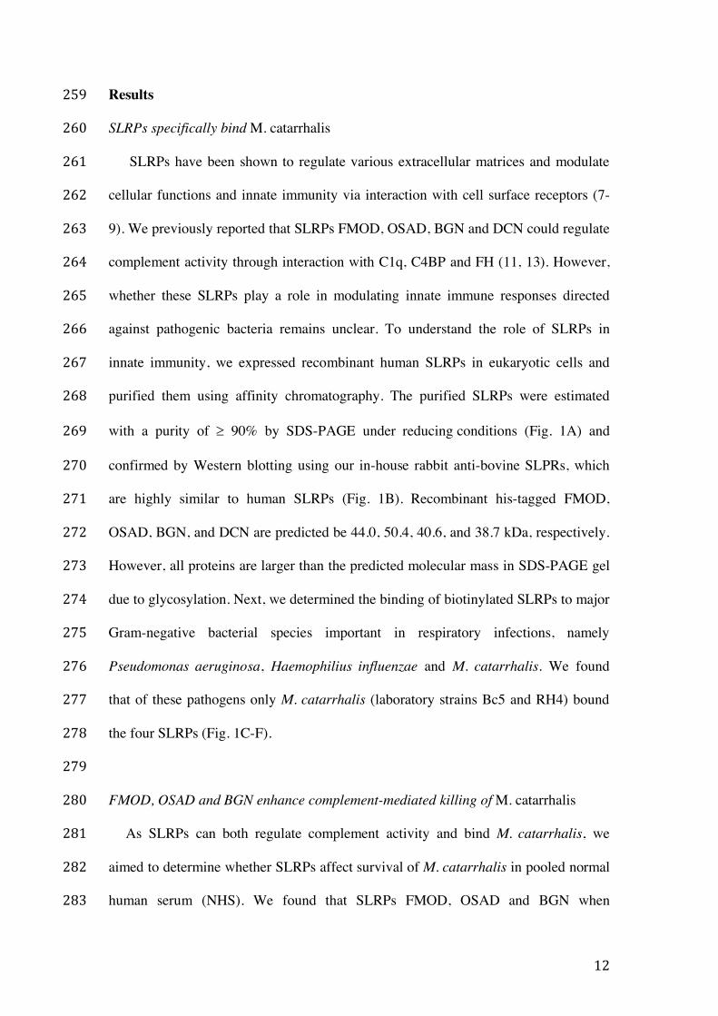
12
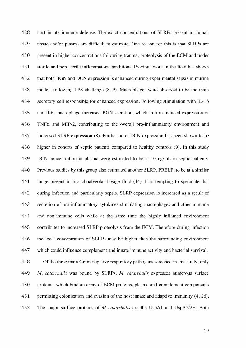
Results 259
SLRPs specifically bind M. catarrhalis 260
SLRPs have been shown to regulate various extracellular matrices and modulate 261
cellular functions and innate immunity via interaction with cell surface receptors (7-262
9). We previously reported that SLRPs FMOD, OSAD, BGN and DCN could regulate 263
complement activity through interaction with C1q, C4BP and FH (11, 13). However, 264
whether these SLRPs play a role in modulating innate immune responses directed 265
against pathogenic bacteria remains unclear. To understand the role of SLRPs in 266
innate immunity, we expressed recombinant human SLRPs in eukaryotic cells and 267
purified them using affinity chromatography. The purified SLRPs were estimated 268
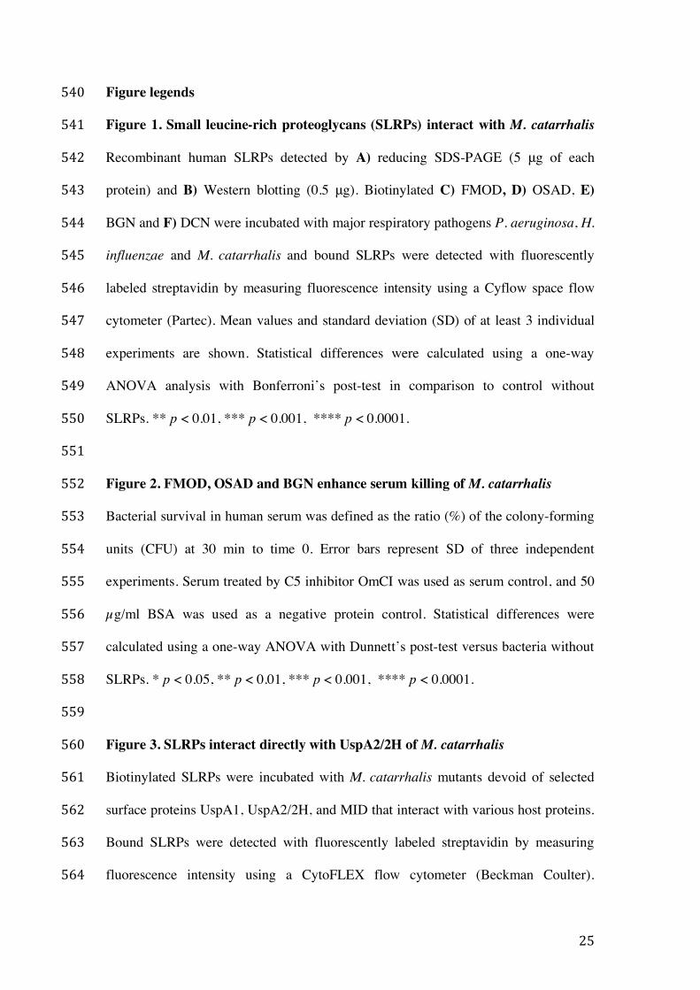
with a purity of ³ 90% by SDS-PAGE under reducing conditions (Fig. 1A) and 269
confirmed by Western blotting using our in-house rabbit anti-bovine SLPRs, which 270
are highly similar to human SLRPs (Fig. 1B). Recombinant his-tagged FMOD, 271
OSAD, BGN, and DCN are predicted be 44.0, 50.4, 40.6, and 38.7 kDa, respectively. 272
However, all proteins are larger than the predicted molecular mass in SDS-PAGE gel 273
due to glycosylation. Next, we determined the binding of biotinylated SLRPs to major 274
Gram-negative bacterial species important in respiratory infections, namely 275
Pseudomonas aeruginosa, Haemophilius influenzae and M. catarrhalis. We found 276
that of these pathogens only M. catarrhalis (laboratory strains Bc5 and RH4) bound 277
the four SLRPs (Fig. 1C-F). 278
279
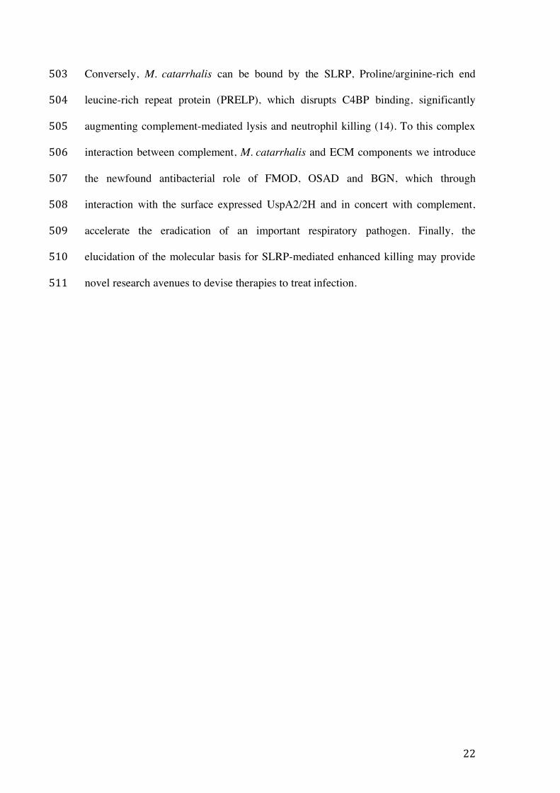
FMOD, OSAD and BGN enhance complement-mediated killing of M. catarrhalis 280
As SLRPs can both regulate complement activity and bind M. catarrhalis, we 281
aimed to determine whether SLRPs affect survival of M. catarrhalis in pooled normal 282
human serum (NHS). We found that SLRPs FMOD, OSAD and BGN when 283

13
supplemented at 50µg/mL significantly decreased survival of both M. catarrhalis 284
RH4 (Fig. 2A) and Bc5 (Fig. 2B) in NHS. Despite being not statistically significant, 285
DCN led to a slight reduction in survival in the Bc5 strain compared to BSA, but no 286
difference was observed in strain RH4, suggesting that DCN does not enhance 287
complement-mediated killing of M. catarrhalis. Furthermore, inhibition of MAC 288
formation by previous treatment of serum with the C5 inhibitor OmCI prevented 289
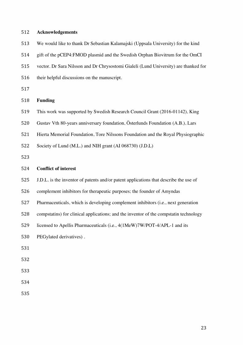
killing of M. catarrhalis under any SLRP condition illustrating that SLRPs enhance 290
killing through complement mediated lysis (Fig 2A-B). Lastly, no antimicrobial 291
activity was observed when SLRPs were incubated with M. catarrhalis in GVB++ 292
buffer in the absence of serum, confirming that the enhanced killing was mediated by 293
complement. To verify that excess unbound SLRPs were not causing a by-stander 294
complement activation effect and contributing to enhanced killing, we also measured 295
the effect of washing bacteria following SLRP binding prior to incubation with serum 296
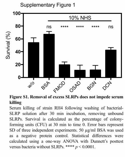
(Fig. S1). As in the above results, a significant decrease in survival was observed for 297
FMOD, OSAD and BGN but not DCN, indicating the SLRPs bound to the bacterial 298
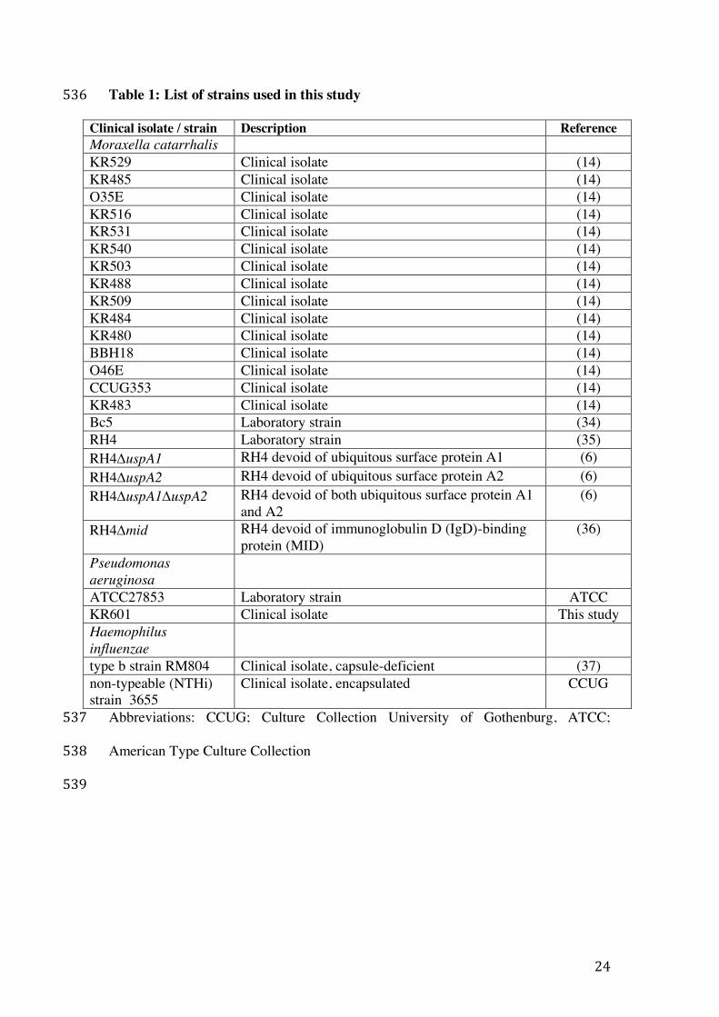
surface promoted enhanced bacterial killing in the presence of serum. 299
300
SLRPs interact directly with UspA2/2H of M. catarrhalis 301
M. catarrhalis interacts with human proteins via major surface proteins such as 302
UspA1/A2, MID, and outer membrane porins such as OmpCD and Mha (4). Given 303
that previous work has shown that UspA1, UspA2 and MID can interact with soluble 304
extracellular matrix proteins we investigated the interaction of wild type (RH4) and 305
isogenic mutants lacking the above surface proteins with biotinylated SLRPs through 306
flow cytometry (Fig. 3A-D). We found that deletion of the uspA2 gene resulted in a 307
significant decrease in binding of all SLRPs in question highlighting the importance 308

14
of UspA2 as a ligand for SLRP binding. No difference in binding was observed with 309
neither the uspA1 nor mid mutants. 310
To further elucidate the interaction between SLRPs and M. catarrhalis we 311
employed a direct biochemical binding assay using immobilized recombinant UspA2, 312
derived from strain RH4, and increasing concentrations of biotinylated SLRPs and 313
BSA (Fig. 3E-F). In accordance with our binding results above employing wild-type 314
and uspA2 mutant, all four SLRPs bound UspA2, with the highest affinity observed 315
for BGN (Kd = 89 ±11 nM), similar affinities seen for FMOD (Kd = 202 ±21 nM) 316
and OSAD (Kd = 231 ±28 nM) and the lowest affinity seen for DCN (Kd = 293 ±32 317
nM). 318
319
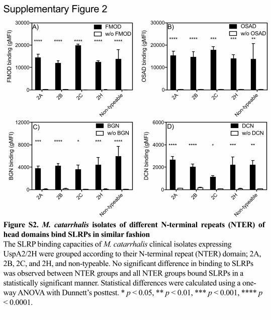
SLRPs bind to the majority of clinical isolates of M. catarrhalis 320
To determine the clinical relevance of M. catarrhalis interaction with SLRPs we 321
evaluated the binding capacity of a panel of clinical isolates (n=16) to all four SLRPs 322
(Fig. 4A-D). These clinical isolates were chosen based on their respective differences 323
in the N- terminal sequence motif of the UspA2 protein, in order to capture a 324
significant diversity of important clinical M. catarrhalis strains. This domain is 325
classified into the different groups 2A, 2B, 2C and ‘nontypeable’ based on the domain 326
distribution and sequence similarity (25). We found that the overwhelming majority 327
of clinical isolates bound all four SLRPs whereby there was a general trend for 328
increased binding in the order of FMOD ≥OSAD >BGN >DCN. However, isolates 329
that express UspA2/2H with different N-terminal repeats of head domains showed no 330
significant difference in binding of SLRPs (Fig. S2). 331
332
333
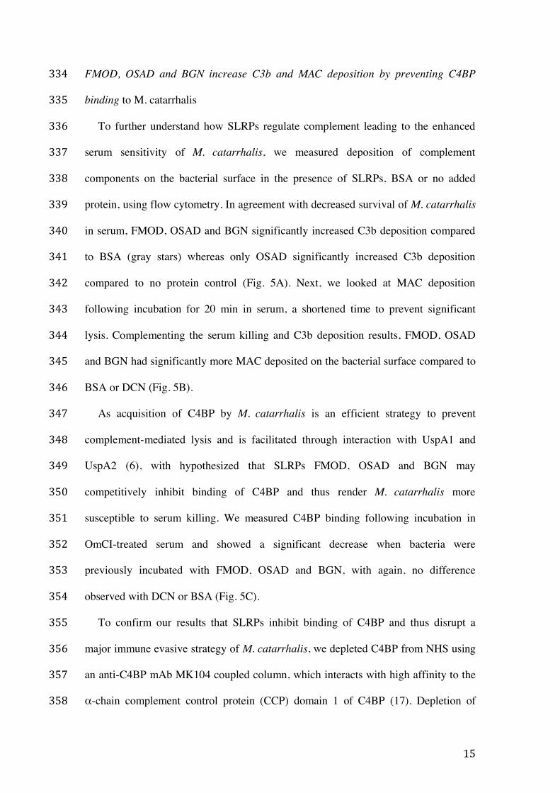
15
FMOD, OSAD and BGN increase C3b and MAC deposition by preventing C4BP 334
binding to M. catarrhalis 335
To further understand how SLRPs regulate complement leading to the enhanced 336
serum sensitivity of M. catarrhalis, we measured deposition of complement 337
components on the bacterial surface in the presence of SLRPs, BSA or no added 338
protein, using flow cytometry. In agreement with decreased survival of M. catarrhalis 339
in serum, FMOD, OSAD and BGN significantly increased C3b deposition compared 340
to BSA (gray stars) whereas only OSAD significantly increased C3b deposition 341
compared to no protein control (Fig. 5A). Next, we looked at MAC deposition 342
following incubation for 20 min in serum, a shortened time to prevent significant 343
lysis. Complementing the serum killing and C3b deposition results, FMOD, OSAD 344
and BGN had significantly more MAC deposited on the bacterial surface compared to 345
BSA or DCN (Fig. 5B). 346
As acquisition of C4BP by M. catarrhalis is an efficient strategy to prevent 347
complement-mediated lysis and is facilitated through interaction with UspA1 and 348
UspA2 (6), with hypothesized that SLRPs FMOD, OSAD and BGN may 349
competitively inhibit binding of C4BP and thus render M. catarrhalis more 350
susceptible to serum killing. We measured C4BP binding following incubation in 351
OmCI-treated serum and showed a significant decrease when bacteria were 352
previously incubated with FMOD, OSAD and BGN, with again, no difference 353
observed with DCN or BSA (Fig. 5C). 354
To confirm our results that SLRPs inhibit binding of C4BP and thus disrupt a 355
major immune evasive strategy of M. catarrhalis, we depleted C4BP from NHS using 356
an anti-C4BP mAb MK104 coupled column, which interacts with high affinity to the 357
α-chain complement control protein (CCP) domain 1 of C4BP (17). Depletion of 358
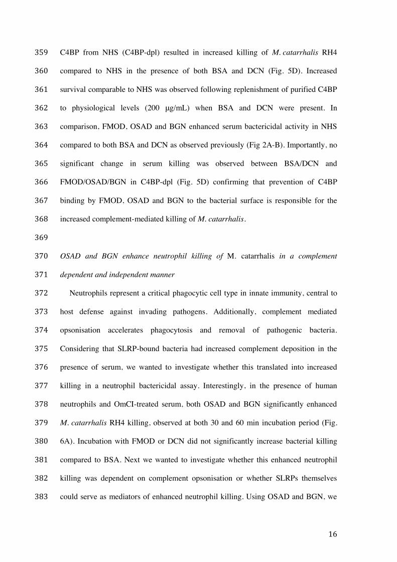
16
C4BP from NHS (C4BP-dpl) resulted in increased killing of M. catarrhalis RH4 359
compared to NHS in the presence of both BSA and DCN (Fig. 5D). Increased 360
survival comparable to NHS was observed following replenishment of purified C4BP 361
to physiological levels (200 μg/mL) when BSA and DCN were present. In 362
comparison, FMOD, OSAD and BGN enhanced serum bactericidal activity in NHS 363
compared to both BSA and DCN as observed previously (Fig 2A-B). Importantly, no 364
significant change in serum killing was observed between BSA/DCN and 365
FMOD/OSAD/BGN in C4BP-dpl (Fig. 5D) confirming that prevention of C4BP 366
binding by FMOD, OSAD and BGN to the bacterial surface is responsible for the 367
increased complement-mediated killing of M. catarrhalis. 368
369
OSAD and BGN enhance neutrophil killing of M. catarrhalis in a complement 370
dependent and independent manner 371
Neutrophils represent a critical phagocytic cell type in innate immunity, central to 372
host defense against invading pathogens. Additionally, complement mediated 373
opsonisation accelerates phagocytosis and removal of pathogenic bacteria. 374
Considering that SLRP-bound bacteria had increased complement deposition in the 375
presence of serum, we wanted to investigate whether this translated into increased 376
killing in a neutrophil bactericidal assay. Interestingly, in the presence of human 377
neutrophils and OmCI-treated serum, both OSAD and BGN significantly enhanced 378
M. catarrhalis RH4 killing, observed at both 30 and 60 min incubation period (Fig. 379
6A). Incubation with FMOD or DCN did not significantly increase bacterial killing 380
compared to BSA. Next we wanted to investigate whether this enhanced neutrophil 381
killing was dependent on complement opsonisation or whether SLRPs themselves 382
could serve as mediators of enhanced neutrophil killing. Using OSAD and BGN, we 383
17
repeated the neutrophil bactericidal assays with either OmCI-treated serum (inhibiting 384
complement at the C5 level) or compstatin-treated serum (inhibiting complement at 385
the C3 level). At 30 min we observed only a decrease in bacterial survival in the 386
OmCI-treated serum conditions and not in the presence of compstatin (Fig. 6B). 387
Surprisingly, after 60 min we observed a statistically significant decrease in survival 388
both with the OmCI- and compstatin-treated sera compared to BSA. This suggests 389
that the main mechanism of SLRP-dependent enhanced neutrophil killing is via 390
complement activation. After a prolonged incubation time, however, SLRPs promote 391
a bactericidal killing effect in concert with neutrophils, which is independent of 392
complement. 393
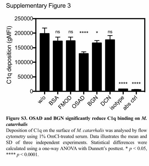
As compstatin-treated serum still contains C1q, which can act as an opsonin and 394
promote phagocytosis, and as previous work has shown that SLRPs can interact with 395
C1q (11, 12), we investigated the binding of C1q from serum in the presence of 396
SLRPs (Supp Fig. 3). We observed no difference in binding of C1q to the bacterial 397
surface when bacteria were incubated with FMOD or DCN compared to BSA. In 398
contrast, a significant reduction in C1q binding was shown when bacteria were bound 399
with OSAD and BGN. Therefore, these results indicate that enhanced neutrophil 400
killing under compstatin-treated serum conditions in the presence of OSAD and BGN 401
was not due to increased C1q binding. 402

18
Discussion 403
M. catarrhalis causes significant morbidity in children and COPD patients, and is 404
responsible for a plethora of respiratory infections and occasionally, systemic diseases 405
(26). The exact molecular mechanisms governing M. catarrhalis pathogenicity are not 406
fully understood. However, mounting evidence suggests that immune evasion, 407
directed primarily at circumventing the complement system, is an essential feature of 408
pathogenic strains (4-6, 14, 21, 27). Therefore, future treatment intervention directed 409
at hampering complement inhibitor recruitment is a promising avenue of research. In 410
this study, we highlight a novel antimicrobial role displayed by specific SLRPs, 411
namely FMOD, OSAD and BGN and in detail revealed the molecular mechanisms 412
resulting in enhanced innate immunity against M. catarrhalis. 413
SLRPs such as BGN and DCN are considered bi-functional proteoglycans, acting 414
both as central structural components of the ECM and danger-associated molecular 415
patterns (DAMPs) stimulating immune reactions (28). SLRPs are abundantly present 416
in the ECM and distributed in numerous tissues throughout the body (7). Previous 417
immumohistochemical analysis has shown that BGN and DCN are expressed in the 418
human lung and bronchial tissue (29-31). Furthermore, mining of the Human Protein 419
Atlas (www.proteinatlas.org) (32), a genome-wide analysis of RNA and protein 420
expression from samples representing major tissues and organs, confirmed the 421
expression of all SLRPs used in this study in lung tissue. RNA expression data 422
generated from 320 individual tissue samples showed that for this set of SLRPs, BGN 423
and DCN had the highest expression, followed by FMOD with OSAD having the 424
lowest expression (Suppl Fig 4). Combined, these expression data and previous 425
immunohistochemical analysis indicate that these SLRPs are present in sites 426
anatomically important for M. catarrhalis infection and therefore may play a role in 427
19
host innate immune defense. The exact concentrations of SLRPs present in human 428
tissue and/or plasma are difficult to estimate. One reason for this is that SLRPs are 429
present in higher concentrations following trauma, proteolysis of the ECM and under 430
sterile and non-sterile inflammatory conditions. Previous work in the field has shown 431
that both BGN and DCN expression is enhanced during experimental sepsis in murine 432
models following LPS challenge (8, 9). Macrophages were observed to be the main 433
secretory cell responsible for enhanced expression. Following stimulation with IL-1β 434
and Il-6, macrophage increased BGN secretion, which in turn induced expression of 435
TNFα and MIP-2, contributing to the overall pro-inflammatory environment and 436
increased SLRP expression (8). Furthermore, DCN expression has been shown to be 437
higher in cohorts of septic patients compared to healthy controls (9). In this study 438
DCN concentration in plasma were estimated to be at 10 ng/mL in septic patients. 439
Previous studies by this group also estimated another SLRP, PRELP, to be at a similar 440
range present in bronchoalveolar lavage fluid (14). It is tempting to speculate that 441
during infection and particularly sepsis, SLRP expression is increased as a result of 442
secretion of pro-inflammatory cytokines stimulating macrophages and other immune 443
and non-immune cells while at the same time the highly inflamed environment 444
contributes to increased SLRP proteolysis from the ECM. Therefore during infection 445
the local concentration of SLRPs may be higher than the surrounding environment 446
which could influence complement and innate immune activity and bacterial survival. 447
Of the three main Gram-negative respiratory pathogens screened in this study, only 448
M. catarrhalis was bound by SLRPs. M. catarrhalis expresses numerous surface 449
proteins, which bind an array of ECM proteins, plasma and complement components 450
permitting colonization and evasion of the host innate and adaptive immunity (4, 26). 451
The major surface proteins of M. catarrhalis are the UspA1 and UspA2/2H. Both 452

20
UspA1 and A2/2H interact with C4BP, however UspA2/2H is more strongly 453
expressed than UspA1 and therefore plays a more prominent role in C4BP binding 454
and in conferring a complement resistant phenotype (6). Through mutational analysis 455
we determined that all four SLRPs bound to M. catarrhalis predominantly through 456
UspA2/2H. This suggested that SLRP binding to UspA2/2H could competitively 457
inhibit C4BP binding resulting in reduced complement inhibition. Using flow 458
cytometry we illustrated that prior binding of FMOD, OSAD, BGN but not DCN to 459
M. catarrhalis effective reduced C4BP binding thus explaining the increased serum 460
sensitivity. 461
Given the similarity between BGN and DCN it is surprising that BGN and not 462
DCN competitively inhibits C4BP binding. Both BGN and DCN are members of the 463
class I SLRP family, possessing significant homology at both the protein and genetic 464
level. BGN contains two N-terminal tissue-specific chondroitin/dermatan sulfate side 465
chains whereas DCN contains one, and both differ in the pattern and level of 466
glycosylation (7). These differences permit both SLRPs to perform different tasks in 467
terms of ECM maintenance and cell signaling and possibly binding to different 468
regions of UspA2, resulting in differential inhibition of C4BP. Our results show that 469
M. catarrhalis can bind both DCN and C4BP simultaneously. UspA2 is a trimeric 470
autotransporter adhesin, which interacts with C4BP specifically at the CCP2, CCP5 471
and CCP7 domains (6). UspA2 is composed of a globular head and stalk domain and 472
therefore it is feasible that DCN but not the other SLRPs bind to specific regions 473
within UspA2 that are not required for C4BP binding. Future biochemical studies are 474
required to fully confirm this hypothesis. 475
Incubation of bacteria with SLRPs in the presence of serum resulted in significant 476
opsonisation with C3b/iC3b (Fig. 5A). As these opsonins are recognized by 477

21
complement receptors present on neutrophils we wished to examine where this 478
resulted in enhanced neutrophil bactericidal killing. Here we observed that only 479
OSAD and BGN effectively increased neutrophil killing of M. catarrhalis but not 480
FMOD or DCN. This was surprising considering that FMOD enhanced C3b 481
opsonisation in the presence of serum. Therefore, we checked whether this enhanced 482
neutrophil killing was independent of complement by using compstatin-treated serum, 483
effectively blocking complement at the C3 level. We observed that the majority of the 484
neutrophil killing was complement (opsonisation) mediated (Fig. 6A). However with 485
prolonged incubation both OSAD and BGN enhanced killing in a complement 486
(opsonisation) independent manner. It is known that certain ECM proteins such as the 487
SLRP lumican can enhance phagocytosis by interacting with both bacteria and 488
phagocytes via surface expressed integrins (33). Additionally, it has been shown that 489
other SLRPs such as BGN and DCN can bind to toll-like receptors expressed on 490
professional phagocytes and induce a pro-inflammatory response (8, 9). Two 491
questions arise that require future molecular dissection: 1) Can FMOD and OSAD 492
interact with professional phagocytes and induce an immune response analogous to 493
BGN and DCN and 2) can the SLRPs in question mediate an interaction between 494
bacteria and phagocytes which facilitates enhanced phagocytosis and subsequent 495
killing. As such future molecular characterization is underway to elucidate fully the 496
mechanisms of SLRPs mediated neutrophil bactericidal activity. 497
Recent work by our lab has shown that respiratory pathogens such as M. 498
catarrhalis can interact with ECM components whereby two opposing scenarios may 499
result, namely attenuated or enhanced complement activity. M. catarrhalis interacts 500
with cartilage oligomeric matrix protein (COMP) preventing complement deposition 501
and interfering with complement-independent phagocytosis, enhancing survival (21). 502
22
Conversely, M. catarrhalis can be bound by the SLRP, Proline/arginine-rich end 503
leucine-rich repeat protein (PRELP), which disrupts C4BP binding, significantly 504
augmenting complement-mediated lysis and neutrophil killing (14). To this complex 505
interaction between complement, M. catarrhalis and ECM components we introduce 506
the newfound antibacterial role of FMOD, OSAD and BGN, which through 507
interaction with the surface expressed UspA2/2H and in concert with complement, 508
accelerate the eradication of an important respiratory pathogen. Finally, the 509
elucidation of the molecular basis for SLRP-mediated enhanced killing may provide 510
novel research avenues to devise therapies to treat infection. 511
23
Acknowledgements 512
We would like to thank Dr Sebastian Kalamajski (Uppsala University) for the kind 513
gift of the pCEP4:FMOD plasmid and the Swedish Orphan Biovitrum for the OmCI 514
vector. Dr Sara Nilsson and Dr Chrysostomi Gialeli (Lund University) are thanked for 515
their helpful discussions on the manuscript. 516
517
Funding 518
This work was supported by Swedish Research Council Grant (2016-01142), King 519
Gustav Vth 80-years anniversary foundation, Österlunds Foundation (A.B.), Lars 520
Hierta Memorial Foundation, Tore Nilssons Foundation and the Royal Physiographic 521
Society of Lund (M.L.) and NIH grant (AI 068730) (J.D.L) 522
523
Conflict of interest 524
J.D.L. is the inventor of patents and/or patent applications that describe the use of 525
complement inhibitors for therapeutic purposes; the founder of Amyndas 526
Pharmaceuticals, which is developing complement inhibitors (i.e., next generation 527
compstatins) for clinical applications; and the inventor of the compstatin technology 528
licensed to Apellis Pharmaceuticals (i.e., 4(1MeW)7W/POT-4/APL-1 and its 529
PEGylated derivatives) . 530
531
532
533
534
535
24
Table 1: List of strains used in this study 536
Clinical isolate / strain Description Reference Moraxella catarrhalis KR529 Clinical isolate (14) KR485 Clinical isolate (14) O35E Clinical isolate (14) KR516 Clinical isolate (14) KR531 Clinical isolate (14) KR540 Clinical isolate (14) KR503 Clinical isolate (14) KR488 Clinical isolate (14) KR509 Clinical isolate (14) KR484 Clinical isolate (14) KR480 Clinical isolate (14) BBH18 Clinical isolate (14) O46E Clinical isolate (14) CCUG353 Clinical isolate (14) KR483 Clinical isolate (14) Bc5 Laboratory strain (34) RH4 Laboratory strain (35) RH4DuspA1 RH4 devoid of ubiquitous surface protein A1 (6) RH4DuspA2 RH4 devoid of ubiquitous surface protein A2 (6) RH4DuspA1DuspA2 RH4 devoid of both ubiquitous surface protein A1
and A2 (6)
RH4Dmid RH4 devoid of immunoglobulin D (IgD)-binding protein (MID)
(36)
Pseudomonas aeruginosa
ATCC27853 Laboratory strain ATCC KR601 Clinical isolate This study Haemophilus influenzae
type b strain RM804 Clinical isolate, capsule-deficient (37) non-typeable (NTHi) strain 3655
Clinical isolate, encapsulated CCUG
Abbreviations: CCUG; Culture Collection University of Gothenburg, ATCC; 537
American Type Culture Collection 538
539
25
Figure legends 540
Figure 1. Small leucine-rich proteoglycans (SLRPs) interact with M. catarrhalis 541
Recombinant human SLRPs detected by A) reducing SDS-PAGE (5 μg of each 542
protein) and B) Western blotting (0.5 μg). Biotinylated C) FMOD, D) OSAD, E) 543
BGN and F) DCN were incubated with major respiratory pathogens P. aeruginosa, H. 544
influenzae and M. catarrhalis and bound SLRPs were detected with fluorescently 545
labeled streptavidin by measuring fluorescence intensity using a Cyflow space flow 546
cytometer (Partec). Mean values and standard deviation (SD) of at least 3 individual 547
experiments are shown. Statistical differences were calculated using a one-way 548
ANOVA analysis with Bonferroni’s post-test in comparison to control without 549
SLRPs. ** p < 0.01, *** p < 0.001, **** p < 0.0001. 550
551
Figure 2. FMOD, OSAD and BGN enhance serum killing of M. catarrhalis 552
Bacterial survival in human serum was defined as the ratio (%) of the colony-forming 553
units (CFU) at 30 min to time 0. Error bars represent SD of three independent 554
experiments. Serum treated by C5 inhibitor OmCI was used as serum control, and 50 555
µg/ml BSA was used as a negative protein control. Statistical differences were 556
calculated using a one-way ANOVA with Dunnett’s post-test versus bacteria without 557
SLRPs. * p < 0.05, ** p < 0.01, *** p < 0.001, **** p < 0.0001. 558
559
Figure 3. SLRPs interact directly with UspA2/2H of M. catarrhalis 560
Biotinylated SLRPs were incubated with M. catarrhalis mutants devoid of selected 561
surface proteins UspA1, UspA2/2H, and MID that interact with various host proteins. 562
Bound SLRPs were detected with fluorescently labeled streptavidin by measuring 563
fluorescence intensity using a CytoFLEX flow cytometer (Beckman Coulter). 564

26
UspA2/2H mutant showed significantly reduced binding of all SLRPs (A-D). 565
Biotinylated SLRPs E) (FMOD and OSAD) and F) (BGN and DCN) bind 566
immobilized recombinant UspA2 with differing affinities. Error bars represent SD of 567
three independent experiments. Statistical differences were calculated using a one-568
way ANOVA with Dunnett’s posttest versus wild type (RH4) bacteria. ** p < 0.01, 569
*** p < 0.001, **** p < 0.0001. 570
571
Figure 4. SLRPs bind to M. catarrhalis clinical isolates expressing UspA2/2H 572
The highly diverse head domains of UspA2/2H are classified into N-terminal repeats 573
(NTER) 2A, 2B, 2C, and 2H, and non-typeable. All tested clinical strains bound 574
SLRPs at varying degrees. Negative control consisted of bacterial straining with 575
Streptavidin Alexa Fluor 647 in the absence of biotinylated proteins. Error bars 576
represent the SD of three individual experiments. 577
578
Figure 5. FMOD, OSAD and BGN increase complement deposition through 579
inhibition of C4BP binding 580
Deposition of complement components A) C3b B) MAC and C) C4BP on M. 581
catarrhalis RH4 was analyzed using flow cytometry. D) Bacterial survival in 5 % 582
C4BP depleted sera (C4BP dpl) with matched survival in 5 % NHS and C4BP dpl 583
replenished with C4BP (C4BP dpl + C4BP) at physiological concentrations. Error 584
bars represent SD of three (A-C) and 5 (D) independent experiments. Serum treated 585
by C5 inhibitor OmCI or heat-inactivated was used as serum control, and BSA was 586
used as a negative protein control. Grey stars (BSA) and black starts (without protein 587
w/o) indicate statistical calculations using a one-way ANOVA with Dunnett’s post-588
test. * p < 0.05, ** p < 0.01, *** p < 0.001, **** p < 0.0001, ns, not significant. 589

27
Figure 6. SLRPs OSAD and BGN enhance neutrophil killing of M. catarrhalis 590
Human neutrophils were incubated with M. catarrhalis RH4 in the presence of A) 591
OmCI-treated serum and SLRPs or BSA or B) OmCI or compstatin-reated serum and 592
OSAD, BGN or BSA for 30 or 60 min at 37°C and 5%CO2. Following incubation 593
total viable bacteria was enumerated following lysis of neutrophils and bacterial 594
survival was calculated by diving CFU at time 30 or 60 with that of time 0. Graphs 595
are presented as the mean and SD of 5 independent experiments and analyzed using a 596
two-way ANOVA with Bonferroni post-hoc tests comparing SLRP/BSA condition to 597
that of no protein control (without, w/o) A) or SLRPs to BSA control B). * p < 0.05, 598
** p < 0.01, *** p < 0.001, **** p < 0.0001. 599
600
Abbreviations: BHI, brain-heart infusion; BGN, biglycan; C4BP, C4b-binding 601
protein; CCP, complement control protein; DCN, decorin; dpl, depleted serum; FH, 602
factor H; FMOD, fibromodulin; MAC, membrane attack complex; NHS, normal 603
human serum; NTER, N-terminal repeat; OSAD, osteoadherin; SLRP, short leucine-604
rich proteoglycan; Usp, ubiquitous surface protein. 605
606
607
608
609
610
611
612
613
614
28
References 615
1. Merle,N.S.,S.E.Church,V.Fremeaux-Bacchi,andL.T.Roumenina.2015.616ComplementSystemPartI-MolecularMechanismsofActivationandRegulation.617FrontImmunol6:262.6182. Blom,A.M.,L.Kask,andB.Dahlback.2001.Structuralrequirementsfor619thecomplementregulatoryactivitiesofC4BP.JBiolChem276:27136-27144.6203. Blom,A.M.,T.Hallstrom,andK.Riesbeck.2009.Complementevasion621strategiesofpathogens-acquisitionofinhibitorsandbeyond.MolImmunol46:6222808-2817.6234. Su,Y.C.,B.Singh,andK.Riesbeck.2012.Moraxellacatarrhalis:from624interactionswiththehostimmunesystemtovaccinedevelopment.Future625Microbiol7:1073-1100.6265. Murphy,T.F.,andG.I.Parameswaran.2009.Moraxellacatarrhalis,a627humanrespiratorytractpathogen.ClinInfectDis49:124-131.6286. Nordstrom,T.,A.M.Blom,A.Forsgren,andK.Riesbeck.2004.The629emergingpathogenMoraxellacatarrhalisinteractswithcomplementinhibitor630C4bbindingproteinthroughubiquitoussurfaceproteinsA1andA2.JImmunol631173:4598-4606.6327. McEwan,P.A.,P.G.Scott,P.N.Bishop,andJ.Bella.2006.Structural633correlationsinthefamilyofsmallleucine-richrepeatproteinsand634proteoglycans.JStructBiol155:294-305.6358. Schaefer,L.,A.Babelova,E.Kiss,H.J.Hausser,M.Baliova,M.Krzyzankova,636G.Marsche,M.F.Young,D.Mihalik,M.Gotte,E.Malle,R.M.Schaefer,andH.J.637Grone.2005.Thematrixcomponentbiglycanisproinflammatoryandsignals638throughToll-likereceptors4and2inmacrophages.JClinInvest115:2223-2233.6399. Merline,R.,K.Moreth,J.Beckmann,M.V.Nastase,J.Zeng-Brouwers,J.G.640Tralhao,P.Lemarchand,J.Pfeilschifter,R.M.Schaefer,R.V.Iozzo,andL.641Schaefer.2011.Signalingbythematrixproteoglycandecorincontrols642inflammationandcancerthroughPDCD4andMicroRNA-21.SciSignal4:ra75.64310. Happonen,K.E.,D.Heinegard,T.Saxne,andA.M.Blom.2012.644Interactionsofthecomplementsystemwithmoleculesofextracellularmatrix:645relevanceforjointdiseases.Immunobiology217:1088-1096.64611. Sjoberg,A.P.,G.A.Manderson,M.Morgelin,A.J.Day,D.Heinegard,andA.647M.Blom.2009.Shortleucine-richglycoproteinsoftheextracellularmatrix648displaydiversepatternsofcomplementinteractionandactivation.MolImmunol64946:830-839.65012. Groeneveld,T.W.,M.Oroszlan,R.T.Owens,M.C.Faber-Krol,A.C.Bakker,651G.J.Arlaud,D.J.McQuillan,U.Kishore,M.R.Daha,andA.Roos.2005.652Interactionsoftheextracellularmatrixproteoglycansdecorinandbiglycanwith653C1qandcollectins.JImmunol175:4715-4723.65413. Happonen,K.E.,A.P.Sjoberg,M.Morgelin,D.Heinegard,andA.M.Blom.6552009.ComplementinhibitorC4b-bindingproteininteractsdirectlywithsmall656glycoproteinsoftheextracellularmatrix.JImmunol182:1518-1525.65714. Liu,G.,D.Ermert,M.E.Johansson,B.Singh,Y.C.Su,M.Paulsson,K.658Riesbeck,andA.M.Blom.2017.PRELPEnhancesHostInnateImmunityagainst659theRespiratoryTractPathogenMoraxellacatarrhalis.JImmunol198:2330-6602340.661
29
15. Kalamajski,S.,D.Bihan,A.Bonna,K.Rubin,andR.W.Farndale.2016.662FibromodulinInteractswithCollagenCross-linkingSitesandActivatesLysyl663Oxidase.JBiolChem291:7951-7960.66416. Dahlback,B.1983.PurificationofhumanC4b-bindingproteinand665formationofitscomplexwithvitaminK-dependentproteinS.BiochemJ209:666847-856.66717. Hardig,Y.,A.Hillarp,andB.Dahlback.1997.Theamino-terminalmodule668oftheC4b-bindingproteinalpha-chainiscrucialforC4bbindingandfactorI-669cofactorfunction.BiochemJ323(Pt2):469-475.67018. WorldMedical,A.2013.WorldMedicalAssociationDeclarationof671Helsinki:ethicalprinciplesformedicalresearchinvolvinghumansubjects.JAMA672310:2191-2194.67319. Potempa,M.,J.Potempa,M.Okroj,K.Popadiak,S.Eick,K.A.Nguyen,K.674Riesbeck,andA.M.Blom.2008.BindingofcomplementinhibitorC4b-binding675proteincontributestoserumresistanceofPorphyromonasgingivalis.JImmunol676181:5537-5544.67720. Tan,T.T.,T.Nordstrom,A.Forsgren,andK.Riesbeck.2005.The678respiratorypathogenMoraxellacatarrhalisadherestoepithelialcellsby679interactingwithfibronectinthroughubiquitoussurfaceproteinsA1andA2.J680InfectDis192:1029-1038.68121. Liu,G.,H.Gradstedt,D.Ermert,E.Englund,B.Singh,Y.C.Su,M.E.682Johansson,A.Aspberg,V.Agarwal,K.Riesbeck,andA.M.Blom.2016.Moraxella683catarrhalisEvadesHostInnateImmunityviaTargetingCartilageOligomeric684MatrixProtein.JImmunol196:1249-1258.68522. Nunn,M.A.,A.Sharma,G.C.Paesen,S.Adamson,O.Lissina,A.C.Willis,686andP.A.Nuttall.2005.ComplementinhibitorofC5activationfromthesofttick687Ornithodorosmoubata.JImmunol174:2084-2091.68823. Mastellos,D.C.,D.Yancopoulou,P.Kokkinos,M.Huber-Lang,G.689Hajishengallis,A.R.Biglarnia,F.Lupu,B.Nilsson,A.M.Risitano,D.Ricklin,andJ.690D.Lambris.2015.Compstatin:aC3-targetedcomplementinhibitorreachingits691primeforbedsideintervention.EurJClinInvest45:423-440.69224. Leffler,J.,M.Martin,B.Gullstrand,H.Tyden,C.Lood,L.Truedsson,A.A.693Bengtsson,andA.M.Blom.2012.Neutrophilextracellulartrapsthatarenot694degradedinsystemiclupuserythematosusactivatecomplementexacerbating695thedisease.JImmunol188:3522-3531.69625. Su,Y.C.,B.M.Hallstrom,S.Bernhard,B.Singh,andK.Riesbeck.2013.697ImpactofsequencediversityintheMoraxellacatarrhalisUspA2/UspA2Hhead698domainonvitronectinbindingandantigenicvariation.MicrobesInfect15:375-699387.70026. Verduin,C.M.,C.Hol,A.Fleer,H.vanDijk,andA.vanBelkum.2002.701Moraxellacatarrhalis:fromemergingtoestablishedpathogen.ClinMicrobiolRev70215:125-144.70327. Hallstrom,T.,T.Nordstrom,T.T.Tan,T.Manolov,J.D.Lambris,D.E.704Isenman,P.F.Zipfel,A.M.Blom,andK.Riesbeck.2011.Immuneevasionof705MoraxellacatarrhalisinvolvesubiquitoussurfaceproteinA-dependentC3d706binding.JImmunol186:3120-3129.70728. Frey,H.,N.Schroeder,T.Manon-Jensen,R.V.Iozzo,andL.Schaefer.2013.708Biologicalinterplaybetweenproteoglycansandtheirinnateimmunereceptors709ininflammation.FEBSJ280:2165-2179.710

30
29. Hallgren,O.,K.Nihlberg,M.Dahlback,L.Bjermer,L.T.Eriksson,J.S.711Erjefalt,C.G.Lofdahl,andG.Westergren-Thorsson.2010.Alteredfibroblast712proteoglycanproductioninCOPD.RespirRes11:55.71330. Weitoft,M.,C.Andersson,A.Andersson-Sjoland,E.Tufvesson,L.Bjermer,714J.Erjefalt,andG.Westergren-Thorsson.2014.Controlledanduncontrolled715asthmadisplaydistinctalveolartissuematrixcompositions.RespirRes15:67.71631. Huang,J.,R.Olivenstein,R.Taha,Q.Hamid,andM.Ludwig.1999.717Enhancedproteoglycandepositionintheairwaywallofatopicasthmatics.AmJ718RespirCritCareMed160:725-729.71932. Uhlen,M.,L.Fagerberg,B.M.Hallstrom,C.Lindskog,P.Oksvold,A.720Mardinoglu,A.Sivertsson,C.Kampf,E.Sjostedt,A.Asplund,I.Olsson,K.Edlund,721E.Lundberg,S.Navani,C.A.Szigyarto,J.Odeberg,D.Djureinovic,J.O.Takanen,S.722Hober,T.Alm,P.H.Edqvist,H.Berling,H.Tegel,J.Mulder,J.Rockberg,P.Nilsson,723J.M.Schwenk,M.Hamsten,K.vonFeilitzen,M.Forsberg,L.Persson,F.Johansson,724M.Zwahlen,G.vonHeijne,J.Nielsen,andF.Ponten.2015.Proteomics.Tissue-725basedmapofthehumanproteome.Science347:1260419.72633. Shao,H.,S.Lee,S.Gae-Scott,C.Nakata,S.Chen,A.R.Hamad,andS.727Chakravarti.2012.Extracellularmatrixlumicanpromotesbacterial728phagocytosis,andLum-/-miceshowincreasedPseudomonasaeruginosalung729infectionseverity.JBiolChem287:35860-35872.73034. Forsgren,A.,M.Brant,A.Mollenkvist,A.Muyombwe,H.Janson,N.Woin,731andK.Riesbeck.2001.IsolationandcharacterizationofanovelIgD-binding732proteinfromMoraxellacatarrhalis.JImmunol167:2112-2120.73335. Christensen,J.J.,J.Ursing,andB.Bruun.1994.Genotypicandphenotypic734relatednessof80strainsofBranhamellacatarrhalisofworldwideorigin.FEMS735MicrobiolLett119:155-159.73636. Nordstrom,T.,J.Jendholm,M.Samuelsson,A.Forsgren,andK.Riesbeck.7372006.TheIgD-bindingdomainoftheMoraxellaIgD-bindingproteinMID738(MID962-1200)activateshumanBcellsinthepresenceofTcellcytokines.J739LeukocBiol79:319-329.74037. Kroll,J.S.,andE.R.Moxon.1988.Capsulationandgenecopynumberat741thecaplocusofHaemophilusinfluenzaetypeb.JBacteriol170:859-864.742 743
P. ae
rugino
sa K
R601
P. ae
rugino
sa ATCC27
853
H. influ
enza
e RM80
4
H. influ
enza
e NTHi36
55
M. cata
rrhali
s Bc5
M. cata
rrhali
s RH4
0
5
10
15
20
25
FMO
D b
indi
ng (g
MFI
) FMODw/o FMOD *** ***
P. ae
rugino
sa K
R601
P. ae
rugino
sa ATCC27
853
H. influ
enza
e RM80
4
H. influ
enza
e NTHi36
55
M. cata
rrhali
s Bc5
M. cata
rrhali
s RH4
0
5
10
15
20
25
BGN
bin
ding
(gM
FI)
BGNw/o BGN
****
**
P. ae
rugino
sa K
R601
P. ae
rugino
sa ATCC27
853
H. influ
enza
e RM80
4
H. influ
enza
e NTHi36
55
M. cata
rrhali
s Bc5
M. cata
rrhali
s RH4
0
10
20
30
OSA
D b
indi
ng (g
MFI
) OSADw/o OSAD
***
***
P. ae
rugino
sa K
R601
P. ae
rugino
sa ATCC27
853
H. influ
enza
e RM80
4
H. influ
enza
e NTHi36
55
M. cata
rrhali
s Bc5
M. cata
rrhali
s RH4
0
1
2
3
4
5
DC
N b
indi
ng (g
MFI
)
DCN****
**
w/o DCN****
A) B)
C) D)
Figure 1
E) F)
SDS-PAGE Western Blot
Figure 1. Small leucine-rich proteoglycans (SLRPs) interact with M. catarrhalis Recombinant human SLRPs detected by A) SDS-PAGE and B) Western blotting. 5 µg of each protein was loaded into wells for reducing SDS-PAGE, and 0.5 µg for Western blotting. Biotinylated C) FMOD, D) OSAD, E) BGN and F) DCN were incubated with major respiratroy pathogens P. aeruginosa, H. influenzae and M. catarrhalis and bound SLRPs were detected with fluorescently labeled streptavidin by measuring fluorescence intensity. Standard error of the mean (SEM) of at least 3 individual experiments is shown. Statistical differences were calculated using a one-way ANOVA analysis with Bonferroni’s posttest in comparison to control without SLRPs. ** p < 0.01, *** p < 0.001, **** p < 0.0001.

w/o 5 50 5 50 5 50 5 50 BSA w/o
FMODOSAD
BGNDCN w/o
FMODOSAD
BGNDCN
0
50
100
150
200Su
rviv
al (%
)
µg/mL
FMOD OSAD BGN DCN 50 µg/mL 50 µg/mL
GVB++10% NHS-OmCI10% NHS
** *** **** ns ns
**** **** **** **** ***
M. catarrhalis RH4A)
w/o 5 50 5 50 5 50 5 50 BSA w/o
FMODOSAD
BGNDCN w/o
FMODOSAD
BGNDCN
0
50
100
150
200
Surv
ival
(%)
FMOD OSAD BGN DCN 50 µg/mL 50 µg/mL
* * * ns ns
GVB++20% NHS-OmCI20% NHS* * * * *
µg/mL
M. catarrhalis Bc5B)
Figure 2
Figure 2. SLRPs enhance serum killing of M. catarrhalis Bacterial survival in human serum was defined as the ratio (%) of the colony-forming units (CFU) at 30 min to time 0. Error bars represent SD of three independent experiments. Serum treated by C5 inhibitor OmCI was used as serum control, and 50 µg/ml BSA was used as a negative protein control. * p < 0.05, ** p < 0.01, *** p < 0.001, **** p < 0.0001 versus w/o SLRPs, one-way ANOVA with Dunnett’s posttest.

RH4
ΔuspA1
ΔuspA2
ΔuspA1ΔuspA2
Δmid
0
30000
60000
90000
120000
FMO
D b
indi
ng (g
MFI
)
*** ***
FMOD
RH4
ΔuspA1
ΔuspA2
ΔuspA1ΔuspA2
Δmid
0
50000
100000
150000
200000
250000
BGN
bin
ding
(gM
FI)
********
BGN
0.0 0.5 1.0 1.5 2.00.0
0.5
1.0
1.5
2.0
2.5
µM of added SLRP/BSA
SLR
P bi
ndin
g (A
bs 4
50nm
)
FMOD
OSAD
BSA
FMOD: Kd = 201.6 ±21 nMOSAD: Kd = 230.6 ±28 nM
RH4
ΔuspA1
ΔuspA2
ΔuspA1ΔuspA2
Δmid
0
50000
100000
150000
200000
250000
OSA
D b
indi
ng (g
MFI
)
*****
OSAD
RH4
ΔuspA1
ΔuspA2
ΔuspA1ΔuspA2
Δmid
0
50000
100000
150000
200000
DC
N b
indi
ng (g
MFI
)
********
DCN
0.0 0.5 1.0 1.5 2.00.0
0.5
1.0
1.5
2.0
2.5
µM of added SLRP/BSA
SLR
P b
indi
ng (A
bs 4
50nm
) BGN
DCN
BGN: Kd = 89.2 ±11 nM
DCN: Kd = 292.6 ±32 nM
BSA
Figure 3A) B)
C) D)
F)E)

KR529
KR485
O35E
KR516
KR531
KR540
KR503
KR488
Bc5
KR509
KR484
KR480
BBH18O46
E
CCUG353
KR483
0
10000
20000
30000
FMO
D b
indi
ng (g
MFI
)
FMODw/o FMOD
2A Head 2B Head 2C Head 2H Head Non-typeable
KR529
KR485
O35E
KR516
KR531
KR540
KR503
KR488
Bc5
KR509
KR484
KR480
BBH18O46
E
CCUG353
KR483
0
3000
6000
9000
12000
BGN
bin
ding
(gM
FI)
BGNw/o BGN
2A Head 2B Head 2C Head 2H Head Non-typeable
KR529
KR485
O35E
KR516
KR531
KR540
KR503
KR488
Bc5
KR509
KR484
KR480
BBH18O46
E
CCUG353
KR483
0
10000
20000
30000
OSA
D b
indi
ng (g
MFI
)
OSADw/o OSAD
2A Head 2B Head 2C Head 2H Head Non-typeable
KR529
KR485
O35E
KR516
KR531
KR540
KR503
KR488
Bc5
KR509
KR484
KR480
BBH18O46
E
CCUG353
KR483
0
1000
2000
3000
4000
5000DCNw/o DCN
BGN
bin
ding
(gM
FI)
2A Head 2B Head 2C Head 2H Head Non-typeable
Figure 4
A) B)
D)C)
Figure 4. SLRPs bind to M. catarrhalis clinical isolates expressing UspA2/2H. The highly diverse head domains of UspA2/2H are classified into N-terminal repeats (NTER) 2A, 2B, 2C, and 2H, and non-typeable. All tested clinical strains bound SLRPs at varying degrees. Negative control consisted of bacterial straining with Streptavidin Alexa Fluor 647 in the absence of biotinylated proteins. Error bars represent the SD of three individual experiments.

w/oBSA
FMODOSAD
BGNDCN HI
isotyp
e
abs c
trl0
30000
60000
90000
120000
150000
C3d
dep
ositi
on (g
MFI
)
**ns ns ns**** *** ns
2.5% NHS-OmCI
w/oBSA
FMODOSAD
BGNDCN
isotyp
e
abs c
trl0
20000
40000
60000
80000
C4B
P de
posi
tion
(gM
FI) ns ns
1.25 % NHS-OmCI
**** ****
** **** ****
w/oBSA
FMODOSAD
BGNDCN
OmCI
isotyp
e
abs c
trl0
5000
10000
15000
20000
25000
MAC
dep
ositi
on (g
MFI
) * ****
**** **** ****
*** nsns
10% NHS
0
20
40
60
80
100
120Su
rviv
al (%
)
FMOD OSAD BGN DCNBSA
NHS
C4BP dpl
C4BP dpl + C4BP
* *
p=0.07
ns ns ns
*
A) B)
C) D)
Figure 5
Figure 5. FMOD, OSAD and BGN increase complement deposition through inhibition of C4BP bindingDeposition of complement components A) C3b B) MAC and C) C4BP on M. catarrhalis was analyzed using flow cytometry. D) Bacterial survival in 5 % C4BP depleted sera (C4BP dpl) with matched survival in 5 % NHS and C4BP dpl replenished with C4BP (C4BP dpl + C4BP) at physiological concentrations. Error bars represent SD of three (A-C) and 5 (D) independent experiments. Serum treated by C5 inhibitor OmCI or heat-inactivated was used as serum control, and BSA was used as a negative protein control. Grey stars (BSA) and black starts (without protein w/o) indicate statistical calculations using a one-way ANOVA with Dunnett’s posttest. * p < 0.05, ** p < 0.01, *** p < 0.001, **** p < 0.0001, ns, not significant.

w/oBSA
FMODOSAD
BGNDCN
0
20
40
60
80
Surv
ival
(%)
30 min
60 min*******
****
p=0.06
BSAOSAD
BGNBSA
OSADBGN
0
50
100
150
200
Surv
ival
(%)
compstatin
OmCI
30 min 60 min** * ** *
** **
Figure 6A)
B)
Figure 6. SLRPs OSAD and BGN enhance neutrophil killing of M. catarrhalisHuman neutrophils were incubated with M. catarrhalis in the presence of A) OmCI-treated serum and SLRPs or BSA or B) OmCI or compstatin-reated serum and OSAD, BGN or BSA for 30 or 60 min at 37°C and 5%CO2. Following incubation total viable bacteria was enumerated following lysis of neutrophils and bacterial survival was calculated by diving CFU at time 30 or 60 with that of time 0. Graphs are presented as the mean and SD of 5 independent experiments and analyzed using a two-way ANOVA with Bonferroni post hoc tests comparing SLRP/BSA condition to that of no protein control (without, w/o) A) or SLRPs to BSA control B). * p < 0.05, ** p < 0.01, *** p < 0.001, **** p < 0.0001.
w/oBSA
FMODOSAD
BGNDCN
0
20
40
60
80
100Su
rviv
al (%
)
ns ns**** **** ****
10% NHS
Supplementary Figure 1
Figure S1. Removal of excess SLRPs does not impede serum killingSerum killing of strain RH4 following washing of bacterial-SLRP solution after 30 min incubation, removing unbound SLRPs. Survival is calculated as the percentage of colony-forming units (CFU) at 30 min to time 0. Error bars represent SD of three independent experiments. 50 µg/ml BSA was used as a negative protein control. Statistical differences were calculated using a one-way ANOVA with Dunnett’s posttest versus bacteria without SLRPs. **** p < 0.0001.
2A 2B 2C 2H
Non-ty
peab
le0
10000
20000
30000
FMO
D b
indi
ng (g
MFI
) FMODw/o FMOD
A)
**** **** **** **** ****
2A 2B 2C 2H
Non-ty
peab
le0
4000
8000
12000
BGN
bin
ding
(gM
FI)
BGNw/o BGN
*** **** **** ****
C)
2A 2B 2C 2H
Non-ty
peab
le0
10000
20000
30000
OSA
D b
indi
ng (g
MFI
)
OSADw/o OSAD
**** **** *** *** **
B)
2A 2B 2C 2H
Non-ty
peab
le0
2000
4000
6000
DC
N b
indi
ng (g
MFI
)
DCNw/o DCN
D)
**** **** *** ***
Supplementary Figure 2
Figure S2. M. catarrhalis isolates of different N-terminal repeats (NTER) of head domains bind SLRPs in similar fashion The SLRP binding capacities of M. catarrhalis clinical isolates expressing UspA2/2H were grouped according to their N-terminal repeat (NTER) domain; 2A, 2B, 2C, and 2H, and non-typeable. No significant difference in binding to SLRPs was observed between NTER groups and all NTER groups bound SLRPs in a statistically significant manner. Statistical differences were calculated using a one-way ANOVA with Dunnett’s posttest. * p < 0.05, ** p < 0.01, *** p < 0.001, **** p < 0.0001.
w/oBSA
FMODOSAD
BGNDCN
isotyp
e
abs c
trl0
50000
100000
150000
200000
250000
C1q
dep
ositi
on (g
MFI
)
****ns ns ns*
**** ****
Supplementary Figure 3
Figure S3. OSAD and BGN significantly reduce C1q binding on M. catarrhalisDeposition of C1q on the surface of M. catarrhalis was analysed by flow cytometry using 1% OmCI-treated serum. Data illustrates the mean and SD of three independent experiments. Statistical differences were calculated using a one-way ANOVA with Dunnett’s posttest. * p < 0.05, **** p < 0.0001.

FMOD OSAD BGN DCN0
40
80
120
0
400
800
1200
1600
RPK
MR
PKM
Supplementary Figure 4
Figure S4. SLRP RNA expression from lung tissue RNA expression of SLRPs from lung tissue samples (n=320) adapted from the Human Protein Atlas program (www.proteinatlas.org) (32). RNA sequencing data is reported in reads per kilobase per million mapped reads (RPKM).


