UNIVERSITI PUTRA MALAYSIA
PREVALENCE AND CHARACTERIZATION OF CHLAMYDIA PSITTACIE FROM BIRDS IN MALAYSIA
PHONG SU FUN
FPV 1998 6

PREVALENCE AND CHARACTERIZATION OF CHLAMYDIA PSITTACIE FROM BIRDS IN MALAYSIA
By
PHONG SU FUN
Thesis Submitted in Fulfilment of the Requirement for the Degree of Master of Science in Faculty of
Veterinary Medicine and Animal Science Universiti Putra Malaysia
February 1998

Dedicated with love and gratitude to:
My husband Wesley Voon and my parents, as
my source of encouragement and support in completion of this study.
ACKNO�EDGEMENTS
I am indebted to Professor Dr. Aini Ideris, the deputy dean of the
faculty of Veterinary Medicine and Animal Science, Universiti Putra Malaysia
(UPM), Serdang, Selangor Darul Ehsan, for kindly supervising me in this
Master research. Her constant guidance and encouragement was the
motivation which helped me in completion of my work.
I am grateful to Dr. Karim Sadun for his participation in laboratory
works and mostly for sharing his knowledge and for his guidance. Thanks
also to Dr. Nadzri Salim for helping in epidemiology studies, especially for
his assistance in data analysis and interpretation.
I am also grateful to Dr. Mangalam Sinniah of the Institute Medical
Research (IMR), Kuala Lumpur for her kind support especially for giving the
McCoy cells. Thanks also are due to the Virology laboratory technician, Ms
Lee Phon Chun of the IMR who constantly supported me technically and
assisting in the McCoy cell culture preparation and chlamydia isolation.
I would like to express my thanks to Mr. Ho Ooi Kuan and Puan
Aminah Jusoh for teaching and assisting me in processing the specimen for
electron microscopy work. Dr. Abdul Rahman Omar, Puan Rodiah Husin, En.
Adam bin Ahmad, Puan Faridah Akmal, En. Mohd Kamarudin and all the
staffs in the Virology laboratory of the Veterinary Faculty, UPM, Serdang for
III

always being so willing to render assistance throughout the course of my
study, and making my time in the Vaccine laboratory an enjoyable one. Not
to forget, En. Fauzi Che Yusuf, the photographer at the Veterinary faculty,
who kindly assisted me in taking and developing the photographs and slides.
Finally, my gratitude goes to everybody who has helped or contributed in one
way or another towards the completion of this study.
iv
T ABLE OF CONTENTS Page
ACKNOWLED GEMENT.......................................................................... ili L IST OF TABLES........................................................................................ viii LIST OF F IGURES.............. ...................................... .......... ...... .................. ix LIST OF PLATES........................................................................................ x L IST OF ABBREV IATIONS................ ............................. ..................... ..... xii ABSTRACT.................................................................................................. xiv ABSTRAK............... ..................................................................................... xvii
CHAPTER
I INTRODUCT ION............................................................................ 1
II LITERATURE REVIEW... .................... .......... ...... .................. .......... 6 Species and Host Range.................................. ................................. 6 Transmission................................................................................... 6 Importance of Chlamydiosis.......... ..................... ..... ....................... 7
Chlamydiosis in Birds. ............... .......................................... 8 Chlamydiosis in Human...................................................... 10
Biology of Chlamydia.................................................... ....... .......... 12 Common Characteristics............... ....................................... 12 Morphological and Biological Characteristics of Chlamydia........................................................... ... ....... ........ 13 The Chlamydial Development Cycle.................................. 16 Antigenic Structure............................................................... 18 Resistance ................................. ,............................................ 19
Diagnosis.......................................................................................... 19 Diagnosis of Chlamydia, Past and Present........................ 20 Tissue Culture .......... ........................................................... 20 Egg Inoculation............... ........................ ............................. 24 Histochemical Staining................. ........ ... ....... ..................... 24 Immunochemical Tests ...... ...................... ............. .............. 25 Polymerase Chain Reaction (PCR) ............ .................... ..... 26 Enzyme Linked Immunosorbent Assay (EL ISA).............. 27 Complement Fixation Test.................................................. 27 Clinical Specimens and Transport Method.......................... 28
Recognition of Inclusion........................................................... ......... 29 Differentiation of Chlamydia Species and Strains.......................... 30
v
III PREVALENCE STUDY ON CHLAMYDIA PSITTACIE INFECTION IN PIGEONS, DOVES AND PARROTS.............. 31 Introduction................................................................................... 31 Materials and Mefuods............................... ......... ....... .................. 32
Source of Samples.......... ......... ........ ....... ............................ 32 Sampling Procedures........................................................ 35 Detection of Antigen............................ ..... ........... .......... .... 35 Test Procedures.................................................................. 37
Results......................... ................................................... ............. ... 38 Discussion......... ... ............................. ............ ........................ ......... 43 Summary........................................................................................ 50
IV ISOLATION AND IDENTIFICATION OF CHLAMYDIA....... 52 Introduction......................... .... .............................................. ........ 52 Materials and Mefuods ... ... ................. ......................................... 53
Collection of Samples.. ................ ....................... ............... 53 Processing of Samples....................................................... 53 Preparation and Propagation of Cell Cultures............... 54 Inoculation in Tissue Culture........................................... 55 Detection of Chlamydia psittacie in Cell Culture........... .. 56 Giemsa Staining..................... ................ ............................ 56 Gimenez Staining..... ............. ............. ...... ........ ................. 57 Identification of Chlamydia psittacie.......... .......... .............. 58 Indirect Immunofluorescence (IIF) Technique............... 58 Indirect Immunoperoxidase (TIP) Technique.................. 59 Preparation of Infected Cells for Transmission Electron Microscopy (TEM).... ..... ..................................... 60 Uranyl Acetate and Lead Citrate Staining...................... 61
Results............................................................................................ 65 Discussion...................................................................................... 76 Summary........................................................................................ 83
V GENERAL DISCUSSION AND CONCLUSION........................ 85
BIBLIOGRAPHy.......... ............................................................................. 92
APPENDICES A : Media and Tissue Culture Solution ............. ................ ......... 102 B : Working Cell Suspension........................................................ 106 C: Chemical for Detection of Chlamydia........................ . . .......... 107
Vl
D: Chemical for Identification of Chlamydia............................. 108
E: Epidemiological Sampling Data.................................... ......... 111
VITA ......................................................................................................... 113
vii

LIST OF TABLES
Table
1. The Order, Family, Common Name, Species and Number
Page
of Birds in the Study......................................................... 34
2. Percentage of Faecal Samples Positive for Chlamydiosis by Source of Birds..................................... ............ ................. 40
3. Frequency Distribution of Faecal Samples Positive for Chlamydiosis by Major Bird Species Group................. 41
4. Frequency Distribution of Faecal Samples Positive for Chlamydiosis According to Whether Samples Came from Birds Kept 2 or More Together or Singly in Cages..... 41
5. Frequency Distribution of Faecal Samples Positive for Chlamydiosis According to Cage Condition................ 41
6. Frequency Distribution of Faecal Samples Tested Positive for Chlamydiosis with Respect to Signs of illness in the Sampled Birds................................................................... 42
7. Frequency Distribution of Faecal Samples taken from Various Species of Birds that Tested Positive for Chlamydiosis by the Clearview Chlamydia Test Kit.... 42
8. Type of Birds, Source, Brief Clinical History of Pertinent Information and Clearview Chlamydia test Results ..... 110
Vlll

LIST OF FIGURES
Figure Page
1. Chlamydia Developmental Cycles (Quinn et aI, 1994)....... 17
IX
lIST OF PLATES
Plate
1. Transmission Electron Photomicrograph of a C. psittacie inclusion (INClUS) in infected 1929 Cells. The Various Morphological Forms of Chlamydiae are Present : Elementary Body (EB), Reticular Body (IB). x 15000.
Page
(Grimes and Wyrick, 1991)................................................... 15
2. A Clinically Infected Spotted Dove. ........... ....................... 63
3. This Spotted Dove Showed Clinical Sign of Diarrhoea
4.
5.
6.
7.
8.
9.
10.
11.
( dirty-wet vent at the cloaca region)............ ....................... 63
Normal McCoy Cell Tissue Culture (x 400 Magnification) ..................................................... ................... 64
Normal McCoy Cells (as negative control) ........................ 64
The Inclusion in the Cytoplasm of the McCoy Cells was Stained Dark Purple in Giemsa Stain (xlOOO Magnification) ........................................................................ 68
The Inclusion in the Cytoplasm of the positive McCoy Cells was Stained as Bright White-green Granular Bodies in Giemsa Stain ( x400 magnification) ................... 68
The Inclusion in the Cytoplasm of the positive McCoy Cells was Stained bluish red in Gimenez Stain (x1000 magnification) ....................................................................... 69
In Indirect Immunofluorescent Technique, the Chlamydia was seen as apple green inclusion in the cytoplasm of the McCoy Cells ( x 1000 magnification) ................................... 69
Chlamydia Inclusion in Indirect Immunofluorescent Technique of the McCoy Cells (x 1000 magnification) ....... 70
Multiple Inclusions of Chlamydia in Indirect Immunofluorescent Technique (xIOOO magnification) ........ 70
x
12. In Indirect Immunoperoxidase Technique, Chlamydia Inclusions in the Cytoplasm of the McCoy Cells (x 400 magnification).............................................................. 71
13. The Major Morphological Features of Intracellular Chlamydia Development (RB, EB, IB) under T. E.M. ( x70 000 magnification)....................................................... 72
14. Multiple Inclusions were seen under Transmission Electron Microscope (x 30 000 magnification)................................... 73
15. Lysis of McCoy Cells and Released of EBs from the Cytoplasm of McCoy Cells (x 40 000 magnification).... ...... 74
16. Normal McCoy cells as negative control ( x 40 000 magnification)........................................................................ 75.
xi
ATP
CDC
C02
CTM
DCF
DNA
EB
ELISA
FCS
FITC
GM
IB
IIF
IIP
IMR
KG
LIP
MEM
LIST OF ABBREVIATIONS
Adenosine Triphosphate
Center For Disease Control
Carbon Dioxide
Chlamydia Transport Medium
Direct Complement Fixation
Deoxyribonucleic Acid
Elementary Body
Enzyme Linked Immunosorbent Assay
Fetal Calf Serum
Fluorescein Isothiocyanate
Growth Medium
Hydrogen Peroxide
Intermediate Body
Indirect Immunofluorescence
Indirect Immunoperoxidase
Institute Medical Research
Kilogram
Lipopolysaccharide
Minimum Essential Media
xii
mg
min
ml
mm
MM
MOMP
nm
PBS
peR
RB
RFLP
RNA
SBL
Milligram
Minute
Milliliter
Millimeter
Maintenance Medium
Major Outer Membrane Proteins
Nanometer
Phosphate Buffer Saline
Polymerase Chain Reaction
Reticulate Body
Restriction Fragment Length Polymorphism
Ribonucleic Acid
Sick Bird Look
SOS-PAGE Sodium Dodecyl Sulphate-polyacrylamide Gel Electrophoresis
SPG Sucrose Phosphate Glutamate
TEM Transmission Electron Microscopy
UPM Universiti Putra Malaysia
J.lg Microgram
J.lm Micrometer
J.lI Microliter
xiii

Abstract of thesis presented to the Senate of Universiti Putra Malaysia in fulfilment of the requirements for the degree of Master of Science.
PREVALENCE AND CHARACTERIZATION OF CHLAMYDIA PSIITACIE FROM BIRDS IN MALAYSIA
Chairperson:
Faculty:
By
PHONG SU FUN February 1998
Professor Dr. Aini Ideris, Ph.D.
Veterinary Medicine and Animal Science
Chlamydia psittacie causes a range of clinical syndromes, from life
threatening systemic disease to an inapparent infection in birds and is a
zoonotic disease. The disease is important in many countries with infection
rates ranging from 10 - 40 % and carrier rate that could exceed 90 %. In
Malaysia, no study on avian chlamydiosis has been done. Therefore, this
study is conduded to determine the epidemiological and biological
properties of Chlamydia psittacie in birds from Malaysia. The surveillance on
prevalence rate of Chlamydia psittacie in pigeons, doves and parrots was
carried out in the areas of Klang Valley. One hundred and fifty two (152)
fresh faecal samples were collected from different species of birds with
different management background. The birds were grouped into four
categories, namely, pet bird shops, parks, individual pet birds owner and
xiv
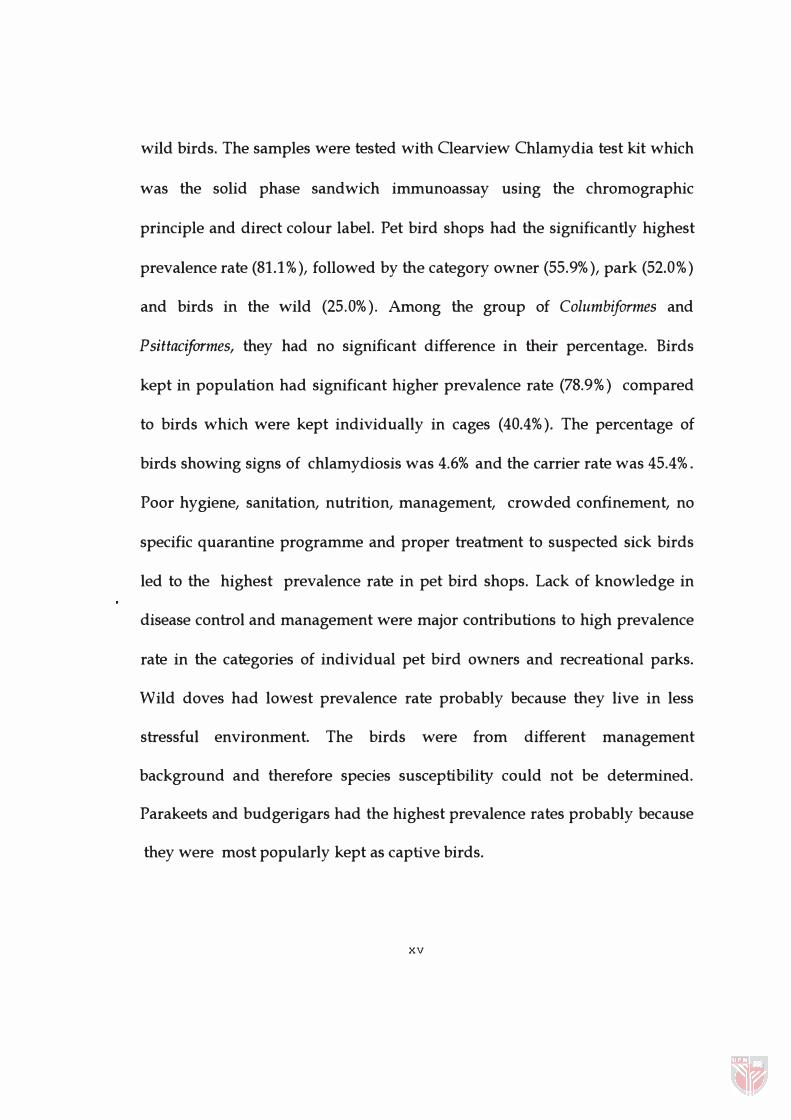
wild birds. The samples were tested with Clearview Chlamydia test kit which
was the solid phase sandwich immunoassay using the chromographic
principle and direct colour label. Pet bird shops had the significantly highest
prevalence rate (81.1 %), followed by the category owner (55.9%), park (52.0 %)
and birds in the wild (25.0%). Among the group of Columbiformes and
Psittaciformes, they had no significant difference in their percentage. Birds
kept in population had significant higher prevalence rate (78.9%) compared
to birds which were kept individually in cages (40.4%). The percentage of
birds showing signs of chlamydiosis was 4.6% and the carrier rate was 45.4%.
Poor hygiene, sanitation, nutrition, management, crowded confinement, no
specific quarantine programme and proper treatment to suspected sick birds
led to the highest prevalence rate in pet bird shops. Lack of knowledge in
disease control and management were major contributions to high prevalence
rate in the categories of individual pet bird owners and recreational parks.
Wild doves had lowest prevalence rate probably because they live in less
stressful environment. The birds were from different management
background and therefore species susceptibility could not be determined.
Parakeets and budgerigars had the highest prevalence rates probably because
they were most popularly kept as captive birds.
xv
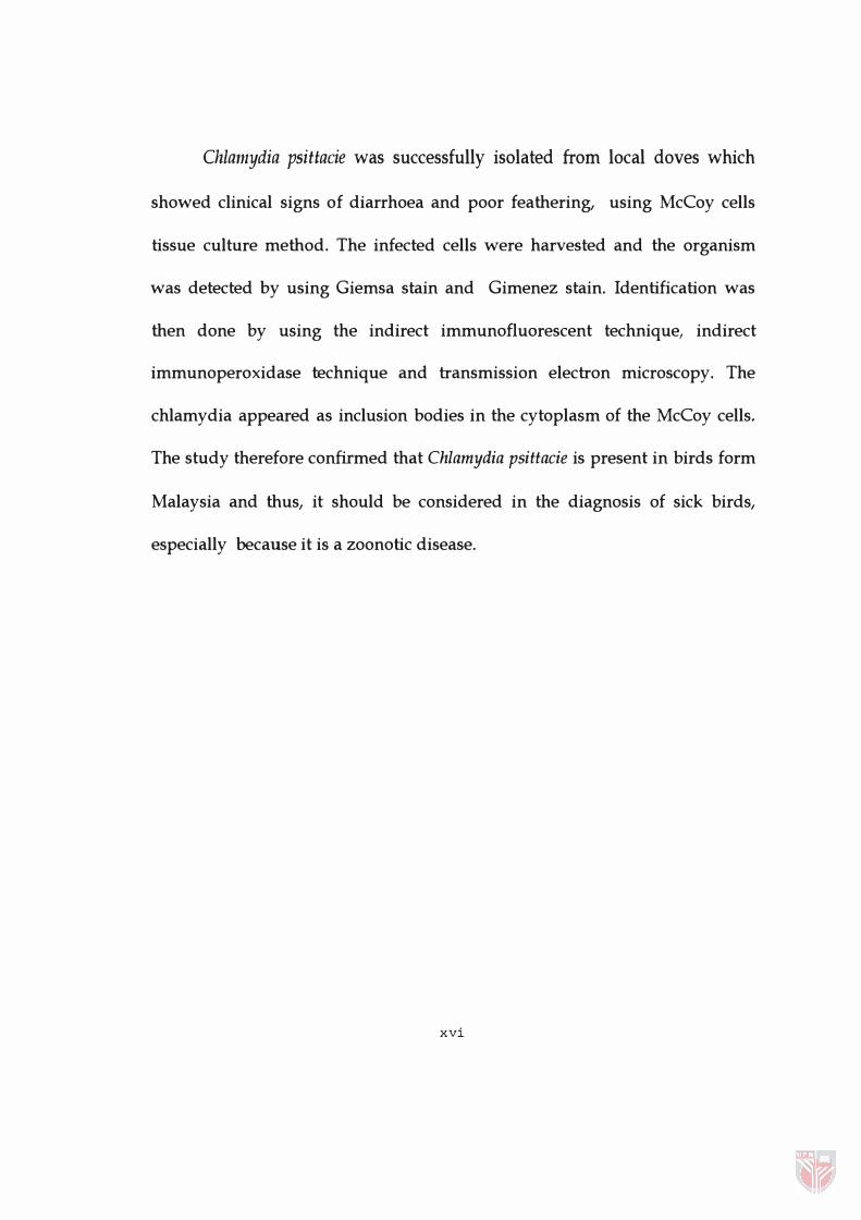
Chlamydia psittacie was successfully isolated from local doves which
showed clinical signs of diarrhoea and poor feathering, using McCoy cells
tissue culture method. The infected cells were harvested and the organism
was detected by using Giemsa stain and Gimenez stain. Identification was
then done by using the indirect immunofluorescent technique, indirect
immunoperoxidase technique and transmission electron microscopy. The
chlamydia appeared as inclusion bodies in the cytoplasm of the McCoy cells.
The study therefore confirmed that Chlamydia psittacie is present in birds form
Malaysia and thus, it should be considered in the diagnosis of sick birds,
especially because it is a zoonotic disease.
xvi
Abstrak tesis yang dikemukakan kepada Senat Universiti Putra Malaysia bagi memenuhi syarat untuk Ijazah Master Sains.
PREVALENS DAN PENCIRIAN CHLAMYDIA PSITTACIE PADA BURUNG-BURUNG 01 MALAYSIA
Pengerusi:
Fakulti:
Oleh
PHONG SU FUN Februari 1998
Professor Dr. Aini Ideris, Ph.D.
Kedoktoran Veterinar dan Sains Petemakan
Chlamydia psittacie boleh menyebabkan pelbagai tanda klinikal
daripada jangkitan sistem yang mengancam nyawa kepada yang tidak nyata
pada burung dan ia juga adalah satu penyakit zoonotik. Penyakit ini adalah
penting di negara-negara lain, seperti mana yang telah dilaporkan oleh Fudge
(1990) bahawa kadar jangkitan chlamydia pada burung kesayangan adalah
antara 10 hingga 40 % dan kadar pembawa boleh melebihi 90%. Di Malaysia,
tiada kajian dijalankan terhadap penyakit chlamydia avian. Oleh itu, satu
kajian telah dijalankan untuk menentukan ciri-ciri epidemiologi dan biologi
Chlamydia psittacie pada burung-burung di Malaysia. Tinjauan terhadap
kadar jangkitan yang disebabkan oleh Chlamydia psittacie pada burung-
burung merpati, tekukur dan nuri telah dijalankan di sekitar kawasan
Lembah Kelang. Sebanyak 152 sampel najis segar burung telah diambil dari
kumpulan burung yang berlainan dari segi spesis dan pengurusannya.
xvii
Burung-burung itu dikumpulkan kepada 4 kategori, iaitu dari kedai-kedai
burung, taman burung, burung yang dipelihara oleh orang perseorangan dan
burung-burung liar. Sampel tersebut kemudiannya diuji dengan kit ujian
Clearview Chlamydia yang berdasarkan prinsip kromografi dan label
perwarnaan terus. Burung-burung dari kedai-kedai burung didapati
mempunyai kadar jangkitan keertian tertinggi (81.1%), diikuti oleh
kumpulan pemilik perseorangan (55.9%), taman burung ( 52.0%) dan burung
liar (25.0%). Didapati burung Columbiformes dan Psittaciformes, tidak
mempunyai kadar perbezaan yang tererti dalam kadar jangkitan. Burung
burung yang dipelihara secara kelompok (78.9%) mempunyai kadar jangkitan
yang lebih tinggi dan tererti berbanding dengan burung-burung yang
dipelihara secara berasingan di dalam sangkar (40.4%). Pada keseluruhannya,
hanya 4.6 % burung yang menunjukkan tanda-tanda chlamydiosis dan kadar
pembawa pula adalah sebanyak 45.4 %. Faktor-faktor kebersihan, pemakanan
dan pengurusan yang tidak memuaskan, sangkar yang sesak, tiada
pengamalan spesifik dalam program kuarantin dan rawatan yang betul,
menyebabkan kadar jangkitan yang tinggi di kedai-kedai burung.
Kekurangan pengetahuan dalam pengawalan penyakit dan pengurusan
adalah paling mungkin menyebabkan kadar jangkitan yang tinggi di
kumpulan pemilik perseorangan dan di taman burung. Burung-burung liar
mempunyai kadar jangkitan yang terendah mungkin disebabkan kehidupan
xviii

yang kurang tekanan. Disebabkan kesemua burung adalah dari
latarbelakang pengurusan yang berbeza, jadi kadar kejangkitan antara spesis
tidak dapat ditentukan. Burung parakeet dan budgerigar mempunyai kadar
jangkitan yang tinggi mungkin disebabkan burung-burung tersebut lebih
diminati untuk dipelihara.
Chlamydia psittacie telah berjaya diasingkan dari burung-burung
tekukur yang menunjukkan tanda-tanda klinikal penyakit chlamydiosis
seperti cirit-beret dan bulu kusut dengan menggunakan teknik tisu kultur sel
McCoy. Sel-sel yang dijangkiti dikumpulkan dan diuji untuk menges an
kehadiran chlamydia dengan menggunakan perwarna Giemsa dan Gimenez.
Identifikasi chlamydia dijalankan dengan menggunakan teknik
immunoberpendaflouran tidak langsung, immunoperoksidase tidak langsung
dan mikroskop elektron pemancaran. Chlamydia kelihatan sebagai jasad di
dalam sitoplasma sel McCoy. Dengan itu, kajian ini telah mengesahkan
kehadiran chlamydia di Malaysia. Jadi, penyakit ini patut dipertimbangkan
semasa membuat diagnosis pada burung yang sakit, terutamanya kerana ia
merupakan penyakit zoonotik.
xix
CHAPTER 1
INTRODUCTION
Caged and aviary birds such as parrots, pigeons and doves are
increasingly popular in Malaysia. They are mostly kept as pet birds by
individual owners or in recreational parks. Knowledge in birds management,
breeding, feeding and disease control are still lacking and most birds die
without proper investigation and diagnosis of disease. Among the common
diseases encountered in the pet birds in many countries is chlamydiosis, which
is caused by Chlamydia psittacie (Fudge, 1990). In Malaysia, no research has
been done on chlamydiosis in the birds, therefore Malaysian isolates of
Chlamydia spp. and the prevalence rate are unknown. Furthermore its
importance in relation to human and bird health in Malaysia is also unknown
although in other countries, chlamydiosis is a threat to their pet birds industry
(Fudge, 1989; 1990).
Chlamydia psittacie is an obligate intracellular coccoid Gram -
negative organism. Chlamydia infection in birds, mammals, or other animals is
generally referred to as chlamydiosis. The disease in human caused by
2
chlamydia contracted from psittacine birds was called pSittacosis (Cullen, 1993;
Grimes and Wyrick, 1991 ; Salisch et al, 1996). Parrot fever, is another
commonly used synonym. Ornithosis, a term first used in 1941 (Meyer, 1941)
was introduced to describe chlamydia infection in humans contracted from non
psittacine birds.
Chlamydiosis can occur with a range of clinical syndromes from
life threatening systemic disease to an inapparent infection (Grimes, 1985;
Grimes and Wyrick, 1991). It is a very common chronic infection of psittacine
birds and is of public health significance because of the popularity of psittacine
birds kept as pets and the increased placement of these birds in home for the
aged. Many of the birds become chronically infected but show no clinical signs
until stressed. These birds often shed chlamydial organism intermittently and
serve as source of infection for human and other birds.
Clinically, Chlamydia psittacie causes a wide range of disease,
including, conjunctivitis, pneumonitis, infertility, abortion, enteric infection,
polyarthritis and meningitis (Timms, 1989). Psittacine birds with active
chlamydiosis can exhibit a variety of clinical signs including somnolence,
respiratory distress, diarrhoea, weight loss and inactive. These clinical signs are
non-specific and may be called a' cold' by the client or the veterinarian

3
(Fudge, 1990). The survivor will become an asymptomatic carrier. Carrier
birds are less active, show poor feathering and poor reproductive performance.
, Chlamydiosis is also a common chronic infection in pigeons
(Andersen and Tappe, 1989). Gross lesions of uncomplicated chlamydiosis in
pigeons are fibrinous exudates on thickened air sacs, peritoneal serosa and
occasionally on epicardium. The liver is usually swollen, soft and discoloured.
The spleen may be enlarged, soft and dark. The amount of urate in cloacal
contents is higher than normal if catarrhal enteritis occurs. In less severe
infection it may involve only the liver or air sacs. Some heavily infected
shedders however may show no lesion at all (Grimes and Wyrick, 1991).
Chlamydiosis is of great interest and concern to avian veterinarians
because it 1) is difficult to diagnose and treat; 2) causes Significant mortality and
morbidity in birds, especially exotic birds which are highly valued, both
monetarily and as companion animals; and 3) is potentially transmissible to
human (Harrison, 1989). Since this disease is known to be a zoonotic disease
(Andrewes and Walton, 1976; Salisch et al, 1996).
The disease in human has an incubation period of one to two
weeks, sometimes longer. The onset may be sudden, with chills, or it may be
insidious. Patchy bronchopneumonia is a characteristic of the disease, with an
irritating, usually non-productive cough. Some myocardial damage is common.
4
In severe cases there may be nausea, vomiting and other symptoms. The disease
may run a short course or last for several months. Infected but apparently
normal birds may also transmit infection to man. In one incident, 26 people
who entered a room containing apparently healthy parrots contracted psittacosis
and five of them died (Andrewes and Walton, 1976). The organism has also
been responsible for numerous laboratory infections (Andrewes and Walton,
1976). People who are in constant contact with birds are the pet bird breeders,
pet bird owners, workers in recreational park with birds and pet shops
workers.
The objectives of this study are therefore ;
1. to study the epidemiological aspects of avian chlamydiosis in three common
species of birds in the Klang Valley (parrots, pigeons and doves).
The epidemiological aspects are :
a. The prevalence rate of chlamydia infected birds from different source ( pet
shops, recreational areas, pet birds household and wild) in the Klang Valley
and from two (2) orders of birds, the ColumbiJormes and PsittaciJormes.
b. The frequency distribution of chlamydia infected birds in asymptomatic
carriers and clinically infected birds; birds kept in crowded confinement, not

5
crowded confinement, more than one birds'in one cage (population) and single
bird in one cage (single) and finally in various species of birds.
2. to culture, isolate and identify the Chlamydia from positive samples.


