
Title Studies on Calcium Signaling in Hemoprotozoan Parasites( 本文(Fulltext) )
Author(s) Ehab Elnour Ahmed Mossaad
Report No.(DoctoralDegree) 博士(獣医学) 甲第431号
Issue Date 2015-03-13
Type 博士論文
Version ETD
URL http://hdl.handle.net/20.500.12099/50997
※この資料の著作権は、各資料の著者・学協会・出版社等に帰属します。

I
Studies on Calcium Signaling in Hemoprotozoan
Parasites
2014
The United Graduate School of Veterinary Sciences, Gifu University
(Obihiro University of Agriculture and Veterinary Medicine)
Ehab Elnour Ahmed Mossaad
I
Table of contents
Table of contents I
List of Figures III
Abbreviations IV
General introduction 1
1. Overview of human malaria 1
2. Overview of bovine babesiosis 3
3. Overview of calcium signaling 4
4. Calcium signaling in apicomplexan parasites 7
5. Objectives of the present study 9
Chapter I 14
Simultaneous administration of 2-aminoethyl diphenylborinate reverses chloroquine-resistance in Plasmodium falciparum 14
1.1 Introduction 14
1.2 Materials and Methods 16
1.3 Results and Discussion 21
1.4 Summary 23
Chapter II 27
An in vivo application of the IP3 receptor blocker 2-APB as an effective resistance reverser to chloroquine-resistant Plasmodium chabaudi 27
2.1 Introduction 27
2.2 Materials and Methods 29
2.3 Results and Discussion 31
2.4 Summary 32
Chapter III 34
Calcium ions are involved in egress of Babesia bovis merozoites from bovine erythrocytes 34
3.1 Introduction 34
3.2 Materials and Methods 36

II
3.3 Results and Discussion 39
3.4 Summary 43
General Discussion 48
Conclusion 52
Acknowledgments 53
References 56

III
List of Figures
Fig. 1 Ca2+ signaling dynamics and homeostasis in eukaryotic cell………10
Fig. 2 Categorization of countries as malaria free, eliminating or controlling
malaria……………………………………………………………….11
Fig. 3 The life cycle of malaria parasite……………………………………12
Fig. 4 The life cycle of Babesia……………………………………………13
Fig. 5 Dose-dependent activities of 2-APB and chloroquine (CQ) on P.
falciparum …………………………………………………………..24
Fig. 6 Ca2+ imaging of P. falciparum………………………………………25
Fig. 7 H+ imaging of P. falciparum………………………………………...26
Fig. 8 In vivo activities of chloroquine (CQ) and 2-APB on chloroquine
resistant (CQR) P. chabaudi AS (30 CQ) strain…………………….33
Fig. 9 Light microscopic observation of A23187-treated B. bovis………...44
Fig. 10 Effect of A23187 on B. bovis culture………………………………..45
Fig. 11 Effect of thapsigargin (Tg) on B. bovis culture……………………...46
Fig. 12 Ca2+ imaging of B. bovis merozoites………………………………...47
IV
Abbreviations
2-APB
2-aminoethyl diphenylborinate
ACT
Artemisinin-based combination therapy
AFV Acidic Food Vacuole
B. bovis
Babesia bovis
B. divergens
Babesia divergens
CA Concanamycin A
Ca2+ Calcium ion
CQ Chloroquine
CQR
Chloroquine-resistant
DMSO Dimethyl sulfoxide
EDTA Ethylendiaminetetraacetic acid
ER
Endoplasmic reticulum
fura 2-AM
Fura 2-acetoxymethylester
H+ Hydrogen ion
HEPES 4-(2-hydroxyethyl)-1-piperazineethanesulfonic acid
IP3
Inositol 1,4,5-trisphosphate
IP3R
IP3 inositol 1,4,5-trisphosphate receptor
mmol
Millimole
Na+ Sodium ion
NCX
Sodium calcium exchangers

V
P. chabaudi
Plasmodium chabaudi
P. falciparum
Plasmodium falciparum
P. knowlesi
Plasmodium knowlesi
P. malariae
Plasmodium malariae
P. ovale Plasmodium ovale
P. vivax
Plasmodium vivax
PIP2
Phosphatidylinositol 4,5-bisphosphate
PLC
Phospholipase C
PM
Plasma membrane
RFU
Relative fluorescent unit
rpm
Round per minute
RYR
Ryanodine receptor
RT
Room temperature
SERCA Sarco/endoplasmic reticulum Ca2+–ATPase
SR Sarcoplasmic reticulum
T. gondii Toxoplasma gondii
Tg Thapsigargin
U Unit
vol
Volume
VP Verapamil
WHO
World Health Organization
Wt Weight
λ
Lambda
VI
Unit abbreviations
μl
Microliter
μM Micromolar
μs Microsecond
g Gram
Kg Kilogram
L
Liter
mg
Milligram
min Minute
ml Milliliter
mM Millimolar
ms Millisecond
nm Nanometer
nM Nanomolar
ºC Degree Celsius
s
Second
1
General introduction
1. Overview of human malaria
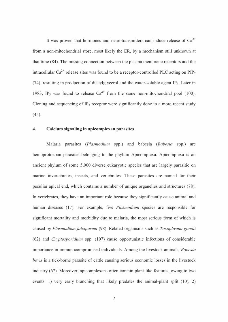
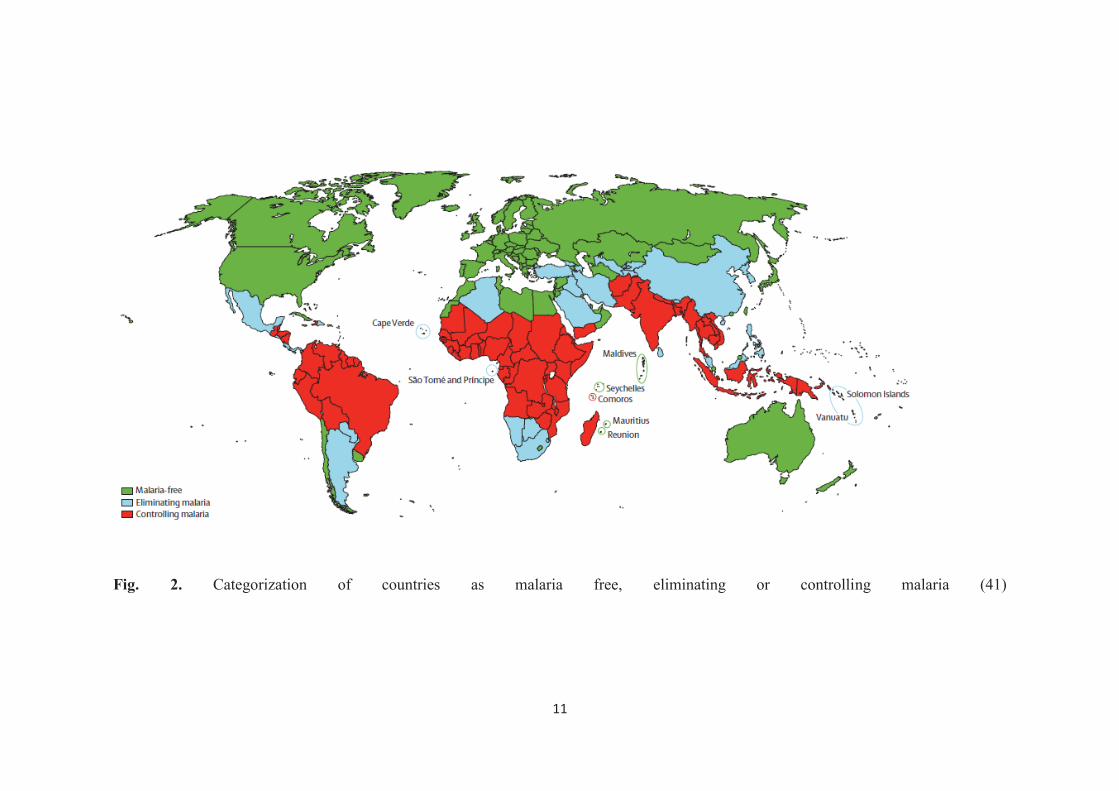
Malaria is a life-threatening disease (7) persisting in more than 90 countries
inhabited by 3.4 billion people. WHO estimated 207 million cases of malaria worldwide
in 2012 with 627,000 malaria deaths (114). Despite several notable attempts are being
made to control and eradicate the disease, it still continues to remain as a major public
health problem all in many areas in the world (50). So far, nearly half of the countries in
the world have already eleminated malaria. However, the disease is still endemic in 99
countries and of which, 67 are controlling malaria while the other 32 are following
elimination strategy (Fig. 2; (41)).
Human malaria is caused by the hemoprotozoan parasites of five species of the
genus Plasmodium of the phylum Apicomplexa including Plasmodium knowlesi,
Plasmodium malariae, Plasmodium ovale, Plasmodium vivax and P. falciparum.
Among these species, P. falcifarum is known to be often lethal whereas P. vivax and P.
falciparum are the principal agents of morbidity and mortality in malaria because of
their vast geographical distribution (48).
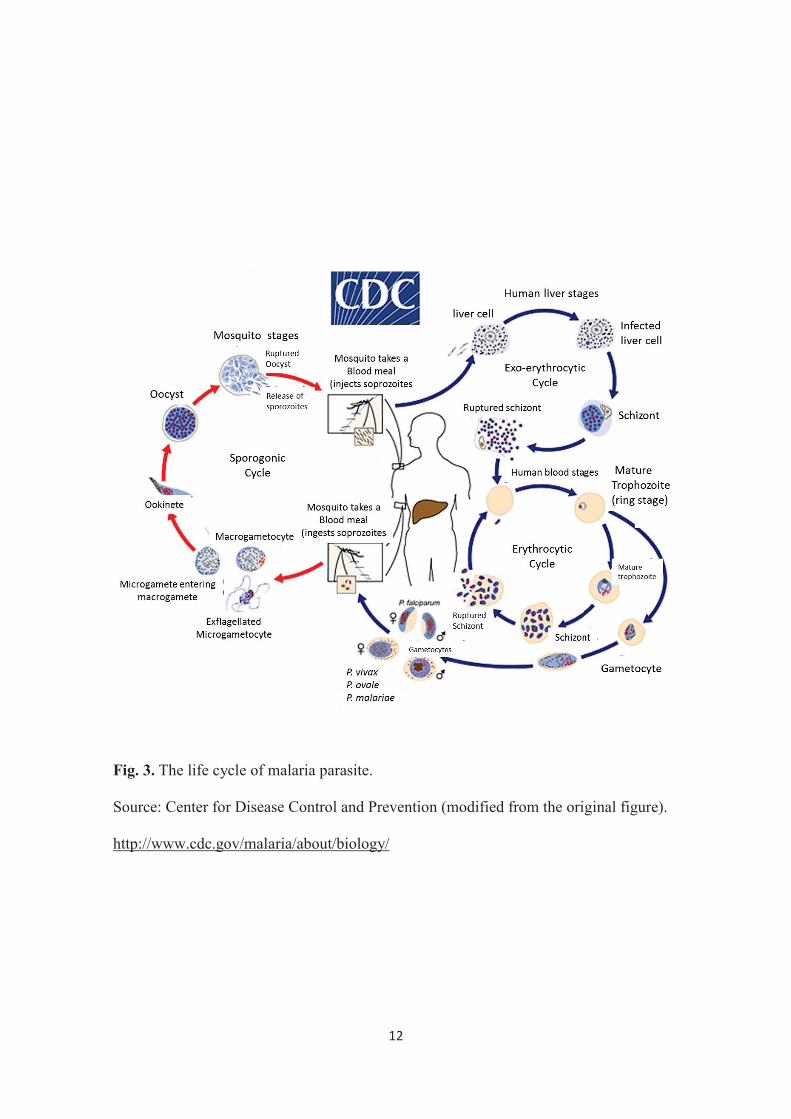
The main vectors of malaria transmission to humans are Anopheles gambiae and
Anopheles stephensi (59) which serve as the definitive hosts of the parasite. In the
complex life cycle of Plasmodium (Fig. 3), the parasite undergoes sporogony inside the
female Anopheles mosquito producing the motile infective sporozoites. These
sporozoites will then be transmitted by the mosquito to the intermediate human host
2
through blood feeding. The sporozoites travel through the blood vessels to the liver cells
where the asexual reproduction known as exoerythrocytic schizogony will occur
producing many merozoites. Merozoites then will infect erythrocytes and initiate
erythrocytic schizogony (96). All the symptoms and pathology of malaria are attributed
to the intraerythrocytic stages of the Plasmodium life cycle. Because Plasmodium
parasites cannot replicate outside the host cells, their ability to recognize and invade
erythrocytes is an essential step for both parasite survival and malaria pathogenesis
(115).
Case detection and treatment have been important parts of malaria control.
However, the development and spread of parasite resistance to a wide range of
antimalarial agents has presented a major obstacle to successful disease management in
malaria-endemic areas. This has also probably contributed to the resurgence of infection
and increase in malaria-related deaths in the recent years (57). Resistance to almost all
commonly used antimalarials including chloroquine (CQ), sulphadoxine-pyrimethamine,
artemisinin, amodiaquine, mefloquine and quinine, has been observed in P. falciparum
(57, 34). Besides drug resistance, cases of insecticide resistance in the mosquito vectors
were also seen to dramatically increase throughout the last decade (27). In addition, the
lack of an effective vaccine (64) is also considered to be a great challenge in malaria
control. Effective management of malaria as suggested by the WHO focuses on long-
lasting insecticidal nets, indoor residual spraying of insecticide, intermittent preventive
therapy in pregnancy and artemisinin-based combination therapy (81).
3
2. Overview of bovine babesiosis
Bovine babesiosis otherwisely known as ‘redwater fever’, ‘cattle tick fever’,
‘Spanish fever’ or ‘Texas cattle fever’ (42) is caused by the tick-transmitted
hemoprotozoan Babesia bovis, Babesia bigemina and Babesia divergens. This parasitic
disease commonly results in substantial cattle morbidity and mortality in endemic areas.
Babesia spp. are unicellular eukaryotes which have been defined as piriform,
round, or rod-shaped parasites (2). The genus Babesia belongs to the phylum
Apicomplexa, class Sporozoasida (18). Its life cycle (Fig. 4), involves an invertebrate
host, an Ixodidae tick, for sexual reproduction of the parasite and a vertebrate host,
wherein the parasite undergoes asexual reproduction exclusively within erythrocytes
(43). Inside the tick, sporozoites, the vertebrate-infective life stage, are generated by
sporogony in the salivary glands and are later injected with the tick’s saliva into the
bloodstream of a cow during a bloodmeal. Sporozoites then invade the erythrocytes,
transform into haemoglobin-feeding trophozoites, asexually divide into typically two
pear-shaped merozoites, and eventually lyse their host cells to invade new ones
repeating it’s asexual propagation cycle (63).
Babesia bovis causes acute disease in a course of 3 to 7 days characterized by
fever (>40ºC) usually present for several days before other signs become obvious. This
is followed by inappetence, depression, increased respiratory rate, weakness and a
reluctance to move. Haemoglobinuria is often present leading to anemia in some cases;
hence, the disease is known as redwater. Muscle wasting, tremors and recumbency
develop in advanced cases followed terminally by coma (18). The fever during
infections may cause pregnant cattle to abort (24) and bulls to show reduced fertility
4
lasting 6 to 8 weeks (30). Cerebral babesiosis is manifested by a variety of signs of
central nervous system involvement and the outcome is almost invariably fatal (18)
The disease causes serious economic losses in the livestock industry (67). Costs
due to babesiosis are incurred not only from mortality, ill-thrift, abortions, loss of
milk/meat production and from control measures (such as acaricide treatments, purchase
of vaccines and therapeutics), but also through its significant impact on international
cattle trade (18).
Despite the fact that chemotherapy is still considered the base for treatment and
control, the high prevalence of infection worldwide and the emergence of drug
resistance (108) have spurred an interest in developing more effective control measures.
So far, only live attenuated vaccine is available with a reasonably long-lasting
protection. However, the possible spread of silent pathogens such as leukemia virus,
difficulties in standardizing the vaccine dose, and the risk of reversion of virulence have
restricted the use of this type of vaccine in many regions of the world (103).
3. Overview of calcium signaling
Calcium is the most abundant mineral in the human body with a total
approximately 1 kg in an average adult and 99% of which is in the skeleton in the form
of calcium phosphate salts. The extracellular fluid contains approximately 22.5 mmol,
of which about 9 mmol is in the plasma (70).
Calcium ion (Ca2+) is a highly versatile intracellular signal that regulates various
cellular functions (15, 25). Ca2+ signaling system works through different ways to
regulate cellular processes that function over a wide dynamic range (Fig. 1; (14)). Ca2+

5
triggers cardiac contraction in microseconds (82), whereas on the other hand, Ca2+ has
to operate over minutes to hours to control other events such as oocyte activation at
fertilization (86) and gene expression (33). One of the great challenges is to understand
how these widely different Ca2+ signaling systems can be set up to control so many
divergent cellular processes.
The level of intracellular Ca2+ is determined by a balance between the ‘on’
reactions that introduce Ca2+ into the cytoplasm and the ‘off’ reactions through which
this signal is removed by the combined action of buffers, pumps and exchangers (Fig. 1;
(14)). During the ‘on’ reaction, a small proportion of the Ca2+ binds to the effectors that
are responsible for stimulating various Ca2-dependent processes (Fig. 1; (14)). Ca2+
homeostasis is a very important and crucial feature of Ca2+ signaling. It is the
mechanism by which the body maintains adequate calcium levels. Cells avoid a net loss
or gain of Ca2+ by ensuring that the fluxes occurring during the ‘on’ and ‘off’ reactions
are always balanced (14).
Cells utilize several types of Ca2+ influx (entry) channel, which can be
categorized on the basis of their activation mechanisms (15). Voltage-operated Ca2+
channels are employed by excitable cell types such as muscle and neuronal cells. Other
types of channels include receptor-operated Ca2+ channels comprised of a range of
structurally and functionally diverse channels that are particularly present on secretory
cells and at nerve terminals. Mechanically activated Ca2+ channels are present on many
cell types and respond to cell deformation. Such channels convey information into the
cell concerning the stress/shape changes that a cell is experiencing. A typical example
of mechanically induced Ca2+ signaling was observed in epithelial cells from the trachea,
6
where deformation of a single cell led to a radial Ca2+ wave that synchronized the Ca2+-
sensitive beating of cilia on many neighboring cells (19). Another type is store-operated
Ca2+ channels which are activated in response to depletion of the intracellular Ca2+ store,
either by physiological Ca2+-mobilizing messengers or pharmacological agents (20).
Ca2+ is also released from intracellular stores by several distinct types of
messenger-activated channels. The binding of many hormones, and growth factors to
specific receptors on the plasma membrane (PM) leads to the activation of
phospholipase C (PLC) which catalyses the hydrolysis of phosphatidylinositol 4,5-
bisphosphate (PIP2) to produce the intracellular messengers inositol 1,4,5-trisphosphate
(IP3) and diacylglycerol. IP3 is highly mobile in the cytoplasm and diffuses into the cell
interior where it binds to specific receptors (IP3R) on the endoplasmic reticulum (ER)
and sarcoplasmic reticulum (SR). This results in the mechanism known as IP3-induced
Ca2+ release (15).
Since Ca2+ influx was not enough to explain how electrically non-excitable cells,
devoid of voltage-gated Ca2+ channels, execute numerous Ca2+-dependent processes,
different studies were done for a better understanding of this mechanism. Investigations
on muscle cells have identified an intracellular source of Ca2+ associated with a
specialized intracellular organelle, the SR. This organelle was initially described in
1902 (73), and then rediscovered (87) identifying this organelle as the ER present in a
variety of non-muscle cells. The function of the SR as an intracellular Ca2+
storage/release site (36) and its role in the initiation of muscle contraction through Ca2+-
induced Ca2+ release (37, 40) have since then identified. Furthermore, the role of
mitochondrion in Ca2+ accumulation was also made known in various studies (75, 76).
7
It was proved that hormones and neurotransmitters can induce release of Ca2+
from a non-mitochondrial store, most likely the ER, by a mechanism still unknown at
that time (84). The missing connection between the plasma membrane receptors and the
intracellular Ca2+ release sites was found to be a receptor-controlled PLC acting on PIP2
(74), resulting in production of diacylglycerol and the water-soluble agent IP3. Later in
1983, IP3 was found to release Ca2+ from the same non-mitochondrial pool (100).
Cloning and sequencing of IP3 receptor were significantly done in a more recent study
(45).
4. Calcium signaling in apicomplexan parasites
Malaria parasites (Plasmodium spp.) and babesia (Babesia spp.) are
hemoprotozoan parasites belonging to the phylum Apicomplexa. Apicomplexa is an
ancient phylum of some 5,000 diverse eukaryotic species that are largely parasitic on
marine invertebrates, insects, and vertebrates. These parasites are named for their
peculiar apical end, which contains a number of unique organelles and structures (78).
In vertebrates, they have an important role because they significantly cause animal and
human diseases (17). For example, five Plasmodium species are responsible for
significant mortality and morbidity due to malaria, the most serious form of which is
caused by Plasmodium falciparum (98). Related organisms such as Toxoplasma gondii
(62) and Cryptosporidium spp. (107) cause opportunistic infections of considerable
importance in immunocompromised individuals. Among the livestock animals, Babesia
bovis is a tick-borne parasite of cattle causing serious economic losses in the livestock
industry (67). Moreover, apicomplexans often contain plant-like features, owing to two
events: 1) very early branching that likely predates the animal-plant split (10), 2)
8
acquisition of a secondary endosymbiont derived from engulfment of an algal cell (110).
As such, signaling pathways in apicomplexan parasites contain both conserved and
unique features as compared to other eukaryotic cells (17).
Ca2+ homeostasis and storage have been studied mainly in T. gondii and
Plasmodium spp. using the fluorescent calcium indicator fura 2-AM (fura 2-
acetoxymethylester). In T. gondii, the cytosolic Ca2+ concentration in the tachyzoites
was measured at 70 ± 6 nM (79). On the other hand, the concentrations obtained in
Plasmodium chabaudi and P. falciparum using fura 2-AM loaded free parasites were
also at nanomolar levels performed in single-cell imaging experiments (49).
In addition to known eukaryotic Ca2+ stores including acidocalcisomes, the ER,
Golgi apparatus and mitochondria, apicomplexan parasites contain other unique
compartments that potentially could contribute to diverse Ca2+ transients necessary for
crucial functions within the parasites. These include the apicoplast, a remnant plastid
derived from a secondary endosymbiotic event, and various acidic organelles such as
the recently described plant-like vacuole (78).
Apicomplexans have highly polarized cells that are specialized for regulated
secretion and directed entry into their host cells (28, 94). Ca2+ controls a number of
critical events in their life cycles including secretion of adhesins, gliding motility cell
invasion, and egress (79). Ca2+ also influences developmental processes that occur at
distinct stages in their complex life cycles (5).
9
Ca2+ regulation in parasitic protozoa differs in several aspects from the processes
that occur in other eukaryotic cells, providing great opportunities for targeting them for
new therapies (78).
5. Objectives of the present study
Therefore, given this general background, the objectives of this study are: 1) To
study the involvement of Ca2+ signaling in reversing the chloroquine-resistance in the
malaria parasites to provide a new therapeutic strategy against the resistant parasites. 2)
To study the involvement of Ca2+ in egress of B. bovis merozoites from bovine
erythrocytes as it is considered one of the most important steps in the parasite’s life
cycle.

10
Fig. 1. Ca2+ signaling dynamics and homeostasis in eukaryotic cells (14)
11
Fig. 2. Categorization of countries as malaria free, eliminating or controlling malaria (41)
12
Fig. 3. The life cycle of malaria parasite.
Source: Center for Disease Control and Prevention (modified from the original figure).
http://www.cdc.gov/malaria/about/biology/

13
Fig. 4. The life cycle of Babesia.
Source: Full-Babesia (modified from the original figure).
http://fullmal.hgc.jp/bb/docs/aboutdb.html
14
Chapter I
Simultaneous administration of 2-aminoethyl diphenylborinate
reverses chloroquine-resistance in Plasmodium falciparum
1.1 Introduction
Malaria continues to be a worldwide public health problem causing significant
morbidity and mortality and its resistance to existing antimalarial drugs is a growing
problem (113). The emergence, followed by the spread throughout most malaria-
endemic regions, of P. falciparum parasites resistant to the antimalarial drug
chloroquine (CQ) has worsened the global malaria situation (106). Malaria is caused by
a protozoan parasites of the genus Plasmodium, its blood stage replicates inside the host
erythrocytes (8) and responsible for most of the clinical symptoms of the disease (97).
Understanding the signaling pathways governing the parasite’s blood stage growth may
aid in discovering new therapeutic targets for antimalarial drugs.
Ca2+ is a ubiquitous intracellular signal responsible for controlling a wide range
of cellular activities in eukaryotic cells (15). In protozoan parasites, Ca2+-mediated
signaling controls various vital functions such as protein secretion, motility, cell
invasion and differentiation (83, 95). In the malaria parasite, Plasmodium, the
intracellular signaling pathways controlling the activity of Ca2+ dependant protein
kinases has extensively been studied (95, 35). The role of Ca2+ signaling underlying
modulation of Plasmodium cell cycle has also been widely investigated including the
effect on protease activity (11, 90). Moreover, some research groups tried to investigate
how Ca2+ signaling is triggered by demonstrating the important role of the host derived

15
hormone melatonin and its derivatives that elicit a rise in cytosolic Ca2+ in Plasmodium
(99, 54). It has also been demonstrated recently, for the first time, the spontaneous Ca2+
oscillation in P. falciparum (39). Furthermore, it has also been shown that the blockage
of this oscillation in the trophozoite stage by 2-aminoethyl diphenylborinate (2-APB)
which inhibits IP3-induced Ca2+ release (72, 116, 117), caused severe degeneration and
breakdown of successive asexual reproduction in the intraerythrocytic parasites,
resulting in their death (39).
The antimalarial drug CQ is thought to exert its toxic effect in the
intraerythrocytic parasite at the digestive vacuole (44). The compound was also found to
induce Ca2+ release and disrupt Ca2+ and H+ homeostasis in cytoplasm of P. chabudai
cells (88, 53). Therefore, with regard to disruption of intracellular Ca2+ homeostasis, it
was hypothesized that the potentiation of the CQ activity could be achieved in the
malaria parasite with the simultaneous administration of 2-APB.
In this chapter the potential of 2-APB in reversing CQ-resistance in P.
falciparum was examined. This resulted in a complete reversal of CQ-resistance in P.
falciparum K-1 strain.

16
1.2 Materials and Methods
Parasite culture. Chloroquine resistant (CQR) K-1 strain of P. falciparum was
cultured with the modified method of Trager and Jensen (105) using a multi-gas
incubator (5% O2 and 5% CO2) in RPMI 1640 medium (Life Technologies Japan Co.,
Tokyo, Japan) supplemented with 0.5% Albumax (Life Technologies Japan), 25 mM
HEPES, 24 mM sodium bicarbonate, 0.5 g/L L-glutamine, 50 mg/L hypoxanthine, 25
μg/L gentamycin and human erythrocyte (from healthy Japanese volunteers) at a
hematocrit of 2% in 4 ml cultures. Growth synchronization was achieved with 5% D-
sorbitol (68). P. falciparum K-1 strain was kindly provided by Prof. Shigeyuki Kano.
In vitro drug susceptibility test. The assessment of the outcome of the in vitro
drug susceptibility test was done using the SYBR Green I method (61). P. falciparum-
infected erythrocytes were cultured with the standard method using a multi-gas
incubator (5% O2 and 5% CO2). After reaching 1.5% ring form parasitemia, the
parasites were synchronized with 5% D-sorbitol for 30 min at room temperature (RT)
and washed with RPMI 1640 medium twice by centrifugation at 1,000× g for 5 min.
Then, the erythrocytes were resuspended in the culture medium at 2% hematocrit. One
hundred μl of the erythrocyte suspension was then replaced in each well of a tissue
culture plate (96-well flat bottom, Corning Japan Co., Tokyo, Japan) in triplicate. For
the CQ sensitivity test, chloroquine diphosphate (Sigma Aldrich Japan Co., Tokyo,
Japan) was added to the parasite culture (100 μl in total) in each well to give a series of
dilutions from 10,240 to 1.25 nM. The sensitivity tests for 2-APB (obtained from
Laboratory for Developmental Neurobiology, RIKEN Brain Science Institute), was
mixed with dimethyl sulfoxide (DMSO, Sigma Aldrich Japan) before being added to the

17
culture to give a series of dilutions from 200 to 25 μM. The simultaneous addition of the
two compounds was performed by adding 50 μM 2-APB to the serially diluted CQ.
After 72 h of incubation, each test plate was removed from the incubator and 100 μl of
lysis buffer [130.1 mM Tris-HCl (pH 7.5), 10 mM EDTA, 0.016 (wt/vol) saponin and
1.6 (vol/vol) Triton X-100] containing SYBR green I (Life Technologies Japan) (2×
final concentration) was added directly to each well in the plates and gently mixed. The
plates were then covered with aluminum foil and incubated for another 24 h at RT in the
dark. Relative fluorescent units (RFU) per well were determined using Fluoroskan
Ascent (Thermo Fisher Scientific K.K., Yokohama, Japan) with excitation and emission
wavelength bands set at 485 and 530 nm, respectively. The concentration of anti-
malarial drug inhibiting parasite growth by 50% (IC50) was calculated using the probit
method as described previously (102).
Fluorescence Ca2+ imaging. Fluorescence Ca2+ imaging was performed as
described previously (39). A culture of P. falciparum-infected erythrocytes was diluted
10-fold with RPMI 1640, without phenol red, culture medium (Invitrogen Japan Co.,
Tokyo, Japan), which served as the imaging medium. The infected erythrocytes were
collected from the 1 ml aliquot by centrifugation (1,000× g for 5 min at RT) and
resuspended in 350 μl of the imaging medium. Loading solution was prepared by
adding 10 μM Fluo-4 AM (Invitrogen Japan) and 100-fold dilution of PowerLoad
(Invitrogen Japan) to the imaging medium and was used for the loading of Fluo-4 AM
to the parasite cells. A suspension of erythrocytes (350 μl) was mixed with 150 μl of
loading solution to give a final concentration of 3 μM of Fluo-4 AM and then shaken at
200 rpm for 30 min at 37°C with a TAITEC bioshaker BR-22UM (TAITEC, Tokyo,
18
Japan). Erythrocytes were then mixed with 10 ml of the imaging medium, centrifuged
(1,000× g for 5 min at RT) and resuspended in 1.2 ml of the imaging medium. A
suspension of erythrocytes (200 μl) was applied in a 35 mm glass-bottomed dish
(MatTek Co., Ashland, MA, USA) that had been coated with 1 mg/ml poly-L-lysine
before use. After 30 min incubation in a humidified multi-gas water-jacketed incubator
at 37ºC, suspended erythrocytes were removed by gentle washing with the imaging
medium. The glass-bottomed dish was then placed in the culture chamber of a Leica
confocal microscope (TCSSP5, Leica Microsystems, Wetzlar, Germany).
Sequential time lapse imaging of Fluo-4 AM and transparent images was
performed using the Leica confocal microscopy system (Leica Microsystems) with a
40× oil immersion objective lens and excitation at 488 nm (Argon laser) for Fluo-4 AM
and transparent images. Emissions were collected using the true spectral detection
method developed by Leica Microsystems. Images were captured every 5–15 s for 300–
600 s. Specific Fluo-4 AM fluorescence in a parasite (F) was calculated by the
subtraction of background fluorescence and normalized by the average fluorescence
obtained before the tested compound was added (F0).
Fluorescence H+ imaging. Fluorescence H+ imaging was performed as
described previously (91). Briefly, a culture of P. falciparum-infected erythrocytes was
diluted 10-fold with RPMI 1640, phenol red (-), culture medium (Invitrogen Japan),
which served as the imaging medium. The infected erythrocytes were collected from the
1 ml aliquot by centrifugation (1,000× g for 5 min at RT) and resuspended in 500 μl of
the imaging medium. The parasite’s cytoplasm was then loaded with 5 μM cSNARF-5F
AM (Invitrogen) for 40 min, the stock solution of the dye was prepared in Pluronic® F-

19
127 (20% (w/v) solution in DMSO . Pluronic® F-127 was used to facilitate AM dye
entry and allow for efficient loading while maintaining cell’s integrity. The parasite
suspension was then shaken at 200 rpm for 30 min at 37°C with a TAITEC bioshaker
BR-22UM (TAITEC, Tokyo, Japan). Erythrocytes were then mixed with 10 ml of the
imaging medium, centrifuged (1,000× g for 5 min at RT) and resuspended in 1.2 ml of
the imaging medium. A suspension of erythrocytes (200 μl) was applied in a 35 mm
glass-bottomed dish (MatTek Co., Ashland, MA, USA) that had been coated with 1
mg/ml poly-L-lysine before use. After 40 min incubation in a humidified multi-gas
water-jacketed incubator at 37ºC, suspended erythrocytes were removed by gentle
washing with the imaging medium. The glass-bottomed dish was then placed in the
culture chamber of a Leica confocal microscope (TCSSP5, Leica Microsystems,
Wetzlar, Germany).
Sequential time lapse imaging for cytoplasmic pH ratio determination of
cSNARF-5F AM was performed using the Leica confocal microscopy system (Leica
Microsystems) with a 40× oil immersion objective lens and excitation at 488 nm (Argon
laser). A dual-emissions were collected in two different wave lengths (λ1= 580 and λ2 =
640) using the true spectral detection method developed by Leica Microsystems. Images
were captured every 5 s for 300–600 s. The specific fluorescence ratio of cSNARF-5F
AM in the cytoplasm (R) was calculated by subtraction of background fluorescence
separately from each channel and the fluorescence ration (R) was calculated R (λ1/λ2).
Perfusion system. A manipulator system (type YOU-4, Narishige Co., Ltd.,
Tokyo, Japan), Perista pump (SJ-1211h- NO, 483313- ATTO Co, Tokyo, Japan) and
Enomoto Micro Pump (model MV-6005VP, Enomoto Micro Pump Mfg. Co., Ltd.,

20
Tokyo, Japan) were used to add and remove CQ, thapsigargin (Tg) (Sigma Aldrich
Japan) and concanamycin A (CA) (Sigma Aldrich Japan) continuously to and from the
parasite preparation during the live cell imaging process.
21
1.3 Results and Discussion
In order to investigate the potential of 2-APB in reversing CQ-resistance in K-1
strain, the IC50 values of both 2-APB and CQ were assessed by in vitro drug sensitivity
test using SYBR Green I method, and were found to be 73.5 ± 3 μM and 1050 ± 95 nM,
respectively (Fig. 5A and B). The addition of the suboptimal dose of 2-APB (50 μM) to
the series of CQ concentrations, which causes a minimum effect on in vitro growth of
the parasite (Fig. 5A) potentiated antimalarial effect of CQ against the CQR K-1 strain
and resulted in a significant decline in IC50 from 1050 ± 95 nM to 14 ± 2 nM (Fig. 5B).
This suggested that 2-APB completely reversed the CQ-resistance in CQR P.
falciparum K-1 strain. This could be due partial blockage of IP3 pathway for Ca2+
release which is critical for the blood stage development of the parasite using 2-APB
(39) and the involvement of CQ in Ca2+ homeostasis disturbance (88, 53).
To figure out the mechanism, live cell Ca2+ imaging with confocal laser
scanning microscopy was performed by loading the trophozoite stage with the calcium
sensitive indicator Fluo-4 AM. It was previously observed that CQ induces Ca2+ release
to the cytoplasm of P. chabaudi (88) as well as P. falciparum (6). Thus, in this study,
the fact that CQ-induced Ca2+ release causes disturbance in Ca2+ homeostasis in the
parasite’s cytoplasm was considered, and these observations were utilized to investigate
the role of CQ in this strategy. In this study, the CQ-induced Ca2+ release to the
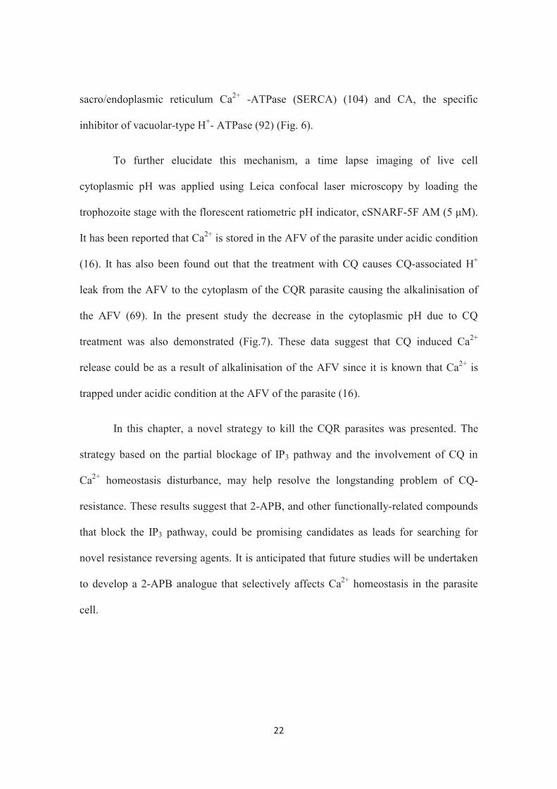
cytoplasm of the CQR K-1 strain was confirmed (Fig. 6). It has also been demonstrated
that there are two Ca2+ stores in the parasite, namely, the ER (89) and the acidic food
vacuole (AFV) (47). Moreover, In this study, the CQ-induced Ca2+ release was
demonstrated to be from the AFV by using Tg, the specific inhibitor of
22
sacro/endoplasmic reticulum Ca2+ -ATPase (SERCA) (104) and CA, the specific
inhibitor of vacuolar-type H+- ATPase (92) (Fig. 6).
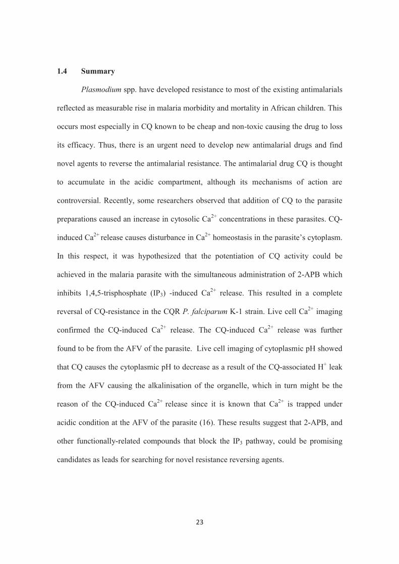
To further elucidate this mechanism, a time lapse imaging of live cell
cytoplasmic pH was applied using Leica confocal laser microscopy by loading the
trophozoite stage with the florescent ratiometric pH indicator, cSNARF-5F AM (5 μM).
It has been reported that Ca2+ is stored in the AFV of the parasite under acidic condition
(16). It has also been found out that the treatment with CQ causes CQ-associated H+
leak from the AFV to the cytoplasm of the CQR parasite causing the alkalinisation of
the AFV (69). In the present study the decrease in the cytoplasmic pH due to CQ
treatment was also demonstrated (Fig.7). These data suggest that CQ induced Ca2+
release could be as a result of alkalinisation of the AFV since it is known that Ca2+ is
trapped under acidic condition at the AFV of the parasite (16).
In this chapter, a novel strategy to kill the CQR parasites was presented. The
strategy based on the partial blockage of IP3 pathway and the involvement of CQ in
Ca2+ homeostasis disturbance, may help resolve the longstanding problem of CQ-
resistance. These results suggest that 2-APB, and other functionally-related compounds
that block the IP3 pathway, could be promising candidates as leads for searching for
novel resistance reversing agents. It is anticipated that future studies will be undertaken
to develop a 2-APB analogue that selectively affects Ca2+ homeostasis in the parasite
cell.
23
1.4 Summary
Plasmodium spp. have developed resistance to most of the existing antimalarials
reflected as measurable rise in malaria morbidity and mortality in African children. This
occurs most especially in CQ known to be cheap and non-toxic causing the drug to loss
its efficacy. Thus, there is an urgent need to develop new antimalarial drugs and find
novel agents to reverse the antimalarial resistance. The antimalarial drug CQ is thought
to accumulate in the acidic compartment, although its mechanisms of action are
controversial. Recently, some researchers observed that addition of CQ to the parasite
preparations caused an increase in cytosolic Ca2+ concentrations in these parasites. CQ-
induced Ca2+ release causes disturbance in Ca2+ homeostasis in the parasite’s cytoplasm.
In this respect, it was hypothesized that the potentiation of CQ activity could be
achieved in the malaria parasite with the simultaneous administration of 2-APB which
inhibits 1,4,5-trisphosphate (IP3) -induced Ca2+ release. This resulted in a complete
reversal of CQ-resistance in the CQR P. falciparum K-1 strain. Live cell Ca2+ imaging
confirmed the CQ-induced Ca2+ release. The CQ-induced Ca2+ release was further
found to be from the AFV of the parasite. Live cell imaging of cytoplasmic pH showed
that CQ causes the cytoplasmic pH to decrease as a result of the CQ-associated H+ leak
from the AFV causing the alkalinisation of the organelle, which in turn might be the
reason of the CQ-induced Ca2+ release since it is known that Ca2+ is trapped under
acidic condition at the AFV of the parasite (16). These results suggest that 2-APB, and
other functionally-related compounds that block the IP3 pathway, could be promising
candidates as leads for searching for novel resistance reversing agents.

24
Fig. 5. Dose-dependent activities of 2-APB and chloroquine (CQ) on P. falciparum
synchronized cultures of CQR K-1 strain. Various concentrations of 2-APB (A).
Various concentrations of CQ plus DMSO as solvent control (black line) and CQ plus
50 μM of 2-APB (red line) (B). The statistical significance of differences between
treatments was assessed with student’s t-test. **P<0.004 and ***P<0.0005. Results are
presented as the mean ± SD of three independent experiments. RFU denotes relative
fluorescence units in SYBR Green I assay (See Materials and Methods).
25
Fig. 6. Ca2+ imaging of P. falciparum analyzed by confocal microscopy. Trophozoite
stages were loaded with Fluo-4 AM, and fluorescence in the parasite cytoplasm (F/F0)
was calculated (see Materials and Methods). Treatment with chloroquine (CQ) (10 μM)
caused an increase in mean fluorescence ratio of 0.8 ± 0.3 (n = 10). Treatment with
concanamycin A (CA) (100 nM) didn’t show any change in the fluorescence ratio
suggesting that CQ and CA induce Ca2+ release from the same compartment (AFV).
Treatment with thapsigargin (Tg) (2 μM) caused an increase in the mean fluorescence
ratio of 0.9 ± 0.2 (n = 6). Data are representative of eight similar experiments.
26
Fig. 7. H+ imaging of P. falciparum analyzed by confocal microscopy. Trophozoite
stages were loaded with cSNARF-5F AM (5 μM), and fluorescence ration (R) was
calculated R (λ1/λ2) (see Materials and Methods). Treatment with 10 μM chloroquine
(CQ) caused a decrease in the fluorescence ratio of 0.9 ± 0.2 (n = 9). Treatment with
imaging medium (pH 7.8) (served as control) caused an increase in fluorescence ratio of
1.7 ± 0.5 (n = 9) suggesting that CQ causes cytoplasmic pH to decrease. Data are
representative of 6 similar experiments.

27
Chapter II
An in vivo application of the IP3 receptor blocker 2-APB as an effective
resistance reverser to chloroquine-resistant Plasmodium chabaudi
2.1 Introduction
Malaria is a major cause of illness and death in children and adults in tropical
countries. The spread of resistance to available antimalarial drugs such as chloroquine is
a major threat to malaria control. The discovery of artemisinin as an antimalarial drug
which was isolated from the leaves of the sweet wormwood, Artemisia annua, had a
great impact on treatment of malaria and more potent derivatives of artemisinin were
subsequently developed. Presently, artemisinin-based combination therapy (ACT) is
recommended by the WHO as the first-line therapy for P. falciparum malaria (113).
However, the cost of artemisinins limits their use in the developing world (112) and the
emergence of artemisinin-resistant malaria has also been recently reported (34). The
development of new, efficacious, affordable drugs remains crucial. Extensive searches
for novel compounds have met with only limited success (29). Therefore, to combat
malaria, there is an urgent need to develop new antimalarial drugs (65, 56, 46) and find
novel agents to reverse antimalarial resistance.
Following the discovery that the Ca2+ channel-blocker verapamil (VP) restores
chloroquine-sensitivity in CQR strains (66, 71), there has been considerable interest in
such so-called resistance-reversing agents (93). In this connection, it is worth to mention
that there are a number of reports of attempts to reverse CQ-resistance in vitro through
the use of Ca2+ channel antagonists such as VP (71, 32, 3). However, all of these studies
28
focused on reversing CQ-resistance through the blockage of CQ efflux from the acidic
food vacuole of resistant parasites.
In chapter I of this study, CQ-resistance was successfully reversed in P.
falciparum by a novel strategy which mainly targets Ca2+ homeostasis through the
partial blockage of IP3 pathway for Ca2+ release which is critical for the blood stage
development of the parasite (39). That was achieved by using 2-APB which inhibits IP3
-induced Ca2+ release. In this study, the strategy was applied in vivo to the rodent
malaria mouse infection model, CQR P. chabaudi AS (30 CQ) strain, to confirm the
universality of my finding.

29
2.2 Materials and Methods
Mice and parasite. 6 week-old female ICR mice (CLEA Japan, Tokyo, Japan; 3
mice/group), were infected with 5 × 10⁶ parasitized erythrocytes of the CQR P.
chabaudi AS (30 CQ) strain by the intraperitoneal injection. (Plasmodium chabaudi
chabaudi AS (30 CQ) was obtained through the MR4 as part of the BEI Resources
Repository, NIAID, NIH:, MRA-744, deposited by D Walliker).
In vivo drug susceptibility test. The in vivo simultaneous administration of CQ
and 2-APB was performed as previously described (77) with some modifications.
Briefly, mice were infected by the intraperitoneal injection of 5 × 10⁶ parasitized
erythrocytes of the CQR P. chabaudi AS (30 CQ) strain. To first evaluate the
antimalarial activity of CQ and 2-APB, CQ (3 mg/kg), 2-APB (0.1 and 1 mg/kg) and
DMSO (as solvent control) were administered separately via intraperitoneal injection to
different groups of mice at days 0, 1 and 2. Thin blood films were prepared at days 1-4
and stained with Giemsa. The number of parasitized erythrocytes per 10,000
erythrocytes in each stained preparation was counted with the mean values obtained
from 3 preparations used as an index of parasitemia (%). Antimalarial activity was
evaluated at day 4 as follows: Normalized parasitemia= (Parasitemia in the compound-
treated group) / (Parasitemia in DMSO control group). To evaluate the CQ-resistance
reversing activity with 2-APB, CQ (3 mg/kg) and 2-APB (0.1 mg/kg) were
simultaneously administered to a group of mice. A group receiving CQ (3 mg/kg) and
VP, a Ca2+ channel antagonists, (Wako Chemical Co, Osaka, Japan; (20 mg/kg) served
as a positive control, while a group receiving CQ (3 mg/kg) alone served as negative
control. The three treatments were administered via intraperitoneal injection at days 0, 1
30
and 2 after the infection. The potential of 2-APB in reversing CQ-resistance was
evaluated at day 4 as follows: Normalized parasitemia= (Parasitemia in the two
compound-treated group) / (Parasitemia in the CQ-treated group). The animal
experiments in this study were carried out in compliance with the Guide for Animal
Experimentation at Obihiro University of Agriculture and Veterinary Medicine
(Permission number 25-71).
31
2.3 Results and Discussion
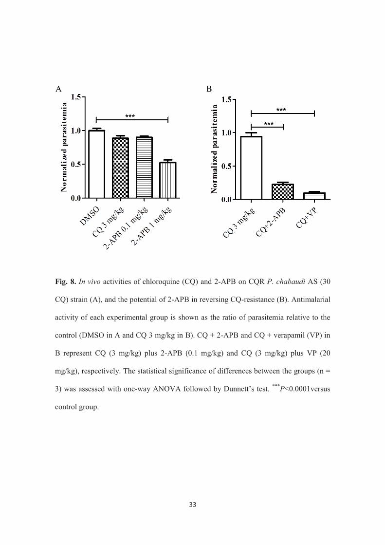
The antimalarial activity of CQ (3 mg/kg) and 2 different doses of 2-APB (1
mg/kg and 0.1 mg/kg) was initially tested against the CQR P. chabaudi AS (30 CQ)
strain by intraperitoneal injection. Results showed that 1 mg/kg of 2-APB exhibited
antimalarial effect but not the lower concentration of 2-APB (0.1 mg/kg) (Fig. 8A).
Furthermore, the simultaneous intraperitoneal injection of a lower concentration of 2-
APB (0.1 mg/kg), which presented minimal antimalarial activity, produced a CQ-
resistance reversing effect in the mice (Fig. 8B). It is noteworthy that the potency of 2-
APB as a CQ-resistance reverser was equivalent to that of verapamil (20 mg/kg,
intraperitoneal injection) (Fig. 8B). Verapamil is the first Ca2+ channel antagonist that is
reported to exhibit a resistance reversing effect in CQ-resistant P. falciparum (71).
These outcomes suggest the complete reversal of CQ-resistance in vivo with 2-APB.
These data has strengthened the in vitro outcomes confirming the universality the
strategy.
32
2.4 Summary
Drug resistance against most of the available antimalarial drugs has worsened
the global malaria situation. In search for novel agents to reverse CQ-resistance in vitro,
2-APB was introduced as an effective compound through a novel strategy in chapter I.
In this chapter, the same strategy was applied in vivo to the rodent malaria mouse
infection model, CQR P. chabaudi AS (30 CQ) strain, to confirm the universality of my
finding in chapter I. Firstly, CQ (3mg/kg) and (2-APB 0.1 mg/kg) were separately
intraperitoneal-injected to different groups of mice to check their antimalarial activity.
Both two compounds didn’t show significant antimalarial activity. Simultaneous
intraperitoneal administration of lower concentration of 2-APB (0.1 mg/kg) which
presented minimal antimalarial activity produced CQ-resistance reversing effect in the
mice. This is probably due to the disturbance of Ca2+ homeostasis in the parasite cell.
These results strengthened the in vitro finding that 2-APB, and other functionally
related compounds that block IP3 pathway, could be promising candidates as leads for
searching for novel resistance reversing agents. To the best of our knowledge, this is the
first observation of a CQ-resistance reversing effect induced by an IP3 receptor inhibitor
in the malaria parasite.
33
Fig. 8. In vivo activities of chloroquine (CQ) and 2-APB on CQR P. chabaudi AS (30
CQ) strain (A), and the potential of 2-APB in reversing CQ-resistance (B). Antimalarial
activity of each experimental group is shown as the ratio of parasitemia relative to the
control (DMSO in A and CQ 3 mg/kg in B). CQ + 2-APB and CQ + verapamil (VP) in
B represent CQ (3 mg/kg) plus 2-APB (0.1 mg/kg) and CQ (3 mg/kg) plus VP (20
mg/kg), respectively. The statistical significance of differences between the groups (n =
3) was assessed with one-way ANOVA followed by Dunnett’s test. ***P<0.0001versus
control group.
34
Chapter III
Calcium ions are involved in egress of Babesia bovis merozoites from
bovine erythrocytes
3.1 Introduction
As explained in the General Introduction, B. bovis is a tick-borne
hemoprotozoan parasite of cattle that causes serious economic losses in the livestock
industry (67). Although vaccine development has been the subject of intense focus, to
date, only a live attenuated vaccine, with some restrictions, has been introduced to the
field in certain regions of the world (31). Chemotherapy remains one of the main
components of control strategies against babesiosis, and current drugs used against
bovine babesiosis include diminazene aceturate and imidocarb. It has been proven that
these drugs reduce the risk of severe infection in endemic areas. However, the
withdrawal of many anti-babesia drugs from the market for various reasons (80) has
made the search for new potent chemotherapeutic agents highly important.
Understanding the signaling pathways governing the parasite’s growth in the
erythrocytic stages may help in strategizing new control measures to combat babesiosis.
Ca2+ is a ubiquitous intracellular signal messenger that is responsible for
controlling a wide range of cellular activities in eukaryotic cells (15). In protozoan
parasites, Ca2+-mediated signaling controls various vital functions, such as protein
secretion, motility, cell invasion and differentiation (26, 83, 94, 111). In contrast to the
Plasmodium and Toxoplasma parasites, little is understood about Ca2+ signaling in
Babesia, with the exception of a small amount of information on the involvement of
Ca2+ in the invasion of erythrocytes by the merozoites of B. divergens and equine
35
Babesia parasites (85, 101). The inhibitory effect of Ca2+ dependent protein kinase
inhibitor on the in vitro growth of B. bovis has also been reported (21). While egress
(release) of Plasmodium merozoites and Toxoplasma tachyzoites from their host cells
has been studied intensively in terms of Ca2+ signaling (4, 51, 58, 83), at the present
time, there are no available data showing the role of Ca2+ in the egress of Babesia
parasites. Therefore, in this chapter, the involvement of Ca2+ in the egress of B. bovis
merozoites from infected erythrocytes was investigated. Calcium ionophore A23187
and Tg, an inhibitor of SERCA, which have been used in various studies to artificially
increase Ca2+ concentration in the cytosol of apicomplexan parasite cells (12, 38, 58),
were found to induced egress of B. bovis from host erythrocytes. In addition, changes in
intracellular Ca2+ concentration after these treatments were also observed using the live
cell Ca2+ imaging technique with confocal laser scanning microscopy.
36
3.2 Materials and Methods
Parasite culture. B. bovis (Texas strain) (60) was maintained in a serum-free
GIT medium (Wako Pure Chemical Industries Ltd., Osaka, Japan) supplemented with
10% bovine erythrocytes, 60 U/ml of penicillin G, 60 g/ml of streptomycin and 0.15
g/ml of amphotericin B (Sigma Aldrich Japan Co., Tokyo, Japan) (complete culture
medium) using a continuous microaerophilic stationary-phase culture system (1). The
animal experiments in this study were carried out in compliance with the Guide for
Animal Experimentation at Obihiro University of Agriculture and Veterinary Medicine
(Permission number 25-78-3).
In vitro egress assay. The effect of calcium ionophore A23187 (Sigma Aldrich
Japan) and thapsigargin (Tg) (Sigma Aldrich Japan) on the egress of the parasite from
infected erythrocytes was examined using a method for measuring drug activity as
previously described (22, 23) with some modifications. Briefly, the parasite culture was
diluted with a fresh complete culture medium to obtain a parasitemia of 4-7% in a 1.5
ml plastic tube. A23187 or Tg, which had been dissolved in DMSO, was added to the
culture in the tube at 1 nM to 10 μM or 1.25 - 5 μM, respectively. The mixture was then
incubated in humidified multi-gas water-jacketed incubator with cap open at 37ºC for
indicated periods of time. In parallel, normal culture supplemented with the same
concentration of DMSO was prepared as control. All of the experiments were carried
out in triplicate for each compound. Parasitemia was monitored by counting
approximately 1,000 erythrocytes in a Giemsa-stained thin smear, while the percentage
of extracellular merozoites was calculated as the ratio of extracellular merozoites to the
37
entire parasite population (extracellular and intraerythrocytic merozoites) in
approximately 500 parasites.
Fluorescence Ca2+ imaging. Fluorescence Ca2+ imaging was performed as
described in chapter I with some modifications. In brief, a culture of B. bovis-infected
erythrocytes was diluted 20-fold with RPMI 1640, phenol red (-), culture medium
(Invitrogen Japan), which served as the imaging medium. The infected erythrocytes
were collected from the 1 ml aliquot by centrifugation (1,000× g for 5 min at RT) and
resuspended in 350 μl of the imaging medium. Loading solution was prepared by
adding 10 μM Fluo-4 AM (Invitrogen Japan) and 100-fold dilution of PowerLoad
(Invitrogen Japan) to the imaging medium and was used for the loading of Fluo-4 AM
to the parasite cells. A suspension of erythrocytes (350 μl) was mixed with 150 μl of
loading solution to give a final concentration of 3 μM of Fluo-4 AM and then shaken at
200 rpm for 15 min at 37°C with a TAITEC bioshaker BR-22UM (TAITEC).
Erythrocytes were then mixed with 10 ml of the imaging medium, centrifuged (1,000× g
for 5 min at RT) and resuspended in 1.2 ml of the imaging medium. A suspension of
erythrocytes (200 μl) was applied in a 35 mm glass-bottomed dish (MatTek) that had
been coated with 1 mg/ml poly-L-lysine before use. After 30 min incubation in a
humidified multi-gas water-jacketed incubator at 37ºC, suspended erythrocytes were
removed by gentle washing with the imaging medium. The glass-bottomed dish was
then placed in the culture chamber of a Leica confocal microscope (TCSSP5, Leica
Microsystems). Sequential time lapse imaging of Fluo-4 AM and transparent images
was performed using the Leica confocal microscope system (Leica Microsystems) with
a 40× oil immersion objective lens and excitation at 488 nm (Argon laser) for Fluo-4
38
AM and transparent images. Emissions were collected using the true spectral detection
method developed by Leica Microsystems. Images were captured every 5–15 s for 200–
300 s. Specific Fluo-4 AM fluorescence in a parasite (F) was calculated by the
subtraction of background fluorescence and normalized by the average fluorescence
obtained before the tested compound was added (F0).
Perfusion system. A23187 and Tg were added and removed continuously to and
from the parasite preparation during the live cell imaging process using the same
perfusion system descried in chapter I (Materials and Methods).
39
3.3 Results and Discussion
In order to investigate the effect of the increase in cytosolic Ca2+ concentration
on the egress of B. bovis merozoites from bovine erythrocytes, Giemsa-stained smears
of the parasite culture were prepared after 10 min incubation in vitro with two different
concentrations of A23187 (1 and 10 μM). In Plasmodium, Toxoplasma and Neospora
parasites, micromolar concentrations of A23187 have induced egress (12, 38, 58),
however, these treatments resulted in the emergence of rarely seen degenerated and dot-
shaped parasites (Fig. 9B and C) in the control culture of B. bovis (Fig. 9A). The
parasite was therefore incubated with low concentrations of A23187 (1, 10 and 100 nM),
and Giemsa-stained smears were prepared every 10 min until 30 min after the
treatments. In this experiment, it was found that 10 min incubation with 1 - 100 nM
A23187 resulted in a significantly lower parasitemia in all concentrations in comparison
to control without A23187. Extending the incubation with the A23187 for another 10
min resulted in an increase in parasitemia. After 30 min incubation, parasitemia was
significantly higher in all concentrations as compared to control (Fig. 10A). These
findings suggest that A23187 induces the parasite’s egress from and consequent
invasion to erythrocytes. To distinguish the A23187 effect on the egress from the
invasion step, cultures incubated with 10 nM A23187 were then monitored for free
merozoites (merozoites outside erythrocytes). Giemsa-stained smears were prepared
every 1 min from the parasite culture for 10 min after A23187 treatment. The results
showed that ratio of free merozoites to total parasites began increasing from 1 min after
treatment upto 10 min after treatment (Fig. 10B). To confirm this observation and
determine the time point suitable for observing the egress, I compared the culture for
free merozoites at 5 and 10 min after A23187 treatment and found that 10 min

40
incubation gave clearer difference in the ratio of free merozoites (Fig. 10C) between test
and control parasites. These data suggest that A23187 induces the parasite’s egress from
infected erythrocytes.
To further examine whether the parasite’s egress can be induced by the
increased cytosolic Ca2+ concentration in the parasite, the parasite culture was incubated
with Tg, an inhibitor of the uptake of cytosolic Ca2+ to the ER by specific inhibition of
SERCA. The parasite was first incubated with different concentrations of Tg (1.25, 2.5
and 5 μM), and Giemsa-stained smears were prepared after 90 min incubation. In this
experiment, It was found that all tested concentrations showed significantly higher
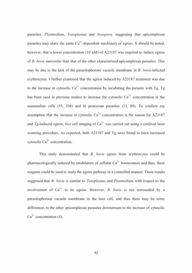
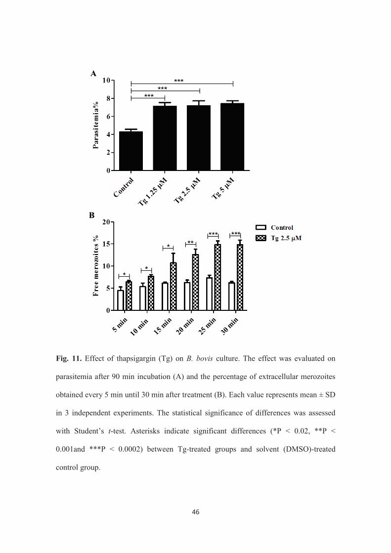
parasitemia in comparison to control (Fig. 11A). These data suggest that Tg increases
parasitemia as a result of egress acceleration, followed by the reinvasion of the egressed
merozoites into new erythrocytes. To investigate whether Tg can induce egress, parasite
culture incubated with 2.5 μM of Tg was monitored for free meroziotes for 30 min, and
Giemsa-stained smears were prepared every 5 min. The results from this experiment
revealed that, in comparison to non-treated control, Tg-treatment significantly increased
the ratio of free merozoites to total parasites at all tested time points and that the
increase of the ratio was clearer with 25 and 30 min of treatment (Fig. 11B). These
results indicate that the increase in cytosolic Ca2+ concentration most probably induces
the parasite’s egress and suggest a Ca2+ signaling pathway in the egress of this parasite.
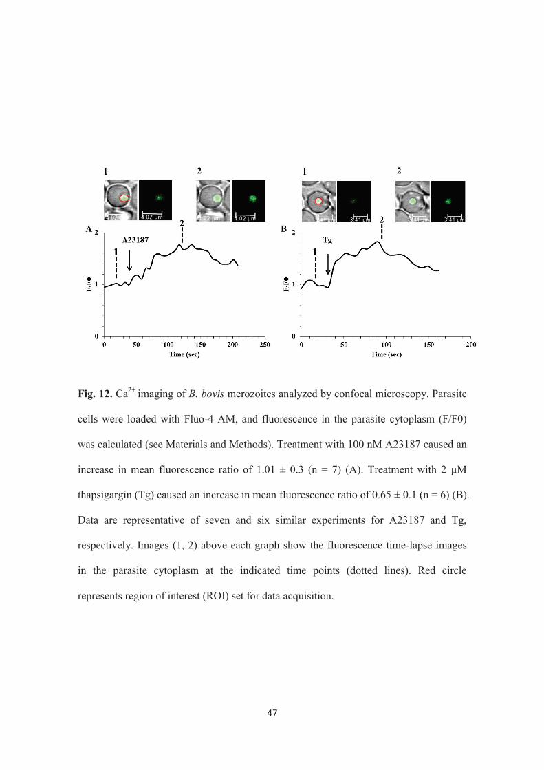
To confirm the effects of treatment with A23187 and Tg toward egress of B.
bovis merozoites, time lapse imaging of live cell Ca2+ was applied using Leica confocal
laser microscopy by loading the parasite cell with the Ca2+ sensitive indicator Fluo-4
AM. The addition of 100 nM of A23187 or 2 μM of Tg to the parasite preparation
41
induced an increase in cytosolic Ca2+ concentration of the parasite cells (Fig. 12A and
B). These findings suggest that the induced egress by these two cytoplasmic Ca2+
modulators might be, at least, due to their effect in increasing cytosolic Ca2+
concentration. The cause-and-effect link between the increase of cytosolic Ca2+ and the
merozoite egress needs to be proved in the future studies.
The information available on Ca2+ signaling components in apicomplexan
parasites is still fragmentary and insufficient. Important features of their life cycle, such
as motility, host cell invasion and egress from infected cells, are known to be linked
with Ca2+ (78). Obligate intracellular parasites like T. gondii replicate inside its host cell,
but at some point need to exit the cell by rupturing the infected host cell in order to
infect other cells. This rapid egress process is still poorly understood. However, it is
known that calcium ionophores like A23187 can stimulate the process (38). T. gondii
mutants with delayed egress have been isolated and found to have elevated intracellular
Ca2+ level (9). In the schizont stage of P. falciparum, it has also been observed that
intracellular Ca2+ level was increased just prior to parasite egress and that A23187
artificially induced the egress (58). Synchronization of Plasmodium and Toxoplasma
parasites cultures has made the study of egress easier. However, this may not be the
case in the Babesia parasite, wherein the limitation in tools for obtaining synchronized
cultures (101) might hamper the study of egress. To overcome this difficulty, we
adopted a criterion for evaluating the egressed parasite in compound-treated culture
through parasitemia and by counting the free merozoites to provide direct evidence of
the parasite being outside the cell as a result of the treatment. The data obtained here
with A23187 treatment were consistent with those obtained in the other apicomplexan
42
parasites, Plasmodium, Toxoplasma and Neospora, suggesting that apicomplexan
parasites may share the same Ca2+-dependent machinery of egress. It should be noted,
however, that a lower concentration (10 nM) of A23187 was required to induce egress
of B. bovis merozoite than that of the other characterized apicomplexan parasites. This
may be due to the lack of the parasitophorous vacuole membrane in B. bovis-infected
erythrocytes. I further examined that the egress induced by A23187 treatment was due
to the increase in cytosolic Ca2+ concentration by incubating the parasite with Tg. Tg
has been used in previous studies to increase the cytosolic Ca2+ concentration in the
mammalian cells (55, 104) and in protozoan parasites (13, 88). To confirm my
assumption that the increase in cytosolic Ca2+ concentration is the reason for A23187
and Tg-induced egress, live cell imaging of Ca2+ was carried out using a confocal laser
scanning procedure. As expected, both A23187 and Tg were found to have increased
cytosolic Ca2+ concentration.
This study demonstrated that B. bovis egress from erythrocytes could be
pharmacologically induced by modulators of cellular Ca2+ homeostasis and thus, these
reagents could be used to study the egress pathway in a controlled manner. These results
suggested that B. bovis is similar to Toxoplasma and Plasmodium with respect to the
involvement of Ca2+ in its egress. However, B. bovis is not surrounded by a
parasitophorous vacuole membrane in the host cell, and thus there may be some
differences to the other apicomplexan parasites downstream to the increase of cytosolic
Ca2+ concentration (4).
43
3.4 Summary
Egress is an important feature of the life cycle of B. bovis. At some point the
parasite needs to exit the cell by rupturing the infected host cell in order to infect other
cells to continue its life cycle. In spite of the importance of egress as a crucial step in the
parasite’s life cycle, to date, there is no available information in the mechanism of
egress of B. bovis merozoites. In this chapter, the involvement of Ca2+ in egress of B.
bovis merozoites from infected bovine erythrocytes was investigated. The increase in
cytosolic Ca2+ concentration induced by A23187 and Tg was found to accelerate the
parasite’s egress. Time lapse imaging of live cell Ca2+ revealed that these treatments
induced an increase in cytosolic Ca2+ concentration of the parasite cells. The data
suggest the involvement of Ca2+ and a Ca2+ signaling pathway in the egress of this
parasite. The data provided here is the first report on the parasite egress and therefore,
provide information to better understand the mechanism of the egress pathway and its
molecular components in Babesia parasites. Further studies would therefore elicit new
therapeutic and prevention strategies against babesiosis.

44
Fig. 9. Light microscopic observation of A23187-treated B. bovis in an in vitro culture.
Micrographs were taken after 10 min incubation with DMSO solvent-control (A), 1 μM
A23187 (B) and 10 μM A23187 (C). The A23187 treatments showed a higher number
of degenerated and dot shaped parasite (black arrows) than the control. Scale bars
indicate 10 μm.

45
Fig. 10. Effect of A23187 on B. bovis culture. The effect was evaluated on parasitemia
(A), percentage of extracellular merozoites (number of free merozoites/ number of free
merozoites + number of intraerythrocytic parasite × 100) obtained every 1 min until 10
min after treatment (B) and the percentage of extracellular merozoites obtained every 5
min until 10 min after treatment (C). Each value represents mean ± SD in 3 independent
experiments. The statistical significance of differences was assessed with Student’s t-
test. Asterisks indicate significant differences (*P < 0.01, **P < 0.005 and ***P <
0.0002) between A23187-treated groups and solvent (DMSO)-treated control group.
46
Fig. 11. Effect of thapsigargin (Tg) on B. bovis culture. The effect was evaluated on
parasitemia after 90 min incubation (A) and the percentage of extracellular merozoites
obtained every 5 min until 30 min after treatment (B). Each value represents mean ± SD
in 3 independent experiments. The statistical significance of differences was assessed
with Student’s t-test. Asterisks indicate significant differences (*P < 0.02, **P <
0.001and ***P < 0.0002) between Tg-treated groups and solvent (DMSO)-treated
control group.
47
Fig. 12. Ca2+ imaging of B. bovis merozoites analyzed by confocal microscopy. Parasite
cells were loaded with Fluo-4 AM, and fluorescence in the parasite cytoplasm (F/F0)
was calculated (see Materials and Methods). Treatment with 100 nM A23187 caused an
increase in mean fluorescence ratio of 1.01 ± 0.3 (n = 7) (A). Treatment with 2 μM
thapsigargin (Tg) caused an increase in mean fluorescence ratio of 0.65 ± 0.1 (n = 6) (B).
Data are representative of seven and six similar experiments for A23187 and Tg,
respectively. Images (1, 2) above each graph show the fluorescence time-lapse images
in the parasite cytoplasm at the indicated time points (dotted lines). Red circle
represents region of interest (ROI) set for data acquisition.

48
General Discussion
Although hemoprotozoan parasites of the phylum Apicomplexa like most
eukaryotes, utilize second messenger signaling cascades including Ca2+ to coordinate
cell functions (6), and Ca2+ is known to be relevant for several vital functions in these
parasites, the information available on Ca2+ signaling components in these parasites is
still fragmentary and insufficient (78). The current study’s main thrust was to further
understand the Ca2+ signaling in two hemoprotozoan parasites from the phylum
Apicomplexa namely Plasmodium and Babesia. This study consists of two parts: 1)
Reversing of CQ-resistance by potentiation of the antimalarial activity of CQ in vitro
against CQR P. falciparum K-1 strain and in vivo against CQR P. chabaudi AS (30 CQ)
strain by using 2-APB (Chapters I and II); and 2) the involvement of Ca2+ in egress of B.
bovis merozoites from bovine erythrocytes (Chapter III).
In the first chapter, the potential of 2-APB in reversing CQ-resistance in CQR P.
falciparum K-1 strain was examined. This resulted in a complete reversal of CQ-
resistance in the parasite. There are already a number of reports of attempts to reverse
CQ-resistance in vitro through the use of Ca2+ channel antagonists such as verapamil
(71, 32, 3). However, all of the studies focused on reversing CQ-resistance through the
blockage of CQ efflux from the acidic food vacuole of resistant parasites wherein the
compound is thought to exert its effect. In this study, a novel strategy was presented
wherein, the possible involvement of CQ itself in the disruption of Ca2+ and H+
homeostasis in the cytoplasm of P. chabaudi cell reported in previous studies (88, 53),
might be potentiated with 2-APB which blocks the IP3 pathway for Ca2+ release. This
49
probably resulted in the disturbance of Ca2+ homeostasis in the cytoplasm of the
resistant parasite, and consequently reversal of CQ-resistance in the CQR human
malaria parasite P. falciparum K-1 strain. Attempts have then been made to investigate
the mechanism using live cell Ca2+ imaging technique. The CQ-induced Ca2+ release
(88, 53) was demonstrated. It was then found that CQ induces Ca2+ release from the
AFV of the parasite. Further investigation was done using live cell imaging of
cytoplasmic pH confirming that CQ causes the decrease of cytoplasmic pH as a result of
the CQ-associated H+ leak from the AFV. This leads to the alkalinisation of the
organelle (69), and therefore it might be the reason behind the CQ-induced Ca2+ release
since it is known that Ca2+ is trapped under acidic condition at the AFV of the parasite
(16). Since the strategy depends mainly on the blockage of the IP3 pathway for Ca2+
release and the drawback of CQR parasite that CQ induces Ca2+ release, the parasite
might not further develop resistance. IP3 receptor has yet to be identified in malaria
parasites, however pharmacological data clearly demonstrate that these parasites
maintain intracellular Ca2+ stores (13, 52, 109). Moreover, the IP3 pathway has also
been demonstrated in P. chabaudi (88). This available information on Ca2+ signaling in
the malaria parasites leads to the outcome of this study. However, further studies are still
required to elucidate the relationship between Ca2+ homeostasis and CQ-resistance in
malaria parasites as well as developing 2-APB analogue that selectively affect Ca2+
homeostasis in the parasite cell.
In the second chapter, the strategy was applied, in vivo, to the mouse malaria
infection model, CQR P. chabaudi AS (30 CQ) strain, to confirm the universality of the
obtained result in chapter I. Interestingly, the minimum concentration of 2-APB that
50
affects the parasite growth, significantly reversed the CQ-resistance in CQR P.
chabaudi. The potency of 2-APB as CQ-resistance reverser was equivalent to that of
verapamil, the first drug reported to exhibit the resistance reversing effect in CQR P.
falciparum (71). These results strengthened the in vitro finding that 2-APB, and other
functionally-related compounds that block the IP3 pathway, could be promising
candidates as leads for searching for novel resistance reversing agents. To the best of
my knowledge, this is the first observation of a CQ-resistance reversing effect induced
by an IP3 receptor inhibitor in the malaria parasites.
In the third and last chapter, a first report on egress of B. bovis merozoites from
infected bovine erythrocytes was presented. This was achieved by using tow Ca2+
modulators; A23187 and Tg. Both two compounds were found to increase cytosolic
Ca2+ concentration in a wide range of eukaryotic cells including apicomplexan parasites
(12, 38, 58). The Ca2+ -dependent induced egress obtained by A23187 and Tg
treatments was consistent with that obtained in the other apicomplexan parasites,
Plasmodium, Toxoplasma and Neospora, suggesting that apicomplexan parasites may
share the same Ca2+-dependent machinery of egress.
Results of this study showed that B. bovis egress from erythrocytes could be
pharmacologically induced by modulators of cytosolic Ca2+ concentration and thus,
these reagents could be used to study the egress pathway in a controlled manner. Further
studies to investigate the egress pathway downstream to the increase of cytosolic Ca2+
concentration might therefore elicit new therapeutic and prevention strategies targeting
the molecular components of the egress pathway.

51
In general, this study demonstrated the involvement of Ca2+ in tow important
features of two hemoprotozoan parasites of the phylum Apicomplexa. The study
demonstrated that the disturbance in Ca2+ homeostasis obtained by the simultaneous
administration of 2-APB and CQ to the CQ-resistant malaria parasites has resulted in a
complete reversal of CQ-resistance in these parasites. This may help resolve the
longstanding problem of CQ-resistance. On the other hand, Ca2+ was also demonstrated,
for the first time, to be involved in egress of B. bovis merozoites from infected
erythrocytes. This could help in developing new strategies targeting the components of
the egress pathway. This may lead to the blockage of the egress of the parasite and
consequently the erythrocytic life cycle of the parasite.
52
Conclusion
In this study, the potential of 2-APB, which inhibits IP3 pathway for Ca2+ release,
in reversing CQ-resistance in CQR malaria parasites was examined. This probably
resulted in the disturbance of Ca2+ homeostasis in the cytoplasm of the resistant parasite,
and consequently reversal of CQ-resistance in P. falciparum and P. chabaudi. In
addition, the CQ-induced Ca2+ release was demonstrated using live cell Ca2+ imaging
and it was found that CQ induces Ca2+ release from the AFV of the parasite. Also, live
cell imaging of cytoplasmic pH showed that CQ causes the cytoplasmic pH to decrease
as a result of the CQ-associated H+ leak from the AFV. This leads to the alkalinisation
of the organelle which might be the reason of the CQ-induced Ca2+ release since it is
known that Ca2+ is stored under acidic condition at the AFV of the parasite. These data
suggest that 2-APB, and other functionally related compounds that block IP3 pathway,
could be promising candidates as leads for searching for novel resistance reversing
agents.
With regards to the role of Ca2+ in egress of B. bovis merozoites from the
infected bovine erythrocytes, the increase in cytosolic Ca2+ concentration by Ca2+
ionophore A23187 and Tg, which inhibits SERCA, was found to induce the parasite’s
egress. This suggests the involvement of Ca2+ in this crucial step in the life cycle of the
parasite, giving a good opportunity for future studies to target the components of the
egress pathway for new therapies and prevention strategies.
53
Acknowledgments
I am heartily thankful to have studied and done my research work at the National
Research Center for Protozoan Diseases (NRCPD), Obihiro University of Agriculture
and Veterinary Medicine (OUAVM).
I would like to express my special appreciation and thanks to my advisor
Professor Shin-ichiro Kawazu, for accepting me as a PhD student. I would like to thank
him for encouragement, motivation, guidance and unlimited support generously offered
me throughout my stay in Japan. His advices on both researches as well as on my career
have been priceless. Because of all that, writing this thesis was possible. Indeed, I will
forever be indebted to him.
I am thankful to Prof. Yasutake Shimizu of Gifu University and Prof Hiroshi
Suzuki of NRCPD as my co-supervisors for their help and making my studies possible.
I also humbly acknowledge Prof. Tadashi Itagaki of Iwate University and Prof. Tetsuya
Furuya of Tokyo University of Agriculture and Technology for carefully reviewing this
dissertation.
I sincerely thank Prof. Noboru Inoue whom I was benefited from his advices
throughout my PhD course and for all the professors of NRCPD for their advices during
my progress reports.
I am grateful to Prof. Shigeyuki Kano of the National Center for Global health
and Medicine for providing the P. falciparum parasite, Prof. Satoru Kawai of
Laboratory of Tropical Medicine and Parasitology, Dokkyo Medical University for
electron microscopy analysis of my experiments, Prof. Katsuhiko Mikoshiba of

54
Laboratory for Developmental Neurobiology, RIKEN Brain Science Institute for
providing 2-APB and carful reviewing of my manuscripts, Dr. Masahiro Enomoto of
Princess Margaret Cancer Centre, Toronto, Canada for the helpful suggestions in Ca2+
imaging experiments and carful reviewing of my manuscripts, Prof. Osamu Kaneko of
Institute of Tropical Medicine, Nagasaki University (NEKKEN) for the manuscript
reviews as well as his advice, comments and suggestions on my research work and Dr.
Masahito Asada of (NEKKEN) for his help and giving me some insights in the
experiments.
I am also grateful to the Hokkaido Kushiro and Sapporo Red Cross Blood
Centers for supplying human red blood cells used for culturing the malaria parasites. It
is also highly acknowledged that the following reagent was obtained through the MR4
as part of the BEI Resources Repository, NIAID, NIH: Plasmodium chabaudi chabaudi
AS (30 CQ), MRA-744, deposited by D Walliker.
I would also like to extend my gratitude to my labmates especially Dr. Jose Ma.
Angeles (Joma) for his continuous help throughout my study course, Dr. Hassan Hakimi,
Dr. Keisuke Suganuma (Kero), Dr. Miho Usui, Dr. Alaa Terkawi, Dr Dusit
Laohasinnarong, Mrs. Nguyen Thu Thuy, Ms. Wakako Furuyama, Mrs. Hirono
Masuda-Suganuma, Ms. Shino Yamasaki, Dr. Mo Zhou, Dr. Ruttayaporn Ngassaman
(Thom) for their various assistances in one way or another giving me some insights in
the experiments during the course of my doctoral studies ; to other staffs and students of
the Research Unit of Advanced Preventive Medicine and NRCPD for their assistance,
friendship and companionship for providing kind environment for study.
55
My sincere Thanks go to my director, Sudan University of Science and
Technology for granting me a paid study leave and Japanese Government for offering
me the highly competitive Monbukagakusho (MEXT) scholarship to pursue knowledge
in Japan. For sure, I will endeavour to impart the same to my country’s students while
maintaining collaborations with Japan.
To the current and former international students in Japan, I am glad to have
enjoyed their friendship and company throughout my stay in Japan. The good memories
with them will be in mind forever.
I would also like to express my sincerest gratitude to all the staff of Gifu
University, NRCPD and international Students Affairs Section of OUAVM for their
kind help, guidance and support at any moment in time I needed their help.
A special thanks to my family. Words cannot express how grateful I am to my
mother Saida Awad and father Elnour Mossaad for all of the sacrifices that they have
made on my behalf. Their prayer for me was what sustained me thus far. At the end, I
would like express appreciation to my beloved wife May Khojali who spent sleepless
nights, taking care of me and our kids and supporting me in the moments when there
was no one to answer my queries.
56
References
1) Aboulaila M, Nakamura K, Govind Y, Yokoyama N, Igarashi I (2010)
Evaluation of the in vitro growth-inhibitory effect of epoxomicin on Babesia
parasites. Vet. Parasitol. 167: 19-27.
2) Adl SM, Simpson AG, Farmer MA, Andersen RA, Anderson OR, Barta JR,
Bowser SS, Brugerolle G, Fensome RA, Fredericq S, James TY, Karpov S,
Kugrens P, Krug J, Lane CE, Lewis LA, Lodge J, Lynn DH, Mann DG, Mc
Court RM, Mendoza L, Moestrup O, Mozley-Standridge SE, Nerad TA, Shearer
CA, Smirnov AV, Spiegel FW, Taylor MF (2005) The new higher level
classification of eukaryotes with emphasis on the taxonomy of protists. J.
Eukaryot. Microbiol. 52: 399-451.
3) Adovelande J, Delèze J, Schrével J (1998) Synergy between two calcium
channel blockers, verapamil and fantofarone (SR33557), in reversing
chloroquine resistance in Plasmodium falciparum. Biochem. Pharmacol. 55:
433-440.
4) Agarwal S, Singh MK, Garg S, Chitnis CE, Singh S (2013) Ca2+ -mediated
exocytosis of subtilisin-like protease 1: a key step in egress of Plasmodium
falciparum merozoites. Cell Microbiol. 15: 910-921.
5) Alano P and Billker O (2005) Gametocytes and Gametes Chapter 10. In:
Molecular Approaches to Malaria (Sherman I ed.). Washington: ASM Press.
57
6) Alves E, Bartlett PJ, Garcia CR, Thomas AP (2011) Melatonin and IP3-induced
Ca2+ release from intracellular stores in the malaria parasite Plasmodium
falciparum within infected red blood cells. J. Biol. Chem. 286: 5905-5912.
7) Arama C and Troye-Blomberg M (2014) The path of malaria vaccine
development: challenges and perspectives. J. Intern. Med. 275:456-466. doi:
10.1111/joim.12223.
8) Aravind L, Iyer LM, Wellems TE, Miller LH (2003) Plasmodium biology:
genomic gleanings. Cell. 115:771-785.
9) Arrizabalaga G, Ruiz F, Moreno S, Boothroyd JC (2004) Ionophore-resistant
mutant of Toxoplasma gondii reveals involvement of a sodium/hydrogen
exchanger in calcium regulation. J. Cell Biol. 165: 653-662.
10) Baldauf SL (2003) The deep roots of eukaryotes. Science. 300:1703-1706.
11) Barale JC, Blisnick T, Fujioka H, Alzari PM, Aikawa M, Braun-Breton C,
Langsley G (1999) Plasmodium falciparum subtilisin-like protease 2, a
merozoite candidate for the merozoite surface protein 1-42 maturase. Proc. Natl.
Acad. Sci. U.S.A. 96:6445-6450.
12) Behrendt JH, Taubert A, Zahner H, Hermosilla C (2008) Studies on synchronous
egress of coccidian parasites (Neospora caninum, Toxoplasma gondii, Eimeria
bovis) from bovine endothelial host cells mediated by calcium ionophore
A23187. Vet. Res. Commun. 32: 325-332.
13) Beraldo FH, Mikoshiba K, Garcia CR (2007) Human malarial parasite,

58
Plasmodium falciparum, displays capacitative calcium entry: 2-aminoethyl
diphenylborinate blocks the signal transduction pathway of melatonin action on
the P. falciparum cell cycle. J. Pineal Res. 43: 360-364.
14) Berridge MJ, Bootman MD, Roderick HL (2003) Calcium signalling: dynamics,
homeostasis and remodelling. Nat. Rev. Mol. Cell Biol. 4:517-529.
15) Berridge MJ, Lipp P, Bootman MD (2000) The versatility and universality of
calcium signalling. Nature Rev. Mol. Cell Biol. 1: 11-21.
16) Biagini GA, Bray PG, Spiller DG, White MR, Ward SA (2003) The digestive
food vacuole of the malaria parasite is a dynamic intracellular Ca2+ store. J. Biol.
Chem. 278:27910-27915.
17) Billker O, Lourido S, Sibley LD (2009) Calcium-dependent signaling and
kinases in apicomplexan parasites. Cell Host Microbe. 5:612-622. doi:
10.1016/j.chom.2009.05.017.
18) Bock R, Jackson L, de Vos A, Jorgensen W (2004) Babesiosis of cattle.
Parasitology. 129: S247-269.
19) Boitano S, Dirksen ER, Sanderson MJ (1992) Intercellular propagation of
calcium waves mediated by inositol trisphosphate. Science. 258:292-295.
20) Bootman MD, Collins TJ, Peppiatt CM, Prothero LS, MacKenzie L, De Smet P,
Travers M, Tovey SC, Seo JT, Berridge MJ, Ciccolini F, Lipp P (2001) Calcium
signaling - an overview. Semin. Cell Dev. Biol. 12:3-10.
59
21) Bork S, Das S, Okubo K, Yokoyama N, Igarashi I (2006) Effects of protein
kinase inhibitors on the in vitro growth of Babesia bovis. Parasitology. 132: 775-
779.
22) Bork S, Yokoyama N, Matsuo T, Claveria FG, Fujisaki K, Igarashi I (2003)
Clotrimazole, ketoconazole, and clodinafop-propargyl as potent growth
inhibitors of equine Babesia parasites during in vitro culture. J. Parasitol. 89:
604-606.
23) Bork S, Yokoyama N, Matsuo T, Claveria FG, Fujisaki K, Igarashi I (2003)
Growth inhibitory effect of triclosan on equine and bovine Babesia parasites.
Am. J. Trop. Med. Hyg. 68: 334-340.
24) Callow LL (1984) Piroplasms. In Animal Health in Australia, Protozoal and
Rickettsial Diseases, vol. 5. Animal Health in Australia, pp. 121-160. Canberra,
Australian Bureau of Animal Health, AGPS.
25) Carafoli E, Santella L, Brance D, Brisi M (2001) Generation, control, and
processing of cellular calcium signals. Crit. Rev. Biochem. Mol. Biol. 36: 107-
260.
26) Carruthers VB, Giddings OK, Sibley LD (1999) Secretion of micronemal
proteins is associated with Toxoplasma invasion of host cells. Cell Microbiol. 1:
225-235.
27) Choi KS, Christian R, Nardini L, Wood OR, Agubuzo E, Muleba M, Munyati S,
Makuwaza A, Koekemoer LL, Brooke BD, Hunt RH, Coetzee M (2014)

60
Insecticide resistance and role in malaria transmission of Anopheles funestus
populations from Zambia and Zimbabwe. Parasit. Vectors. 7(1):464.
28) Cowman AF and Crabb BS (2006) Invasion of red blood cells by malaria
parasites. Cell. 124:755-766.
29) Dar O, Khan MS, Adagu I (2008) The potential use of methotrexate in the
treatment of falciparum malaria: in vitro assays against sensitive and multidrug-
resistant falciparum strains. Jpn. J. Infect. Dis. 61:210-211.
30) de Vos AJ and Potgieter FT (1994) Bovine babesiosis. In Infectious Diseases of
Livestock (ed. Coetzer JAW, Thomson GR, Tustin RC), pp. 278-294. Capetown,
Oxford University Press.
31) de Waal DT and Combrink MP (2006) Live vaccines against bovine babesiosis.
Vet. Parasitol. 138: 88-96.
32) Deloron P, Basco LK, Dubois B, Gaudin C, Clavier F, Le Bras J, Verdier F
(1991) In vitro and in vivo potentiation of chloroquine against malaria parasites
by an enantiomer of amlodipine. Antimicrob. Agents Chemother. 35: 1338-1342.
33) Dolmetsch RE, Xu K, Lewis RS (1998) Calcium oscillations increase the
efficiency and specificity of gene expression. Nature. 392: 933-936.
34) Dondorp AM, Yeung S, White L, Nguon C, Day NPJ, Socheat D, von Seidlein L
(2010) Artemisinin resistance: current status and scenarios for containment. Nat.
Rev. Microbiol. 8:272-280. doi: 10.1038/nrmicro2331.
61
35) Dvorin JD, Martyn DC, Patel SD, Grimley JS, Collins CR, Hopp CS, Bright AT,
Westenberger S, Winzeler E, Blackman MJ, Baker DA, Wandless TJ,
Duraisingh MT (2010) A plant-like kinase in Plasmodium falciparum regulates
parasite egress from erythrocytes. Science. 328: 910-912. doi:
10.1126/science.1188191.
36) Ebashi S and Lipmann F (1962) Adenosine triphophate-linked concentration of
calcium ions in a particulate fraction of rabbit muscle. J. Cell Biol. 14: 389-400.
37) Endo M, Tanaka M, Ogawa Y (1970) Calcium induced release of calcium from
the sarcoplasmic reticulum of skinned skeletal muscle fibers. Nature. 228: 34-36.
38) Endo T, Sethi KK, Piekarski G (1982) Toxoplasma gondii: calcium ionophore
A23187-mediated exit of trophozoites from infected murine macrophages. Exp.
Parasitol. 53: 179-188.
39) Enomoto M, Kawazu S, Kawai S, Furuyama W, Ikegami T, Watanabe J,
Mikoshiba K (2012) Blockage of spontaneous Ca2+ oscillation causes cell death
in intraerythrocitic Plasmodium falciparum. PLoS One. 7:e39499.
40) Fabiato A, Fabiato F (1975) Contractions induced by a calcium-triggered release
of calcium from the sarcoplasmic reticulum of single skinned cardiac cells. J.
Physiol. 249: 469-495.
41) Feachem RG, Phillips AA, Hwang J, Cotter C, Wielgosz B, Greenwood BM,
Sabot O, Rodriguez MH, Abeyasinghe RR, Ghebreyesus TA, Snow RW (2010)
Shrinking the malaria map: progress and prospects. Lancet. 376:1566-1578.
62
42) Florin-Christensen M, Suarez CE, Rodriguez AE, Flores DA, Schnittger L
(2014) Vaccines against bovine babesiosis: where we are now and possible
roads ahead. Parasitology. 1-30.
43) Florin-Christensen M. and Schnittger L (2009) Piroplasmids and ticks: a long-
lasting intimate relationship. Front. Biosci (Landmark Ed) 14, 3064-3073.
44) Francis SE, Sullivan DJ Jr, Goldberg DE (1997) Hemoglobin metabolism in the
malaria parasite Plasmodium falciparum. Annu. Rev. Microbiol. 51: 97-123.
45) Furuichi T, Yoshikawa S, Miyawaki A, Wada K, Maeda N, Mikoshiba K (1989)
Primary structure and functional expression of the inositol 1,4,5-trisphosphate-
binding protein P400, Nature. 342: 32-38.
46) Gamo FJ, Sanz LM, Vidal J, de Cozar C, Alvarez E, Lavandera JL, Vanderwall
DE, Green DV, Kumar V, Hasan S, Brown JR, Peishoff CE, Cardon LR, Garcia-
Bustos JF (2010) Thousands of chemical starting points for antimalarial lead
identification. Nature. 465:305-310.
47) Garcia CR, Ann SE, Tavares ES, Dluzewski AR, Mason WT, Paiva FB (1998)
Acidic calcium pools in intraerythrocytic malaria parasites. Eur. J. Cell Biol. 76:
133-138.
48) Garcia CR, de Azevedo MF, Wunderlich G, Budu A, Young JA, Bannister L
(2008) Plasmodium in the postgenomic era: new insights into the molecular cell
biology of malaria parasites. Int. Rev. Cell Mol. Biol. 266:85-156.
49) Garcia CR, Dluzewski AR, Catalani LH, Burting R, Hoyland J, Mason WT
63
(1996) Calcium homeostasis in intraerythrocytic malaria parasites. Eur. J. Cell
Biol. 71:409-413.
50) Gardner MJ, Hall N, Fung E, White O, Berriman M, Hyman RW, Carlton JM,
Pain A, Nelson KE, Bowman S, Paulsen IT, James K, Eisen JA, Rutherford K,
Salzberg SL, Craig A, Kyes S, Chan MS, Nene V, Shallom SJ, Suh B, Peterson J,
Angiuoli S, Pertea M, Allen J, Selengut J, Haft D, Mather MW, Vaidya AB,
Martin DM, Fairlamb AH, Fraunholz MJ, Roos DS, Ralph SA, McFadden GI,
Cummings LM, Subramanian GM, Mungall C, Venter JC, Carucci DJ, Hoffman
SL, Newbold C, Davis RW, Fraser CM, Barrell B (2002) Genome sequence of
the human malaria parasite Plasmodium falciparum. Nature. 419:498-511.
51) Garg S, Agarwal S, Kumar S, Yazdani SS, Chitnis CE, Singh S (2013) Calcium-
dependent permeabilization of erythrocytes by a perforin-like protein during
egress of malaria parasites. Nat. Commun. 4: 1736.
52) Gazarini ML and Garcia CR (2004) The malaria parasite mitochondrion senses
cytosolic Ca2+ fluctuations. Biochem. Biophys. Res. Commun. 321:138-144.
53) Gazarini ML, Carlos AO Sigolo, Regina P Markus, Andrew P Thomas, Celia RS
Garcia (2007) Antimalarial drugs disrupt ion homeostasis in malarial parasites.
Mem. Inst. Oswaldo Cruz. 102: 329-334.
54) Gazarini ML, Thomas AP, Pozzan T, Garcia CR (2003) Calcium signaling in a
low calcium environment: how the intracellular malaria parasite solves the
problem. J. Cell Biol. 161:103-110.
64
55) Gerasimenko J, Peng S, Gerasimenko O (2014) Role of acidic stores in secretory
epithelia. Cell Calcium. 55: 346-354.
56) Giha HA (2010) Artemisinin derivatives for treatment of uncomplicated
Plasmodium falciparum malaria in Sudan: too early for too much hope. Parasitol.
Res. 106:549-552.
57) Gilbert Kokwaro (2009) Ongoing challenges in the management of malaria.
Malaria Journal. 8(Suppl 1):S2 doi:10.1186/1475-2875-8-S1-S2.
58) Glushakova S, Lizunov V, Blank PS, Melikov K, Humphrey G, Zimmerberg J
(2013) Cytoplasmic free Ca2+ is essential for multiple steps in malaria parasite
egress from infected erythrocytes. Malar. J. 12: 41.
59) Hay SI, Sinka ME, Okara RM, Kabaria CW, Mbithi PM, Tago CC, Benz D
(2010) Developing global maps of the dominant Anopheles vectors of human
malaria. PLoS Med. 7(2):e1000209.
60) Hines SA, Palmer GH, Jasmer DP, McGuire TC, McElwain TF (1992)
Neutralization-sensitive merozoite surface antigens of Babesia bovis encoded by
members of a polymorphic gene family. Mol. Biochem. Parasitol. 55: 85-94.
61) Johnson JD, Dennull RA, Gerena L, Lopez-Sanchez M, Roncal NE, Waters NC
(2007) Assessment and continued validation of the malaria SYBR green 1-based
fluorescence assay for use in malaria drug screening. Antimicrob. Agents
Chemother. 51:1926-1933.
62) Joynson DH and Wreghitt TJ (2001) Toxoplasmosis: A comprehensive clinical

65
guide. (Tim G. Wreghitt. Ed.), Cambridge University Press; ISBN-10:
0521443288. 2001.
63) Kakoma I and Mehlhorn H (1994) Babesia of domestic ruminants. In Parasitic
Protozoa (ed. Kreier, J. P. and Baker, J. R.), Vol. 7, pp. 141-216. Academic
Press, New York, NY, U.S.A.
64) Karunamoorthi K (2014) Malaria vaccine: a future hope to curtail the global
malaria burden. Int. J. Prev. Med. 5:529-538.
65) Kaur K, Jain M, Kaur T, Jain R (2009) Antimalarials from nature. Bioorg. Med.
Chem. 17:3229-3256.
66) Krogstad DJ, Gluzman IY, Kyle DE, Oduola AMJ, Martin SK, Milhous WK,
Schlesinger PH (1987) Efflux of chloroquine from Plasmodium falciparum:
mechanism of chloroquine resistance. Science. 238:1283-1285.
67) Kuttler KL (1988) Worldwide impact of babesiosis. pp. 2-22. In: Babesiosis of
domestic animals and man (Ristic, M. ed.), CRC Press Inc, Boca Raton, FL.
68) Lambros C and Venderberg JP (1976) Synchronization of Plasmodium
falciparum erythrocytic stages in culture. J. Parasitol. 65: 418-420.
69) Lehane AM, Hayward R, Saliba KJ, Kirk K (2008) A verapamil-sensitive
chloroquine-associated H+ leak from the digestive vacuole in chloroquine-
resistant malaria parasites. J. Cell Sci. 121:1624-1632.
70) Marshall WJ (1995) Clinical Chemistry (3rd ed). London: Mosby. ISBN 0-7234-
66
2190-0.
71) Martin SK, Oduola AMJ, Milhous WK (1987) Reversal of chloroquine
resistance in Plasmodium falciparum by verapamil. Science. 235:899-901.
72) Maruyama T, Kanaji T, Nakade S, Kanno T, Mikoshiba K (1997) 2APB, 2-
aminoethoxydiphenyl borate, a membrane-penetrable modulator of Ins (1,4,5)
P3-induced Ca2+ release. J. Biochem. 122: 498-505.
73) Mazzarello P, Calligaro A, Vannini V, Muscatello U (2003) The sarcoplasmic
reticulum: its discovery and rediscovery. Nat. Rev. Mol. Cell Biol. 4:69-74.
74) Michell RH, Kirk CJ, Jones LM, Downes CP, Creba JA (1981) The stimulation
of inositol lipid metabolism that accompanies calcium mobilization in stimulated
cells: defined characteristics and unanswered questions. Philos. Trans. R. Soc.
Lond. B: Biol. Sci. 296: 123-138.
75) Mitchell P (1961) Coupling of phosphorylation to electron and hydrogen transfer
by a chemi-osmotic type of mechanism. Nature. 191: 144-148.
76) Mitchell P (1966) Chemiosmotic coupling in oxidative and photosynthetic
phosphorylation. Biol. Rev. Camb. Philos. Soc. 41:445-502.
77) Miyata Y, Fujii H, Osa Y, Kobayashi S, Takeuchi T, Nagase H (2011) Opioid δ
receptor antagonist 7-benzylidenenaltrexone as an effective resistance reverser
for chloroquine-resistant Plasmodium chabaudi. Bioorg. Med. Chem. Lett. 21:
4710-4712.
67
78) Moreno SN, Ayong L, Pace DA (2011) Calcium storage and function in
apicomplexan parasites. Essays Biochem. 51:97-110.
79) Moreno SN and Zhong L (1996) Acidocalcisomes in Toxoplasma gondii
tachyzoites. Biochem. J. 313:655-659.
80) Mosqueda J, Olvera-Ramirez A, Aguilar-Tipacamu G, Canto GJ (2012) Current
advances in detection and treatment of babesiosis. Curr. Med. Chem. 19: 1504-
1518.
81) Murray CK and Bennett JW (2009) Rapid Diagnosis of Malaria. Interdiscip.
Perspect. Infect. Dis. 2009:415953.
82) Näbauer M, Ellis-Davies GC, Kaplan JH, Morad M (1989) Modulation of Ca2+
channel selectivity and cardiac contraction by photorelease of Ca2+. Am. J.
Physiol. 256:H916-20.
83) Nagamune K, Hicks LM, Fux B, Brossier F, Chini EN, Sibley LD (2008)
Abscisic acid controls calcium-dependent egress and development in
Toxoplasma gondii. Nature. 451: 207-210.
84) Nielsen SP and Petersen OH (1972) Transport of calcium in the perfused
submandibular gland of the cat. J. Physiol. 223: 685-697.
85) Okubo K, Wilawan P, Bork S, Okamura M, Yokoyama N, Igarashi I (2006)
Calcium-ions are involved in erythrocyte invasion by equine Babesia parasites.
Parasitology. 133: 289-294.

68
86) Ozil JP. And Swann K (1995) Stimulation of repetitive calcium transients in
mouse eggs. J. Physiol. 483: 331-346.
87) Palade GE and Porter KR (1954) Studies on the endoplasmic reticulum I. Its
identification in cells in situ. J. Exp. Med. 100: 641-656.
88) Passos AP, Garcia CR (1998) Inositol 1,4,5-trisphosphate induced Ca2+ release
from chloroquine-sensitive and -insensitive intracellular stores in the
intraerythrocytic stage of the malaria parasite P. chabaudi. Biochem. Biophys.
Res. Commun. 245:155-160.
89) Passos AP and Garcia CR (1997) Characterization of Ca2+ transport activity
associated with a non-mitochondrial calcium pool in the rodent malaria parasite
P. chabaudi. Biochem. Mol. Biol. Int. 42:919-925.
90) Perrin BJ and Huttenlocher A (2002) Calpain. Int. J. Biochem. Cell Biol.
34:722-725.
91) Rohrbach P (2012) Quantitative fluorescent live cell imaging of intracellular
Ca2+ and H+ ions in malaria parasites. Methods in Enzymology 505: 469-483.
92) Saliba KJ, Allen RJ, Zissis S, Bray PG, Ward SA, Kirk K (2003) Acidification
of the malaria parasites’ digestive vacuole by a H+-ATPase and a H+-
pyrophosphatase. J. Biol. Chem. 278: 5605-5612.
93) Schalkwyk DA and Egan TJ (2006) Quinoline-resistance reversing agents for
the malaria parasite Plasmodium falciparum. Drug. Resist. Updat. 9:211-226.
69
94) Sibley LD (2004) Invasion strategies of intracellular parasites. Science.
304:248-253.
95) Siden-Kiamos I, Ecker A, Nybäck S, Louis C, Sinden RE, Billker O (2006)
Plasmodium berghei calcium-dependent protein kinase 3 is required for ookinete
gliding motility and mosquito midgut invasion. Mol. Microbiol. 60: 1355-1363.
96) Sinden RE and Gilles HM (2002) The malaria parasites. In: Essential
malariology. (Warrell DA and Gilles HM ed). Fourth edition. London, Arnold,
pp 270-271.
97) Singh S, Alam MM, Pal-Bhowmick I, Brzostowski JA, Chitnis CE (2010)
Distinct external signals trigger sequential release of apical organelles during
erythrocyte invasion by malaria parasites. PLoS Pathog. 6(2): e1000746.
doi:10.1371/journal.ppat.1000746.
98) Snow RW, Guerra CA, Noor AM, Myint HY, Hay SI (2005) The global
distribution of clinical episodes of Plasmodium falciparum malaria. Nature.
434:214-217.
99) Srinivasan V, Spence DW, Moscovitch A, Pandi-Perumal SR, Trakht I, Brown
GM, Cardinali DP (2010) Malaria: therapeutic implications of melatonin. J.
Pineal Res. 48:1-8. doi: 10.1111/j.1600-079X.2009.00728.x.
100) Streb H, Irvine RF, Berridge MJ, Schulz I (1983) Release of Ca2+ from a
nonmitochondrial intracellular store in pancreatic acinar cells by inositol-1,4,5-
trisphosphate, Nature. 306: 67-69.
70
101) Sun Y, Moreau E, Chauvin A, Malandrin L (2011) The invasion process of
bovine erythrocyte by Babesia divergens: knowledge from an in vitro assay. Vet.
Res. 42: 62.
102) Taguchi N, Hatabu T, Yamaguchi H, Suzuki M, Sato K, Kano S (2004)
Plasmodium falciparum: selenium-induced cytotoxicity to P. falciparum. Exp.
Parasitol. 106:50-55.
103) Terkawi MA, Ratthanophart J, Salama A, AbouLaila M, Asada M, Ueno A,
Alhasan H, Guswanto A, Masatani T, Yokoyama N, Nishikawa Y, Xuan X,
Igarashi I (2013) Molecular characterization of a new Babesia bovis
thrombospondin-related anonymous protein (BbTRAP2). PLoS One.
8(12):e83305.
104) Thastrup O, Cullen PJ, Drobak BK, Hanley MR, Dawson AP (1990)
Thapsigargin, a tumor promoter, discharges intracellular Ca2+ stores by specific
inhibition of the endoplasmic reticulum Ca2+-ATPase. Proc. Natl. Acad. Sci. U.
S. A. 87:2466-7240.
105) Trager W and Jensen JB (1979) Human malaria parasite in continuous culture.
Science. 193:673-675.
106) Trape JF (2001) The public health impact of chloroquine resistance in Africa.
Am. J. Trop. Med. Hyg. 64 (1-2 Suppl): 12-17.
107) Tzipori S and Widmer G (2008) A hundred-year retrospective on
cryptosporidiosis. Trends Parasitol. 24:184-189.
71
108) Uilenberg G (2006) Babesia—A historical overview. Vet. Parasitol. 138: 3-10.
doi:10.1016/j.vetpar.2006.01.035.
109) Varotti FP, Beraldo FH, Gazarini ML, Garcia CR (2003) Plasmodium
falciparum malaria parasites display a THG-sensitive Ca2+ pool. Cell Calcium.
33:137-144.
110) Waller RF and McFadden GI (2005) The apicoplast: a review of the derived
plastid of apicomplexan parasites. Curr. Issues Mol. Biol. 7:57-79.
111) Wetzel DM, Chen LA, Ruiz FA, Moreno SN, Sibley LD (2004) Calcium-
mediated protein secretion potentiates motility in Toxoplasma gondii. J. Cell Sci.
117: 5739-5748.
112) White NJ (2008) "Qinghaosu (artemisinin): The price of success". Science. 320
(5874): 330-334. Doi:10.1126/science.1155165.
113) World Health Organaization (2011) World Malaria Report 2010. (WHO) (http://
www.who.int/malaria/publications/atoz/9789241564106/en/index.html).
114) World Health Organaization (2013) World Malaria Report 2012. (WHO)
http://www.who.int/malaria/publications/world_malaria_report_2013/report/en/
115) Wright GJ, Rayner JC (2014) Plasmodium falciparum erythrocyte invasion:
combining function with immune evasion. PLoS Pathog. 10(3):e1003943.
116) Yoshikawa S, Tanimura T, Miyawaki A, Nakamura M, Yuzaki M, Furuichi T,
Mikoshiba K (1992) Molecular cloning and characterization of the inositol
72
1,4,5-trisphosphate receptor in Drosophila melanogaster. J. Biol. Chem.
267:16613-16619.
117) Zhou H, Iwasaki H, Nakamura T, Nakamura K, Maruyama T, Hamano S, Ozaki
S, Mizutani A, Mikoshiba K (2007) 2-Aminoethyl diphenylborinate analogues:
selective inhibition for storeoperated Ca2+ entry. Biochem. Biophys. Res.
Commun. 352:277-282.


