Download - Tissue engg

TISSUE ENGINEERING
Arushe Tickoo
B.Tech Biotech

Tissue Engineering
Tissue Engineering is the in vitro development (growth) of tissues or
organs to replace or support the function of defective or injured body
parts.
Tissue Engineering is using a persons cells to create a new artificial
fully alive tissue or organ that can replace or improve/heal the old
one in the body.

Principle
1. Tissue engineering implies the addition of suitable cell types to a
suitable support matrix, through which an organised and functional
tissue is formed (resembling the tissue engineering)
2. Epithelial and endothelial cell layers organize themselves easily in
vitro, but connective tissues do not form appropriate structures
spontaneously.
3. Simple concept: Building material (e.g., extracellular matrix or
biodegradable polymer), seed it with living cells and bathe it with
growth factors. When the cells multiply, they fill up the scaffold and
grow into three-dimensional tissue, and once implanted in the body,
the cells recreate their intended tissue functions.


Initially approved by FDA for treatment of venous leg ulcers, later on in 2000 for
treatment of diabetic foot ulcers.
Apligraf contains two types of cells – an outer layer of protective skin cells, and
an inner layer of cells contained within collagen. Both types of cells contain
substances similar to those found in human skin. Apligraf does not contain
certain things in skin such as hair follicles, sweat glands or blood vessels.
Pro-osteon is a coralline hydroxyapatite, bone like graft used to fill defects in
bone.
In 1960’s artificial skin was being used to treat burn victims, later on modified
into synthetic fibres for artificial skin grafts.
1n 1990’s pro-osteon coral derived bone graft material was introduced and 1996
integra’s was approved for as an tissue regeneration product.
In 1998 “Apligraf ” approved for treatment of ulcers,

PRO OSTEON
Pro Osteon
Implant 500
Apligraf
(artificial
skin)

Cell substrate and Support material
Nature of the support material depends on the information and
suitability for the adherent cell types.
It is divided into 5 broad classes:
Traditional
Abiotic
materials
(metals and
ceramics)
Bio- prostheses
Natural
materials are
modified to
make them
biologically
inert
Synthetic
Resorbable
polymers are
used
Semi-natural Natural
polymersNatural
material are
conjugated
with synthetic
material
Biomolecule
s such as
proteins and
polysacchari
des are used

BIOINERT RESORBABLE BIOACTIVE
Support material
Bio-Compatibility
1. No material can be totally inert when implanted but the group known as “bioinert”
only provoke the formation of scar tissues (eg: stainless steel in artificial hips)
2. Resorbable materials dissolve when implanted with generation of harmless
dissolution products (eg: polymers like PLLA using suturing)
Poly(L-lactic) acid is a biodegradable thermoplastic a aliphatic polyester derived
from renewable resources, such as corn starch (in the United States), tapioca roots, chips
or starch (mostly in Asia), or sugarcane.
3. Bioactive material stimulate a biological response from the body (eg: synthetic
hydroxyapatite ceramics and bioactive glasses.

Bioprostheses
1. It is formed by the extensive cross-linking of natural tissue eg: porcin heart
valves and tendons
2. These are designed and fabricated primarily to function as long as possible
independently and without modification by surrounding tissue
eg: collagen based connective tissue stabilized by glutaraldehyde, can survive
unchanged for many years.
Traditional support materials are not used because they do not
integrate within reasonable period.

Prosthetic heart
valves

1. Resorbable polymers are used which are hydrolysed and then
phagocytosed, the greatest advantage of such material is their easy
and cheap production in a controllable & reproducible manner at
large scale.
2. Less compatibility than natural polymers
3. Synthetic polymers used are PGA [poly(glycolic acid)],
PLA[Poly(l-lactic acid)], polycarbonate, polycaprolactone.
4. PLA, PGA and PLGA[poly(lactic-co-glycolic acid) are most widely
used, PLA is amorphous and hydrophobic degrading to release lactic
acid.

Semi natural & natural substrate
Natural macromolecules are cross-linked polysaccharides,
chemically cross-linked polysaccharide is mammalian
hyaluronan, stabilized by benzyl esterification of increasing
number of side chains.

Collagen sponges are also used, prepared from various insoluble and
aggregated collagen eg: collagen scaffold in tubular shape, with smooth
muscles and endothelial cells
Tissue engineered
heart valve
Tissue engineered
vascular graft

Types of cells
1. Autologous cells are obtained from the same individual to which
they will be re-implanted. Autologous cells have the fewest
problems with rejection and pathogen transmission, however in
some cases might not be available
2. Allogeneic cells come from the body of a donor of the same
species.
3. Xenogenic cells are these isolated from individuals of another
species. In particular animal cells have been used quite extensively in
experiments aimed at the construction of cardiovascular implants.
4. Syngenic or isogenic cells are isolated from genetically identical
organisms, such as twins, clones, or highly inbred research animal
models

Stem cells are undifferentiated cells with the ability to divide in culture and
give rise to different forms of specialized cells. According to their source
stem cells are divided into "adult" and "embryonic" stem cells, the first class
being multipotent and the latter mostly pluripotent; some cells are
totipotent, in the earliest stages of the embryo.
Scaffolds
Cells are often implanted or 'seeded' into an artificial structure capable of
supporting three-dimensional tissue formation. Scaffolds usually serve at
least one of the following purposes:
1. Allow cell attachment and migration
2. Deliver and retain cells and biochemical factors
3. Enable diffusion of vital cell nutrients and expressed products

Allow the manipulation of
cells to form as correctly
shaped
Structures that are able to
support 3-D cell structures
Scaffold

Stem cells
Undifferentiated cells with ability to divide in culture & give rise to
different forms of specialized cells.
Characteristic Features:
1. They are capable of dividing & renewing themselves for long
periods
2. They are unspecialized
3. They can give rise to specialized cell types.
Pluripotent
cellsTotipotent
cells
Multipotent
cells

Stem cells could be:
1. Embryonic stem cells
2. Adult stem cells

Embryonic stem cell lines are cultures of cells derived from epiblast
tissue of inner cell mass of a blastocyst or earlier morula stage
embryos — approximately 4 to 5 days old in humans & consisting of
50–150 cells. ES cells are pluripotent & give rise during
development to all derivatives of 3 primary germ layers:
1. ectoderm,
2. endoderm &
3. mesoderm.
Embryonic stem cells


Totipotent stem cells can differentiate into embryonic & extra embryonic
cell types. Such cells can construct a complete, viable organism. These cells
are produced from fusion of an egg & sperm cell. Eg: Fertilized egg
Multipotent stem cells give rise to a limited range of cells within a tissue
type. Eg: Hematopoietic stem cells
Pluripotent stem cells are descendants of totipotent cells & can
differentiate into nearly all cells, but cannot give rise to an entire
organism. i.e. cells derived from any of three germ layers


Unipotent cells can produce only one cell type, their own, but have the property
of self-renewal, which distinguishes them from non-stem cells. E.g. muscle stem
cells.

Stem Cells (Hematopoietic)

Mesenchymal
Mesenchymal stem cells, or MSCs, are multipotent stromal cells
that can differentiate into a variety of cell types, including:
osteoblasts (bone cells), chondrocytes (cartilage cells), and
adipocytes (fat cells).

There are three basic steps in tissue engineering.
1. The first step is actually getting the base cells to work with.
2. The second step is putting the altered cells into a scaffold in order to
incubate the cells.
3. The final step is to put the newly created cells or organ into use.
STEPS INVOLVED:


.
TISSUE ENGINEERING

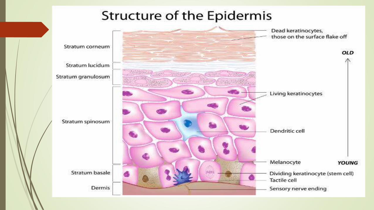
95% of the cells in the epidermis are keratinocytes. These cells are
found in the basal layer of the stratified epithelium that comprises
the epidermis, and are sometimes referred to as basal cells, or basal
keratinocytes.
Tissue Engineered Skin
Tissue engineering of skin became feasible in 1975 with the
demonstration that sheets of human keratinocytes could be grown in
the laboratory in a suitable form for grafting. This was a simple,
cohesive sheet of cells cultured from the donor on a feeder layer of
fibroblasts .
The epithelial component is able to regenerate in culture, since the
cells grow as a continuous sheet over a suitable surface, producing a
continuous layer which progresses to form cornified layers.

Keratinocytes cells in skin

Though both scar tissue and normal skin are made with collagen
proteins, they look different because of the way the collagen is arranged.
In regular skin, the collagen proteins overlap in many random directions,
but in scar tissue, they generally align in one direction. This makes the
scar have a different texture than the surrounding skin. Scar tissue is also
not as flexible as normal skin, and does not have a normal blood supply,
sweat glands, or hair
Various forms of implantable skin substitutes
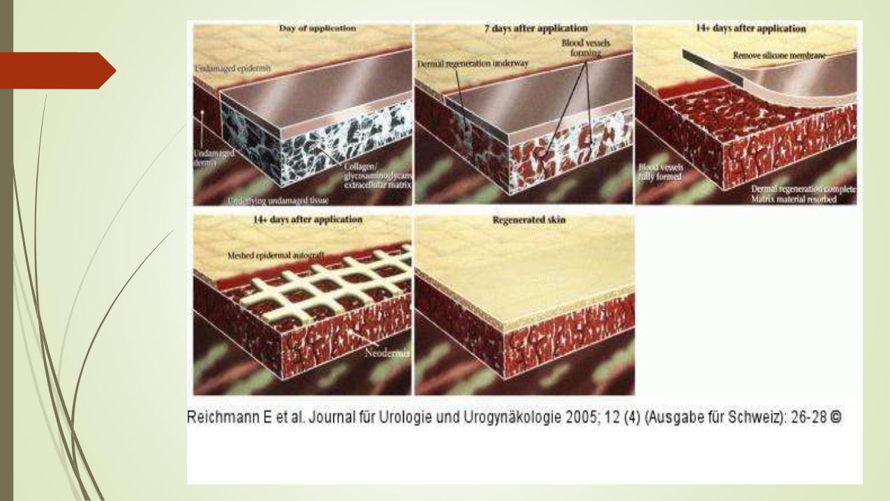
1. Integra consists of insoluble bovine collagen type I and the
glycosaminoglycan chondroitin sulfate. This can be covered in a
keratinocyte sheet at the time of implantation.
2. Dermagraft consists of PGA polymer mesh of suitable pore size,
seeded with human dermal fibroblasts from neonatal foreskins.
3. Apligraf consists of human dermal fibroblasts seeded into a type I
collagen gel and allowed to contract under tension.

Artificial skin

Human urothelial cells and bladder smooth muscle cells can be
cultured.
The criteria are that the final structures need to form elastic tubes
or bladders, and the implant should not allow crystal formation
from urine or harbour local infections.
Support materials tested have included resorbable polymers
[polyglycolic acid) and poly(lactic-co-glycolic acid) co-polymer:
PGA and PLGA and cross-linked collagen sponges.
Urothelial and bladder muscle cells seeded onto PGA scaffolds
formed urothelium-like, vascularized bilayered tissues when
implanted
Tissue Engineered Urothelium
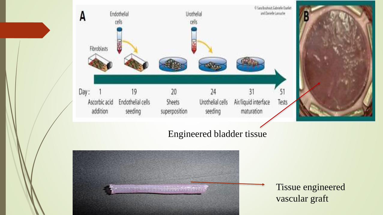
Tissue engineered
vascular graft
Engineered bladder tissue

ARTIFICIAL BONE GRAFTS: PRO OSTEON
Safe, strong, and cost effective bone grafts are now performed
using synthetic material known as Pro Osteon Implant 500.
It is sterile, biocompatible (meaning the body’s immune system
does not reject it), and it is easily sculpted to fill a defect in
fractured bones.
Pro Osteon mimics the internal structure of human bone. This
synthetic material is made by subjecting a common, non-decorative
form to coral to a patented chemical process which converts the
coral to hydroxyapatite, the same mineral content of human bone.
The porous, interconnected structure of the coral remains intact,
providing an ideal matrix through which new bone tissue can grow.
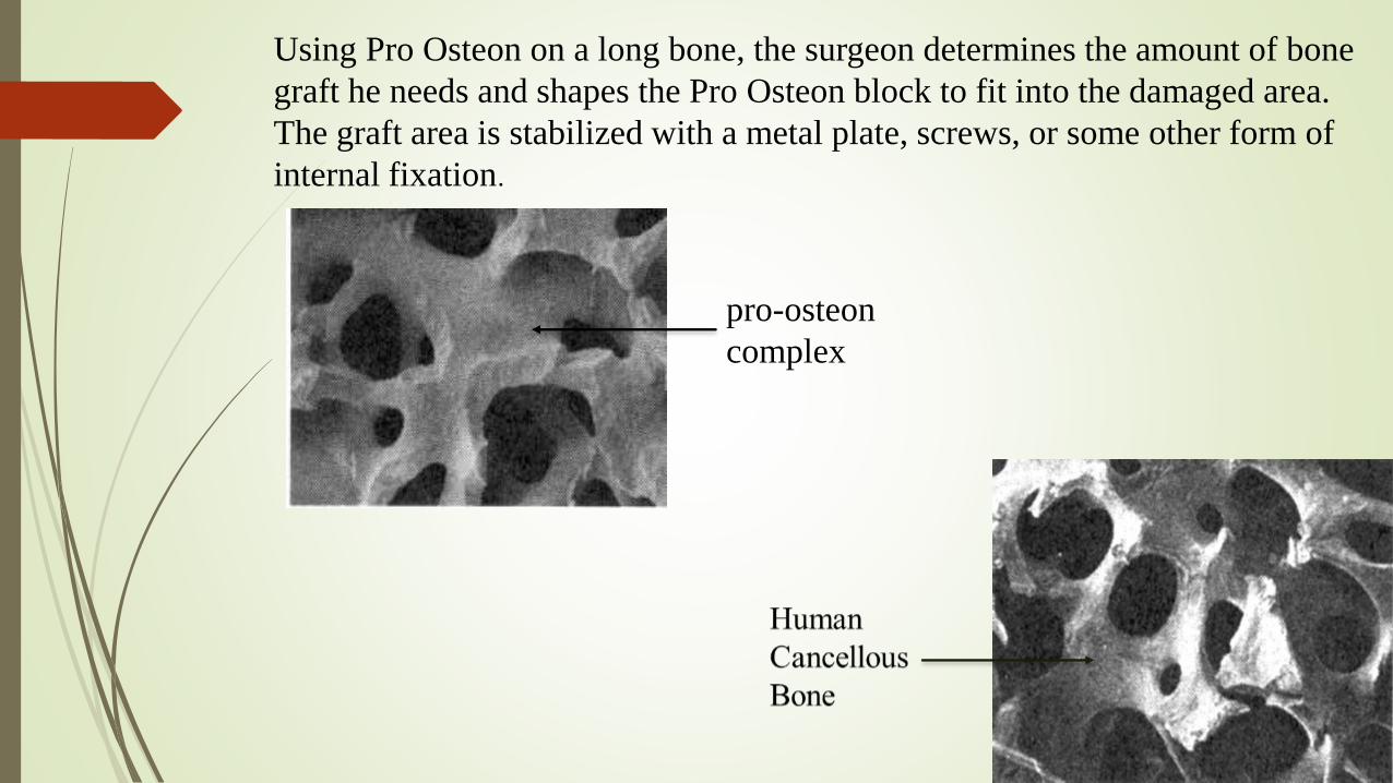
Using Pro Osteon on a long bone, the surgeon determines the amount of bone
graft he needs and shapes the Pro Osteon block to fit into the damaged area.
The graft area is stabilized with a metal plate, screws, or some other form of
internal fixation.
pro-osteon
complex

Bioengineered Tissue Implants Regenerate Damaged Knee Cartilage
ScienceDaily(July 5, 2006)
Cartilage was removed from 23 patients with an average age of 36 years.
After growing the cells in culture for 14 days, the researchers seeded them
onto scaffolds made of esterified hyaluronic acid, grew them for another 14
days on the scaffolds, and then implanted them into the injured knees of the
study patients.
Cartilage regeneration was seen in ten of 23 patients, including in some
patients with pre-existing early osteoarthritis of the knee secondary to
traumatic injury. Maturation of the implanted, tissue-engineered cartilage was
evident as early as 11 months after implantation

Tissue engineered organs
(A)(B)
(C)
(A): tissue engineered heart valve
(B): tissue engineered vascular graft
(C): tissue engineered human ear

Tissue engineered
artificial skin

Ear growing on the
mouse


