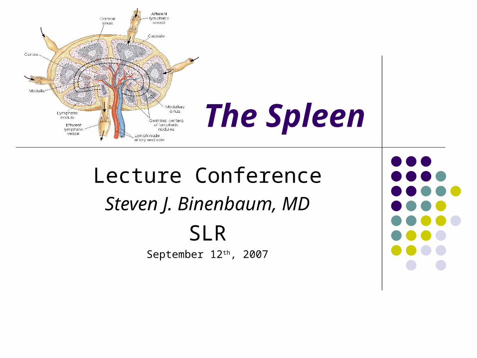
The Spleen
Lecture ConferenceSteven J. Binenbaum, MD
SLRSeptember 12th, 2007

Splenectomy for Hematologic DiseasesSplenectomy for Hematologic Diseases
Rarely cures the disease
Alleviates symptoms
Corrects hematologic abnormalities
Staging & Diagnosis
Splenectomy for Hematologic DiseasesSplenectomy for Hematologic Diseases
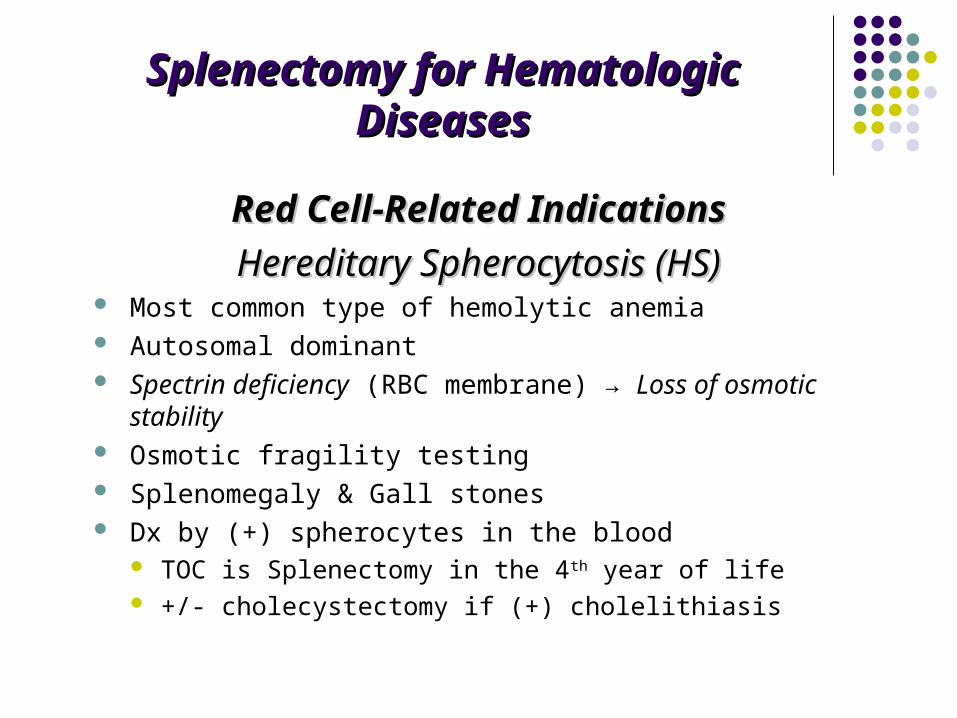
Red Cell-Related IndicationsRed Cell-Related Indications
Hereditary Spherocytosis (HS)Hereditary Spherocytosis (HS) Most common type of hemolytic anemia Autosomal dominant Spectrin deficiency (RBC membrane) → Loss of osmotic stability Osmotic fragility testing Splenomegaly & Gall stones Dx by (+) spherocytes in the blood
TOC is Splenectomy in the 4th year of life +/- cholecystectomy if (+) cholelithiasis

Splenectomy for Hematologic DiseasesSplenectomy for Hematologic Diseases
Autoimmune Hemolytic Anemia (AIHA)Autoimmune Hemolytic Anemia (AIHA) Warm-antibody autoimmune HA Autoantibody opsonization and phagocytosis Intravascular destruction or by spleen macrophages Treatment Of Choice
#1 is Corticosteroids 1-2 mg/kg/day#1 is Corticosteroids 1-2 mg/kg/day PRBC’s for severe anemia SplenectomySplenectomy (80% favorable clinical response) if:
medical tx fails Intolerance to steroids or its side-effects

Splenectomy for Hematologic DiseasesSplenectomy for Hematologic Diseases
Pyruvate kinase DeficiencyPyruvate kinase Deficiency Autosomal-recessive disease Splenomegaly TOC
SplenectomySplenectomy Reduces PRBC’s requirements
G6PD DeficiencyG6PD Deficiency Splenomegaly is rare Splenectomy is not indicated!

Splenectomy for Hematologic DiseasesSplenectomy for Hematologic Diseases
Sickle Cell Disease (SCD)Sickle Cell Disease (SCD) B-globin gene A→T substitution (Hb S/SS) autosomal dominant Sickling of RBCs in tissues with low O2 tension Red & White pulps Splenic microinfarcts
Painful Abscess Infections Anemia
Splenectomy (palliative)Splenectomy (palliative) Acute sequestration crises (recurrence = 40%-50% with 20% Mortality rate) Rapid hypersplenism Abscess formation

Splenectomy for Hematologic DiseasesSplenectomy for Hematologic Diseases
ThalassemiaThalassemia Thalassemia major (homozygous B) autosomal-dominant disease Decreased expression of beta-chains Pallor, Growth retardation, head enlargement Splenectomy indicated if:
Symptomatic splenomegaly Anemia Pain due to infarctions increased PRBC’s requirements (>200 ml/kg/year)
↑ rate of infections after splenectomy Risk vs. Benefit

Splenectomy for Hematologic DiseasesSplenectomy for Hematologic DiseasesPlatelet Related IndicationsPlatelet Related Indications
ITPITP Spleen is not enlarged ♀ > ♂; Renal insufficiency children vs. adults
Self-limited in children (70%) Splenectomy is for rare cases
Insidious onset in adults ↓ Plts < 10,000/mm3
#1 TOC is PO Steroids 1-1.5 mg/kg/d (up to 70% response)
IVIG 1 g/kg/d x2-3 days Splenectomy if fail steroid tx
TTPTTP Splenomegaly Microvascular thrombosis Petechiae, fever, neurologic changes #1 TOC
Plasmaphoresis Splenectomy is #2
Durable remission? Avoid PLT transfusions
↑ morbidity

White Cell - Related IndicationsWhite Cell - Related Indications
LeukemiaLeukemia CLL & Hairy cell leukemia (HCL) Splenectomy
improves cytopenias (75%) Ameliorates symptomatic splenomegaly
Non-Hodgkin’s Lymphoma (NHL)Non-Hodgkin’s Lymphoma (NHL) Splenectomy
Painful splenomegaly Cytopenia
No role for staging

White Cell - Related IndicationsWhite Cell - Related Indications
Hodgkin’s DiseaseHodgkin’s Disease Current indications for surgical staging:
Stage I or Stage II with NS histology and w/o B-symptoms
Staging procedure Wedge liver biopsy LN sampling:
Retroperitoneal Mesenteric Hepatoduodenal Splenectomy

Bone Marrow – Related Indications
Myelofibrosis & Myeloproliferative disorders Splenectomy for symptoms due to enlarged spleen

Splenectomy ComplicationsSplenectomy Complications
LLL atelectasis, pneumonia, effusion Hemorrhage (mostly with laparoscopic) Intraabdominal abscess (LUQ) Pancreatitis or fistula formation DVT PVT
Hemolytic anemia or myeloproliferative dz with splenomegaly
OPSI

Splenectomy ComplicationsSplenectomy Complications
OPSI Incidence ≈1% (up to 5%); 50% Mortality Encapsulated gram-positive bacteria = Streptococcus Risk factors
Children < 15 yrs old Immunosupression Hematologic dz (thalassemia, SCD, etc) Highest within the first 2 yrs post splenectomy
Pnemococcus, H.influenza, meningococcusPnemococcus, H.influenza, meningococcus vaccination 7-14 days prior to splenectomy
Pneumovax booster Q5yrs and annual H.influenza immunizations Abx prophylaxis for children x2 yrs post splenectomy Lower incidence of OPSI in adults (vs children) and after trauma


Tumors, Cysts, and Tumors, Cysts, and Abscesses of the SpleenAbscesses of the Spleen
Steven J. Binenbaum, MD
September 12th, 2007
SLR

Malignant TumorsMalignant Tumors
Spleen - mostly secondary involvement non-Hodgkin’s Lymphoma – most common malignancy
Main Tx: Chemo +/- RT
Spleen is the primary site 10% Hodgkin’s disease 30% of resected spleens (staging procedure) have (+) histology
Hairy cell leukemia Resect for symptomatic splenomegaly
Improved survival
CML & CLL symptomatic splenomegaly = splenectomy

Malignant TumorsMalignant Tumors
AngiosarcomaAngiosarcoma Nonlymphoid malignant tumor of the spleen Early metastatic disease Aggressive with rapid growth Spontaneous splenic rupture and hemolytic anemia Palliation
Benign TumorsBenign Tumors Hemangioma
Risk of rupture + platelet sequestration (Syndrome?) No tx unless symptomatic
Hamartoma Lymphangioma
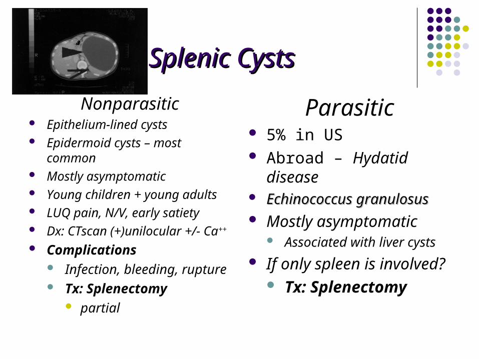
Splenic CystsSplenic Cysts
Nonparasitic Epithelium-lined cysts Epidermoid cysts – most common Mostly asymptomatic Young children + young adults LUQ pain, N/V, early satiety Dx: CTscan (+)unilocular +/- Ca++
Complications Infection, bleeding, rupture Tx: Splenectomy
partial
Parasitic 5% in US Abroad – Hydatid disease Echinococcus granulosusEchinococcus granulosus Mostly asymptomatic
Associated with liver cysts
If only spleen is involved? Tx: Splenectomy

Splenic CystsSplenic Cysts
Splenic PseudocystsSplenic Pseudocysts Lack epithelial lining Account for most cystic splenic dz in US
Pancreatic pseudocyst Posttraumatic
Splenectomy is indicated when: Size >10 cm or symptomatic

Splenic AbscessSplenic Abscess Uncommon, but fatal
Erode into adjacent structures Most are secondary in etiology
Bacterial endocarditis Intrabdominal infections (pyelo-, etc) IVDA Infected splenic hematoma Infected splenic infarctions (embolizations, ischemia, etc)
S/S: fever, WBC; 50% (+) blood cultures
Dx by CT scan + IV contrast Staphylococcus & Streptococcus E.coli, Salmonella, anaerobes Tx:Tx:
Splenectomy + IV Abx Percutaneous drainage

Splenic Salvage Procedures: Splenic Salvage Procedures: Therapeutic OptionsTherapeutic Options
SLR
September 12th, 2007

Nonoperative Management of Splenic Nonoperative Management of Splenic TraumaTrauma
Indications for initial nonoperative management hemodynamic stability absence of peritonitis CT scan
No contrast extravasation absence of other injuries
Transfusions - >2 PRBC’s

Protocol for Nonoperative Management
Grade I & IIGrade I & II Awake + alert, isolated injury
monitored observation BR, H/H q6h, serial abdominal exams Regular floor in 48º If remain stable and asymptomatic – D/C in 5 days F/U CT scan in 4 wks
Avoid prophylactic and therapeutic heparinization Grade III, IV, & VGrade III, IV, & V
Monitored observation x5 days Repeat CT scan Transfer to floor if stable F/U CT scan in 6-8 wks after discharge


SplenorrhaphySplenorrhaphy
Topical Hemostasis Small injuries (I & II)
Bovie electrocautery Argon beam Gelfoam Surgicel Avitene
Suture Repair & Partial Resection Segmental blood supply Monofilament sutures
Pledgeted horizontal mattress sutures
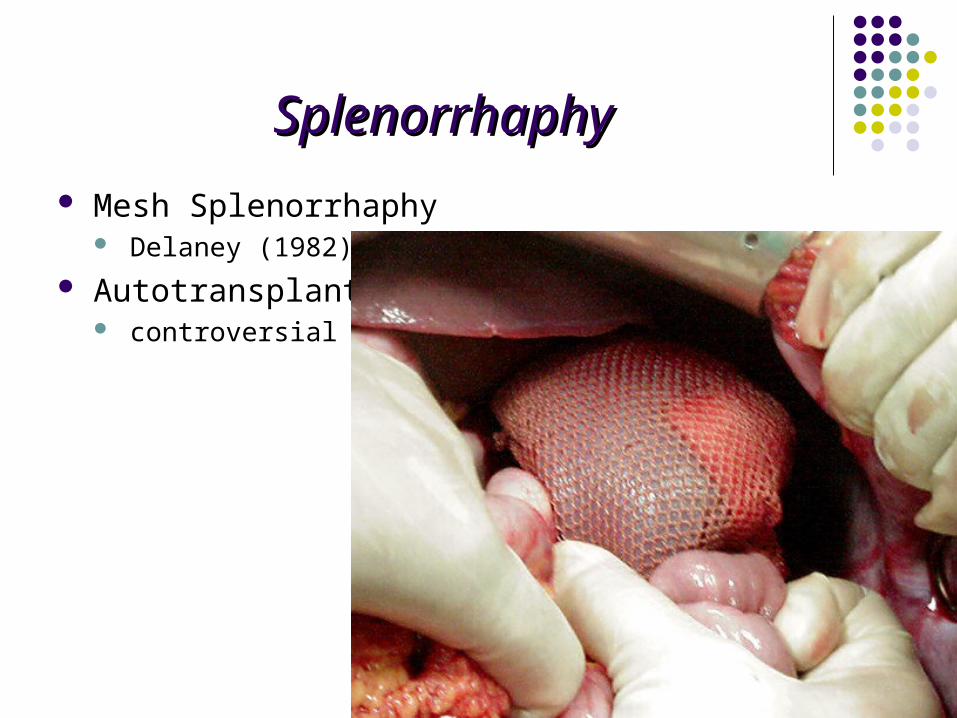
SplenorrhaphySplenorrhaphy
Mesh Splenorrhaphy Delaney (1982)
Autotransplantation controversial


