
THE MOLECULAR FUNCTION OF mCLCA1, mCLCA2 and mCLCA4
IN MURINE LIFE
A Dissertation
Presented to the Faculty of the Graduate School
of Cornell University
In Partial Fulfillment of the Requirements for the Degree of
Doctor of Philosophy
by
Kai Su Greene
May 2009

© 2009 Kai Su Greene

THE MOLECULAR FUNCTION OF mCLCA1, mCLCA2 and mCLCA4
IN MURINE LIFE
Kai Su Greene, Ph. D.
Cornell University 2009
Chloride channel, calcium-activated proteins (CLCAs) have been reported to
regulate chloride transport and be involved in the pathophysiology of diseases, such as
cystic fibrosis, asthma, airway inflammation and cancer. They have been cloned or
isolated from multiple species, incluing human, mouse, bovine, equine, rat, porcine
and canine. The aim of this research was to understand the function of this group of
genes in murine life, focusing on mCLCA4 and its highly homologous family
members, mCLCA1 and mCLCA2.
mCLCA4 is one of six members in the murine genome cloned to date, and is
highly expressed in smooth muscle. To begin to understand the function of this gene
family, I investigated the cellular processing and regulatory sequences of mCLCA4
proteins. The full length mCLCA4 gene product [125 kilo-Dalton (kD)] is made in the
endoplasmic reticulum and is cleaved to 90 kD and 40 kD fragments. Both fragments,
90 kD N- terminal and 40 kD C- terminal fragments are secreted out of the cell and
associate with the cell membrane. A specific diarginine motif is the retention signal
while a dileucine motif is the forward trafficking signal during mCLCA4 secretion.
While secretion of mClCA4 excludes this gene product as a channel protein, its
association with the membrane may be consistent with a role as a regulator of chloride
conductance.
To further understand mCLCA4 gene function, I generated mCLCA4 knock-

out mice. These mice displayed no gross phenotype and bred normally. Specific lung
challenge experiments are being undertaken by collaborators to examine the effect of
airway challenge on mClCA4 null mice. To further study the function of mCLCA4 in
vivo, mCLCA1, mCLCA2, mCLCA4 triple knock-out mice were made. A 112 kilo
base pair (kb) sequence was deleted from chromosome 3 of the murine genome using
bacterial artificial chromosomal (BAC) recombineering techniques. Quantitative PCR
was used to screen for positive embryonic stem cell clones that were then injected into
blastocysts using standard techniques. Highly chimeric mice were bred to C57Bl/6J
mice to produce heterozygous offspring. Currently, the triple gene knock-out mice
survive to birth, but further phenotypic evaluation is needed.

iii
BIOGRAPHICAL SKETCH
Kai Su Greene was born and raised in Changchun, China. She went to Jilin
University after high school and obtained her B.S with a biochemistry major and
chemistry minor. She worked as a researcher, instructor and then promoted to director
of the Jilin Province Diagnostic Center where she trained the technicians from hospital
diagnostic laboratories in Jilin Province. Kai spent three years at Showa University on
a visiting scholar fellowship and Tokyo University as a graduate student in Japan.
Kai came to United States in 1993. She was invited by and worked for Dr.
Leslie Pick to study Drosophila genetics at the Mt. Sinai Medical School, New York,
NY. After five years of New York City life, she took a job at Wyeth Inc., a
pharmaceutical company as a research scientist in Princeton, NJ. She moved to
Endicott, New York to join her husband and started to work for Dr. Kotlikoff at
Cornell University in year 2000. 2002 was a special year for Kai, she became a US
citizen and stared her PhD program. Currently she lives with her husband Dr.
Raymond Greene and son Thom Greene in Ithaca, NY.
After completing her degree, she will continue to be a researcher in the field of
biology and continue to contribute to scientific discoveries to save human life.

iv
To my parents, Shuzhen Ma and Dongfeng Su, for their love and for reminding me the
value of education. I would like to dedicate my dissertation to them

v
ACKNOWLEDGMENTS
I would like to thank my advisor, Dr. Michael I. Kotlikoff, for his academic
guidance and support of my PhD research. Without him I would not even have thought
to start my PhD at my age. He invested a great deal of knowledge, resources and
encouragement to help me succeed. He gave me the chance to work independently and
solve the problems on my own. I am very grateful for this. I am also indebted to all my
dissertation committee members for their advice and support in the past six and half
years. Every committee member has played an important role in my research and in
my graduate student life. They are Dr. Teresa Gunn, Dr. Ellis Loew, Dr. Mark
Roberson (in alphabetical order) and former committee member Dr. JunLin Guan.
Also I would like to thank all the Kotlikoff lab members (past and present) for their
assistance, support and help in many ways. They are Dr. N. Yvonne Tallini for helping
with my thesis writing; Dr. Gwendolyn Spizz for helping with my A exam proposal
writing; Dr. Michael Craven, Rorbert Doran, Dr. Guangju Ji, Chunlei Huan, Jane Lee,
Shaun Reining, Dr. Mark Rishniw, Dr. Bo Shui, Dr. Yongxiao Wang, Dr. Hongbo Xin
(in alphabetical order).
I would like to thank Dr. Randolph Elble and Dr. Bendicht Pauli for
collaborating with us on CLCA gene family studies. I would also like to thank Mr.
Robert Munroe in Dr. John Schementi lab, Cornell University for injecting my
mCLCA1,2,4 knock out ES cell clone and Ms. Keyu Deng in Transgenic Facility at
Cornell University for mCLCA4 knock-out ES cell clone injecting. There are so many
friends and colleagues in Biomedical Sciences Department and Vet school that
supported me and helped me, I really appreciate them all.
I would also like to thank The Employee Degree Program of Cornell
University. It encouraged me to start my PhD program.

vi
I am indebted to Dr. Isao Niki for his academic guidance, friendship as well as
financial support during my Japan years. I am also grateful for Dr. Leslie Pick who
invited me to this country and trained me to become an independent researcher.
I am really grateful to and thank my family, without their support I would not
have reached this point. My husband, Dr. Raymond Greene, was always fully
supportive and encouraged me on my good or bad days. My son, Thom Greene, is my
energy source and accompanied me on the weekends while I worked in the lab. My
parents, Donfeng Su and Shuzhen Ma, always believe in the importance of education
and supported me. My father-in-law, Dr. Thom R. Greene, always supported me and
encouraged me. To all my relatives who supported me during these years, my sister-
in-law Deborah Greene Modra, my brother Libing Su and his family, my sister Qi Su
and her family, thank you.

vii
TABLE OF CONTENTS
Biographical Sketch ………………………………………………………………iii
Dedication …………………………………………………………………………iv
Acknowledgements ……………………………………………………………….v
Table of contents …………………………………………………………………vii
List of figures ……………………………………………………………………...x
List of Tables ……………………………………………………………………..xii
Chapter 1 Introduction 1
1.1 CLCA family: mammalian species and expression..……………..2
1.2 CLCAs common features…………………………………………8
1.2.1 Chloride conductance……………………………..8
1.2.2 The structure of CLCA proteins and their cellular
location……………………………………………11
1.3 The relationship between CLCAs and asthma…………………...14
1.4 The relationship between CLCAs and cystic fibrosis……………15
1.5 The function and contribution in cell adhesion, tumor and cell ....17
1.6 Summary of CLCAs functions…………………………………..19
1.7 References………………………………………………………..20
Chapter 2 The regulation and secretion of mCLCA4 protein 28
2.1 Abstract ………………………………………………………… 29
2.2 Introduction ……………………………………………………...29

viii
2.3 Materials and Methods ………………………………………….31
2.4 Results …………………………………………………………..36
2.5 Discussion ………………………………………………………52
2.6 References ………………………………………………………55
Chapter 3 mCLCA4 knock-out mice and its phenotype 58
3.1 Abstract ………………………………………………………...59
3.2 Introduction …………………………………………………….59
3.3 Materials and Methods …………………………………………60
3.4 Results ………………………………………………………….67
3.5 Discussion ……………………………………………………...67
3.6 References ……………………………………………………...74
Chapter 4 Generating mCLCA1, 2 and 4 triple knock-out 76
4.1 Abstract …………………………………………………………77
4.2 Introduction ……………………………………………………. 77
4.3 Materials and Methods ………………………………………….79
4.4 Results …………………………………………………………. 92
4.5 Discussion ……………………………………………………...102
4.6 References ……………………………………………………...107
Chapter 5 Summery and future research direction 108
5.1 Summery ……………………………………………………….109

ix
5.2 Future direction and studies..……………………………………110
5.3 Reference ………………………………………………………..116

x
LIST OF FIGURES
Figure 1.1 Orthology summary map of CLCA genes, human and mouse
genome loci, knock-out mice, and gene functions ……………………6
Figure 1.2 Phylogenetic tree of human and mouse CLCA members …………….9
Figure 1.3 Human and mouse CLCA protein structure diagram…………………13
Figure 2.1 Proteolytic processing and cellular localization of mCLCA4………...37
Figure 2.2 The 90 kD N -terminal fragment and C -terminal
fragment are secreted into the extracellular space ...............................40
Figure 2.3 Mutation of putative phosphorylation site slows
proteolytic cleavage………………………………………………….42
Figure 2.4 Forward traffic and ER retention signals ……………………………45
Figure 2.5 Glycosylation patterns of mutant proteins …………………………..48
Figure 2.6 mCLCA4 dileucine sequences are required for forward
trafficking of secreted proteins ...........................................................50
Figure 3.1 Diagram of mCLCA4 knockout mouse and homologous
recombination in ES cells ...................................................................62
Figure 3.2 Genotyping strategy for mCLCA4 …………………………………..66
Figure 3.3 Southern blot and PCR genotyping ………………………………….68
Figure 4.1 Diagram of BAC clone RP24-271-G16 …………………………….81
Figure 4.2 Schematic generating BAC-KO124-galk targeting vector …………..83
Figure 4.3 Diagram depicting generation of BAC-KO124-neo/kan
targeting vector for ES cells .................................................................87
Figure 4.4 Homologous recombination between BAC-KO124-Neo/kan
and one chromosome 3 allele in ES cells ………………………….. 94
Figure 4.5 Diagram of the real-time PCR strategy ……………………………...95

xi
Figure 4.6 qPCR using KO124KO4 probe …………………………………….97
Figure 4.7 qPCR results ……………………………………………………….98
Figure 4.8 PCR strategy and results to genotype mCLCA1,2,4 mice …………100
Figure 4.9 Evaluation of mCLCA1,2,4 eyes …………………………………..104
Figure 5.1 Diagram depicting strategy to make mCLCA1,2,4,6,7,8
knock out mice ……………………………………………………..114
xii
LIST OF TABLES
Table 3.1 Blood chemistry test results from mCLCA4-/- mice
and littermate controls ………………………………………..71
Table 3.2 Blood CBC test results from mCLCA4-/- mice and
littermate controls …………………………………………….72
Table 4.1 Evaluation of body weight in mCLCA1,2,4 knock
out offspring …………………………………………………..106

1
CHAPTER 1
INTRODUCTION OF CLCA FAMILY IN ALL SPECIES

2
Calcium-activated chloride currents play an important role in diverse cellular
physiological functions, such as neuronal excitability, regulation of vascular tone,
epithelial secretion, fast block to polyspermy and olfactory transduction (24). They
have been reported in many cell types such as smooth muscle cells (13-15, 37),
cardiac myocytes (35, 78), exocrine glands (7, 22, 62), epithelial cells (5, 16, 49) and
endothelial cells (51). The genes involved in these channels have been sought and
studied for many years, but the molecular identity of these channels remains unknown.
Two candidate gene families contribute to the calcium-activated chloride channel
activity, CLCA and Bestrophin (56, 64). Recently, Yang et al. (70) discovered that the
transmembrane protein 16A (TMEM16, also called ANO1) may also be a chloride
channel protein. This dissertation focuses on the function of the CLCA family in
murine life, concentrating on mCLCA4 and two highly homologous family members,
mCLCA1 and mCLCA2.
1.1 CLCA family: mammalian species and expression
CLCAs have been reported in the literature for seven mammalian species:
human, murine, rat, bovine, equine, porcine and canine. Since the complete sequence
of the human genome, CLCA putative orthologs were discovered in more than 30
species as listed on the NCBI Gene database and the Ensemble Gene Tree View (52).
The CLCA genomic organization in many species is conserved. The genes cluster
together on the same chromosome, but little information is available about the
promoters that drive their expression. So far, the following have been cloned and
reported: four members from human (hCLCA1, hCLCA2, hCLCA3 and hCLCA4),
six plus two predicted members from murine (mCLCA1, mCLCA2, mCLCA3,
mCLCA4, mCLCA5 and mCLCA6; ESTs EG622139 and AI747448 are predicted),
two members from bovine (bCLCA1, and bCLCA2), one member from porcine

3
(pCLCA1), one member from equine (eCLCA1), two members from rat (rCLCA1 and
rCLCA2) and one from canine (cCLCA1).
The first cDNA of the CLCA family was reported by Cunningham and
colleagues (17) using the polyclonal antibody αp38 on a bovine tracheal cDNA
library. It was originally named CaCC (calcium-activated chloride channel) and later
renamed to bCLCA1 to match the CLCA nomenclature. The full length cDNA of this
protein is 3001 base pair (bp) and codes for a 903 amino-acid (aa) protein product that
is 140 kDa. The 140 kDa protein product is translated and glycosylated, then cleaved
to 90 kDa and 38/32 kDa fragments.
The first CLCA protein was purified from lung endothelial cell matrix extracts
using the monoclonal antibody 6D3 by Zhu et al. (76) and was determined to be a 90
kDa fragment of the lung endothelial cell adhesion molecule-1 (Lu-ECAM-1) protein.
Later, the gene of this protein was cloned by the same group, from a bovine gene
expression library and renamed bCLCA2 (21, 77). bCLCA2 codes for a 130 kDa
glycosylated precursor protein and is cleaved into two fragments: 90 and 30 kDa.
bCLCA1 and bCLCA2 isoforms share 88% identity, but their tissue distribution is
different—bCLCA1 is expressed in epithelial cells while bCLCA2 in endothelial cells.
There are two other bovine CLCA genes, bCLCA3 and bCLCA4. All four of these
genes are clustered on bovine chromosome 3 (52).
All four human CLCA members are clustered on chromosome 1, 1p31-1p22
(Figure 1.1). hCLCA1 was cloned by Gruber et al. (30) from a human genomic library
using bCLCA2 cDNA as the probe. It was also isolated by Angel and colleagues (4)
along with the human CLCA family genes, namely hCaCC-1 (the same as hCLCA1),
hCaCC-2 (hCLCA4) and hCaCC-3 (hCLCA3). The hCLCA1 gene spans 31 kb and
codes for a 914 aa protein. After glycosylation this protein is 125 kDa and is then
cleaved into 90 kDa and 38-40 kDa products in the HEK293 cell. hCLCA1 is highly

4
expressed in small intestine, colon mucosa and appendix, and weakly expressed in
uterus, stomach, testis, kidney and fetal spleen. At the cellular level, the expression is
detected in basal crypt epithelia and goblet cells by Northern blotting and in situ
hybridization (30, 47).
hCLCA2 was cloned from a human lung cDNA library with bCLCA2 cDNA
as the probe (34). The 2832 bp cDNA is the predicted coding sequence for a 943 aa
polypeptide. It is translated to a 120 kDa protein product after glycosylation, and
subsequently cleaved into 86 kDa and 34 kDa fragments. Northern blot analysis
showed that hCLCA2 is only expressed in trachea and mammary gland tissues.
Interestingly, by Northern blot, hCLCA2 expression was not detected in lung,
although it was cloned from lung cDNA. This indicates that hCLCA2 has a very low
expression level in lung tissue (34, 47).
hCLCA3 was cloned from a human spleen cDNA library using Lu-ECAM-1
(bCLCA2) cDNA as a probe (32). hCLCA3 is expressed in many tissues, such as
lung, trachea, spleen, thymus and mammary gland as detected by reverse transcriptase
polymerase chain reaction (RT-PCR). Unlike other CLCA members, hCLCA3 cDNA
is 3.6 kb with two internal stop codons and encodes 37 kDa and 22 kDa products,
respectively. The N-terminal product (37 kDa) is secreted out of cell and the function
of this truncated product remains unknown (32).
Using a homology search in the EST database Agnel et al. (4) identified a
potentially new human CLCA family member, hCLCA4 (previously referred to as
hCaCC2). hCLCA4 was subsequently cloned using the 5’ rapid amplification of
cDNA ends (RACE) approach. The hCLCA4 cDNA is 3.3 kb and encod a 917 aa
protein product. This gene is expressed in multiple areas of the brain such as the
amygdala, caudate nucleus, cerebral cortex, frontal lobe, hippocampus, medulla
oblongata, occipital lobe, putamen, substantia nigra, temporal lobe, thalamus,

5
acumbens and spinal cord. The highest level of hCLCA4 expression is in the colon.
The weakest expression is in stomach, testis, small intestine, appendix, salivary gland
and mammary gland. It also is moderately expressed in the bladder, uterus, prostate
and trachea (4, 47).
Only one member has been cloned and expressed from full-length cDNA from
ileal gene porcine expression library, pCLCA1 (26, 47). pCLCA1 is a 3.1 kb cDNA
and has a 2.7 kb open reading frame (ORF) which encodes a 917 aa protein product
cleaved to two polypeptides. The protein product is 78% homologous to hCLCA1.
The mRNA of pCLCA1 is expressed in several exocrine epithelial tissues, such as the
ileum (crypt and villus epithelia), the trachea and salivary glands.
Base on the rat genome sequence (27, 66) and bioinformatics information (66,
67), there are five CLCA members in rat. Yamazaki and colleagues (69) cloned full
length rCLCA1 [Patel et al renamed it rCLCA2 (52)] cDNA from salivary cells.
rCLCA1 cDNA is 3.3 kb in length and the predicted ORF encodes a 903 aa protein. A
year after, Yoon et al (71) cloned the same gene from rat brain and named it
rbCLCA1. The mRNA of rCLCA1 was expressed in the submandibular gland (SMG),
ileum and lung. In the SMG, rCLCA1 protein was only detected in the striated duct
and not in the acinar cells by immunostaining with anti-rCLCA polyclonal antibody.
At the cDNA level, rCLCA1 is highly homologous to mCLCA1and mCLCA2 (87%),
mCLCA4 (84%), and hCLCA1 (70%) (69). Jeong et al (38) cloned another rat CLCA
gene named rbCLCA2 [Patel et al renamed it rCLCA1 (52)]. The full-length cDNA of
rbCLCA2 is 2895 bp and codes for a 902 aa protein.
To date only one CLCA member has been cloned from equine (6). Total
mRNA was extracted and purified from horse rectal mucosa, conserved region primers
from hCLCA1 and mCLCA3 were used to PCR the isoform from the mRNA pool.

6
Figure 1.1 Orthology summary map of CLCA genes, human and mouse
genome loci, knock-out mice, and gene functions. Double end arrow shows CLCA
genes orthology between species. Human and mouse CLCA gene loci based on NCBI
Map View. Three single gene knock-out mice have been generated, mCLCA3 (59),
mCLCA5 (Deltagen, Inc.) and mCLCA4 (chapter 3). Summary of potential CLCA
protein function.
86.60MChr.1Human 1p31-1p22
86.65M 86.70M 86.75M 86.80M 86.85M 86.90M 86.95M
Chr.3Mouse3H2-H3 144.75M 144.70M 144.65M 144.55M 144.50M 144.40M144.60M
mCLCA4 mCLCA2 mCLCA1
hCLCA3hCLCA4hCLCA1hCLCA2
mCLCA5 mCLCA3 mCLCA6
AsthmaCystic fibrosis
Tumor suppressorCancer
CancerCell adhesionTumor suppressor
Knock-out Yes Yes no Yes no no
liver wt/body wt high no phenotype no phenotype
Function
bCLCA2 bCLCA1 eCLCA1 pCLCA1rCLCA1 cCLCA1

7
The cDNA of eCLCA1 is 2.9 kb (GenBank accession NO. AY524856) and encodes a
913 aa protein product. In vitro translation of eCLCA1 cDNA followed by microsomal
membrane experimental methods showed eCLCA protein is a 120 kDa when
glycosylated. A 70 kDa and a 80 kDa fragment have been detected from colon mucosa
by antibody (a-eCa1) (6). eCLCA1 is highly homologous to hCLCA1 and mCLCA3
(82.4% and 73.5% respectively at the polypeptide level) (6). At the protein level,
eCLCA1 is expressed in mucous-producing cells of the respiratory and intestinal
tracts, cutaneous sweat glands and renal mucous glands.
Loewen et al. (48) partially cloned cCLCA1 from canine retinal pigment
epithelium using pCLCA1 specific primers. The partially predicted aa sequence of
cCLCA1 is highly homologous to pCLCA1 and hCLCA1 (48). The mRNA and
protein expression pattern of cCLCA1 is unknown.
Six murine members of CLCA (mCLCA1 to mCLCA6) have been cloned and
characterized. There are two additional potential members in the murine genome:
AI747448 (or mCLCA7) and EG622139 (or mCLCA8), which are on the same
chromosome location as the other family members (NCBI database). All the
mCLCAs are clustered on chromosome 3 H2-H3 (Figure 1.1). mCLCA1 was cloned
from a mouse lung cDNA library using bCLCA2 cDNA as a probe (25). It is a 3.1 kb
cDNA and the protein product is 125 kDa after glycosylation. mCLCA2 was cloned
from mouse mammary gland mRNA pool using suppression subtractive hybridization
method (42). mCLCA4 was cloned by Elble and colleagues (18) using degenerate
primers based on bCLCA2. mCLCA1, mCLCA2 and mCLCA4 are not only located
next to each other on mouse chromosome 3 but also share 70-80% identity at protein
level and are orthologous to hCLCA3 (18, 47, 52) (Figure 1.2). mCLCA3, also named
Gob-5, was isolated from mouse intestinal goblet cells (41). The mCLCA3 cDNA is 3
kb long and encodes 913 aa protein that shares homology with hCLCA 1 and pCLCA1

8
(28, 41) (Figure 1.2). mCLCA 5 and 6 were cloned from mouse eye and intestine
RNA extracts, respectively (23). Both ORFs of mCLCA5 and mCLCA6 are
approximately 2.8 kb but mCLCA6 may have a splice variant that is 2.6 kb.
In summary, CLCA members exist in at least 30 different species’ genomes
and 17 members from seven mammalian species have been published to date. All the
CLCA members from the same species cluster on the same chromosome and the 17
CLCAs discussed have similar protein structure with a very diverse expression pattern
in multiple tissues and organs. All the studied CLCA proteins are cleaved to NH2-
terminus and COOH-terminus fragments after glycosylation.
1.2 CLCAs common features
1.2.1 Chloride conductance
Anionic currents have been detected by either whole cell or single cell patch
clamp techniques following transient transfection of the CLCA cDNA into either
HEK293T or COS-1 cells. The anionic currents are dependent on intracellular Ca2+
concentration and can be blocked by chloride channel blockers, such as 4,4”-
diisothiocyanatostilbene-2,2’-disulfonic acid (DIDS), dithiothreitol (DTT) or Niflumic
(NAF) (30, 47). CLCA family was considered as a candidate of chloride channel
protein based on the finding that bCLCA1 protein appeared to regulate calcium-
activated chloride current in tracheal epithelial cells. Thus these data have led
investigators to conclude that CLCAs regulate of a family of chloride currents
controlled by calcium.
Gruber et al (30) investigated the electrophysiological features of hCLCA1
using whole cell and single cell patch clamp techniques following transient
transfection of hCLCA1 cDNA into HEK293T cells. This group demonstrated the

9
Figure 1.2 Phylogenetic tree of human and mouse CLCA members. The
homology tree was generated using mRNA sequences obtained from GeneBank and
subsequently aligned with Vector-NTI software (Invitrogen). The bar represents 5%
diversity.
hCLCA1 mCLCA3
hCLCA2 mCLCA5
hCLCA3 mCLCA1 mCLCA2
mCLCA4 hCLCA4
mCLCA6

10
whole cell chloride current increased compared to control when intracellular calcium
concentration was increased after an application of ionomycin. A similar experiment
was performed with hCLCA2 cDNA and after administration of ionomycin the whole
cell current as determined by patch clamp increased compared to control. Both of
currents were blocked by DIDS, DTT, NAF and tamoxifen. mCLCA1 cDNA was
transiently transfected into HEK293 cell and a calcium-activated chloride current was
detected by whole cell patch clamp. The current increased with the addition of
ionomycin and 2 mM calcium. The mCLCA1 current was inhibited by DTT, DIDS
and NAF in 2 mM calcium (25). Romio and colleagues (60) injected mCLCA1 cDNA
into Xenopus oocytes and detected a significant current without a calcium ionophore
compared to water-injected oocytes. The mCLCA1 current in oocytes, but not the
background current, was chloride dependent. Because of the high background current
in Xenopus, it was impossible to detect the effect of calcium on the CLCA1 chloride
conductance in the presence of ionomycin (60). Greenwood et al. (29) compared the
native calcium-activated chloride current in murine portal vein with the mCLCA1
cloned from murine portal vein myocytes. They found that the two channels shared
permeability similar to various anions, but the mCLCA1 channel current was not time-
dependent while the native one was. Moreover, the mCLCA1 channel showed a lower
sensitivity to calcium than the native channel. When they co-expressed mCLCA1 and
β-subunit of the big potassium channel (mKCNMB1), the current significantly
changed from time-independent to time-dependent and was more sensitive to calcium
than without the mKCNB1 subunit. Using the mammalian two-hybrid system they
showed that mCLCA1 and mKCNMB1 subunit interacted with each other but it
remains unknown whether these two proteins interact in native vascular smooth
muscle cells. These results suggested not only that mCLCA1 is a chloride current

11
regulated by calcium but that mCLCA1 current activity can be modified by other
proteins (29, 47).
In summary, many CLCA members from different species produce chloride
currents in response to changes in cellular calcium concentration. However, the
mechanism of how these proteins contribute to the chloride currents remains
unknown.
1.2.2. The structure of CLCA proteins and their cellular location
Epitope insertion in concert position followed by immunohistochemistry and
hydropathy techniques resulted in several initial models of the CLCA protein
structure. They were predicted to be transmembrane proteins (30-32, 54). Recently,
the region including the transmembrane-spanning segments was also predicted using
Simple Modular Architecture Research Tool (SMART) and it suggested that CLCA
proteins contained von Willebrand factor domain A (VWA), an extracellular soluble
domain (47). However, the Markov models for transmembrane segments and protein
folding prediction suggested that CLCA proteins are soluble extracellular molecules
with VWA domains and are not transmembrane proteins except the hCLCA2 COOH-
terminal fragment (20, 52).
It was discovered that all the members of CLCA family are processed similarly
using transient transfection of the CLCA cDNA into mammalian cell line such as
HEK293T and COS cells (Chapter 2), (9-11, 18, 20, 25). They all code and
glycosylate an approximately 130 kDa full-length precursor, then are cleaved to ~ 90
kDa (NH2-terminus) and ~ 40 kDa (COOH-terminus) fragments. Both fragments, as
with hCLCA1, mCLCA3 and mCLCA4, are secreted into the media and associate
with the cell membrane. For hCLCA2, the NH2-terminal fragment was released into
the media and the COOH-terminal fragment remained in the membrane. Some CLCA

12
proteins were cleaved inside the cell such as mCLCA4 (Chapter 2), and some were
cleaved outside the cell, such as hCLCA2 (20) (Figure 1.3).
The 90 kDa N-terminus fragment contains a cysteine-rich (also called N-
terminal hydrolase) domain in the first 280 aa which is only found in the CLCA family
(52, 55). Bioinformatics analysis by Pawlowski et al. (55) suggested the N-terminal
hydrolase domain (N-term) contains a zinc metalloprotease domain that potentially
functions as a hydrolase and protease. This prediction is supported by hCLCA3
structure—truncated protein with only N-terminal hydrolase domain. The CLCA
proteins may possibly self-cleave during their processing (55). However, the mutant
protein for this experiment may have misfolded and the cleavage of this protein group
is still not certain (52). Moreover, the VWA domain is followed by the N-terminal
hydrolase (N-Term) domain in this 90 kDa molecule. The VWA domain is involved in
protein-protein interactions and contains a metal ion-dependent adhesion site
(MIDAS) motif (68). In human and mice, all CLCA members have an N-Term domain
and all members except hCLCA3 and EG622139 (one of the predicted genes) contain
a VWA domain in their 90 kDa fragment (52). The 35 kDa COOH-terminal fragment
has a conserved fibronectin type III (Fib3) domain that was predicted using JPRED 3
(a secondary structure prediction software), except hCLCA3 and EG622139. The main
function of the Fib3 domain is for protein-protein interaction, but no studies have been
published to date of how this domain actions in CLCA (52).
In summary, CLCA proteins are ~130 kDa precursors and cleaved to 90 kDa
NH2-terminal and 40 kDa COOH-terminal fragments. The cleavage occurs
intracellular in some CLCA family members (mCLCA4, Chapter 2) and extracellular
in others (20). CLCA family members contain a hydrolase and a VWA domain in the
NH2-terminal fragment while a Fib3 domain is found in the COOH-terminal fragment.

13
Figure 1.3 Structure of human and mouse CLCA proteins diagram. Each
protein contains an N-terminal hydrolase domain (N-Term) (blue) that is cysteine-rich
and conserved in all members. Note other common features shared between members.
Abbreviations: von Willebrand factor type A, VWA domain; Fibronectin type III, Fib3
(orange); transmembrane, TM (light blue); glycosylphatidylinositol anchor, GPI
(yellow). “//” denotes predicted proteolytic cleavage sites.
Human CLCAs Mouse CLCAs
hCLCA1
hCLCA2
hCLCA4
hCLCA3
mCLCA3
mCLCA5
mCLCA1
mCLCA2
mCLCA4
mCLCA6
AI747448
EG622139
N-Term VWA Fib3
N-Term VWA Fib3
N-Term VWA Fib3
N-Term VWA Fib3
N-Term VWA Fib3N-Term
N-Term
N-Term
N-Term
N-Term
N-Term
VWAVWA
VWA
VWA
N-Term VWA Fib3
Fib3Fib3
Fib3
Fib3
TM TM
TM
TMGPI
Human CLCAs Mouse CLCAs
hCLCA1
hCLCA2
hCLCA4
hCLCA3
mCLCA3
mCLCA5
mCLCA1
mCLCA2
mCLCA4
mCLCA6
AI747448
EG622139
N-Term VWA Fib3
N-Term VWA Fib3
N-Term VWA Fib3
N-Term VWA Fib3
N-Term VWA Fib3N-Term
N-Term
N-Term
N-Term
N-Term
N-Term
VWAVWA
VWA
VWA
N-Term VWA Fib3
Fib3Fib3
Fib3
Fib3
TM TM
TM
TMGPI

14
1.3 The relationship between CLCAs and asthma
Asthma is a chronic inflammatory disorder of the airway. Many cells and
elements are involved in the hyperactivity associated with asthma, such as mast cells,
eosinophils, T lymphocytes--especially T helper type 1 and 2 (Th1/Th2)
subpopulation-cells, neutrophils, cytokines and epithelial cells. As with many other
human diseases, asthma is complex disease with multifactorial and/or genetic issues.
The main features of asthma are airflow obstruction, bronchial hyper-responsiveness,
airway smooth muscle spasm, mucosal edema, airway inflammation and airway
remodeling (12, 43).
Evidence suggests that some CLCA gene family members are overexpressed in
bronchial allergic asthma and may play a direct role in mucus production and
differentiation in goblet cells (36, 63). CLCA overexpression appears to be through
cytokine production of T lymphocytes, specifically Th1 and Th2 (72).
hCLCA1 has been found to be upregulated by interleukin-9 (IL-9) and
regulates the expression of soluble mucus in airways (63). In IL-9 transgenic mice,
mCLCA3 was induced by Th2 cytokines, IL-9, IL-4 and IL-13, but not by interferon-γ
(INF-γ) (72). Moreover, hCLCA1 and mCLCA3 were induced in human primary lung
culture by Th2 cytokines.
hCLCA1 and its murine homolog mCLCA3 are upregulated in human asthma
patients and a murine homolog model of lung tissue disease, respectively (36, 63, 72).
Overexpression of hCLCA1 and mCLCA3 resulted in mucus overproduction, goblet
cell metaplasia and worsening of the asthma phenotype (50). Reduction in the
asthmatic phenotype occurred with an adenoviral mCLCA3 mRNA antisense
(mediated repression of mCLCA3 expression). Goblet cell metaplasia and mucin
production were inhibited by the functional block of hCLCA1 and mCLCA3 chloride
channel activity with chloride channel blocker (such as Niflumic acid) resulting in

15
reduced airway inflammation in ovalbumin allergen challenged mice (50, 73). In
hCLCA1, single nucleotide polymorphisms (SNP) are associated with the
susceptibility of human asthma. Kamada et al. (39) identified eight SNPs in hCLCA1
and one was associated with an increased risk of asthma as determined by screening
children with and without asthma. Based on the above evidence, mCLCA3 and
hCLCA1 are promising therapeutic targets for asthma. Recurrent airway obstruction
(RAO) in horses has been used as a model to study human asthma. Range and
colleagues (57) observed that eCLCA1 is overexpressed in RAO horse airways as
determined by Northern blot, Western blot and real-time PCR techniques. In addition,
RAO horses showed goblet cell metaplasia in bronchioles and goblet cell hyperplasia
in bronchi and the trachea and eCLCA1 protein as part of airway mucins.
To study the biological role of CLCA proteins, Patel et al. (53) developed an
animal model of inflammatory airway disease. Sendai virus, a mouse parainfluenza
virus, was used as the infectious agent and resulted in inflammation of small airways.
With this model, they found that mCLCA3 gene expression was sufficient for the
development of mucous cell metaplasia but did not result in airway hyperreactivity.
However, mCLCA3 null mice mucous cell metaplasia was no different when
compared to wildtype mice and this may be the result of compensation by other CLCA
proteins, namely, mCLCA5 and mCLCA6 (52, 53).
1.4 The relationship between CLCAs and cystic fibrosis
Cystic fibrosis (CF) is a common secretary epithelia disorder that is a fatal
multisystem disease due to genetic mutations of the chloride channel protein CF
transmembrane conductance regulator (CFTR). Many organs are affected in this
disease, such as lung, large and small intestine, sweat glands, pancreas, cutaneous and
reproductive organs. The clinical symptoms include heavy airway mucus with poor

16
hydration and obstruction, bacterial lung infection, pancreatic insufficiency, and
obstruction of the small intestine (44, 61, 65). However, the complexity of CF
symptoms and clinical phenotype suggest this disease is, in part, regulated by other
modulators which remain unknown.
Several laboratories have identified some CLCA family members as potential
CF modulators. Ritzka et al. (58) used polymorphic microsatellite markers covering 40
Mbp region of human CLCA locus from chromosome 1p in CF patients to display
CFTR-independent residual chloride conductance in gastrointestinal epithelia. The
results showed that hCLCA1 and some of hCLCA4 are associated with CFTR-
independent, DIDS-sensitive, residual chloride conductance in the rectal mucosa of
CF patients. Hoshino and colleagues (36) collected bronchial mucosa endoscopic
biopsy from 10 CF patients and six healthy control individuals and found that
hCLCA1 overexpression in CF lungs was associated with mucus overproduction.
Lastly, Leverkoehne et al. (45) used qPCR to quantify mCLCA1-mCLCA4 expression
levels in murine CF small intestines compared to wildtype animals and found that
mCLCA3 mRNA copy number was increased threefold in all CF mice and mCLCA2
mRNA copy number was increased due to transcriptional upregulation. Moreover,
Brouillard et al. (11) discovered that expression of mCLCA3 proteins was reduced in
cystic fibrosis knock-out mice by native/SDS-PAGE analysis. These results suggest
that hCLCA1, hCLCA4 and mCLCA3 may play a role in CF disease processes.
In summary, some CLCA family members such as hCLCA1, hCLCA4 and
mCLCA3 are thought to play an important role as potential modulators in CF. Thus,
the CLCA family may be a pharmaceutical target for patients with CF.

17
1.5 The function and contribution in cell adhesion, tumor, and cancer
CLCA proteins such as bCLCA2, hCLCA2, mCLCA1, mCLCA2 and
mCLCA5 have been reported to play a role in cell adhesion and regulating the
development of tumors and cancer.
Over the past decade, research suggests that some members of the CLCA
family may be involved in cell adhesion. bCLCA2 was identified as the endothelial
adhesion molecule which binds hematogeneous tumor cells and this action leads to
vascular arrest before tumor growth and invasion. Studies in mice with melanomas
showed that CLCA proteins may promote metastasis to the lung (74-76). 90 kDa
bCLCA2 was tightly bound to murine high lung-metastatic B16-F10 melanoma cells
compared to low lung-metastatic counterparts B16-L8-F10 and B16-F0. B16
melanoma cell binding to Lu-ECAM-1(90 kDa bCLCA2) was blocked by Lu-ECAM-
1 antibody mAb6D3 and was also competitively inhibited by soluble Lu-ECAM-
1(74).
hCLCA2 is also expressed in endothelial cells derived from different lung
vascular compartments (2, 76) and is critical for colonization in human breast cancer
cell lines (2). These results suggested that CLCA binding to endothelia is an important
part of hematogeneous metastasis and lung colonization. In addition, it has been
shown that hCLCA2 interacted with β4-integrin in MDA-MB-231 breast cancer cell
line and that mCLCA1 interacted with focal adhesion kinase; both interactions
mediate early metastatic growth (1-3).
Little information is available on the role CLCA proteins play in tumor
formation. CLCA gene expression is frequently reduced in tumor cell lines and may
contribute to tumor growth (19, 33, 40). Elble et al (19) transfected mCLCA2 into
HC11 (mammary epithelial cell line) and found that the rate of apoptosis of serum-
starved cells increased significantly compared with control cells. In apoptosis-resistant

18
tumor cell lines (JC and CSML-0) and in HC11 cells selected for resistance to
detachment-induced apoptosis (anoikis), mCLCA1 message was at least thirtyfold less
and mCLCA2 function was lost, most likely by disrupted splicing. As a tumor
suppresser, the mechanism(s) of CLCA proteins for this purpose is not clear. It has
been suggested CLCAs act through the modulation of ion channels in the transformed
cell by a proapoptotic signal (19).
Gruber and Pauli (33) used in situ hybridization to study the relationship
between hCLCA2 and human breast cancer and found that hCLCA2 can be detected in
normal breast epithelium tissues such as acini and small ducts but was absent in breast
tumor tissue. A similar pattern was also observed in normal and tumor cell lines.
hCLCA2 was detected in nontransformed human mammary epithelial cell line such as
MCF10A and in the nontumorigenic cell line MDA-MB-453. However, hCLCA2 was
not detected in the tumorigenic cell lines such as MDA231, MDA-MB-435, MDA-
MB-468 and MCF7. These results were confirmed by Li et al. (46). They confirmed
that hCLCA2 was not present in tumorigenic cell lines and that overexpression of
hCLCA2 in CLCA2-negative cell lines reduced tumorigenicity and metastasis
capacity. The mechanism of hCLCA2 silencing in breast cancer and tumor cell lines
may be due to hypermethylation of the hCLCA2 promoter region (46). These results
all suggest that hCLCA2 plays a tumor-suppression role in breast cancer.
Interestingly, another study showed the hCLCA2 gene is deleted in mantle cell
lymphoma specimens (8), again confirming a potential role of CLCA2 in cancer.
The mouse ortholog of hCLCA2, mCLCA5 was expressed in the immortalized
cell line HC11 and correlated with either slow or arrested cell growth. Without growth
factors or anchorage of the cells, mCLCA5 expression was increased, and the
apoptosis effector Bax was increased in parallel. mCLCA5 was down-regulated in

19
metastatic mammary tumor cell lines (CSML-100 and 4T1). This study indicated that
hCLCA2 and mCLCA5 are not only orthologous in sequence but also in function (9).
In conclusion, CLCA members hCLCA2, mCLCA1, mCLCA2 and mCLCA5
play a role in tumor suppression through an apoptotic pathway and in cell adhesion
through β-integrin signaling. However, the details of this mechanism(s) remain
unknown. bCLCA2 mediates lung metastasis of murine melanomas; again, details of
the pathway remain unknown.
1.6 Summary of CLCA function
The CLCA gene family clusters together on the same chromosome and code
for protein products with similar structure, but they are functionally diverse. The
precursor protein is cleaved into two fragments, both of which are secreted and
associate with the cell membrane. They modulate chloride current but do not appear to
be the pore-forming structure of chloride channels themselves. Their involvement in
the pathophysiology of disease can be divided into two categories. One group, that
includes hCLCA1, mCLCA3 and mCLCA5, contributes to asthma, CF and secreted
diseases; the other group, such as hCLCA2, mCLCA1, mCLCA2 and mCLCA5, is
related to cancer, tumor and cell adhesion functions. However, how CLCAs interact
and modulate other protein groups remains largely unknown. This dissertation focuses
on the functional role of mCLCA4 and its highly homologous isoforms mCLCA1 and
mCLCA2 in murine physiology.

20
REFERENCE
1. Abdel-Ghany M, Cheng HC, Elble RC, Lin H, DiBiasio J, and Pauli BU. The interacting binding domains of the beta(4) integrin and calcium-activated chloride channels (CLCAs) in metastasis. J Biol Chem 278: 49406-49416, 2003. 2. Abdel-Ghany M, Cheng HC, Elble RC, and Pauli BU. The breast cancer beta 4 integrin and endothelial human CLCA2 mediate lung metastasis. J Biol Chem 276: 25438-25446, 2001. 3. Abdel-Ghany M, Cheng HC, Elble RC, and Pauli BU. Focal adhesion kinase activated by beta(4) integrin ligation to mCLCA1 mediates early metastatic growth. J Biol Chem 277: 34391-34400, 2002. 4. Agnel M, Vermat T, and Culouscou JM. Identification of three novel members of the calcium-dependent chloride channel (CaCC) family predominantly expressed in the digestive tract and trachea. FEBS Lett 455: 295-301, 1999. 5. Anderson MP, Sheppard DN, Berger HA, and Welsh MJ. Chloride channels in the apical membrane of normal and cystic fibrosis airway and intestinal epithelia. Am J Physiol 263: L1-14, 1992. 6. Anton F, Leverkoehne I, Mundhenk L, Thoreson WB, and Gruber AD. Overexpression of eCLCA1 in small airways of horses with recurrent airway obstruction. J Histochem Cytochem 53: 1011-1021, 2005. 7. Arreola J, Melvin JE, and Begenisich T. Activation of calcium-dependent chloride channels in rat parotid acinar cells. J Gen Physiol 108: 35-47, 1996. 8. Balakrishnan A, von Neuhoff N, Rudolph C, Kamphues K, Schraders M, Groenen P, van Krieken JH, Callet-Bauchu E, Schlegelberger B, and Steinemann D. Quantitative microsatellite analysis to delineate the commonly deleted region 1p22.3 in mantle cell lymphomas. Genes Chromosomes Cancer 45: 883-892, 2006. 9. Beckley JR, Pauli BU, and Elble RC. Re-expression of detachment-inducible chloride channel mCLCA5 suppresses growth of metastatic breast cancer cells. J Biol Chem 279: 41634-41641, 2004. 10. Bothe MK, Braun J, Mundhenk L, and Gruber AD. Murine mCLCA6 is an integral apical membrane protein of non-goblet cell enterocytes and co-localizes with the cystic fibrosis transmembrane conductance regulator. J Histochem Cytochem 56: 495-509, 2008.

21
11. Brouillard F, Bensalem N, Hinzpeter A, Tondelier D, Trudel S, Gruber AD, Ollero M, and Edelman A. Blue native/SDS-PAGE analysis reveals reduced expression of the mClCA3 protein in cystic fibrosis knock-out mice. Mol Cell Proteomics 4: 1762-1775, 2005. 12. Busse WW and Lemanske RF, Jr. Asthma. N Engl J Med 344: 350-362, 2001. 13. Byrne NG and Large WA. The action of noradrenaline on single smooth muscle cells freshly dispersed from the guinea-pig pulmonary artery. Br J Pharmacol 91: 89-94, 1987. 14. Byrne NG and Large WA. Action of noradrenaline on single smooth muscle cells freshly dispersed from the rat anococcygeus muscle. J Physiol 389: 513-525, 1987. 15. Byrne NG and Large WA. Membrane ionic mechanisms activated by noradrenaline in cells isolated from the rabbit portal vein. J Physiol 404: 557-573, 1988. 16. Clancy JP, McCann JD, Li M, and Welsh MJ. Calcium-dependent regulation of airway epithelial chloride channels. Am J Physiol 258: L25-32, 1990. 17. Cunningham SA, Awayda MS, Bubien JK, Ismailov, II, Arrate MP, Berdiev BK, Benos DJ, and Fuller CM. Cloning of an epithelial chloride channel from bovine trachea. J Biol Chem 270: 31016-31026, 1995. 18. Elble RC, Ji G, Nehrke K, DeBiasio J, Kingsley PD, Kotlikoff MI, and Pauli BU. Molecular and functional characterization of a murine calcium-activated chloride channel expressed in smooth muscle. J Biol Chem 277: 18586-18591, 2002. 19. Elble RC and Pauli BU. Tumor suppression by a proapoptotic calcium-activated chloride channel in mammary epithelium. J Biol Chem 276: 40510-40517, 2001. 20. Elble RC, Walia V, Cheng HC, Connon CJ, Mundhenk L, Gruber AD, and Pauli BU. The putative chloride channel hCLCA2 has a single C-terminal transmembrane segment. J Biol Chem 281: 29448-29454, 2006. 21. Elble RC, Widom J, Gruber AD, Abdel-Ghany M, Levine R, Goodwin A, Cheng HC, and Pauli BU. Cloning and characterization of lung-endothelial cell adhesion molecule-1 suggest it is an endothelial chloride channel. J Biol Chem 272: 27853-27861, 1997.

22
22. Evans MG and Marty A. Calcium-dependent chloride currents in isolated cells from rat lacrimal glands. J Physiol 378: 437-460, 1986. 23. Evans SR, Thoreson WB, and Beck CL. Molecular and functional analyses of two new calcium-activated chloride channel family members from mouse eye and intestine. J Biol Chem 279: 41792-41800, 2004. 24. Fuller CM. Calcium-activated Chloride Channels. San Diego: Academic Press, 2002. 25. Gandhi R, Elble RC, Gruber AD, Schreur KD, Ji HL, Fuller CM, and Pauli BU. Molecular and functional characterization of a calcium-sensitive chloride channel from mouse lung. J Biol Chem 273: 32096-32101, 1998. 26. Gaspar KJ, Racette KJ, Gordon JR, Loewen ME, and Forsyth GW. Cloning a chloride conductance mediator from the apical membrane of porcine ileal enterocytes. Physiol Genomics 3: 101-111, 2000. 27. Gibbs RA, Weinstock GM, Metzker ML, Muzny DM, Sodergren EJ, Scherer S, Scott G, Steffen D, Worley KC, Burch PE, Okwuonu G, Hines S, Lewis L, DeRamo C, Delgado O, Dugan-Rocha S, Miner G, Morgan M, Hawes A, Gill R, Celera, Holt RA, Adams MD, Amanatides PG, Baden-Tillson H, Barnstead M, Chin S, Evans CA, Ferriera S, Fosler C, Glodek A, Gu Z, Jennings D, Kraft CL, Nguyen T, Pfannkoch CM, Sitter C, Sutton GG, Venter JC, Woodage T, Smith D, Lee HM, Gustafson E, Cahill P, Kana A, Doucette-Stamm L, Weinstock K, Fechtel K, Weiss RB, Dunn DM, Green ED, Blakesley RW, Bouffard GG, De Jong PJ, Osoegawa K, Zhu B, Marra M, Schein J, Bosdet I, Fjell C, Jones S, Krzywinski M, Mathewson C, Siddiqui A, Wye N, McPherson J, Zhao S, Fraser CM, Shetty J, Shatsman S, Geer K, Chen Y, Abramzon S, Nierman WC, Havlak PH, Chen R, Durbin KJ, Egan A, Ren Y, Song XZ, Li B, Liu Y, Qin X, Cawley S, Cooney AJ, D'Souza LM, Martin K, Wu JQ, Gonzalez-Garay ML, Jackson AR, Kalafus KJ, McLeod MP, Milosavljevic A, Virk D, Volkov A, Wheeler DA, Zhang Z, Bailey JA, Eichler EE, Tuzun E, et al. Genome sequence of the Brown Norway rat yields insights into mammalian evolution. Nature 428: 493-521, 2004. 28. Gibson A, Lewis AP, Affleck K, Aitken AJ, Meldrum E, and Thompson N. hCLCA1 and mCLCA3 are secreted non-integral membrane proteins and therefore are not ion channels. J Biol Chem 280: 27205-27212, 2005. 29. Greenwood IA, Miller LJ, Ohya S, and Horowitz B. The large conductance potassium channel beta-subunit can interact with and modulate the functional properties of a calcium-activated chloride channel, CLCA1. J Biol Chem 277: 22119-22122, 2002.

23
30. Gruber AD, Elble RC, Ji HL, Schreur KD, Fuller CM, and Pauli BU. Genomic cloning, molecular characterization, and functional analysis of human CLCA1, the first human member of the family of Ca2+-activated Cl- channel proteins. Genomics 54: 200-214, 1998. 31. Gruber AD and Pauli BU. Clustering of the human CLCA gene family on the short arm of chromosome 1 (1p22-31). Genome 42: 1030-1032, 1999. 32. Gruber AD and Pauli BU. Molecular cloning and biochemical characterization of a truncated, secreted member of the human family of Ca2+-activated Cl- channels. Biochim Biophys Acta 1444: 418-423, 1999. 33. Gruber AD and Pauli BU. Tumorigenicity of human breast cancer is associated with loss of the Ca2+-activated chloride channel CLCA2. Cancer Res 59: 5488-5491, 1999. 34. Gruber AD, Schreur KD, Ji HL, Fuller CM, and Pauli BU. Molecular cloning and transmembrane structure of hCLCA2 from human lung, trachea, and mammary gland. Am J Physiol 276: C1261-1270, 1999. 35. Harvey RD and Hume JR. Autonomic regulation of a chloride current in heart. Science 244: 983-985, 1989. 36. Hoshino M, Morita S, Iwashita H, Sagiya Y, Nagi T, Nakanishi A, Ashida Y, Nishimura O, Fujisawa Y, and Fujino M. Increased expression of the human Ca2+-activated Cl- channel 1 (CaCC1) gene in the asthmatic airway. Am J Respir Crit Care Med 165: 1132-1136, 2002. 37. Janssen LJ and Sims SM. Acetylcholine activates non-selective cation and chloride conductances in canine and guinea-pig tracheal myocytes. J Physiol 453: 197-218, 1992. 38. Jeong SM, Park HK, Yoon IS, Lee JH, Kim JH, Jang CG, Lee CJ, and Nah SY. Cloning and expression of Ca2+-activated chloride channel from rat brain. Biochem Biophys Res Commun 334: 569-576, 2005. 39. Kamada F, Suzuki Y, Shao C, Tamari M, Hasegawa K, Hirota T, Shimizu M, Takahashi N, Mao XQ, Doi S, Fujiwara H, Miyatake A, Fujita K, Chiba Y, Aoki Y, Kure S, Tamura G, Shirakawa T, and Matsubara Y. Association of the hCLCA1 gene with childhood and adult asthma. Genes Immun 5: 540-547, 2004. 40. Kim JA, Kang YS, and Lee YS. Role of Ca2+-activated Cl- channels in the mechanism of apoptosis induced by cyclosporin A in a human hepatoma cell line. Biochem Biophys Res Commun 309: 291-297, 2003.

24
41. Komiya T, Tanigawa Y, and Hirohashi S. Cloning and identification of the gene gob-5, which is expressed in intestinal goblet cells in mice. Biochem Biophys Res Commun 255: 347-351, 1999. 42. Lee D, Ha S, Kho Y, Kim J, Cho K, Baik M, and Choi Y. Induction of mouse Ca(2+)-sensitive chloride channel 2 gene during involution of mammary gland. Biochem Biophys Res Commun 264: 933-937, 1999. 43. Lemanske RF, Jr. and Busse WW. 6. Asthma. J Allergy Clin Immunol 111: S502-519, 2003. 44. Leverkoehne I, Holle H, Anton F, and Gruber AD. Differential expression of calcium-activated chloride channels (CLCA) gene family members in the small intestine of cystic fibrosis mouse models. Histochem Cell Biol 126: 239-250, 2006. 45. Leverkoehne I, Horstmeier BA, von Samson-Himmelstjerna G, Scholte BJ, and Gruber AD. Real-time RT-PCR quantitation of mCLCA1 and mCLCA2 reveals differentially regulated expression in pre- and postnatal murine tissues. Histochem Cell Biol 118: 11-17, 2002. 46. Li X, Cowell JK, and Sossey-Alaoui K. CLCA2 tumour suppressor gene in 1p31 is epigenetically regulated in breast cancer. Oncogene 23: 1474-1480, 2004. 47. Loewen ME and Forsyth GW. Structure and function of CLCA proteins. Physiol Rev 85: 1061-1092, 2005. 48. Loewen ME, Smith NK, Hamilton DL, Grahn BH, and Forsyth GW. CLCA protein and chloride transport in canine retinal pigment epithelium. Am J Physiol Cell Physiol 285: C1314-1321, 2003. 49. Morris AP and Frizzell RA. Ca(2+)-dependent Cl- channels in undifferentiated human colonic cells (HT-29). II. Regulation and rundown. Am J Physiol 264: C977-985, 1993. 50. Nakanishi A, Morita S, Iwashita H, Sagiya Y, Ashida Y, Shirafuji H, Fujisawa Y, Nishimura O, and Fujino M. Role of gob-5 in mucus overproduction and airway hyperresponsiveness in asthma. Proc Natl Acad Sci U S A 98: 5175-5180, 2001. 51. Nilius B, Prenen J, Szucs G, Wei L, Tanzi F, Voets T, and Droogmans G. Calcium-activated chloride channels in bovine pulmonary artery endothelial cells. J Physiol 498 ( Pt 2): 381-396, 1997. 52. Patel AC, Brett TJ, and Holtzman MJ. The Role of CLCA Proteins in Inflammatory Airway Disease. Annu Rev Physiol, 2008.

25
53. Patel AC, Morton JD, Kim EY, Alevy Y, Swanson S, Tucker J, Huang G, Agapov E, Phillips TE, Fuentes ME, Iglesias A, Aud D, Allard JD, Dabbagh K, Peltz G, and Holtzman MJ. Genetic segregation of airway disease traits despite redundancy of calcium-activated chloride channel family members. Physiol Genomics 25: 502-513, 2006. 54. Pauli BU, Abdel-Ghany M, Cheng HC, Gruber AD, Archibald HA, and Elble RC. Molecular characteristics and functional diversity of CLCA family members. Clin Exp Pharmacol Physiol 27: 901-905, 2000. 55. Pawlowski K, Lepisto M, Meinander N, Sivars U, Varga M, and Wieslander E. Novel conserved hydrolase domain in the CLCA family of alleged calcium-activated chloride channels. Proteins 63: 424-439, 2006. 56. Qu Z, Wei RW, Mann W, and Hartzell HC. Two bestrophins cloned from Xenopus laevis oocytes express Ca(2+)-activated Cl(-) currents. J Biol Chem 278: 49563-49572, 2003. 57. Range F, Mundhenk L, and Gruber AD. A soluble secreted glycoprotein (eCLCA1) is overexpressed due to goblet cell hyperplasia and metaplasia in horses with recurrent airway obstruction. Vet Pathol 44: 901-911, 2007. 58. Ritzka M, Stanke F, Jansen S, Gruber AD, Pusch L, Woelfl S, Veeze HJ, Halley DJ, and Tummler B. The CLCA gene locus as a modulator of the gastrointestinal basic defect in cystic fibrosis. Hum Genet 115: 483-491, 2004. 59. Robichaud A, Tuck SA, Kargman S, Tam J, Wong E, Abramovitz M, Mortimer JR, Burston HE, Masson P, Hirota J, Slipetz D, Kennedy B, O'Neill G, and Xanthoudakis S. Gob-5 is not essential for mucus overproduction in preclinical murine models of allergic asthma. Am J Respir Cell Mol Biol 33: 303-314, 2005. 60. Romio L, Musante L, Cinti R, Seri M, Moran O, Zegarra-Moran O, and Galietta LJ. Characterization of a murine gene homologous to the bovine CaCC chloride channel. Gene 228: 181-188, 1999. 61. Schwiebert EM, Morales MM, Devidas S, Egan ME, and Guggino WB. Chloride channel and chloride conductance regulator domains of CFTR, the cystic fibrosis transmembrane conductance regulator. Proc Natl Acad Sci U S A 95: 2674-2679, 1998. 62. Smith PM and Gallacher DV. Acetylcholine- and caffeine-evoked repetitive transient Ca(2+)-activated K+ and C1- currents in mouse submandibular cells. J Physiol 449: 109-120, 1992.

26
63. Toda M, Tulic MK, Levitt RC, and Hamid Q. A calcium-activated chloride channel (HCLCA1) is strongly related to IL-9 expression and mucus production in bronchial epithelium of patients with asthma. J Allergy Clin Immunol 109: 246-250, 2002. 64. Tsunenari T, Sun H, Williams J, Cahill H, Smallwood P, Yau KW, and Nathans J. Structure-function analysis of the bestrophin family of anion channels. J Biol Chem 278: 41114-41125, 2003. 65. Turcios NL. Cystic fibrosis: an overview. J Clin Gastroenterol 39: 307-317, 2005. 66. Twigger SN, Pruitt KD, Fernandez-Suarez XM, Karolchik D, Worley KC, Maglott DR, Brown G, Weinstock G, Gibbs RA, Kent J, Birney E, and Jacob HJ. What everybody should know about the rat genome and its online resources. Nat Genet 40: 523-527, 2008. 67. Wheeler DL, Barrett T, Benson DA, Bryant SH, Canese K, Chetvernin V, Church DM, Dicuccio M, Edgar R, Federhen S, Feolo M, Geer LY, Helmberg W, Kapustin Y, Khovayko O, Landsman D, Lipman DJ, Madden TL, Maglott DR, Miller V, Ostell J, Pruitt KD, Schuler GD, Shumway M, Sequeira E, Sherry ST, Sirotkin K, Souvorov A, Starchenko G, Tatusov RL, Tatusova TA, Wagner L, and Yaschenko E. Database resources of the National Center for Biotechnology Information. Nucleic Acids Res 36: D13-21, 2008. 68. Whittaker CA and Hynes RO. Distribution and evolution of von Willebrand/integrin A domains: widely dispersed domains with roles in cell adhesion and elsewhere. Mol Biol Cell 13: 3369-3387, 2002. 69. Yamazaki J, Okamura K, Ishibashi K, and Kitamura K. Characterization of CLCA protein expressed in ductal cells of rat salivary glands. Biochim Biophys Acta 1715: 132-144, 2005. 70. Yang YD, Cho H, Koo JY, Tak MH, Cho Y, Shim WS, Park SP, Lee J, Lee B, Kim BM, Raouf R, Shin YK, and Oh U. TMEM16A confers receptor-activated calcium-dependent chloride conductance. Nature 455: 1210-1215, 2008. 71. Yoon IS, Jeong SM, Lee SN, Lee JH, Kim JH, Pyo MK, Lee BH, Choi SH, Rhim H, Choe H, and Nah SY. Cloning and heterologous expression of a Ca2+-activated chloride channel isoform from rat brain. Biol Pharm Bull 29: 2168-2173, 2006. 72. Zhou Y, Dong Q, Louahed J, Dragwa C, Savio D, Huang M, Weiss C, Tomer Y, McLane MP, Nicolaides NC, and Levitt RC. Characterization of a

27
calcium-activated chloride channel as a shared target of Th2 cytokine pathways and its potential involvement in asthma. Am J Respir Cell Mol Biol 25: 486-491, 2001. 73. Zhou Y, Shapiro M, Dong Q, Louahed J, Weiss C, Wan S, Chen Q, Dragwa C, Savio D, Huang M, Fuller C, Tomer Y, Nicolaides NC, McLane M, and Levitt RC. A calcium-activated chloride channel blocker inhibits goblet cell metaplasia and mucus overproduction. Novartis Found Symp 248: 150-165; discussion 165-170, 277-182, 2002. 74. Zhu D, Cheng CF, and Pauli BU. Blocking of lung endothelial cell adhesion molecule-1 (Lu-ECAM-1) inhibits murine melanoma lung metastasis. J Clin Invest 89: 1718-1724, 1992. 75. Zhu D and Pauli BU. Correlation between the lung distribution patterns of Lu-ECAM-1 and melanoma experimental metastases. Int J Cancer 53: 628-633, 1993. 76. Zhu DZ, Cheng CF, and Pauli BU. Mediation of lung metastasis of murine melanomas by a lung-specific endothelial cell adhesion molecule. Proc Natl Acad Sci U S A 88: 9568-9572, 1991. 77. Zhu DZ and Pauli BU. Generation of monoclonal antibodies directed against organ-specific endothelial cell surface determinants. J Histochem Cytochem 39: 1137-1142, 1991. 78. Zygmunt AC and Gibbons WR. Calcium-activated chloride current in rabbit ventricular myocytes. Circ Res 68: 424-437, 1991.

28
CHAPTER 2
MCLCA4 ER PROCESSING AND SECRETION REQUIRES LUMINAL
SORTING MOTIFS*
----The regulation and secretion of mCLCA4 protein
* This article was published in Am J Physiol Cell Physiol 295: C279-C287, 2008
First published May 21, 2008; doi: 10.1152/ajpcell.00060.2008
Chunlei Huan**, Kai Su Greene**, Bo Shui, Gwendolyn Spizz, Haitao Sun,
Robert M. Doran, Partricia J. Fisher, Mark S. Roberson, Randolph C. Elble, and
Michael I. Kotlikoff
** Chunlei Huan and Kai Su Greene contributed equally to this study.

29
2.1 Abstract
Ca2+-activated Cl- channel (CLCA) proteins are encoded by a family of highly
related and clustered genes in mammals that are markedly upregulated in
inflammation and have been shown to affect chloride transport. Here we describe the
cellular processing and regulatory sequences underlying murine (m) CLCA4 proteins.
The 125-kDa mCLCA4 gene product is cleaved to 90 kDa and 40 kDa fragments and
the NH2- and COOH-terminal fragments are secreted, where they are found in cell
media and associated with the plasma membrane. The 125-kDa full-length protein is
only found in the endoplasmic reticulum (ER), and specific luminal diarginine
retention and dileucine forward trafficking signals contained within the CLCA4
sequence regulate export from the ER and proteolytic processing. Mutation of the
dileucine luminal sequences resulted in ER trapping of the immaturely glycosylated
125-kDa peptide, indicating that proteolytic cleavage occurs following recognition of
the trafficking motifs. Moreover, the mutated dileucine and diarginine signal
sequences directed processing of a secreted form of enhanced green fluorescent
protein in a manner consistent with the effects on mCLCA4.
2.2 Introduction
Ca2+-activated Cl- channel (CLCA) proteins are highly upregulated in human
asthma and animal models of mucosal inflammation (15, 17, 23). Since their initial
cloning in bovine epithelium (1) and endothelium (4), numerous mammalian CLCA
genes have been described with distinct, tissue-specific expression patterns (2, 13, 18).
Originally described as integral membrane proteins that may be chloride channels,
CLCAs have recently been shown to undergo extensive processing, including
proteolytic cleavage and secretion (3, 6, 14), suggesting a more complex role than the
regulation of chloride permeability and mucous secretion. In the mouse, the highly

30
homologous mCLCA genes are tightly clustered on Chromosome 3 (H3) in a manner
that may enable coordinated transcriptional regulation, and the marked alteration in
expression of CLCA genes in several disease states has provoked further interest in
their function (12, 20, 23). mCLCA4, which is most closely related to mCLCA2 and
mCLCA1 (the orthologs of hCLCA3), is expressed in epithelial and smooth muscle
cells (2) and contains conserved family features including predicted proteolytic
cleavage and serine phosphorylation sites, although neither the processing nor
phosphorylation of mCLCA4 have been experimentally demonstrated.
Post-translational processing in the endoplasmic reticulum (ER) is critical for
the proper trafficking of proteins to their cellular targets, including the plasma
membrane, secretory vesicles, and other organelles (21). Proper assembly and
processing occurs through the folding and glycosylation of the native peptide and the
recognition of specific motifs by chaperone proteins (5). With respect to proteins that
traverse the secretory pathway, specific glycosylations occur that are recognized by
carbohydrate binding proteins that serve to sort and direct the peptides to the
appropriate destination (10). Specific motifs regulating ER retention and forward
trafficking have recently been demonstrated in the integral membrane, sodium-
dependent glutamate subtype glial transporter (GLT-1), consisting of dileucine (LL)
pairs N-terminal to a diarginine (RxR) sequence (11). In the GLT-1 transporter, 3
dileucine pairs occur as part of a pattern of heptad leucines closely followed by a
highly conserved diarginine sequence that is conserved in glutamate transporters and
found in several channel or receptor proteins (11). The former sequences regulate the
forward transport of the full–length GLT-1 translation product in monomeric and
multimeric forms for endosomal transport to the plasma membrane and the latter RxR
sequence serves as an ER retention motif (11).

31
We have utilized heterologous expression of tagged mCLCA4 proteins to
determine the intracellular processing and cellular localization of the full length and
processed peptides. Tagged mCLCA4 proteins specific for the N and C –terminal
fragments were created that were processed in the same way as native proteins. We
report that the full length mCLCA4 proteins are cleaved into 90 and 40 kDa fragments
within the ER compartment, that both peptides are secreted and associate with the
plasma membrane, and that internal dileucine and diarginine sequences serve as ER
trafficking signals in mCLCA4 proteins.
2.3 Materials and Methods
2.3.1 Cell Culture and Heterologous Expression
HEK293T and CHO cells were grown in DMEM (Invitrogen, Carlsbad, CA)
supplemented with 10% fetal bovine serum, 1% penicillin/streptomycin (Invitrogen,
Carlsbad, CA) and 1% L-glutamine (Invitrogen, Carlsbad, CA). Cells were
transfected with mCLCA4 constructs cloned into the mammalian expression vector
pIRES2-EGFP (Clontech Laboratories, Mountain View, CA), using Lipofectamine
2000 (Invitrogen, Carlsbad, CA) and the manufacturer’s protocol (for transient
transfection of adherent cells using a half dosage of Lipofectamine 2000). For
immunocytochemistry experiments, cells were initially transfected with myc-tagged
and other constructs. Twenty-four hours later, transfected cells were plated onto slide
chambers coated with 17 µg/ml poly-D-lysine in the same growth medium. Twenty-
four hours following plating, transfected cells were washed 3 times with phosphate
buffered saline (PBS), then fixed with 2% paraformaldehyde. After fixation, cells
were washed with PBS, blocked with 10% nonfat dried milk in Tris-buffered saline
(TBS), and permeabilized with 0.05% Triton X-100. For Myc-tagged mCLCA4
constructs, mCLCA4 expression was determined by sequential incubation with 4A6

32
anti-Myc antibody (1:100, Upstate, Lake Placid, NY), and Texas Red conjugated (or
FITC conjugated) horse anti-mouse IgG secondary antibody (1:80, Vector
Laboratories, Burlingame, CA). Slides were visualized with an Olympus Fluoview
(FV5-PSU) confocal microscope. The hCLCA2ss-EGFP-4L/4A construct was made
by fusing EGFP with the putative trafficking sequences. Expression was detected
using an EGFP polyclonal antibody (1:8, Chemican, Temecula, CA), followed by a
FITC conjugated goat anti-rabbit IgG secondary antibody (1:80, Vector, Burlingame,
CA). Cells were imaged using a Zeiss Meta confocal microscope.
2.3.2 mCLCA4 Expression
All insertions were performed by PCR-based, site-directed mutagenesis
(ExSite, Strategene, La Jolla, CA). Myc insertions were expressed and proteins
separated by gel electrophoresis to insure proper processing of mCLCA4 to 90 and 40
kDa fragments. NH2-terminal specific tagged mutants were produced by insertion of
the Myc tag between Ser21 and Ser22 (mCLCA4-21myc). COOH-terminal specific
mutants were produced by inserting a Myc tag between Asn713 and Asp714 (mCLCA4-
713myc). Both N and COOH-terminal Myc insertion mutants were found to be
processed normally. Amino acid substitutions were produced by site-directed
mutagenesis of mClCA4 as previously described (16). All mutants were confirmed by
DNA sequencing. To test the activity of mCLCA4 trafficking sequences, 120 bp
sequences containing the putative dileucine and diarginine trafficking signals (amino
acids 565-605), or the respective mutants (2R/2A or 4L/4A), were inserted into the C-
terminus of EGFP cDNA containing the N-terminal hCLCA2 (secretion) signal
sequence (hCLCA2ss), by PCR amplification of the pcDNA3.1Zeo vector (3) using a
primer containing the putative trafficking sequences. The three DNA constructs
(hCLCA2ss-EGFP-WT or 2R/2A or 4L/4A-pcDNA3.1Zeo) were transfected into

33
HEK293T cells as above. EGFP fluorescence was detected at 24 and 48 h and cells
imaged using a confocal (Zeiss Meta) or widefield epifluorescence (Nikon TE300)
microscopes. Cell lysates were collected using the same amount of RIPA buffer and
15ug protein was used for immunoblot in each condition. Seventy-two hours after
transfection, the media was collected for immunopricipetation and immunoblot as
described below.
2.3.3 Protein Preparation and Immunoblotting
After 48 h transfection, cells were washed with ice-cold PBS and lysed in (150
µl) RIPA buffer (20 mM Tris buffer at pH 7.5 containing 10% glycerol, 150 mM
NaCl, 1 mM EDTA, 1 mM EGTA, 1% Triton 100-X, 5 mM sodium β-
glycerophosphate, 50 mM sodium fluoride, 5 mM sodium pyrophosphate, 1 mM
sodium vanadate, 0.1% SDS, 0.5% sodium deoxycholate, 1.46 µM pepstatin A, 10.5
µM leupeptin, 960 µM benzamidine, 1.53 µM aprotinin, and 570 µM
phenylmethylsulfonylfloride). Samples were centrifuged (34,000 rpm for 30 min at
4ºC) to remove insoluble cellular debris and the supernatants were boiled in
denaturing sample buffer (200 mM Tris buffer at pH 6.8 containing 8% SDS, 40%
sucrose, 0.4% bromophenol blue, and 100 mM DTT) and proteins resolved by SDS-
PAGE on a 10% gradient gel. Gels were electroblotted to PVDF membranes and the
membranes were blocked in TBS containing 0.1% Triton X-100 and 5% non-fat dried
milk. Subsequently, the membranes were immunostained with the anti-Myc antibody
4A6 (1:1000, Upstate, Lake Placid, NY), or anti-EGFP antibody (1:1000, Chemicon,
Temecula, CA), or anti-α-tubulin antibody (1:10,000, Sigma, Saint Louis, MO),
washed extensively then exposed to a horseradish peroxidase-conjugated goat anti-
mouse secondary antibody (1:3000; Bio-Rad, Hercules, CA). Immunoblots were
visualized by enhanced chemiluminescence (Pierce Biotechnology, Rockford, IL) on a

34
Kodak Image Station 440 (NEN Life Science Products, Boston, MA). For
immunoprecipitation (IP), culture media was collected 48 h and 72 h after transfection
and pre-cleared with protein A/G-agarose beads (Santa Cruz Biotechnology, Santa
Cruz, CA) at 4°C for 30 min. Precleared media was then incubated overnight with an
anti-Myc antibody (4A6) (1:100), followed by incubation with protein A/G-agarose
beads. Protein A/G-agarose beads were washed extensively in RIPA buffer and boiled
in an equal volume of 2X SDS loading buffer, followed by Western blotting as
described above.
2.3.4 ER-Golgi transport inhibition
Twenty-four hours post transfection of mCLCA4-21-Myc HEK293T cells
were washed and treated with 2 µg/ml Brefeldin A (BFA; 0.02% Ethanol in DMEM);
control wells were washed and exposed to the same medium without BFA. BFA –
treated cells were either exposed to the drug for 24 h (BFA condition), or the
inhibition reversed by wash after 4 h and exposure to 0.02% Ethanol in DMEM for an
additional 20 h (Wash). After 24 h, the medium was collected for
immunoprecipitation; cells were lysed for protein extraction and equal concentrations
of protein/sample were used for Western blotting. All experiments were performed in
triplicate.
2.3.5 Subcellular Fractionation
Forty-eight hours after transfection cells were washed with cold PBS and
scraped into 1 ml of homogenization buffer containing 250 mM sucrose, 10 mM Tris
at pH 8.0, 50 mM NaF, 1.9 mM benzamidine, 1.1 mM phenylmethylsulfonylfluoride,
and 1 mM EDTA, with 2.92 μM pepstatin A, 21 μM leupeptin, and 3 μM aprotinin.
Cells were homogenized and centrifuged at 600 g for 10 minutes to remove nuclei and

35
the supernatants transferred to a new tube and subsequently centrifuged at 100,000 g
for 60 minutes to pellet membrane fractions. Supernatants were adjusted to 100 mM
NaCl and 1% Triton X-100; this fraction was designated S2 and was immunoblotted
as described above. The pellets were washed in homogenization buffer, centrifuged at
100,000 g for 30 minutes and the pellets were solubilized in 150 µl of homogenization
buffer containing 100 mM NaCl and 1% Triton X-100; this fraction was designated P2
and was immunoblotted as described above.
2.3.6 In Vitro Deglycosylation
Deglycosylation assays were carried out with peptide-N-glycosidase F
(PNGase) and endoglycosidase H (Endo H) using proprietary buffers provided (New
England Biolabs, Ipswich, MA). Briefly, cells were lysed as described above and
centrifuged at 13,000 rpm for 25 min, and the supernatants were mixed with
denaturation buffer (2 µl to 60 µl cell lysate). Samples were boiled for 10 min, divided
into thirds, and treated with either G7/NP-40 buffer and PNGase, G5 buffer and endo
H, or with no enzymes. After incubation, all samples were boiled for 5 min, subjected
to SDS-PAGE and immunoblotted, as described above.
2.3.7 Statistical Analysis
The density of proteins in images of Western blots was quantitated using
ImageJ. Statistical comparisons were made by student’s T test or one-way ANOVA
(student-neuman-keuls) using SigmaStat, with between group p values < 0.05
considered statistically significant.

36
2.4. Results
2.4.1. mCLCA4 proteins undergo proteolytic cleavage and membrane
association.
To determine the degree to which the 909 amino acid, full length mCLCA4
translation product undergoes intracellular processing and the fate of the processed
peptides, NH2- and COOH-terminal Myc insertion mutants were created, expressed in
HEK293T, and analyzed by Western blotting and immunocytochemistry. Expression
of the NH2-terminal Myc construct (mCLCA4-21Myc) and analysis of cell lysates
demonstrated immunoreactive bands of approximately 125 and 90 kDa (Figure 2.1A),
whereas expression of the C -terminal insertion clone (mCLCA4-713) resulted in
immunoreactive bands of 125 and 40 kDa, and NH2 and COOH-fragment Myc
insertion clones (mCLCA4-21,713Myc) produced all 3 bands (Figure 2.1A). In time
course experiments in which the proportion of 90 kDa relative to 125 kDa protein (90
kDa/125 kDa ratio) was examined 24 and 48 h following transfection, the proportion
of 90 kDa protein increased progressively with time (Figure 2.1B; see also Figure
2.3A). These results are consistent with the endocytic cleavage of the 125 kDa full
length peptide to 90 kDa and 40 kDa NH2 and COOH-terminal peptides, respectively.
The cellular localization of the Myc-tagged mCLCA4 proteins was examined
by immunocytochemistry in cells transfected with bicistronic constructs that included
EGFP. As shown in Figure 2.1C, both NH2 and COOH-terminal fragments are
associated with the plasma membrane. This pattern of localization cannot be explained
by immunodetection of the parent 125-kDa fragment, as the full length peptide is only
found in the ER. As shown in Figure 2.1D, prominent nuclear rim staining was
observed along with plasma membrane localization, consistent with ER localization of
the full length fragment and similar to findings for hCLCA1 and hCLCA2 (3, 7).

37
Figure 2.1. Proteolytic processing and cellular localization of mCLCA4. A.
Western blot of lysates from HEK293T cells expressing amino and carboxy terminal
Myc-tagged mCLCA4 cDNA. mCLCA4 proteins migrate as a 125 kDa full length
protein that is cleaved to a 90 kDa NH2-terminal and a 40 kDa COOH-terminal
fragment. Note that the apparent ratio of 125 and 90 kDa proteins in the blot at right is
distorted by the double labeling of the full length peptide only. B. The ratio of 90 kDa
to 125 kDa proteins (mClCA4-21Myc) in 3 separate western blot experiments as in A
after 24 and 48 h. The relative amount of proteolytic product was significantly
increased at 48 h compared to 24 h (p<0.05; see also Figure 2.3A) C. HEK293T Cells
were transfected with a mCLCA4-IRES-EGFP plasmid and immunostained, revealing
a prominent plasma membrane expression pattern for both NH2 and COOH-terminal
specific Myc-tagged proteins. D. HEK293T cells were transfected with mCLCA4-
21Myc-IRES-EGFP plasmid, followed by anti-myc immunostaining. Nuclear rim
staining (arrows) is consistent with ER localization of the full length protein.

38

39
2.4.2. Secretion of the 90 kDa NH2 and 40 kDa COOH–terminal mCLCA4
peptides.
As previous reports indicated that NH2 and COOH-terminal fragments from
some CLCAs are secreted (3, 6, 8), we attempted to immunoprecipitate Myc-tagged
mCLCA proteins from media surrounding transfected cells. As shown in Figure 2.2A,
Western blots of the immunoprecipitates revealed the presence of the 90 kDa NH2 and
40kDa COOH-terminal peptides in the cell media. Moreover, exposure of cells to the
ER-Golgi transport inhibitor Brefeldin A (BFA, 2 μg/ml for 24 h), markedly reduced
the amount of protein present in the media, whereas washout of BFA (4 h) reversed
this effect (Figure 2.2B). Thus mCLCA4 is processed into two peptides, both of
which are secreted and associate with the plasma membrane.
2.4.3 mCLCA4 is not phosphorylated at RARSPT but mutation alters
proteolytic cleavage
mCLCA1, mCLCA2, mCLCA4, bCLCA1, and bCLCA2 proteins contain a
predicted phosphorylation hotspot (R592ARSPT597) near the COOH terminus of the
NH2-terminal peptide (2). To further understand the significance of this motif, we
mutated S595 and T597 residues to A (mCLCA4-21MycAA) or D (mCLCA4-
21MycDD). Our original goal was to determine whether these residues undergo
phosphorylation by serine/threonine kinases. Transfection of cells with these
constructs and Western blotting of proteins failed to demonstrate a shift in
electrophoretic mobility of the 90 kDa fragment; these studies included extensive
serum deprivation alone or additional exposure to forskolin, phorbol ester,
methacholine, ionomycin, or several protein kinase inhibitors (data not shown), and
were paired with control experiments in which phosphorylation produced the expected

40
Figure 2.2. The 90 kDa NH2-terminal fragment and COOH-terminal fragment
are secreted into the extracellular space. A. Media from HEK293T cells transfected
with Myc–tagged cDNAs was immunoprecipitated with a Myc antibody and
immunoblotted with the same antibody. Note detection of both the 90 kDa NH2- and
40 kDa COOH-terminal peptides in the cell media. B. Experiments were performed as
in Panel A. Continuous exposure to 2 µg/ml Brefeldin A (BFA) for 24 h markedly
inhibited secretion of the 90 kDa fragment, whereas replacement of BFA after 4 h with
DMEM and further incubation for 20 h (wash) reversed the inhibition. Lysates are
shown next to immunoprecipitates for identification of the proteins.

41
mobility shift in similar size proteins (data not shown). In the experiments mCLCA4-
21MycAA mutant clones displayed a consistent proteolytic phenotype, however.
Western blotting indicated that the mCLCA4- 21MycAA mutant underwent less
proteolytic cleavage than either wildtype or mCLCA4-21MycDD proteins, resulting in
a significant shift in the ratio of the 90 to 125 kDa proteins (Figure 2.3A, B). This
finding was observed over a time course of 12 – 72 hours (data not shown) and in both
HEK293T and CHO cells (Figure 2.3C). The apparent shift in the degree of
proteolytic cleavage of the parent 125 kDa translation product was not due to a shift in
the localization of the fragments. Subcellular fractionation of cellular lysate indicated
that both proteins were almost exclusively associated with cellular membranes, and
that a relative decrease in production of the 90 kDa fragment was observed within this
membrane pellet (Figure 2.3D). Similarly, immunoprecipitation of the 90 kDa
fragment from media of transfected cells demonstrated that the 90 kDa fragment of the
mCLCA4-21MycAA mutant was secreted into the media (Figure 2.3E), indicating that
a marked change in distribution or folding did not occur.
2.4.3. Dileucine and diarginine sequences regulate ER retention and export of
mCLCA4.
The alteration in proteolytic processing observed in the mCLCA4-21MycAA
mutant suggested the possibility that the RARSPT sequence might be involved in the
regulation of ER export, resulting in less efficient export of the parent 125 kDa full–
length protein and an attendant decrease in processing to the 90 kDa NH2-terminal
fragment. Examination of the neighboring sequence indicated the presence of two
upstream dileucines that have been shown to regulate ER retention and export in
combination with an RxR motif (11), which is contained within the targeted RARSPT
sequence. The RAR sequence is shared between mCLCA1, mCLCA2, and mCLCA4,
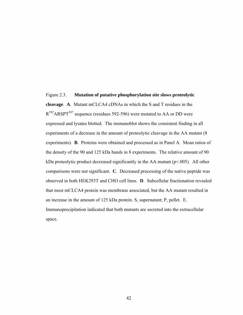
42
Figure 2.3. Mutation of putative phosphorylation site slows proteolytic
cleavage. A. Mutant mCLCA4 cDNAs in which the S and T residues in the
R592ARSPT597 sequence (residues 592-596) were mutated to AA or DD were
expressed and lysates blotted. The immunoblot shows the consistent finding in all
experiments of a decrease in the amount of proteolytic cleavage in the AA mutant (8
experiments). B. Proteins were obtained and processed as in Panel A. Mean ratios of
the density of the 90 and 125 kDa bands in 8 experiments. The relative amount of 90
kDa proteolytic product decreased significantly in the AA mutant (p<.005). All other
comparisons were not significant. C. Decreased processing of the native peptide was
observed in both HEK293T and CHO cell lines. D. Subcellular fractionation revealed
that most mCLCA4 protein was membrane associated, but the AA mutant resulted in
an increase in the amount of 125 kDa protein. S, supernatant; P, pellet. E.
Immunoprecipitation indicated that both mutants are secreted into the extracellular
space.

43

44
but is not preserved in other mCLCAs, whereas the dileucine sequences are unique to
mCLCA4. LI sequences are found in mCLCA1 and mCLCA2 (Figure 2.4A),
however, and may play a similar role. In other proteins, cytoplasmic dileucines appear
to serve as an ER export or forward trafficking signal through the binding of
chaperones, whereas the RxR is an ER retention signal (11). To determine the
relevance of these sequences we created additional mutants that eliminated the more
COOH-terminal dileucine (2L/2A), both dileucine sequences (4L/4A), the RxR
sequence (2R/2A), or both the dileucine and RxR sequences (4L2R/6A) (Figure 2.4B).
Mutation of either the C -terminal dileucine or both dileucine pairs prevented forward
trafficking of mCLCA4 proteins to the cell membrane (Figure 2.4C), indicating a
potent role of these sequences in forward trafficking. However mutation of the
putative RxR retention sequence also resulted in perinuclear localization of mCLCA4
(Figure 2.4C).
To further understand the role of these motifs we examined the glycosylation
pattern of putatively ER–trapped proteins in lysates from transfected cells. In vitro
deglycosylation of tagged proteins from mCLCA4 –expressing cells demonstrated that
the 125 kDa protein was sensitive to both PNGase F and Endo H (Figure 2.5A),
indicating that the full length mClCA4 protein is immaturely glycosylated and resides
within the ER compartment, as PNGase F cleaves both mature and immature N -
linked glycans, whereas Endo H digests high-mannose carbohydrates found in the ER
or medial Golgi. Conversely, the 90 kDa and 40 kDa cleaved fragments were sensitive
to PNGase F, but markedly less sensitive to Endo H, suggesting that a minor
component of these proteins are immaturely glycosylated (Figure 2.5A), consistent
with export from the ER and mature glycosylation. The 125 kDa full length protein
displayed a deglycosylation pattern consistent with ER trapping (Figure 2.5B) and the

45
Figure 2.4. Forward traffic and ER retention signals. A. Alignments of the
amino acid sequence of the highly related mCLCAs identifies the dileucine signals in
mCLCA4 and the diarginine signals that are unique to these three genes. Similar
signals in GLT-1 are shown for comparison. Regions of identity are shown in grey;
sequence numbering is shown for mCLCA4. B. Alanine mutations of the transport
and retention signals are shown in red. C. Immunolocalization of the dileucine and
diarginine mutants. Note the perinuclear localization of all mutants and an absence of
immunoreactive protein at the membrane.

46

47
90 kDa NH2–terminal fragment was not present in the lysate of 2L/2A and 4L/4A
dileucine mutants. Similarly, mutation of the RxR sequence (2R/2A) resulted in the
apparent loss of the 90 kDa fragment in the lysate. To further test whether ER trapping
of the export mutants was complete, we immunoprecipitated proteins from the cell
media of tagged (NH2-terminal fragment specific) wildtype and ER mutant
transfectants. These experiments demonstrated that the ER export mutants (2L/2A and
4L/4A) did not produce the 90 kDa secreted protein, whereas the 90 kDa cleavage
produce was abundant in the 2R/2A ER retention mutant (Figure 2.5C). Our failure to
detect the 90 kDa peptide of 2R/2A in cell lysates (Figure 2.5B), despite its presence
in the media, is likely due to the augmented forward trafficking effected by mutation
of the retention signal. Unlike GLT-1, the combined mutant (4L2R/6A) did not rescue
forward trafficking, as the 90 kDa fragment was not observed in the lysate or in the
media (data not shown). Moveover, the mCLCA4-21MycAA and mCLCA4-
21MycDD mutants were deglycosylated in a manner consistent with predominately
mature glycosylation of the 90 and 40 kDa fragments. The ratio of 90 kDa to 125 kDa
protein in the cell lysate and medium for the WT, AA mutant and DD mutant was
81%, 60% and 79%, respectively, consistent with less complete processing of the AA
mutant. This was not a result of block of secretion of the mutants, as WT, AA, and
DD proteins were found in the medium (Figure 2.5D).
To more definitively test the function of putative mCLCA4 trafficking signal
sequences, we created chimeric proteins in which a 40 amino acid mCLCA4 peptide
containing the wildtype, LL, or R-R mutants was placed immediately COOH-terminal
to a secreted EGFP construct (3) (Figure 2.6A). Consistent with the mCLCA4
mutational results, 48 h after transfection chimeric EGFP proteins containing the
4L/4A mutation markedly accumulated within the cell, whereas WT and 2R/2A
mutant proteins were poorly retained in cells and in those cases were diffusely

48
Figure 2.5. Glycosylation pattern of mutant proteins. A. PNGaseF or Endo H
deglycosylation of N and C -terminal fragment tagged mCLCA4 proteins indicated
that the 125 kDa full length protein is immaturely glycosylated (sensitive to both
enzymes), whereas the 90 and 40 kDa fragments were only partially sensitive to Endo
H, indicating more mature glycosylation. B. The dileucine and diarginine mutants
demonstrate marked ER or early Golgi trapping as there is no observable 90 kDa
fragment and the 125 kDa protein displays an immature glycosylation pattern. C.
Mutation of the diarginine (RxR) motif does not result in ER trapping, as the 90 kDa
fragment is detected in the media by immunoprecipitation, indicating highly efficient
forward trafficking. Conversely, mutation of the dileucine signals results in complete
loss of forward trafficking. D. The AA R592ARAPA597 mutant displays a
glycosylation pattern consistent with delayed processing, but not ER trapping, as the
90 kDa proteolytic fragment from this mutant, as well as the DD mutant, is resistant to
Endo H. Media lanes were by IP.

49

50
Figure 2.6. mCLCA4 dileucine sequences are required for forward trafficking
of secreted proteins. A. Schematic of construct used to examine mCLCA4 dileucine
and diarginine sequences. The N -terminal signal sequence (SS) from hCLCA2 was
used to direct EGFP to secretory compartments. A 40 amino acid fragment of
mCLCA4 was positioned C -terminal to EGFP, similar to its location with respect to
the N -terminal mCLCA4 fragment. Dileucine (LL) and diarginine (RxR) sequences
are shown. B. Widefield (above, 10X) and confocal (below, 40X) images of
HEK293T cells 48 h after transfection (scaled to equivalent fluorescence intensity).
The expression pattern indicates EGFP trapping in the 4L/4A mutant (approximately
70% of the cells showed EGFP localized to the cytosol and 30% exhibited a more
vesicular staining pattern) and are consistent with rapid secretion of EGFP by WT and
RxR mutant cells. Below, confocal images show a predominate cytosolic pattern in
EGFP –retaining WT and 2R/2A cells, and a vesicular pattern in cells transfected with
the 4L/4A mutant. Fluorescence of WT and 2R/2A images was substantially lower and
is scaled to show roughly equivalent maximum intensity as 4L/4A. C.
Immunoprecipitation of GFP from medium 72 h after transfection; quantification of
the EGFP band is shown below (representative of 3 experiments). Little or no 4L/4A
protein was detected in the media in these experiments, further supporting vesicle
trapping, whereas slightly more 2R/2A protein was detected, consistent with rapid
secretion of the EGFP constructs in which the retention signal has been mutated. The
“C” lane indicates non –transfected control.

51

52
present within the cytosol (Figure 2.6B). Moreover, immunoprecipitation and
immunoblotting indicated substantial WT and 2R/2A protein in the cell media,
indicating efficient forward trafficking and secretion, but accumulation in the media of
4L/4A mCLCA4 mutant protein was markedly reduced (Figure 2.6C).
These results confirm the function of dileucine and RxR ER regulatory
sequences in mClCA4. No secreted protein was detected in dileucine mutants,
indicating a complete block of forward trafficking, whereas secreted protein was
detected in RxR mutants, indicating that the motif functions as an ER retention signal.
These observations are consistent with the role of the RxR motif in GLT-1 (11).
Moreover, the decrease in processing efficiency observed in the RARSPT mutant
mCLCA4-21MycAA likely results from conformational effects on the RxR retention
sequence imposed by mutation of the neighboring S595 and/or T597 residues.
2.5. Discussion
We have used epitope insertion and immunodetection to examine the
processing and fate of mCLCA proteins. Our results indicate that mCLCA4 cDNA
encodes a full length protein of approximately 125 kDa that is proteolytically cleaved
into large NH2-terminal (90 kDa) and smaller COOH-terminal (40 kDa) fragments.
The data are consistent with cleavage within the ER or early in the trans Golgi
network, as the full length mClCA4 protein displays an ER –like immunoreactive
pattern, does not traffic to the plasma membrane, and is immaturely glycosylated
(Figure 2.5D). Both the N and C -terminal cleavage products are secreted and found
in the media, where they rapidly associate with the plasma membrane, and protein
isolated from cell lysates displays a mature glycosylation pattern (Figures 2.1B, 2.5A),
further suggesting that only the proteolytic products are forward trafficked beyond the
ER.

53
The topology and cellular fate of CLCA proteins has undergone substantial
refinement since the initial modeling indicating multiple transmembrane segments (1,
7, 9). More recent structural analysis indicates a single COOH-terminal
transmembrane segment (22) and a conserved NH2-terminal hydrolase domain (19).
This prediction has been supported by experimental data indicating the secretion of a
CLCA fragment into the extracellular space (3, 6, 14), although differences exist as to
whether the NH2-terminal fragment (6) or both the NH2- and COOH-terminal
fragments (3) are secreted extracellularly, with the latter data inconsistent with a single
transmembrane domain. For mCLCA3, the two proteins are reported to remain
physically associated and secreted into the extracellular space (14), whereas the
COOH-terminal fragment of the highly related human homologue hCLCA1 does not
appear to be associated with the secreted NH2-terminal cleaved peptide, and is not
found in the extracellular media (6).
Based on results from hCLCA2, Elble et al (3) have suggested a general model
of CLCA processing in which the full–length precursor is transported to the cell
surface where it undergoes cleavage by an endopeptidase, resulting in release of the
NH2-terminal fragment, while the COOH-terminal fragment with its transmembrane
segment remains as an integral membrane protein. Our data are in general consistent
with this model, although it appears that both proteins remain associated with the
plasma membrane and that the full–length precursor of mCLCA4 does not traffic to
the plasma membrane, since the 125 kDa fragment is sensitive to EndoH digestion
(Figure 2.5A,B,D). Also, unlike hCLCA2, we were unable to demonstrate tight
association between the two fragments by co-immunoprecipitation (data not shown).
We have also extended these observations by determining the luminal motifs
that regulate trafficking of mCLCA4. Kalandze et al (11) described cytosolic
diarginine retention and dileucine forward trafficking signals regulating the trafficking

54
of GLT-1, an intrinsic membrane protein. Interestingly, mutation of all of the
dileucine motifs in GLT-1 was required to abolish forward trafficking, as 2L/2A and
4L/4A proteins were still maturely glycosylated and inserted in the plasma membrane.
The 6L/6A GLT-1 mutation resulted in ER trapping, but also resulted in markedly less
full length protein in the lysate, suggesting misfolding and degradation. By contrast,
secretion of the 90 kDa NH2-terminal peptide was completely inhibited in 2L/2A and
4L/4A mutants of the luminal protein mCLCA4, and high amounts of ER trapped 125
kDa protein were observed (Figure 2.5B). Mutation of the diarginines also produced
an apparent loss of 90 kDa product in the protein lysate, however protein was readily
detected in the cell medium, suggesting augmented forward trafficking associated with
loss of the RxR retention signal. The apparent absence of the 90 kDa peptide from the
plasma membrane suggests that the RxR motif may also be required for surface
association. Chimeric constructs clearly demonstrated the role of the dileucine
sequences in forward trafficking (Figure 2.6B, C) and the prominent vesicular pattern
of EGFP expression in 4L/4A mutants confirms the importance of these sequences as
retention signals. As CLCA proteins appear to undergo distinct processing (3, 6, 14),
these motifs may underlie significant functional differences in these family members.
Studies designed to determine the function of the separate membrane–associated
fragments should provide more clarity with respect to the role of CLCAs in ion
transport and secretion.

55
REFERENCE
1. Cunningham SA, Awayda MS, Bubien JK, Ismailov, II, Arrate MP, Berdiev BK, Benos DJ, and Fuller CM. Cloning of an epithelial chloride channel from bovine trachea. J Biol Chem 270: 31016-31026, 1995. 2. Elble RC, Ji G, Nehrke K, DeBiasio J, Kingsley PD, Kotlikoff MI, and Pauli BU. Molecular and functional characterization of a murine calcium-activated chloride channel expressed in smooth muscle. J Biol Chem 277: 18586-18591, 2002. 3. Elble RC, Walia V, Cheng HC, Connon CJ, Mundhenk L, Gruber AD, and Pauli BU. The putative chloride channel hCLCA2 has a single C-terminal transmembrane segment. J Biol Chem 281: 29448-29454, 2006. 4. Elble RC, Widom J, Gruber AD, Abdel-Ghany M, Levine R, Goodwin A, Cheng HC, and Pauli BU. Cloning and characterization of lung-endothelial cell adhesion molecule-1 suggest it is an endothelial chloride channel. J Biol Chem 272: 27853-27861, 1997. 5. Ellgaard L and Helenius A. Quality control in the endoplasmic reticulum. Nat Rev Mol Cell Biol 4: 181-191, 2003. 6. Gibson A, Lewis AP, Affleck K, Aitken AJ, Meldrum E, and Thompson N. hCLCA1 and mCLCA3 are secreted non-integral membrane proteins and therefore are not ion channels. J Biol Chem 280: 27205-27212, 2005. 7. Gruber AD, Elble RC, Ji HL, Schreur KD, Fuller CM, and Pauli BU. Genomic cloning, molecular characterization, and functional analysis of human CLCA1, the first human member of the family of Ca2+-activated Cl- channel proteins. Genomics 54: 200-214, 1998. 8. Gruber AD and Pauli BU. Molecular cloning and biochemical characterization of a truncated, secreted member of the human family of Ca2+-activated Cl- channels. Biochim Biophys Acta 1444: 418-423, 1999. 9. Gruber AD, Schreur KD, Ji HL, Fuller CM, and Pauli BU. Molecular cloning and transmembrane structure of hCLCA2 from human lung, trachea, and mammary gland. Am J Physiol 276: C1261-1270, 1999. 10. Hebert DN, Garman SC, and Molinari M. The glycan code of the endoplasmic reticulum: asparagine-linked carbohydrates as protein maturation and quality-control tags. Trends Cell Biol 15: 364-370, 2005.

56
11. Kalandadze A, Wu Y, Fournier K, and Robinson MB. Identification of motifs involved in endoplasmic reticulum retention-forward trafficking of the GLT-1 subtype of glutamate transporter. J Neurosci 24: 5183-5192, 2004. 12. Leverkoehne I, Holle H, Anton F, and Gruber AD. Differential expression of calcium-activated chloride channels (CLCA) gene family members in the small intestine of cystic fibrosis mouse models. Histochem Cell Biol 126: 239-250, 2006. 13. Loewen ME and Forsyth GW. Structure and function of CLCA proteins. Physiol Rev 85: 1061-1092, 2005. 14. Mundhenk L, Alfalah M, Elble RC, Pauli BU, Naim HY, and Gruber AD. Both cleavage products of the mCLCA3 protein are secreted soluble proteins. J Biol Chem 281: 30072-30080, 2006. 15. Nakanishi A, Morita S, Iwashita H, Sagiya Y, Ashida Y, Shirafuji H, Fujisawa Y, Nishimura O, and Fujino M. Role of gob-5 in mucus overproduction and airway hyperresponsiveness in asthma. Proc Natl Acad Sci U S A 98: 5175-5180, 2001. 16. Nara M, Dhulipala PD, Wang YX, and Kotlikoff MI. Reconstitution of beta-adrenergic modulation of large conductance, calcium-activated potassium (maxi-K) channels in Xenopus oocytes. Identification of the camp-dependent protein kinase phosphorylation site. J Biol Chem 273: 14920-14924, 1998. 17. Patel AC, Morton JD, Kim EY, Alevy Y, Swanson S, Tucker J, Huang G, Agapov E, Phillips TE, Fuentes ME, Iglesias A, Aud D, Allard JD, Dabbagh K, Peltz G, and Holtzman MJ. Genetic segregation of airway disease traits despite redundancy of calcium-activated chloride channel family members. Physiol Genomics 25: 502-513, 2006. 18. Pauli BU, Abdel-Ghany M, Cheng HC, Gruber AD, Archibald HA, and Elble RC. Molecular characteristics and functional diversity of CLCA family members. Clin Exp Pharmacol Physiol 27: 901-905, 2000. 19. Pawlowski K, Lepisto M, Meinander N, Sivars U, Varga M, and Wieslander E. Novel conserved hydrolase domain in the CLCA family of alleged calcium-activated chloride channels. Proteins 63: 424-439, 2006. 20. Ritzka M, Stanke F, Jansen S, Gruber AD, Pusch L, Woelfl S, Veeze HJ, Halley DJ, and Tummler B. The CLCA gene locus as a modulator of the gastrointestinal basic defect in cystic fibrosis. Hum Genet 115: 483-491, 2004.

57
21. Teasdale RD and Jackson MR. Signal-mediated sorting of membrane proteins between the endoplasmic reticulum and the golgi apparatus. Annu Rev Cell Dev Biol 12: 27-54, 1996. 22. Whittaker CA and Hynes RO. Distribution and evolution of von Willebrand/integrin A domains: widely dispersed domains with roles in cell adhesion and elsewhere. Mol Biol Cell 13: 3369-3387, 2002. 23. Zhou Y, Dong Q, Louahed J, Dragwa C, Savio D, Huang M, Weiss C, Tomer Y, McLane MP, Nicolaides NC, and Levitt RC. Characterization of a calcium-activated chloride channel as a shared target of Th2 cytokine pathways and its potential involvement in asthma. Am J Respir Cell Mol Biol 25: 486-491, 2001.

58
CHAPTER 3
THE FUNCTIONAL STUDY OF MCLCA4 IN MURINE LIFE
-----mCLCA4 knock-out

59
3.1 Abstract
mCLCA4 is highly expressed in smooth muscle and other cells, and following
transient transfection of mCLCA4 cDNA into HEK293T cells a chloride current is
detected. This suggests that mCLCA4 plays a role in regulating of chloride currents.
To better understand the function of mCLCA4 in murine life, I generated mice lacking
CLCA4 expression via gene targeting of the mCLCA4 gene locus. A 4.8 kb genomic
sequence, that included exons 1 to 3, was deleted from the mouse genome and resulted
in no mCLCA4 gene expression and no protein production. There was no obvious
gross phenotype in the mCLCA4 homozygous knock-out mice and they appeared to
breed normally.
3.2 Introduction
As candidate calcium-activated chloride channel proteins, the CLCA family
has drawn the attention of our smooth muscle electrophysiological research laboratory,
especially mCLCA4, one of the murine members that is highly expressed in smooth
muscle cells. To understand mCLCA4 function, in vitro protein processing and
trafficking were studied (See Chapter 2). To determine the function of mCLCA4 in
vivo I generated mice lacking the mCLCA4 gene.
The calcium-activated chloride channel protein family was originally described
as an integral membrane protein that may act as a chloride channel (5, 7, 15). Recent
studies showed that these family members are soluble secreted proteins associated
with the plasma membrane (see Chapter 2)(3, 4, 12), suggesting this group of proteins
plays a more complex role than just regulating chloride currents. The most convincing
study recently showed that some members (such as hCLCA1, mCLCA3 and eCLCA1)
play a role in the pathophysiology observed in asthma and cystic fibrosis (8, 11, 13,
14, 16). The other CLCA proteins are involved in tumor suppression (such as hCLC2,

60
mCLCA1 and mCLCA2) (2, 6). To date, there are no functional studies on the
involvement of the mCLCA4 protein in any disease processes.
As one of the six cloned mouse CLCA members that cluster on mouse
chromosome 3, mCLCA4 is highly and preferentially expressed in organs that contain
a high percentage of smooth muscle cells, such as aorta, stomach, uterus, bladder and
intestine (1). It was cloned from RNA isolated from mouse bladder and stomach
smooth muscle layers that were dissected free from the mucosa followed by reverse
transcription PCR based on bCLCA2 cDNA as primers. Transient transfection studies
of mCLCA4 cDNA in HEK293T cells demonstrated the presence of an anionic
current when cells were exposed to increased intracellular calcium by administration
of ionomycin or methacholine. The current characteristics were similar to the smooth
muscle chloride current found in many tissues (1, 9, 18). The full-length mCLCA4
cDNA is 2.7 kb and codes for a 909 aa protein precursor. It yields ~125 kDa product
after glycosylation in HEK293T cells that is cleaved into 90 kDa NH2-terminal and 40
kDa COOH-terminal fragments. Both protein fragments of mCLCA4 are secreted out
of the cell and associate with the plasma membrane (see Chapter 2).
To understand the function of mCLCA4 in murine life, mice lacking mCLCA4
gene expression were generated. Gross phenotypic characterization of mCLCA4 null
mice was completed by a pathologist at Cornell University’s Veterinary School of
Medicine and no gross phenotype was observed.
3.3 Materials and Methods
3.3.1 Generation of mCLCA4 null mice
3.3.1.1 Generating the targeting vector
A 17.3 kb mCLCA4 genomic DNA fragment with partial restriction enzyme
digestion information was provided by Keith Nehrke at the Aab Institute for

61
Biomedical Research, University of Rochester. It contained a 7 kb 5’ flanking
sequence including exons 1 to 6 that was verified by restriction enzyme mapping and
partial sequence analysis. The deletion sequence was designed to eliminate a 4.8 kb
fragment that included exons 1 to 3 of the mCLCA4 gene. Two homologous arms,
upstream of the deletion fragment, were designed and generated from the 17.3 kb
genomic DNA fragment. A 2.5 kb Short-arm fragment was released by EcoRV and
XbaI enzyme sites and a 6 kb Long-arm fragment was removed by EcoRV and KpnI
sites (Figure 3.1A). The pKO-Neo/loxp targeting vector contained
Neomycin/Kanamycin (Neo/kan) positive and thymidine kinase (TK) negative
selection genes. Homologous arms were subcloned into pKO-Neo/loxp vector through
XhoI* (* blunted end of XhoI site to match EcoRV site) and XbaI sites for the short
arm (located before Neomycin gene) and SalI* (* blunted end of SalI site to match
EcoRV site) and KpnI sites for the Long-arm (after Neomycin gene). The targeting
vector-PKO-Neo/loxp containing two homologous arms was verified by sequencing
(Figure 3.1B).
3.3.1.2 ES Cell Targeting
The feeder dependent R1 mouse embryonic stem (ES) cell line
[129X1/SvJX129S1.Sv-+p+Tyr-cMgfs1-J/+(129)C129] was obtained from the Nagy
Laboratory (http://www.mshri.on.ca/nagy/ ). The neomycin resistant feeder cells were
generated in our lab. The ES cells were grown in DMEM media supplemented with
15% ES qualified fetal calf serum (Hyclone), 1x L-glutamine (Gibco/Invitrogen), 1x
nonessential amino acids (Gibco/Invitrogen), 1x sodium pyruvate (Gibco/Invitrogen),
1x β-mercaptoethanol (Gibco/Invitrogen), 1x Penicillin/Streptomycin
(Gibco/Invitrogen) and 1000 U/ml leukemia inhibitory factor (LIF) (ESGRO,
Millipore). After the targeting fragment was electroporated into the ES cells, 48 hours

62
Figure 3.1 Diagram of mCLCA4 knock-out mouse and homologous
recombination in ES cells. A. Diagram of 17.3 kb mCLCA4 genomic DNA fragment
used to make mCLCA4 knock-out mice. A 4.8 kb fragment including exon 1 to 3 was
deleted; the homologous arms were 2.5 kb with EcoRV/XbaI digestion sites and 6 kb
with EcoRV/KpnI digestion sites, respectively. B. Diagram of homologous
recombination between targeting vector and mouse genomic DNA mCLCA4 gene
locus in ES cells. A 4.8 kb of mCLCA4 sequence was replaced by the loxP-neo-loxP
fragment after the homologous recombination occurred between the two arms. Note:
XhoI* or SalI* indicated that the PGK-Neo vector was digested by XhoI or SalI
following by filling in with dNTPs to make blunted end to match EcoRV site during
construction of the targeting vector.

63
KpnI SacI
ScaI
EcoRV XbaI EcoRV
4.8kb delete region
6kb Long Arm
2.5kb short Arm
Exon
A. 17.3 kb mCLCA4 genomic DNA fragment
KpnI SacI
ScaI
EcoRV XbaI EcoRV
4.8kb delete region
6kb Long Arm
2.5kb short Arm
Exon
A. 17.3 kb mCLCA4 genomic DNA fragment
Genomic allele
NotI
EcoRV KpnI
4.8kb deletion region
EcoRV XbaI
Long armShort arm
SacI
MCS
Targeting vector
loxploxp
MIC-TK PGK-Neo
XhoI* EcoRVXbaI SalI* KpnI
NotI
B. Homologous recombination between Genomic allele and Targetting vector
Genomic allele
NotI
EcoRV KpnI
4.8kb deletion region
EcoRV XbaI
Long armShort arm
SacI
MCS
Targeting vector
loxploxp
MIC-TK PGK-Neo
XhoI* EcoRVXbaI SalI* KpnI
NotI
B. Homologous recombination between Genomic allele and Targetting vector

64
later 150 ng/ml G418 sulfate (selection drug for Neomycin gene, Gibco/Invitrogen)
and 2 μM gancyclovir (selection drug for TK gene, Roche) were added to the media
(150 ng/ml and 2 µM). The plates were coated with 0.1% gelatin (Chemicon) for 30
minutes at room temperature and liquid removed. The feeder cells were plated at a
density of 106 cells per 100 mm gelatin coated plates and cultured in a 370 C 5% CO2
incubator for at least two hours before use. The ES cells were plated on feeder cells
and cultured in a 370C, 5% CO2 incubator. ES cell media was changed daily and cells
split when plates reached confluence.
The targeting vector was linearized with NotI in order to increase the
homologous recombination efficiency in ES cells. 25 µg linearized targeting vector
DNA (15 kb) was added to 8x10⁶ ES cells in 0.8 ml phosphate buffered saline (PBS)
and transferred to an electroporation cuvette with a 0.4 cm gap. The ES cell-DNA
mixture was electroporated using the following conditions: 240 V, 500 µF using a
BioRad Gene Pulser with Capacitance Extender. The ES cells in the cuvette were
placed on ice for 20 minutes then plated on four 100 mm neomycin resistant feeder
(NRF) plates with 10 ml DMEM media plus supplements. Forty eight hours after
electroporation, the ES cells were selected with G418 (200 µg/ml) and gancyclovir (2
µM) in DMEM media for 10 days. The individual ES cell colonies were picked and
were grown in 96 well plates with feeder cells. The colony plates were split and two
plates were frozen in liquid nitrogen with 2x ES Freezing Media (Chemicon
International) and one plate used for DNA.
3.3.1.3 Positive ES cell clones screening
Standard Southern blot techniques were used to screen the ES cell for positive
clones. ES cell DNA was extracted based on the protocol in Gene Targeting (10) and
digested with EcoRV and SacI restriction enzymes. The DNA mixes were run on 0.8%

65
agarose gels and transferred from the gels to blotting membranes. A 500 bp DNA
fragment in the genomic region (out of long-arm region) was used as the probe (probe
5) to distinguish the wildtype allele (7.5 kb band) and the knock-out allele (9.2 kb
band) (Figure 3.2). Neomycin gene fragment (Neo-probe, 600bp) was used as a
second probe to confirm the positive clones by only detecting the knock-out allele
band (9.2 kb) on the same Southern blot.
3.3.1.4 ES cell clone injection and breeding strategy.
One positive ES cell clone (1H8) was injected into 3.5 days blastocysts of
C57Bl/6J at the Cornell Transgenic Core Facility using standard microinjection
techniques (10). Two highly chimeric founders (one approximately 90% and the other
approximately 80%) were obtained and mated to C57BL/6J to obtain heterozygous
mice. Heterozygous mice were identified using PCR with the following primers sets
“a” (GGCCAGGTGTGCACACCTGTAA) and “b”
(GCTGGGGATGCGGTGGGCTCTA) with a resultant 410 bp product from the
targeted allele on “a” and “c” (CCCACGCCATTTTCAACTGGATTTG) to amplify a
370 bp product from the wildtype allele. This result was also confirmed by Southern
blot with probe5 and Neo-probe. Heterozygous mice were mated to produce
homozygous null allele mCLCA4 mice, identified by PCR with the same primer sets
above and verified by Southern blot with probe 5.
3.3.2 Phenotypic evaluation.
Evaluation of six mCLCA4 mice (two wildtype, two heterozygotes, two
homozygotes) at six weeks of age was performed by Dr. Ana Alcaraz at Cornell
University’s Department of Anatomical Pathology at the Veterinary School of
Medicine. Multiple organs (such as heart, liver, brain, stomach, thymus, lung, large

66
Figure 3.2 Diagram of mCLCA4 homozygous, heterozygous and wildtype
genotyping scheme. The targeted allele and wildtype allele of mCLCA4 gene region
are shown. Southern blot screening yielded a 9.2 kb band in the targeted genomic
allele while a 7.5 kb band would be detected for the wildtype allele. PCR using
primers “a” and “b” produced a 410 bp product from mice carrying the targeted
genomic allele while PCR using primers “c” and “d” only detected a 370 bp product
from the wildtype allele. Heterozygous mice have both 410 bp and 370 bp PCR
products.
Targeted genomic allele
PGK-Neo
Wildtype genomic allele
EcoRV
EcoRV
SacI
SacIShort arm region
Short arm region
Long arm region
Long arm region
Deletion region
7.5 kb
9.2 kb
a b
a c
Neo-probe Probe 5
Probe 5
Targeted genomic allele
PGK-Neo
Wildtype genomic allele
EcoRV
EcoRV
SacI
SacIShort arm region
Short arm region
Long arm region
Long arm region
Deletion region
7.5 kb
9.2 kb
a b
a c
Neo-probe Probe 5
Probe 5

67
and small intestines, bladder, and ovary) were weighed for comparison to total body
weight. Blood chemistry and complete blood count (CBC) were tested in the
Diagnostic Laboratory at Cornell University’s Veterinary School of Medicine.
3.4 Results
3.4.1 Generation and Gross Phenotypic Evaluation of mCLCA4 null mice
Two chimeric mCLCA4 founder lines were generated by replacing a 4.8 kb
fragment including a portion of the mCLCA4 5’ region and exons 1 to 3, resulting in a
gene that can not be expressed (Figure 3.3). Homozygous, heterozygous and wildtype
mice were identified by PCR (420 bp, 370 bp plus 410 bp, or 370 bp, products
respectively) using specific primers (Figure 3.3). Mice were systemically evaluated,
comparing wildtype littermate controls to heterozygous and homozygous null mice.
Body weight, body size and behavior were not different between the three genotypes.
Even after aging to 14 months, no gross differences could be detected. Next, organ
weights such as liver, spleen, stomach, large and small intestine, and lung tissues,
were evaluated, but no apparent differences were observed. Table 1 shows the blood
chemistry profiles obtained from wildtype and null mCLCA4 mice; no obvious
differences were observed. Table 2 shows the complete blood count (CBC) result. The
homozygous mCLCA4 null mice appeared in normal Mendelian ratios, indicating no
reduction in viability.
3.5 Discussion
3.5.1 Rationale for Generation of mCLCA4-/- mice
mCLCA4 was cloned from a mouse smooth muscle RNA pool and
characterized by Elbe et al (1). It is highly expressed in organs that contain a
significant amount of smooth muscle cells (1). HEK293T cells transiently transfected

68
Figure 3.3 Southern blot and PCR genotyping. A. Southern blot screening
results from ES cells using probe 5 and confirmed with Neo-probe. With probe 5, two
ES cell clones (1H8 and 1G10) were identified by a 9.2 kb band (targeted genomic
allele) and 7.5 kb band (wildtype genomic allele band). Note other ES cells only
showed 7.5 kb wildtype band. With Neo-probe, only the targeted genomic allele 9.2
kb band was detected in these two clones. B. Mouse tail DNA PCR genotyping
results. Homozygous mice only yield a 410 bp band, wildtype mice 370 bp band and
heterozygous littermates 410 bp and 370 bp bands.

69
A.
A B C
9.2 kb7.5 kb
Neo-probe Probe 5 Probe 5
1H8 1G10 1G10 1H8
A B C
9.2 kb7.5 kb
Neo-probe Probe 5 Probe 5
1H8 1G10 1G10 1H8
+/+ -/- +/- M
400 bp300 bp
+/+ -/- +/- M
400 bp300 bp
B.

70
Table 3.1 Blood chemistry test results from mCLCA4 null mice and their littermates
Blood Chemistry Test Result for mCLCA4
WT male
mCLCA4 (-/-) male
WT female
mCLCA4(-/-) female
SODIUM (mEq/L) 149 152 156 154 POTASSIUM
(mEq/L) 8.5 7.3 6.9 7.4
CHLORIDE (mEq/L) 112 109 116 113 BICARB (mEq/L) 21 23 16 32
ANION GAP (mEq/L) 25 27 31 16 Na/K 18 21 23 21
UREA (mg/dL) 33 28 18 37 CREAT-rb (mg/dL) 0.2 0.2 0.2 0.2 CALCIUM (mg/dL) 10.3 10.3 8.9 10.5
PHOSPHATE (mg/dL) 8.9 8 9 5.7
MAGNES-xb (mEq/L) 1.9 1.9 1.6 1.9
gTOT Prot (g/dL) 5.3 6.1 4.5 4.4 ALB-blk (g/dL) 3.2 3.3 2.8 3
GLOBULIN (g/dL) 2.1 2.8 1.7 1.4 A/G 1.52 1.18 1.65 2.14
GLUCOSE (mg?dL) 234 253 213 177 ALT/P5P (U/L) 52 55 18 40 AST/P5P (U/L) 256 129 142 76
ALK PHOS (U/L) 89 60 57 39 GGT (U/L) < 3 < 3 < 3 < 3
TOT BILI (mg/dL) 0.2 0.2 2.4 0.1 DIR BILI (mg/dL) 0 0 0 0 IND BILI (mg/dL) 0.2 0.2 2.4 0.1 AMYLASE (U/L) 2762 3309 2089 3915
CHOLESTER (mg/dL) 115 186 87 110
hCK (U/L) 1521 449 765 457 IRON (ug/dL) 195 207 142 323 TIBC (ug/dL) 325 413 250 471
%SAT 60 50 57 69 LIPEMIA 13 21 20 25
HEMOLYSIS 379 786 153 123 ICTERUS 0 0 0 0

71
Table 3.2 Blood CBC test results from mCLCA4 null mice and their littermates.
Blood CBC Test Result for mCLCA4
WT male mCLCA4 (-/-) male WT female mCLCA4 (-/-)
female HCT (%) 49 52 47 52 HB (g/dL) 15.5 15.8 14.5 16
RBC (mill/uL) 10.2 10.7 9.1 10.2 MCV (fL) 48 49 52 51 MCH (pg) 15 15 16 16
MCHC (g/dL) 32 30 31 31 RDW (%) 11.8 14.8 13.6 12.4
WBC (thou/uL) 4.3 4.6 7.7 4.9
SEGMENT N (thou/uL) 1.4 0.3 0.6 0.6
BAND N (thou/uL) 0 0 0 0
LYMPH (thou/uL) 2.5 3.9 6.9 3.7
MONO (thou/uL) 0.3 0.3 0.2 0.4
EOSIN (thou/uL) 0.1 0.1 0 0.2
BASO (thou/L) 0 0 0 0
PLAT SMEAR adeq adeq Low adeq PLAT
(thou/uL) 1555 660 737 410
MPV (fL) 7.3 6.9 7.3 6.3 TP-REF (g/dL) 6.8 6.7 5.1 5.9
RBC morphololgy
polychromasia-moderate
Howell-jolly bodies-few,
poly
polychromasia-moderate
polychromasia-moderate
Parasites none seen none seen none seen none seen
WBC Exam no significant abnormalities
no significant abnormalities
no significant abnormalities
no significant abnormalities
Plasma Appearance normal normal normal normal

72
with mCLCA4 cDNA display a significant chloride current (1). This protein is made
in the ER and cleaved inside the cell, and then both fragments are secreted out of the
cell and associated with the plasma membrane (See Chapter 2). Taken together, these
results suggest that mCLCA4 may be a regulator of a chloride current. To better
understand the function of mCLCA4, mCLCA4 knock-out mice were created by
deleting the first three exons and a portion of the 5’ region of the mCLCA4 gene from
the mouse genome resulting in gene disruption at the genomic level (Figure 3.1).
3.5.2 Lack of mCLCA4 does not result in a gross phenotype
mCLCA4-/- mice did not show any abnormal phenotype from birth to 14
months of age as evidenced by the lack of statistical differences in the ratios obtained
from multiple organs compared to body weight. This suggests these mice do not have
gross cardiac, lung, liver or kidney dysfunction. Even though only two littermate
wildtype controls and null animals were taken for the blood chemistry and CBC
panels, the mCLCA4 null values were similar to the wildtype mice and published
mouse values for normal mouse strains (17). The chemistry profiles suggested that
mCLCA4 mice have normal renal and liver function while the CBC values indicated
mCLCA4 null mice do not suffer from anemia, infection or leukemia. Just to note, the
blood samples were hemolyzed which artificially raised the potassium value due to the
release of cellular contents into the serum and may explain the high potassium values
observed across all the samples. Possible reasons a mCLCA4 null phenotype was not
observed include: 1) loss of mCLCA4 was compensated for by other highly
homologous family members, such as mCLCA1 and/or mCLCA2; 2) a subtle
phenotype is present but may only be observed under certain conditions of stress;
and/or 3) mCLCA4 is not necessary to sustain murine life.

73
3.5.3 Further study plan
I am convinced that this group of genes plays critical and diverse roles in
murine life. Studies are being undertaken by collaborators (Michael Holtzman et al. at
Washington University, School of Medicine, St. Louis, MO) to examine the effect of
airway challenge on mCLCA4 null mice. In addition, if the CLCAs are critical genes,
a loss of function in family member may be compensated for by the other family
members. For example, Patel et al. (14) showed that mCLCA5, mCLCA6 and
mCLCA7 (AI747448) may compensate for loss of mCLCA3. This may be the case in
mCLCA4-/- mice, where I would predict the most highly related family members
mCLCA1 and/or mCLCA2, could compensate for loss of mCLCA4. These three
genes, which cluster on mouse chromosome 3, share 80% identity at the protein level.
Since there are no antibodies available for mCLCA1, mCLCA2 or mCLCA4, further
experiments to evaluate functional compensation are limited. To test my hypothesis
that mCLCA1 and mCLCA2 genes compensate for mCLCA4’s function, I generated
mCLCA1,2,4 triple knock-out mice, which will be discussed in Chapter 4.
In summary, I generated mCLCA4 gene knock-out mice but no gross
phenotype was observed. To further understand the mCLCA4 gene and other family
member functions, I generated triple gene knock-out (mCLCA1, mCLCA2 and
mCLCA4) mice, discussed in Chapter 4.

74
REFERENCE
1. Elble RC, Ji G, Nehrke K, DeBiasio J, Kingsley PD, Kotlikoff MI, and Pauli BU. Molecular and functional characterization of a murine calcium-activated chloride channel expressed in smooth muscle. J Biol Chem 277: 18586-18591, 2002. 2. Elble RC and Pauli BU. Tumor suppression by a proapoptotic calcium-activated chloride channel in mammary epithelium. J Biol Chem 276: 40510-40517, 2001. 3. Elble RC, Walia V, Cheng HC, Connon CJ, Mundhenk L, Gruber AD, and Pauli BU. The putative chloride channel hCLCA2 has a single C-terminal transmembrane segment. J Biol Chem 281: 29448-29454, 2006. 4. Gibson A, Lewis AP, Affleck K, Aitken AJ, Meldrum E, and Thompson N. hCLCA1 and mCLCA3 are secreted non-integral membrane proteins and therefore are not ion channels. J Biol Chem 280: 27205-27212, 2005. 5. Gruber AD, Elble RC, Ji HL, Schreur KD, Fuller CM, and Pauli BU. Genomic cloning, molecular characterization, and functional analysis of human CLCA1, the first human member of the family of Ca2+-activated Cl- channel proteins. Genomics 54: 200-214, 1998. 6. Gruber AD and Pauli BU. Tumorigenicity of human breast cancer is associated with loss of the Ca2+-activated chloride channel CLCA2. Cancer Res 59: 5488-5491, 1999. 7. Gruber AD, Schreur KD, Ji HL, Fuller CM, and Pauli BU. Molecular cloning and transmembrane structure of hCLCA2 from human lung, trachea, and mammary gland. Am J Physiol 276: C1261-1270, 1999. 8. Hauber HP, Tsicopoulos A, Wallaert B, Griffin S, McElvaney NG, Daigneault P, Mueller Z, Olivenstein R, Holroyd KJ, Levitt RC, and Hamid Q. Expression of HCLCA1 in cystic fibrosis lungs is associated with mucus overproduction. Eur Respir J 23: 846-850, 2004. 9. Janssen LJ and Sims SM. Spontaneous transient inward currents and rhythmicity in canine and guinea-pig tracheal smooth muscle cells. Pflugers Arch 427: 473-480, 1994. 10. Joyner A. Gene Targeting, 2000. 11. Kamada F, Suzuki Y, Shao C, Tamari M, Hasegawa K, Hirota T, Shimizu M, Takahashi N, Mao XQ, Doi S, Fujiwara H, Miyatake A, Fujita K, Chiba Y,

75
Aoki Y, Kure S, Tamura G, Shirakawa T, and Matsubara Y. Association of the hCLCA1 gene with childhood and adult asthma. Genes Immun 5: 540-547, 2004. 12. Mundhenk L, Alfalah M, Elble RC, Pauli BU, Naim HY, and Gruber AD. Both cleavage products of the mCLCA3 protein are secreted soluble proteins. J Biol Chem 281: 30072-30080, 2006. 13. Nakanishi A, Morita S, Iwashita H, Sagiya Y, Ashida Y, Shirafuji H, Fujisawa Y, Nishimura O, and Fujino M. Role of gob-5 in mucus overproduction and airway hyperresponsiveness in asthma. Proc Natl Acad Sci U S A 98: 5175-5180, 2001. 14. Patel AC, Brett TJ, and Holtzman MJ. The Role of CLCA Proteins in Inflammatory Airway Disease. Annu Rev Physiol, 2008. 15. Pauli BU, Abdel-Ghany M, Cheng HC, Gruber AD, Archibald HA, and Elble RC. Molecular characteristics and functional diversity of CLCA family members. Clin Exp Pharmacol Physiol 27: 901-905, 2000. 16. Range F, Mundhenk L, and Gruber AD. A soluble secreted glycoprotein (eCLCA1) is overexpressed due to goblet cell hyperplasia and metaplasia in horses with recurrent airway obstruction. Vet Pathol 44: 901-911, 2007. 17. Schnell MA, Hardy C, Hawley M, Propert KJ, and Wilson JM. Effect of blood collection technique in mice on clinical pathology parameters. Hum Gene Ther 13: 155-161, 2002. 18. Wang Q, Hogg RC, and Large WA. Properties of spontaneous inward currents recorded in smooth muscle cells isolated from the rabbit portal vein. J Physiol 451: 525-537, 1992.

76
CHAPTER 4
A FUNCTIONAL STUDY OF MCLCA1, MCLCA2 AND MCLCA4 IN
MURINE LIFE
------ mCLCA1, mCLCA2 and mCLCA4 genes triple knock-out

77
4.1 Abstract
mCLCA1, mCLCA2 and mCLCA4 share high identity at the gene level,
mRNA level and the protein level. The three of them are clustered next to each other
on mouse chromosome 3 (H2-H3). mCLCA4 null mice did not show any obvious
phenotype from one day old to >1 year of age. Potentially, the loss of function of
mCLCA4 protein in mCLCA4-/- mice was compensated for by other family members,
namely mCLCA1 and/or mCLCA2 proteins. To test this hypothesis, I created
mCLCA1, mCLCA2, mCLCA4 triple knock-out mice; as these highly homologous
genes are contiguous on chromosome 3, the generation of these mice required the
development of a bacterial artificial chromosome (BAC) deletion strategy. Using BAC
recombineering methods, I deleted a 112 kb specific region of chromosome 3 from a
BAC containing all 3 genes, and then targeted the modified BAC to the appropriate
location in ES cells by homologous recombination. Using polymerase chain reaction
(PCR) and real-time quantitative PCR (qPCR), I developed a method to screen the ES
cell positive clones. Three os seven chimeric male founders obtained and mated to
C57Bl/6J females. To date three gene triple knock-out mice have been born and initial
characterizations started.
4.2. Introduction
The CLCA family of proteins is encoded by a group of genes clustered
together on the same chromosome. Human CLCAs are on chromosome 1, 1p22-1p31
region, while mouse CLCAs are on chromosome 3 (H2-H3) (NCBI-MGI data base).
mCLCA1 and mCLCA2 share 95% homology at the protein level. mCLCA4 shares
80% at the protein level with either mCLCA1 or mCLCA2. mCLCA1, mCLCA2 and
mCLCA4 are orthologous to hCLCA3, which is truncated at the protein level because
of a stop codon in the mRNA.

78
Three single gene knock-out mice have been generated for the mCLCA family,
mCLCA3 knock-out mice were generated by Robichaud et al. (2), mCLCA5 knock-
out mice were generated by Deltagen, Inc. (San Carlos, CA) and mCLCA4 knock-out
mice were generated by me. Evaluation of the mCLCA5 mice showed liver weight
versus body weight ratio was higher in knock-out mice compared to wildtype
littermates (MGI data base). There was no obvious phenotype observed in mCLCA3
[MGI data base, (2)] nor mCLCA4 knock-out mice (See Chapter 3). The observed
normal phenotype may due to: 1) the function of these genes being compensated for
by other family members; 2) the presence of subtle phenotypes, not yet detected; 3)
the function of these genes being compensated for by some other unknown genes; 4)
the genes are not essential for normal physiology. It seems unlikely that these genes
are not necessary for murine life based on the exsitance of orthologous members
existing in more than 30 species. My hypothesis is that these genes play an important
role with variable functions in the mouse and that loss of function in knock-out mice is
compensated for by other CLCA members.
I examined this hypothesis by generating mCLCA1, mCLCA2, mCLCA4
triple gene knock-out mice. Due to the large region needed to be deleted and the long
homologous arms involved, the traditional knock-out method was not suitable for this
purpose. The strategy I developed utilized the BAC recombineering (recombination-
mediated genetic engineering) technique for the 3 gene deletion in bacteria
(http://recombineering.ncifcrf.gov).
SW102 bacterial strain created by Warming et al. (7) at NCI
(http://recombineering.ncifcrf.gov/) was used for this work. It contains 3 λ prophage
genes, gem, bet and exo, that are driven by a λPL promoter and a galk selection
system. The λPL promoter is controlled by a temperature-sensitive repressor cI857
and it is activated at 42˚C and inactivated at 32˚C. Exonuclease (exo) creates single-

79
stranded overhangs on the 5’ to 3’ on the linearized DNA. Bet proteins protect the
overhangs and help the recombination process. Gem products inhibit E.Coli RecRCD
protein to prevent linear DNA degradation. The SW102 bacterial strain is kept in 32˚C
normally and the three recombination genes will be expressed sufficiently when the
strain is heat-shocked at 42˚C making this bacterial stain ideal for recombineering.
In the galk selection system, the galactokinase (galk) gene is deleted and the
galactose operon is modified but remains functional in SW102 cells. Therefore the
galk gene can be added with the targeting construct to the cells and thereby the ability
to grow on the plates with only galactose as carbon source. In this case, the galk
selection has the advantage over kanamycin selection due to the high background
issue. The disadvantage of the galk selection system is that there is no galk selection
system in ES cells and it has to be replaced by a neomycin/kanamycin cassette.
Regular PCR and Relative Quantification (RQ) real-time PCR method were
used for screening ES cell positive clones after ES cell targeting. Seven, nearly 100%
chimera (base on the coat color) male founder mice were obtained and mated to
produce homozygous null mCLCA1, mCLCA2 and mCLCA4 mice. Further analysis
will be required to determine if a phenotype is present.
4.3. Materials and Methods
4.3.1 BAC clone handling and confirmation
BAC clone RP24-271-G16 (Children’s Hospital Oakland Research Institute)
was chosen based on its size (192.6 kb) and it containing the mCLCA1, mCLCA2,
mCLCA4 and EG622139 genes. The DH10B bacterial strain containing RP24-271-
G16 was streaked onto chloramphenicol (CM)(Chemicon) (12.5µl/ml) /Luria-Bertani
(LB) (selection for BAC Bone) plates and incubated at 37°C overnight. Six colonies
were picked and grown in 5 ml CM/LB(12.5µl/ml) liquid overnight. Each bacterial

80
culture (0.2-0.5 ml) was frozen in 20% glycerol and stored in a -80°C freezer. The
remaining culture media was used for mini-prep DNA using the standard mini-prep
DNA protocol (http://recombineering.ncifcrf.gov/). The BAC sequence was confirmed
by: 1) digesting the BAC DNA with SpeI restriction enzyme and showing the same
bands pattern as the BAC sequence in Vector-NTI program; 2) obtaining two end PCR
products from BAC vector to BAC end region (PCR-C and PCR-D) and sequencing
them [primers: RP24-T7 (TAATACGACTCACTATAGGGAGA) and G16R1
(GCTGTGTCCCCAAGACCAGCTA)], 699 bp); Sp6-BAC-end [RP24-Sp6
(ATTTAGGTGACACTATAG) and G16-F1 (GTACTTCATAGCCTCAGTAATA),
453bp]; 3) generating ans sequencing two PCR products from the BAC middle region
[primers: PCR-A (GGTCTACTATGTGACAGAATAG and
GCTAATCAGACGAATCTGCCTG, 544 bp) and PCR-B
(TGCTGTCCATGCTCAGGCAAAG and ACAGTATCAGCACCTTTATAATG,
531 bp)] (Figure 4.1.)
4.3.2 SW102-RP24-271-G16 BAC strain generation
The bacterial recombineering strain SW102 and galactokinase (galk) selection
vector were obtained from National Institute of Cancer (NIC) (Frederick, VA National
Institute of Health). BAC mini prep DNA and SW102 bacterial competent cells
without drug selection were made according to the NIC BAC recombineering web site
protocol (http://recombineering.ncifcrf.gov/). The BAC DNA (3μl of 50μl mini prep)
was electroporated into 40 µl SW102 competent cells (1.75kV, 25μF, 200 Ω), then 1
ml of LB was added into the DNA-cell mix immediately and transferred into 15 ml
culture tube. The tube was shaken at 32˚C for 1 hour. The cells were placed on
CM/LB (12.5µg/ml) plates then incubated at 32˚C for 2-3 days until the BAC colonies
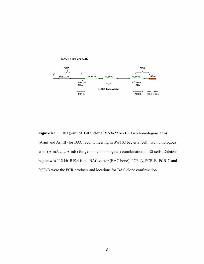
81
Figure 4.1 Diagram of BAC clone RP24-271-G16. Two homologous arms
(ArmI and ArmII) for BAC recombineering in SW102 bacterial cell; two homologous
arms (ArmA and ArmB) for genomic homologous recombination in ES cells. Deletion
region was 112 kb. RP24 is the BAC vector (BAC bone). PCR-A, PCR-B, PCR-C and
PCR-D were the PCR products and locations for BAC clone confirmation.
mCLCA4 mCLCA2 mCLCA1
112 KB deletion region
BAC-RP24-271-G16
ArmA ArmB
ArmI51bp
ArmII71bp
EG622139 RP24
PCR-A PCR-B PCR-C PCR-D
mCLCA4 mCLCA2 mCLCA1
112 KB deletion region
BAC-RP24-271-G16
ArmA ArmB
ArmI51bp
ArmII71bp
EG622139 RP24
PCR-A PCR-B PCR-C PCR-D

82
appeared. BAC mini prep DNA was made from the BAC colonies and sequence
confirmed by sequencing four PCR products, two BAC ends and two products around
the arms with the same primers as described (Figure 4.1).
4.3.3 ArmI-galk-ArmII targeting fragment construction
The total 112 kb fragment that included the partial mCLCA4 5’ region,
mCLCA4 gene, mCLCA2 gene and the beginning 22.5 kb of mCLCA1 gene was
deleted from BAC RP24-271-G16. Two homologous arms were designed and used to
construct the targeting fragment, ArmI (51 base pair) located just before 112 kb
deletion region and ArmII (71 base pair) located just after 112 kb deletion region
(Figure 4.1). Two specific long oligos were made by IDT (Integrated DNA
Technologies). Oligo1: ArmI (51bp) plus 20 bp of beginning em7 promoter for galk
gene
(CTTCACAAAACTCCTTTTACAGGTATTATTGTTCTTACTTTACAGGTAAGA-
GATATCGAATTCCTGTTGAC); Oligo2: ArmII (71bp) plus the last 20 bp of galk
gene from galk vector
(CCGAGCAGCACCTGGATAGCTAGGGTGCTCAGTCGTCTGACTATCGCCAG
CTACCCACAACACCCACCAA-TCAGCACTGTCCTGCTCCTT).1.3 kb of PCR
product (named ArmI-galk-ArmII) was generated by PCR primers (Oligo1 and
Oligo2) with galk vector as the template. PCR cycling conditions were: 94˚C 2 min,
94˚C 30sec, 58˚C 30 sec, 72˚C 1.5 min, repeat 5 cycles from second step, 94˚C 30 sec
and 72˚C 1.5 min for 35 cycles. The PCR product was purified from a 0.8% agarose
(Invitrogen) gel following the Qiagen Kit Gel Extraction Protocol and sequenced
(Figure 4.2 A).

83
Figure 4.2 Schematic generating BAC-KO124-galk targeting vector.
A. Generating ArmI-galk-ArmII targeting fragment by PCR. B. Homologous
recombination occurs between BAC-RP24-271-G16 and ArmI-galk-ArmII in SW102
cells and results in a BAC-KO124-galk vector with 112 kb sequence deletion. PCR
product crossing the arms, PCR(i) and PCR(ii) are shown (double end arrows).

84
Em7 galK
Galk vector
20bp 20bpArmII 71bp
ArmI 51bp
Em7 galk
PCR
ArmI 51bp ArmII 71bp
ArmI-galk-ArmII
A
Em7 galK
Galk vector
20bp 20bpArmII 71bp
ArmI 51bp
Em7 galk
PCR
ArmI 51bp ArmII 71bp
ArmI-galk-ArmII
A
Em7 galkArmI 51bp ArmII 71bp
112kb (mCLCA124)
ArmI-galk-ArmII
BAC-RP24-271-G16
51bp 71bp 22 Kb48 kb
Recombination in SW102
Em7 galk48 kb 22 kb
BAC-KO124-galk
51bp 71bp
B
RP24
PCR (i) PCR (ii)
Em7 galkArmI 51bp ArmII 71bp
112kb (mCLCA124)
ArmI-galk-ArmII
BAC-RP24-271-G16
51bp 71bp 22 Kb48 kb
Recombination in SW102
Em7 galk48 kb 22 kb
BAC-KO124-galk
51bp 71bp
B
RP24
PCR (i) PCR (ii)

85
4.3.4 Recombineering the targeting fragment and BAC in SW102 cells.
The SW102-RP24-271-G16 competent cells were made with CM selection and
3μl of 1.3 kb targeting fragment (ArmI-Galk-ArmII) was electroporated into the
competent cells followed by plating on galk selection plates and colonies were
observed after 3-4 days. Positive galk gene clones appeared bright pink on
MacConkey Agar (Acumedia Manufactuers) plates. PCR products crossing the arms
PCR(i): bef-armI-F (GGCACTAAACCATTAACTAAGT) and pgalk-R
(GCGAACTTTACGGTCATCGCG) yielded a 393 bp product; PCR(ii): pgalk-F
(GGTGCCTGCCGTACAGCAAGCT) and aft-armII-R
(GAGAGTATACAGCACAGTTAA) yielded a 271 bp product; and the PCR products
of the BAC ends (the same primers as listed in 4.3.1.) were generated and confirmed
by sequencing (Figure 4.2 B)
4.3.5 Replacing galk gene with Neomycin gene.
Due to the further selection needed in ES cells, the galk gene in the BAC
targeting construct was replaced by a Neomycin cassette. Two long oligos, (i) OligoA:
ArmI (51 bp) plus 20 bp [before the first FRT site in PBS-neomycin/kanamycin (PBS-
Neo/Kan) vector]
(CTTCACAAAACTCCTTTTACAGGTATTATTGTTCTTACTTTACAGGTAAGA-
GGATCTAGATAACTGATCAGC) and (ii) OligoB: ArmII (71bp) plus 20bp after
second FRT site in PBS-Neo/Kan vector
(CCGAGCAGCACCTGGATAGCTAGGGTGCTCAGTCGTCTGACTATCGCCAG
CTACCCACAACACCCACCAA-CCATGGAGAAGTTACTATTC) were generated
by IDT. The neomycin targeting fragment was made by PCR using OligoA and
OligoB with PBS-Neo/Kan vector (self made) as a template. The PCR conditions

86
were: 94˚C 2 min, 94˚C 30 sec, 58˚C 30 sec, 72˚C 1 min 30 sec, repeat 5 cycles from
step2, 94˚C 30 sec, 72 ˚C 1 min 30 sec, repeat 35 cycles from step 4, 72˚C for 10 min
then hold at 4˚C. The neomycin targeting fragment (ArmI-Neo/kan-ArmII) PCR
product was purified and sequenced. SW102-BAC-KO124-galk competent cells were
made as described and 3µl of neomycin targeting fragment (ArmI-Neo/kan-ArmII)
was electroporated into the competent cells. The cells were plated on Kan
(25µl/ml)/CM(12.5µ/ml)/LB plates and incubated at 32˚C for 2-3 days. Six colonies
were picked and checked by PCR with cross arm primers [PCR-a:
(GGTCTACTATGTGACAGAATAG) / (CTCGTCTTGCAGTTCATTCAG) and
generated 899 bp; PCR-b: (GCAGCGCATCGCCTTCTATCGC)/
(CATTATAAAGGTGCTGATACTGT) and yield 538 bp product] and with the ends
primers. The PCR products were sequenced to confirm the BAC construct. For easy
handling, the BAC-KO124-Neo construct plasmid DNA was electroporated into
DH10 electro competent cells (Invitrogen) (Figure 4.3).
4.3.6 ES cell culture and handling.
E14Tg2A.4 feeder independent ES cells line (BayGenomics) used for this
work was generated from 129P2/olaHsd strains with Pink-eyed chinchilla coat color
(6). ES cells were grown in GMEM base media supplemented with 15% fetal calf
serum (EmbryoMax, Chemicon), 1x L-glutamine (Gibco/Invitrogen), 1x NEAA (non
essential amino acids) (Gibco/Invitrogene), 1x Na pyruvate (Gibco/Invitrogen), 1x β-
mercaptoethanol (Gibco/Invitrogen), 1x Penicillin/Streptomycin (Gibco/Invitrogen)
and 1000U/ml Leukemia Inhibitory Factor (LIF) (ESGRO, Millipore). The ES cell
media was changed daily and the ES cells were split every other day to keep 60%-
80% confluency. The 10 cm culture plates were coated with 0.1% gelatin (Chemicon)
for 1 hour before replating the ES cells. Twenty-four hours after electroporation of the

87
Figure 4.3 Diagram depicting generation of BAC-KO124-neo/kan targeting
vector for ES cells. A. The same homologous arms (ArmI/51bp and ArmII/71bp)
plus a 20 bp sequence from PBS-Neo/kan vector were used were used to generate
ArmI-Neo/kan-ARMII targeting fragment by PCR. B. ArmI-Neo/kan-ArmII targeting
fragment was transfected into SW102-BAC-KO124-galk competent cells and resulted
in SW102-BAC-KO124-Neo/kan. C. Two PCR products (PCR-a and PCR-b) crossing
the arms plus two BAC ends PCR products were generated and sequenced to confirm
the BAC sequence.

88
20bp 20bpArmII 71bpArmI 51bp
pkansv40 Neo/kan
PBS-Neo/Kan
FRT FRT
Pkan sv40 Neo/kanArmI 51bp ArmII 71bp
ArmI-Neo/kan-ArmII
Em7 galk48 kb 22 kb
BAC-KO124-galk
51bp 71bp RP24
Pkan sv40 Neo/kanRP2422kb48 kb
BAC-KO124-Neo/kan
PCR
Homologous recombination in SW102
A
B
C
PCR-a PCR-b
20bp 20bpArmII 71bpArmI 51bp
pkansv40 Neo/kan
PBS-Neo/Kan
FRT FRT
Pkan sv40 Neo/kanArmI 51bp ArmII 71bp
Pkan sv40 Neo/kanArmI 51bp ArmII 71bp
ArmI-Neo/kan-ArmII
Em7 galk48 kb 22 kb
BAC-KO124-galk
51bp 71bp RP24
Pkan sv40 Neo/kanRP2422kb48 kb
BAC-KO124-Neo/kan
PCR
Homologous recombination in SW102
A
B
C
PCR-a PCR-b

89
ES cells, G418 (200ng/ml) was added to the GMEM media for the neomycin positive
selection.
4.3.7 ES cell targeting
ES cells (2x10⁷) and linerized BAC-KO124-Neo targeting DNA fragment (24
µg) were mixed in 0.8 ml PBS solution in 0.4 cm gap Gene Pulser Cuvette (Bio-RAD)
and the mixture was electroporated (3µF, 800V, 0.04 sec). After resting on ice for 30
minute, the ES cells were plated on 4 gelatin (10%) coaded plates with GMEM
supplemented media and incubated at 37˚C, 5% CO2. The G418 (200µg/ml in GMEM
media) drug selection was started on the second day following electroporation and
continued for 7-8 days. Three hundred and fifty colonies were picked at days 9-10 and
transferred to 48-well gelatin coated plates. Half of the cells were frozen in liquid
nitrogen with 2X frozen media (Chemicon) and half of the cells were harvest.
4.3.8 Homologous recombination positive clone detection.
DNA from ES cell clones was made according to the protocol in Gene
Targeting book (1). The random insertion clones were excluded by one of the four
BAC bone (vector) PCR products, 1) T7-BAC-end [RP24-T7
(TAATACGACTCACTATAGGGAGA) and G16R1
(GCTGTGTCCCCAAGACCAGCTA)], 699 bp; 2) Sp6-BAC-end [RP24-Sp6
(ATTTAGGTGACACTATAG) and G16-F1 (GTACTTCATAGCCTCAGTAATA)]
453 bp]; 3) BAC bone fragment-1 [RP24-12187(ATCCTGGTGTCCCTGTTG) and
RP24-12691(CGGAAATCGTCGTGGTAT), 505 bp]; and 4) BAC bone fragment-2
[RP243229 (AATACAACGGCTATCACG) and RP24-3823
(AGCTTCTGGCTTTCTTTAC), 595 bp]. The clones without BAC bone PCR bands
were checked by PCR to determine if they contained the Neomycin using the

90
following primer pairs: NeoGeneF2 (GCAGCGCATCGCCTTCTATCGC) and aft-
A2-R (ACAGTATCAGCACCTTTATAATG), 443 bp product.
The clones without BAC bone but containing the Neomycin gene were then
screened by real-time quantification PCR (qPCR) Relative Quantification (RQ)
method. Three qPCR primers/probe sets (TaqMan probes) were made by ABI
(Applied Biosystems) and used to screen positive clones: 1) ConKO124 ( Forward
primer-AGGTGCCATAGCAGATGAATGAAT, Reverse primer-
CACATAAGTTACCTGTAGCTATTTCAGGAA; reporter-FAM probe
CCCAACACAATTTCA ); 2) KO124KO4 (Forward primer-
GTGACTTTTAAGTGACCAGGACAAGA; Reverse primer-
ATCATGAGTGTGCTCGAAGCT; reporter-FAM probe-
ATGTGAGCCCAACTCC); and proKO124 (Forward primer-
CGTTGGCCTTAGAAGAGTGTGA; Reverse prime-
GCTCTGACTAGTTGAATGACCCTAA; reporter-FAM probe
CCCCAGCCGGATTAT ). In each reaction, 10 ng of sample DNA, 1X TaqMan
universal PCR Master Mix (Applied Biosystems, manufactured by Roche) and 1X
TaqMan probe (250 nM probe and 900 nM primers) were used. The was performed
using the 7500 Real Time PCR System (Applied Biosystems) with the following
cycling conditions: 50°C for 2 min, 95°C for 10 min, 95°C for 15 sec, 60°C for 1 min
repeat 40 cycles from step 3. Every sample was run parallel with TaqMan Mouse β-
actin (ACTB, Applied Biosystems) probe as an endogenous control to normalize
variation in concentrations between samples to make the data comparable. RQ Study
software included in the 7500 Real Time PCR System was used for the data analysis.

91
4.3.9 ES cell injection and mating strategy.
The positive clone 1C3 was injected into 3.5 days B6 blastocysts by Robert
Munroe in Department of Biomedical Sciences, Cornell University using standard
techniques (1) and resulted in 7 chimeric male founders. Three chimeric male mice
were mated to C67Bl/6J females. To generate homozygous mCLCA1,2,4 triple knock-
out mice, heterozygous (mCLCA1,2,4+/-) males and females were mated and
mCLCA1,2,4-/- mice were obtained.
4.3.10 Genotyping strategy to distinguish mCLCA4 offspring.
Four PCR products were used to distinguish mCLCA1,2,4-/-, mCLCA1,2,4+/-
and wildtype mCLCA1,2,4+/+ mice. PCR products: i) Primers NeoGeneF2(c) and aft-
A2-R(d) (538bp); ii) WT-genotypeF1(a) (GGTCTACTATGTGACAGAATAG) and
Neo-VecR(b) (CCTCGTCTTGCAGTTCATTCAG), 899 bp; iii) WT-genotype-F2(f)
(TGCTGTCCATGCTCAGGCAAAG) and aft-A2-R(d), 531 bp; and d) WT genotype-
F1(a) and WT-genotype-R1(e) (GCTAATCAGACGAATCTGCCTG), 400 bp. The
homozygous mice DNA will give PCR products “a/b” and “c/d” but no “a/e” and “f/d”
products. The heterozygous mice DNA will give all 4 PCR products. The wild type
mice DNA will give PCR product “a/e” and “f/d” but no products for “a/b” and “c/d”.
(Figure 4.8).
4.3.11 Phenotypic evaluation of mCLCA1,2,4 knock-out mice
Body weights were measured at 3, 6 and 10 weeks of age. Initial histological
evaluation of multiple organs was performed on two 6 week old mCLCA1,2,4-/- and
wildtype littermates by Bendicht Pauli from Molecular Medicine Department at
Cornell University.

92
4.4. Results
4.4.1. Generating BAC-KO124-Neo targeting construct by deleting 112kb
fragment in BAC-RP24-271-G16
The BAC RP24-271-G16 was used for the triple gene knock-out construct. It
contains 182 kb of mouse genomic sequence (including mCLCA1, mCLA2, mCLCA4
and EG622139 genes) and 10.6 kb RP24 vector. A 112 kb fragment was deleted from
the mouse genome that included 74% (22.5 kb/30.4kb) of mCLCA1gene, all of
mCLCA2 and all of mCLCA4 genes.
To take the advantage of the galk selection system I initially made a BAC-
KO124-galk vector. Fifty one base pair sequence located before the 112 kb deletion
region (ArmI) and 71 base pair sequence located after the 112 kb deletion region
(ArmII) were used for the homologous recombination Arms for BAC recombineering
(Figure 4. 2). Homologous recombination occurred between the BAC-RP24-271-G16
and ArmI-galK-ArmII targeting fragment in SW102 cells as evidenced by the fact that
all the colonies that were picked from the galk plates (20/20) had the smaller size of
insert fragment with NotI enzyme digestion and correct PCR products [PCR(i) and
PCR(ii)] (Figures 4.2). The 112 kb sequence including three quarter of mCLCA1
gene, all of the mCLCA2 and all of the mCLCA4 was successfully excluded from
BAC-RP24-271-G16 and named BAC-KO124-galk (Figure 4.2). These data
demonstrate the efficiency of the galk system for generating targeting vectors.
Since ES cell do not contain a galk selection system, BAC-KO124-galk could
not be used for ES cell targeting purpose. To make the ES cell targeting construct the
galk promoter em7 and galk gene were replaced by the Psv40 promoter and Neomycin
gene in BAC-KO124-galk BAC construct through one round of BAC recombineering,
to produce targeting vector, named BAC-KO124-Neo (Figure 4.3).

93
4.4.2. ES cell targeting and detection of homologous recombination
After the 112 kb deletion, 48 kb (ArmA) and 22 kb (ArmB) of the genomic
region remained in the BAC and were the homologous arms for recombination in the
ES cells (Figure 4.3). To increase the likelihood of homologous recombination the
BAC-KO124-Neo targeting vector DNA was prepared in large scale and linearized by
the PI-SCEI restriction enzyme. The linearized BAC targeting fragment was
electroporated into E14Tg2A ES cells and 350 clones were analyzed.
Due to the long homologous arms, traditional methods such as Southern Blot
can not be used to distinguish homologously recombined clones from wild type or
random insertion clones. I used two methods to determine which ES cells had
homologous recombination occurring in one allele: regular PCR and qPCR. Initially, I
used regular PCR and reasoned that if the linearized construct was randomly inserted
into the genome, the BAC vector would be part of the insertion in the majority of
cases. To take advantage of this idea, I used PCR products from the BAC vector to
exclude random insertion clones (Figure 4.4). Fifty-six clones without BAC vector
were detected by PCR products but contained the neomycin gene (the selection gene
for G418 drug positive clones) PCR product (Figure 4.4).
Figure 4.5 shows the strategy used to confirm the PCR results in the 56
potential homologous recombined clones identified using regular PCR. Regular ES
cell DNA was used as a calibrator in data analysis. The mCLCA4 knock-out construct
was a 4.8 kb deletion and shared a 4.2 kb deletion window with mCLCA1,2,4 knock-
out clones. mCLCA1,2,4-/-and mCLCA1,2,4+/-mice tail DNA and the injected knock-
out mCLCA4 ES cell clone (1H8) were used as controls. (i) TaqMan Probe “Con” is
ocated in the 4.8 kb ArmA region (non-deletion region) and every sample except the
random insertion clones have the same Relative Quantification (RQ) value as ES cell

94
Figure 4.4 Homologous recombination between BAC-KO124-Neo/kan
and one chromosome 3 allele in ES cells. When homologous recombination occurred
a 112 kb genomic region was deleted from chromosome 3 (targeted ES cell allele).
“1”, “2”, “3” and “4” show the PCR product positions from the BAC bone (RP24) and
indicate that ES cell clones had random insertion. “5” is neomycin gene PCR product
which indicated ES cell clones contained neomycin gene.
Pkan sv40 Neo/kanRP24
112 kb
BAC-KO124-Neo/kan
ES cell chromosom 3 (one allele)
Homologous recombination in ES cell
Pkan sv40 Neo/kan
Targeted ES cell allele
1 2 34
5
Pkan sv40 Neo/kanRP24
112 kb
BAC-KO124-Neo/kan
ES cell chromosom 3 (one allele)
Homologous recombination in ES cell
Pkan sv40 Neo/kan
Targeted ES cell allele
1 2 34
5

95
Figure 4.5 Diagram of the Real-time PCR strategy. Regular E14 ES cell DNA
was used as a calibrator. mCLCA4 knock-out (KO4) ES cell clone (1H8) DNA and
mCLCA4+/-, mCLCA4-/- mice tail DNA were used as control to screen the
homologous recombination ES cell clones of mCLCA1,2,4 knock-out. β-actin probe is
the endogenous control to normalize the concentration variation of the samples. (i)
The Con probe located in the non-deleted (ArmA) region and non-random insertion
samples will show the same RQ value as ES cell DNA. Random insertion clones will
show higher RQ value compared to ES cell DNA. (ii) KO124KO4 probe is located in
the deleted window of mCLCA4 and mCLCA1,2,4 knock-out. mCLCA4+/- (KO4+/-)
tail DNA, 1H8 DNA and positive mCLCA1,2,4 ES cell clone DNA should show half
of RQ value as ES cell DNA. mCLCA4-/- (KO4-/-) tail DNA should show zero RQ
value comparing with ES cell DNA. Random insertion clones will show the same RQ
value as ES cell DNA. (iii) KO124 probe located in the mCLCA1,2,4 deletion region.
Only mCLCA1,2,4 positive clones will have half of the RQ value as ES cell DNA.
Others will show the same RQ value as ES cell DNA.
4.8kb deletion
112kb deletion
actinKO124
KO4
KO124
KO124KO4Con
4.8kb deletion
112kb deletion
actinKO124
KO4
KO124
KO124KO4Con

96
DNA. Random insertion samples had a higher RQ value than ES cell sample DNA
(such as sample 41) (Figure 4.6 A ). (ii) TaqMan Probe KO124KO4 is located in the
shared deletion region and products from both knock-out mCLCA4 and knock-out
mCLCA1,2,4 ES cell positive clone DNA should have half RQ value of the wildtype
ES cell DNA. All other mCLCA1,2,4 ES cell non-positive clones had the same RQ
values as wildtype ES cell (Figure 4.6). (iii) TaqMan probe KO124 is located in the
112 kb deletion but not in the mCLCA4 deletion region. In this case, mCLCA1,2,4-/-
controls showed the same RQ value as wildtype ES cell sample. mCLCA1,2,4
homologous recombined clones showed half RQ value of wildtype and the other
mCLCA1,2,4 clones showed the same RQ value as wildtype. Two clones, 1C3 and
2B, were determined to have undergone homologous recombination resulting in the
removal of mCLCA1,2,4 (Figure 4.7). These data demonstrate the utility of using two
PCR strategies to confirm homologous recombination in ES cells following BAC
recombination.
4.4.3. Generating mCLCA 1,2,4 homozygous knock-out mice
One knock-out mCLCA1,2,4 ES cell clone (1C3) was injected into 3.5 days
blastocysts of C57Bl/6 mice and seven nearly 100% chimeric male founder mice were
obtained. Figure 4.8 shows the strategy to genotype the offspring by PCR and
representative examples. Three founder males were mated to C56Bl/6J females
generating normal size litters, 3-6 pups/litter. Six pairs of heterozygous mCLCA1,2,4
knock-out mice (mCLCA1,2,4+/-) were mated and both male and female homozygous
mCLCA1,2,4-/- mice were born. Figure 4.8 shows the PCR strategy genotype the
offsprings and the mice tail genotype results.

97
Figure 4.6 using KO124KO4 probe. Raw data from mCLCA1,2,4 ES cell clone
screening. 1C3 and 2B3 are the clones with one deleted allele. KO4ES is the
mCLCA4 knock-out ES cell clone (same as 1H8). “41” is the random insertion clone.
E14 ES is the ES cell DNA sample as the calibrator. Clones, 5A1-5D1, are non-
positive but without BAC bone clones.

98
Figure 4.7 qPCR results. A. Con probe, clone 41 have a higher PCR RQ value
than ES cell DNA, suggesting random insertion, while all other clones had the same
RQ value as ES cells. B. with KO124 probe, only two ES cell positive clones (1C3
and 2B3) showed half RQ value of ES cell DNA while others showed the same value
as ES cell DNA. Note, the higher RQ value of 41, KO(+/-) probably due to the non-
specific primer binding. C. With KO124KO4 probe, two mCLCA1,2,4 ES cell
positive clones (1C3, 2B3), mCLCA4 ES cell positive clone (1H8), mCLCA4+/-
[KO4(+/-)] tail DNA showed half RQ value of ES cell DNA while mCLCA4-/- [KO4(-
/-)] tail DNA showed zero RQ value compared to the ES cell DNA (See Figure 4.5).
The numbers under the bottom of each panel indicated the number of time the
experiment repeated.

99
(18) (18) (12) (15) ( 9) (9) (9)
(18) (18) (12) (15) (9) (9) (9)
Rel
ativ
e Q
uant
ifica
tion
Rel
ativ
e Q
uant
ifica
tion
0
0.5
1
1.5
2
2.5
3
E14 1C3-1 2B3 1H8-I 41 KO4(-/-) KO4(+/-)
KO124KO4
(15) (15) (9) (15) (15) (15) (15)
Rel
ativ
e Q
uant
ifica
tion
KO124
0
0.5
1
1.5
2
2.5
E14 1C3-1 2B3 1H8-I 41 KO4(-/-) KO4(+/-)
Con
0
0.5
1
1.5
2
2.5
3
3.5
E14 1C3-1 2B3 1H8-I 41 KO4(-/-) KO4(+/-)
(18) (18) (12) (15) ( 9) (9) (9)
(18) (18) (12) (15) (9) (9) (9)
Rel
ativ
e Q
uant
ifica
tion
Rel
ativ
e Q
uant
ifica
tion
0
0.5
1
1.5
2
2.5
3
E14 1C3-1 2B3 1H8-I 41 KO4(-/-) KO4(+/-)
KO124KO4
(15) (15) (9) (15) (15) (15) (15)
Rel
ativ
e Q
uant
ifica
tion
KO124
0
0.5
1
1.5
2
2.5
E14 1C3-1 2B3 1H8-I 41 KO4(-/-) KO4(+/-)
KO124
0
0.5
1
1.5
2
2.5
E14 1C3-1 2B3 1H8-I 41 KO4(-/-) KO4(+/-)
Con
0
0.5
1
1.5
2
2.5
3
3.5
E14 1C3-1 2B3 1H8-I 41 KO4(-/-) KO4(+/-)
Con
0
0.5
1
1.5
2
2.5
3
3.5
E14 1C3-1 2B3 1H8-I 41 KO4(-/-) KO4(+/-)
A
B
C

100
Figure 4.8 PCR strategy and results to genotype mCLCA1,2,4 mice. A.
Location of four sets of primer to genotype offspring. B. Examples of PCR bands
obtained from tail DNA of mCLCA1,2,4 knock-out mating offspring with genotyping
primers in Panel A. mCLCA1,2,4-/- mice have 899 bp (a/b) and 538 bp (c/d) bands
(left panel). mCLCA1,2,4+/- mice have all four bands (middle panel). mCLCA1,2,4+/+
mice have two PCR products, 400 bp (a/e) and 531 bp (f/d) (right panel).

101
A
B
KO allele
Neo-R 1.4 KB
a b c d
WT allele
112 KB
a e f d
KO allele
Neo-R 1.4 KB
a b c d
WT allele
112 KB
a e f d
a/e c/d a/b f/d M a/e c/d a/b f/d M
-/- +/- +/+
a/e c/d a/b f/d Ma/e c/d a/b f/d M a/e c/d a/b f/d M
-/- +/- +/+
a/e c/d a/b f/d M

102
4.4.4 Characterization of mCLCA1,2,4-/- mice
mCLCA1,2,4-/- mice survive to at least 2 months of age. Table 4.1 shows
weight taken at 3 different time points. Even though our mCLCA1,2,4-/- numbers do
not allow us to perform statistical analysis it appears the female mice are smaller at 6
and 12 weeks of age compared to littermate mCLCA1,2,4+/- and mCLCA1,2,4+/+.
Organs were taken from two mCLCA1,2,4-/-and two mCLCA1,2,4+/+ littermate
controls at 6 weeks of age. Hematoxylin and eosin staining of multiple tissues, such as
brain, heart, liver, small and large intestine, ovary, bladder, thymus, lung, stomach, etc
will be examined. I have observed postnatal day one mCLCA1,2,4-/- pup with eyes that
appeared opaque compared to wildtype littermates (Figure 4.9 A). Histological
evaluation of this pup showed hypopigment of the retainal epithelium (Figure 4.9 B).
This potential phenotype will need to be confirmed in more mCLCA1,2,4-/- mice and
their wildtype littermates.
4.5. Discussion
In this chapter I discussed two techniques that I developed and used
successfully namely the deletion of a large region of genomic DNA through BAC
recombineering and a PCR/qPCR strategy to screen ES cell clones. The BAC
recombineering technique has become a very useful tool for various purposes, such as
generating transgenic and knock-out constructs. Over the past several years I have
played a key role in developing transgenic mouse BAC recombineering technologies
in our laboratory by taking advantage of the full length gene promoter (5’ flanking
region of gene of interest in BAC) to drive reporter genes such as EGFP(3, 4). BAC
recombineering technology offers several advantages to produce triple gene knock-out
mice. First, BAC clones hold large fragments of genomic DNA. Second, mCLCA1,
mCLCA2 and mCLCA4 cluster together on the same chromosome. Third, a large

103
Table 4.1 Evaluation of body weight in mCLCA1,2,4 knock-out offspring.
Two mCLCA1,2,4-/- female showed the weight lost in 6 weeks. * Note, only two
animals obtained in this time and their weights were shown individulely.
mCLCA1,2,4+/+ weight (gm)
mCLCA1,2,4+/- weight (gm)
mCLCA1,2,4-/- weight (gm)
Age(wks)/Sex(number) F(8) M(8) F(15) M(15) *F(2) M(3) 3 15 ± 2 16 ± 2 14.9 ± 2 16 ± 2 16 ; 12.5 13 ± 3 6 23 ± 3.5 25 ± 3 22 ± 2 24 ± 3 21.8; 18 25 ± 2 10 28 ± 3 34 ± 3 24 ± 4 32 ± 3 23 ; -- --

104
A B
Figure 4.9 Evaluation of mCLCA1,2,4 eyes. A. Wide-field image of 1 day old
pup eyes. mCLCA1,2,4+/+ (left) and mCLCA1,2,4-/- (right). Note the opacity of the
mCLCA1,2,4-/- eye compared to the mCLCA1,2,4+/+ eye. B. Right is mCLCA1,2,4-/-
pup eye and left is WT pup littermate (20X). The red oval area shows the retinal
pigment epithelium cells.

105
deletion can be accomplished by BAC recombineering. Unfortunately, the high
percentage of identity shared between mCLCA gene family made it very difficult to
find unique sequences for the homologous arms that were used for BAC
recombineering. In this study I successfully deleted 112 kb specific region from a
BAC and the construct still contained enough genomic DNA for the homologous
arms.
Due to the large homologous arms, traditional colony screening methods such
as Southern blotting could not be used to detect whether recombination had occurred.
I used two methods to screen for homologously recombined positive clones: regular
PCR and qPCR (5). In the majority of cases, ES clones with random insertion of the
linearized BAC construct were excluded by detecting the BAC bone (vector) using
multiple sets of primers to the BAC bone. Once clones were excluded due to random
insertion the remaining clones were then screened for the neomycin gene to further
confirm the clones. The second step involved a qPCR strategy to quantitatively detect
the difference between two wildtype alleles versus one wildtype allele and one knock-
out allele with specific TaqMan probes within and outside the deleted region. The
combination of these two PCR strategies resulted in the identification of 2
mCLCA1,2,4 knock-out ES clones—1C3 and 2B3.
The real-time PCR system uses fluorescent-based PCR chemistry to provide
quantitative detection of DNA sequence with real-time analysis. The PCR product of
the region of interst is quantitatively detected by a fluorescent labeled probe. There are
two types of quantitative real-time PCR: absolute and relative. Relative quantification
(RQ) type suited my purpose and was used for this study. It determines the change of
targeted nucleic acid sequence of samples (ES cell clones) relative to the same
sequence in the calibrator sample (unmodified E14 ES cell DNA). RQ provides
accurate comparison between the samples and the calibrator and good results can be
obtained (Figure 4.7).

106
The finished targeting construct of knock-out mCLCA1,2,4 (BAC-KO124-
Neo) for ES cell is 80.6 kb. It contains 48 kb and 22 kb homology arms separated by
neomycin gene and contains the BAC vector. In general, large deletions in the genome
need large homologous arms in ES cell during the recombination process, but the ratio
between the length of the deletion and the length of the arms remains unknown. Using
a linearized targeting construct turned out to be a critical step as after screening over
250 non-linearized targeting clones not one homologous recombined clone was found
when circular targeting DNA was used. Once the DNA was linearized, I obtained two
mCLCA1,2,4 knock-out clones after screening the first 100 targeted clones. Thus,
linearizing DNA is a critical step for homologous recombination to occur in ES cells.
The E14Tg2A ES cell line was advantageous for this study compared to the R1
feeder dependent ES cell line used for the mCLCA4 knock-out mice (Chapter 3).
E14Tg2A ES cells are a feeder independent cell line which decreases cost and time
associated with feeder cells used for R1 ES cells. It has a fast growth rate and is
capable of germ line transmission.
In conclusion, this chapter details the galk and neomycin selection systems
combined with BAC recombineering techniques to produce a large deletion of
genomic DNA. ES cell clones screening strategy used both regular PCR and qPCR.
mCLCA1,2,4 triple gene knock-out homozygote mice have been obtained,
demonstrating the utility of this new method to produce null mice. mCLCA1,2,4-/-
mice are not embryonic lethal and survive until at least 12 weeks. Further studies are
ongoing to evaluate the gross phenotype and behavior of these animals.

107
REFERENCE
1. Joyner A. Gene Targeting, 2000. 2. Robichaud A, Tuck SA, Kargman S, Tam J, Wong E, Abramovitz M, Mortimer JR, Burston HE, Masson P, Hirota J, Slipetz D, Kennedy B, O'Neill G, and Xanthoudakis S. Gob-5 is not essential for mucus overproduction in preclinical murine models of allergic asthma. Am J Respir Cell Mol Biol 33: 303-314, 2005. 3. Tallini YN, Greene KS, Craven M, Spealman A, Breitbach M, Smith J, Fisher PJ, Steffey M, Hesse M, Doran RM, Woods A, Singh B, Yen A, Fleischmann BK, and Kotlikoff MI. c-kit expression identifies cardiovascular precursors in the neonatal heart. Proc Natl Acad Sci U S A 106: 1808-1813, 2009. 4. Tallini YN, Shui B, Greene KS, Deng KY, Doran R, Fisher PJ, Zipfel W, and Kotlikoff MI. BAC transgenic mice express enhanced green fluorescent protein in central and peripheral cholinergic neurons. Physiol Genomics 27: 391-397, 2006. 5. Valenzuela DM, Murphy AJ, Frendewey D, Gale NW, Economides AN, Auerbach W, Poueymirou WT, Adams NC, Rojas J, Yasenchak J, Chernomorsky R, Boucher M, Elsasser AL, Esau L, Zheng J, Griffiths JA, Wang X, Su H, Xue Y, Dominguez MG, Noguera I, Torres R, Macdonald LE, Stewart AF, DeChiara TM, and Yancopoulos GD. High-throughput engineering of the mouse genome coupled with high-resolution expression analysis. Nat Biotechnol 21: 652-659, 2003. 6. Ward CM, Stern P, Willington MA, and Flenniken AM. Efficient germline transmission of mouse embryonic stem cells grown in synthetic serum in the absence of a fibroblast feeder layer. Lab Invest 82: 1765-1767, 2002. 7. Warming S, Costantino N, Court DL, Jenkins NA, and Copeland NG. Simple and highly efficient BAC recombineering using galK selection. Nucleic Acids Res 33: e36, 2005.

108
CHAPTER 5
SUMMARY AND FUTURE RESEARCH DIRECTION

109
5.1. Summary
Chloride channel, calcium activated gene family members exist in more than
30 species (18). Originally, it was proposed that CLCAs were the integral channel
protein, but later studies have proved this to be incorrect [(9, 11, 17, 22), chapter 2].
All the members of this family have a similar structure but they are expressed in
different tissues (4, 7, 10, 12, 13, 18, 20). The proteins are cleaved and secreted out of
the cell and associate with the plasma membrane [(11, 17), chapter 2]. Many studies
have been focused on the role CLCAs play in the pathophysiology of human diseases.
Some CLCAs functions may be related to asthma, such as hCLCA1 and mCLCA3
(14, 19); some of them are involved in CF, such as hCLCA1, hCLCA4 and mCLCA3
(6, 15, 21), and others (hCLCA2, mCLCA1 and mCLCA2) may play a role in tumor
suppression, cancer and cell adhesion (1-3, 5, 16, 23-25). However, molecular level
function of this group of genes remains unknown.
I started this study to understand the function of one murine CLCA member,
mCLCA4. The mCLCA4 gene is located on mouse chromosome 3, clustering with
seven other family members (5 genes were cloned and 2 gene were predicted). It is
highly expressed in smooth muscle cells. Transient transfection studies indicated that
mCLCA4 may be related to a chloride channel (8). mCLCA4 gene spans 26 kb with a
2.7 kb mRNA transcript that codes for a 909 aa peptide (8). mCLCA4 protein contains
a N-terminal hydrolase domain, a VWA domain and a Fib3 domain (Figure 1.3).
In Chapter 2, I described the cellular processing and regulatory sequences
underlying mCLCA4 proteins. After glycosylation the 125 kDa protein product was
cleaved to 90 kDa NH2- and 30 kDa COOH-terminal fragments, and both fragments
were secreted into the cell media and associated with the cell membrane. The full-
length 125-kDa protein was only found in the ER. I discovered specific luminal

110
diarginine retention and dileucine forward trafficking signals in the CLCA4 sequence
that regulate export from the ER and proteolytic processing (Chapter 2).
To study the function of mCLCA4 in vivo, I generated a mouse lacking
mCLCA4 gene expression. These animals displayed no gross phenotype and bred
normally. Specific challenge experiments are being undertaken by collaborators
(Michael Holtzman et al. at Washington University) to examine the effect of airway
challenge on mClCa4 null mice. My hypothesis for why mCLCA4 null mice have no
overt phenotype is that mCLCA1 and/or mCLCA2 compensate for loss of mCLCA4
(Chapter 3).
To further understand the role of the mCLCA4 gene and its highly homologous
family members, I generated mCLCA1, mCLCA2, mCLCA4 triple knock-out mice. I
developed a novel method to delete a large fragment of DNA from the mouse genome
based on BAC recombineering techniques and successfully used it to delete three
linked genes. I also developed a novel PCR and qPCR based ES cell clone screening
method. The triple gene knock-out null mice survive to at least six months of age, but
the homozygous with homozygous mating has not obtained offspring yet. It is too
soon to conclude whether the male or female animal is sterile or it is just take time to
get the progenys. Phenotype studies had been started with few mice and are continuing
with more (Chapter 4).
5.2 Future direction and studies
5.2.1 mCLCA1,2,4 triple gene knock-out mice phenotype study
Currently, preliminary studies are underway to determine whether any
phenotype exists in mCLCA1,2,4-/- mice. A few interesting potential phenotypes have
been observed. First, it appears that adult female mCLCA1,2,4-/- mice may be
underweight compared to wildtype littermate controls. Second, some null animals may

111
have hypopigmentation of the eye. Third, the homozygous male and female mating
has not produced the offspring as yet, suggesting that one or both sexes may be sterile.
However, these observations need to be confirmed by increasing the number of
animals in the study. I am collaborating with Bendicht Pauli in Molecular Medicine
Department, Cornell University, to determine the phenotypes of mCLCA1,2,4-/- mice.
5.2.2. mCLCA1,2,4-/- Physiological Function: Predictions
Based on the literature, CLCAs play roles in asthma (hCLCA1 and
mCLCA3)(14, 19), and in cancer (hCLCA4, mCLCA1 and mCLCA2)(1-3, 5, 23-25).
mCLCA4 is expressed at high levels in airway smooth muscle (8) and it may be
involved in airway disease. Future study should investigate whether mCLCA4,
mCLCA1 and /or mCLCA2 play roles in asthma or cancer suppression and, if so,
through what pathway it acts. These questions may be answered using mCLCA4
knock-out mice and mCLCA1,2,4 triple gene knock-out mice. It will also be of
interest to determine what domain(s) in the protein structures are the key domain(s) for
the function. Specific challenge studies for asthma (19) and cancer in the two lines of
knock-out mice I have gnearated will help elucidate the roles of these genes in these
two diseases. For asthma challenge studies, I will continue collaboration with Michael
Holtzman et al. at Washington University to determine if these three genes contribute
to asthma susceptibily in accordance with his animal model.
CLCA proteins are the calcium activated chloride channel regulators. Although
the mechanism of regulation is not clear, CLCA proteins will still become the useful
biomarkers and therapeutic targeting molecules for asthma, cancer and chloride
currents related diseases.

112
5.2.3. Generating additional CLCA knock-out mice
Eight mCLCA genes cluster together on mouse chromosome 3. They have
with similar protein structure but divers tissue/organ expression patterns. This fact
suggested that mCLCA genes play different roles in different tissues but that other
family members may compensate if the function of one is lost. This group of genes
may function as a team and compensate for each other when the function of one is
lost. To understand the in vivo functions of all eight genes, deletion of the whole
family from mouse genome may necessary. However, deletion of a large specific
region (total ~340 kb) from mouse genome is not yet possible with current technology.
It is possible, nonetheless, to delete more members from the mouse genome based on
the technique I developed and used in my study, as described below.
Since the BAC recombineering technique was successfully used for this study,
we can now generate a mouse with additional CLCA members deleted from the
genome and discover the function of these genes in murine life. For example, a BAC
containing mCLCA6, AI747448 (mCLCa7) and EG622139 (mCLCA8) genes could
be chosen and these three genes deleted using the galk selection system with two
genomic regions across the deletion area remaining called ArmC-Neo-ArmD (Figure
5.1A). To take advantage of the mCLCA1,2,4 BAC knock-out construct (ArmA-Neo-
ArmB) with three genes deleted, ArmD of BAC-ArmC-Neo-ArmD construct would be
replaced by ArmB through gap repair. The Neomycin gene of BAC-ArmC-Neo-ArmB
would be replaced by a puromycin gene for further 1C3 ES cell (the ES cell line with
mCLCA1,2,4 knock-out) selection since this ES cell already contains a neomycin
gene, named BAC-ArmC-Puromycin-ArmB (Figure 5.1B-D). ES cell 1C3
(mCLCA1,2,4, knock-out clone) could used for the ES cell source for targeting. This
line of knock-out mice will be very useful for further understanding the molecular
function of this group of genes.

113
5.2.4. CLCA family and its future
The CLCA family has drawn the attention of researchers for many years and in
many ways. The family members have been excluded as chloride channel integral
proteins, but they still play roles in regulating chloride currents. The function of some
of the members is related to asthma and cystic fibrosis diseases, while others are tumor
suppressors. It is still unknown, however, how these proteins function at the molecular
level and the roles they play in disease. The answers will be taken a few steps further
with my contribution of mCLCA4 knock-out and mCLCA1,2,4 knock-out mice.

114
Figure 5.1 Diagram depicting strategy to make mCLCA1,2,4,6,7,8 knock-out
mice. A. Choose a BAC that contains mCLCA6, mCLCA7 and mCLCA8 and a free
region crossing the genes. B-C. Use BAC recombineering/gap repair to replace ArmD
in BAC-KO378-Neo construct with ArmB in BAC-KO124-Neo construct after
deletion of three genes with galk selection and replacing galk gene with neomycin
gene. D. With one round of BAC recombineering, Neomycin gene will be replaced by
the puromycin gene for further ES cell selection.

115
mCLCA6 mCLCA8
Deletion region
ArmCArmD
ArmI ArmII
A
B
Neo-R
BAC-KO378-Neo
Neo-R
BAC-KO124-Neo
ArmD
ArmBArmA
ArmC
CNeo-R
Part-RP24
ArmBArmC
BAC-KO278-Neo-ArmB
CM
D
Puiomycine-RArmC ArmB
BAC-KO378-Puio
CM
CM
mCLCA7mCLCA6 mCLCA8
Deletion region
ArmCArmD
ArmI ArmII
A
B
Neo-R
BAC-KO378-Neo
Neo-R
BAC-KO124-Neo
ArmD
ArmBArmA
ArmC
CNeo-R
Part-RP24
ArmBArmC
BAC-KO278-Neo-ArmB
CM
D
Puiomycine-RArmC ArmB
BAC-KO378-Puio
CM
CM
mCLCA7

116
REFERENCE
1. Abdel-Ghany M, Cheng HC, Elble RC, Lin H, DiBiasio J, and Pauli BU. The interacting binding domains of the beta(4) integrin and calcium-activated chloride channels (CLCAs) in metastasis. J Biol Chem 278: 49406-49416, 2003. 2. Abdel-Ghany M, Cheng HC, Elble RC, and Pauli BU. The breast cancer beta 4 integrin and endothelial human CLCA2 mediate lung metastasis. J Biol Chem 276: 25438-25446, 2001. 3. Abdel-Ghany M, Cheng HC, Elble RC, and Pauli BU. Focal adhesion kinase activated by beta(4) integrin ligation to mCLCA1 mediates early metastatic growth. J Biol Chem 277: 34391-34400, 2002. 4. Agnel M, Vermat T, and Culouscou JM. Identification of three novel members of the calcium-dependent chloride channel (CaCC) family predominantly expressed in the digestive tract and trachea. FEBS Lett 455: 295-301, 1999. 5. Balakrishnan A, von Neuhoff N, Rudolph C, Kamphues K, Schraders M, Groenen P, van Krieken JH, Callet-Bauchu E, Schlegelberger B, and Steinemann D. Quantitative microsatellite analysis to delineate the commonly deleted region 1p22.3 in mantle cell lymphomas. Genes Chromosomes Cancer 45: 883-892, 2006. 6. Brouillard F, Bensalem N, Hinzpeter A, Tondelier D, Trudel S, Gruber AD, Ollero M, and Edelman A. Blue native/SDS-PAGE analysis reveals reduced expression of the mClCA3 protein in cystic fibrosis knock-out mice. Mol Cell Proteomics 4: 1762-1775, 2005. 7. Danahay H, Atherton H, Jones G, Bridges RJ, and Poll CT. Interleukin-13 induces a hypersecretory ion transport phenotype in human bronchial epithelial cells. Am J Physiol Lung Cell Mol Physiol 282: L226-236, 2002. 8. Elble RC, Ji G, Nehrke K, DeBiasio J, Kingsley PD, Kotlikoff MI, and Pauli BU. Molecular and functional characterization of a murine calcium-activated chloride channel expressed in smooth muscle. J Biol Chem 277: 18586-18591, 2002. 9. Elble RC, Walia V, Cheng HC, Connon CJ, Mundhenk L, Gruber AD, and Pauli BU. The putative chloride channel hCLCA2 has a single C-terminal transmembrane segment. J Biol Chem 281: 29448-29454, 2006. 10. Elble RC, Widom J, Gruber AD, Abdel-Ghany M, Levine R, Goodwin A, Cheng HC, and Pauli BU. Cloning and characterization of lung-endothelial cell adhesion molecule-1 suggest it is an endothelial chloride channel. J Biol Chem 272: 27853-27861, 1997.

117
11. Gibson A, Lewis AP, Affleck K, Aitken AJ, Meldrum E, and Thompson N. hCLCA1 and mCLCA3 are secreted non-integral membrane proteins and therefore are not ion channels. J Biol Chem 280: 27205-27212, 2005. 12. Gruber AD, Elble RC, Ji HL, Schreur KD, Fuller CM, and Pauli BU. Genomic cloning, molecular characterization, and functional analysis of human CLCA1, the first human member of the family of Ca2+-activated Cl- channel proteins. Genomics 54: 200-214, 1998. 13. Gruber AD and Pauli BU. Molecular cloning and biochemical characterization of a truncated, secreted member of the human family of Ca2+-activated Cl- channels. Biochim Biophys Acta 1444: 418-423, 1999. 14. Kamada F, Suzuki Y, Shao C, Tamari M, Hasegawa K, Hirota T, Shimizu M, Takahashi N, Mao XQ, Doi S, Fujiwara H, Miyatake A, Fujita K, Chiba Y, Aoki Y, Kure S, Tamura G, Shirakawa T, and Matsubara Y. Association of the hCLCA1 gene with childhood and adult asthma. Genes Immun 5: 540-547, 2004. 15. Leverkoehne I and Gruber AD. The murine mCLCA3 (alias gob-5) protein is located in the mucin granule membranes of intestinal, respiratory, and uterine goblet cells. J Histochem Cytochem 50: 829-838, 2002. 16. Li X, Cowell JK, and Sossey-Alaoui K. CLCA2 tumour suppressor gene in 1p31 is epigenetically regulated in breast cancer. Oncogene 23: 1474-1480, 2004. 17. Mundhenk L, Alfalah M, Elble RC, Pauli BU, Naim HY, and Gruber AD. Both cleavage products of the mCLCA3 protein are secreted soluble proteins. J Biol Chem 281: 30072-30080, 2006. 18. Patel AC, Brett TJ, and Holtzman MJ. The Role of CLCA Proteins in Inflammatory Airway Disease. Annu Rev Physiol, 2008. 19. Patel AC, Morton JD, Kim EY, Alevy Y, Swanson S, Tucker J, Huang G, Agapov E, Phillips TE, Fuentes ME, Iglesias A, Aud D, Allard JD, Dabbagh K, Peltz G, and Holtzman MJ. Genetic segregation of airway disease traits despite redundancy of calcium-activated chloride channel family members. Physiol Genomics 25: 502-513, 2006. 20. Pawlowski K, Lepisto M, Meinander N, Sivars U, Varga M, and Wieslander E. Novel conserved hydrolase domain in the CLCA family of alleged calcium-activated chloride channels. Proteins 63: 424-439, 2006.

118
21. Ritzka M, Stanke F, Jansen S, Gruber AD, Pusch L, Woelfl S, Veeze HJ, Halley DJ, and Tummler B. The CLCA gene locus as a modulator of the gastrointestinal basic defect in cystic fibrosis. Hum Genet 115: 483-491, 2004. 22. Whittaker CA and Hynes RO. Distribution and evolution of von Willebrand/integrin A domains: widely dispersed domains with roles in cell adhesion and elsewhere. Mol Biol Cell 13: 3369-3387, 2002. 23. Zhu D, Cheng CF, and Pauli BU. Blocking of lung endothelial cell adhesion molecule-1 (Lu-ECAM-1) inhibits murine melanoma lung metastasis. J Clin Invest 89: 1718-1724, 1992. 24. Zhu D and Pauli BU. Correlation between the lung distribution patterns of Lu-ECAM-1 and melanoma experimental metastases. Int J Cancer 53: 628-633, 1993. 25. Zhu DZ, Cheng CF, and Pauli BU. Mediation of lung metastasis of murine melanomas by a lung-specific endothelial cell adhesion molecule. Proc Natl Acad Sci U S A 88: 9568-9572, 1991.


![KD-A645 / KD-R640 / KD-R540 / KD-R440 - Car Audio ...santafeautosound.com/uploads/product-manuals/JVC KD-R540.pdfKD-A645 / KD-R640 / KD-R540 / KD-R440 GET0829-001A [J/JW] ENGLISH ESPAÑOL](https://cdn.vdocuments.us/doc/165x107/5aaf5da87f8b9a25088d67c4/kd-a645-kd-r640-kd-r540-kd-r440-car-audio-kd-r540pdfkd-a645-kd-r640.jpg)


