
The Eye


Major Parts of the Eye
• Cornea -– clear, transparent portion of the outer coat of
the eyeball through which light passes to the lens.
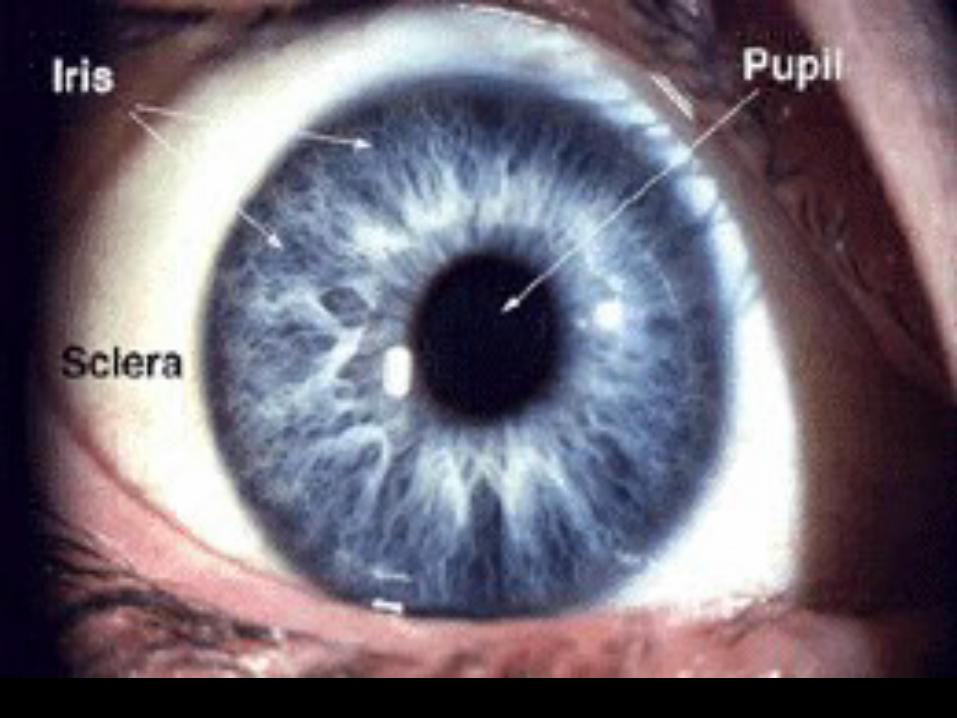
• Iris -– gives our eyes color
– smooth muscles that open or close an apeture called the PUPIL.

• Pupil - Opening in middle of iris.
• Lens– Eye's primary light-focusing
structure located behind pupil.
– Muscles attach to it that contract causing lens to contract enabling lens to focus light.

•Draw this

Aqueous vs Vitreous Humour
• aqueous is a clear, watery
• anterior and posterior chamber (anterior compartment)


• vitreous is a transparent, colorless mass of soft, gelatinous material
• behind the lens - posterior “compartment”
• Note: your book only mentions anterior compartment (aqueous) and posterior compartment (vitreous)

Eye layers
• Conjunctiva – is a clear membrane covering the white of the
eye (sclera)
• Sclera – is the white of the eye
• Choroid– carries blood vessels, is the inner coat between
the sclera and the retina

Retina
• A thin membrane on back of eye that contains light-sensitive receptors (rods & cones).


• Cones– Light receptors that are sensitive only in
bright light.– They can distinguish form & colour very
well.
• Rods– Light receptors that are sensitive in dim
light.– They cannot distinguish colour &
therefore, you only see shades of gray in dim light.


• Macula – is a small area in the retina that provides our most
central, acute vision.
– Small depressed area in centre of macula directly in line with centre of cornea & lens is called the FOVEA.
•Fovea is the region of keenest vision.
•Light-sensitive cones are concentrated in fovea.

• Optic Nerve
– Carries nerve impulses to the brain created when light strikes rods & cones.

Blind spot• the point on the retina where the
approximately 1 million axons converge on the optic nerve, there are no rods or cones. This spot, called the blind spot, is thus insensitive to light.

Eye Muscles
• Movement of the eye is controlled by six muscles:
• Medial rectus- lies on the inner side
• Lateral rectus- lies on the outer side
• Superior rectus- lies above the eye
• Inferior rectus- lies below the eye
• Superior oblique- lies above and runs obliquely
• inferior oblique- lies below and runs obliquely


Why do we see?
– Light reflected from object enters your eye.
– Cornea & lens bend light rays together.– Rays cross & focus on retina which
appears upside down & backward.– Light strikes rods & cones creates
nerve impulses that are carried by optic nerve to brain to be interpreted.
– Retinal image reversed in brain so we see object right side up.

Retinal Layers
• Composed of three layers:
– 1. Ganglion cell layer– 2. Bipolar cell layer– 3. Rod and cone cell layer

• rods and cones - actual photoreceptors
• ganglion cells - transmit to the brain; the axons of these ganglion cells make up the optic nerve.
• bipolar cells - process input from photoreceptors and transmit the signal to ganglion cells.


