
THE EFFECT OF DIFFERENT IMPACT EXERCISE TRAINING ON DEFORMATIONAL BEHAVIOR AND FUNCTIONAL ADAPTATION OF
ARTICULAR CARTILAGE
A THESIS SUBMITTED TO THE GRADUATE SCHOOL OF SOCIAL SCIENCES
OF MIDDLE EAST TECHNICAL UNIVERSITY
BY
ÖZGÜR ÇELİK
IN PARTIAL FULFILLMENT OF THE REQUIREMENTS FOR
THE DEGREE OF DOCTOR OF PHILOSOPHY IN
THE DEPARTMENT OF PHYSICAL EDUCATION AND SPORTS
JANUARY 2010

Approval of the Graduate School of Social Sciences
_________________________
Prof. Dr. Sencer Ayata Director
I certify that this thesis satisfies all the requirements as a thesis for the degree of Doctor of Philosophy.
_________________________
Assoc. Prof. Dr. M. Settar Koçak Head of Department
This is to certify that we have read this thesis and that in our opinion it is fully adequate, in scope and quality, as a thesis for the degree of Doctor of Philosophy.
_________________________
Prof. Dr. Feza Korkusuz Supervisor
Examining Committee Members Prof. Dr. Gülfem Ersöz (AU, BESYO) _____________________
Prof. Dr. Feza Korkusuz (METU, PES) _____________________
Assoc. Prof. Dr. Settar Koçak (METU, PES) _____________________
Assoc. Prof. Dr. Aydiner Kalacı (MKU, BESYO) _____________________
Assist. Prof. Dr. Hilmi Taşer (MKU, BESYO) _____________________

iii
I hereby declare that all information in this document has been obtained and presented in accordance with academic rules and ethical conduct. I also declare that, as required by these rules and conduct, I have fully cited and referenced all material and results that are not original to this work. Name, Last Name : Özgür Çelik
Signature :

iv
ABSTRACT
THE EFFECT OF DIFFERENT IMPACT EXERCISE TRAINING ON
DEFORMATIONAL BEHAVIOR AND FUNCTIONAL ADAPTATION OF
ARTICULAR CARTILAGE
Çelik, Özgür
Ph.D., Department of Physical Education & Sports
Supervisor: Prof. Dr. Feza Korkusuz
January 2010, 159 pages
The objective of the present study was to investigate deformational behavior
and functional adaptation of articular cartilage by comparing the changes of
biochemical osteoarthritis markers’ concentrations due to 30-min exercise after
12-weeks of regular high impact, impact or non-impact exercise.
Blood samples were drawn from 44 healthy sedentary males immediately
before, immediately after and 0.5 h after a 30-min moderate walking exercise.
Osteoarthritis biomarkers’ (Serum COMP and CTX-I) concentrations were
determined with enzyme-linked immunosorbent assay. After the first
measurements, participants were randomly assigned to running, cycling,
swimming, and control groups. All groups except for control group trained for
12 weeks. After 12-weeks, post tests were applied.
Multivariate tests indicated a significant fatigue and resting effect on serum
COMP concentration in all groups at pre- and post-tests. Therefore, pair wise

v
comparisons were conducted in order to assess the differences across all groups
and conditions. Results indicated significant differences in post-test
measurements among phases of groups except for running group. However,
fatigue or resting did not change the concentration of serum CTX-I in any
groups during the tests.
According to results, moderate walking activity has an influence on the increase
of serum COMP concentrations of young sedentary men. However, 12 weeks
regular weight-bearing high impact physical exercise decreases the
deformational effect of walking activity by functional adaptation of articular
cartilage to specific environmental requirements.
Key words: COMP, CTX-I, Running, Cycling, Swimming.

vi
ÖZ
FARKLI DARBELERDEKİ EGZERSİZLERİN EKLEM KIKIRDAĞININ
DEFORMASYONEL DAVRANIŞINA VE FONKSİYONEL
ADAPTASYONUNA ETKİSİ
Çelik, Özgür
Doktora, Beden Eğitimi ve Spor Bölümü
Tez Yöneticisi : Prof. Dr. Feza Korkusuz
Ocak 2010, 159 sayfa
Bu çalışmanın amacı, 12 hafta süreyle yapılan darbeli, yüksek darbeli veya
darbesiz egzersizlerden sonra, 30 dakikalık egzersizin eklem kıkırdağında sebep
olduğu deformasyonel davranışı ve fonksiyonel adaptasyonu, serum osteoartrit
işaretleyicilerinin konsantrasyonundaki değişikliği değerlendirerek araştırmaktı.
Kan örnekleri, 44 sağlıklı sedanter erkekten 30 dakikalık orta şiddette yürüme
egzersizinden hemen önce, hemen sonra ve yarım saat sonra alındı. Osteartrit
biyo-işaretleyicilerinin (Serum KOMP ve CTX-I) konsantrasyonu ELIZA
yöntemi ile belirlendi. İlk ölçümlerin ardından katılımcılar rasgele örneklem
metodu ile koşu, bisiklet, yüzme ve kontrol gruplarına ayrıldı. Kontrol grubu
haricinde tüm gruplar 12 hafta boyunca antrenman yaptı. 12 hafta sonunda, son
testler uygulandı.

vii
Çokdeğişkenli testler tüm gruplarda ilk ve son testlerde yorgunluğun ve
dinlenmenin serum KOMP konsantrasyonu üzerinde anlamlı bir etkisi
olduğunu gösterdi. Bu nedenle, hangi grupların ortalamasının değiştiğini
değerlendirmek için ikili karşılaştırmalar uygulandı. Koşu grubu hariç diğer
grupların son test ölçümlerindeki fazlarda da anlamlı farklar olduğu ortaya
çıktı. Ancak testler sırasında yorgunluk ve dinlenme serum CTX-I
konsantrasyonunu değiştirmedi.
Sonuçlara göre, orta şiddetli yürüme aktivitesi genç sedanter erkeklerin serum
KOMP konsantrasyonunu yükseltebilecek etkiye sahiptir. Ancak, 12 hafta
süreyle düzenli olarak yapılan, vücudunun ağırlığının taşındığı ve yüksek
çarpma etkisi yaratan egzersizler, eklem kıkırdağının belirli çevresel
gereksinimlere fonksiyonel adaptasyonu sayesinde yürüme aktivitesinin sebep
olduğu deformasyonel etkiyi düşürmektedir.
Anahtar Kelimeler: KOMP, CTX-I, Koşu, Bisiklet, Yüzme

viii
To Göşeğağ, Aslan & Sırma For their unconditional love

ix
ACKNOWLEDGEMENTS
I would like to express my gratitude and sincere appreciation to my advisor, Dr.
Korkusuz for his encouragement, guidance and enthusiasm. Thanks for your
countless ideas for developing this study, editing and re-editing each chapter.
I would also like to acknowledge and thank two of my committee members Dr.
Ersöz and Dr. Koçak, for their initial guidance in instructing me in the design
and theory of my research and formalizing of my final dissertation.
I have a great deal of thanks to give to my forbearing wife Sırma and my
faithful friend Yaşar Salcı. Sırma was very creative in paraphrasing my writings
and very patient in lonely weekends and Yaşar demonstrated outstanding
performance in laboratory measurements.
Also, I wish to thank Emre Ak and Ahmet Yıldırım for their assistance in my
presentations and defences.
Finally, I would like to thank a number of people in METU Physical Education
and Sports Department for their contribution towards this thesis.
This study was supported by:
The fund of METU Scientific Research Projects (METU BAP). Grant No:
BAP-06-07-03-00-17
The Scientific and Technological Research Council of Turkey (TUBİTAK).
Grand No: 107S112

x
TABLE OF CONTENTS
TITLE PAGE ....................................................................................................... i
APPROVAL PAGE ............................................................................................ ii
PLAGIARISM PAGE. ...................................................................................... iii
ABSTRACT ....................................................................................................... iv
ÖZ ....................................................................................................................... vi
DEDICATION PAGE ...................................................................................... viii
ACKNOWLEDGEMENTS ............................................................................... ix
LIST OF TABLES ........................................................................................... xiii
LIST OF FIGURES ........................................................................................... xv
CHAPTER 1 ........................................................................................................ 1
INTRODUCTION ........................................................................................... 1
1.1 Background of the Study ................................................................... 1
1.2 Rationale of the Study ....................................................................... 5
1.3 Research Questions ........................................................................... 6
1.4 Purpose of the Study ......................................................................... 6
1.5 Research Hypotheses ........................................................................ 6
1.6 Delimitations ..................................................................................... 7
1.7 Limitations ........................................................................................ 7
1.8 Assumptions ...................................................................................... 8
1.9 Definition and Abbreviation of Terms .............................................. 8
CHAPTER 2 ...................................................................................................... 10
LITERATURE REVIEW .............................................................................. 10
2.1 Structure of Knee Joint and Articular Cartilage ............................ 10
2.2 Evaluation of Knee Joint and Articular Cartilage .......................... 12
2.3 Loading on Articular Cartilage and Knee Joint ............................. 35
2.4 Sports and Body .............................................................................. 40
2.5 Sport and Articular Cartilage ......................................................... 45

xi
CHAPTER 3 ...................................................................................................... 62
MATERIALS AND METHODS .................................................................. 62
3.1 Overall Research Design ................................................................ 62
3.2 Participants ..................................................................................... 63
3.3 Interventions .................................................................................... 65
3.4 Physical and Physiological Measurements ..................................... 67
3.4.1 Body Mass Index (BMI) ............................................................... 67
3.4.2 Endurance (VO2max) Measurements .......................................... 67
3.4.3 Isokinetic Strength Measurements ............................................... 68
3.5 Blood Tests ...................................................................................... 69
3.5.1 Blood Sampling ........................................................................... 69
3.5.2 Biochemical Analysis of the Blood Samples. ............................... 70
3.6 Statistical Analyses ......................................................................... 71
CHAPTER 4 ...................................................................................................... 73
RESULTS ...................................................................................................... 73
4.1 Demographic Information of the Participants ................................ 73
4.1.1 Participants ................................................................................. 73
4.1.2 Body Mass Index ......................................................................... 74
4.1.3 Endurance (VO2max) Measurements .......................................... 76
4.1.4 Isokinetic Strength Measurements ............................................... 78
4.2 Serum COMP Concentration Measurements .................................. 79
4.2.1 Serum COMP level differences among groups at pre- and post-
test measurements ...................................................................................... 80
4.2.2 Serum COMP level changes due to acute exercise at pre- and
post-test measurements (Deformational Behavior of Cartilage) .............. 82
4.2.3 Serum COMP level changes within groups at pre- and post-test
measurements ............................................................................................ 87
4.3 Serum CTX-I Concentration Measurements ................................... 92
4.3.1 Serum CTX-I level differences among groups at pre- and post-test
measurements ............................................................................................ 92
4.3.2 Serum CTX-I level changes due to acute exercise at pre- and post-
test measurements ...................................................................................... 94

xii
4.3.3 Serum CTX-I level changes within groups at pre- and post-test
measurements ............................................................................................ 97
CHAPTER 5 .................................................................................................... 101
DISCUSSION ............................................................................................. 101
5.1 Physical and Biomotor abilities of participants. .......................... 102
5.2 Serum COMP levels ...................................................................... 104
5.3 Serum CTX-I levels ....................................................................... 113
CHAPTER 6 .................................................................................................... 116
SUMMARY, CONCLUSIONS & RECOMMENDATIONS..................... 116
6.1 Summary ........................................................................................ 116
6.2 Conclusions ................................................................................... 117
6.3 Recommendations ......................................................................... 118
REFERENCES ................................................................................................ 120
APPENDICES ................................................................................................. 137
A. INFORMED CONSENT FORM ........................................................... 138
B: ETHIC COMMITY APPROVAL .......................................................... 139
C. DATA COLLECTION SHEET .............................................................. 140
D. VO2max EVALUATION SHEET .......................................................... 141
E. ISOKINETIC STRENGTH EVALUATION SHEET ............................ 142
F. TÜRKÇE ÖZET ...................................................................................... 143
G. CURRICULUM VITAE ........................................................................ 154

xiii
LIST OF TABLES
Table 1. Physical characteristics of participants ............................................... 74
Table 2. Simple Effect Analysis of Time for BMI ............................................ 75
Table 3. Simple Effect Analysis of Time for VO2max ..................................... 77
Table 4. Simple Effect Analysis of Dominant quadriceps PT/BW ................... 78
Table 5. Pre-test Serum COMP concentrations (U/l) ........................................ 79
Table 6. Post-test Serum COMP concentrations (U/l) ...................................... 80
Table 7. Mean differences of serum COMP concentration between pairs of different states in running group. ...................................................................... 83
Table 8. Mean differences of serum COMP concentration between pairs of different states in cycling group. ....................................................................... 84
Table 9. Mean differences of serum COMP concentration between pairs of different states in swimming group ................................................................... 85
Table 10. Mean differences of serum COMP concentration between pairs of different states in control group ......................................................................... 85
Table 11. Simple Effect Analysis of Time for Serum COMP Level at Rec. ... 88
Table 12. Simple Effect Analysis of Time for Serum COMP Level at Fat ...... 90
Table 13. Simple Effect Analysis of Time for Serum COMP Level at Reg. .... 91
Table 14. Pre-test Serum CTX-I concentrations (ng/mL) ................................. 92
Table 15. Post-test Serum CTX-I concentrations (ng/mL) ............................... 92
Table 16. Mean differences of serum CTX-I concentration between pairs of different states in running group. ...................................................................... 95

xiv
Table 17. Mean differences of serum CTX-I concentration between pairs of different states in cycling group. ....................................................................... 95
Table 18. Mean differences of serum CTX-I concentration between pairs of different states in swimming group. .................................................................. 96
Table 19. Mean differences of serum CTX-I concentration between pairs of different states in control group. ........................................................................ 97
Table 20. Simple Effect Analysis of Time for Serum CTX-I Level at Rec ...... 98
Table 21. Simple Effect Analysis of Time for Serum CTX-I Level at Fat ....... 99
Table 22. Simple Effect Analysis of Time for Serum CTX-I Level at Reg .... 100

xv
LIST OF FIGURES
Figure 1. Structure of Human Knee Cartilage ................................................... 12
Figure 2.Flow chart of research design. ............................................................ 64
Figure 3. A sample of heart rate during a training session. ............................... 66
Figure 4. Design of Blood Sampling ................................................................. 70
Figure 5. Multivariate and Univariate Comparisons of Blood Samples. .......... 72
Figure 6. Body Mass Index Differences. ........................................................... 76
Figure 7. Maximum Oxygen Consumption Differences. .................................. 77
Figure 8. Dominant Legs, Quadriceps Strength Differences. ........................... 79
Figure 9. Serum COMP concentrations at different states during pre-test measurements. ................................................................................................... 81
Figure 10. Serum COMP concentrations at different states during post-test measurements. ................................................................................................... 82
Figure 11. Pair-wise comparisons for different states for each group at pre-test measurements .................................................................................................... 86
Figure 12. Pair-wise comparisons for different states for each group at post-test measurements .................................................................................................... 87
Figure 13. Pre- and post-test serum COMP level changes for each group ....... 88
Figure 14. Pre- and post-test serum COMP level changes for each group. ...... 89
Figure 15. Pre- and post-test serum COMP level changes for each group. ...... 91
Figure 16. Serum CTX-I concentrations at different states during pre-test measurements .................................................................................................... 93

xvi
Figure 17. Serum CTX-I concentrations at different states during post-test measurements .................................................................................................... 94
Figure 18. Pre- and post-test serum CTX-I level changes for each group ........ 98
Figure 19. Pre- and post-test serum CTX-I level changes for each group ........ 99
Figure 20. Pre- and post-test serum CTX-I level changes for each group ...... 100

1
CHAPTER 1
INTRODUCTION
1.1 Background of the Study
About 140 years ago famous biologist Charles Darwin (1872) stated that the
ability of tissues to emerge and maintain their structure in accordance with
specific environmental requirements has been called “Functional Adaptation”
(Darwin, as cited in Eckstein, Hudelmaier, & Putz, 2006). Processes of
functional adaptation have been regarded as occurring during the development
of the central nervous system (e.g. the visual cortex), internal organs (e.g. the
kidney) and in tissues with primarily mechanical functions, such as muscle and
bone (Eckstein, et al., 2006).
Today almost all the principles of physiological conditioning and sports
training theories are based on functional adaptation. According to ACSM’s
guidelines (2001) there are numerous effects of regular physical activity that
can be called as functional adaptation such as improvement in cardiovascular
and respiratory functions, reduction in coronary artery disease risk factors,
control of hypertension, decrease of obesity, and descent of cancer risk.
Moreover, effect of regular exercise is most obvious in muscle and bone. A
muscle hypertrophy elicited by using moderate loads, high volume and short to
moderate rest periods at training (Baechle & Earle, 2000). In addition, in a twin
study, it is revealed that some part of the variability of the peak bone mineral

2
content and density are determined by genotype, but other part is determined by
life style and epigenetic factors, such as exercise, diet (Pocock et al., 1987). On
the other hand, prolonged skeletal unloading in long-duration spaceflight has
shown consistent loss of bone mineral, especially at the lower skeleton (Lang,
2006; LeBlanc, Spector, Evans & Sibonga1, 2007). These findings are evidence
of well defined mechanism of functional adaptation of muscle and bone to
mechanical load.
Cartilage tissue has also mechanical function like bone or muscle tissues.
Physical forces are known to influence the synthesis, assembly and degradation
of the cartilage extracellular matrix (Giannoni, Siegrist, Hunziker & Wong,
2003). Cyclic loading is the most common mode of loading in human lower
limb joint. These joints are subjected on average to between 1 and 4 million
load cycles per year (Barker & Seedhom, 1997; Seedhom & Wallbridge, 1985).
However, the functional adaptations of a cartilage tissue especially to physical
exercises were the subjects that had not been searched for enough. Up to now,
the effect of exercise on articular cartilage tissue has traditionally been
examined in animal models (Lammi et al., 1993), and until recent times there
have been limited researches about the relation between human cartilage and
exercise.
There is no exact combination of sport exercise that will be beneficial for
cartilage health and preventive precautions. Moreover, there are some exercise
recommendations for arthritis treatment and rehabilitation (Gordon, 1993;
Zhang et. al, 2008). In these recommendations patients were encouraged to
undertake regular aerobic, muscle strengthening and range of motion exercises.
Also, there are many recommendations about exercises for general health
(ACSM, 2001; Cavill, Kahlmeier & Racioppi, 2006). Swimming, cycling and

3
running are some of the most recommended aerobic exercise types for both
arthritis and general health in literature. These three sport kinds are also
relatively most accessible and common sports all over the world.
According to Austin and Noble (1994), swimming is the best kind of exercise
which is beneficial for all the systems and functions. Swimming incorporates
both the upper and lower body musculature. Since it is a nonweight-bearing
activity, the chances of exacerbating arthritis are extremely low (Gordon,
1993). Evidence for pain relief and improvements in stiffness in patients with
symptomatic hip osteoarthritis following exercise in water was also reported by
Cochrane, Davey & Edwards (2005). In addition to benefits of swimming on
arthritis cases, Katz & Bruning (1993) claimed that enough swimming helps
improving circulatory and respiratory functions, gives stronger and firmer
muscles, increases flexibility, helps losing weight, aids physical therapy and has
many other benefits. Maglischo & Brennan (1985) also listed the benefits of
swimming as appearance benefits, other muscular and skeletal changes,
improved flexibility, circulatory benefits, respiratory benefits, psychological
benefits.
Cycling is another exercise type which is commonly recommended for
treatment of arthritis and general health. Cycling has become a favorite
recreational sport all over the world. The total number of cyclist who exercises
regularly in US is estimated as more than 50 million by the Bicycle Institution
of America (Carmichael and Burke, 1994). And one of the most popular home
equipment exercise item is a stationary bicycle; more than three million are
bought every year (Carmichael and Burke, 1994). Both kinds of cycling have
similar benefits but in road cycling, roads tend to go up and down
unexpectedly. Also an additional mild joint stress by outdoor cycling on

4
shoulder, elbow and wrist might occur (Gordon, 1993). Therefore, stationary
bicycle is preferable in some cases. Carmichael and Burke (1994) listed the
benefits of cycling as cardiovascular fitness benefits improved flexibility,
improved body composition and improved muscular endurance and strength.
In literature there are lots of sources explaining the similar benefits of running
on general health like swimming and cycling. In the report of surgeon general,
running is one of the most pronounced physical exercise types. Especially long-
distance running is currently one of the most popular fitness activities,
involving an estimated 30 million people in the United States alone
(Hofmann,Wortler & Imhoff, 2004). Also, according to Anderson and his
collaborates (2006) Running and jogging, like walking, is one of the best
aerobic activities. They states that running is just extension of walking, and will
add intensity for physical activity and also takes less time for the same aerobic
and calorie-burning benefits. One another advantage of running is that bones
and joints get more pressure exerted on during running therefore, they grow
stronger and this affords better protection against osteoporosis (Anderson et. al.,
2006). Also, running is a easy way for most people to get regular exercise
because it doesn’t require special facilities or equipment except for a proper
shoe.
As a result all these three exercises are almost most common and recommended
exercises. The effect of different exercises, durations, intensities and densities
on general health studied extensively in the literature. And as a general
expectation a well-organized exercise program, with these kinds of exercises, is
the most beneficial instruction to achieve the sense of well-being. On the other
hand, the effect of recommended exercise interventions on the articular
cartilage is not clear enough. Previous studies limited with the animal and

5
cadaveric studies or injured or elite athletes evaluations. But in the designing of
the present study we examined the effect of most recommended exercise
interventions on articular cartilage of knee joint.
1.2 Rationale of the Study
As a mechanical tissue, mechanosensitivity of cartilage tissue has not been
investigated extensively unlike bone or muscle. Main reason of this was the
lack of non-invasive methods that allow human articular cartilage to be studied
directly in vivo. However, with quantitative magnetic resonance imaging and
validated biochemical joint damage markers, investigations on these topics
have become popular (Eckstein, Hudelmaier, Putz, 2006; Garnero et al., 2002;
de Jong et al., 2008).
Preliminary researches about the effect of exercise on articular cartilage were
generally on animal models (Lammi et al, 1993). Recent researches tested
effect of physical exercise on human articular cartilage. In this researches
investigators generally, compared structure of elite athletes’ articular cartilage
with those of non-athletes (Eckstein et al, 2002; Muhlbauer et al, 2000) or
effect of one bout of training on articular cartilage (Kersting, Stubendorff,
Schmidt & Bruggemann, 2005; Mundermann, Dyrby, Andriacchi & King
2005). However, results of these researches are not enough to conclude
longitudinal adaptation or deformation of articular cartilage due to physical
exercise. Also evidence of the effect of different exercise on articular cartilage
is still unclear.
Therefore, this study would explain the short term (deformational behavior) and
long term (functional adaptation) changes of articular cartilage of healthy non-
athlete men due to regular exercise. Another important role of this study would

6
be to determine which type of physical exercise could be better or not for knee
cartilage. If alterations in articular cartilage are evident in any type of exercise
after 12 weeks regular exercise, then further studies investigating the effect of
different quantity and quality of exercise could be worthwhile.
1.3 Research Questions
Does 12-weeks of high, medium and low impact exercises effect the (a)
deformational behavior (b) functional adaptation of human articular cartilage?
1.4 Purpose of the Study
The purpose of this study was to compare changes of biochemical markers of
cartilage degradation levels during a 30-min exercise after 12-weeks of regular
high, medium or non impact exercises.
1.5 Research Hypotheses
1. There will be no significant difference in deformational behavior of the
knee cartilage due to 12-week
High-impact (running) exercise
Medium-impact (cycling) exercise
Low-impact (swimming) exercise

7
2. There will be no significant difference in functional adaptation of the
knee cartilage due to 12-week
High-impact (running) exercise
Medium-impact (cycling) exercise
Low-impact (swimming) exercise
1.6 Delimitations
1. Participants consisted of 18-25 years old male non-athletes.
2. Volume of the exercises was the same for all experimental groups.
3. Intensity of the exercises was the same for all experimental groups.
4. Participants who had sustained previous lower extremity injuries were
excluded from the study.
5. All measurements were performed using the same set-up throughout the
course of testing for all participants.
1.7 Limitations
1. Participants of this study were not selected randomly.
2. Participants were limited to those students taking General Physical
Conditioning course as an elective course
3. Participant group was composed of only male subjects.
4. Morphological changes of articular cartilage were not observed.
5. Cycling and running exercises were performed on stationary ergometer
and treadmill.
6. Daily activities of the participants were not controlled.
7. Interaction between groups was not controlled

8
1.8 Assumptions
1. The participants participated the tests at fully recovered state
2. The participants gave their best effort during performance tests
3. The participants of experimental groups did not participate in any extra
physical exercises during intervention period
4. The participants of control group did not participate any physical
exercise during intervention period
1.9 Definition and Abbreviation of Terms
The following are definitions of terms that were operationally defined
throughout this study.
Maximal oxygen consumption (VO2max): The maximum amount of
oxygen that can be consumed per minute during maximal exercise; also
known as aerobic power and maximal oxygen consumption rate.
Peak Torque (PT): The maximum torque that can be obtained from a
maximum muscle contraction.
Body Mass Index (BMI): A controversial statistical measurement which
compares a person's weight and height.
Maximum Heart Rate (HRmax): The highest number of times a human
heart can contract in one minute.
Heart Rate Reserve (HRR): The difference between a person's measured
or predicted maximum heart rate and resting heart rate

9
Rating of Perceived Exertion (RPE): A methods for determining
exercise intensity levels. Perceived exertion is how hard one feel his
body is working.
Western Ontario and McMaster Universities (WOMAC): A set of
standardized questionnaires used to evaluate the condition of
osteoarthritis patients.
Kellgren and Lawrence K-L Grade: An x-ray grading system for
osteoarthritis that varies from 1 (mild) to severe (grade 4)
American College of Sports Medicine (ACSM): The largest sports
medicine and exercise science organization in the world
Enzyme-linked immunosorbent assay (ELISA): A biochemical
technique used mainly in immunology to detect the presence of an
antibody or an antigen in a sample.
Joint Space Narrowing (JSN): Under pathologic conditions, occurrence
of abnormal gap between components of a joint.
Osteoarthritis (OA): A group of diseases and mechanical abnormalities
entailing degradation of joints, including articular cartilage and the
subchondral bone next to it.
Rheumatoid Arthritis (RA): A chronic, systemic inflammatory disorder
that may affect many tissues and organs, but principally attacks the
joints producing an inflammatory synovitis that often progresses to
destruction of the articular cartilage and ankylosis of the joints.

10
CHAPTER 2
LITERATURE REVIEW
The purpose of this study was to evaluate the cartilage and bone degradation
biomarker changes during a 30-min exercise due to 12-weeks of regular high,
medium or non impact exercises. In order to be aware of the responses of
articular cartilage to physical exercises, it is essential to review related literature
about the cartilage and sport.
This chapter gives basic information about the cartilage and related researches
which are fundamental studies constituted background of this study. In this part,
literature about (1) Structure of Knee Joint and Articular Cartilage, (2)
Evaluation of Knee Joint and Articular Cartilage, (3) Loading on Articular
Cartilage and Knee Joint, (4) Sports and Body, (5) Sports and Articular
Cartilage, will be mentioned.
2.1 Structure of Knee Joint and Articular Cartilage
Cartilage is one of the rigid connective tissues. It supports parts, provides
frameworks and attachments, protects underlying tissues, and forms structural
models for many developing bones (Hole, 1989, p. 99). According to Hall,
(2005, p. 5) cartilage is deposited by cartilage-forming cells (chondroblasts,
chondrocytes) and removed by mono- and multinucleated chondroclasts.
Cartilage cells are separated from one another by pericellular and extracellular
matrices. Unlike bone cells, chondrocytes lack connecting cell processes. Hall
also explained that most chondrocytes continue to divide throughout life,

11
although in some cartilages (mammalian articular cartilages, for example), the
number of dividing cells may be less than one percent of the chondrocytes
population.
The hydrated ECM of vertebrate cartilage is primarily composed of
glycosaminoglycans, notably chondroitin sulphates and proteoglycans. The
major collagen is type II, composed of three αII chains, depicted as α1(I)2αII.
Some types of vertebrate cartilages contain additional collagens; for example,
type I in articular, fibro and secondary cartilages, and type X in hypertrophic
cartilage (Hall, 2005, p. 5).
In their review, Lu & Mow (2008) explained that articular cartilage is a layer of
low-friction, load bearing soft tissue that overlies the articulating bony ends in
diarthrodial joints. It provides the joint with essential biomechanical functions,
such as wear resistance, load bearing, and shock absorption for eight decades or
more. However, articular cartilage is an avascular, supporting and articular
skeletal tissue, primarily consists of extracellular matrix with a sparse
population of cells and it lacks blood vessels, lymphatic vessels and nerves.
Knee joint is the largest joint in the body and it carries the body weight. There
are four bones (tibia, fibula, femur and patella) and four ligaments: Medial
Collateral Ligament (MCL), Lateral Collateral Ligament (LCL), Anterior
Cruciate Ligament (ACL), Posterior Cruciate Ligament (PCL) in a knee joint.
Each knee joint has also two crescent-shaped menisci. Also, quadriceps and the
hamstrings are two main muscle groups of the knee. By contraction of these
muscle groups, the main movements of the knee joint occur between the femur,
patella and tibia. Each of these moved bones are covered by articular cartilage
which is an extremely hard, smooth substance designed to decrease the
frictional forces as movement occurs between the bones (Figure 1).

12
©1999 by The Center for Orthopedics & Sports Medicine http://www.arthroscopy.com
Figure 1. Structure of Human Knee Cartilage
2.2 Evaluation of Knee Joint and Articular Cartilage
Although the knee joint may look like a simple joint, it is one of the most
complexes in all joints. In this complex region, articular cartilage is another
complex structure to evaluate. However, a good assessment of the knee joint is
important for prevention and rehabilitation of the health. In literature there have
been several methods used to assess knee structure and the changes of articular
cartilage. New technologies, such as advanced imaging techniques, arthroscopic
examinations of joint, biochemical markers of degeneration process, are being
used in clinical studies (Kuettner & Goldberg, 1994, p. 481). Also, in literature
there are some other methods used to evaluate cartilage of knee like ultrasound
or vibration, gait analysis but these latter techniques are not common. Most
generally, all these methods have been used to evaluate the structure of
cartilage or arthritis which is a disease process that involves uncoupling of the
normal balance of degradation and repair in the articular cartilage and
subchondral bone (Kuettner & Goldberg, 1994, p. 481).

13
A very recent but seldom method used in knee cartilage evaluation is vibration
method. In a pilot study, Salari and collaborates (2008) used vibration
arthrography to evaluate the degree of osteoarthritis in patients. Researchers
claimed that vibration arthrography can quantify and visualize the mechanical
properties of the knee joint through functional vibration analysis. Result of their
study showed that some of the physiological and mechanical properties of the
knee cause by abnormalities inside the knee joint are reflected in the vibration
signals of the knee.
Another rarely used but old method is ultrasonography. In 1984 Aisen and
collaborates assessed real-time ultrasound as a means of evaluating
osteoarthritis of the knee. After establishing the validity of the method in an
excised bovine knee, 7 asymptomatic individuals, 10 arthritic patients and
several patients with other conditions were examined by researchers. At the end
of the study Aisen and collaborates claimed that sonography can be used to
measure the thickness of the articular cartilage in men, as well as to detect
changes on its surface and internal characteristics and early changes of arthritis
may be revealed in this manner. Backhaus et al., (1999) compared
ultrasonography with some other methods in diagnosis of the arthritis at the
finger joints. 32 patients without radiologic signs of destructive arthritis of the
evaluated hand and wrist, and 28 patients with radiographs revealing erosions
of the evaluated hand and/or wrist were examined in this study. Their data
indicated that MRI and ultrasound are valuable diagnostic methods in patients
with arthritis who have normal findings on radiologic evaluation. In a recent
study Ito and his collaborates (2007), examined the influence of the leg muscle
weakness and body mass index on ultrasonography of the knee joint cartilage in
middle-aged women. In this study also, researchers mentioned that early
screening of the joint cartilage for maintenance and promotion of the health is

14
very important and ultrasound still is a good method for early screening of
cartilage deformation.
Kinetic and kinematic characteristics of gait in knee arthritis are another
popular research topic. Knee arthritis is a prevalent knee joint degenerative
pathology. It is mainly responsible for pain, which occurs when biomechanical
constraints are exerted on the knee. Therefore, Viton et al., (1998) analyzed gait
patterns of knee arthritis patients. They expected to observe, in unilateral knee
arthritis patients, asymmetrical results in the timing of the gait initiation process
and in the associated ground reaction forces. Their experimental group was
composed of 12 patients with unilateral symptomatic knee arthritis and they
excluded patients with pathology that could influence stance and gait. And 12
healthy matched hikers participated in this study as control group. Result of the
study demonstrated that the timing of the movement patterns were different in
two populations. The initial postural phase was longer when the supporting
limb was the affected one than when the sound limb was supporting and than in
control subjects. And total movement duration was longer for knee arthritis
patients than for control subjects. Researchers concluded that knee arthritis
patients develop new posturo motor strategies. Subsequent years, some other
researchers investigated the relationship between knee cartilage problems and
gait characteristics. However, these researches could not clarify the
degenerative condition of articular cartilage.
Bliunas et al., (2002) tested the hypothesis that the peak external knee
adduction moment during gait is increased in a group of ambulatory subjects
with knee osteoarthritis. Thirty-one participants with radiographic evidence of
knee osteoarthritis and medial compartment cartilage damage and thirty-one
normal participants (asymptomatic control subjects) with a comparable age,

15
weight and height distribution were tested. In this study, significant differences
in the sagittal plane knee motion and peak external moments between the
normal and knee osteoarthritis groups were identified using t-tests. Results
showed that osteoarthritis group walked with a greater than normal peak
external knee adduction moment (P=0.003). The finding of a significantly
greater than normal external knee adduction moment in the knee osteoarthritis
group lends support to the hypothesis that an increased knee adduction moment
during gait is associated with knee osteoarthritis. Same year, Gok, Ergin &
Yavuzer (2002) compared the mechanics of gait in 13 patients with early
medial arthrosis of the knee and 13 normal controls, by measuring gait events,
kinematic and kinetic parameters. Gok and his collaborate also found some
differences between the osteoarthritis patients and healthy counterparts during
gait analyses. Their findings indicated that computerized gait analysis can be
used to reveal various mechanical abnormalities accompanying arthrosis of the
knee joint even at early stages. Some of these abnormalities may have etiologic
implications, but others may represent secondary changes developed in part as a
compensatory mechanism in knee osteoarthritis.
Mundermann, Dyrby, Hurwitz, Sharma & Andriacchi (2004) investigated to
potential strategies to reduce medial compartment loading in patients with knee
osteoarthritis of varying severity. Their objective was to determine whether
reducing walking speed is a strategy used by patients with knee osteoarthritis of
varying disease severity to reduce the maximum knee adduction moment. Their
findings demonstrated that patients with less-severe OA adapt a walking style
that differs from that of patients with more-severe OA and controls. In their
study Gok et al., (2002) claimed that gait analysis can be used to reveal knee
osteoarthritis even early stages. In addition to this, Mundermann and
collaborates observed that gait analysis is a useful method for discriminate the

16
level of severity of knee osteoarthritis. At the another study, Mundermann,
Dyrby & Andriacchi (2005) tested the hypothesis that gait changes related to
knee osteoarthritis of varied severity are associated with increased loads at the
ankle, knee, and hip. Also in this study Mundermann et al. observed different
gait characteristics between osteoarthritis patients and control subjects.
However, reducing the load at the knee was successful strategy in only patients
with less severe knee osteoarthritis. Therefore, Mundermann and her
collaborates again demonstrated that gait analyses is an acceptable method for
detection and differentiation of knee osteoarthritis.
Radiographic evaluation of knee joint is relatively more common method.
Direct measurement of cartilage thickness by radiography is not possible,
fundamental principle during radiographic measurement is measuring joint
space narrowing. Medial tibia-femoral compartment of the knee and
anteroposterior weight bearing view of both knees were the most studied and
accepted methods in radiographic assessments of joint space narrowing
(Kuettner & Goldberg, 1994, p. 485). During 1990s radiographic evaluation of
knee joint status or osteoarthritis of the knee joint is believed to be the most
common manifestation of pathology in this joint and different grading systems
have been used, for example the Kellgren & Lawrence system and the Ahlbäck
classification (Petersson, Boegard, Saxne, Silman & Svensson, 1997). Petersson
and coworkers used radiographic measurements and used these two different
radiographic grading systems to determine the prevalence of tibiofemoral
radiographic knee osteoarthritis (OA) in people aged 35–54 years associated
with chronic knee pain. The prevalence of radiographic tibiofemoral OA
combined with chronic knee pain in people aged 35–54 years was around 1% as
estimated by either the Kellgren & Lawrence or the Ahlbäck classifications
systems. This association between grading systems in radiographic

17
measurements in this relatively young age groups demonstrated the accuracy of
the methods.
During radiographic evaluation of the knee, the progression of the osteoarthritis
can be monitored by measuring the minimum joint space width between the
edges of the femoral condyle and the tibial plateau. However, Duryea, Peterfy,
Gordon & Genant (2000) claimed that this method needs a trained physician
using a graduated magnifying lens and is prone to the subjectivity and variation
associated with observer’s measurements. Therefore, Duryea and his
collaborates developed a software that performs this measurement
automatically on digitalized radiographs. The reproducibility of software
measurements was representing an improvement of approximately a factor of 2
over manual measurement. The algorithm also showed excellent agreement
with the hand-drawn contours and with minimum joint space width determined
by the manual method. This software have been used subsequent years by
different researchers (Duryea, Zaim & Genand 2003; Lindsey et al., 2004) but
positioning of knee is still a reproducibility limitation of the method. In addition
to these, Backhaus et al., (1999) reported that cartilage destruction in some
arthritis patients is detected by MRI but not by radiological assessment. Also,
Kuettner & Goldberg (1995, p. 10) explained that the majority of epidemiologic
and clinical studies of the arthritis over 40 years have used plain radiographs to
detect different features. The atlas-based Kellgren and Lawrence criteria, which
differentiate four grades of disease, have been the “gold standart” until recently.
However, Kuettner & Goldberg summarized three major problems of
radiographic diagnostic criteria: 1. they lack sensitivity; joint damage needs to
be extensive before changes are seen on the plain radiographs. 2. The emphasis
is on bone changes; bony abnormalities are easier to see and grade than the
changes in the joint space that constitutes the only way cartilage disease can be
18
detected. 3. Radiographic scoring is subjective and has poor reproducibility.
Also, Raynauld et al., (2004) evaluated the changes in volume of osteoarthritic
knee cartilage over a two-year period with the use of MRI and correlated the
MRI changes with radiologic changes. In their study, no statistical correlation
between loss of cartilage volume and radiographic changes was seen.
Therefore, researchers indicated that radiological changes are not sensible
enough to identify patients with rapidly progressing disease.
Magnetic Resonance Imaging (MRI) is a relatively new technology but today it
is widely considered as a sensitive and reliable diagnostic method to study the
morphological changes of joint cartilage. According to Potter, Linklater, Allen,
Hannafin & Haas (1998) MRI has a sensitivity of 87 %, a specificity of 94 %,
an accuracy of 92 %, a positive predictive value of 85 % and a negative
predictive value of 95 % for the detection of a chondral lesion. Interobserver
variability was minimum. Researchers reported that with use of this method, it
is possible to asses all articular surfaces of the knee accurately. Also Eckstein et
al., (1996) analyzed the accuracy and precision with which the quantitative
distribution of articular cartilage can be determined in the knee joint using MRI.
They found the reproducibility of intraobserver and interobserver very high in
both the specimens and the volunteers. In 1998, Eckstein and collaborates
investigated the in vivo reproducibility of volume and thickness measurements
from replicated data sets, applying three-dimensional (3D) post-processing
methods. Because, previous studies suggested that MR imaging is capable of
providing accurate data on knee joint cartilage volume and thickness in vitro,
but the reproducibility of these data in living subjects had not been analyzed
rigorously. Eckstein and coworkers concluded that MR imaging can be used to
determine cartilage volume and thickness in the knee joints of living subjects
with high precision, provided that a fat-suppressed gradient-echo sequence with

19
adequate resolution and 3D digital image processing are used. After the
evaluation of accuracy of MRI for cartilage thickness measurements, Cohen et
al., (1999) assessed the 3D accuracy of MRI for measuring articular surface
topographies and cartilage thicknesses of knee joints, by comparison with the
calibrated stereo-photogrammetric (SPG) method. In this study, for each bone
of the knee, accuracies were most favorable in the patella, followed by the
femur and then the tibia. Their results demonstrated that clinical MRI can
provide accurate measurements of cartilage topography, thickness, contact
areas and surface curvatures of the knee.
Subsequent years, Faber et al., (2001) compared the cartilage thickness,
volume, and articular surface areas of the knee joint of young healthy, non-
athletic female and male individuals with MRI method. In this study, women
displayed smaller cartilage volumes than men. Differences in cartilage volume
are primarily due to differences in joint surface areas (epiphyseal bone size),
not to differences in cartilage thickness. Their finding demonstrated significant
gender differences in cartilage volume and surface area of men and women,
which need to be taken into account when retrospectively estimating articular
cartilage loss in patients with symptoms of degenerative joint disease.
Magnetic Resonance Imaging was also used to follow progressive cartilage loss
in knee at longitudinal researches. In a longitudinal MRI study Biswal et al.,
(2002) evaluated the risk factors for progressive cartilage loss in the knee in
forty-three patients. Result of this study showed that the presence of meniscal
and ACL tears was associated with more rapid cartilage loss. Also, researchers
reported that MRI can detect interval cartilage loss in patients over a short
period.

20
Hohe, Ateshian, Reiser, Englmeier & Eckstein, (2002) worked on the
development of an MR-based technique for quantitative analysis of joint
surface size, surface curvature, and joint incongruity and for assessing its
reproducibility under in vivo imaging conditions. By this study, Hohe and
collaborates 1. implemented a technique for quantitative determination of the
size, curvature, and incongruity of articular surfaces from MRI; 2. determined
the interscan reproducibility of these parameters in the human knee under in
vivo imaging conditions; and 3. assessed differences in these parameters
between different compartments of the human knee as well as between different
individuals. Researchers claimed that MRI will permit identification of the
specific role of surface size, curvature, and incongruity as potential risk factors
for osteoarthritis.
Hunter, March & Sambrook (2003) examined the association between knee
pain and MRI cartilage volume. In this cross-sectional study, researchers
assessed the association between knee symptoms and MRI cartilage volume in
an unselected, community based population. The participants were 133
postmenopausal females. Femoral, tibial and patella cartilage volumes were
measured using 3D Slicer. In this study qualitative data relating to symptoms,
stiffness, pain, physical dysynovial fluid unction and the quality of life using
the WOMAC were recorded. Results showed that more knee pain was
associated with only severity of patella cartilage reduction. Other MRI cartilage
volume features were not strongly associated with WOMAC sub-scores.
Following years different researchers analyzed the correlation of knee cartilage
volume measured by MRI with several parameters. Nishimura et al., (2005)
evaluated the correlation of some physical characteristics and the articular
cartilage volumes of the patella and femur in the human knee joints of healthy
adults using MRI. They found that cartilage volume was significantly larger in

21
men than in women. However, the volume positively correlated with body
weight, height, leg length, and foot size, without distinction of gender or age.
According to the correlation results, researchers developed a multiple
regression analysis and they concluded that the cartilage volume depends on
physical size regardless of gender, and it can be estimated from factors of
physical size.
In a review article, Eckstein & Glaser (2004) reviewed works on the assessment
of cartilage morphology with quantitative MRI and its relevance to the study of
cartilage anatomy, physiology, deformation, disease status, disease progression,
and the response to treatment. In this review researchers summarized that
quantitative MRI has been shown to provide technically valid and highly
precise information on cartilage morphology, particularly at the knee. Therefore
Eckstein & Glaser recommended that MRI techniques are powerful and
promising tools for cartilage and osteoarthritis research. In another review,
Lang, Noorbakhsh & Yoshioka (2005) explained the current state and recent
developments of MRI at articular cartilage evaluations. In this review,
researchers explained that conventional radiology is widely used in evaluating
the long term progression of osteoarthritis by measuring joint space narrowing.
However, researchers claimed that conventional radiography is limited by its
inability to visualize articular cartilage also during radiographic measurements;
highly standardized positioning procedures and even fluoroscopic control of the
exact position of the joint are required to obtain reproducible data on joint space
narrowing. In their review Lang and his collaborate concluded that MRI offers
the distinct advantage of visualizing the articular cartilage directly. MRI can
detect signal and morphologic changes in the cartilage and has been used to
detect cartilage surface fraying, fissuring, and varying degrees of cartilage
thinning.

22
Another method in the evaluation of the articular cartilage is to use biochemical
markers. Biochemical markers in evaluation of cartilage injury and repair are
more recent method. Taskiran (2007) explained that articular cartilage is a
highly specialized tissue composed of chondrocytes which regulate the
metabolism of extracellular matrix molecules responsible for maintaining
cartilage function. According to Taskiran, she said:
Chondrocytes and synoviocytes are metabolically highly active cells and respond to various factors such as hormones, cytokines, growth factors, and mechanical stresses. Under normal physiological conditions, degradation and synthesis of extracellular matrix molecules are maintained in a state of balance. Any disruption of this balance results in degenerative cartilage diseases such as osteoarthritis and rheumatoid arthritis. Currently, diagnoses of both diseases are based on the assessment of a combination of clinical symptoms and radiological findings. However, degenerative changes in the articular cartilage occur long before radiological changes are observed. Therefore, new laboratory tools are required to detect cartilage degradation in the early phase of the disease, to show the progression of cartilage destruction, and to assess response to treatment. In recent years, there has been an increase in the use of some biochemical markers derived from bone and cartilage for the diagnosis and follow-up of cartilage diseases (Taskiran, 2007).
Also, Kuettner & Goldberg (1995. P. 445) stated that abnormal metabolic
processes begin in the joint cartilage several years before destruction of the
articular surface can be detected radiologically. Also, a noninvasive test for
assessing disease progression, examining responses to treatment and evaluating
long-term prognosis is essential. Although imaging techniques, such as
ultrasonography and MRI, hold promise, the necessary equipments are very
costly and not accessible to all (Kuettner & Goldberg, 1995, p. 445). In their
book, Kuettner & Goldberg indicated that an exciting new approach consists of
measuring cartilage-derived molecules in joint fluid, blood, and urine and then
attempting to correlate the levels of these “markers” with metabolic changes
taking place in articular cartilage evaluation long before clinical signs of

23
osteoarthritis appear. Williams & Spector (2008) also stated that the favored
methods of osteoarthritis assessment are imaging based, with plain radiographs
widely used and magnetic resonance imaging (MRI), which remains relatively
expensive, used much less. Plain films, however, relate poorly to patient
symptoms, and abnormalities occur relatively late in disease.
In a review article Garnero, Rousseau & Delmas (2000) mentioned that the
major clinical manifestations of rheumatoid arthritis and osteoarthritis are
abnormal and degraded cartilage, synovial, and bone tissues, resulting in severe
mobility impairment. Garnero and colleagues explained that specific and
sensitive biochemical markers reflecting abnormalities of cartilage, synovium,
and bone tissues may be useful for the investigation and monitoring of arthritis.
One of the most-used biochemical markers of the cartilage degradation is
Cartilage Oligomeric Matrix Protein (COMP) (Garnero, Rousseau & Delmas,
2000). The COMP is a noncollagenous protein, a glycoprotein, the function of
which is to bind to type II collagen fibres and stabilize the collagen fibre
network in the articular cartilage (Wislowska & Jablonska, 2005). COMP was
first described in 1992 by Hedbom and his collaborates, University of Lund,
Sweden. In their study, Hedbom et al., (1992) explained COMP as a specific
biochemical marker of bovine fetlock joint articular cartilage. In their study,
COMP was found in all cartilages analyzed, but could not be detected in other
tissues by enzyme-linked immunosorbent assay of guanidine HCl extracts.
Within a given cartilage, COMP shows a preferential localization to the
territorial matrix surrounding the chondrocytes. At the end of the same year and
from same institution, Forslind K, Eberhardt K, Jonsson A, Saxne T (1992)
published an article. In this study, two cartilage specific macromolecules,
COMP and proteoglycan were quantified by immunoassay in sera of two

24
groups of patients with rheumatoid arthritis of recent onset to evaluate the
prognostic value of such measurements. Patients with rapidly progressive joint
destruction had increased COMP concentrations initially, which subsequently
decreased. A group with more benign disease, and less extensive joint damage,
had normal COMP levels throughout the study period. Serum concentrations of
proteoglycan were normal in both groups. Therefore measurement of COMP in
serum early in the course of Rheumatoid Arthritis holds promise as a prognostic
marker for development of joint destruction in this disease.
Neidhart et al., (1997) determined the tissue distribution of cartilage oligomeric
matrix protein in man and evaluated COMP in synovial fluid and serum. COMP
was purified from human articular cartilage. Polyclonal antibodies were used to
detect COMP in tissue cryosections and protein extracts. COMP was
determined quantitatively and qualitatively in synovial fluid and serum by
competitive enzyme-linked immunosorbent assay and immunoblotting. Knee
joint synovial fluid was taken from nine cadaveric and six living controls, 52
patients with osteoarthritis, 85 patients with rheumatoid arthritis and 60 patients
with other forms of inflammatory arthritis. According to their results, the
absolute levels of COMP in synovial fluid and serum, and its fragmentation
pattern in synovial fluid, seem to be promising as markers of joint tissue
metabolism. Synovial fluid and serum level of COMP was also assessed by
Kühne et al., (1998). Main objective of their study was to assess whether
changes of COMP serum levels can predict the development of osteoarthritis
following traumatic knee injury. They acquired sera and synovial fluids at
surgery and postoperatively during the first and second year from 30 knee-
injured patients. COMP levels and anti-COMP autoantibodies were quantified
by ELISA. In their study, results indicated that COMP levels in serum and
synovial fluid correlated significantly.

25
In a cross sectional study Garnero et al.,(2001) evaluated biochemical markers
of bone, cartilage, and synovial tissue metabolism in patients with knee
osteoarthritis. They correlated these markers with disease activity and joint
damage. Researchers analyzed the relations between the urinary levels of type
II collagen C-telopeptide (CTX-II) and glucosyl-galactosyl pyridinoline (Glc-
Gal-PYD)-two biochemical markers of type II collagen and synovial tissue
destruction respectively. In this study, researchers also evaluated serum COMP
concentration as a biochemical marker of cartilage degeneration. Garnero and
coworkers confirmed that level of serum COMP concentration is higher in
patients with knee osteoarthritis than in age matched controls, indicating
increased cartilage and synovial tissue turnover.
Larsson et al., (2002) investigated if changes in serum-plasma fibrinogen,
hyaluronan and cartilage oligomeric matrix protein levels can be used to
differentiate between inflammation and cartilage involvement during arthritis.
Fibrinogen was a general inflammation marker, hyaluronan appeared to be a
marker for synovitis, and changes in COMP levels appeared to reflect the
cartilage destruction process according to the results of this study.
Effect of ethnicity and sex differences on the concentration of potential
biochemical markers of osteoarthritis was not described before the Jordan and
collaborates’ study (2003). In their population-based study of osteoarthritis,
they examined associations between serum levels of cartilage oligomeric matrix
protein and ethnicity (African American or Caucasian) and sex. They found that
in adjusted models, COMP was higher in African American women than in
Caucasian women (P = 0.003) and higher in Caucasian men than Caucasian
women (P = 0.0001). There were no statistically significant differences in
serum COMP levels between African American men and women. These results

26
show that serum COMP levels vary by ethnicity and sex and differences should
be considered in the derivation of standards using this, and possibly other,
potential biomarkers of osteoarthritis. Also the heritable determinants of
cartilage oligomeric matrix protein were evaluated in literature (Williams et al.,
2006). In their research design, a classic twin study was conducted. COMP
levels in serum were obtained from healthy female twin volunteers to determine
whether genetic factors influence it. The heritability of COMP was determined
by comparing correlation among 160 monozygotic and 349 dizygotic twin pairs
in this study. Serum levels of COMP showed a correlation of 0.72 among
monozygotic twin pairs and 0.47 in dizygotic pairs. Their findings showed that
heritable factors influence serum levels of the cartilage matrix biomarker
COMP.
Some researchers tried to decrease high serum COMP concentration, which
increased during arthritis, by different treatment applications. Crnkic et al.,
(2003) evaluated changes in serum COMP concentration during a 6-month
period from initiation of treatment of rheumatoid arthritis patients with two
different compound, infliximab or etanercept. According to their results, serum
COMP concentration decreased at third month in both groups of treated patients
and remained low until sixth months. In these patient groups, the pattern of
changes of serum COMP supports the interpretation that infliximab and
etanercept have a joint protective effect. By this study, serum COMP was
shown to have potential as a useful marker for evaluating tissue effects of novel
treatment modalities in rheumatoid arthritis. Wislowska & Jablonska (2005)
compared the serum COMP concentration levels of osteoarthritis and
rheumatoid arthritis patients and no statistical differences were found. This
study demonstrated that serum COMP level is a useful marker of both
osteoarthritis and rheumatoid arthritis.

27
Vilim et al., (2002) correlated serum level of COMP with radiographic
progression of knee osteoarthritis. Serum COMP levels were measured at
baseline and at the end of the study, and levels were correlated with changes in
joint space width, Kellgren-Lawrence grade, and WOMAC indices, over 3
years. Correlation results indicated that serum COMP has the potential to be a
prognostic marker of disease progression. In another longitudinal study, started
at the same period with Vilim and coworkers, nonlinear or phased progression
of knee osteoarthritis based on measurements of serum cartilage oligomeric
matrix protein levels was monitored for five years (Sharif, Kirwan, Elson,
Granell, & Clarke, 2004). Serum COMP levels were measured at study entry
and every 6 months thereafter in 115 patients with knee pain and osteoarthritis
of mainly the tibiofemoral joint. Cartilage loss was determined from knee
radiographs taken at entry and at 24, 36, and 60 months. Most important finding
of logistic regression analysis in this study was on average, a 1-unit increase in
serum COMP levels increased the probability of radiographic progression by
15%. Researchers suggested that serum COMP is related to progressive joint
damage in knee osteoarthritis and sequential measurements of serum COMP
levels may identify patients whose osteoarthritis is likely to progress over the
next year or two.
While some researchers were investigating the effectiveness of COMP as a
marker of progressive joint damage, some other research groups started to
evaluate the changes of COMP concentration by the effect of mechanic loads.
Physical forces are known to influence the synthesis, assembly and degradation
of the cartilage extracellular matrix (Giannoni, Siegrist, Hunziker & Wong,
2003). For example marathon runners have an increased risk of developing
joint disease, and erosive joint lesions can frequently be observed. Therefore,
increased serum level of cartilage oligomeric matrix protein in marathon

28
runners was investigated by Neidhart et al., (2000). They compared the serum
levels of COMP between marathon runners, non-running healthy participants
and patients with various joint diseases, and they examined whether it can be
used as a marker for joint metabolism in sport medicine. In this study Neidhart
and colleagues collected serum samples from eight endurance-trained runners
shortly before the start of a marathon run, after 31 km, 42 km, 2 h after the end,
on the first and on the second morning after the run. For comparison, serum
was obtained from 35 healthy controls and 80 patients with knee joint injury,
rheumatoid arthritis or osteoarthritis. Their results revealed that, the runner's
baseline serum level of COMP was significantly increased when compared with
healthy controls. The elevated levels of COMP were similar to those found in
joint injury or osteoarthritis. During the run, the serum level of COMP rose
significantly and gradually returned to baseline within 24 h. Neidhart concluded
that elevated baseline level of COMP might reflect increased joint matrix
turnover and/or damage due to prior extreme physical training. During the run,
COMP was increasing possibly due to the severe physical strain on joint
structures, associated with the early inflammation. Therefore researchers
suggested that COMP is a marker for distinct aspects of joint metabolism
and/or damage in both disease and sport.
In 2005 Kersting, Stubendorff, Schmidt & Brüggemann, investigated the
relationship between running induced joint loading at the knee, changes in
cartilage volume and serum COMP concentration. During this study, blood
samples and MRI scans were taken before and following a 1h training run in
order to determine knee cartilage volume and serum COMP concentration.
Individual knee joint loading parameters were calculated from positional data
and ground reaction forces, also electromyography was employed to quantify
activity of main muscle groups crossing the knee joint. Analysis showed that

29
changes in cartilage volume and COMP showed significant correlations. Also,
multiple regression revealed that resting COMP, COMP change after exercise
and the time of co-activation of flexor and extensor muscles explain the
variance of cartilage volume changes. By means of results Kersting and
collaborates concluded that muscular co-activation was the main mechanical
parameter related to cartilage changes. Same year, Mündermann, Dyrby,
Andriacchi & King (2005) tested the hypothesis that physiological cyclic
loading during a 30-min walking exercise causes an increase in serum COMP
concentration in a healthy population. Blood samples were drawn from 10
physically active adults immediately before and after, and 0.5h, 1.5h, 3.5h and
5.5h after a 30-min walking exercise on a level outdoor walking track at self-
selected normal speed. A significant increase (9.7%; P=0.003) occurred
immediately after the walking exercise. Also second increase in serum COMP
concentration (7.0%; P=0.024) occurred 5.5h after the walking exercise. Results
of this study showed us that; even a moderate walking activity can significantly
influence serum COMP concentration. The immediate response points to a
diffusion time of COMP fragments from cartilage to the blood of 30 min or
less.
By an experimental study Andersson et al., (2006) proved that COMP increase
temporarily after physical exercise in patients with knee osteoarthritis.
Researchers monitored serum levels of COMP during a randomized controlled
trial of physical exercise vs. standardized rest in individuals with symptomatic
and radiographic knee osteoarthritis. In this study blood samples were collected
from 58 individuals at predefined time points before and after exercise or rest,
one training group and one control group. The physical exercise consisted of a
one-hour supervised session twice a week and daily home exercises. In a
second supplementary study 7 individuals were subjected to the same exercise

30
program and sampling of blood was performed at fixed intervals before,
immediately after, 30 and 60 minutes after the exercise session and then with
60 minutes interval for another five hours after the exercise to monitor the
short-term changes of serum COMP. Results showed that after 60 minutes
exercise serum COMP levels increased (p < 0.001) and after 60 minutes of rest
the serum levels decreased (p = 0.003). Median serum COMP values in samples
obtained prior to exercise or rest at baseline and after 24 weeks did not change
between start and end of the study. In the second study serum COMP increased
immediately after exercise (p = 0.018) and had decreased to baseline levels
after 30 minutes. Serum COMP levels increased during exercise in individuals
with knee osteoarthritis, whereas levels decreased during rest. The increased
serum COMP levels were normalized 30 minutes after exercise session,
therefore it is suggested that samples of blood for analysis of serum COMP
should be drawn after at least 30 minutes rest in a seated position. No increase
was seen after a six-week exercise program indicating that any effect of
individualized supervised exercise on cartilage turnover is transient. These
studies demonstrated that COMP is a sensitive biomarker of cartilage
degradation and increase temporarily after physical exercise.
There are some other biochemical markers of the cartilage degradation. C-
terminal telopeptides of type II collagen (CTX-II) as marker of cartilage
destruction and C-terminal telopeptides of type I collagen (CTX-I) as marker of
bone destruction may be other most common markers in osteoarthritis research.
CTX-II was first used in 2001 by Christgau and his collaborates. In this study,
researchers reported the development of an assay for measurement of the
urinary concentration of collagen type II C-telopeptide fragments. They
developed this assay for providing a specific marker of joint metabolism. And
Christgau and colleagues claimed that this assay showed high technical

31
precision and an ability to differentiate populations with an elevated joint
metabolism from normal controls. This suggests that the assay may have
clinical value in assisting in the diagnosis of joint diseases and in monitoring
progression and therapy in rheumatoid arthritis and osteoarthritis. Today,
manufacturer of CTX-II Elisa kit explains the intended use of marker for in
vitro diagnostic use as an indication of degradation of cartilage and may be
used as an aid for 1. quantitative assessment of disease activity (structural
damage of articular cartilage) in patients with rheumatoid arthritis and
osteoarthritis, 2. prognosis of disease activity in patients with rheumatoid
arthritis and osteoarthritis, and 3. early assessment of long-term effect of
therapy in patients with rheumatoid arthritis.
Same year, Garnero et al, (2001) made a research about this newly developed
biochemical markers of type II collagen. In a part of this study, the relations
between the urinary levels of CTX-II, disease activity and the severity of joint
destruction in patients with knee osteoarthritis was analyzed. CTX-II was also
correlated with pain and physical function (WOMAC index). Results showed
that CTX-II was an important predictor of the WOMAC index and joint
damage. Therefore, researchers claimed that CTX-II may be useful marker of
disease severity in patients with knee osteoarthritis. In 2002, Garnero et al.,
mentioned the limitations of radiological evaluation of rheumatoid arthritis and
they sought to determine whether urinary C-terminal crosslinking telopeptide of
type I (CTX-I) and type II (CTX-II) collagen (markers of bone and cartilage
destruction, respectively) are associated with long-term radiologic progression
in patients with early rheumatoid arthritis. They investigated the relationship
between baseline levels of urinary CTX-I and CTX-II and the mean annual
progression of joint destruction over a median of 4 years. In this study high
baseline level of urinary CTX-I and CTX-II independently predict an increased

32
risk of radiologic progression over 4 years in patients with early rheumatoid
arthritis. Garnero and his collaborates concluded that urinary CTX-I and CTX-
II may be useful for identifying individual rheumatoid arthritis patients at high
risk of progression very early in the disease, before erosions can be detected
radiographically. Then Garnero et al., (2003) published an extended report
about the changes of urinary CTX-II level in patients with rapidly destructive
hip osteoarthritis. They compared type II collagen degradation using CTX-II in
patients with rapidly destructive and those with slowly progressive hip
osteoarthritis. In this study, patients with hip osteoarthritis had higher mean
urinary CTX-II levels than healthy age matched controls. Also researchers
indicated that increased urinary CTX-II correlated significantly with decreased
minimum joint space width assessed by radiograph of the hip. Researchers also
evaluated urinary free deoxypyridinoline, a marker of bone resorption in this
study but there was no significant difference between patients and controls in
measured parameters. Increased urinary CTX-II levels are associated with
rapidly destructive disease in this study. Therefore, results suggest that this
marker might be useful in identifying patients with hip osteoarthritis at high
risk for rapid progression of joint damage.
Forsblad d'Elia et al., (2004) evaluated the effects of hormone replacement
therapy on markers of bone and cartilage metabolism. Also they assessed
whether changes in these markers corresponded to alterations in bone mineral
density and radiographic joint destructions in postmenopausal women with
rheumatoid arthritis. CTX-I was one of the bone turnover markers in this study
and urinary CTX-II and serum COMP were use as cartilage turnover markers.
Treatment with hormone replacement therapy resulted in decrease in CTX-I (P
< 0.001), COMP (P < 0.01), and CTX-II (P < 0.05) at 2 years. Based on these
results, it was indicated that biochemical markers of bone and cartilage turnover

33
may provide a useful tool for assessing novel treatment modalities in arthritis,
concerning both joint protection and prevention of osteoporosis. Subsequent
year, CTX-II was correlated with new cartilage degradation biochemical
marker as a high standard. Charni, Juillet, & Garnero (2005) worked on
developing a new biochemical marker reflecting the degradation of the helical
region of type II collagen and evaluated its clinical performance in patients with
osteoarthritis and rheumatoid arthritis. In this study, results demonstrated that
patients with increased levels of both urinary HELIX-II and CTX-II had the
highest risk of progression.
Ding et al., (2005) evaluated the associations between knee cartilage defects
and knee radiographic osteoarthritis, cartilage volume, bone size and type II
collagen breakdown in adults. In multivariate analysis, the severity and
prevalence of knee cartilage defects were significantly and independently
associated with tibiofemoral osteophytes and tibial bone area. Knee cartilage
defects were inconsistently associated with joint space narrowing after
adjustment for osteophytes but consistently associated with knee cartilage
volume. Lastly, knee cartilage defect severity was significantly associated with
urinary CTX-II. Ding and coworkers summarized that knee cartilage defects
may result in increased cartilage breakdown leading to decreased cartilage
volume and joint space narrowing suggesting an important role for knee
cartilage defects in early knee osteoarthritis. Also, Raissouni, Gossec, Ayral, &
Dougados, (2005) stated that treatment for rheumatoid arthritis should be
initiated early, if possible within the first six months after symptom onset, and
should be selected according to the potential for disease progression. Early
initiation of combination drug therapy may improve quality of life and long-
term outcomes. Therefore, investigators reviewed the literature
comprehensively to identify factors predicting chronic arthritis and joint

34
destruction. In this review Raissouni and collaborates concluded that high
levels of CTX-I and CTX-II strongly predict the development of joint erosion
before the occurrence of radiographical joint damage. However, a threat was
indicated by Garnero, Sornay-Rendu, Arlot, Christiansen, & Delmas, (2004)
when analyzing levels of CTX-II in studies of knee, hip, and hand
osteoarthritis. In a group of postmenopausal women, disc space narrowing and
osteophytes were scored. Femorotibial knee osteoarthritis was assessed by
radiographs. For all women, hand osteoarthritis was assessed by clinical
examination. Also, level of urinary CTX-II was measured. Results revealed that
patients with lumbar spine disc space narrowing, knee osteoarthritis and clinical
hand osteoarthritis had CTX-II levels 80% higher than those of patients with no
lumbar spine disc space narrowing, no radiologic knee osteoarthritis, and no
clinical hand osteoarthritis. Postmenopausal women with lumbar spine disc
degeneration were characterized by increased CII degradation. By this study,
researchers exhibited that Lumbar spine disc degeneration in elderly patients
should be assessed when analyzing levels of CTX-II in studies of knee, hip, and
hand osteoarthritis.
In a very recent study, correlation of CTX-I and CTX-II with radiological
changes at baseline and after 2 years follow-up in patients with ankylosing
spondylitis was evaluated (Vosse et al., 2008). Ankylosing spondylitis is a
chronic inflammatory disease mainly affecting the axial skeleton and is
characterized by ossification of the spinal joints and ligaments. In this study,
eighty-three patients with ankylosing spondylitis were assessed for urinary
CTX-I and CTX-II. Results of both biochemical markers were compared with
baseline scores for radiological damage, and with scores for radiological
progression after 2 years follow-up. Analysis showed that baseline radiological
damage correlated with CTX-II, but not with CTX-I. In multivariate analyses,

35
CTX-II significantly and independently contributed to explaining variation in
radiological damage and progression. It was concluded that cartilage
degradation, and therefore, CTX-II increase plays a role in explaining
radiological-damage and -progression in the spine.
In spite of developing new biochemical markers, COMP is still unique in all
biochemical markers of cartilage degradation or osteoarthritis. In an editorial
article in Arthritis Research & Therapy journal, Williams and Spector (2008)
explained that biomarkers aid the study of osteoarthritis in a number of
different ways and COMP is foremost among hitherto investigated biomarkers
and is most consistently shown to predict knee osteoarthritis progression.
Similar finding were stated by Hunter and collaborates (2007). In their
longitudinal observation study Hunter et al., reported that among subjects with
symptomatic knee osteoarthritis, a single measurement of increased COMP
predicted subsequent cartilage loss on MRI. In addition to this, with the
exception of COMP, none of the other biomarkers was a statistically significant
predictor of cartilage loss.
2.3 Loading on Articular Cartilage and Knee Joint
In daily life, cyclic loading is the most common mode of loading in human
lower limb joints. These joints are subjected to 1 to 4 million load cycles per
year on average during walking activity (Seedhom and Wallbridge, 1985). In
addition to walking activity, in daily life some other activities are loaded on
knee joint. Mundermann, Dyrby, D'Lima, Colwell, & Andriacchi, (2008)
examined the relationship between activity, peak load, medial to lateral load
distribution, and flexion angle at peak load for activities of daily living. They
measured knee joint force simultaneously with motion capture during walking,
chair sit to stand and stand to sit, stair ascending and descending, squatting
36
from a standing position, and golf swings. For all activities, total compressive
load on knee joint exceeded 2 times body weight, and for most activities 2.5
times body weight. This amount of physiological cyclic load causes a
deformational behavior of articular cartilage. In a normal joint, there is a
balance between the production of new collagen and proteoglycans, and the
breakdown of aging molecules. However, mechanical and biochemical
responses of articular cartilage for disuse and overuse have been interesting
subjects for researchers.
The potential for loss of bone mineral mass due to space flight was recognized
by space scientists even before man's first venture into micro-gravity (LeBlanc,
Spector, Evans, & Sibonga, 2007). However, knowledge about the effect of
microgravity or unloading on articular cartilage was limited up to recent years.
Vanwanseele, Lucchinetti, & Stussi (2002) reviewed current data and concepts
concerning the effect of immobilization on articular cartilage in animal models.
They also evaluated the methods to measure articular cartilage changes in
human. In this study, effects of immobilization on morphological, biochemical,
and biomechanical characteristics of articular cartilage were reviewed. Results
revealed that articular cartilage changes in immobilized animals include altered
proteoglycan synthesis, as well as thinning and softening of the tissue. The
overall thickness of articular cartilage in the knee decreases up to 9% after 11
weeks of immobilization. This study summarized that alterations in the
morphologic, biochemical, and mechanical properties of cartilage occur after
unloading and immobilization in animals. However, the findings have been
inconsistent and it is unclear whether such changes also take place in human.
Therefore, Vanwanseele, Eckstein, Knecht, Stüssi, & Spaepen, (2002) tested
the hypothesis that progressive thinning of knee joint cartilage is observed after
spinal cord injury. They assessed the knee cartilage of patients with complete,

37
traumatic spinal cord injury at 6 (n = 9), 12 (n = 11), and 24 months (n = 6)
after injury. Morphologic parameters of the knee cartilage were compared with
those in young, healthy volunteers. Results showed that after 6 months of
injury, the mean articular cartilage thickness was significantly less in the patella
and medial tibia (decrease of 10% and 16%, respectively), but not in the lateral
tibia (decrease of 10%), compared with the MRI findings in healthy volunteers.
After 12 and 24 months of injury, the differences amounted to a reduction of
21% and 23%, in the patella, 24% and 25%, in the medial tibia, and 16% and
19%, in the lateral tibia, respectively. The changes were significant in all 3
surfaces of the spinal cord-injured joint cartilage. This data showed that
progressive thinning of human cartilage occurs in the absence of normal joint
loading and movement.
After this cross-sectional study, Vanwanseele, Eckstein, Knecht, Spaepen, &
Stussi, (2003) performed a longitudinal analysis of cartilage atrophy in all knee
compartments, including the femoral condyles, in spinal cord injury patients
over 12 months. They examined the right knees of 9 patients with complete,
traumatic spinal cord injury shortly after the injury and at 6 and 12 months
post-injury. Results demonstrated that the mean thickness of knee joint cartilage
decreased significantly during the first 6 months after injury (range 5-7%). The
mean change at 12 months was 9% in the patella, 11% in the medial tibia, 11%
in the medial femoral condyle, 13% in the lateral tibia, and 10% in the lateral
femoral condyle. This study indicated that human cartilage atrophies in the
absence of normal joint loading and movement after spinal cord injury, with a
rate of change that is higher than that observed in osteoarthritis.
Immobility studies indicated that regular use of articular cartilage is essential
for its health. We know that daily activities cause some impact on weight
38
bearing sides of articular cartilage. Quantification and characterization of the
deformation behavior of articular cartilage when subjected to physiological
cyclic loading have a fundamental importance. In order to quantify the
responses of cartilage, some researchers evaluated the ground reaction forces,
impact-absorption properties, deformational and adaptational behavior of
articular cartilage.
In a biomechanical study, Hoshino, & Wallace, (1987) investigated load-
absorbing mechanism of 20 cadaveric knees. The impact load was applied
using a weight falling onto the transected proximal femur and the force
transmitted through the knee was measured at the transected distal tibia using a
load transducer. In this study, results showed that the joint has an impact-
absorbing property in each segment.
Articular cartilage is the thin layer of deformable, load-bearing material which
lines the bony ends of all diarthrodial joints. When an external load is applied to
a joint, articular cartilage deforms, resulting in increased joint contact areas and
decreased contact stresses (Setton, Elliott, & Mow, 1999). Setton et al.
explained that, in a healthy joint, articular cartilage may provide load-bearing,
energy dissipation and joint lubrication with little or no signs of wear. With
injury or degeneration related to osteoarthritis, cartilage changes which are
associated with significant loss of mechanical function will occur, with the
potential to cause further progressive degeneration of cartilage.
Abnormal transmission of loads may result in osteoarthritis. Fukuda et al.,
(2000) evaluated the role of the cancellous bone in these processes and load
transmission in the cancellous bone of the tibia under static and impact load. In
this study, the compressive stresses in the subchondral bone, epiphysis and
diaphysis of the tibia of porcine knees were measured under static and impact

39
load using mini-pressure transducers. Tests were repeated after meniscectomy
and after removing the articular cartilage. Results showed that in the intact knee
in all alignments, the highest stress on the medial side was found in the
epiphysis, and in the subchondral bone on the lateral side. After meniscectomy,
a significant increase was observed in the stress on both sides of the
subchondral bone. After the removal of the articular cartilage, the stress in the
subchondral bone increased again, but slightly.
The growth, maintenance and ossification of cartilage are fundamental for
skeletal development and are regulated throughout life by the mechanical cues
that are imposed by physical activities. In their computer modeling study,
Carter & Wong (2003) indicated that local intermittent hydrostatic pressure
promotes cartilage maintenance. Cyclic tensile strains (or shear), however,
promote cartilage growth and ossification. They explained that in the middle
and deep layers of articular cartilage, the cartilage phenotype is maintained by
cyclic fluid pressure. In superficial articular layers the chondrocytes are
exposed to tangential tensile strain in addition to the high fluid pressure.
Researchers concluded that computer model predictions of cartilage
mechanobiology are consistent with the results of in vitro cell, tissue and
molecular biology experiments. According to Wong & Carter (2003) the
histomorphogenesis of articular cartilage is regulated during skeletal
development by the intermittent forces and motions imposed at diarthrodial
joints. In a mature joint, cyclic loads produce cyclic hydrostatic fluid pressure
through the entire cartilage thickness that is comparable in magnitude to the
applied joint pressure. Prolonged physical activity can cause the total cartilage
thickness to decrease about 5%. The topological variation in the
histomorphologic appearance of articular cartilage is influenced by the local
mechanical loading of chondrocytes in the different zones.

40
Henderson et al., (2007) stated that experimental and theoretical researches
show that mechanical stimuli may play a role in morphogenesis. Therefore,
Henderson and collaborates investigated whether theoretically predicted
patterns of stress and strain generated during the growth of a skeletal
condensation are similar to in vivo expression patterns of chondrogenic and
osteogenic genes. Their analysis showed that predicted patterns of compressive
hydrostatic pressure correspond to the expression patterns of chondrogenic
genes, and predicted patterns of tensile strain correspond to the expression
patterns of osteogenic genes. Furthermore, the results of the analysis suggested
that stresses and strains could promote the formation and refinement of stiff
tissue surrounding the condensation, a prediction that is in agreement with an
observed increase in collagen bundling surrounding the cartilage condensation,
as indicated by picro-sirius red staining. These results are consistent with
mechanical stimuli playing an inductive or maintenance role in the developing
cartilage and associated perichondrium and bone collar. This theoretical
analysis provides insight into the potential importance of mechanical stimuli
during the growth of skeletogenic condensations.
2.4 Sports and Body
Shifts from hunting and gathering to agriculture, and then to industry, have
changed physical activity patterns markedly since the Stone Age, which has
improved mankind’s health vitality and longevity. The importance of physical
activity and physical fitness to health and longevity has long been featured
through the writings of the Ancients (Paffenbarger, Blair, & Lee 2001). Also,
MacAuley (1994) reported that the importance of physical development in early
eastern civilizations had been recorded in many tomb drawings. Even in
primitive society, physical culture was important, often ritualized into dance

41
and similar activity (MacAuley, 1994). More than 2000 years ago, Hippocrates
advised us that exercise, through not too much of it, was good for health
(Hippocrates, as cited in Paffenbarger, 2001).
From early ages to today’s knowledge about the physical activity for physical
fitness has developed apace with new methods of researches. According to
Housh et al, (2003) in modern ages, discussion about physical fitness frequently
evolved into arguments about physical fitness for what? Today we have enough
scientific evidence to answer this question with a simple statement: Optimal
level of physical activity and fitness are conducive to lifelong good health. In
this regard, U.S. Department of Health and Human Services published a report
on Physical Activity and Health (1996). Major conclusions of this report were:
1. People of all ages, both male and female, benefit from regular physical
activity.
2. Significant health benefits can be obtained by including a moderate amount
of physical activity (e.g., 30 minutes of brisk walking or raking leaves, 15
minutes of running, or 45 minutes of playing volleyball) on most, if not all,
days of the week. Through a modest increase in daily activity, most Americans
can improve their health and quality of life.
3. Additional health benefits can be gained through greater amounts of physical
activity. People who can maintain a regular regimen of activity that is of longer
duration or of more vigorous intensity are likely to derive greater benefit.
4. Physical activity reduces the risk of premature mortality in general, and of
coronary heart disease, hypertension, colon cancer, and diabetes mellitus in

42
particular. Physical activity also improves mental health and is important for
the health of muscles, bones, and joints.
5. More than 60 percent of American adults are not regularly physically active.
In fact, 25 percent of all adults are not active at all.
6. Nearly half of American youths 12–21 years of age are not vigorously active
on a regular basis. Moreover, physical activity declines dramatically during
adolescence.
7. Daily enrollment in physical education classes has declined among high
school students from 42 percent in 1991 to 25 percent in 1995.
8. Research on understanding and promoting physical activity is at an early
stage, but some interventions to promote physical activity through schools,
worksites, and health care settings have been evaluated and found to be
successful.
Numerous scientific studies confirmed that many health and fitness benefits
derived from regular physical exercises (de Assis et al., 2008; Bucksch, 2005;
Dolezal & Potteiger 1998; Karlsson, Nordqvist & Karlsson, 2008; Inoue et al.,
2008; Salci et al., 2001; Wong et al., 2008). Also, American College of Sports
Medicine (2001) summarized the benefits of regular physical activity and
exercise as improvements in cardiovascular and respiratory functions, reduction
in coronary artery disease risk factors, decrease in mortality and morbidity.
Also, decreased anxiety and depression, enhanced feelings of well-being and,
enhanced performance of work, recreational, and sport activities were reported
as other postulated benefits of regular exercise in this guideline.

43
In their study, Wong and her collaborates (2008) investigated the effects of a
12-week twice weekly additional exercise training, in additional to typical
physical education sessions, on aerobic fitness, body composition and serum C-
reactive protein (CRP) and lipids were analyzed in 13 to 14 year old obese boys
contrasted with a control group. At the end of the study researchers observed
that exercise training significantly improved lean muscle mass, body mass
index, fitness, resting HR, systolic blood pressure and triglycerides in
experimental group. This study supports the value of exercise training program,
to produce physiological benefits in the management of obesity in adolescents.
Relationship between physical activity and bone mineral density has been
another popular subject in recent years. Salci et al. (2001) investigated the
relationship between bone mineral density and physical activity level of
physically active and sedentary women between 19-40 ages. Result of this
study demonstrates that practicing regular physical activity in early ages has an
improving effect on bone mineral density values. Also, sustainability of
exercise-induced increases of bone mineral density was questioned in a review
by Karlsson et al. (2008). In this study, researchers evaluated if exercise-
induced skeletal benefits achieved during growth remain in a long-term
perspective. Publications within the field were searched through Medline using
the search words: exercise, physical activity, bone mass, bone mineral content
(BMC), bone mineral density (BMD) and skeletal structure. Results of this
study demonstrated that benefits of exercise on BMD during growth seem to be
eroded at retirement, but benefits in skeletal structure may possibly be retained
in a longer perspective. Karlsson and his collaborates concluded that exercise
during growth may be followed by long-term beneficial skeletal effects, which
could possibly reduce the incidence of fractures.
44
In a follow up study, Bucksch (2005) examined the effect of moderately intense
physical activity on all cause mortality in a sample composed of men and
women. Findings of the this study confirm that physical activity of moderate
intensity not less than the recommendation of CDC (Centers for Disease
Control and Prevention)/ACSM (American College of Sports Medicine) is
sufficient to prevent premature death, but this effect was only seen in the
female sample. In men, only physical activity of vigorous intensity predicted a
clear risk reduction.
Physical activity and cancer is another popular research topic in literature. In a
very recent study Inoue et al. (2008) prospectively examined the association
between daily total physical activity and subsequent cancer risk in the Japan
Public Health Center based Prospective Study. The decreased risk was more
clearly observed in women than when compared with men, especially among
the elderly and those who regularly engaged in leisure-time sports or physical
exercise. By site, decreased risks were observed for cancers of the colon, liver,
and pancreas in men and for cancer of the stomach in women. Researcher
concluded that increased daily physical activity may be beneficial in preventing
cancer in a relatively lean population.
Lee, Rexrode, Cook, Manson, & Buring, (2001) stated that physically active
women have lower coronary heart disease rates than inactive women. However,
whether the association differs by intensity of activity or in women at high risk
for coronary heart disease is unclear. Therefore, they examined the relation
between physical activity and coronary heart disease among women, including
those at high risk for coronary heart disease. 39372 healthy female health
professionals were participated in this Cohort study from 1992 to 1999. A total
of 244 cases of coronary heart disease occurred during this period. Vigorous

45
activities were associated with lower risk of coronary heart disease. Walking
also predicted lower risk among women without vigorous activities. Measured
data of this investigation indicated that even light-to-moderate activity is
associated with lower coronary heart disease rates in women.
2.5 Sport and Articular Cartilage
The father of medicine, Hippocrates, indicated following: “All parts of the body
which have a function, if used in moderation and exercised in labors in which
each is accustomed, become thereby healthy, well-developed and age more
slowly, but if unused and left idle they become liable to disease, defective in
growth, and age quickly” (Hippocrates, as cited in Leon, Myers, & Connett,
1997). This view was proved for most of the tissues by modern researchers.
During last decades researchers evaluated the changes of bone and muscle by
the effect of exercises (Taaffe, Robinson, Snow, & Marcus, 1997; Salci et al.,
2001). However, permanent effect of exercise on articular cartilage has not
been established clearly for articular cartilage.
Preliminary studies about the effect of physical exercise on articular cartilage
generally performed on animal models. Kiviranta, Tammi, Jurvelin, Saamanen,
& Helminen, (1988) evaluated the effect of moderate running exercise on
glycosaminoglycans and thickness of articular cartilage in the knee joint of
young beagle dogs. The local influences of physical exercise on thickness and
glycosaminoglycan content of canine articular cartilage were measured by
microspectrophotometry. Beagle dogs were divided into runner (n = 6) and
control (n = 8) groups and the training program started at the age of 15 weeks.
At the age of 40 weeks, the samples for histology were taken from 11 different
anatomical locations of the right knee joint. In this study, the thickness of the
uncalcified cartilage increased 19-23% on the lateral condyle and patellar

46
surface of the femur, whereas the enhancement was smaller in other parts of the
trained cartilage. Total glycosaminoglycans were augmented by 28% in the
summits on the femoral condyles, more on the medial than lateral side.
According to these results, researchers judged that enhanced
glycosaminoglycan content and thickness shows that moderate running exercise
locally alters the biological properties of young articular cartilage at regions
bearing the highest loading surplus. In another animal study, Lammi et al.,
(1993) evaluated the adaptation of canine femoral head articular cartilage to
long distance running exercise in young beagles. The effects of long term (one
year), long distance (up to 40 km/day) running on the metabolism of articular
cartilage the biosynthesis of proteoglycans was examined by in vitro labeling of
anterior (weight bearing) and posterior (less weight bearing) areas of the
femoral head from young beagles. Like previous study, the articular cartilage of
the femoral head showed a great capacity to adapt to the increased mechanical
loading in present study.
In another study on canine, Qi & Changlin (2007) examined the association
between levels of COMP, and some other cartilage biomarkers with MRI of
cartilage degeneration in knee joint. Also, they evaluated the effects of
movement training with different intensity on cartilage of knee joint. 20 adult
canines were divided into light training, intensive training and control groups.
The training lasted for 10 weeks. MRI examinations were performed regularly
(2nd, 4th, 6th, 8th, 10th week) to investigate the changes of articular cartilage in
the canine knee, while concentrations of biomarkers were measured. They
found significant differences between training groups and control group in MRI
results. However, there was no significant difference between training groups.
Elevations of levels of COMP, MMP-1, MMP-3, TIMP-1, MMP-3/TIMP-1
were seen in serum and synovial fluid after training, and their levels had

47
obvious association with knee MRI grades of cartilage lesion. Furthermore,
there were statistically significant associations between biomarkers’ levels in
serum and in synovial fluid. Long-time and high-intensity movement training
induces cartilage degeneration in knee joint. Within the intensity extent applied
in this study, knee cartilage degeneration caused by light or intensive training
has no difference in MR imaging, but has a comparatively obvious difference in
biomarkers’ level. To detect articular cartilage degeneration in early stage and
monitor pathological process, the associated application of several biomarkers
has a very good practical value, and can be used as a helpful supplement to
MRI. In another study, Qi, Changlin, & Zefeng, (2007) evaluated the cartilage
injury of knee joint in a rabbit model under high-intensity jumping training.
They used matrix metalloproteinase-1 (MMP-1), matrix metalloproteinase-3
(MMP-3), and tissue inhibitor of matrix metalloproteinase-1 (TIMP-1) in
synovial fluid to predict early sports injury of articular cartilage effectively.
Rabbits were divided into two groups of untreated control and jumping training
groups. Researchers measured the concentrations of biological markers at 4th
and 8th weeks. Results demonstrated that sulfated GAG content, thickness of
subchondral bone, and Mankin grades in training group were significantly
higher than control at the end of the 4 weeks. After 8 weeks, the training group
had a further increase in the articular cartilage injury and the level of these
biomarkers was significantly associated with the severity of the articular
cartilage pathology. According to this result, researchers concluded that
repetitive and high-intensity jumping movement may induce sports injury in the
knee joint cartilage.
Brommer et al., (2005) evaluated the functional adaptation of articular cartilage
under the influence of loading in foals. Osteochondral plugs were drilled out
from impact-loading and constant loading sites of stillborn, 5 months, 18

48
months and mature horses. Researchers observed that fetal cartilage was
significantly thicker compared to the other ages with no further age-dependent
differences in cartilage thickness from age 5 months onwards. According to
these findings, after 18 months age, no major adaptations seem to occur.
Therefore researchers concluded that functional adaptation of biomechanical
properties takes place early in life for horses.
Early studies about the effect of sport or exercise on human articular cartilage
were commonly cross-sectional studies. Generally, current conditions of elite
athletes or industrial workers were compared with control counterparts in these
studies. One of pioneer study, Mühlbauer, Lukasz, Faber, Stammberger &
Eckstein, (2000) compared knee joint cartilage thickness of triathletes with
those of physically inactive volunteers. The right knee joints of nine male
triathletes and nine inactive male volunteers were imaged with a fat-suppressed
gradient echo sequence. It is reported that in the patella, the femoral trochlea,
and the lateral femoral condyle, the mean and maximal cartilage thickness
values were slightly higher in triathletes, but they were somehow lower in the
medial femoral condyle, and in the medial and lateral tibial plateau but these
differences were not statistically significant. Researchers claimed that these
results are unexpected in view of the functional adaptation observed in other
musculoskeletal tissues, such as muscle and bone, in which a more obvious
relationship with the magnitude of the applied mechanical stress has been
observed. However, in this study, a high inter-individual variability of the mean
and the maximal cartilage thickness values in all surfaces was observed, both in
the triathletes and in the inactive volunteers. Functional adaptation of human
joints to mechanical stimuli was again evaluated by comparing triathletes’ and
non-athletes’ cartilage (Eckstein et al., 2002). They examined nine men and
nine women triathletes, and 18 inactive healthy volunteers. Researchers

49
reported that the knee joint cartilage thickness, and signal intensity were not
significantly different between athletes and inactive volunteers, but male
athletes displayed significantly larger knee joint surfaces. Female athletes
displayed a significantly larger medial tibia and the difference in the total knee
surface area reaching borderline significance. According to their findings
Eckstein and his colleagues claimed that joint size can be modulated during
growth, but the thickness of the cartilage does not adapt to mechanical
stimulation.
In a cross sectional study, sports-related differences in biomarkers of bone
resorption and cartilage degradation in endurance athletes were compared
(O'Kane, Hutchinson, Atley, & Eyre 2006). Researchers asked whether
differences in skeletal stresses in college athletes undergoing high-intensity
training for diverse types of aerobic sports affect their skeletal metabolism. In
this study, urinary cross-linked N-telopeptide (NTX) and urinary CTX-II were
used as a bone resorption marker and as a cartilage degradation marker,
respectively. 60 student athletes from crew, cross-country running, swimming
and 16 non-athlete controls were participated in this study. Biomarker levels
were compared after adjusted for BMI. NTX was highest in the rowers, and
higher in rowers and runners than in swimmers or controls. CTX-II was
significantly higher in runners than swimmers and controls, when adjusted for
BMI. Researchers suggested that crew undergo the highest bone remodeling
and runners the highest cartilage degradation. However, in a previous study no
difference in the NTX concentrations among the high impact, medium impact
and nonimpact sport groups were noted (Creighton, Morgan, Boardley, &
Brolinson 2001).

50
Gratzke, Hudelmaier, Hitzl, Glaser, & Eckstein (2007) compared the
morphologic characteristics of knee cartilage and muscle status of professional
weight lifters and sprinters. Their hypothesis was subjects with high muscle
strength display thicker knee cartilage and larger joint surface areas than
nonathletic volunteers, and knee cartilage morphologic characteristics correlate
more strongly with muscle force than with muscle cross-sectional areas. In this
cross-sectional study seven weight lifters and seven bobsled sprinters were
examined and compared with 14 adult nonathletic volunteers. Results showed
that experimental group displayed significantly larger extensor muscle
moments and cross-sectional areas. Also, patellar cartilage thickness of weight
lifters and sprinters were larger than nonathletic volunteers but no significant
differences in the cartilage thickness of the other knee joint cartilage plates or
joint surface areas. Gratzke and collaborates claimed that cartilage thickness
has much less ability, if any, to adapt to mechanical loading than muscle.
Investigations on acute effect of sport exercise on articular cartilage are a very
new subject. First study according to our knowledge was performed by
Eckstein et al., (1998). In this study, Eckstein and collaborates quantified the in
vivo changes of cartilage volume and thickness with MRI after physical
exercise. The patellae of eight volunteers were imaged six times at physical test
by using a spoiled fat-suppressed gradient-echo sequence with an acquisition
time of 4.10 minutes. The volunteers then performed 50 knee bends, and two
more data sets were acquired 3-7 minutes and 8-12 minutes after exercise.
According to the results, a statistically significant decrease in cartilage volume
was observed 3-7 minutes (6.0%) and 8-12 minutes (5.2%) after exercise. They
observed that the deformation was homogeneous throughout the joint surface.
In 1999, Eckstein, Tieschky, Faber, Englmeier, & Reiser, analyzed the
deformation, recovery, and fluid flow in human articular cartilage after

51
dynamic loading. The patellae of seven volunteers were imaged at physical rest
and after performing knee bends with MRI. Patellar cartilage deformation
ranged from 2.4 to 8.6% after 50 knee bends, and from 2.4% to 8.5% after 100
knee bends. Analyses showed that repeated sets of dynamic exercise at intervals
of 15 min did not cause further deformation. The rate of fluid flow during
relaxation ranged from 1.1 to 3.5 mm3/min and was highly correlated with the
individual degree of deformation after knee bends. Latter year Eckstein,
Lemberger, Stammberger, Englmeier, & Reiser, (2000) compared the effect of
static and dynamic loading on patellar cartilage. They hypothesed that static
loading (squatting at a 90 degrees angle) and dynamic loading (30 deep knee
bends) cause different extents and patterns of patellar cartilage deformation.
Twelve healthy volunteers were examined and the volume and thickness of the
patellar cartilage determined before and from 90 to 320s after loading.
Following knee bends, a residual reduction of the patellar cartilage volume (-
5.9+/-2.1%) and of the maximal cartilage thickness (-2.8+/-2.6%) was
calculated. Following squatting, the change of patellar cartilage volume was -
4.7+/-1.6% and that of the maximal cartilage thickness -4.9+/-1.4%. Results
demonstrated that the volume changes were significantly lower after squatting
than after knee bends, but the maximal thickness changes higher.
Hohmann, Wörtler, & Imhoff, (2004) investigated whether external impact
loading in marathon runners creates internal stresses on bone and cartilage that
are demonstrable on MR images. In this study, six recreational and two semi-
professional runners underwent MRI of the hip and knee before and after a
marathon run. Results of this study suggested that the high impact forces in
long-distance running are well tolerated and subsequently do not demonstrate
changes on MR images, because the pre-run and post-run scans failed to
demonstrate marrow oedema, periosteal stress reactions, or joint effusions in

52
seven runners. However, only one patient who underwent a reconstruction of
his anterior cruciate ligament 18 months ago demonstrated a small effusion in
the reconstructed knee before and after the race. Articular cartilage subjected to
the high repetitive loading cycles occurring during long-distance running and
ability to remain intact is still the topic of controversy. Kessler, Glaser, Tittel,
Reiser, & Imhoff, (2006) also, investigated the changes in cartilaginous
volumes of the tibia, patella, and medial and lateral menisci after extreme
dynamic loading as occurs in long-distance runners. 30 male athletes ran
around a predetermined and precisely measured course (5, 10, 20 km). Overall,
researchers observed significant reductions in volume for the patella, tibia, and
menisci. Actual change was observed after a running distance of 5 km.
However, other statistical reduction was observed only for medial meniscus
after 10 and 20 km. On the basis of the results of this study, the authors
assumed that the cartilage is able to adapt well to the loads caused by running.
In an extended report, Eckstein et al., (2006) evaluated the deformational
behavior of patellar and femorotibial cartilage for different types of
physiological activities and tested the hypothesis that in vivo deformation of
cartilage is modified by intense physical exercise. Researchers analyzed the MR
images to determine cartilage volume before and after physical activity (knee
bends, squatting, normal gait, running, cycling) in the patella of 12 volunteers.
Deformation of femorotibial cartilage was investigated in 10 participants (knee
bends, static compression, high impact loading). Also, patellar cartilage
deformation after knee bends was compared in seven professional weight
lifters, seven sprinters, and 14 untrained volunteers. The highest patellar
cartilage deformation was observed after knee bends (-5.9%) and the lowest
after cycling (-4.5%). Tibial cartilage deformation was greatest under high
impact loading (-7%), but small for other activities. Researchers did not find a

53
significant difference between athletes and non-athletic controls. The findings
provide no evidence that adult human cartilage properties are amendable to
training effects in vivo.
Koo & Andriacchi (2007) compared the influence of global functional loads
with local contact anatomy on articular cartilage thickness at the knee.
Researchers isolated the relationship between cartilage thickness predicted by
individual variations in contact surface geometry based on the radii of the
femur and tibia vs. cartilage thickness predicted by individual variations in joint
loading. Knee MR images and the peak knee adduction moments during
walking were obtained from 11 young healthy male participants. Koo &
Andriacchi realized that in general, pressure was higher in the lateral than
medial compartments and cartilage was thicker in the lateral than medial
compartments. Also, the peak knee adduction moment showed a significant
positive linear correlation with medial to lateral thickness ratio in both femur
and tibia. The results of this study suggested that the dynamics of walking is an
important factor to describe individual differences in cartilage thickness for
normal participants.
It is still unclear whether long-distance running has a deleterious effect on joint
health; therefore Kessler, Glaser, Tittel, Reiser, & Imhoff, (2008) examined the
rate of recovery from alterations occurring at the knee joint in marathon
runners. Researchers hypothesized that tibial, patellar, and meniscal
cartilaginous volumes are able to recover adequately from changes due to
repeated loading immediately after cessation. Cartilaginous volume was
measured by MRI before and immediately after the 20 km run and after a
recovery period of 1 hour. Results showed that there was a significant decrease
in cartilage volume after the 20-km run. After 1 hour of rest, no significant

54
reduction of cartilage volume was measured for any sides of knee cartilage.
However, the values recorded for the menisci were borderline, which indicates
that recovery of meniscus volume lags behind that of articular cartilage.
Researchers indicated that a clear tendency toward rapid recovery of the
cartilaginous and meniscal volumes at the knee. The results of this study lead to
the assumption that the cartilage and the menisci are well able to adapt to the
loads caused by running and the articular structures recover rapidly so that
exercise could be continued after a short time without reservation.
Effect of exercise on the biomarkers of cartilage deformation or bone turnover
are relatively new topics in literature. Kersting, Stubendorff, Schmidt, &
Brüggemann, (2005) investigate the relationship between running induced joint
loading at the knee, changes in cartilage volume and serum COMP
concentration. In this study, serum COMP levels and knee cartilage volumes of
experienced runners were tested before and after running and joint loading was
determined using a biomechanical model of the lower extremity. Blood samples
and magnetic resonance imaging scans were taken before and following a 1h
training run. Also, researchers calculated individual knee joint loading
parameters from positional data and ground reaction forces. According to this
results, Kersting and collaborates explained that changes in cartilage volume
and COMP showed significant correlations. However, net joint forces did not
explain the differences in cartilage changes. Kersting and collaborates found
significant difference in serum COMP concentration after 1 hour of running
training. Mundermann, Dyrby, Andriacchi, & King, (2005) tested the
hypothesis that physiological cyclic loading during a 30-min walking exercise
causes an increase in COMP. In their study amount of exercise impact on knee
joint and volume of exercise is absolutely lower than Kersting’s study. Also,
Mundermann and collaborates’ participants were 10 health non-athletes. Blood

55
samples were drawn immediately before and after, and 0.5h, 1.5h, 3.5h and
5.5h after a 30-min walking exercise. On a separate day, blood samples were
drawn from the same 10 participants during 6h while they were resting on a
chair. Researchers compared serum COMP concentrations within the exercise
protocol and within the resting protocol. Mundermann and colleagues observed
a significant increase immediately after the walking exercise. Their study
showed that even a moderate walking activity can significantly influence serum
COMP concentration and the immediate response points to a diffusion time of
COMP fragments from cartilage to the blood of 30 min or less.
After the evaluation of 30 and 60 minutes of exercise on articular cartilage,
Kim, Lee, & Kim (2007) evaluated muscle and cartilage biomarkers and
cytokine secretion during an ultra-marathon (200 km running). Venous blood
samples were taken before, midway and immediately after the race from 54
trained male ultra-marathon runners. In this study, COMP was used to
determine cartilage damage. Serum COMP concentration increased 1.3 fold at
100 km and 3-fold at the end of 200 km. According to results, ultra-marathon
running clearly has a major impact on muscle and cartilage structures.
Researchers concluded that ultra-long running causes a wide range of injury-
related changes and that possibly greater damage occurs in the latter half of the
race. Two years later, again Kim, Lee, & Kim (2009) compared muscle and
cartilage damage after a marathon (42.195 km) and an ultra-marathon (200 km).
Twenty male marathoners and ultra-marathoners participated in their study.
Serum COMP level was increased 1.6-fold at 10 km during a marathon race
and stayed constant up to end of the marathon. This level was significantly
decreased at first day and declined to the pre-race level after 2 days recovery.
However, serum COMP was increased 1.9-fold after a 200-km race and
returned to the pre-race level on day 6. These findings demonstrated that

56
running distance may affect on the impact-stress related damage in cartilage.
Also researchers stated that running distance and the tissue type may affect the
required time for recovery.
Recently, Niehoff et al. (2008) tested the hypothesis that elevation of serum
COMP level depends on loading mode of physical activity. In their study,
researchers investigated whether there is a relationship between the mode of
physical activity and COMP concentration and whether the lymphatic system
contributes directly or indirectly to the rise in serum COMP concentration.
Only five male participants were enrolled in this study. Blood samples were
taken before, after and at seven time points within 1 hour after three different
loading interventions of 30 min duration or on a day of complete rest. The
interventions were running, slow knee bends and lymph drainage, which is a
delicate form of massage that stimulates the body’s lymphatic system. Results
showed that 30 minutes running exercise significantly increased the serum
COMP concentration and this level stayed constant up to 1h following the
exercise. However, slow knee bends and lymphatic drainage had no effect on
the serum COMP concentration. According to these findings, it was concluded
that impact-loading results in a greater exudation of COMP from the joint
cartilage.
There are some studies about the effect of different interventions on the
articular cartilage in literature, but the number of these studies is very limited.
In a community rehabilitation program, Lin, Davey, & Cochrane (2004)
mentioned the effectiveness of water based exercise program on the knee-hip
osteoarthritis. In this study, sixty-six participants were offered a water-exercise
program and forty participants received monthly education material and
quarterly telephone calls. Effectiveness of interventions was evaluated with

57
WOMAC and physical function tests. Post-test results indicated that there was a
significant improvement in physical function and reduction in the perception of
pain of exercise group. This study showed us that 12 months of community-
based water exercise of elderly people with knee/hip osteoarthritis improves
physical function, general mobility and flexibility. Roos & Dahlberg (2005)
mentioned about the positive effects of moderate exercise on knee cartilage in
patients at risk of osteoarthritis. In their study, Ross & Dahlberg evaluated the
effects of moderate exercise on glycosaminoglycan content in knee cartilage in
subjects at high risk of knee osteoarthritis. Forty-five participants were
randomly assigned to supervise exercising group for 3 times weekly for 4
months or to control group. According to researchers, patients at risk of knee
osteoarthritis who begin exercising indicated that adult human articular
cartilage has a potential to adapt to loading change. Remarkable finding of this
study was that supervised exercise may be a good treatment not only to improve
joint symptoms and function, but also to improve the knee cartilage
glycosaminoglycan content in patients at high risk of developing osteoarthritis.
Serum levels of COMP concentration increase temporarily after physical
exercise also in patients with knee osteoarthritis (Andersson et al., 2006). They
monitored serum COMP concentration of patients of knee osteoarthritis during
a physical exercise or rest. Andersson and his collaborates collected blood
samples from training and control groups before and after exercise or rest.
Participants performed one-hour supervised exercise twice a week and daily
home exercises for six weeks. Also in supplementary study 7 individuals were
subjected to the same exercise program and sampling of blood was performed
at fixed intervals before, immediately after, 30 and 60 minutes after the exercise
session and then with 60 minutes interval for another five hours after exercise
to monitor the short-term changes of serum COMP concentration. Serum

58
COMP levels of participants were not significantly different at the beginning of
the study. 60 minutes exercise caused a significant increase in serum COMP
levels and after 60 minutes of rest, serum COMP levels decreased in both
groups. Results also indicated that median serum COMP values in samples
obtained prior to exercise or rest at baseline and after 24 weeks did not change
between start and end of the study. Similar to Mundermann’s (2005) findings
serum COMP was increased immediately after exercise and decreased to
baseline levels after 30 minutes rest. According to the results, researchers
suggested that samples of blood for analysis of serum COMP should be drawn
after at least 30 minutes rest in a seated position. And no increase was seen
after a six-week exercise program which is interpreted as any effect of
individualized supervised exercise on cartilage turnover is permanent.
In a longitudinal study, Foley, Ding, Cicuttini, & Jones, (2007) evaluated the
physical activity and knee structural changes in adult males and females. In this
study, 325 subjects were measured at baseline and approximately two years
later. Measured parameters of this study were physical activity questionnaire,
physical work capacity, lower-limb muscle strength, knee cartilage volume,
tibial plateau area and cartilage defect score. Results of this study indicated that
physical load stimulates the knee cartilage volume and tibial plateau area so
that they are interpreted as dynamic structures. Remarkable comment of
researchers was that physical activity may have both good and bad effects on
the knee, because, greater muscle strength and endurance fitness may be
protective against cartilage loss, but it also may result in a maladaptive
enlargement of subchondral bone in both sexes. Effect of physical activity on
articular knee joint structures was evaluated also in a community-based study
(Racunica et al., 2007). Racunica and collaborates examined the association
between intensity, frequency, and duration of physical activity and knee

59
structures. 297 healthy adults without knee injury history participated in this
study. Tibial cartilage volume, tibiofemoral cartilage defects, and bone marrow
lesions of the participants were measured. Physical activity and anthropometric
data of the participants were followed via questionnaire about ten year. Results
revealed that tibial cartilage volume increased with frequency and duration of
vigorous activity reported 10 years previously, as well as recent vigorous
activity in the 7 days prior to MRI. Recent weight-bearing vigorous activity
increased with tibial cartilage volume and was inversely associated with
cartilage defects. A reduced risk of bone marrow lesions was associated with
regular walking. These findings showed that vigorous physical activity appears
to have a beneficial effect on knee articular cartilage in healthy, community-
based adults with no history of knee injury or disease. Also it is suggested that
physical activity is good for the heart is also good for the knees.
In a cohort study of healthy middle-aged women, Wijayaratne et al., (2008)
searched the determinants of change in patella cartilage volume over two years
among healthy middle-aged women with no clinical knee osteoarthritis. MRI of
one hundred and forty-eight women was performed at baseline and at 2nd year,
to assess patella cartilage and bone volume. Self-reported exercise
questionnaire was used to assess exercise content. Results showed that
participation in exercise associated with a reduced rate of patella cartilage
volume loss. Researchers concluded that increased patella bone volume and
exercise participation tends to be associated with a reduction in the rate of
patella cartilage volume loss.
In addition to studies about the effect of exercise on cartilage markers, Lester et
al. (2009) investigated the influence of exercise mode bone biomarker
responses. According to Lester and collaborates, in rodents, an osteogenic index

60
has been used to predict the osteogenic potential of exercise but has not been
established in humans. Sixty-nine young, healthy college women were enrolled
in this study. Participants were divided into four groups aerobic, resistance,
combined aerobic and resistance and control group and trained for eight weeks.
Researchers collected serum biomarkers and BMD before, during and after
eight weeks of training. Small changes in volumetric and areal BMD were
observed in the distal tibia in the aerobic and combined groups however,
researchers could not find a difference in CTX-I concentrations. Researchers
reported that serum CTX-I concentrations were similar in all four groups pre-
training and remained stable throughout the training programs.
Some research groups published review articles about the effect of exercise on
articular cartilage but still there is no consensus about the deformation and
adaptation of articular cartilage to physical exercise. Initially, Eckstein,
Hudelmaier, & Putz, (2006) published a review about the effects of exercise on
human articular cartilage. In this review Eckstein and collaborates summarized
researches on short-term (deformational behavior) and long-term (functional
adaptation) effects of exercise on human articular cartilage. They explained that
human cartilage deforms very little in vivo during physiological activities and
recovers from deformation within 90 min after loading. As mentioned before,
Eckstein indicated that cartilage undergoes some type of atrophy (thinning)
under reduced loading conditions, such as with postoperative immobilization
and paraplegia. They claimed that in contrast to findings about reduced loading,
increased loading is not associated with increased average cartilage thickness.
However, in a very recent review, Hunter & Eckstein, (2009) mentioned about
the benefits of exercise on weight control, disease management advantages for
cardiovascular disease and diabetes, in addition to improving psychological
well-being amongst an array of other benefits. Researchers indicated a

61
community perception that exercise is potentially deleterious to one's joints.
However, researchers indicated that in the absence of joint injury there is no
evidence to support for the common misconception that exercise is deleterious
to one's joints. They concluded that exercise has positive salutary benefits for
joint tissues in addition to its other health benefits.

62
CHAPTER 3
MATERIALS AND METHODS
This study was designed to determine if deformational behavior and functional
adaptation of articular cartilage are changed by moderate amount of high-
impact, impact or non-impact physical exercise programs. The chapter begins
with the (1) Overall Design of the Study, and covers (2) Participants, (3)
Interventions (4) Physical and Physiological Measurements, (5) Blood Tests,
and (6) Statistical Analysis.
3.1 Overall Research Design
A Randomized Control Trial study design where comparison of four groups,
consisting of 12 participants each, was selected for this study. The design was
chosen to examine the possible differences among four groups of participants in
terms of deformational behavior and functional adaptation of articular cartilage
(Figure 2).
At the beginning and the end of the study, each participant participated a
serious of tests. All the tests were performed in the laboratories of the Middle
East Technical University at Ankara. These tests composed of physical and
physiological tests, knee exercise and blood collection tests. This blood samples
were analyzed in a biochemistry laboratory and determined the level of
biochemical markers of cartilage or bone degradation. Significance of changes
was compared by statistical methods.

63
3.2 Participants
48 male students from various departments of the Middle East Technical
University and aged between 19-25 years (Mage = 21.8, SD= 1.9) were
participated in this study. Participants were voluntarily enrolled in this study. 36
of the participants were randomly assigned to exercise groups whereas
remaining participants (n=12) recruited as a control group. To meet the
requirements of the study, participants had to be healthy males with a sedentary
life style who had no regular exercise backgrounds but had intermediate level
swimming and cycling experience. Availability of participants for the tests and
appropriateness of the schedule of the exercises for students were other major
inclusion criteria. Criteria for exclusion were: osteoarthritis, rheumatoid
arthritis or other inflammatory joint disease, knee arthroplasty intra-articular
steroid injection malalignment of the knees (varus/valgus) larger than 15 °,
recent (6 months) fracture of lower extremity, lack of understanding of the
study (dementia, language problems), and abuse of drugs or alcohol. Also,
students from physical education and sports department were not accepted to
this study. During intervention period 2 participants were excluded from the
study because of the health problems. Also 2 participants were excluded from
the study at biochemical analyses. The exact nature of the studies’ aim was
explained to each voluntary participant and written consent and protocol of the
study was approved by the Ethical Comity.

64
Applications for studyn > 100
Eligibility screeningn = 48
Pre-test measurements
Randomization
Cyclingn = 12
(12 weeks)
Runningn = 12
(12 weeks)
Controln = 12
(12 weeks)
Swimmingn = 12
(12 weeks)
Post-test measurements
Biochemical analysis
REPORTING
Statistical Analysis
Excluded 1participant
Excluded 1participant
Excluded 2participant
Figure 2.Flow chart of research design.

65
3.3 Interventions
At the beginning and the end of the study, each participant took part in a
serious of tests. These tests were moderate intensity walking tests for blood
sampling, isokinetic leg strength measurements, VO2max measurements, Body
Mass Index (BMI) measurements.
After first measurements, participants were randomly and equally assigned to
swimming (n=12), running (n=12), cycling (n=12) and control groups (n=12).
All exercise groups participated in sessions of 40 minutes per day, 3 days per
week for 12 weeks. Each session started with a 5 minute warm-up, continued
with a main set for 30 minutes at their individual target heart rate zone (60-70
% of heart rate reserve) and finished with a 5 min. cool down period (Figure 3).
The individual target heart rate was determined according to the American
College of Sport Medicine’s (ACSM) Guidelines for Exercise Testing and
Prescription (ACSM, 2001). Rather than skill acquisition volume and intensity
of exercise were emphasized in all exercise groups. Swimming exercises were
composed of front crawl swimming and kicking drills. All participants in
swimming group had at least intermediate level experience and during main
sets they performed swimming skills in a random manner at their individual
heart rate zone. Cyclers had to continue exercise between 60-80 RPM and
resistance of cycle ergometers (Monark E 834) were adjusted individually to
keep each participant in his target heart rate zone. Participants in the running
group performed sessions on the treadmill (Quinton Q65) with 1.5 % incline
and speed of the treadmill was determined according to individual heart rate
zone. Throughout the 12 weeks Heart Rate Reserve (HRR) were held constant
(60-70%) while speed of the exercises was increased according to individual

66
progression. The target heart rate zone was determined by the Karvonen
formula (Bompa, 1999, p.87).
Figure 3. A sample of heart rate during a training session.
Participants in the running and cycling groups attended sessions in the human
performance laboratory one by one, three times a week throughout their weekly
schedule. This was every Monday, Wednesday and Friday as determined by the
instructor. The swimming group performed exercise sessions in the indoor
swimming pool according to the similar schedule at Tuesdays, Thursdays and
Saturdays. The heart rate of the participants was measured and stored by heart
rate monitors (Polar, Vantage NV Heart Rate Monitor) during exercise
sessions. Throughout the 12-week period, the control group was told not to
participate in any organized or structured exercise and to continue their daily
life activities.

67
3.4 Physical and Physiological Measurements
3.4.1 Body Mass Index (BMI)
BMI is the ratio of body weight to height squired (Heyward & Stolarczyk
1996). To calculate BMI, the body weight was measured in kilograms by Seca
767 electronic column scale (Seca gmbh & co. Hamburg, Germany) and the
height was measured by Harpenden Stadiometer (Holtain ltd. Crosswell, United
Kingdom) and converted from centimeters to meters (cm/100).
3.4.2 Endurance (VO2max) Measurements
In this study, VO2max was measured by performing standard Bruce Protocol
(Bruce, Kusumi & Hosmer 1973) on a Jager LE 200 CE treadmill. The exercise
workloads gradually progressed in increments from moderate to maximal
intensity. In particular, the Bruce-Protocol has a very low start speed, but a
large incline (10 %). The speed and incline were increased simultaneously
every third minute (Fredriksen, Ingjer, Nystad & Taulow, 1998). The
participants were considered to have reached their VO2max if maximal heart
rate was reached, Rating of Perceived Exertion (RPE) were reached to 17 or
more and attainment of a respiratory exchange ratio of 1.15 or greater. Oxygen
uptake was calculated from measures of oxygen and carbon dioxide in the
expired air and minute ventilation, and the maximal level was determined at or
near test completion. Measurements were performed with VIASYS Healthcare
ergospirometry line, the MasterScreen CPX (Wuerzburg, Germany). Results
were presented as ml/kg/min (mililiters of oxygen per kilogram of body weight
per minute).

68
3.4.3 Isokinetic Strength Measurements
Isokinetic strength data were recorded with the Biodex System Dynamometer
(Biodex Medical Inc, Shirley, NY) to assess strength of the quadriceps and
hamstrings muscle groups. Participants were placed in a comfortable upright-
seated position on the Biodex dynamometer chair and was secured using thigh,
pelvis and torso straps in order to minimize extraneous body movements. The
lateral femoral epicondyle was used as the bony landmark for matching the axis
of rotation of the knee joint with the axis rotation of the dynamometer
resistance adapter. The participants were placed in a position that allowed for a
comfortable and unrestricted motion of knee extension and flexion from a
position of 90 degrees of flexion to terminal extension. Gravity correction was
obtained by measuring the torque exerted on the dynamometer resistance
adapter with the knee in a relaxed state at terminal extension. Values for the
isokinetic variables measured were automatically adjusted for gravity for the
Biodex Advantage Software Rev. 3.27.
Before the measurements of physical performance, the participants warmed up
on a bicycle ergometer for 5 minutes and then stretched their body parts. Before
the test trials, participants were instructed to perform their maximum efforts
and participants did five isokinetic concentric knee flexion and extension at
60o/sec with their dominant limbs. The dominant leg was defined as the
preferred kicking leg. Peak torque to body weight (PT/BW) was chosen for
strength measures.

69
3.5 Blood Tests
3.5.1 Blood Sampling
Blood obtaining procedures were modified from Mundermann (2005).
Participants were asked to limit their physical activity 48 h prior to the
experiment. On the day of the experiment, participants consumed breakfast
within 1 h of waking, and the experiment started within 3-4 h of waking.
Participants were seated on a chair for 15 min immediately before the
experiment. Five-milliliter blood samples were drawn by a certified research
nurse from the same antecubital vein immediately before, immediately after,
and 0.5 h, after a 30-min walking exercise. Participants consumed lunch
immediately following the 1.5-h blood draw. During the walking exercise of the
test protocol, participants walked at the pace of 5 km/h with 1.5 % grade on
treadmill. After walking exercise participants sat on an office chair for 30-min
and were asked to perform a minimal amount of physical activity. By this
procedure blood samples were taken at rested, degenerated and regenerated
state of the knee cartilage.
Blood sample collection and storage procedures were done as described
previously (Andersson et al, 2006). Venous blood samples were obtained from
vena mediana cubitii. After clotting for 60 min at room temperature, they were
centrifuged at 5000 rpm for 10 minutes. The serum samples were stored at -
20°C until all the serum samples were obtained. The samples were then stored
at -80°C for long-term storage until analysis. General procedure of blood
sampling is tabulated at figure 4.

70
30m
in W
alki
ngE
xerc
ise 30
minRest
Blo
od S
ampl
ing
Blo
od S
ampl
ing
Blo
od S
ampl
ing
30m
in W
alki
ngE
xerc
ise 30
minRest
Blo
od S
ampl
ing
Blo
od S
ampl
ing
Blo
od S
ampl
ing
12 Weeks InterventionBiochemical
Analysis
Storageat
-80°C
Figure 4. Design of Blood Sampling
3.5.2 Biochemical Analysis of the Blood Samples. Enzyme-Linked Immuno-
sorbent Assay (ELISA)
Serum COMP and CTX-I levels were analyzed with a sandwich-ELISA
(COMP® ELISA; AnaMar Medical AB, Lund, Sweden) and (Serum
CrossLaps® ELISA; Immunodiagnostic Systems Nordic A/S, Herlev,
Denmark) respectively.
According to manufacturers, principle of the procedure for COMP ELISA is:
In the competitive COMP ELISA, bovine COMP is used to coat the microtiterplates and serum from rats as calibrators. A polyclonal antisera directed against COMP from rats is used as the primary antibody and is incubated together with samples and calibrators directly in the microtiterplate. After the wash a secondary antibody is added to the well. The plate is incubated, developed and read at 450 nm. The response is inversely proportional to the concentration of Animal COMP in the sample.
And principle of the procedure for CTX-I ELISA (2008) is:
The Serum CrossLaps® ELISA is based on two highly specific monoclonal antibodies against the aminoacid sequence of EKAHD-ß-GGR, where the aspartic acid residue (D) is ß-isomerized. In order to obtaina specifi c signal in the Serum CrossLaps® ELISA, two chains of

71
EKAHD-ß-GGR must be cross linked. Standards, control, or unknown serum samples are pipetted into the appropriate microtitre wells coated with streptavidin, followed by application of a mixture of a biotinylated antibody and a peroxidase conjugated antibody. Then, a complex between CrossLaps antigens, biotinylated antibody and peroxidase conjugated antibody is generated, and this complex binds to the streptavidin surface via the biotinylated antibody. Following the one step incubation at room temperature, the wells are emptied andwashed. A chromogenic substrate is added and the colour reaction is stopped with sulfuric acid. Finally, the absorbance is measured.
All ELISA analysis was performed in a private institution and assay procedures
were followed according to the manufacturers’ guidelines.
3.6 Statistical Analyses
The statistical analysis were performed using the computer program SPSS. The
data were analyzed with separate 4x2 (groups and time) mixed repeated-
measures ANOVA design with kinetic, isokinetic, explosive leg power, and
proprioceptive sense values as the dependent measures. Separate analyses of
variances was chosen because all of the dependent variables are of autonomous
interest.
Bonferroni-adjusted paired-samples t-tests were employed for post-hoc
analyses. A corrected p-value, which was p = .05 divided by number of
comparisons for each dependent variable, was thus employed. All cross-
evaluations of blood samples were tabulated at figure 5.

72
R1
C4
S4
C5
S5
Co4 Co5 Co6
S6
C6
R6R5R4
Co3
S3
C3
Co2Co1
S2S1
R3
C2
R2
C1
Figure 5. Multivariate and Univariate Comparisons of Blood Samples.

73
CHAPTER 4
RESULTS
This chapter, which was divided into three main sections, presents the results of
the study. The first section, describing the demographic information and
physiological adaptation of the participants, is the non-hypothesized part of the
study. The second section provides data related with the serum COMP
concentrations. And the last section includes results related with the serum
CTX-I concentration.
4.1 Demographic Information of the Participants
4.1.1 Participants
Descriptive information of participant’s was gathered at the beginning of the
study. Statistically significant differences were not observed among the
running, cycling, swimming and control groups’ age, height and BMI at the
pre-test measurements (Table 1).

74
Table 1. Physical characteristics of participants N
# Age (yrs) Height (m) Weight
(kg) BMI
(kg/m2)
Groups
Running 11 20.7±1.3 1.77±.1 72.6±9.0 23.1±2.0
Cycling 11 21.1±1.5 1.79±.1 71.0±8.3 22.1±2.7
Swimming 11 22.8±1.9 1.75±.1 75.2±8.3 24.7±2.8
Control 11 22.6±2.1 1.78±.1 74.6±4.0 23.7±1.3
Total 44 21.8±1.9 1.77±.1 73.4±7.6 23.4±2.4
During the implementation period 32 training sessions were performed.
Specifically participation rates of participants in running, cycling and
swimming groups were 86%, 84% and 90%, respectively.
4.1.2 Body Mass Index
The results of this study showed that running, cycling or swimming exercises of
40 minutes per day, 3 days per week for 12 weeks resulted in a significant
decrease in Body Mass Index.
Multivariate analysis showed that there was no significant main effect for BMI
measurement for Group (F (1,40) = 2.41, p > .05, Eta-squared (η2) = .15) or
Time by Group interactions (F (3,40) = 1.18, p > .05, η2= .08). Significant (F
(1,40) = 20.24, p < .05) main effect (η2= .34) for Time however was obtained.
Body mass index decreased significantly in all exercising groups.
Based on multivariate analysis, it is only possible to examine the main effect
for the Time variable, a within-subjects factor. An overall change in BMI of all
participants was observed from pre-test to post-test via multivariate analysis.
However, in order to find out which group changed significantly, a univariate
simple effect analysis was performed for the time within level of treatment by
using a syntax command in SPSS. Simple effect analysis showed that BMI of

75
the participants decreased significantly with time in the running, the cycling
and the swimming groups (Table 2).
Table 2. Simple Effect Analysis of Time for BMI Groups DF F p
Running 1 9.6 .00
Cycling 1 4.3 .05
Swimming 1 9.3 .00
Control 1 .6 .44
BMI results indicated that the mean body fat percent was 23.1±2.0 in the
running group, 22.1±2.7 in the cycling group, 24.7±2.8 in the swimming group
and 23.7±1.3 in the control group at pre-tests. At the post-test measurements,
body fat percents decreased 3.2 %, 2.2 %, 2.9 %, 0.8 %, respectively and
became 22.3±2.0, 21.6±2.4, 24.0±3.1, 23.5±1.5. Difference in body fat
percentage between pre- and post-test was given in Figure 6.
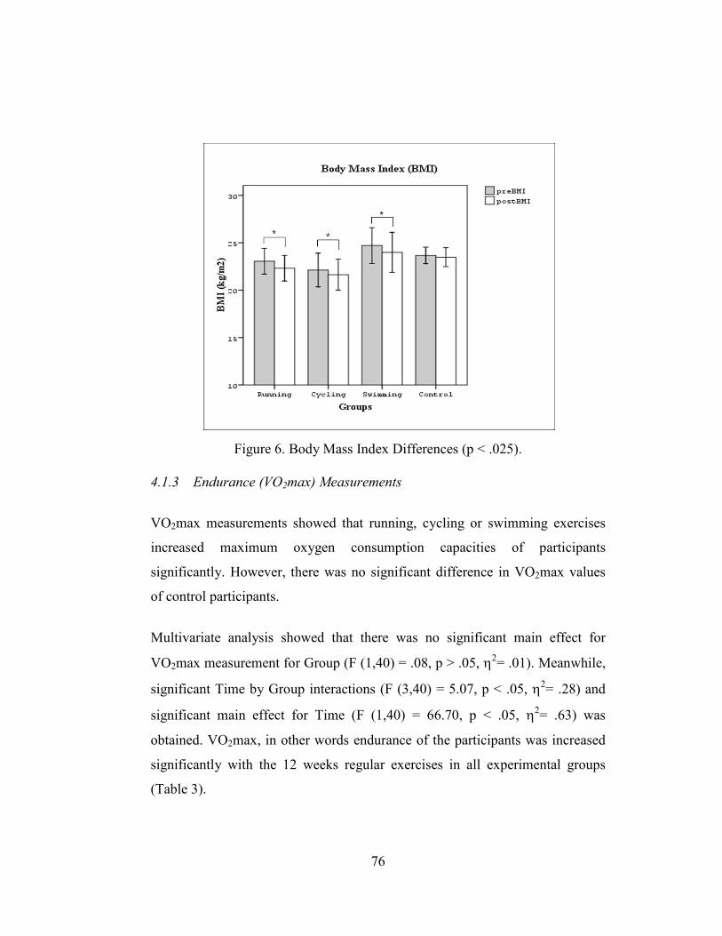
76
Figure 6. Body Mass Index Differences (p < .025).
4.1.3 Endurance (VO2max) Measurements
VO2max measurements showed that running, cycling or swimming exercises
increased maximum oxygen consumption capacities of participants
significantly. However, there was no significant difference in VO2max values
of control participants.
Multivariate analysis showed that there was no significant main effect for
VO2max measurement for Group (F (1,40) = .08, p > .05, η2= .01). Meanwhile,
significant Time by Group interactions (F (3,40) = 5.07, p < .05, η2= .28) and
significant main effect for Time (F (1,40) = 66.70, p < .05, η2= .63) was
obtained. VO2max, in other words endurance of the participants was increased
significantly with the 12 weeks regular exercises in all experimental groups
(Table 3).

77
Table 3. Simple Effect Analysis of Time for VO2max Groups DF F p
Running 1 31.0 .00
Cycling 1 11.0 .00
Swimming 1 38.4 .00
Control 1 1.6 .22
VO2max results indicated that the mean maximum oxygen consumption was
46.2±3.4 in the running group, 47.1±5.1 in the cycling group, 45.6±3.8 in the
swimming group, and 47.3±3.6 in the control group at pre-tests. Percent
decreases between pre- and post-test measurements for VO2max were 10.6%,
6.2%, 12.0%, 2.3 %, respectively and became 51.1±5,8, 50.0±4,9, 51.1±4.3,
48.4±3.6. Difference between pre- and post-tests was given at Figure 7.
Figure 7. Maximum Oxygen Consumption Differences (p < .025).
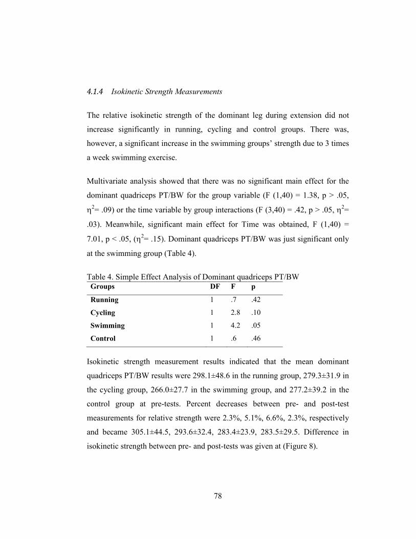
78
4.1.4 Isokinetic Strength Measurements
The relative isokinetic strength of the dominant leg during extension did not
increase significantly in running, cycling and control groups. There was,
however, a significant increase in the swimming groups’ strength due to 3 times
a week swimming exercise.
Multivariate analysis showed that there was no significant main effect for the
dominant quadriceps PT/BW for the group variable (F (1,40) = 1.38, p > .05,
η2= .09) or the time variable by group interactions (F (3,40) = .42, p > .05, η2=
.03). Meanwhile, significant main effect for Time was obtained, F (1,40) =
7.01, p < .05, (η2= .15). Dominant quadriceps PT/BW was just significant only
at the swimming group (Table 4).
Table 4. Simple Effect Analysis of Dominant quadriceps PT/BW Groups DF F p
Running 1 .7 .42
Cycling 1 2.8 .10
Swimming 1 4.2 .05
Control 1 .6 .46
Isokinetic strength measurement results indicated that the mean dominant
quadriceps PT/BW results were 298.1±48.6 in the running group, 279.3±31.9 in
the cycling group, 266.0±27.7 in the swimming group, and 277.2±39.2 in the
control group at pre-tests. Percent decreases between pre- and post-test
measurements for relative strength were 2.3%, 5.1%, 6.6%, 2.3%, respectively
and became 305.1±44.5, 293.6±32.4, 283.4±23.9, 283.5±29.5. Difference in
isokinetic strength between pre- and post-tests was given at (Figure 8).

79
Figure 8. Dominant Legs, Quadriceps Strength Differences (p < .025).
4.2 Serum COMP Concentration Measurements
During pre- and post-test measurements, serum samples were taken at the
recovery, the fatigue and the regeneration phases. Descriptive statistics of
Serum COMP concentrations are presented in Tables 5 and 6.
Table 5. Pre-test Serum COMP concentrations (U/l) Recovery Fatigue Regeneration (M±SD) (M±SD) (M±SD) Running (N=11) 10.07±1.8 12.93±2.9 10.71±2.2
Cycling (N=11) 9.74±2.3 12.39±1.9 11.00±1.9
Swimming (N=11) 9.68±1.6 12.42±1.8 10.99±1.4
Control (N=11) 9.49±1.4 12.52±1.1 10.54±1.4

80
Table 6. Post-test Serum COMP concentrations (U/l) Recovery Fatigue Regeneration (M±SD) (M±SD) (M±SD) Running (N=11) 9.76±1.6 11.34±2.6 10.32±1.8
Cycling (N=11) 9.44±2.4 11.86±1.7 10.32±2.1
Swimming (N=11) 9.35±1.5 12.23±1.3 10.34±1.5
Control (N=11) 9.66±1.3 12.41±1.1 10.52±1.3
4.2.1 Serum COMP level differences among groups at pre- and post-test
measurements
During pre-test measurements, serum COMP concentrations at the recovery
state were not significantly different among groups. Similarly, there were no
significant differences among groups after the 30 minute walking exercise. For
the regeneration period, the same consistency was also valid. Main effect for
group variable at the recovery state (F (3,40) = .19, p = .90), at the fatigue state
(F (3,40) = .17, p = .92) or at the regeneration state (F (3,40) = .18, p = .91) is
presented at Figure 9.

81
Figure 9. Serum COMP concentrations at different states during pre-test
measurements.
Similar findings were also observed during post-test measurements. Serum
COMP concentrations at the recovery state, after 30 minutes walking exercise
and at regeneration were not significantly different among groups. Serum
COMP concentration results showed no significant main effect for group
variable during post-test measurements at the recovery state (F (3,40) = .13, p =
.99), at the fatigue state (F (3,40) = .79, p = .51) or at the regeneration state (F
(3,40) = .04, p = .99) (Figure 10).

82
Figure 10. Serum COMP concentrations at different states during post-test
measurements.
4.2.2 Serum COMP level changes due to acute exercise at pre- and post-test
measurements (Deformational Behavior of Cartilage)
Multivariate tests indicate a significant fatigue or resting effect on serum
COMP concentration in all experimental and control groups. Therefore, pair
wise comparisons were conducted in order to assess the mean differences.
Differences are tabulated in Figures 11 and 12.
Results indicated that 30 minutes of walking exercise significantly increased
the serum COMP concentration in the running group at pre-tests. Also, after

83
fatigue, 30 minutes regeneration decreased serum COMP concentration
significantly in this group. However, in the post-tests measurements differences
after fatigue or regeneration were not significant in the running group. Pair wise
comparisons are presented in Table 7.
Table 7. Mean differences of serum COMP concentration between pairs of different states in running group.
Running Mean Diff. Std. Err. Sig.a
Pre
-Tes
t Recovery-Fatigue -2.87* .85 .02
Recovery-Regeneration -.64 .36 .32
Fatigue- Regeneration 2.23* .02 .02
Pos
t-T
est Recovery-Fatigue -1.57 .67 .12
Recovery-Regeneration -.55 .29 .27
Fatigue- Regeneration 1.02 .42 .11
Based on estimated marginal means *. The mean difference is significant at the .05 level a. Adjustment for multiple comparisons: Bonferroni
In the cycling group, results indicated that 30 minutes of walking exercise
significantly increased the serum COMP concentration at pre-tests. Also, after
fatigue, 30 minutes regeneration decreased serum COMP concentration
significantly in the cycling group. In this group, 12 weeks low-impact cycling
exercise did not change the adaptation capacity of the cartilage. Similar to pre-
test analysis, serum COMP concentration at fatigue and regeneration increased
and decreased respectively during post-tests measurements. Pair wise
comparisons are presented in Table 8.
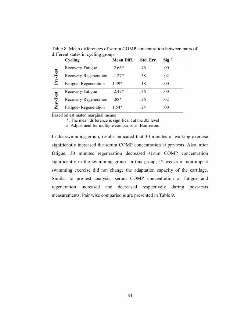
84
Table 8. Mean differences of serum COMP concentration between pairs of different states in cycling group.
Cycling Mean Diff. Std. Err. Sig. a P
re-T
est Recovery-Fatigue -2.66* .46 .00
Recovery-Regeneration -1.27* .38 .02
Fatigue- Regeneration 1.39* .18 .00
Pos
t-T
est Recovery-Fatigue -2.42* .36 .00
Recovery-Regeneration -.88* .26 .02
Fatigue- Regeneration 1.54* .24 .00
Based on estimated marginal means *. The mean difference is significant at the .05 level a. Adjustment for multiple comparisons: Bonferroni
In the swimming group, results indicated that 30 minutes of walking exercise
significantly increased the serum COMP concentration at pre-tests. Also, after
fatigue, 30 minutes regeneration decreased serum COMP concentration
significantly in the swimming group. In this group, 12 weeks of non-impact
swimming exercise did not change the adaptation capacity of the cartilage.
Similar to pre-test analysis, serum COMP concentration at fatigue and
regeneration increased and decreased respectively during post-tests
measurements. Pair wise comparisons are presented in Table 9.

85
Table 9. Mean differences of serum COMP concentration between pairs of different states in swimming group
Swimming Mean Diff. Std. Err. Sig. a P
re-T
est Recovery-Fatigue -2.74* .48 .00
Recovery-Regeneration -1.31* .31 .01
Fatigue- Regeneration 1.43* .38 .01
Pos
t-T
est Recovery-Fatigue -2.88* .39 .00
Recovery-Regeneration -.99* .33 .04
Fatigue- Regeneration 1.90* .28 .00
Based on estimated marginal means *. The mean difference is significant at the .05 level a. Adjustment for multiple comparisons: Bonferroni
Results indicated that 30 minutes of walking exercise significantly increased
the serum COMP concentration of the control participants at pre-tests
measurements. Also, after fatigue, 30 minutes regeneration decreased serum
COMP concentration significantly. Response of the articular cartilage to 30
minutes walking exercise stayed constant during the 12 weeks period. Pair wise
comparisons are presented in Table 10.
Table 10. Mean differences of serum COMP concentration between pairs of different states in control group
Control Mean Diff. Std. Err. Sig. a
Pre
-Tes
t Recovery-Fatigue -3.03* .42 .00
Recovery-Regeneration -1.05* .28 .01
Fatigue- Regeneration 1.98* 36 .00
Pos
t-T
est Recovery-Fatigue -2.74* .32 .00
Recovery-Regeneration -.86* .23 .01
Fatigue- Regeneration 1.89* .30 .00
Based on estimated marginal means *. The mean difference is significant at the .05 level a. Adjustment for multiple comparisons: Bonferroni

86
Figure 11. Pair-wise comparisons for different states for each group at pre-test
measurements

87
Figure 12. Pair-wise comparisons for different states for each group at post-test
measurements
4.2.3 Serum COMP level changes within groups at pre- and post-test
measurements
Changes of serum COMP concentrations between pre- and post-tests
measurements were also evaluated. According to multivariate tests, serum
COMP concentration during recovery did not decrease significantly in any of
the groups (Figure 13). Statistical computations showed that in the recovery
state, serum COMP concentration showed no significant main effect for Group
(F (1,40) = .13, p > .05, η2= .01), Time by Group interactions (F (3,40) = 57, p
> .05, η2= .04) and Time variable (F (1,40) = 1.39, p > .05, η2= .03) (Table 11).
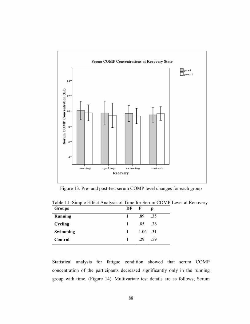
88
Figure 13. Pre- and post-test serum COMP level changes for each group
Table 11. Simple Effect Analysis of Time for Serum COMP Level at Recovery Groups DF F p
Running 1 .89 .35
Cycling 1 .85 .36
Swimming 1 1.06 .31
Control 1 .29 .59
Statistical analysis for fatigue condition showed that serum COMP
concentration of the participants decreased significantly only in the running
group with time. (Figure 14). Multivariate test details are as follows; Serum

89
COMP concentration measurement results in fatigue states showed no
significant main effect for Group (F (1,40) = .09, p > .05, η2= .01). Meanwhile,
significant Time by Group interactions (F (3,40) = 4.97, p < .05, η2= .27) and
significant main effect for Time (F (1,40) = 15.74, p < .05, η2= .28) was
obtained (Table 12).
Figure 14. Pre- and post-test serum COMP level changes for each group.

90
Table 12. Simple Effect Analysis of Time for Serum COMP Level at Fatigue Groups DF F p
Running 1 27.2 .00
Cycling 1 3.0 .09
Swimming 1 .4 .54
Control 1 .1 .71
During the regeneration states of pre- and post-tests, serum COMP
concentrations of the participants decreased significantly in the cycling and the
swimming groups (Figure 15). Results in the regeneration states showed no
significant main effect for Group (F (1,40) = .03, p > .05, η2= .00), or time by
group interactions (F (3,40) = .96, p > .05, η2= .07). Meanwhile, significant
main effect for Time was obtained, F (1,40) = 7.57, p < .05, (η2= .16) (Table
13).

91
Figure 15. Pre- and post-test serum COMP level changes for each group.
Table 13. Simple Effect Analysis of Time for Serum COMP Level at Regeneration Groups DF F p
Running 1 1.5 .23
Cycling 1 4.6 .04
Swimming 1 4.3 .05
Control 1 .0 .96

92
4.3 Serum CTX-I Concentration Measurements
Pre- and post-test serum samples were analyzed for the assessment of serum
CTX-I concentration changes at recovery, fatigue and regeneration phases.
Descriptive statistics of serum CTX-I concentrations are presented in Tables 14
and 15.
Table 14. Pre-test Serum CTX-I concentrations (ng/mL) Recovery Fatigue Regeneration (M±SD) (M±SD) (M±SD) Running (N=11) .45±.07 .49±.09 .45±.08
Cycling (N=11) .48±.10 .51±.12 .49±.10
Swimming (N=11) .43±.12 .46±.12 .41±.09
Control (N=11) .44±.07 .45±.06 .42±.09
Table 15. Post-test Serum CTX-I concentrations (ng/mL) Recovery Fatigue Regeneration (M±SD) (M±SD) (M±SD) Running (N=11) .46±.10 .48±.11 .46±.15
Cycling (N=11) .50±.14 .51±.21 .47±.13
Swimming (N=11) .43±.09 .46±.12 .40±.12
Control (N=11) .42±.07 .48±.07 .44±.07
4.3.1 Serum CTX-I level differences among groups at pre- and post-test
measurements
During pre-test measurements, serum CTX-I concentration analysis showed no
significant differences among groups at the recovery state. Also, there were no
significant differences among groups after 30 minute walking exercise. The
same consistency was also valid at the regeneration measurements. Main effect

93
for group variable at recovery state (F (3,40) = .67, Sig. of F = .58), at fatigue
state (F (3,40) = .59, Sig. of F = .51) or at regeneration state (F (3,40) = 1.89,
Sig. of F = .15) are presented in Figure 16.
Figure 16. Serum CTX-I concentrations at different states during pre-test
measurements
Serum CTX-I concentrations at the recovery state, 30 minute walking exercise
and after 30 minutes regeneration, were also not significantly different among
groups. Serum CTX-I concentration results showed no significant main effect
for the group variable during post-test measurements at the recovery state (F
(3,40) = 1.24, Sig. of F = .31), at the fatigue state (F (3,40) = .28, Sig. of F =
.84) or at the regeneration state (F (3,40) = .70, Sig. of F = .56) (Figure 17).

94
Figure 17. Serum CTX-I concentrations at different states during post-test
measurements
4.3.2 Serum CTX-I level changes due to acute exercise at pre- and post-test
measurements
Multivariate tests indicate that fatigue or resting effect was not significant on
serum CTX-I concentration in all experimental and control groups.
Nevertheless, pair wise comparisons were conducted in order to assess the level
of statistical differences. Results indicated that a significant difference among
the different states of measurements was not observed in any of the groups.
Results indicated that 30 minutes of walking exercise did not affect the serum
CTX-I concentration in the running group significantly at pre-tests. Also, after
30 minutes regeneration, serum CTX-I concentration did not change
significantly in this group. Similar results were observed at post tests.

95
Differences after fatigue or regeneration were not significant in the running
group. Pair wise comparisons are presented in Table 16.
Table 16. Mean differences of serum CTX-I concentration between pairs of different states in running group.
Running Mean Diff.
Std. Err. Sig.a
Pre
-Tes
t Recovery-Fatigue -.04 .02 .37
Recovery-Regeneration .00 .01 1.00
Fatigue- Regeneration .04 .02 .07
Pos
t-T
est Recovery-Fatigue -.03 .02 .52
Recovery-Regeneration -.00 .03 1.00
Fatigue- Regeneration .02 .03 1.00
Based on estimated marginal means a. Adjustment for multiple comparisons: Bonferroni
Also in the cycling group, results indicated that there were no significant
differences among recovery, fatigue or regeneration states in the pre and the
post tests. Pair wise comparisons are presented in Table 17.
Table 17. Mean differences of serum CTX-I concentration between pairs of different states in cycling group.
Cycling Mean Diff.
Std. Err. Sig.
Pre
-Tes
t Recovery-Fatigue -.03 .01 .07
Recovery-Regeneration -.01 .01 1.00
Fatigue- Regeneration .02 .02 .77
Pos
t-T
est Recovery-Fatigue -.02 .03 1.00
Recovery-Regeneration .03 .02 .77
Fatigue- Regeneration .04 .04 .96
Based on estimated marginal means a. Adjustment for multiple comparisons: Bonferroni
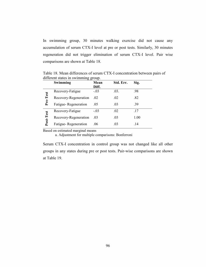
96
In swimming group, 30 minutes walking exercise did not cause any
accumulation of serum CTX-I level at pre or post tests. Similarly, 30 minutes
regeneration did not trigger elimination of serum CTX-I level. Pair wise
comparisons are shown at Table 18.
Table 18. Mean differences of serum CTX-I concentration between pairs of different states in swimming group.
Swimming Mean Diff.
Std. Err. Sig.
Pre
-Tes
t Recovery-Fatigue -.03 .03. .98
Recovery-Regeneration .02 .02 .82
Fatigue- Regeneration .05 .03 .39
Pos
t-T
est Recovery-Fatigue -.03 .02 .17
Recovery-Regeneration .03 .03 1.00
Fatigue- Regeneration .06 .03 .14
Based on estimated marginal means a. Adjustment for multiple comparisons: Bonferroni
Serum CTX-I concentration in control group was not changed like all other
groups in any states during pre or post tests. Pair-wise comparisons are shown
at Table 19.

97
Table 19. Mean differences of serum CTX-I concentration between pairs of different states in control group.
Control Mean Diff.
Std. Err. Sig. P
re-T
est Recovery-Fatigue -.01 .00 .19
Recovery-Regeneration .02 .03 1.00
Fatigue- Regeneration .03 .03 .84
Pos
t-T
est Recovery-Fatigue -.05 .02 .11
Recovery-Regeneration -.02 .01 .57
Fatigue- Regeneration .04 .01 .062
Based on estimated marginal means a. Adjustment for multiple comparisons: Bonferroni
4.3.3 Serum CTX-I level changes within groups at pre- and post-test
measurements
Changes of serum CTX-I concentrations between pre- and post-tests
measurements were also evaluated. According to multivariate tests, serum
CTX-I concentration during recovery did not decrease significantly in any of
the groups (Figure 18). Statistical computations showed that in the recovery
state serum CTX-I concentration showed no significant main effect for Group
(F (1,40) = 1.22, p > .05, η2= .84), Time by Group interactions (F (3,40) = .27,
p > .05, η2= .02) and Time variable (F (1,40) = .03, p > .05, η2= .00) (Table
20).

98
Figure 18. Pre- and post-test serum CTX-I level changes for each group
Table 20. Simple Effect Analysis of Time for Serum CTX-I Level at Recovery Groups DF F p
Running 1 .12 .73
Cycling 1 .38 .54
Swimming 1 .00 .97
Control 1 .34 .56
Statistical analysis for the fatigue condition showed that serum CTX-I
concentration of the participants did not decrease significantly in any of the
groups (Figure 19). Serum CTX-I concentration measurement in the fatigue
state showed no significant main effect for Group (F (1,40) = .63, p > .05, η2=
.05), Time by Group interactions (F (3,40) = .11, p > .05, η2= .01) and Time
variable (F (1,40) = .18, p > .05, η2= .01) (Table 21).

99
Figure 19. Pre- and post-test serum CTX-I level changes for each group
Table 21. Simple Effect Analysis of Time for Serum CTX-I Level at Fatigue Groups DF F p
Running 1 .00 .96
Cycling 1 .01 .91
Swimming 1 .01 .91
Control 1 .47 .50
During regeneration states of pre- and post-tests, serum CTX-I concentrations
of the participants did not change significantly in any of the groups (Figure 20).
Detailed statistics showed that serum CTX-I concentration measurement results
in the regeneration states showed no significant main effect for Group (F (1,40)
= 1.43, p > .05, η2= .10), Time by Group interactions (F (3,40) = .31, p > .05,
η2= .02) and Time variable (F (1,40) = .03, p > .05, η2= .00) (Table 22).

100
Figure 20. Pre- and post-test serum CTX-I level changes for each group
Table 22. Simple Effect Analysis of Time for Serum CTX-I Level at Regeneration Groups DF F p
Running 1 .21 .65
Cycling 1 .33 .57
Swimming 1 .03 .87
Control 1 .41 .53

101
CHAPTER 5
DISCUSSION
The purpose of this study was to compare changes of deformational behavior
and functional adaptation of articular cartilage and bone after 12 weeks
moderate amount of high-impact, impact or non-impact physical exercise
program by measuring biochemical markers of cartilage and bone degradation.
This chapter discusses effects of different exercise interventions on some
biomotor abilities and short term (deformational behavior) or long term
(functional adaptation) changes of knee articular cartilage and bone. After an
extensive literature review, two main hypotheses and six sub-hypotheses were
constructed. In the light of our findings, these hypotheses are discussed in this
chapter.
Participants of this study were 44 healthy male university students. Inclusion
criteria were no previous history of lower extremity surgery and refrainment of
regular sports activity.
Participants trained for 12 weeks according to the ACSM’s guidelines for
endurance development. All three activities were continuous cyclic exercises
but impact levels were changing from low to high.

102
The results of the present study showed that all endurance exercises
significantly changed body composition and endurance capacities of
participants but changes in isokinetic strength capacity increased significantly
only in the swimming group. Biochemical marker of cartilage degeneration
(COMP) measurements showed that 30 minutes walking exercise did not
significantly increase the level of cartilage degeneration after 12 weeks of high
impact running exercise. However, level of CTX-I was not affected by 30
minutes of exercise neither in pre-post tests, nor in any groups.
5.1 Physical and Biomotor abilities of participants
General implication of all kinds of endurance exercises is decreasing body mass
index (BMI) and increasing maximum oxygen consumption (VO2max). In the
present study, groups performed three different endurance type activities for 12-
weeks. Forms of activities were different but most importantly quantitative
elements of the exercises were similar. Volume of exercises were 40 minutes
per session, densities were three days a week for 12 weeks and intensity of
exercises were 60-70 % of heart rate reserves. This amount of exercise caused a
significant decrease in BMI and a significant increase in VO2max in all
experimental groups. However, isokinetic strength significant increased only in
the swimming group.
Changes in BMI and VO2max can partially be explained by functional
adaptation (Darwin., 1872). Hippocrates, also claimed that all parts of the body
that have a function, if used in moderation and exercised in labour in which
each is well developed and age more slowly (Hippocrates, as cited in
Paffenbarger, 2001). Therefore improvement in maximal oxygen consumption
and decrease in fatness were expectable results. In a similar study design,
Scharhag-Rosenberger et al. (2009) observed a significant decrease in the body

103
weights of healthy untrained subjects after six months of moderate intensity
jogging/walking exercises. Additionally, significant increase in VO2max
occurred at third months of intervention and continued along the 12 months
intervention. However, Scharhag-Rosenberger and collaborates did not use a
control group in their study. In our study, we applied three different exercise
interventions in three groups and additionally a control group participated in the
tests but did not attend in any physical exercise during the 12 weeks of
assessment. VO2max improved in all groups but this change was significant
only in the exercise groups. Changes of VO2max among experimental groups
were not significantly different in our study. Lieber, Lieber, & Adams (1989)
investigated the effects of run-training and swim-training at similar absolute
intensities on treadmill VO2max. In their randomized control trial, they
observed that runners, swimmers, and controls experienced a significant
increase in treadmill VO2max over the 11 1/2-week study period. However,
28% and 25% increases in VO2max observed for the runners and swimmers,
respectively; these were significantly greater than the 5% increase observed for
the controls.
In this study different types of exercises were performed for 12 weeks.
Common trait of these exercises was cyclic-endurance type of exercises.
Therefore, improvement of isokinetic strength was not a certain prospect for
our study. However, isokinetic strength increased significantly in the swimming
group. This change may be explained by the nature of aquatic resistance.
Isokinetic contraction can be produced by special designed machines but
according to Fox, Bowers, & Foss (1988) arm stroke during freestyle
swimming is a good example of natural isokinetic contraction. Moreover, some
researchers attempted to establish reliable swimming simulators which were
isokinetic dry-land swim benches or they used isokinetic swim benches as

104
reliable representative of water resistance (Swaine, 1997; Konstantaki,
Trowbridge, & Swaine 1998). Our findings are also parallel with the findings of
Gehlsen, Grigsby, & Winant (1984). Although swimming is an endurance type
of sport, Gehlsen and collaborates observed that aquatic fitness programs
increased isokinetic peak torque values of knee extensor and flexor muscles.
5.2 Serum COMP levels
Differences between serum COMP levels of different groups at each state were
not statistically significant in pre-test measurements. Similarly, there were no
significant differences in serum COMP concentrations between the groups at
each state during post-test. Thirty minutes moderate walking exercise increased
serum COMP concentration in participants at pre-test measurements. After
thirty minutes walking, participants rested on o chair for thirty minutes. This
regeneration period caused a significant decrease of serum COMP
concentrations.
This kind of serum COMP responses to acute exercise was also observed by
Mundermann and her collaborates (2005). In their study, serum COMP levels
of ten adults increased significantly after 30-min outdoor walking at their self-
selected normal speed. Mundermann’s study was the first attempt to link serum
COMP concentration to physiological cyclic loading in vivo. Similar findings
were observed by Andersson et al. 2006. They observed that serum COMP
levels increased during exercise in individuals with knee osteoarthritis, whereas
levels decreased during rest.
Kersting et al. (2005) also investigated the relationship between the effect of
running on cartilage volume and serum COMP concentration. Similar to our
findings, resting COMP level was lower than fatigue serum COMP level.

105
However, this elevation was not significant in their study. This contradictory
finding of the study might be related to the time of blood sampling. In their
study, Kersting and collaborates collected post-exercise blood samples 25 min
after training which was immediately after the post-activity MRI. In spite of
this delayed sampling, general findings of their study were parallel with the
findings of the present study. Multiple regression analysis showed that resting
COMP and COMP changes after 1 h running explained the variance of cartilage
volume changes.
Acute effect of physical exercise on articular cartilage was also evaluated by
MRI studies. Findings of MRI studies were generally parallel to our
biochemical measurements. Significant amount of deformational behavior after
exercise was observed morphologically in these studies. Eckstein and
collaborates (1998) mentioned a deformational behavior of articular cartilage
after repetitive knee bending exercise. After several sets of knee bending
exercises, a statistically significant decrease in cartilage volume was observed.
In a subsequent study, Eckstein et al (1999) observed a deformation of articular
cartilage after 50 knee bending ranged from 2.4 to 8.6 %. More repetition or
repeated sets of exercise with intervals did not cause further deformation.
Eckstein and his colleagues (2000) also compared the effect of dynamic versus
static loading on deformational behavior of articular cartilage. In this study,
researchers observed a significant reduction of articular cartilage volume after
dynamic or static loading. However, the volume changes were significantly
lower after static loading than after dynamic loading. The maximal thickness of
cartilage also decreased significantly due to both types of loading.
Eckstein et al., (2005) evaluated the effect of different types of activity. They
compared effect of knee bends, squatting, normal gait, running and cycling.

106
Finding of this study demonstrated that knee bending as high impact loading
caused highest deformation and normal gait caused lowest deformation on
patellar cartilage deformation. This result mentioned that articular cartilage
deformation is directly dose dependent response. In contrast to Eckstein and
collaborates, in a very recent study, Niehoff et al., (2008) observed that 30 min
running exercise enhanced the serum COMP concentration but slow knee bends
did not cause any significant change.
Articular cartilage contributes to transferring enormous loads as uniformly as
possible from one skeletal segment to the next. According to Kessler et al.,
(2006) whether it manages this task when subjected to the high repetitive
loading cycles occurring during long-distance running and can remain intact is
still the topic of controversy. Therefore, in recent years, possible deleterious
effects of competitive exercises, especially long distance running, have been the
subject of interest (Hofmann,Wortler & Imhoff, 2004; Kim, Lee & Kim, 2007;
Kessler et al., 2008; Kim, Lee & Kim, 2009). Hofmann et al investigated
whether external impact loading in marathon running creates an internal stress
on bone and cartilage that are demonstrable on MRI. MR images did not
demonstrate any deleterious signs after post marathon run. Hofmann suggested
that high impact forces, created by long distance running, are well tolerated and
changes are not detected by MR images, whereas our results showed that there
is a significant degradation of articular cartilage according to elevation of
biochemical markers at serum after lower extremity exercise. In another long-
distance running study Kessler et al., (2008) refute the assertion of Hofmann.
They found a significant reduction after running even moderate distances and
also 60 minutes recovery was sufficient for significant regeneration of cartilage
volume.

107
MRI studies shows that competitive long distance running decreases the
volume and thickness of articular cartilage at knee region morphologically.
From another perspective, Kim, Lee & Kim (2007) evaluated the biomarkers of
muscle and cartilage damage during a 200 km run. They measured the serum
COMP content before, midway and immediately after the race. Serum COMP
level increased 1.3-fold at 100 km and 3-fold at the finish of the 200 km
running race. This result mentioned that ultra-marathon running event may be
detrimental for articular cartilage and may increase the incidence of
degenerative conditions in later life. Kim, Lee & Kim (2009) also compared the
cartilage damage between different long distance running competitions. They
also evaluated serum COMP level changes after recovery periods. Similar
increase was observed after marathon run like ultra-marathon. However, serum
COMP level declined to the pre-race level after 2 days recovery after marathon
race. Researchers observed that serum COMP only returned to the pre-race
level on day 6. This amount of impact exercise may have detrimental effect on
cartilage and the time for recovery may extend for many days.
It seems that the physical activity causes a deformational effect on articular
cartilage. Our finding about deformational behavior of articular cartilage was
parallel with the findings of other studies. The literature presents that extremely
long duration of exercise causes more serious deformation in joint cartilage and
a longer recovery period is needed for regeneration. However, intensity of our
walking test during blood sampling is relatively lower and duration of protocol
was shorter. Our results and literature support the idea that 30 minutes of
exercise is enough to observe a significant deformation in articular cartilage and
30 minutes of resting is an adequate period for recovery. In addition to the
effect of exercise on articular cartilage, literature supports our finding about the
108
high sensitivity of the COMP measurement technique as a consistent biomarker
for cartilage degeneration.
In our study, after pre-test measurements, participants were divided into four
groups randomly. Each group accomplished their groups exercise requirements
for a 12 weeks intervention period. After interventions, post-test measurements
were performed and similar serum COMP responses were observed. During
post-test measurements, thirty minutes walking exercise increased serum
COMP levels of the cycling and the swimming groups significantly. However,
this increase was not significant in the running group. Also, the serum COMP
concentration at fatigue state was significantly lower in the post-test
measurement than that of the pre-test measurement in the running group.
However, pre- and post-test serum COMP concentration difference, after thirty
minutes of walking exercise, were not significant neither in the cycling nor in
the running groups.
Although it is obvious that immobilization has a deterioration effect on
cartilage, there is no consensus on the functional adaptation ability of articular
cartilage for fitness exercise in the literature. It is known that unloading during
long space flight decreases the BMD and increases the bone fracture risks
(Lang, 2006; LeBlanc, Spector, Evans & Sibonga, 2007). Articular cartilage
also has mechanical functions like the bone tissue. Vanwanseele, Lucchinetti &
Stussi (2002) mentioned a 9% decrease in articular cartilage thickness after 11
weeks of immobilization. Also Vanwanseele, Eckstein, Knecht, Stussi, &
Spaepen, (2002) evaluated the tinning of knee cartilage. Their data showed that
progressive atrophy of human cartilage occurs in spinal cord-injured patients
because of absence of normal joint loading. Six years ago, Vanwanseele,
Eckstein, Knecht, Spaepen, & Stussi (2003) firstly reported a longitudinal

109
analysis of cartilage atrophy in patients with spinal cord-injured. They reported
that human cartilage atrophies during unloading is higher than that observed in
osteoarthritis patients.
In these spinal cord-injury studies, not only skeletal unloading induced defect
was occurred, but also restricted joint motion was another possible treat for
cartilage defect. In a very recent study, Tomiya et al., (2009) investigated the
effect of long-term skeletal unloading without restriction of joint motion on the
patella. They observed that a full-thickness cartilage defect was revealed in
88.6% of tail suspended rats. Their finding shows that impact or loading is
another important component of healthy cartilage. In our study, subjects were
active and they were subjected to some different amount of impact load on
articular cartilage in addition to daily life. Parallel with the findings of the
Tomiya, we observed a better adaptation in high-impact running group.
In addition to unloading and cartilage relationship, effects of overloading and
sport-exercise on articular cartilage of animals and human with patients, healthy
subjects and athletes were also evaluated.
In preliminary studies, researchers found positive effect of exercise on articular
cartilage on animal studies. Kiviranta, Tammi, Jurvelin, Saamanen, &
Helminen, (1988) measured the local influences of physical exercise on
thickness of canine articular cartilage. Findings of their study demonstrated that
moderate running exercise locally alters the biological properties of young
articular cartilage at regions of bearing the highest loading surplus. Also, in
1993, Lammi and collaborates examined the effects of long-term long-distance
running on the metabolism of articular cartilage of young beagles. Post-test
measurements at their study showed that the articular cartilage of the femoral
head showed a great capacity to adapt to the increased mechanical loading.

110
According to Hunter & Eckstein (2009), exercise has positive salutary benefits
for joint tissue in addition to its other health benefits. Also, therapeutic exercise
is an invaluable component to a comprehensive treatment program for patients
with osteoarthritis (Stitik, Gazzillo, & Foye 2007). Therefore, effects of
exercise in some arthritis patients were also evaluated. Lin, Davey, &
Cochrane, (2004) examined the effectiveness of a community-based water
exercise program on measures of self-reported health and physical function
with knee-hip osteoarthritis. Results showed that older people with knee/hip
OA gained modest improvements in measures of physical function, pain,
general mobility and flexibility after participating in 12 months of community-
based water exercise. In addition to improvements in physical function due to
physical exercise, Ross & Dahlberg (2005) also indicated that adult human
articular cartilage has a potential to adapt to loading change. Moderate exercise
may be a good treatment not only to improve joint symptoms and function, but
also to improve the knee cartilage glycosaminoglycan content, an important
aspect of the biomechanical properties of cartilage, in patients at high risk of
developing OA.
In addition to previous sports related studies, Wijayarante et al., (2008)
investigated the determinants of change in patellar cartilage volume among
healthy middle-aged women. In this study self-reported exercise was assessed
by questionnaire. Researchers observed that annual loss of patella cartilage
volume was 1.6% and age was positively associated with patella cartilage
volume loss. They also found that fortnightly participation in exercise
promoting an increased heart and respiratory rate for at least 20 min also tended
to be associated with a reduced rate of patella cartilage volume loss. Results of
their study revealed that exercise participation tends to be associated with a
reduction in the rate of patella cartilage volume loss. Similarly, Koo &

111
Andriacchi (2007) claimed that individual variations in cartilage thickness can
be associated with both individual variations in joint loading associated with
activities of daily living as well as individual differences in the anatomy of the
contacting surfaces of the joint. They suggested that the dynamics of walking is
an important factor to describe individual differences in cartilage thickness for
normal subjects.
Morphologic characteristics of knee cartilage of elite athletes and inactive
subjects were compared in some studies (Muhlbauer et al., 2000; Eckstein et
al., 2002; Eckstein et al., 2006; Eckstein, Hudelmaier, & Putz 2006; Gratzke et
al., 2007). MRI results of Muhlbauer showed that mean or maximal thickness
of knee cartilage was not significantly different in triathletes when compared
with inactive volunteers. Also, Eckstein et al., (2002) could not find a
significant difference between the knee joint cartilage thickness of triathletes
and inactive volunteers. This finding is unexpected in the view of the functional
adaptation observed in other musculoskeletal tissues and seems like
controversial with our results. However, results of Gratzke et al., (2007) explain
this controversial condition somehow. In their study, weight lifters and sprinters
displayed significantly greater cartilage thickness only in patella than
nonathletic volunteers but findings suggested that cartilage thickness has much
less ability to adapt to mechanical loading than muscle. However, as mentioned
in a review of Eckstein, Hudelmaier, & Putz (2006) whereas ‘more’ muscle
provides more tensile strength, and ‘more’ bone provides higher structural
compressive and bending strength and hence better protection against fractures,
‘more’ cartilage is not known to be associated with improved mechanical
competence of joints. Therefore, functional adaptation of articular cartilage may
not be directly related with the enlargement of the cartilage morphology.

112
Patellar cartilage deformation after physical exercise was compared in some
professional athletes and untrained volunteers by Eckstein et al (2006). In this
study, researchers evaluated both the effect of exercise on deformational
behavior and functional adaptation of articular cartilage. They found a
significant deformation after physical exercise. However, no significant
difference was found between athletes and non-athletic controls. Therefore,
researchers claimed that functional adaptation of human articular cartilage is
limited or not. The results of our study refute this statement. A significant
functional adaptation was observed in high impact running group in our study
as reported in some previous studies. Also finding of Qi & Changlin (2007)
supports our results and explains the weakness of the study of Eckstein and
colleagues. According to Qi & Changlin, long-time and high-intensity
movement training induces cartilage degeneration in knee joint. Within the
intensity extent applied in their study, knee cartilage degeneration caused by
light training or intensive training has no difference in MR imaging, but has a
comparatively obvious difference in biomarkers level. This finding
demonstrates that several biomarkers have a very good practical value in
detection of articular cartilage degeneration in early stages.
Fole et al., (2007) suggested that knee cartilage volume and tibial plateau are
dynamic structures that can respond to physical stimuli. Fole also claimed that
muscle strength and endurance fitness may be protective against cartilage loss.
In their longitudinal study, Racunica et al., (2007) observed that vigorous
physical activity appears to have a beneficial effect on knee articular cartilage
in healthy adults with no history of knee injury or disease. Therefore, they
concluded that a beneficial effect of physical activity for diseases associated
with aging and suggests that exercise that is good for the heart is also good for
the knees.

113
5.3 Serum CTX-I levels
Similar to serum COMP concentrations, differences between serum CTX-I
concentrations of different groups at each state were insignificant in pre-test
measurements. Also, there were no significant differences between the groups
at each state during post-test serum CTX-I concentrations. In contrast to COMP
responses, thirty minutes moderate walking exercise did not increase serum
CTX-I concentration significantly in all sedentary participants at pre-test
measurements. Regeneration period after fatigue caused a slight decrease of
serum CTX-I concentrations in participants but this decrease was insignificant.
Post-test measurements showed that serum CTX-I responses to exercise were
similar to pre-test results. Post-test results showed that slight changes after 12
weeks exercise intervention were not significant in any exercise groups or the
control group.
CTX-I also is a biomarker of collagen degradation but it is specific for bone.
Everts &Buttle (2008) explained that degradation of type I and II collagen
results in release of C-terminal telopeptides. According to Garnero et al.,
(2001), CTX-I levels decreased in patients with knee OA. They suggested that
knee OA appears to be characterized by a systemic decrease of bone turnover.
In 2002, Garnero and his collaborates also observed that high baseline levels of
urinary CTX-I and CTX-II independently predict an increased risk of radiologic
progression over 4 years in patients with early RA, especially those without
radiologic joint damage. Therefore, they concluded that CTX-I and CTX-II
may be useful for identifying individual RA patients at high risk of progression
very early in the disease, before erosions can be detected radiographically. Also
in their review, Raissouni, Gossec, Ayral, & Dougados (2005) mentioned that

114
high levels of CTX-I and CTX-II strongly predict the development of joint
erosions before the occurrence of radiological joint damage.
Measurement of CTX-I changes are also accepted as a useful tool of
prosecution of treatment in some arthritis. Bjarnason & Christiansen (2000)
suggested that early CTX-I measurements predict long-term preservation of
bone mass during hormone replacement therapy. Also, in their study, d'Elia et
al (2004) evaluated the effects of hormone replacement therapy on markers of
bone and cartilage metabolism in rheumatoid arthritis patients. In this study
treatment with hormone replacement therapy resulted in decrease in CTX-I and
COMP at 2 years. Researchers concluded that CTX-I reflected bone turnover
very sensitively and they suggested that biochemical markers of bone and
cartilage turnover may provide a useful tool for assessing novel treatment
modalities in arthritis, concerning both joint protection and prevention of
osteoporosis.
In spite of its high sensitivity in the diagnosis of the arthritis or prosecution of
treatment, sensitivity of bone specific biomarkers in the sport related studies are
controversial. In a preliminary study Bennell et al (1998) evaluated bone
turnover in stress fractured athletes and they emphasized that single and
multiple use of biomarkers are not clinically useful in predicting the likelihood
of stress fractures in athletes. Also Bennell et al., (1997) evaluated bone
turnover in different sports groups in a 12-month longitudinal study. Bennell et
al observed that levels of bone markers were not predictive of subsequent
changes in bone metabolism.
O’Kane, Hutchinson, Atley, & Eyre (2006) compared the sport-related
differences in biomarkers of bone resorption and cartilage degradation in
endurance athletes. As we know, only O’Kane and friends found that athletes

115
in-training in different sports show significant differences in these markers. In
our study, however, we could not find a significant difference between the
levels of CTX-I in different endurance exercise groups. The main probable
reason of this finding is the fact that inactive volunteers took part in our study.
Moreover, this 12-week duration and fitness level exercises were not enough to
stimulate the accumulation of serum CTX-I concentration. Our findings are
parallel with some other researches. Creighton, Morgan, Boardley, & Brolinson
(2001) evaluated markers of bone turnover in female athletes comparing three
impact groups, high impact, medium impact and nonimpact, with sedentary
age-matched controls. In this study no differences in a marker of bone
resorption were noted. Also in a very recent study, Lester et al., (2009) sought
to determine whether aerobic, resistance, or combined aerobic and resistance
exercise programs conducted over eight weeks and compared to a control group
could produce changes in biochemical markers of bone turnover indicative of
bone formation. Similar to our findings, Lester and his colleagues did not find a
significant change in the level of CTX-I. The results of their study demonstrate
that during eight weeks physical training program CTX-I concentrations were
similar in all four groups pre-training and remained stable throughout the
training programs. This stability of CTX-I responses against the physical
exercise is also related with the sensitivity of the marker. According to
Whipplle et al., (2004), markers of bone degradation are disadvantageous
because of the lack of sensitivity and tremendous variability between
individuals. Also, Whipplle reported that there is no definite statistical data to
correlate to a gold standard when used to assess markers response to exercise.

116
CHAPTER 6
SUMMARY, CONCLUSIONS & RECOMMENDATIONS
6.1 Summary
The objective of the present study was to investigate deformational behavior
and functional adaptation of articular cartilage by the changes of serum level of
Cartilage Oligomeric Matrix Protein (COMP) and C-terminal telopeptides of
collagen type I (CTX-I) during a 30-min acute exercise after 12-weeks of
regular high impact, impact and non-impact endurance exercise.
In this study, results showed that 12-weeks endurance exercise elicited
improvements of some important health related parameters in all exercise
groups. BMI of participants decreased and VO2max increased significantly in
all exercise groups.
In the analysis of serum COMP comparisons, multivariate tests indicated a
significant fatigue or resting effect on serum COMP concentration in all
experimental and control groups at pre- and post-tests. According to this
finding post-hoc pair-wise comparisons were applied. These univariate tests
indicated significant differences during post test measurements among rest,
fatigue and regeneration phases in all groups except running.

117
On the other hand, multivariate analysis showed that fatigue or resting could
not elicit a significant change of serum CTX-I concentrations in any
experimental or control groups at the beginning or the end of the intervention
period.
6.2 Conclusions
In this study, experimental groups achieved significant increase in endurance
performance and they reached higher capacities on consumption of oxygen.
Also, BMI of the experimental groups decreased significantly. These
improvements are evidence of beneficial effects of regular exercise for general
health.
Also, we hypothesized that all the body systems adapt their functions according
to environmental stimulus; therefore, we expected a functional adaptation of
weight bearing articular cartilage due to regular sports activities. At the
beginning of the study, 30 minutes moderate walking activity increased serum
cartilage specific OA biomarker concentration but not bone specific OA
biomarker concentration of young sedentary men. 12 weeks regular high,
moderate or low impact physical exercise could not change the behavior of
CTX-I concentration in serum. However, weight-bearing high impact physical
exercise (running) caused a decrease in the effect of moderate activities on
serum COMP accumulation.
This kind of decrease in the accumulation of cartilage deformation marker after
12 weeks of regular high impact loading explains the functional adaptation
ability of articular cartilage to specific environmental requirements.
118
According to the results of this study, regular moderate volume and intensity of
endurance exercise are recommended for general health benefits. In addition to
general health benefits, exercises like running or jogging, which create enough
stimuli for functional adaptation, are recommended for the consolidation of
articular cartilage tissue in healthy young men. Also high impact exercises can
be added into the training programs of low-impact sports athletes for their
future general health.
6.3 Recommendations
Up to date, literature focused on cartilage of animals, cartilage of patients and
differences of athletes’ cartilage from normal population. Our investigation is
one of the first cause-effect studies about the responses of the healthy human
articular cartilage. We also examined the responses of the articular cartilage
biochemically.
In this study, we examined the effect of endurance exercise on knee articular
cartilage of healthy young men. However, further studies about the effect of
exercise on human articular cartilage are necessary.
Besides the articular cartilage of healthy male, deformational behavior and
functional adaptation of females’ articular cartilage is a valuable subject to
examine because other similar tissues like muscle or bone of females have
different responses to exercise than males.
Another important issue is the maturation of participants. Responses of articular
cartilage for exercise during developmental ages should be examined in further
studies. Response of articular cartilage at developmental ages and elderly ages

119
may be different. Therefore, this issue should be evaluated in different research
designs.
Moreover, we strongly encourage examining the effect of this kind of exercise
on different patient groups. We tested adaptation of healthy cartilage. In animal
models, researches showed that intact cartilage is more durable than impaired
articular cartilage. Therefore, we do not have enough evidence to recommend
the impact exercise for patients according to the findings of our study.
Effect of endurance type exercises are evaluated in this study. Effect of speed,
strength or concurrent strength-endurance fitness activities will be significant in
further studies since concurrent fitness programs have become popular for
general health in recent years.
In this study, we used biochemical markers to observe changes in articular
cartilage. Another important recommendation for further researchers is that
findings about structural changes in articular cartilage will be reinforced by
morphological examination like MR imaging.

120
REFERENCES
Aisen, A. M., McCune, W. J., MacGuire, A., Carson, P. L., Silver, T. M., Jafri, S. Z., et al. (1984). Sonographic evaluation of the cartilage of the knee. Radiology, 153(3), 781-784.
American College of Sports Medicine (ACSM), (2001). Guidelines for Exercise Testing and Prescription, Johnson, E.P., Lippincott, Williams and Wilkins. Baltimore, Pp: 137-164.
Andersson, M. L. E., Thorstensson, C. A., Roos, E. M., Petersson, I. F., Heinegård, D., & Saxne, T. (2006). Serum levels of cartilage oligomeric matrix protein (COMP) increase temporarily after physical exercise in patients with knee osteoarthritis. BMC Musculoskeletal Disorders, 7, 98. doi: 10.1186/1471-2474-7-98
Austin, G., & Noble, J. (1994). Swimming for fitness (2nd ed.). London A & C Black.
Backhaus, M., T. Kamradt, D. Sandrock, D. Loreck, J. Fritz, K. J. Wolf, et al. (1999). Arthritis of the finger joints: A comprehensive approach comparing conventional radiography, scintigraphy, ultrasound, and contrast-enhanced magnetic resonance imaging. Arthritis & Rheumatism, 42(6), 1232-1245. doi: 10.1002/1529-0131(199906)42:6<1232::AID-ANR21>3.0.CO;2-3
Baechle, T. R., & Earle, R. W. (2000). Essentials of strength training and conditioning (2nd ed.). Champaign, IL: Human Kinetics.
Baliunas, A. J., Hurwitz, D. E., Ryals, A. B., Karrar, A., Case, J. P., Block, J. A., et al. (2002). Increased knee joint loads during walking are present in subjects with knee osteoarthritis. Osteoarthritis and Cartilage / OARS, Osteoarthritis Research Society, 10(7), 573-579.
Barker, M. K., & Seedhom, B. B. (1997). Articular cartilage deformation under physiological cyclic loading--apparatus and measurement technique. Journal of Biomechanics, 30(4), 377-381.
Bennell, K. L., Malcolm, S. A., Brukner, P. D., Green, R. M., Hopper, J. L., Wark, J. D., et al. (1998). A 12-month prospective study of the relationship between stress fractures and bone turnover in athletes. Calcified Tissue International, 63(1), 80-85.

121
Bennell, K. L., Malcolm, S. A., Khan, K. M., Thomas, S. A., Reid, S. J., Brukner, P. D., et al. (1997). Bone mass and bone turnover in power athletes, endurance athletes, and controls: a 12-month longitudinal study. Bone, 20(5), 477-484.
Biswal, S., Hastie, T., Andriacchi, T. P., Bergman, G. A., Dillingham, M. F., & Lang, P. (2002). Risk factors for progressive cartilage loss in the knee: a longitudinal magnetic resonance imaging study in forty-three patients. Arthritis and Rheumatism, 46(11), 2884-2892. doi: 10.1002/art.10573
Bjarnason, N. H., & Christiansen, C. (2000). Early response in biochemical markers predicts long-term response in bone mass during hormone replacement therapy in early postmenopausal women. Bone, 26(6), 561-569. doi: 10.1016/S8756-3282(00)00272-6
Bompa, T. O (1999). Periodization: Theory and methodology of training (4th ed.) Champaign, IL: Human Kinetics.
Brommer, H., Brama, P. A. J., Laasanen, M. S., Helminen, H. J., van Weeren, P. R., & Jurvelin, J. S. (2005). Functional adaptation of articular cartilage from birth to maturity under the influence of loading: a biomechanical analysis. Equine Veterinary Journal, 37(2), 148-154.
Bruce, R. A., Kusumi, F., & Hosmer, D. (1973). Maximal oxygen intake and nomographic assessment of functional aerobic impairment in cardiovascular disease. American Heart Journal, 85(4), 546-562.
Bucksch, J. (2005). Physical activity of moderate intensity in leisure time and the risk of all cause mortality. British Journal of Sports Medicine, 39(9), 632-638. doi: 10.1136/bjsm.2004.015768
Carmichael, C., & Burke, E. R (1994). Fitness cycling. Champaign, IL: Human Kinetics.
Carter, D. R., & Wong, M. (2003). Modelling cartilage mechanobiology. Philosophical Transactions of the Royal Society of London. Series B, Biological
Sciences, 358(1437), 1461-1471. doi: 10.1098/rstb.2003.1346
Cavill, N., Kahlmeier, S., & Racioppi, F. (2006). Physical activity and health in Europe (s. 47). Copenhagen,World Health Organization (WHO) Regional Office for Europe.

122
Charni, N., Juillet, F., & Garnero, P. (2005). Urinary type II collagen helical peptide (HELIX-II) as a new biochemical marker of cartilage degradation in patients with osteoarthritis and rheumatoid arthritis. Arthritis and Rheumatism, 52(4), 1081-1090. doi: 10.1002/art.20930
Christgau, S., Garnero, P., Fledelius, C., Moniz, C., Ensig, M., Gineyts, E., et al. (2001). Collagen type II C-telopeptide fragments as an index of cartilage degradation. Bone, 29(3), 209-215.
Cohen, Z. A., McCarthy, D. M., Kwak, S. D., Legrand, P., Fogarasi, F., Ciaccio, E. J., et al. (1999). Knee cartilage topography, thickness, and contact areas from MRI: in-vitro calibration and in-vivo measurements. Osteoarthritis and
Cartilage / OARS, Osteoarthritis Research Society, 7(1), 95-109. doi: 10.1053/joca.1998.0165
Creighton, D. L., Morgan, A. L., Boardley, D., & Brolinson, P. G. (2001). Weight-bearing exercise and markers of bone turnover in female athletes. J Appl Physiol, 90(2), 565-570.
Cochrane, T., Davey, R. C., & Matthes Edwards, S. M. (2005). Randomised controlled trial of the cost-effectiveness of water-based therapy for lower limb osteoarthritis. Health Technology Assessment (Winchester, England), 9(31), iii-iv, ix-xi, 1-114.
Crnkic, M., Månsson, B., Larsson, L., Geborek, P., Heinegård, D., & Saxne, T. (2003). Serum cartilage oligomeric matrix protein (COMP) decreases in rheumatoid arthritis patients treated with infliximab or etanercept. Arthritis Research & Therapy, 5(4), R181-185. doi: 10.1186/ar760
Darwin, C. (1872). The Origin of Species, 6th edit. New York: New America Library.
de Assis, M. A., de Mello, M. F., Scorza, F. A., Cadrobbi, M. P., Schooedl, A. F., da Silva, S. G., et al. (2008). Evaluation of physical activity habits in patients with posttraumatic stress disorder. Clinics (São Paulo, Brazil), 63(4), 473-478.
de Jong, Z., Munneke, M., Vilim, V., Zwinderman, A. H., Kroon, H. M., Ronday, H. K., et al.,(2008). Value of serum cartilage oligomeric matrix protein as a prognostic marker of large-joint damage in rheumatoid arthritisdata from the RAPIT study. Rheumatology, 47(6), 868-871. doi: 10.1093 /rheumatology/ken052.

123
Ding, C., Garnero, P., Cicuttini, F., Scott, F., Cooley, H., & Jones, G. (2005). Knee cartilage defects: association with early radiographic osteoarthritis, decreased cartilage volume, increased joint surface area and type II collagen breakdown. Osteoarthritis and Cartilage / OARS, Osteoarthritis Research Society, 13(3), 198-205. doi: 10.1016/j.joca.2004.11.007
Dolezal, B. A., & Potteiger, J. A. (1998). Concurrent resistance and endurance training influence basal metabolic rate in nondieting individuals. Journal of Applied Physiolog, 85(2), 695-700.
Duryea, J., Li, J., Peterfy, C. G., Gordon, C., & Genant, H. K. (2000). Trainable rule-based algorithm for the measurement of joint space width in digital radiographic images of the knee. Medical Physics, 27(3), 580-591.
Duryea, J., Zaim, S., & Genant, H. (2003). New radiographic-based surrogate outcome measures for osteoarthritis of the knee. OSTEOARTHRITIS AND CARTILAGE, 11(2), 102-110. doi: 10.1053/joca.2002.0866
Eckstein, F., Faber, S., Mühlbauer, R., Hohe, J., Englmeier, K., Reiser, M., et al. (2002). Functional adaptation of human joints to mechanical stimuli. Osteoarthritis and Cartilage / OARS, Osteoarthritis Research Society, 10(1), 44-50. doi: 10.1053/joca.2001.0480
Eckstein, F., Gavazzeni, A., Sittek, H., Haubner, M., Lösch, A., Milz, S., et al. (1996). Determination of knee joint cartilage thickness using three-dimensional magnetic resonance chondro-crassometry (3D MR-CCM). Magnetic Resonance
in Medicine: Official Journal of the Society of Magnetic Resonance in Medicine
/ Society of Magnetic Resonance in Medicine, 36(2), 256-265.
Eckstein, F., Hudelmaier, M., & Putz, R. (2006). The effects of exercise on human articular cartilage. Journal of Anatomy, 208(4), 491-512. doi: 10.1111/j.1469-7580.2006.00546.x
Eckstein, F., Lemberger, B., Gratzke, C., Hudelmaier, M., Glaser, C., Englmeier, K., et al. (2005). In vivo cartilage deformation after different types of activity and its dependence on physical training status. Annals of the Rheumatic Diseases, 64(2), 291-295. doi: 10.1136/ard.2004.022400
Eckstein, F., Lemberger, B., Stammberger, T., Englmeier, K. H., & Reiser, M. (2000). Patellar cartilage deformation in vivo after static versus dynamic loading. Journal of Biomechanics, 33(7), 819-825.

124
Eckstein, F., Tieschky, M., Faber, S., Englmeier, K. H., & Reiser, M. (1999). Functional analysis of articular cartilage deformation, recovery, and fluid flow following dynamic exercise in vivo. Anatomy and Embryology, 200(4), 419-424.
Eckstein, F., Tieschky, M., et al. (1998). Effect of physical exercise on cartilage volume and thickness in vivo: MR imaging study. Radiology, 207(1), 243-248.
Eckstein, F., Westhoff, J., et al. (1998). In vivo reproducibility of three-dimensional cartilage volume and thickness measurements with MR imaging. AJR. American Journal of Roentgenology, 170(3), 593-597.
Eckstein, F., & Glaser, C. (2004). Measuring cartilage morphology with quantitative magnetic resonance imaging. Seminars in Musculoskeletal
Radiology, 8(4), 329-353. doi: 10.1055/s-2004-861579
Everts, V., & Buttle, D. J. (2008). Methods in studying ECM degradation. Methods (San Diego, Calif.), 45(1), 86-92. doi: 10.1016/j.ymeth.2008.01.009
Faber, S. C., Eckstein, F., Lukasz, S., Mühlbauer, R., Hohe, J., Englmeier, K. H., et al. (2001). Gender differences in knee joint cartilage thickness, volume and articular surface areas: assessment with quantitative three-dimensional MR imaging. Skeletal Radiology, 30(3), 144-150.
Foley, S., Ding, C., Cicuttini, F., & Jones, G. (2007). Physical activity and knee structural change: a longitudinal study using MRI. Medicine and Science in
Sports and Exercise, 39(3), 426-434. doi: 10.1249/mss.0b013e31802d97c6
Forsblad d'Elia, H., Christgau, S., Mattsson, L., Saxne, T., Ohlsson, C., Nordborg, E., et al. (2004). Hormone replacement therapy, calcium and vitamin D3 versus calcium and vitamin D3 alone decreases markers of cartilage and bone metabolism in rheumatoid arthritis: a randomized controlled trial [ISRCTN46523456]. Arthritis Research & Therapy, 6(5), R457-468. doi: 10.1186/ar1215
Forslind, K., Eberhardt, K., Jonsson, A., & Saxne, T. (1992). Increased serum concentrations of cartilage oligomeric matrix protein. A prognostic marker in early rheumatoid arthritis. British Journal of Rheumatology, 31(9), 593-598.
Fox, E. L., Bowers, R. W., & Foss, M. L. (1988). Physiological Basis of Physical Education and Athletics (4th ed.).Dubuque, Brown & Benchmark.

125
Fredriksen, P. M., Ingjer, F., Nystad, W., & Thaulow, E. (1998). Aerobic endurance testing of children and adolescents--a comparison of two treadmill-protocols. Scandinavian Journal of Medicine & Science in Sports, 8(4), 203-207.
Fukuda, Y., Takai, S., Yoshino, N., Murase, K., Tsutsumi, S., Ikeuchi, K., et al. (2000). Impact load transmission of the knee joint-influence of leg alignment and the role of meniscus and articular cartilage. Clinical Biomechanics (Bristol, Avon), 15(7), 516-521.
Garnero, P., Conrozier, T., Christgau, S., Mathieu, P., Delmas, P. D., & Vignon, E. (2003). Urinary type II collagen C-telopeptide levels are increased in patients with rapidly destructive hip osteoarthritis. Annals of the Rheumatic Diseases, 62(10), 939-943.
Garnero, P., Piperno, M., Gineyts, E., Christgau, S., Delmas, P. D., & Vignon, E. (2001). Cross sectional evaluation of biochemical markers of bone, cartilage, and synovial tissue metabolism in patients with knee osteoarthritis: relations with disease activity and joint damage. Annals of the Rheumatic Diseases, 60(6), 619-626.
Garnero, P., Rousseau, J. C., & Delmas, P. D. (2000). Molecular basis and clinical use of biochemical markers of bone, cartilage, and synovium in joint diseases. Arthritis and Rheumatism, 43(5), 953-968. doi: 10.1002/1529-0131(200005)43:5<953::AID-ANR1>3.0.CO;2-Q
Garnero, P., Landewé, R., Boers, M., Verhoeven, A., Van Der Linden, S., Christgau, S., et al. (2002). Association of baseline levels of markers of bone and cartilage degradation with long-term progression of joint damage in patients with early rheumatoid arthritis: the COBRA study. Arthritis and Rheumatism, 46(11), 2847-2856. doi: 10.1002/art.10616
Garnero, P., Sornay-Rendu, E., Arlot, M., Christiansen, C., & Delmas, P. D. (2004a). Association between spine disc degeneration and type II collagen degradation in postmenopausal women: the OFELY study. Arthritis and Rheumatism, 50(10), 3137-3144. doi: 10.1002/art.20493
Gehlsen, G. M., Grigsby, S. A., & Winant, D. M. (1984). Effects of an aquatic fitness program on the muscular strength and endurance of patients with multiple sclerosis. Physical Therapy, 64(5), 653-657.

126
Giannoni, P., Siegrist, M., Hunziker, E. B., & Wong, M. (2003). The mechanosensitivity of cartilage oligomeric matrix protein (COMP). Biorheology, 40(1-3), 101-109.
Gordon, N. F. (1993). Arthritis: your complete exercise guide . Champaign, IL : Human Kinetics.
Gök, H., Ergin, S., & Yavuzer, G. (2002). Kinetic and kinematic characteristics of gait in patients with medial knee arthrosis. Acta Orthopaedica Scandinavica, 73(6), 647-652. doi: 10.1080/000164702321039606
Gratzke, C., Hudelmaier, M., Hitzl, W., Glaser, C., & Eckstein, F. (2007). Knee cartilage morphologic characteristics and muscle status of professional weight lifters and sprinters: a magnetic resonance imaging study. The American Journal of Sports Medicine, 35(8), 1346-1353. doi: 10.1177 /0363546507299746
Hall, B.K. (2005). Bones and cartilage: developmental and evolutionary skeletal biology. San Diego, Calif: Elsevier Academic Press.
Hedbom, E., Antonsson, P., Hjerpe, A., Aeschlimann, D., Paulsson, M., Rosa-Pimentel, E., et al. (1992). Cartilage matrix proteins. An acidic oligomeric protein (COMP) detected only in cartilage. The Journal of Biological
Chemistry, 267(9), 6132-6136.
Henderson, J. H., de la Fuente, L., Romero, D., Colnot, C. I., Huang, S., Carter, D. R., et al. (2007). Rapid growth of cartilage rudiments may generate perichondrial structures by mechanical induction. Biomechanics and Modeling
in Mechanobiology, 6(1-2), 127-137. doi: 10.1007/s10237-006-0038-x
Hohe, J., Ateshian, G., Reiser, M., Englmeier, K., & Eckstein, F. (2002). Surface size, curvature analysis, and assessment of knee joint incongruity with MRI in vivo. Magnetic Resonance in Medicine: Official Journal of the Society of
Magnetic Resonance in Medicine / Society of Magnetic Resonance in Medicine, 47(3), 554-561.
Hohmann, E., Wörtler, K., & Imhoff, A. B. (2004). MR imaging of the hip and knee before and after marathon running. The American Journal of Sports Medicine, 32(1), 55-59.
127
Housh, T.J., Housh, D.J. & DeVries, H.A. (2003). Applied exercise and sport physiology. Scottsdale, Arizona: Holcomb Hathaway, Publishers, Inc.
Hoshino, A., & Wallace, W. A. (1987). Impact-absorbing properties of the human knee. The Journal of Bone and Joint Surgery. British Volume, 69(5), 807-811.
Hunter, D. J., March, L., & Sambrook, P. N. (2003). The association of cartilage volume with knee pain. Osteoarthritis and Cartilage / OARS, Osteoarthritis Research Society, 11(10), 725-729.
Hunter, D., Li, J., LaValley, M., Bauer, D., Nevitt, M., DeGroot, J., et al. (2007). Cartilage markers and their association with cartilage loss on magnetic resonance imaging in knee osteoarthritis: the Boston Osteoarthritis Knee Study. Arthritis Research & Therapy, 9(5), R108. doi: 10.1186/ar2314
Hunter, D. J., & Eckstein, F. (2009). Exercise and osteoarthritis. Journal of Anatomy, 214(2), 197-207. doi: 10.1111/j.1469-7580.2008.01013.x
Inoue, M., Yamamoto, S., Kurahashi, N., Iwasaki, M., Sasazuki, S., & Tsugane, S. (2008). Daily total physical activity level and total cancer risk in men and women: results from a large-scale population-based cohort study in Japan. American Journal of Epidemiology, 168(4), 391-403. doi: 10.1093/aje/kwn146.
Ito, K., Kimura, Y., Tajika, A., Fuchioka, S., Iwasaka, T., Nishiyama, T. (2007). Ultrasonographic changes of the knee joint cartilage associated with physical characterization in middle aged women: 6-month observational survey. J. Phys.Ther. Sci.,19, 277-282.
Jordan, J. M., Luta, G., Stabler, T., Renner, J. B., Dragomir, A. D., Vilim, V., et al. (2003). Ethnic and sex differences in serum levels of cartilage oligomeric matrix protein: the Johnston County Osteoarthritis Project. Arthritis and Rheumatism, 48(3), 675-681. doi: 10.1002/art.10822
Karlsson, M. K., Nordqvist, A., & Karlsson, C. (2008). Sustainability of exercise-induced increases in bone density and skeletal structure. Food & Nutrition
Research, 52. doi: 10.3402/fnr.v52i0.1872
Katz, J., & Bruning, N. (1993). Swimming for total fitness: a progressive aerobic program (2nd ed.). New York: Doubleday.

128
Kersting, U. G., Stubendorff, J. J., Schmidt, M. C., & Brüggemann, G. (2005). Changes in knee cartilage volume and serum COMP concentration after running exercise. Osteoarthritis and Cartilage / OARS, Osteoarthritis Research Society, 13(10), 925-934. doi: 10.1016/j.joca.2005.06.005
Kessler, M. A., Glaser, C., Tittel, S., Reiser, M., & Imhoff, A. B. (2006). Volume changes in the menisci and articular cartilage of runners: an in vivo investigation based on 3-D magnetic resonance imaging. The American Journal of Sports Medicine, 34(5), 832-836. doi: 10.1177/0363546505282622
Kessler, M. A., Glaser, C., Tittel, S., Reiser, M., & Imhoff, A. B. (2008). Recovery of the menisci and articular cartilage of runners after cessation of exercise: additional aspects of in vivo investigation based on 3-dimensional magnetic resonance imaging. The American Journal of Sports Medicine, 36(5), 966-970. doi: 10.1177/0363546507313093
Kim, H. J., Lee, Y. H., & Kim, C. K. (2007). Biomarkers of muscle and cartilage damage and inflammation during a 200 km run. European Journal of Applied Physiology, 99(4), 443-447. doi: 10.1007/s00421-006-0362-y
Kim, H. J., Lee, Y. H., & Kim, C. K. (2009). Changes in serum cartilage oligomeric matrix protein (COMP), plasma CPK and plasma hs-CRP in relation to running distance in a marathon (42.195 km) and an ultra-marathon (200 km) race. European Journal of Applied Physiology, 105(5), 765-770. doi: 10.1007 /s00421-008-0961-x
Kiviranta, I., Tammi, M., Jurvelin, J., Säämänen, A. M., & Helminen, H. J. (1988). Moderate running exercise augments glycosaminoglycans and thickness of articular cartilage in the knee joint of young beagle dogs. Journal of Orthopaedic Research: Official Publication of the Orthopaedic Research
Society, 6(2), 188-195. doi: 10.1002/jor.1100060205
Konstantaki, M., Trowbridge, E. A., & Swaine, I. L. (1998). The relationship between blood lactate and heart rate responses to swim bench exercise and women's competitive water polo. Journal of Sports Sciences, 16(3), 251-256.
Koo, S., & Andriacchi, T. P. (2007). A comparison of the influence of global functional loads vs. local contact anatomy on articular cartilage thickness at the knee. Journal of Biomechanics, 40(13), 2961-2966. doi: 10.1016/j.jbiomech. 2007.02.005

129
Kuettner, K. E., Goldberg, V. M., & Surgeons, A. A. O. O. (1995). Osteoarthritic disorders: workshop, Monterey, California, April 1994. Rosemont, IL: The Academy.
Kühne, S. A., Neidhart, M., Everson, M. P., Häntzschel, H., Fine, P. R., Gay, S., et al. (1998). Persistent high serum levels of cartilage oligomeric matrix protein in a subgroup of patients with traumatic knee injury. Rheumatology
International, 18(1), 21-25.
Lammi, M. J., Häkkinen, T. P., Parkkinen, J. J., Hyttinen, M. M., Jortikka, M., Helminen, H. J., et al. (1993). Adaptation of canine femoral head articular cartilage to long distance running exercise in young beagles. Annals of the Rheumatic Diseases, 52(5), 369-377.
Lang, P., Noorbakhsh, F., & Yoshioka, H. (2005). MR imaging of articular cartilage: current state and recent developments. Radiologic Clinics of North America, 43(4), 629-639, vii. doi: 10.1016/j.rcl.2005.02.009
Lang, T. F. (2006). What do we know about fracture risk in long-duration spaceflight? Journal of Musculoskeletal & Neuronal Interactions, 6(4), 319-321.
Larsson, E., Erlandsson Harris, H., Lorentzen, J. C., Larsson, A., Månsson, B., Klareskog, L., et al. (2002). Serum concentrations of cartilage oligomeric matrix protein, fibrinogen and hyaluronan distinguish inflammation and cartilage destruction in experimental arthritis in rats. Rheumatology (Oxford, England), 41(9), 996-1000.
LeBlanc, A. D., Spector, E. R., Evans, H. J., & Sibonga, J. D. (2007). Skeletal responses to space flight and the bed rest analog: a review. Journal of Musculoskeletal & Neuronal Interactions, 7(1), 33-47.
Lee, I. M., Rexrode, K. M., Cook, N. R., Manson, J. E., & Buring, J. E. (2001). Physical activity and coronary heart disease in women: is "no pain, no gain" passé? JAMA: The Journal of the American Medical Association, 285(11), 1447-1454.
Leon, A. S., Myers, M. J., & Connett, J. (1997). Leisure time physical activity and the 16-year risks of mortality from coronary heart disease and all-causes in the Multiple Risk Factor Intervention Trial (MRFIT). International Journal of Sports Medicine, 18 Suppl 3, S208-215.

130
Lester, M. E., Urso, M. L., Evans, R. K., Pierce, J. R., Spiering, B. A., Maresh, C. M., et al. (2009). Influence of exercise mode and osteogenic index on bone biomarker responses during short-term physical training. Bone. doi: 10.1016 /j.bone.2009.06.001
Lieber, D. C., Lieber, R. L., & Adams, W. C. (1989). Effects of run-training and swim-training at similar absolute intensities on treadmill VO2max. Medicine
and Science in Sports and Exercise, 21(6), 655-661.
Lin, S. Y., Davey, R. C., & Cochrane, T. (2004). Community rehabilitation for older adults with osteoarthritis of the lower limb: a controlled clinical trial. Clinical Rehabilitation, 18(1), 92-101.
Lindsey, C. T., Narasimhan, A., Adolfo, J. M., Jin, H., Steinbach, L. S., Link, T., et al. (2004). Magnetic resonance evaluation of the interrelationship between articular cartilage and trabecular bone of the osteoarthritic knee. Osteoarthritis and Cartilage, 12(2), 86-96. doi: 10.1016/j.joca.2003.10.009
Lu, X. L., & Mow, V. C. (2008). Biomechanics of articular cartilage and determination of material properties. Medicine and Science in Sports and
Exercise, 40(2), 193-199. doi: 10.1249/mss.0b013e31815cb1fc.
MacAuley, D. (1994). A history of physical activity, health and medicine. Journal of the Royal Society of Medicine, 87(1), 32-35.
Maglischo, E. W., & Brennan, C. F. (1985). Swim for the health of it. Palo Alto : Mayfield Pub. Co.
Mühlbauer, R., Lukasz, T. S., Faber, T. S., Stammberger, T., & Eckstein, F. (2000). Comparison of knee joint cartilage thickness in triathletes and physically inactive volunteers based on magnetic resonance imaging and three-dimensional analysis. The American Journal of Sports Medicine, 28(4), 541-546.
Mündermann, A., Dyrby, C. O., Hurwitz, D. E., Sharma, L., & Andriacchi, T. P. (2004). Potential strategies to reduce medial compartment loading in patients with knee osteoarthritis of varying severity: reduced walking speed. Arthritis and Rheumatism, 50(4), 1172-1178. doi: 10.1002/art.20132
Mündermann, A., Dyrby, C. O., & Andriacchi, T. P. (2005). Secondary gait changes in patients with medial compartment knee osteoarthritis: increased load

131
at the ankle, knee, and hip during walking. Arthritis and Rheumatism, 52(9), 2835-2844. doi: 10.1002/art.21262
Mündermann, A., Dyrby, C. O., Andriacchi, T. P., & King, K. B. (2005). Serum concentration of cartilage oligomeric matrix protein (COMP) is sensitive to physiological cyclic loading in healthy adults. Osteoarthritis and Cartilage / OARS, Osteoarthritis Research Society, 13(1), 34-38. doi: 10.1016/j.joca. 2004.09.007
Mündermann, A., Dyrby, C. O., D'Lima, D. D., Colwell, C. W., & Andriacchi, T. P. (2008). In vivo knee loading characteristics during activities of daily living as measured by an instrumented total knee replacement. Journal of Orthopaedic Research: Official Publication of the Orthopaedic Research Society, 26(9), 1167-1172. doi: 10.1002/jor.20655
Neidhart, M., Hauser, N., Paulsson, M., DiCesare, P. E., Michel, B. A., & Häuselmann, H. J. (1997). Small fragments of cartilage oligomeric matrix protein in synovial fluid and serum as markers for cartilage degradation. British Journal of Rheumatology, 36(11), 1151-1160.
Neidhart, M., Müller-Ladner, U., Frey, W., Bosserhoff, A. K., Colombani, P. C., Frey-Rindova, P., et al. (2000). Increased serum levels of non-collagenous matrix proteins (cartilage oligomeric matrix protein and melanoma inhibitory activity) in marathon runners. Osteoarthritis and Cartilage, 8(3), 222-229. doi: 10.1053/joca.1999.0293
Niehoff, A., Kersting, U., Helling, S., Dargel, J., Maurer, J., Thevis, M., et al. (2008). Elevation of serum cartilage oligomeric matrix protein (comp) level depends on loading mode of physical activity – preliminary results. Journal of Biomechanics, 41, S116-S116. doi: 10.1016/S0021-9290(08)70116-7
Nishimura, K., Tanabe, T., Kimura, M., Harasawa, A., Karita, K., & Matsushita, T. (2005). Measurement of articular cartilage volumes in the normal knee by magnetic resonance imaging: can cartilage volumes be estimated from physical characteristics? Journal of Orthopaedic Science: Official Journal of the Japanese Orthopaedic Association, 10(3), 246-252. doi: 10.1007/s00776-005-0889-5
O'Kane, J. W., Hutchinson, E., Atley, L. M., & Eyre, D. R. (2006). Sport-related differences in biomarkers of bone resorption and cartilage degradation in

132
endurance athletes. Osteoarthritis and Cartilage / OARS, Osteoarthritis
Research Society, 14(1), 71-76. doi: 10.1016/j.joca.2005.08.003
Paffenbarger, R. S., Blair, S. N., & Lee, I. (2001). A history of physical activity, cardiovascular health and longevity: the scientific contributions of Jeremy N Morris, DSc, DPH, FRCP. Int. J. Epidemiol., 30(5), 1184-1192. doi: 10.1093/ije/30.5.1184
Petersson, I. F., Boegård, T., Saxne, T., Silman, A. J., & Svensson, B. (1997). Radiographic osteoarthritis of the knee classified by the Ahlbäck and Kellgren & Lawrence systems for the tibiofemoral joint in people aged 35-54 years with chronic knee pain. Annals of the Rheumatic Diseases, 56(8), 493-496.
Pocock, N. A., Eisman, J. A., Hopper, J. L., Yeates, M. G., Sambrook, P. N., & Eberl, S. (1987). Genetic determinants of bone mass in adults. A twin study. The Journal of Clinical Investigation, 80(3), 706-710. doi: 10.1172/JCI113125
Potter, H. G., Linklater, J. M., Allen, A. A., Hannafin, J. A., & Haas, S. B. (1998). Magnetic resonance imaging of articular cartilage in the knee. An evaluation with use of fast-spin-echo imaging. The Journal of Bone and Joint Surgery. American Volume, 80(9), 1276-1284.
Qi, C., & Changlin, H. (2007). Levels of biomarkers correlate with magnetic resonance imaging progression of knee cartilage degeneration: a study on canine. Knee Surgery, Sports Traumatology, Arthroscopy: Official Journal of the ESSKA, 15(7), 869-878. doi: 10.1007/s00167-006-0280-x
Qi, C., Changlin, H., & Zefeng, H. (2007). Matrix metalloproteinases and inhibitor in knee synovial fluid as cartilage biomarkers in rabbits: the effect of high-intensity jumping exercise. The Journal of Surgical Research, 140(1), 149-157. doi: 10.1016/j.jss.2006.12.556
Racunica, T. L., Teichtahl, A. J., Wang, Y., Wluka, A. E., English, D. R., Giles, G. G., et al. (2007). Effect of physical activity on articular knee joint structures in community-based adults. Arthritis and Rheumatism, 57(7), 1261-1268. doi: 10.1002/art.22990
Raissouni, N., Gossec, L., Ayral, X., & Dougados, M. (2005). New concepts in the diagnosis and treatment of recent-onset polyarthritis. Joint, Bone, Spine: Revue Du Rhumatisme, 72(2), 119-123. doi: 10.1016/j.jbspin.2004.04.009

133
Raynauld, J., Martel-Pelletier, J., Berthiaume, M., Labonté, F., Beaudoin, G., de Guise, J. A., et al. (2004). Quantitative magnetic resonance imaging evaluation of knee osteoarthritis progression over two years and correlation with clinical symptoms and radiologic changes. Arthritis and Rheumatism, 50(2), 476-487. doi: 10.1002/art.20000
Roos, E. M., & Dahlberg, L. (2005). Positive effects of moderate exercise on glycosaminoglycan content in knee cartilage: a four-month, randomized, controlled trial in patients at risk of osteoarthritis. Arthritis and Rheumatism, 52(11), 3507-3514. doi: 10.1002/art.21415
Salari, S., Wu, H., Webber, C., Fuentes, C.O., Ogilvie R.A., Bobba, R., et al. (2008). Can vibration analysis be used to evaluate knee osteoarthritis? Osteoarthritis and Cartilage, 16(supp. 4), S129.
Salci, Y., Tansel, B., Celik, O., Karakaya, H., Sarioglu, T., Korkusuz F. (2001). Relationship Between Bone Mineral Density and Physical Activity Level of Active and Sedentary Women Between 19-40yrs. Old. Hacettepe Journal of Sport Sciences, 12(2), 3-8.
Scharhag-Rosenberger, F., Meyer, T., Walitzek, S., & Kindermann, W. (2009). Time course of changes in endurance capacity: a 1-yr training study. Medicine
and Science in Sports and Exercise, 41(5), 1130-1137. doi: 10.1249 /MSS.0b013e3181935a11
Seedhom, B. B., & Wallbridge, N. C. (1985). Walking activities and wear of prostheses. Annals of the Rheumatic Diseases, 44(12), 838-843.
Setton, L. A., Elliott, D. M., & Mow, V. C. (1999). Altered mechanics of cartilage with osteoarthritis: human osteoarthritis and an experimental model of joint degeneration. Osteoarthritis and Cartilage / OARS, Osteoarthritis Research Society, 7(1), 2-14. doi: 10.1053/joca.1998.0170
Sharif, M., Kirwan, J. R., Elson, C. J., Granell, R., & Clarke, S. (2004). Suggestion of nonlinear or phasic progression of knee osteoarthritis based on measurements of serum cartilage oligomeric matrix protein levels over five years. Arthritis and Rheumatism, 50(8), 2479-2488. doi: 10.1002/art.20365
Stitik, T. P., Gazzillo, G., & Foye, P. M. (2007). State of the Art Review: Osteoarthritis and Therapeutic Exercise. American Journal of Lifestyle
Medicine, 1(5), 360-366. doi: 10.1177/1559827607303245.
134
Swaine, I. L. (1997). Time course of changes in bilateral arm power of swimmers during recovery from injury using a swim bench. British Journal of Sports Medicine, 31(3), 213-216.
Swaine, I. L. (2000). Arm and leg power output in swimmers during simulated swimming. Medicine and Science in Sports and Exercise, 32(7), 1288-1292.
Taaffe, D. R., Robinson, T. L., Snow, C. M., & Marcus, R. (1997). High-impact exercise promotes bone gain in well-trained female athletes. Journal of Bone and Mineral Research: The Official Journal of the American Society for Bone
and Mineral Research, 12(2), 255-260.
Taskiran, D. (2007). [Biochemical markers in cartilage injury and repair]. Acta Orthopaedica Et Traumatologica Turcica, 41 Suppl 2, 6-12.
Tomiya, M., Fujikawa, K., Ichimura, S., Kikuchi, T., Yoshihara, Y., & Nemoto, K. (2009). Skeletal unloading induces a full-thickness patellar cartilage defect with increase of urinary collagen II CTx degradation marker in growing rats. Bone, 44(2), 295-305. doi: 10.1016/j.bone.2008.10.038
U.S. Department of Health and Human Services. Physical Activity and Health: A Report of the Surgeon General. Atlanta, GA: U.S. Department of Health and Human Services, Centers for Disease Control and Prevention, National Center for Chronic Disease Prevention and Health Promotion, 1996.
Vanwanseele, B., Eckstein, F., Knecht, H., Spaepen, A., & Stüssi, E. (2003). Longitudinal analysis of cartilage atrophy in the knees of patients with spinal cord injury. Arthritis and Rheumatism, 48(12), 3377-3381. doi: 10.1002 /art.11367
Vanwanseele, B., Eckstein, F., Knecht, H., Stüssi, E., & Spaepen, A. (2002). Knee cartilage of spinal cord-injured patients displays progressive thinning in the absence of normal joint loading and movement. Arthritis and Rheumatism, 46(8), 2073-2078. doi: 10.1002/art.10462
Vanwanseele, B., Lucchinetti, E., & Stüssi, E. (2002). The effects of immobilization on the characteristics of articular cartilage: current concepts and future directions. Osteoarthritis and Cartilage / OARS, Osteoarthritis Research Society, 10(5), 408-419. doi: 10.1053/joca.2002.0529

135
Vilím, V., Olejárová, M., Machácek, S., Gatterová, J., Kraus, V. B., & Pavelka, K. (2002). Serum levels of cartilage oligomeric matrix protein (COMP) correlate with radiographic progression of knee osteoarthritis. Osteoarthritis and Cartilage / OARS, Osteoarthritis Research Society, 10(9), 707-713.
Viton J.-M., Timsit M., Mesure S., Granier P., Franceschi J.-P., Delarque A., et al. (1998). Gait analysis in knee arthritis patients. Journal of Biomechanics, 31, 47. doi: 10.1016/S0021-9290(98)80096-1
Vosse, D., Landewé, R., Garnero, P., van der Heijde, D., van der Linden, S., & Geusens, P. (2008). Association of markers of bone- and cartilage-degradation with radiological changes at baseline and after 2 years follow-up in patients with ankylosing spondylitis. Rheumatology (Oxford, England), 47(8), 1219-1222. doi: 10.1093/rheumatology/ken148
Whipple, T. J., Le, B. H., Demers, L. M., Chinchilli, V. M., Petit, M. A., Sharkey, N., et al. (2004). Acute effects of moderate intensity resistance exercise on bone cell activity. International Journal of Sports Medicine, 25(7), 496-501. doi: 10.1055/s-2004-820942
Wijayaratne, S. P., Teichtahl, A. J., Wluka, A. E., Hanna, F., Bell, R., Davis, S. R., et al. (2008). The determinants of change in patella cartilage volume--a cohort study of healthy middle-aged women. Rheumatology (Oxford, England), 47(9), 1426-1429. doi: 10.1093/rheumatology/ken244
Williams, F., & Spector, T. (2008). Biomarkers in osteoarthritis. Arthritis Research & Therapy, 10(1), 101. doi: 10.1186/ar2344.
Williams, F. M. K., Andrew, T., Saxne, T., Heinegard, D., Spector, T. D., & MacGregor, A. J. (2006). The heritable determinants of cartilage oligomeric matrix protein. Arthritis and Rheumatism, 54(7), 2147-2151. doi: 10.1002 /art.21931
Wisłowska, M., & Jabłońska, B. (2005). Serum cartilage oligomeric matrix protein (COMP) in rheumatoid arthritis and knee osteoarthritis. Clinical Rheumatology, 24(3), 278-284. doi: 10.1007/s10067-004-1000-x
Wong, P. C. H., Chia, M. Y. H., Tsou, I. Y. Y., Wansaicheong, G. K. L., Tan, B., Wang, J. C. K., et al. (2008). Effects of a 12-week exercise training programme on aerobic fitness, body composition, blood lipids and C-reactive protein in

136
adolescents with obesity. Annals of the Academy of Medicine, Singapore, 37(4), 286-293.
Wong, M., & Carter, D. R. (2003). Articular cartilage functional histomorphology and mechanobiology: a research perspective. Bone, 33(1), 1-13. doi: 10.1016 /S8756-3282(03)00083-8
Zhang, W. (2008). OARSI recommendations for the management of hip and knee osteoarthritis, Part II: OARSI evidence-based, expert consensus guidelines. Osteoarthritis Cartilage, 16(2), 137-62.

137
APPENDICES

138
A. INFORMED CONSENT FORM
AYDINLATILMIŞ ONAM FORMU
Bu çalışmanın amacı 12 hafta süre ile üç farklı türde, fakat benzer süre ve şiddetlerde egzersiz yapan yetişkin erkeklerin Elisa yöntemi ile kandaki COMP oranına bakılarak kıkırdak deformasyonunun karşılaştırılması ve kıkırdak dokusunun, yapılan akut fiziksel egzersize karşı adaptasyon gelişiminin karşılaştırılmasıdır. Oluşan değişiklikler biyokimyasal işaretciler kullanılarak gözlemlenecektir. Bu çalışmanın sonuçları, literatürde eksik kalan düzenli egzersiz ve kıkırdak yapısı arasındaki ilişkiyi ortaya koyacaktır. Literatürde yer alan çalışmalarda göze çarpan eksikliklerden: tek grupla yapılan veya tek tip egzersizle yapılan kesitsel çalışmalarda eksik kalan farklı antrenman türlerinin ve adaptasyonun etkisi bu çalışma ile doldurulacaktır. Testler ODTÜ, Sağlık ve Rehberlik Merkezi laboratuvarlarında gerçekleştirilecektir. Katılımcılar 7 farklı teste ilk-test ve son-test olmak üzere 12 hafta ara ile toplam 2 kez girecek olup ölçümler yaklaşık 4 saat sürecektir. Ölçümlerde VO2max, İzokinetik kuvvet, eklem pozisyon algılaması, iki farklı turde vücut kompozisyonu, kemik mineral yoğunlugu, esneklik ve kanda kıkırdak isaretleyicilerini inceleyen tesler yapılacaktır. Bu araştırmada ölçülecek olan saptanacak olan kemik mineral kaybının belirtileri erken yaşlarda alınması gereken önlemlerle ilgili size rehber olacaktır. Ayrıca araştırmanın bulguları başka insanların yararına kullanılabilir. Yukarıda yer alan ve araştırmadan önce gönüllüye verilmesi gereken bilgileri gösteren Aydınlatılmış Onam Formu adlı metni kendi anadilimde okudum ya da bana okunmasını sağladım. Bu bilgilerin içeriği ve anlamı, yazılı ve sözlü olarak açıklandı. Aklıma gelen bütün soruları sorma olanağı tanındı ve sorularıma doyurucu cevaplar aldım. Çalışmaya katılmadığım ya da katıldıktan sonra çekildiğim durumda, hiçbir yasal hakkımdan vazgeçmiş olmayacağım. Bu koşullarla, söz konusu araştırmaya hiçbir baskı ve zorlama olmaksızın gönüllü olarak katılmayı kabul ediyorum. Bu metnin imzalı bir kopyasını aldım. Gönüllünün Adı- Soyadı: Yaş ve Cinsiyeti: İmzası: Adresi (varsa telefon ve/veya fax numarası): Tarih: Açıklamaları Yapan Araştırmacı- Hekimin Adı- Soyadı: İmzası: Görevi: Tarih:
139
B: ETHIC COMMITY APPROVAL

140
C. DATA COLLECTION SHEET
Date : .... /.... / 2007
Group S C R Co
Name, Surname :
Weight :
Height :
Date of Birth :
Dominant Leg :
Lower Extremity Injury : Yes No
Resting Heart Rate :
Supine Blood Pressure :

141
D. VO2max EVALUATION SHEET

142
E. ISOKINETIC STRENGTH EVALUATION SHEET

143
F. TÜRKÇE ÖZET
FARKLI DARBELERDEKİ EGZERSİZLERİN EKLEM KIKIRDAĞININ
DEFORMASYONEL DAVRANIŞINA VE FONKSİYONEL
ADAPTASYONUNA ETKİSİ
Giriş
Yaklaşık 140 yıl önce, ünlü biyolog Charles Darwin (1872) dokuların,
yapılarını çevresel etkilere karşı koruyabilmelerini veya geliştirme becerilerini
“Fonksiyonel Adaptasyon” olarak adlandırmıştı. Bu adaptasyon, merkezi sinir
sisteminde ve iç organlarda ortaya çıkabileceği gibi, mekanik görevler üstlenen
kemik ve kas dokularında da gözlenmektedir (Eckstein, Hudelmaier, & Putz,
2006).
Kıkırdak dokusu da kemik ve kas dokuları gibi, bedenin gerçekleştirdiği
hareketlere bağlı olarak, ağırlık taşımakta veya çarpmalara maruz kalmaktadır.
Günlük döngüsel hareketler insanların en çok alt ekstemitelerinde yük
oluşturmaktadır ve bu bölgedeki eklemler yılda ortalama bir ilâ dört milyon kez
vucüt ağırlığını taşıyacak döngüsel hareket gerçekleştirmektedir (Barker &
Seedhom, 1997; Seedhom & Wallbridge, 1985). Yapılan düzenli sportif
aktivtelerle bu rakamların daha da artacağı açıktır. Bu kadar yüksek oranda
fiziksel etkiye maruz kalmasına rağmen, insan kıkırdağının fiziksel egzersizlere
olan reaksiyonları son zamanlara kadar yeterince araştırılmamıştı. Günümüze
kadar, bu konuda en popüler araştırmalar hayvan çalışmaları ile sınırlı kalmış,
insan kıkırdağının egzersize göstermiş olduğu cevapları araştıran çalışmalar
oldukça sınırlı sayıda kalmıştı.

144
Kıkırdak dokusunun fonksiyonel adaptasyonu ile ilgili çalışmaların kısıtlı
kalmasının temel sebepleri eklemlerin karmaşık yapısı ve kıkırdak
değerlendirme yöntemlerinin kolay uygulanamaması ve ölçüm hassasiyetinin
yeterince gelişmemiş olması idi. Fakat günümüzde kıkırdak dokusunun
morfolojik yapısını ve değişimlerini oldukça güvenilir şekilde gösteren
magnetik rezonans görüntüleme teknikleri ve yapısal değişimlerini gösteren son
derece geçerli ve güvenilir biyokimyasal işaretleyiciler geliştirilmiştir. Bu
yenilikler sayesinde, günümüzde artikular kıkırdakta oluşan yapısal ve
morfolojik değişimleri hassas şekilde belirlemek mümkün hale gelmiştir.
Bu yeni değerlendirme metodlarını kullanarak yapılan güncel çalışmalar
genellikle artrit hastalarında egzersiz tavsiyelerinin etkinliğini değerlendirmek
üzere yapılmış çalışmalardan oluşmaktadır (Gordon, 1993; Zhang ve ark.,
2008). Bu tavsiyeler hastalara düzenli olarak yapılan aerobik dayanıklılık, kas
kuvveti ve esnekliği geliştirici çalışmaları önermektedir. Benzer tavsiyeler
genel sağlığın korunması ve iyileştirilmesi için de önerilmiştir (ACSM, 2001;
Cavill, Kahlmeier & Racioppi, 2006). Tüm bu tavsiyelerde koşu, bisiklet ve
yüzme hem genel sağlıkta hem de osteoartritlilerin rehabilitasyonunda en çok
ismi geçen egzersiz türleri olarak göze çarpmaktadır.
Sonuç olarak, farklı süre, şiddet veya sıklıkta yapılansa bile bu tür egzersizler
halk sağlığı ve osteoartritlilerin rehabilitasyonu için tavsiye edilmektedir, fakat
bu egzersizlerin sağlıklı insan kıkırdağına etkileri net olarak ortaya
konulamamıştır. Önceki çalışmalar, hayvan modelleri, kadavra değerlendir-
meleri veya sporcu gruplarının farklarını ortaya koymak ile sınırlı kalmıştır. Bu
çalışma, genel sağlık için en sık tavsiye edilen egzersiz türlerinin aynı şiddet,
hacim ve sıklıkta uygulandığında insan diz eklem kıkırdağında oluşturduğu
degişiklikleri ortaya çıkarmak üzere tasarlandı.

145
Bu amaçla iki temel konu sorgulandı:
1. 12 hafta süre ile düzenli olarak yapılan ve diz ekleminde farklı
seviyelerde çarpma etkisi oluşturan üç farklı spor dalının diz
kıkırdağının deformasyonel davranışına etkisi nedir?
2. 12 hafta süre ile düzenli olarak yapılan ve diz ekleminde farklı
seviyelerde çarpma etkisi oluşturan üç farklı spor dalının diz
kıkırdağının fonksiyonel adaptasyonuna etkisi nedir?
Materyal ve Metod
Bu çalışma, 12 hafta süre ile düzenli olarak yapılan yüksek ve orta çarpma
etkisi oluşturan ve hiç çarpma etkisi oluştumayan fiziksel egzersizlerin diz
eklem kıkırdağının deformasyonel davranışına ve fonksiyonel adaptasyonuna
olan etkisini araştırmak üzere planlandı.
Konunun araştırılması için randomize kontrollü bir araştırma tasarımı
kullanıldı. Bu çalışmaya toplam 48 sağlıklı erkek katılımcı ile başlandı.
Katılımcıların yaşları 19-25 arasıydı. Katılımcılar çalışmada gönüllü olarak yer
aldı. Bu katılımcıların 36 tanesi egzersiz grubu, 12 tanesi ise kontrol grubunda
yer aldı. Katılımcılarda aranan genel özellikler; sağlıklı erkek olmaları, sedanter
bir yaşam tarzlarının olması ve spor geçmişlerinin olmamasıydı. Çalışmaya
katılımı engelleyen kriterler; osteoartrit, romatoidartrit gibi inflamatuvar
hastalığı olmaları, son altı ayda alt ektremitelerde kemik kırığı yaşamış olmaları
ve ilaç veya alkol bağımlısı olmalarıydı. İki katılımcı çalışmanın egzersiz
periyodunda, iki katılımcı da biyokimyasal analizler sırasında çalışmadan
çıkarıldı.
146
Tüm katılımcılar çalışmanın başında ve sonunda bir dizi teste tabi tutuldular.
Tüm testler Orta Doğu Teknik Üniversitesi laboratuvarlarında gerçekleştirildi.
Testler fiziksel uygunluk testleri ve kıkırdak değerlendirme testleriydi.
Ön-testlerin ardından egzersiz grubunda yer alan 36 katılımcı rastgele örneklem
yöntemi ile eşit üç gruba ayrıldı. Bu gruplar: yüksek çarpma etkisine sahip koşu
egzersizi yapacak grup, orta seviyede çarpma etkisine yaratan bisiklet egzersizi
yapacak grup ve diz ekleminde çarpma etkisi yaratmayan yüzme egzersizi
yapacak gruptu.
Tüm egzersiz grupları, her seansı 40 dakika süren, haftada üç günlük egzersiz
programlarına 12 hafta süreyle düzenli olarak katıldılar. 40 dakikalık egzersiz
seansları, 5 dakika ısınma, 30 dakika ana set ve 5 dakika soğuma evrelerinden
oluştu. Egzersizlerin içeriği American College of Sport Medicine’s (ACSM)
kılavuzlarında belirtilen kriterlere göre düzenlendi (ACSM, 2001) ve egzersizin
şiddeti Karvonen formülü kullanılarak her katılımcının bireysel kalp atım hızı
rezervinin %60-70’inde yapılacak şekilde ayarlandı (Bompa, 1999, syf.87).
Yüzme egzersizleri olimpik yüzme havuzunda, koşu ve bisiklet egzersizleri ise
laboratuvar ortamında koşubandı ve bisiklet ergometresi üzerinde yapıldı.
Egzersizler süresince kalp atım hızları kaydedildi. 12 haftalık egzersiz
uygulaması süresince kontrol grubu herhangi bir sportif faaliyete katılmadı,
ayrıca günlük faaliyetlerine herhangi bir kısıtlama getirilmedi.
12 haftalık antrenman periyodunun ardından son-testler uygulandı ve tüm
katılımcılar ön-testlerde uygulanan fiziksel uygunluk ölçümlerine ve kıkırdak
değerlendirme ölçümlerine tekrar tabi tutuldular.
Katılımcıların fiziksel uygunluk değerlendirmeleri, vucüt kitle indeksleri (VKİ),
maksimum oksijen kullanma kapasiteleri (VO2max) ve maksimum izokinetik

147
kuvvetleri ölçülerek ve 12 hafta sonunda bu parametrelerdeki değişimler
karşılaştırılarak yapıldı. Vucüt kitle indeksi, vucüt ağırlığının, boyun karesine
bölünmesiyle hesaplandı (Heyward & Stolarczyk 1996). Katılımcıların
VO2max’ı standart Bruce protokolü (Bruce, Kusumi & Hosmer 1973)
uygulanarak, VIASYS Healthcare ergospirometry line, the MasterScreen CPX
(Wuerzburg, Almanya) sistemleri ile direk olarak ölçüldü. VO2max değerleri
ml/kg/dak cinsinden ifade edildi. İzokinetik kuvvet ölçümleri Biodex Sistem
Dinamometresi (Biodex Medical Inc, Shirley, NY) kullanılarak ölçüldü.
Katılımcılar dominant bacakları ile 60 o/sn lik açısal hızla toplam beş tane
bükülme ve düzelme hareketi gerçekleştirdiler. Ölçümlerde kuadriseps kas
grubunun ulaştığı en yüksek torkun vucüt ağırlığına oranı (PT/VA) maximum
izokinetik kuvvet olarak kabul edildi.
Katılımcıların kıkırdak yapılarında oluşan değişimler, biyokimyasal osteoartrit
isaretleyicilerinden kıkırdağa özel Kıkırdak Oligomerik Matriks Protein
(KOMP) ve kemiğe özel Tip-I kollagenin C-terminal Telopeptidlerin (CTX-I)
kullanılarak değerlendirildi. Bu işaretleyicilerin kandaki yogunluklarının farklı
yorgunluk ve dinlenme seviyelerinde, ön ve son testlerdeki değişimleri
karşılaştırıldı. Kan alım ve işleme prosedürü Munderman’dan (2005) modifiye
edildi.
Katılımcılar kan testlerine gelmeden 48 saat önce fiziksel olarak yorucu olan
faaliyetlere ara verdiler. Kan testine gelen katılımcılar 15 dakika sandalyede
oturarak dinlendi. Ardından katılımcılara orta şiddette yarım saatlik yürüme
egzersizi yaptırıldı. Bu egzersizin hemen öncesinde, hemen sonrasında ve yarım
saatlik oturarak dinlenme sonrasında 5 mililitre venöz kan örneği alındı.
Serumu ayrıştırılan kan örnekleri biyokimyasal analizler yapılana kadar -80°C’
de korundu.

148
Biyokimyasal işaretleyicilerin konsantrasyonları özel bir enstitüde ELİSA
yöntemi kulanılarak analiz edildi. Bu analizlerde KOMP ve CTX-I
konsantrasyon değerlendirmeleri biyokimyasal kitlerin üreticisi olan
kuruluşların belirttiği prosedürlere uygun olarak gerçekleştirildi (sırasıyla,
COMP® ELISA; AnaMar Medical AB, Lund, İsveç ve Serum CrossLaps®
ELISA; Immunodiagnostic Systems Nordic A/S, Herlev, Danimarka).
İstatistiksel analizler SPSS programı kullanılarak yapıldı. Toplanan fiziksel
uygunluk ve biyokimyasal verileri ayrı ayrı 4x2 (grup ve zaman) “mixed
repeated measures ANOVA” tasarımı ile test edildi. Post-hoc testler Bonferroni
düzeltmeli eşleştirilmiş t testi kullanılarak yapıldı.
Sonuçlar
Fiziksel ölçüm sonuçları, katılımcıların her grupta benzer özelliklere sahip
olduğunu göstermiştir. Tüm katılımcıları sağlıklı genç erkekler olan bu
çalışmada VKİ’ler tüm gruplarda normal sınırlar içinde yer almaktaydı. 12
hafta süresince koşu grubunun antrenmanlara katılımı %86, bisiklet grubunun
%84, yüzme grubunun ise %90 dı.
Normal aralıkta yer alan VKİ değerlerinde 12 hafta antrenman programı
sonunda tüm egzersiz gruplarında anlamlı seviyede düşüş gözlendi (p < 0.05).
Ayrıca dayanıklılık performansı ölçümleri, düzenli dayanıklılık egzersizlerinin
oksijen kullanma kapasitesini anlamlı olarak geliştirdiğini gösterdi (p < 0.05).
12 hafta sonunda koşu grubunda VO2max değerleri 10.6%, bisiklet grubunda
6.2%, yüzme grubunda ise 12.0% arttığı gözlemlendi. Kontrol grubunda ise
VKİ ve VO2max değerlerinde anlamlı bir farklılığa rastlanmadı. İzokinetik
kuvvet ölçümleri, 12 haftalık dayanıklılık türü egzersizin sadece yüzme
grubunda kuvvet değerlerini anlamlı olarak artırdığını gösterdi (p < 0.05).
149
KOMP verilerinin analizi yarım saatlik yürüme egzersizinin, sağlıklı genç
sedanter erkeklerin diz eklem kıkırdağında deformasyonel bir davranışa sebep
olduğunu gösterdi (p < 0.05). 12 haftalık egzersiz sonunda yarım saatlik
yürüme egzersizinin yüksek çarpma etkisine sahip koşu egzersizi yapan grupta
artık deformasyonel bir davranışa sebep olamadığı gözlemlendi (p < 0.05).
CTX-I verilerinin analizi yarım saatlik yürüme egzersizinin, sağlıklı genç
sedanter erkeklerin kemiğe özel osteortrit işaterleyicisi olan CTX-I
konsantrasyonuna etki etmediğini gösterdi. Ayrıca, 12 haftalık egzersiz
sonunda da yarım saatlik yürüme egzersizinin hiçbir grupta CTX-I birikimine
sebep olmadığı gözlemlendi.
Tartışma
Egzersiz gruplarında yer alan katılımcılar 12 hafta süresince ACSM tavsiyeleri
doğrultusunda dayanıklılık egzersizleri yaptı. Bu egzersizlerin tamamı
devinimsel hareketlerden oluşan aerobik tipte egzersizlerdi fakat diz ekleminde
oluşturdukları çarpma etkisi biribirinden farklıydı.
Çalışmanın sonuçları, tüm egzersiz türlerinin VKİ değerlerini düşürdüğünü,
VO2max değerlerini ise arttırdığını gösterdi. İzokinetik kuvvet ise sadece
yüzme grubunda arttı. Bu bulgular, 12 hafta süreyle yapılan egzersizin, sporun
fiziksel uygunluk parametreleri üzerindeki etkilerini genel olarak yansıtacak
şekilde uygulandığını göstermektedir. VKİ ve VO2max değerlerinde
gözlemlenen değişimler kısmen fonksiyonel adaptasyonla açıklanabilir
(Darwin, 1872). Bu bulgular güncel literatürle de paralellik göstermektedir.
Çok yeni bir çalışmada, Kindermann (2009) bizim tasarımımıza benzeyen bir
araştırmada 12 haftalık düzenli aerobik egzersizin bazı fiziksel uygunluk
parametrelerine etkisini araştırmıştır. Kindermann çalışmasında yaptığı ara

150
ölçümlerde, VO2max değerlerinde anlamlı bir artışa üçüncü ayda ulaşıldığını,
vucüt ağırlığının ise altıncı ayda anlamlı olarak düştüğünü gözlemlemiştir.
Benzer bir çalışmada Lieber, Lieber, & Adams (1989) 11.5 hafta süre ile
yapılan koşu ve yüzme egzersizinin koşu bandı üzerinde yapılan VO2max
testine olan etkisini araştırmışlardır. Bu çalışmada da araştırmacılar bizim
çalışmamızda olduğu gibi yaklaşık 12 haftalık antrenmanın VO2max değerini
anlamlı olarak artırdığını gözlemlemişlerdir. Bu çalışmada tüm egzersiz
grupları döngüsel hareketlerden oluşan dayanıklılık egzersizleri yaptılar.
Dolayısıyla bu tür bir antrenman programının, izokinetik kuvvetin artışına
sebep olması her zaman beklenen bir bulgu değildi. Bu tür bir adaptasyon su
sporlarının doğasında bulunan izokinetik benzeri akuatik dirence karşı yapılan
egzersizin sebep olduğu bir gelişim olarak değerlendirilebilir. İzokinetik kas
kasılması özel olarak tasarlanmış makinalarla oluşturulabilen bir direnç türü
olmakla beraber Fox, Bowers, & Foss’e (1988) göre doğada isokinek kasılmaya
benzer verilebilecek en iyi örneklerden birisi yüzme hareketindeki kulaçlardır.
Yüzme grubunda gözlenen izokinetik kuvvetteki artışın bu benzerlikten
kaynaklanmış olabileceği düşünülmektedir.
Ön-testlerde yarım saatlik egzersizin, sağlıklı genç sedanter erkeklerin serum
KOMP konsantrasyonlarını anlamlı olarak artırdığı gözlemlendi. Bu bulgu
literatürle paralellik bir bulgudur. Akut koşu egzersizinin serum KOMP
konsantrasyonunu anlamlı olarak artırdığını Kersting ve arkadaşları (2005)
daha önce belirtmişlerdi. Aynı yıl Mundermann ve ekibi (2005) daha hafif bir
egzersiz olan günlük yürüme hızında yarım saatlik egzersizin bile serum
KOMP konsantrasyonunu anlamlı olarak artırdığını gözlemlemişlerdir.
Ayrıca, akut egzersizin kıkırdağın morfolojik yapısına olan etkileri de farklı
araştırmacılar tarafından araştırılmıştır. Bu tür MR çalışmalarının bulguları,

151
genellikle biyokimysal işaretleyici bulguları ile paralellik göstermektedir.
Eckstein ve arkadaşları (1998) çok tekrarlı diz bükme hareketi yapan bireylerin
MR görüntülerinde, kıkırdak kalınlığında geçici olarak anlamlı miktarda
incelme olduğunu gözlemledi. Sonraki yıl, Eckstein ve arkadaşları (1999)
yapılan farklı egzersizlerin ardından kıkırdak kalınlığının farklı miktarlarda
inceldiğini ortaya koydu. Bu bulgu kıkırdakta gözlemlenen deformasyonel
davranışın yapılan egzersizin çarpma dozuna göre değiştiğini de
göstermektedir.
12 hafta sonunda yarım saatlik yürüyüş egzersizinin, yüksek çarpma etkisine
sahip koşu egzersizi yapan grupta serum KOMP seviyesini, dolayısıyla
kıkırdağın deformasyonel davranışını azalttığı gözlemlendi. Gözlemlenen bu
değişim kıkır dokusunun fonksiyonel adaptasyonu olarak değerlendirilebilir.
Literatürde eklem kıkırdağının fonksiyonel adaptasyonu ile ilgili ortak bir kanı
yoktur.
Eklem kıkırdağının fonksiyonel adaptasyonu ile ilgili olarak ilk çalışmalar
hayvan denekler üzerinde yapılan çalışmalardı. Kiviranta ve arkadaşlarının
(1988) fiziksel egzersizin atlarda lokal olarak kıkırdak dokusunu nasıl
etkilediğini araştırdıkları çalışmalarında en fazla adaptasyonun, eklemin yük
taşıyan bölgelerinde meydana geldiğini gözlemlediler. İnsan deneklerle yapılan
çalışmalar ise genellikle elit sporcuların eklem kıkırdak morfolojileri ile
sedanter bireylerin eklem kıkırdaklarının karşılaştırılması yoluyla yapılmaya
çalışıldı. Muhlbauer ve arkadaşları (2000) elit triatletlerin ortalama veya
maksimum diz kıkırdak kalınlıklarının sedanter bireylerden anlamlı olarak
farklı olmadığını gözlemledi. Fakat Muhlbauer’in araştırmasının ve benzeri
çalışmaların zayıf yönü, elit sporcuların bu eklemlerinde meydana gelen aşırı
yüklenmenin fonksiyonel adaptasyonun negatif yönde gelişme ihtimalini göz

152
ardı etmeleriydi. Ayrıca elit sporcuların genetik yapılarından gelen farklılıklar
kimi zaman antrenmanla değiştirilebilecek özelliklerden daha fazla belirleyici
farklılıklar doğurabileceği göz önüne alınması gereken etkenlerdendir.
Bu araştırmacıların aksine, güncel çalışmalar bizim çalışmamızda ortaya
konulan bilgileri doğrulamaktadır. Foley ve arkadaşları (2007) diz kıkırdak
ekleminin dinamik bir yapı olduğunu ve fiziksel egzersize olumlu cevaplar
verdiğini savunmuştu. Ayrıca Foley, kas kuvveti ve dayanıklılık antrenmanının
kıkırdak kaybına karşı koruyucu etkisinin olduğunu savunmuştur. Ayrıca,
boylamsal bir çalışmada Racunica ve arkadaşları (2007) şiddetli fiziksel
aktvitelerin sakatlık geçmişi olmayan sağlıklı bireylerde kıkırdak sağlığı için
faydalı olduğunu ortaya koymuşlardır.
Kemik dokusuna özel osteoartrit işaretleyicisi olan CTX-I verileri yarım saatlik
yürüyüş egzersizinin, bu biyokimyasalın serumdaki artışını tetikleyecek
şiddette veya sürede bir uyarıcı etkiye sahip olmadığını gösterdi. 12 haftalık
egzersiz sonunda da CTX-I verileri ön testlerle paralel sonuçlar göstermiştir. Bu
sabitliğin sebebi osteoartrit ve romatoid tedavisinin takibinde oldukça etkili
olan CTX-I (Bjarnason & Christiansen, 2000) değerlendirmelerinin, sporla
ilgili çalışmalarda yüksek standartlarda hassasiyete sahip olmadığıdır (Whipplle
ve ark. 2004).
Sonuç ve Öneriler
Bu çalışmada, tüm egzersiz gruplarında dayanıklılık performansı anlamlı
seviyede arttı ve katılımcılar daha yüksek oksijen kullanma kapasitesine
ulaştılar. Ayrıca tüm egzersiz gruplarında VKİ değerleri anlamlı olarak düştü.
Bu gelişmeler, düzenli olarak yapılan fiziksel egzersizlerin genel sağlık üzerine
olumlu etkilerini göstermektedir.

153
Bu çalışmanın en önemli hipotezi, tüm diğer dokular gibi kıkırdak dokusunun
da çevresel uyaranlara karşı fonksiyonel bir adaptasyon göstermesi idi. Ön-
testlerde spor geçmişi olmayan katılımcıların diz eklem kıkırdağının yarım
saatlik yürüyüş sonucunda deforme olduğu gözlenmiştir. Hipotezimizi
sorgulamak için diz ekleminin hareketli olduğu 12 haftalık egzersizler
uygulandı. Uygulanan bu programın sonucunda 30 dakikalık egzersizin diz
eklemine en çok darbeyi oluşturan koşu grubunda artık deformasyona sebep
olmadığı gözlemlendi. Bu bulgu, eklem kıkırdak dokusunun da fonksiyonel
adaptasyon geliştirme yeteneğini ortaya koymaktadır.
Bu çalışmanın tüm bulguları düzenli olarak yapılan orta şiddet ve hacimdeki
dayanıklılık egzersizlerinin, genel sağlığın geliştirilmesi için tavsiye
edilebilecek nitelikler taşıdığını göstermektedir. Genel sağlık tavsiyelerine ek
olarak, bu çalışmanın bulguları göstermektedir ki, vucüt ağırlığını taşıyarak
yaptığımız egzersizler diz kıkırdak ekleminde fonksiyonel adaptasyon
geliştirebilecek miktarda uyarı oluşturmaktadır. Bu tür egzersizlerin, genç
erkekler için kıkırdak dokusunun yapısını güçlendiren bir etkiye sahip olduğu
söylenebilir. Ayrıca, bu tür yüksek darbeli egzersizlerin, vücut ağırlığının
taşınmadığı spor dallarında mücadele eden sporcuların kıkırdak sağlığı
açısından, genel antrenman programlarının içerisinde yer ayrılması tavsiye
edilebilir.
Bu çalışmanın sınırlılıkları sadece genç, sağlıklı, erkek katılımcılardan
oluşması, sadece dayanıklılık antrenmanlarının uygulanması ve sadece yapısal
değişimlerin değerlendirilmesi idi. Bu yüzden, ileride yapılacak çalışmaların
farklı gruplarda, farklı antrenman türlerinin kıkırdakta oluşturduğu şekilsel ve
yapısal değişimlerin değerlendirildiği çalışmalar olması tavsiye edilebilir.

154
G. CURRICULUM VITAE
Name Özgür ÇELİK Address Physical Education and Sports Dep., no:409
Faculty of Education, Middle East Technical University 06531 Çankaya Ankara/TURKEY
Telephone (office) (mobile)
+90 (312) 210 40 22 +90 (532) 230 59 45
E-mail [email protected]
Date of Birth May 08th, 1976
Place of Birth Gürün
Marital Status Married
Nationality Turkish
Current Occupation Research Assistant
Research interests: Training theory and Endurance performance
Prevention and rehabilitation of sports injuries
Joint loading during sport activities
Adaptation and degeneration of cartilage under physiological conditions
Formal Education: 2003 - 2010 PhD. at Middle East Technical University - Physical Education
and Sports Department
1999 - 2002 M.S. at Middle East Technical University - Physical Education and Sports Department
1995 - 1999 B.S. at Middle East Technical University - Physical Education and Sports Department

155
Languages: Turkish Native
English Fluent
Professional Experience: 2000 - 2010 Middle East Technical University, Research Assistant
2000 - present Middle East Technical University, Triathlon Team Coach
2009 - present Middle East Technical University, Sports Club, Swimming Team Head Coach
2005 - 2008 Ekol Swimming and Triathlon Club, Swimming Team Head Coach
1998 - 2006 Middle East Technical University, Swimming Team Coach
2001 - 2002 Baskent University, Swimming Team Coach
2000 - 2001 Reyhan Swimming Specialization Sports Club, Swimming Team Coach
1999 - 2000 Mustafa Kemal University, Research Assistant
1994 - 1999 METU Swimming Pool, Swimming Teacher
1994 - 1999 METU Swimming Pool, Life Guard
Professional Activities and Qualifications: 2001 Coaching Certificate by the Turkish National Swimming
Federation, Directorate General of Youth and Sports
1997 Life Saving Certificate Turkish Under Water Sports, Life-Saving, Water Ski and Fin Swimming Federation - Silver Sportsman
2003 - 2004 Member of Turkish Triathlon Federation Technical Council
2004 - 2008 National Team Coach of Turkish Triathlon Team
1998 - 2003 Member of Turkish National Triathlon Team
2002 - 2004 Member of Turkish Swimming Federation Education Council
2001 - 2003 Member of Turkish Triathlon Federation Education Council
2003 - 2004 Member of Turkish Triathlon Federation Technical Council

156
Teaching Experience: Undergraduate Courses
4530213 Track and Field I
4530214 Track and Field II
4530225 General Physical Conditioning
4530307 Intermediate Swimming
4530331 Training Theory
Computer Skills: Microsoft Office All Microsoft Office applications
Program and Software SPSS 15.0
Lunar DPX pro, DXA system Software
MasterScreen CPX gas analyzer Software
Biodex isokinetic system, Advantage Software Rev. 3.27
Publications: Papers Published in Refereed International Journals:
Ozdemir R.A., Celik O., & Asci F.H (2009). Exercise interventions and their effects on physical self perceptions of male university students. International Journal of Psychology. (in-press).
Celik, O., Salcı, Y., Manisalı, M., & Korkusuz, F. (2009). The effect of hip rotation on bone mineral density of the proximal femur measured by dual energy X-ray absorptiometry. Joint Diseases & Related Surgery. 20(2): 71-77.
Tastan, M, Celik, O., Weber G.W, Karasozen B, Korkusuz F. (2006). Mathematical modeling of proximal femur geometry and bone mineral density. Joint Diseases & Related Surgery. 17(3):128-136
Celik, O., Kosar N, Korkusuz F, Bozkurt M. (2005). Reliability and validity of the modified Conconi test on Concept II rowing ergometers. J Strength Cond Res;19:871-877
Bachev, V., Neykov, S., Celik, O., Calupsis, S., Surucu, B. (2001). Comparative analysis of results of competitive rowing and 2000 M Concept II ergometer tests. Sport and Science (Cnopt & Hayka). 5, 140-146

157
Papers Published in Refereed National Journals:
Celik, O., Ince, M.L., Kirazci, S. (2002). A group of triathletes’ physical, physiological and performance changes due to training: a four year longitudinal study. Hacettepe Journal of Sport Sciences. 8 (1), 17-24.
Salci, Y., Tansel, B., Celik, O., Karakaya, H., Sarioglu, T., Korkusuz F. (2001). Relationship Between Bone Mineral Density and Physical Activity Level of Active and Sedentary Women Between 19-40yrs. Old. Hacettepe Journal of Sport Sciences. 12 (2), 3-8.
Celik, O., Kocak, S. (2001). Stroke Patterns and Swimming Times of Male Swimmers in 100M Events at 2000 University Games. Marmara Sports Research Review. 5 (2-3), 93-103.
International Congress and Presentations:
Celik, O., Y. Salci, F. Korkusuz, A. Kalaci. (2008). Changes in Serum Cartilage Oligomerıc Matrix Protein Concentration after 12 Weeks Impact or Non-Impact Exercises. 2008 World Congress on Osteoarthritis. Rome, Italy: 18-21 September.
Ozdemir, R.A., Celik, O., Asci H.F. (2007). Effects of Different Exercise Interventions on Physical Self Perceptions and Some Physiological Parameters of Male University Students. International Mediterranean Sports Science Congress. Antalya, Turkey: 09-11 November.
Tastan M., Celik O., Weber G.-W., Korkusuz F., Karasozen B. (2007) New Approaches in Mathematical Modeling of Proximal Femur Geometry and Bone Mineral Density. International Symposium on Health Informatics and Bioinformatics Turkey '07, HIBIT, Antalya, Turkey, (http://hibit.ii.metu.edu.tr/07/index.html) 30 April - 2 May.
Avsar, P.A., Celik, O., Korkusuz F. (2005). The Effect of Different Cycling Cadence on Knee Joint Proprioception in Triathletes. The 46th ICHPER.SD Anniversary World Congress. Istanbul, Turkey: 9-13 November.
Coskun, O. O., Ozberk, Z.N., Salci, Y., Celik, O., Altun, O., Akin, S., Bayramoglu, A., Korkusuz, F. (2005). Evaluation of Physiological Characteristics of Elite Basketball Players. The 46th ICHPER.SD Anniversary World Congress. Istanbul, Turkey: 9-13 November.

158
Celik, O., Kosar S. N., Korkusuz F. (2002). Reliability and Validity of Modified Conconi Test on Rowing Ergometers. 7th International Sports Sciences Congress. Antalya, Turkey: 27-29 October.
Bachev, V., Calupsis, S., Diafas V., Celik, O. (2002). Optimum Results of Visual Feedback of Some Rowing Ergometer Tests. 7th. Annual Congress of the European Collage of Sport Science. Athens, Greece: 24-28 July.
Bachev, V., İnce M.L., Tuzun M., Celik, O., Agascioglu E., Surucu B., Neykov, S., Calupsis, S. (2002). The Analysis of Interval Training Content and Ergometer Performances of a Group of Rowers. National Sports Academy, Anniversary Scientific Conference. Sofia, Bulgaria
Bachev, V., Celik, O., Calupsis, S., Sürücü, B. (2001). Comparative Analysis of Results of Competitive Rowing and 2000 M Concept II Ergometer Tests. Balkan Scientific Conference. Sofia, Bulgaria: 25-26 April.
Celik, O., Kocak S. (2000). The Effect of Stroke Patterns on Swimming Velocity. 6th International Sports Sciences Congress. Ankara, Turkey: 3-5 November.
Celik, O., Ince M.L., Kirazci, S. (2000). A Group of Triathletes’ Physical, Physiological and Performance Changes Due To Training: A Four Year Longitudinal Study. 6th International Sports Sciences Congress. Ankara, Turkey: 3-5 November.
Salci, Y., Tansel, B., Celik, O., Karakaya, H., Sarioglu, T., Korkusuz F. (2000). Comparison of Bone Mineral Density of Physically Active and Sedentary Women Between 19-40 Years Old. 6th International Sports Sciences Congress. Ankara, Turkey: 3-5 November.
National Congress and Presentations:
Sozeri, B., Kocak, S., Celik, O. (2009). Turkcell Süper Lig’teki Dört Büyük Futbol Kulübünün Online Promosyonel Aktivitelerinin İncelenmesi. III. Ulusal Futbol ve Bilim Kongresi, 9-11 Ocak, Antalya / Türkiye.

159
Projects:
2007 Kalaci A.., O. Celik, Y. Salci. The Investigation of Changes in Articular Cartilage and Blood Cartilage Parameters Among the Sedantery Men Related with 12-weeks Exercise. The Scientific and Technological Research Counsel of Turkey-107S112
2006 Korkusuz F., O. Celik, Y. Salci. Effects of Different Sports Background on Articular Cartilage on Recreational Athletes. BAP- 06-07-03-00-17
2005 Kocak, S., S. Yildirim, Y. Salci, O. Celik, M. Ücgül, S. Toker. Design and development project on web-based training theory course context. BAP-05-04-02
2001 Kirazci, S., O. Celik, F. Korkusuz & N. Kosar. Reliability and Validity of Modified Conconi Test on Rowing Ergometers. AFP-2001-05-04-03.
Awards:
14-18 Oct 2009 Free Communication, Young Investigator Award. European Federation of Sports Medicine Associations (EFSMA)
November 2009 The Scientific & Technological Research Council of Turkey. International publication encouragement award
October 2006 The Scientific & Technological Research Council of Turkey. International publication encouragement award
Current Participations:
14-18 Oct 2009 6th European Sports Medicine Congress, Antalya-Turkey
18-21 Sep 2008 World Congress on Osteoarthritis, Rome-Italy
18-20 April 2008 Clinical Orthopedics and Related Research, Writing Workshop, Istanbul-Turkey
16-25 June 2008 Head Coach of National Triathlon Team Camp for World University Triathlon Championship
26-30 June 2008 Technical Delegate of World University Triathlon Championship, Erdek-Turkey


