
The Circulatory System37-1BIO 1004Flora

Functions of Circulatory System Small Organisms vs. large organisms
(multi-cellular) Humans and other vertebrates have
closed circulatory systems Blood is enclosed in blood vessels
Human CS consists of blood vessels, heart, and blood that travels through them

The Heart Located in the center of the thoracic
cavity Surrounding the heart is protective
tissue called pericardium The thick layer of muscle that makes up
the heart is called myocardium The contraction of the myocardium is what
forces blood through the blood vessels

The Heart Cont. Dividing the right side of the heart from the left
side of the heart is a barrier called the septum Prevents the mixing of oxygen poor and oxygen
rich blood On each side of the septum are two chambers
Atrium – upper chamber that receives the blood Ventricle – pumps blood out of the heart
* The heart contains two atria and two ventricles

The Heart

Circulation Through the Body Heart functions as two separate pumps Right side of the heart pumps blood
from the heart to the lungs – called pulmonary circulation
Oxygen rich blood then flows into the left side of the heart and is pumped to the rest of the body – called systemic circulation
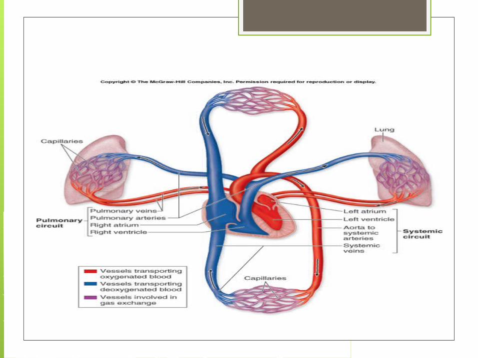
Circulation

Circulation Through the HeartThe Process Blood enters through right and left atria As heart contracts, blood flows into the
ventricles and then to body or lungs Valves – prevent blood from having a reverse
flow When ventricles contract, the valves close
preventing blood flow back into the atria There are also valves at the exits of the
ventricles that prevent blood from flowing back into the heart
Heart Beat The contraction of the heart begins in a
small group of cardiac muscle cells called the sinoatrial node/SA Node (located in right atrium)
Called the pacemaker because these cells set the pace for the heart by contracting
This signal is picked up by fibers called the atrioventricular node (AV node) and this allows the ventricles to contract

Blood Vessels Blood leaving the left side of the heart is
oxygen rich The aorta carries oxygen rich blood from
the heart to the body 3 types of blood vessels
Arteries Capillaries veins

Blood Vessels Arteries – large vessels that carry blood from
the heart to tissues in the body Except for pulmonary – all arteries carry
oxygen rich blood Arteries have thick walls that allow for the high
pressures of blood passing through these canals
Capillaries – smallest blood vessels Nutrient transport, oxygen transport to tissues,
and removal of waste products is done by the capillaries

Blood Vessels Cont. Veins – carry oxygen poor blood back to
the heart Thick walls to withstand pressure Lack of exercise will weaken vein walls
causing them to stretch, thus weakening the valves causing blood to pool in the veins Known as varicose veins

Blood Pressure Force of blood on the artery walls is known as
blood pressure Pressure is measured with a sphygmomanometer Systolic pressure – 1st # - force felt in arteries
when ventricles contract Diastolic pressure – force felt in arteries when
ventricles contract Kidneys also help regulate blood pressure
Hormones cause kidneys to remove more water when blood pressure is too high
Reduces volume and reduces pressure
Diseases Atherosclerosis – fatty deposits/plaque build
up on the inner walls of the arteries Often builds up in coronary arteries which
bring oxygen rich blood back to heart, and then the heart cells will die from lack of oxygen
Blood clots can break free and travel to the brain and then cause blockage – known as a stroke Brain cells die from lack of oxygen

Diseases Cont. High blood pressure – known as
hypertension Weakens and damages heart and blood
vessels Increases risk of heart attack and stroke
Diet Exercise NO smoking Low saturated fat Watch cholesterol Obesity causes heart to contract harder,
thus wearing out heart faster


