
The Biomechanics and Evolutionof High-Speed Throwing
The Harvard community has made thisarticle openly available. Please share howthis access benefits you. Your story matters
Citation Roach, Neil. 2012. The Biomechanics and Evolution of High-SpeedThrowing. Doctoral dissertation, Harvard University.
Citable link http://nrs.harvard.edu/urn-3:HUL.InstRepos:9822375
Terms of Use This article was downloaded from Harvard University’s DASHrepository, and is made available under the terms and conditionsapplicable to Other Posted Material, as set forth at http://nrs.harvard.edu/urn-3:HUL.InstRepos:dash.current.terms-of-use#LAA

© 2012 Neil Thomas Roach
All rights reserved.

iii
Advisor: Daniel Lieberman Neil Thomas Roach
The Biomechanics and Evolution of High-‐Speed Throwing
Abstract
Throwing with power and accuracy is a uniquely human behavior and a
potentially important mode of early hunting. Chimpanzees, our closest living
relatives, do occasionally throw, although with much less velocity. At some point in
our evolutionary history, hominins developed the ability to produce high
performance throws. The anatomical changes that enable increased throwing ability
are poorly understood and the antiquity of this behavior is unknown.
In this thesis, I examine how anatomical shifts in the upper body known to
occur during human evolution affect throwing performance. I propose a new
biomechanical model for how humans amplify power during high-‐speed throwing
using elastic energy stored and released in the throwing shoulder. I also propose
and experimentally test a series of functional hypotheses regarding how four key
shifts in upper body anatomy affect throwing performance: increased torso
rotational mobility, laterally oriented shoulders, lower humeral torsion, and
increased wrist hyperextensability.

iv
These hypotheses are tested by collecting 3D body motion data during
throws performed by human subjects in whom I varied anatomical parameters
using restrictive braces to examine their effects on throwing kinematics. These data
are broken down using inverse dynamics analysis into the individual motions,
velocities, and forces acting around each joint axis. I compare performance at each
joint across experimental conditions to test hypotheses regarding the relationship
between skeletal features and throwing performance. I also developed and tested a
method for predicting humeral torsion using range of motion data, allowing me to
calculate torsion in my subjects and determine its effect on throwing performance.
My results strongly support an important role for elastic energy storage in
powering humans’ uniquely rapid throwing motion. I also found strong
performance effects related to anatomical shifts in the torso, shoulder, and arm.
When used to interpret the hominin fossil record, my data suggest high-‐speed
throwing ability arose in a mosaic-‐like fashion, with all relevant features first
present in Homo erectus. What drove the evolution of these anatomical shifts is
unknown, but as a result the ability to produce high-‐speed throws was available for
early hunting and likely provided an adaptive advantage in this context.

v
Table of Contents
Abstract………………………………………………………………………………………………...... iii List of Figures………………………………………………………………………………………….. vii List of Tables…………………………………………………………………………………………… viii Acknowledgements…………………………………………………………………………………. ix Chapter 1 – An Introduction………………………………………………………………….. 1 1.1. Why throwing?......................................................................................................... 1 1.2. Thesis Overview...................................................................................................... 1 1.3. Literature Cited....................................................................................................... 6 Chapter 2 – The Effect of Humeral Torsion on Rotational Range of Motion in the Shoulder and Throwing Performance……………………..
7
2.1. Chapter Summary……………………………………………………………………… 7 2.2. Introduction……………………………………………………………………………… 9 2.3. Model………………...……………………………………………………………………… 13 2.4. Materials and Methods…….………………………………………………………… 17 2.5. Results…………………………….………………………………………………………… 22 2.6. Discussion……………………….………………………………………………………… 25 2.7. Acknowledgements………….………………………………………………………… 31 2.8. Literature Cited……………….………………………………………………………… 32 Chapter 3 – Elastic Energy Storage in the Shoulder and the Evolution of High-‐Speed Throwing in Homo…………………………………………………..
37
3.1. Chapter Summary……………………………………………………………………… 37 3.2. Introduction……………………………………………………………………………… 38 3.3. Model………………...……………………………………………………………………… 39 3.4. Materials and Methods…….………………………………………………………… 40 3.5. Results…………………………….………………………………………………………… 44 3.6. Discussion……………………….………………………………………………………… 48 3.7. Acknowledgements………….………………………………………………………… 50 3.8. Literature Cited……………….………………………………………………………… 51 Chapter 4 – The Biomechanics of Power Generation during Rapid, Overhand Throwing in Humans…………………………………………………..…
54
4.1. Chapter Summary……………………………………………………………………… 54 4.2. Introduction……………………………………………………………………………… 55 4.3. Model………………...……………………………………………………………………… 58 4.4. Hypotheses……………….…….………………………………………………………… 60 4.5. Materials and Methods……….……………………………………………………… 65 4.6. Results…………………………….………………………………………………………… 71 4.7. Discussion……………………….………………………………………………………… 80

vi
4.8. Acknowledgements………….………………………………………………………… 89 4.9. Literature Cited……………….………………………………………………………… 90 Chapter 5 – Throwing and the Evolution of the Hominin Forelimb……… 95 5.1. Chapter Summary……………………………………………………………………… 95 5.2. Introduction……………………………………………………………………………… 96 5.3. Who throws?……...……………………………………………………………………… 99 5.4. How do we throw?…………..………………………………………………………… 104 5.5. Who threw first?.............……….……………………………………………………… 110 5.6. Why do we throw?…….…….………………………………………………………… 135 5.7. What questions remain?…………………….………………………………………. 142 5.8. Acknowledgements………….………………………………………………………… 144 5.9. Literature Cited……………….………………………………………………………… 145 Appendix 1 – Supplemental Data for Chapter 2..................................................... 168 Appendix 1 – Supplemental Material for Chapter 3............................................ 169

vii
List of Figures
Figure 2.1: Humeral torsion vs. retroversion……………………………………………. 10 Figure 2.2: Predicting humeral torsion from rotational range of motion…… 15 Figure 2.3: Measuring humeral torsion using computed tomography............. 19 Figure 2.4: Measuring range of motion at the shoulder…….………………………. 20 Figure 2.5: Torsion prediction regressions………………...…….………………………. 24 Figure 3.1: Model of throwing differences between Pan and Homo…...………. 41 Figure 3.2: Shoulder and elbow power during throwing……………...…...………. 45 Figure 3.3: Brace-‐restricted power during throwing…………………...…...………. 47 Figure 3.4: Humeral torsion and throwing performance……………...…...………. 49 Figure 4.1: Free-‐body diagram of the arm during cocking……….…...…...………. 59 Figure 4.2: Critical upper body motions during throwing.…………...…...………. 61 Figure 4.3: Reflective markers and kinetic model…………......………...…...………. 66 Figure 4.4: Arm-‐cocking and acceleration phases…………......………...…...………. 70 Figure 4.5: Kinematic and kinetic data for 4 critical motions..……...…...………. 72 Figure 5.1: Chimpanzee throwing…………………………………….....……...…...………. 100 Figure 5.2: Arm-‐cocking and acceleration phases…………......………...…...………. 103 Figure 5.3: Critical upper body motions during throwing.…………...…...………. 106 Figure 5.4: Free-‐body diagram of the arm during cocking……….…...…...………. 108 Figure 5.4: Humeral torsion………………………………………….……….…...…...………. 125 Supplemental Figure 3.1: Reflective markers…...………….……….…...…...………. 170 Supplemental Figure 3.2: Kinetic model……...…...………….……….…...…...………. 174 Supplemental Figure 3.3: Residual analysis – full trial…...………….……….….... 177 Supplemental Figure 3.4: Residual analysis – critical period…......……….….... 177 Supplemental Figure 3.5: Phase duration………………………….…......……….….... 183 Supplemental Figure 3.6: Maximum ball speed………………….…......……….….... 188 Supplemental Figure 3.7: Shoulder sham power……….……….…......……….….... 189 Supplemental Figure 3.8: Free-‐body diagram of the arm during cocking…. 196 Supplemental Figure 3.9: Static optimization…………………………………………. 197

viii
List of Tables
Table 4.1: Segment definitions…………………..……………………………………………. 67 Table 4.2: Torso restriction data………………..……………………………………………. 73 Table 4.3: Clavicle restriction data………………..…………………………………………. 75 Table 4.4: Shoulder restriction data………………..………….……………………………. 77 Table 4.5: Wrist restriction data…….…………..……………………………………………. 79 Supplemental Table 2.1: Measurement reliability data…………………………… 168 Supplemental Table 3.1: Segment definitions…………….…………………………… 172 Supplemental Table 3.2: Joint definitions………………….…………………………… 175 Supplemental Table 3.3: Cutoff frequency data………….…………………………… 179 Supplemental Table 3.4: Shoulder muscle volumes…...…………………………… 185 Supplemental Table 3.5: Modeled power comparison….….……………………… 186 Supplemental Table 3.6: Chimpanzee throwing velocity….……………………… 191

ix
Acknowledgements
The process of completing a PhD is long, at times difficult, and could not be
done alone. I owe a great debt of gratitude to many friends, colleagues, and mentors
for their support and encouragement during the past seven years. You have made
my time in graduate school interesting and enjoyable. Thank you!
First and foremost, I would like to thank my advisor, Dan Lieberman. Over
the last seven years Dan has been my strongest advocate, provided good advice (and
a healthy dose of nudging), read innumerable drafts of variable quality, and has
always been available to talk about ideas, plans, or anything else that was on my
mind. That I frequently find myself in academic talks thinking, “What is your model?”
is terrifying, but also a testament to the sound, experimentalist approach to science
that Dan has imparted to me. I am forever indebted to him for his continued belief in
me and my abilities, for his patience in the face of my epic stubbornness, and for his
being an excellent mentor and friend.
Many thanks are owed to my thesis committee: Richard Wrangham, for his
constant encouragement and for always advocating on behalf of chimpanzee
throwing abilities; Andy Biewener, whose biomechanical expertise and careful
review of my work has made this thesis immeasurably better; L Mahadevan, whose
ideas about throwing have changed my thinking; Tom Gill III, whose guidance
through complicated hospital procedures and IRB protocols made a difficult process
seamless; and finally Susan Larson, whose work I have followed and learned from
x
over the years, and whose careful reading and interrogation of my research has
helped strengthen my work and clarified my thinking.
Completing this dissertation would not have been possible without the help
and excellent ideas of my collaborators. Madhu Venkadesan was my partner in
throwing related crimes during his time at Harvard. His ideas and help were critical
in my thinking about elastic energy storage, the heart of my thesis. I count Madhu a
good friend and look forward to our continued collaboration. Many thanks are due
to Tom Gill IV, whose help and financial support made data collection at MGH
possible. I would also like to thank Bill Palmer, who reached out numerous time on
my behalf to cut through hospital red tape and get me access to hospital equipment.
Finally, I would like to thank Mike Rainbow, whose biomechanical expertise made
difficult data analysis easy. Mike’s excellent ideas and careful review of my work
have also vastly improved my writing and thinking. I look forward to our continued
work together.
Many individuals were also vital in making my data collection and analysis
possible. Leia Stirling and the Wyss Institute Motion Capture Lab were incredibly
generous in allowing me to collect my kinematic data in their facility. Processing this
data was a titanic endeavor and would never have been completed if not for the
hundreds of hours spent by many Harvard students who tirelessly turned tiny little
dots on a computer screen into my thesis data. For this I am indebted to Matt
Vincent, Gulus Emre, Randi Griffin, Adanma Ekeledo, Wesley Gordon, and George

xi
Karas. I am also very grateful to the many subjects who have participated in my
experiments for kindly sharing their valuable time with me.
I am also very grateful to have been a part of the close-‐knit Human
Evolutionary Biology department these past years. I have greatly enjoyed all of the
many beer hours, Tuesday lunch talks, and getting to know and interact with so
many interesting people. Special thanks are due to the members of the Skeletal
Biology Lab past and present (Brian Addison, Meir Barak, Eric Castillo, Adam Daoud,
Maureen Devlin, Carolyn Eng, Brenda Frazier, Kristi Lewton, Herman Pontzer, Dave
Raichlen, Campbell Rolian, Madhu Venkadesan, Anna Warrener, Katherine
Whitcome, Sara Wright, and Katie Zink) who taught me a great many things and
made my life at Harvard enjoyable. I count you all as good friends and will (and do)
miss seeing you all every day.
To Meg Lynch, Meg Jarvi, and Laura Christoffels (the best departmental staff
anywhere): Thank you, Thank you, a thousand times…Thank you. Your help sorting
out the intricacies of university bureaucracy, grant renewals, paycheck issues, and
the thousands of other tasks I have bothered you with has been invaluable.
Many thanks are owed to my other mentors, David Pilbeam and Sally
McBrearty. Throughout my time at Harvard, David has always been there to provide
good advice, listen to me vent, and help me when I needed it. David has generously
written countless recommendations, made many phone calls, and procured many
ASPR dollars on my behalf. His help and support have always been graciously given
and were always much appreciated. Sally McBrearty has been my mentor and friend

xii
since I walked into her office at UConn in the fall of 2001, declared that I was
interested in human evolution, and asked if she would advise me. Sally took me
under her wing, brought me to do fieldwork in Kenya (an experience that changed
the course of my life), and treated and trained me as one of her own graduate
students. Sally’s unshakable belief in me, her continued support, and the doors she
has opened for me are debts I will never be able to repay. I am forever grateful.
I also want to thank my parents, Brian and Kathy Roach, and siblings, Lee,
Jessica, and Thomas, for their encouragement, support, and love during this process.
While my parent’s interest and love of the natural world was passed to all of us kids,
my father’s fascination with dinosaur bones has mutated a bit in its passage to me.
Nonetheless, I always had an interested audience during visits home, who thought
what I worked on was cool and encouraged me when I needed it most.
Finally, I owe the greatest debt of gratitude to my wife, Amanda Lobell.
Meeting Amanda in graduate school was by far the best thing that has ever
happened to me and she has filled my time in Cambridge with love, caring, and
unreasonable amounts of fun. Amanda’s impact on this thesis is not quantifiable. She
has acted as my research assistant, my editor, my confidant, my advisor, and my
therapist. She has been there for me through every step of this process. I owe her
more than I can hope to repay (although she reminds me frequently that a vacation
would be a good way to start). I consider myself immensely lucky to call her my best
friend, my colleague, and my wife.

1
Chapter 1 – An Introduction
1.1 Why throwing?
How and why do humans throw projectiles with both considerable accuracy
and high velocities? Human throwing ability is unique among animals and universal
to all human cultures. High-‐speed throwing was likely important during human
evolution, enabling hominin throwers to hunt more effectively and safely, to drive
off predators, and to interact aggressively with other individuals from a safer
distance than more direct forms of fighting. Despite the potential adaptive
importance of high performance throwing, little is known about the evolution of this
unique and critical behavior. This thesis investigates the biomechanics of how
humans produce high-‐speed throws and examines how differences in skeletal and
soft tissue anatomy affect throwing performance. The results of these analyses are
applied to the hominin fossil record in hopes of gaining insight into the evolutionary
history and antiquity of high-‐speed throwing.
1.2 Thesis Overview
This dissertation includes four related studies. Chapter 2 considers the
effects of humeral torsion on the range of rotational motion in the shoulder and
throwing performance. Chapter 3 proposes and tests a model for elastic energy
storage in the shoulder. Chapter 4 presents and tests a model of anatomical
variations that improve the biomechanics of power generation during rapid,
overhand throwing in humans. Finally, Chapter 5 integrates the evidence from

2
Chapters 2-‐4 with fossil and archaeological evidence to review how and when
throwing evolved and how selection for throwing may have helped modify the
hominin upper body and forelimb.
In Chapter 2, I examine the effect of humeral torsion of rotational range of
motion at the shoulder. Humeral torsion is a highly variable angular measure that
quantifies the difference between the orientation of the humeral head and the axis
around which flexion and extension occur in the elbow. Previous studies have
shown that throwing athletes have lower levels of humeral torsion than non-‐
throwing athletes, and lower levels of torsion in their throwing versus non-‐throwing
arms (1-‐4). Some of these studies have also shown that throwing athletes with
higher than expected torsion are more likely to experience pain and injuries to their
shoulders (1). While these studies have attempted to link torsion values with
biomechanically meaningful measures such as rotational range of motion in the
shoulder, they have only partially confirmed these relationships. Chapter 2
investigates the relationship between humeral torsion and active range of motion by
regressing actual computed tomography (CT) derived humeral torsion values
against predicted values calculated from range of motion data (both goniometric
and 3D kinematic data). My findings show that range of motion measures are
significantly correlated with underlying humeral torsion values and can be used to
predict these values when medical imaging is not possible.
In Chapter 3, I propose a biomechanical model for how humans achieve rapid
throws in part by using elastic energy stored in the ligaments, tendons, and muscles

3
crossing the shoulder. According to the model, elastic energy is used to amplify
power production by muscles, resulting in very rapid motions in the forelimb. I
further hypothesize that anatomical differences between humans and chimpanzees
compromise chimpanzees’ ability to use this elastic energy storage mechanism and
help to explain why our closest extant relatives throw so poorly. I use three lines of
evidence to test these hypotheses: 1. a kinetic analysis of humeral rotation during
throwing; 2. a quantitative comparison of actual power production at the shoulder
with modeled estimates of shoulder muscle power generation capacity; 3. an
experimental perturbation of the proposed elastic energy storage mechanism. The
results from these studies show that during normal throwing, the humerus first
externally rotates passively, stretching the elastic elements at the shoulder.
Furthermore, during this stretching a large opposing muscular force is generated by
the shoulder muscles, resulting in a sustained period of energy absorption during
which elastic energy is stored. When released, this energy can account for more than
half of the total energy required to generate the rapid internal rotation of the
humerus that follows. Mathematical modeling of shoulder joint power shows that
forces generated by shoulder muscles alone are insufficient to explain the rapid
motions of the arm, confirming the importance of the elastic energy mechanism.
Finally, a perturbation of this elastic storage mechanism using a shoulder brace
shows that storage of elastic energy at the shoulder affects the velocity of the arm
and of the projectile. We infer from these data that a mobile waist and hips, “relaxed”
laterally oriented shoulder, and low humeral torsion all increase throwing
performance by enabling more elastic energy storage and release. The presence of

4
all of these functionally important morphologies in Homo erectus suggests that these
hominins could throw projectiles with high-‐speed, and may have done so during
hunting.
Chapter 4 focuses on power generation, by testing how variations in upper
body skeletal anatomy would affect the four key motions that previous studies (5)
have shown to contribute the most to projectile velocity: 1. internal rotation of the
humerus; 2. elbow extension; 3. torso rotation; 4. wrist flexion. The performance
effects of the throwing motion were perturbed using braces that restrict particular
movements, allowing me to quantify the effects of specific components of normal
throwing using an observed vs. expected framework. I use three-‐dimensional
inverse dynamics analysis to decompose the complex throwing motion into the
individual rotations occurring at each joint and the forces that drive those rotations.
These joint performance data are then used to test a set of discrete hypotheses of
how joint motions are interrelated. The results suggest that high-‐velocity during
throwing is achieved using a combination of kinetic energy transfer between
adjacent body segments — a “kinetic chain” (6-‐9) — combined with substantial
power amplification from elastic energy stored at the shoulder. Restriction of
proximal joint motions at the torso and shoulder lead to significant reductions in
throwing performance measures not only in the restricted joint but also more distal
joints. These results provide insight into why the shoulder and elbow frequently
suffer repetitive stress injuries in throwing athletes.

5
Finally, in chapter 5, I review what is known about the mechanics of
throwing and interpret the hominin fossil record in light of these data. I assess my
own functional hypotheses as well as previous functional morphological
interpretations of hominin fossil material relating to throwing performance and
address the biomechanical support for those interpretations. I conclude that the
morphological shifts that enable high-‐speed throwing behavior emerge in a mosaic-‐
like fashion in Australopithecus and early Homo. By Homo erectus, all the relevant
features appear to be present, suggesting that this species had modern human-‐like
throwing performance abilities. I conclude that such throwing ability would have
provided significant adaptive benefits for early hunting, although further research is
necessary to determine the extent to which selection for throwing or other
behaviors led to the evolution of these features.

6
1.3 Literature Cited
1. Pieper HG (1998) Humeral torsion in the throwing arm of handball players. Am J Sports Med 26(2):247-‐253.
2. Osbahr DC, Cannon DL, & Speer KP (2002) Retroversion of the humerus in
the throwing shoulder of college baseball pitchers. Am J Sports Med 30(3):347-‐353.
3. Reagan KM, et al. (2002) Humeral retroversion and its relationship to
glenohumeral rotation in the shoulder of college baseball players. Am J Sports Med 30(3):354-‐360.
4. Chant CB, Litchfield R, Griffin S, & Thain LM (2007) Humeral head
retroversion in competitive baseball players and its relationship to glenohumeral rotation range of motion. J Orthop Sports Phys Ther 37(9):514-‐520.
5. Hirashima M, Kudo K, Watarai K, & Ohtsuki T (2007) Control of 3D limb
dynamics in unconstrained overarm throws of different speeds performed by skilled baseball players. J Neurophysiol 97(1):680-‐691.
6. Hirashima M, Yamane K, Nakamura Y, & Ohtsuki T (2008) Kinetic chain of
overarm throwing in terms of joint rotations revealed by induced acceleration analysis. J Biomech 41(13):2874-‐2883.
7. Higgins JR (1977) Human Movement: an integrated approach (The C.V. Mosby
Company, Saint Louis). 8. Feltner ME (1989) 3-‐Dimensional Interactions in a 2-‐Segment Kinetic
Chain .2. Application to the Throwing Arm in Baseball Pitching. Int J Sport Biomech 5(4):420-‐450.
9. Atwater AE (1979) Biomechanics of overarm throwing movements and of
throwing injuries. Exerc Sport Sci Rev 7:43-‐85.

7
Chapter 2 – The Effect of Humeral Torsion on Rotational Range of Motion in
the Shoulder and Throwing Performance
2.1 Chapter Summary
Several recent studies have found that throwing athletes typically have lower
humeral torsion (retroversion) and a greater range of external rotation at the
shoulder than non-‐athletes. How these two parameters are related is debated. This
study uses data from a sample of both throwers and non-‐throwers to test a new
model that predicts torsion values from range of motion data. The model proposes a
series of predicted regressions, which can help provide new insight into the factors
affecting rotational range of motion at the shoulder.
Humeral torsion angles were measured from computed tomography scans
collected from 25 male subjects. These values are compared to predicted torsion
values for the same subjects calculated from both kinematic and goniometric range-‐
of-‐motion data. Results show that humeral torsion is negatively correlated
(Goniometric: r = -‐0.409, p = 0.047; Kinematic: r = -‐0.442, p = 0.035) with external
rotational range of motion and positively correlated (Goniometric: r = 0.741, p <
0.001; Kinematic: r = 0.559, p = 0.006) with internal rotational range of motion. The
predicted torsion values are highly correlated (Goniometric: r = 0.815, p < 0.001;
Kinematic: r = 0.617, p = 0.006) with actual torsion values. Deviations in the data
away from predicted equations highlight significant differences between high
torsion and low torsion individuals that may have significant functional

8
consequences. The method described here may be useful for non-‐invasively
assessing the degree of torsion in studies of the evolution and biomechanics of the
shoulder and arm, and for testing hypotheses about the etiology of repetitive stress
injuries among athletes and others who throw frequently.

9
2.2 Introduction
Humeral torsion describes the angular difference between the orientation of
the proximal humeral head and the axis of the elbow at the distal humerus. This
angle is measured at the intersection of two lines: one that evenly bisects the
articular surface of the humeral head proximally, and the second being the
transepicondylar line distally (Figure 2.1). In the clinical literature, this angle is
measured in the opposing direction and is referred to as humeral retroversion.
Therefore, a measured increase in retroversion is the same as a decrease in torsion.
These two terms simply represent different assumptions about the neutral position
of this angle (1-‐5). The use of alternative terms has led to some confusion in the
literature (1, 2). For clarity, we will here use exclusively the term humeral torsion
and have translated the results of previous studies cited as needed.
In humans, humeral torsion is highly variable. Torsion values have been
shown to differ between western and non-‐western populations (4, 6-‐8), males and
females (6-‐9), and by side of the body (6, 8, 10). Torsion also varies ontogenetically,
with younger individuals having less torsion, which then increases during postnatal
growth (5, 11-‐13). During normal postnatal ontogeny, torsion steadily increases by
an average of 23.4 degrees until the completion of skeletal growth (11). Much of this
change in torsion seems to occur at the proximal humerus, which is one of the last
bones in the body to fuse: only 20 percent of individuals have achieved fusion by 18
years of age (14).

10
Figure 2.1. Humeral torsion (in blue) is determined by measuring the angle between the orientation of the humeral head and the distal condyle of the humerus. In the clinical literature, the same angle is referred to as humeral retroversion (in yellow) and is measured in the opposite direction.
Many previous functional studies of humeral torsion have focused on the
relationship between torsion and habitual throwing activity (15-‐19). Athletes who
habitually throw tend to have 10-‐20 degrees less torsion in their dominant,
throwing arm compared to their non-‐dominant arm and the arms of non-‐throwing
controls (15-‐25). No statistical difference has been found between arms in non-‐
throwing controls (15, 16, 19). Furthermore, Pieper (19) shows that when throwing
athletes are subdivided into those with and without chronic pain, those reporting

11
chronic pain did not show this reduction in dominant arm torsion. Other studies
have sought to further clarify the relationship of torsion to injury by linking torsion
to rotational range-‐of-‐motion (ROM) in the shoulder (15, 17, 18).
Deficits to rotational ROM in the shoulder have long been suggested as a
potential cause of shoulder injury in throwing athletes (26-‐31). A recent study by
Wilk et al (26) showed that a deficit to the total rotational ROM at the shoulder of as
little as 5 degrees led to a twofold increase in the likelihood of injury. Previous work
has shown that, much like torsion, rotational ROM also differs between dominant
and non-‐dominant arms in throwing athletes (15-‐18, 22, 23, 28, 32, 33). Throwing
athletes typically have an externally shifted ROM arc in the throwing arm, with an
increase in external rotational ROM of between 9-‐13 degrees and a similar deficit to
the internal rotational ROM (15-‐18).
Beyond its relevance to understanding injury, rotational range of motion at
the shoulder may also have significant performance consequences. Glenohumeral
rotation is known to be a significant contributor to power generated during the
throwing motion (22, 34-‐37). Reaching angular velocities in excess of 7000 degrees
per second, the rapid internal rotation of the humerus is the fastest motion
produced by the human body and this rotation can generate very high torques (22,
35, 36). Whether modifications to the rotational ROM allow additional acceleration
of the arm and torque production at the shoulder is unknown and is an important
avenue for future research.

12
What factors contribute to variations in the rotational ROM arc is unclear.
Numerous authors have posited that these modifications result primarily from
changes to the soft tissues of the shoulder capsule (22-‐25, 38-‐43). These same
studies note the high prevalence of severe laxity in the anterior glenohumeral
capsule of throwing athletes. Burkhart and colleagues have further suggested that
this anterior laxity is accompanied by tightening of the ligaments in the posterior
portion of the capsule (31, 38). Still others have suggested that skeletal remodeling
of the proximal humerus is the major cause of modifications to the rotational ROM
at the shoulder (15-‐19). Several studies investigating the relationship between
humeral torsion and rotational ROM have found significant correlations between
these two variables in professional and collegiate throwing athletes (15, 17, 18).
However, these correlations have not been consistent in either strength or
directionality between studies.
Although the studies noted above have all found significant negative
correlations between external rotational ROM and torsion, the relationship between
internal rotational ROM and torsion is less clear. Osbahr et al (17) and Reagan et al
(18) found no statistically significant relationship between internal rotational ROM
and torsion in baseball pitchers. In contrast, Reagan et al (18) found these variables
to be significantly, negatively correlated in baseball players playing field positions.
This result contrasts with another study of baseball players of unspecified positions
in which internal rotational ROM and torsion were significantly, positively
correlated (15).

13
We sought to clarify and generalize the relationship between humeral
torsion and rotational range of motion at the shoulder using a sample of both
collegiate athletes and non-‐athletes. We propose a simple, new model to predict
expected torsion values from ROM data collected using both standard goniometry
and kinematics. Our method is then validated against observed (actual) torsion
values derived from computed tomography (CT). This observed versus expected
framework allows us to generate testable predictions and move beyond simple
strength of correlation results. Using this method, we can begin to address the
complex interplay between how soft and hard tissues affect ROM at the shoulder
joint, how this interplay may relate to injury, and the possible functional
consequences of these tradeoffs. This method also provides a means to assess
ranges of motion in the fossil record, allowing us to test some hypotheses about the
evolution of throwing capabilities.
2.3 Model
We tested a model that uses ROM measures to predict torsion values and vice
versa. Our model is based on a set of simple assumptions.
We begin with the assumption that the total rotational ROM (internal +
external) is not correlated significantly with the degree of torsion, as shown by
previous studies (15, 17, 18). We therefore would expect any angular change at the
external end of the ROM arc must be coupled with a commensurate, opposing
angular change at the internal end of the ROM arc. Thus, while the values for
internal and external rotational ROM will change, the sum of these to values will not.

14
This requires a one-‐to-‐one tradeoff between internal and external rotational ROM,
which accounts for the constant total ROM value, and leads to all regression
predictions between torsion and any range of motion value in our analyses having
an absolute slope of 1.0. This predicted slope allows for changes in torsion to affect
changes in the position of the ROM arc relative to the body without affecting the
total ROM. Furthermore, this assumption of a constant total ROM leads to the
prediction that the regression relationships between external rotational
ROM/torsion and internal rotational ROM/torsion should also be inversely related
(as one increases the other decreases).
Predicting torsion values from ROM data further requires the assumption
that the midpoint of the total ROM arc represents the neutral, resting position of the
humerus in relation to the scapular glenoid. If this is correct, then the location of this
neutral position should provide an indication of the degree of humeral torsion.
Individuals with a total ROM midpoint that is internally shifted are predicted to
have a higher torsion value than individuals with more externally shifted total ROM
midpoints. Using the measured external and internal rotational ROM maxima,
expected torsion values are generated using the formula below (see also Figure 2.2).
Note: The addition/subtraction of 180 degrees in all formulas allows our results to
be reported using a common convention for human torsion values, eg. (7, 44).
Equation 2.1.
Torsion (predicted) =
€
180−χmax Ext ROM − χmax Int ROM
2
$
% &
'
( )

15
Figure 2.2. Torsion is predicted from mean ROM maxima data. The difference between external and internal rotation (in the example – 25 degrees) is equal to the ROM midpoint. The use of the 180-‐degree term allows the torsion value to be reported according to prior convention. Note: Following clinical definition, external rotational ROM is illustrated in blue and internal rotational ROM is illustrated in red.
To test our assumptions, these expected values are regressed against actual torsion
values. If our model is accurate, the predicted torsion values will closely match the

16
actual values. Thus, for the predicted torsion (y)/actual torsion (x) regression we
predict a regression equation of y=x.
Although predicting torsion values from ROM data is useful, when the actual
regressions differ from our predictions it becomes important to accurately address
the discrepancy. By running our model in the reverse direction and predicting
external and internal rotational ROM from actual torsion values we can better
identify the source of the discrepancy. These reverse predictions again rely on the
assumption that the midpoint of the total rotational ROM reflects the underlying
torsion. Given this assumption, a predicted ROM midpoint can be generated from
actual torsion values. By adding/subtracting half of the mean total ROM from this
midpoint, external and internal ROM maxima can be generated. The formula used to
generate predicted external ROM values is:
Equation 2.2.
External ROM (predicted)=
The regression equation derived from this formula is y = -‐x + (180+ ½ total ROM);
with (y) as predicted external ROM and (x) as actual torsion. For our calculations,
we use the mean total ROM value across all subjects (reported below). According to
our model, as torsion increases there should be a commensurate decrease in
external ROM maxima (hence the slope of -‐1.0). This prediction is tested against the
actual external rotational ROM maxima/torsion regression. The formula used to
€
−Actual torsion + 180+total ROM
2#
$ %
&
' (

17
generate predicted internal ROM values is:
Equation 2.3.
Internal ROM (predicted)=
The regression equation derived from this formula is y = x -‐ (180 -‐ ½ total ROM);
with (y) as predicted internal ROM and (x) as actual torsion. Accordingly, as torsion
increases, so too should the internal ROM maxima (hence the slope of 1.0). This
prediction is tested against the actual internal rotational ROM maxima/torsion
regression.
2.4 Materials and Methods
Subjects -‐ Twenty-‐five adult male subjects (ages 18-‐35) were recruited to
participate in the study. Thirteen of the subjects were collegiate athletes (6 baseball
players from a variety of positions, 7 athletes from non-‐throwing sports) and the
remaining 12 subjects were non-‐athletes. The proportions of the study sample were
chosen to maximize the variance in torsion values surveyed and to reduce the
effects of pathological ROM often found in throwing athletes (26, 29, 38).
Institutional review board approval was obtained from both Massachusetts General
Hospital and Harvard University. Subjects provided written consent and completed
an injury history and physical activity questionnaire prior to participation in the
study.
€
Actual torsion− 180− total ROM2
#
$ %
&
' (

18
Computed Tomography (CT) Imaging -‐ All subjects were CT scanned at the
Massachusetts General Hospital Imaging Center using a GE 8-‐slice Lightspeed
Computed Tomography Imager. A low dose scanning technique was used to
minimize radiation exposure to the subjects. Subjects were positioned on the
scanner examination table with their dominant arm fully adducted at their side and
their elbow flexed 90 degrees with their forearm resting on their abdomen. The arm
was then immobilized by wedging a stiff pillow between the arm and the
examination table. Two separate scans were targeted and collected at the shoulder
and the elbow. Each scan covered approximately 5 cm of the arm, capturing each
end of the humerus with 5mm think image slices. The images were processed using
Image J software and the humeral torsion angle calculated (following (45)) by
subtracting the angle of the transepicondylar line from the axis of the humeral head
(Figure 2.3). The humeral torsion angle was calculated using the mean of the three
best superior and inferior scans. The measurement reliability was calculated (see
supplemental data) and the mean torsion angle was used in the analysis as the
actual, CT derived torsion value.
Range of Motion (ROM) measures -‐ Active ROM was measured using both
standard goniometry and three-‐dimensional kinematic imaging. Active ROM, where
subjects rotate their arm to the limits of their ROM using their own muscular power,
was used instead of passive ROM because it most closely approximates the
functional ROM available for actions such as throwing and also reduces the effects of
any anisotropy in the capsular ligaments. The two different methods for collecting

19
ROM data were used to maximize the utility of our method for a variety of research
and clinical contexts.
Figure 2.3. Humeral torsion was measured in Image J by creating transects between the inflection points marking the anatomical neck proximally (A) and along the distal transepicondylar line (B).
For goniometric ROM measurements, subjects were positioned on an
examination table with their dominant arm off the side of the table (Figure 2.4). The
arm was positioned in line with both shoulders in a neutral shoulder
flexion/extension posture. The shoulder was abducted 90 degrees and the elbow
flexed 90 degrees. The subject was instructed to keep their shoulder pressed to the
examination table to prevent scapular movement. The subject then rotated his arm
to both its maximal external and internal rotation position and held this position for
5 seconds for goniometric measurement taken along the length of the forearm. The
measurements were taken using a Jamar 12 ½” goniometer, which was modified to

20
include a weighted plumb line to serve as a vertical arm. The rotational motion and
measurements were repeated twice. Measurement repeatability was calculated
using the interclass correlation (see supplemental material). The mean of both
external and internal ROM maxima measures were taken and the predicted,
goniometry derived torsion value was calculated using equation 1.
Figure 2.4. For the ROM calculations, each subject was measured using a standard goniometer (A) and a kinematic imaging system (B). For the goniometric measurement, the subject was in a supine position on an examining table. For the kinematic measurement, the subject was seating in an armless chair. External rotational ROM is illustrated in blue and internal rotational ROM is illustrated in red.

21
For the kinematic ROM measurements, data was collected using an eight-‐
camera Qualisys Motion Capture 3D Infrared Oqus camera system collecting at
500Hz. All subjects were fitted with nine 25cm passive reflective markers. The
markers were placed on C7 prominens, both hips (at the greater trochanter of the
femora), both scapular acromion, the lateral and medial epicondyles of the
dominant side distal humerus and both ulnar and radial styloid processes in the
dominant side wrist. The subject was then seated in an armless, high backed office
chair in an upright posture with the back firmly against the chair back. The subject
was again instructed to position his arm out to the side of his body, in line with both
shoulders. The shoulder was abducted 90 degrees and the elbow flexed 90 degrees.
The subject was then instructed to again rotate their arm to both its maximal
external and internal rotation position with as little scapular motion as possible (to
limit scapular protraction or retraction). The motion was repeated between 4-‐6
times during 30 seconds of data capture. Raw 3D marker positional data was
processed using Qualisys Task Manager software and exported for analysis to
MATLAB 7.6. Custom written MATLAB code was used to calculate external and
internal ROM maxima while correcting for minor deviations in elbow and shoulder
position. Measurement reliability was calculated and the means of both external and
internal rotational maxima were used to generate predicted, kinematics derived
torsion values using equation 1.
Statistical analysis -‐ Pearson correlation coefficients were calculated and the
presence of outliers tested (using Mahalanobis distances at a 95% confidence

22
interval) with JMP version 5 software. Intraclass correlations were calculated to
assess measurement reliability using SPSS version 19 software.
2.5 Results
Total rotational ROM was calculated first as an initial test of the validity of
our model. Total ROM values, calculated as the external ROM maxima plus the
internal ROM maxima, ranged from 108.5 -‐ 205.5 degrees (Goniometric: mean –
153.4, st. dev. – 18.2; Kinematic: mean – 142.3, st. dev. – 21.5). As hypothesized, no
statistically significant correlation was found between actual torsion and the total
rotational ROM (Goniometric: r = 0.229, p = 0.293; Kinematic: r = 0.328, p = 0.127).
This lack of a significant relationship between torsion and total rotational ROM is
consistent with previous studies (15, 17, 18) and provides support for our model’s
initial assumption.
The measured values of both internal and external rotation ROM were then
regressed against actual, CT derived torsion values. These regressions were
compared to the predicted regression relationship derived from the model to assess
any potential skew in our model.
Measured external ROM values ranged from 70.5-‐120.5 degrees
(Goniometric: mean -‐100.7, st. dev. – 10.9; Kinematic: mean – 105.4, st. dev. – 7.9). In
keeping with previous work (15, 17, 18) and our model’s predictions, external
rotational ROM is significantly negatively correlated with actual torsion using both
goniometric (r = -‐0.409) and kinematic (r = -‐0.442) measures. However, the slope of
the measured external ROM/torsion regression is significantly lower than our

23
predicted slope of -‐1.0 (Figure 2.5A & 2.5D). This deviation from the predicted
equation appears to be driven primarily by lower than predicted external ROM
values in low torsion individuals.
Measured internal ROM values ranged from 1-‐99.5 degrees (Goniometric:
mean -‐51.5, st. dev. – 15.5; Kinematic: mean – 34.8, st. dev. – 22.4). As hypothesized,
internal rotational ROM is significantly positively correlated with actual torsion
using both goniometric (r = 0.741) and kinematic (r = 0.559) measures. The internal
ROM/actual torsion regression equations are not significantly different than the
predicted equation and the predicted slope of 1.0 (Figure 2.5B & 2.5E).
Finally, predicted torsion values were calculated from both ROM data sets
and regressed against actual torsion. The actual (CT derived) torsion values ranged
from 111.6 – 184.1 degrees (mean -‐ 141.1, st. dev. – 18.3). Both predicted torsion
measures are significantly correlated with actual torsion (Figure 2.5C & 2.5F).
Goniometric predictions are more highly correlated (r = 0.815) with actual values
than are the kinematic predictions (r = .617). Beyond the strength of the
correlations, it is worth noting that neither predicted versus actual torsion
regressions has the slope of 1.0 expected in our model. Both predicted/actual
torsion regressions show significantly higher predicted values in individuals with
low actual torsion. This is in keeping with the deviation from expected values found
in the external ROM maxima in low torsion individuals. While the strength of the
correlations varies between the ROM collection methods, the regression equations

24
Figure 2.5. Regression relationships, Pearson’s correlation coefficients (r) and significance values (p) are shown for
all statistically significant param
eters. The red dashed line illustrates the expected regression equations for each
parameter. Note: The num
ber of subjects (N) represented in each graph is variable. For graphs A & C; N = 24 as
goniom
etry data was missing for 1 subject. For graph B; N = 23 due to the same lack of data from
1 subject and the
exclusion of a significant outlier. For graphs D, E & F; N = 23 as maker occlusion prevents calculation for 2 subjects.

25
for both predicted/actual torsion regressions are statistically indistinguishable from
each other.
2.6 Discussion
As predicted, the above results show that the ROM available for internal
rotation increases with humeral torsion. Further, this increase is accompanied by a
decrease in the ROM available for external rotation. This finding supports the
inverse tradeoff between internal and external rotation and humeral torsion found
by Chant et al. (2007). The strength of the correlations reported here between ROM
and torsion are equivalent to previous published values for external rotation ROM
and significantly higher than previously published values for internal rotation ROM
(15, 17, 18). This improved correlation coefficient for the relationship between
internal rotational ROM and torsion may partly result from increasing the variance
in the sample by including non-‐throwers in the study population.
As in previous studies, no significant relationship was found between total
rotational ROM and humeral torsion (15, 17, 18). This result supports the validity of
our model’s assumptions. It should be noted that there is a non-‐significant trend in
this relationship towards a slight increase in total rotational ROM with increased
torsion from both ROM data sets. A power test indicates that significantly larger
samples would be required to test the statistical validity of this trend (needed
sample, Goniometric: 197; Kinematic: 92). If this trend were supported, it would
seem to defy clinical expectations that low torsion throwing athletes might be

26
expected to extend their total rotational ROM with increased laxity of the anterior
shoulder capsule. However, in the light of its current non-‐significance this trend
should be interpreted with caution until a larger, balanced sample of throwers and
non-‐throwers is available.
The strength of the correlation coefficients reported here suggests that the
hard tissue contributions to rotational ROM are significant, but that soft tissue
contributions may also be substantial. However, caution should be exercised before
attributing the remaining variance to soft tissue effects as some of this variance is
likely explained by the co-‐occurrence of other small shoulder motions that
accompany humeral rotation (46). These small scapular motions can reduce the
accuracy of humeral rotation measures and may account for some of the differences
in capsular laxity reported in previous studies (27, 29, 38). Furthermore, given the
near spherical shape of the articular surface of the humeral head and the rotational
nature of the motion investigated here, it seems likely that in a neutral shoulder
flexion/extension posture that the anterior and posterior ligaments of the
glenohumeral capsule would be stretched roughly equally. Only when these
humeral rotations are accompanied by deviations from this neutral
flexion/extension posture, such as during the maximal shoulder extension seen in
the windup of a pitch, would we expect the asymmetrical stretching of the capsular
ligaments and the resulting laxity.
The strength of the predicted torsion values reported here strongly validate
our method of estimating torsion using simple kinematic measures and without the

27
use of costly radioactive imaging. While the goniometric predictions were
significantly stronger in this analysis than the kinematic method, this difference in
strength may very well be due to small scapular protraction/retraction that
occurred during the kinematic imaging trials. While scapular movement was limited
by contact with the table during goniometric data collection, this was not possible
during the kinematic trials. Although our marker setup allowed us to identify
protraction and retraction of the scapula, without additional markers collecting
scapular movement data it was not possible to correct for the minor scapular
rotations that accompanied these movements. Another potential factor affecting the
strength of our regressions could result from not providing external support for the
weight of the arm. Minor deviations in humeral position could introduce noise into
our data. However, due to the short duration of data collection fatigue effects are
unlikely and humeral movements are thought to be quite minimal and evenly
applied to both data sets. While these small movements apparently reduced the
strength of the regressions (more noticeably in the kinematic data), they do not
significantly alter the slope of the regression equations. The consistency of the slope
between both ROM data sets provides further confidence in the predictive model.
A key result is that the predicted torsion values deviate from the predicted
regression equation (y = x), especially in the low torsion individuals. These low
torsion individuals showed more internal rotation than expected and lower external
rotation. In the kinematic ROM measures the high torsion individuals also deviate
from the predicted regression with higher than expected external ROM and lower
internal ROM. What factors are responsible for these residuals is unclear. Covarying

28
changes in the carrying angle of the elbow (45) could be responsible for these
differences, as might some non-‐ligamentous soft tissue constraint such as
differences in muscle mass. Further research is needed to resolve this problem.
Based on our empirical data (reported in Figure 5C & 5F), we propose that a
better linear regression for predicting torsion would be (y = .5x + ½ total ROM).
This regression equation represents a simplification of the empirically derived
slopes (0.4766x, 0.5315x) to a slope of 0.5x and the empirical intercepts (72.505,
76.564) to an intercept equal to half of the total ROM (for our data -‐ 71.2, 76.7
respectively). Using this modified regression equation to predicting torsion from
ROM maxima, we derive:
Equation 2.5.
Torsion (predicted) =
This predictive equation is useful in kinematic studies and when imaging of subjects
is not possible. It could also be a useful clinical tool for quickly identifying throwing
athletes with high torsion values who might be at increased risk of shoulder and
elbow injury.
This non-‐invasive assessment of torsion may be particularly important for
juvenile athletes. Little league players (especially pitchers) have been shown to have
high rates of injury both to the shoulder and elbow (47-‐55). It has been suggested
that these injuries are the result of overuse of the shoulder and arm during the
€
180−χmax Ext ROM − χmax Int ROM
2$
% &
'
( ) − total ROM
2$
% &
'
( )
0.5
$
%
& & & &
'
(
) ) ) )

29
throwing motion (38, 39, 56, 57). It is also possible that the risk of injury in juveniles
is further increased due to the incomplete torsion of their throwing arm. Such
incomplete torsion could lead to compensations in the throwing motion, which in
turn could put additional stresses on downstream joint such as the elbow leading to
injury.
It is also noteworthy that there are some significant differences between the
correlation values reported in this study and other studies (15, 17, 18). These may
in fact represent important differences in functional constraints between differing
study populations. Our study found correlation coefficients for external rotation
ROM and torsion that were equal to or slightly lower than previous studies.
Conversely, for the relationship between internal rotation ROM and torsion we
found significantly higher correlation values. This may be due to the fact that our
study surveyed a wide variety of torsion values and included many non-‐throwers
while previous work has largely focused on throwing athletes. It is possible that
these groups have different functional requirements when it comes to humeral
rotation. Non-‐throwers may require more internal rotational movement for
manipulative tasks and power generation for actions such as pounding and prying.
Conversely, throwing athletes, for whom high projectile velocity is imperative, may
benefit greatly from increased external rotational ROM. An increase in external
rotational ROM could allow throwers to achieve a much greater range of rotational
motion prior to the release of the projectile and still maintain accuracy. Given that
humeral rotation is known to occur very quickly and is a significant contributor to
projectile velocity at release, a change in rotational ROM could have significant

30
performance effects during throwing. These potential differences in functional
constraint may explain some important differences in the literature and are a
relevant avenue of future study.
In short, humeral torsion significantly affects rotational ROM at the shoulder.
Low torsion (high retroversion) is strongly associated with reduced internal
rotational ROM and a greater range of external rotational ROM. We present a simple
metric for assessing humeral torsion using non-‐invasive, easily obtained ROM data
which can be helpful clinically in diagnosing individuals with high risk of shoulder
and elbow injuries and useful for kinematic studies of shoulder motion. This method
also allows for the assessment of rotational ROM at the shoulder from the skeleton
itself. Assessing ROM using skeletal material allows for estimates of ROM at the
shoulder in fossil hominins, which show considerable variability in this feature. For
example, two recently published H. erectus skeletons have been shown to have low
degrees of humeral torsion (58, 59). These values are similar to or lower than those
found in the dominant arm in elite throwing athletes, suggesting a comparable or
even more externally shifted ROM in these hominin shoulders. If it is the case, as we
propose, that greater external rotational ROM at the shoulder increases throwing
velocity, then it is possible that the uniquely human capacity for high performance
throwing may have evolved by 2 million years ago. Testing this hypothesis,
however, requires additional data on how rotational ROM at the shoulder relates to
throwing velocity, as well as how other anatomical changes in the upper body that
occur in H. erectus affect the ability to throw with power and accuracy.

31
2.7 Acknowledgements
I would like to thank Dan Lieberman, Tom Gill, III, Bill Palmer, and Tom Gill,
IV for their help in designing and implementing this study. I would also like to thank
the subjects who volunteered their time, the staff at the MGH Imaging Service,
Michelle Cardillo, Louise Borda & Isabella Suzarte for their help making this study
possible. The authors would also like to thank the two reviewers for their helpful
comments and criticisms. Generous funding for the Qualisys Oqus camera system
was provided by the Hintze Family Charitable Trust and the American School for
Prehistoric Research. External funding was provided by the National Science
Foundation (BCS 0961943).

32
2.8 Literature Cited
1. Larson SG (2007) The definition of humeral torsion: a comment on Rhodes (2006). American Journal of Physical Anthropology 133(2):819-‐820; discussion 820-‐821.
2. Rhodes JA (2007) Humeral torsion and retroversion in the literature: A reply
to Larson. American Journal of Physical Anthropology 133:819-‐821. 3. Hill JA, Tkach L, & Hendrix RW (1989) A study of glenohumeral orientation in
patients with anterior recurrent shoulder dislocations using computerized axial tomography. Orthop Rev 18(1):84-‐91.
4. Evans FG & Krahl VE (1945) The torsion of the humerus: a phylogenetic
study from fish to man. American Journal of Anatomy 76:303-‐307. 5. Krahl VE (1947) The torsion of the humerus; its localization, cause and
duration in man. Am J Anat 80(3):275-‐319. 6. Broca P (1881) Le torsion de l'humerus et le tropometre. Revue
d'Anthropologie 4:193-‐210; 385-‐423. 7. Martin CP (1933) The Cause of Torsion of the Humerus and of the Notch on
the Anterior Edge of the Glenoid Cavity of the Scapula. J Anat 67(Pt 4):573-‐582.
8. Krahl VE & Evans FG (1945) Humeral Torsion in Man. American Journal of
Physical Anthropology 3(3):229-‐253. 9. Edelson G (1999) Variations in the retroversion of the humeral head. J
Shoulder Elbow Surg 8:142-‐145. 10. Kronberg M, Brostrom LA, & Soderlund V (1990) Retroversion of the
humeral head in the normal shoulder and its relationship to the normal range of motion. Clin Orthop Relat Res 253:113-‐117.
11. Cowgill LW (2007) Humeral torsion revisited: a functional and ontogenetic
model for populational variation. Am J Phys Anthropol 134(4):472-‐480. 12. Scheuer L & Black S (2000) Developmental Juvenile Osteology (Elselvier
Academic Press, New York). 13. Edelson G (2000) The development of humeral head retroversion. J Shoulder
Elbow Surg 9(4):316-‐318.

33
14. McKern TW & Stewart TD (1957) Skeletal age changes in young american males. (Quartermaster research & development command, Natick, MA).
15. Chant CB, Litchfield R, Griffin S, & Thain LM (2007) Humeral head
retroversion in competitive baseball players and its relationship to glenohumeral rotation range of motion. J Orthop Sports Phys Ther 37(9):514-‐520.
16. Crockett HC, et al. (2002) Osseous adaptation and range of motion at the
glenohumeral joint in professional baseball pitchers. Am J Sports Med 30(1):20-‐26.
17. Osbahr DC, Cannon DL, & Speer KP (2002) Retroversion of the humerus in
the throwing shoulder of college baseball pitchers. Am J Sports Med 30(3):347-‐353.
18. Reagan KM, et al. (2002) Humeral retroversion and its relationship to
glenohumeral rotation in the shoulder of college baseball players. Am J Sports Med 30(3):354-‐360.
19. Pieper HG (1998) Humeral torsion in the throwing arm of handball players.
Am J Sports Med 26(2):247-‐253. 20. Borsa PA, Dover GC, Wilk KE, & Reinold MM (2006) Glenohumeral range of
motion and stiffness in professional baseball pitchers. Med Sci Sports Exerc 38(1):21-‐26.
21. Borsa PA, et al. (2005) Correlation of range of motion and glenohumeral
translation in professional baseball pitchers. Am J Sports Med 33(9):1392-‐1399.
22. Bigliani LU, et al. (1997) Shoulder motion and laxity in the professional
baseball player. Am J Sports Med 25(5):609-‐613. 23. Brown LP, Niehues SL, Harrah A, Yavorsky P, & Hirshman HP (1988) Upper
extremity range of motion and isokinetic strength of the internal and external shoulder rotators in major league baseball players. Am J Sports Med 16(6):577-‐585.
24. King J, Brelsford HJ, & Tullos HS (1969) Analysis of the pitching arm of the
professional baseball pitcher. Clin Orthop Relat Res 67:116-‐123. 25. Magnusson SP, Gleim GW, & Nicholas JA (1994) Shoulder weakness in
professional baseball pitchers. Med Sci Sports Exerc 26(1):5-‐9.

34
26. Wilk KE, et al. (2011) Correlation of glenohumeral internal rotation deficit and total rotational motion to shoulder injuries in professional baseball pitchers. Am J Sports Med 39(2):329-‐335.
27. Torres RR & Gomes JL (2009) Measurement of glenohumeral internal
rotation in asymptomatic tennis players and swimmers. Am J Sports Med 37(5):1017-‐1023.
28. Ellenbecker TS, Roetert EP, Bailie DS, Davies GJ, & Brown SW (2002)
Glenohumeral joint total rotation range of motion in elite tennis players and baseball pitchers. Med Sci Sports Exerc 34(12):2052-‐2056.
29. Dines JS, Frank JB, Akerman M, & Yocum LA (2009) Glenohumeral internal
rotation deficits in baseball players with ulnar collateral ligament insufficiency. Am J Sports Med 37(3):566-‐570.
30. Burkhart SS, Morgan CD, & Kibler WB (2003) The disabled throwing
shoulder: spectrum of pathology Part III: The SICK scapula, scapular dyskinesis, the kinetic chain, and rehabilitation. Arthroscopy 19(6):641-‐661.
31. Burkhart SS, Morgan CD, & Kibler WB (2003) The disabled throwing
shoulder: spectrum of pathology Part I: pathoanatomy and biomechanics. Arthroscopy 19(4):404-‐420.
32. Ellenbecker TS, Roetert EP, Piorkowski PA, & Schulz DA (1996)
Glenohumeral joint internal and external rotation range of motion in elite junior tennis players. J Orthop Sports Phys Ther 24(6):336-‐341.
33. Kibler WB, Chandler TJ, Livingston BP, & Roetert EP (1996) Shoulder range
of motion in elite tennis players. Effect of age and years of tournament play. Am J Sports Med 24(3):279-‐285.
34. Hirashima M, Kudo K, Watarai K, & Ohtsuki T (2007) Control of 3D limb
dynamics in unconstrained overarm throws of different speeds performed by skilled baseball players. J Neurophysiol 97(1):680-‐691.
35. Fleisig GS, Andrews JR, Dillman CJ, & Escamilla RF (1995) Kinetics of baseball
pitching with implications about injury mechanisms. Am J Sports Med 23(2):233-‐239.
36. Fleisig GS, Barrentine SW, Escamilla RF, & Andrews JR (1996) Biomechanics
of overhand throwing with implications for injuries. Sports Med 21(6):421-‐437.
37. Dillman CJ (1990) Proper mechanics of pitching. Sports Med Update 5:15-‐20.

35
38. Burkhart SS, Morgan CD, & Kibler WB (2000) Shoulder injuries in overhead athletes. The "dead arm" revisited. Clin Sports Med 19(1):125-‐158.
39. Jobe CM, Pink MM, Jobe FW, & Shaffer B (1996) Anterior shoulder instability,
impingement, and rotator cuff tear: theories and concepts. Operative Techniques in Upper Extremity Sports Injuries, ed Jobe FW (Mosby, St. Louis, MO).
40. Kvitne RS & Jobe FW (1993) The diagnosis and treatment of anterior
instability in the throwing athlete. Clin Orthop Relat Res 291:107-‐123. 41. Meister K (2000) Injuries to the shoulder in the throwing athlete. Part two:
evaluation/treatment. Am J Sports Med 28(4):587-‐601. 42. Meister K (2000) Injuries to the shoulder in the throwing athlete. Part one:
Biomechanics/pathophysiology/classification of injury. Am J Sports Med 28(2):265-‐275.
43. Pappas AM, Zawacki RM, & McCarthy CF (1985) Rehabilitation of the pitching
shoulder. Am J Sports Med 13(4):223-‐235. 44. Larson SG (1988) Subscapularis function in gibbons and chimpanzees:
Implications for interpretation of humeral head torsion in hominoids. American Journal of Physical Anthropology 76:449-‐462.
45. Hernigou P, Duparc F, & Hernigou A (2002) Determining humeral
retroversion with computed tomography. J Bone Joint Surg Am 84-‐A(10):1753-‐1762.
46. McCully SP, Kumar N, Lazarus MD, & Karduna AR (2005) Internal and
external rotation of the shoulder: effects of plane, end-‐range determination, and scapular motion. J Shoulder Elbow Surg 14(6):602-‐610.
47. Larson RL, Singer KM, Bergstrom R, & Thomas S (1976) Little League survey:
the Eugene study. Am J Sports Med 4(5):201-‐209. 48. Adams JE (1966) Little league shoulder: osteochondrosis of the proximal
humeral epiphysis in boy baseball pitchers. Calif Med 105(1):22-‐25. 49. Axe MJ, Snyder-‐Mackler L, Konin JG, & Strube MJ (1996) Development of a
distance-‐based interval throwing program for Little League-‐aged athletes. Am J Sports Med 24(5):594-‐602.
50. Keeley DW, Hackett T, Keirns M, Sabick MB, & Torry MR (2008) A
biomechanical analysis of youth pitching mechanics. J Pediatr Orthop 28(4):452-‐459.

36
51. Klingele KE & Kocher MS (2002) Little league elbow: valgus overload injury in the paediatric athlete. Sports Med 32(15):1005-‐1015.
52. Ricci AR & Mason DE (2004) Little league shoulder: case report and
literature review. Del Med J 76(1):11-‐14. 53. Stein CJ & Micheli LJ (2011) Overuse injuries in youth sports. Phys Sportsmed
38(2):102-‐108. 54. Gugenheim JJ, Jr., Stanley RF, Woods GW, & Tullos HS (1976) Little League
survey: the Houston study. Am J Sports Med 4(5):189-‐200. 55. Olsen SJ, 2nd, Fleisig GS, Dun S, Loftice J, & Andrews JR (2006) Risk factors
for shoulder and elbow injuries in adolescent baseball pitchers. Am J Sports Med 34(6):905-‐912.
56. Lyman S, Fleisig GS, Andrews JR, & Osinski ED (2002) Effect of pitch type,
pitch count, and pitching mechanics on risk of elbow and shoulder pain in youth baseball pitchers. Am J Sports Med 30(4):463-‐468.
57. Fleisig GS, et al. (2011) Risk of serious injury for young baseball pitchers: a
10-‐year prospective study. Am J Sports Med 39(2):253-‐257. 58. Larson SG (2007) Evolutionary Transformation of the Hominin Shoulder.
Evolutionary Anthropology 16:172-‐187. 59. Lordkipanidze D, et al. (2007) Postcranial evidence from early Homo from
Dmanisi, Georgia. Nature 449(7160):305-‐310.

37
Chapter 3 -‐ Elastic Energy Storage in the Shoulder and the Evolution of
High-‐Speed Throwing in Homo
3.1 Chapter Summary
Humans are uniquely able to throw overhand with considerable accuracy and high
velocity, with much of the power coming from the shoulder. How and when humans
evolved the ability to generate high-‐speed throws is poorly understood. Here, we show that
much of the power necessary to throw objects with high velocity comes from elastic energy
stored at the shoulder in combination with decreased humeral torsion. Low torsion and
other anatomical features that would have improved throwing performance are all present
two million years ago in Homo erectus, suggesting that throwing ability was selected for
early hunting.
38
3.2 Introduction
Humans are the only species that throws objects with high speed and accuracy.
Some primates, including chimpanzees, our closest extant relatives, occasionally throw
objects with moderate accuracy but little velocity (1). Darwin speculated that the evolution
of bipedalism made high-‐speed throwing possible by freeing the arms (2). Since Darwin, it
is widely believed that hunting, first evident in the archaeological record by at least 1.8
million years ago, was the most likely selective context for the evolution of throwing (3, 4).
However, when, how, and why hominins evolved high-‐powered throwing capabilities
remain subjects of much conjecture (5-‐7).
Throws are powered by rapid, sequential activation of many muscles, starting in the
legs and progressing through the body (8-‐10). At each joint, torques are generated that
accelerate segmental masses, creating rapid angular movements, which accumulate kinetic
energy in the projectile until its release. One especially important movement is internal
(medial) rotation around the long axis of the humerus, which occurs in a few milliseconds
and can exceed 9,000°/sec (10). This rotation is the fastest motion the human body
produces and the largest contributor to projectile velocity (11). Previous research has
focused on the shoulder muscles’ role in generating internal humeral rotation (8, 12).
Although internal rotator muscles are very important in generating this rapid rotation,
these muscles alone may not explain how humans generate so much internal rotational
power. Peak internal rotation moments have been shown to occur well before the humerus
starts to rotate internally (9). In addition, electromyography data show that the internal
rotators are variably active during the critical acceleration phase (13, 14). Further, three of
the human internal rotator muscles appear ill-‐suited to producing the high torques

39
required for rapid rotation because their fibers are oriented so that the majority of the
torque produced is for actions other than humeral rotation (Pectoralis major, Latissimus
dorsi, Deltoid).
3.3 Model
We propose that humans generate much of the power needed for rapid humeral
rotation using several evolutionarily novel features in the shoulder that help store and
release elastic energy. We postulate that energy storage occurs during the arm-‐cocking
phase (Figure 3.1A), which begins with completion of a large step towards the target. As
the foot hits the ground, the arm is already externally rotated, horizontally extended, and
abducted nearly 90° at the shoulder, with forearm flexion approaching 90° at the elbow
(10). As the cocking phase begins, the torso rotates rapidly towards the target and the
major shoulder horizontal flexor, Pectoralis major, is activated (8, 10). Together, these
motions generate large torques around the vertebral axis. When the arm’s mass is
positioned away from the shoulder and elbow prior to the initiation of these torques, the
arm’s moment of inertia is increased around the long axis of the humerus and lags behind
the accelerating torso (Figure 3.1B). Further, flexing the elbow during the cocking phase
allows passive inertial forces to externally rotate the arm, stretching all of the short,
parallel tendons, ligaments, and elastic components of muscles that cross the shoulder
(27), potentially storing elastic energy in the large aggregate cross-‐sectional area of these
structures. Then, when the biceps deactivate and elbow extension begins, the arm’s
moment of inertia is reduced, allowing stretched elements to recoil, releasing energy, and
powering the extremely rapid internal rotation of the humerus.

40
Compared to apes (27), humans have three derived morphological features that can
help store and release elastic energy during throwing. First, humans’ tall, mobile waists
decouple the hips and thorax, permitting more torso rotation than is found in chimpanzees
(15), in turn contributing to the torque needed to load the shoulder’s elastic elements.
Second, humeral torsion, the angle between humeral head orientation and the axis of the
elbow, is 10-‐20° lower in human throwers’ dominant arms compared to chimpanzee
humeri (5). Decreased torsion extends the rotational range-‐of-‐motion (ROM) at the
shoulder externally (16), potentially enabling more elastic energy storage during the
cocking phase. Finally, humans have a more laterally oriented glenohumeral joint, which
aligns the P. major flexion moment around the same axis as the torso rotation moment. This
orientation allows humans to increase the arm’s moment of inertia by abducting the
humerus in line with the torso rotation and shoulder flexion torques, maximizing
resistance to both (Figure 3.1B/C). In contrast, chimpanzees’ more cranially oriented
glenohumeral joint and limited ability to produce torso rotation torque requires them to
maximize inertial loading by abducting their humeri more than humans’ to bring their arm
in line with the P. major flexion moment. This increased abduction, however, forces
chimpanzees to position their elbow in a more extended posture to maximize the arm’s
moment of inertia resulting in a costly reduction in elbow extension during the throw.
3.4 Materials and Methods
Subjects -‐ Data were collected from 24 male subjects (ages 19-‐23). Nineteen of the
subjects were collegiate athletes (16 baseball players, 3 non-‐throwing athletes). Prior to
enrollment in the study, all participants were required to pass a throwing performance task
(27) in order to eliminate poor throwers. For all subjects, we collected weight,

41
B.
τpec
τtorso
τpec
τtorso
D.C.
A.
Arm-cocking phase Acceleration phase
End of Stride Maximum External Rotation Release
Shoulder external rotation Shoulder internal rotation
Elbow !exion Elbow extensionShoulder extension Shoulder !exion
Wrist extension
Torso rotation
Figure 3.1. Arm-‐cocking and acceleration phases of the overhand throw (A). Humans (left) and chimpanzees (right) differ in arm abduction and elbow flexion during throwing (B) because of differences in shoulder orientation, which alters the major line of action of the Pectoralis major (C). Aligning the long axis of the humerus with the major axis of P. major

42
and flexing the elbow maximizes inertia to shoulder flexion torque and loads the elastic ligaments in the shoulder. However, chimpanzee morphology is compromised between maximizing humeral rotation or elbow extension. Signatures of shoulder orientation found in the scapula (D) can be used to reconstruct hominin shoulder orientation.
information on relevant injury/medical history, and basic anthropometric data (height,
segment lengths and circumferences, joint range of motion). Humeral torsion was
estimated using range of motion measures (16). All subjects provided informed written
consent in accordance with the Harvard Committee on the Use of Human Subjects.
Kinematics -‐ Kinematic data were collected at 1000 Hz using an eight-‐camera Vicon
T10s 3D infrared motion capture system (Vicon Inc, Centennial CO, USA). Each subject had
twenty-‐one passive reflective markers taped on the throwing arm and torso (27). Subjects
were given approximately 5 minutes to stretch and warm up before recording. After the
warm up period, subjects were tasked to throw a 144g baseball at a 1m-‐radius target from
10m away. The subject then threw 8-‐10 normal pitches and 8-‐20 pitches using a Donjoy
Shoulder Stabilizer (Donjoy Inc, Vista, CA) brace that restricts external rotational range of
motion at the shoulder. As a sham, data from an intermediate condition in which the brace
was applied but not tightened were also collected (27). Ball speed was measured using a
Sports Radar Model 3600 radar gun. Ball release was timed using a synched FlexiForce
A201 force sensor (Tekscan Inc, Boston MA, USA) collected at 1000 Hz taped to the palmar
side of the distal phalanx of the third digit and synched with a 30 Hz Canon Vixia HV30
digital video camera (Canon Inc, Tokyo, Japan). In order to filter the kinematic data, a
residual analysis (17) of the entire throwing trial and the critical period during the humeral
internal rotation motion was calculated in MATLAB (version R2010b) (27). A Butterworth
2nd order low-‐pass filter (cutoff 25Hz) was applied and marker gaps up to 100 frames were

43
interpolated using C-‐Motion Visual3D software (v4). For analysis, each motion was then
subdivided into five standard phases of the throw: windup/stride, arm cocking, arm
acceleration, arm deceleration, and follow-‐through (18).
Kinetics – Joint Euler angles were calculated and inverse dynamics analyses were
performed using mass distribution data from Dempster (19) in Visual3D. Joint angular
velocities, moments, and power were calculated using each joint’s instantaneous axis of
rotation. The sequence of rotations at each joint is described in the Supplemental text. Joint
work was calculated in MATLAB using the trapz function.
Muscular power modeling – The work of humeral rotation during the acceleration
phase of the throw was calculated as the area under the humeral rotation power curve.
This measurement of work was divided by the duration of the acceleration phase to
calculate average power of humeral rotation. This power value (W) was then standardized
to W kg-‐1 by dividing by the summed maximum masses of all muscles which potentially
contribute to internal rotation at the shoulder: Pectoralis major, Latissimus dorsi, anterior
Deltoid, Subscapularis, and Teres major (27). These masses were calculated by multiplying
the largest recorded volumes for each muscle (20) by the known density (1.06 g/cm3) of
mammalian muscle (21). The resulting average actual power of humeral rotation during
the acceleration phase (W kg-‐1) was then compared to published maximum power values
for skeletal muscle (22-‐24).
Statistics – Kinetic data were standardized to phase length, interpolated, and
resampled using custom MATLAB code to yield comparable data across all trials and
subjects (27). Individual subject means were compared across experimental conditions
using repeated measures ANOVA or MANOVA where appropriate. All statistical analyses

44
were conducted using JMP software (v.5). Differences were considered to be significant at
alpha < 0.05.
3.5 Results
To test this model, we used high-‐speed, 3D kinematic and kinetic data from 20
experienced human throwers to quantify power production at the shoulder during
overhand baseball throwing (25). During the arm-‐cocking phase, the throwers’ humeri
externally rotate 57±15° past the active ROM limit achieved using their own muscular
power, indicating passive stretching of the ligaments, tendons, and muscles crossing the
shoulder. Inverse dynamics analysis shows that during this period, the shoulder produces
an opposing internal rotation torque, causing a sustained period of power absorption
(Figure 3.2). By integrating the area under the power curve during arm-‐cocking, we
calculate that the negative work of shoulder rotation averages -‐201±70J, with an average
power of -‐631±337W. In contrast, the total rotational work of the subsequent internal
rotation motion is 346±120J, with power during acceleration averaging 3,847±1,697W. If
90% of the negative work during arm-‐cocking is stored and returned elastically (26), this
energy can account for 54±15% of the internal humeral rotation work done during a
typical throw.
Without using invasive techniques such as sonomicrometry, we cannot precisely
partition how much of this power is generated by muscles versus how much is returned
from elastic structures. However, to infer how much shoulder rotational power is provided
by elastic energy during throwing, we compared estimates of the maximum power
production capacity of all shoulder internal rotator muscles with actual calculated power
(27). Dividing average shoulder rotation power during acceleration by an estimate of total

45
Time
RELSTR MER
-2000
0
2000
4000
6000
8000
10000
12000
14000 P
ower
(W)
Extension
Flexion
Elbow flexion/extension
External Internal
Humeral rotation
STR MER REL
-12000
-10000
-8000
-6000
-4000
-2000
0
2000
Pow
er (W
)
-300
-200
-100
0
100
200
Wor
k (J
)
C A
-300
-200
-100
0
50
C A
Wor
k (J
)
60% 80% 40% 20%
60% 80% 40% 20%
Figure 3.2. Mean shoulder rotational power (with 95% confidence intervals) shows a sustained period of negative power and work during arm-‐cocking, between stride (STR) and maximum external rotation (MER) -‐ white. This negative work is recovered during acceleration, between MER and release (REL) -‐ gray. Recovered work powers both internal rotation at the shoulder and extension of the elbow.
internal rotator muscle volume, shows that average rotational power per kilogram at the
shoulder is between three and seven times greater than muscle’s maximum isotonic power

46
production capacity (22). Even when the modeled muscle power values are increased to
account for force enhancement during active muscle stretching (28), the average rotational
power we measured still exceeds those increased muscle power estimates by two to four
fold. Therefore, shoulder muscles alone are incapable of powering the rapid internal
rotation motion observed during overhand throwing. Instead, throwing power is amplified
through elastic recoil in a manner similar to the way extremely rapid and forceful
movements are generated in other animals (29, 30).
Elastic energy storage at the shoulder also affects the generation of joint velocity
and power at the elbow. During acceleration, the elbow extends at very high angular
velocities (2,434±552°/sec) despite large amounts of negative power and work (-‐246±63J),
indicating that the triceps are not powering this rapid extension (Figure 3.2). As previous
studies have shown, elbow extension is powered primarily by more proximal segments
(11), especially the shoulder.
A third line of evidence for the importance of elastic energy storage comes from
experimentally limiting shoulder rotational ROM with therapeutic braces (15), which
restricted external rotation by 24±9°. During brace trials, shoulder rotation beyond the
active ROM decreased by 50±36% and shoulder work during arm-‐cocking decreased by
45±17% (rmANOVA p < 0.001) (Figure 3.3). Shoulder rotation work during the
subsequent acceleration phase was not significantly different between conditions, but
average shoulder rotation power during acceleration decreased significantly (-‐16±35%,
rmANOVA p = 0.036). Wearing a shoulder brace also decreased elbow negative work
during acceleration by 20±21% (rmANOVA p < 0.001). Overall, these work and power
reductions from less elastic energy exchange significantly reduced humeral rotation

47
Time STR MER REL
-12000
-10000
-8000
-6000
-4000
-2000
0
2000
STR MER REL
-2000
0
2000
4000
6000
8000
10000
12000
14000
16000
18000 Po
wer
(W)
Pow
er (W
)
-300
-200
-100
0
100
200
Wor
k (J
)
C A
-300
-200
-100
0
50
Wor
k (J
)
C A
**
**
Extension
Flexion
Elbow flexion/extension
External Internal
Humeral rotation
60% 80% 40% 20%
60% 80% 40% 20%
Figure 3.3. Brace restricted mean power (with 95% confidence intervals) for shoulder rotation and elbow flexion/extension are plotted in red alongside normal values in dashed blue. Significant reductions (p < 0.05) in shoulder rotation work occur during arm-‐cocking and in elbow flexion/extension work during acceleration.
angular acceleration (-‐24±29%, rmANOVA p < 0.001) and elbow extension angular velocity
(-‐21±10%, rmANOVA p < 0.001), reducing ball speed by 10±8% (MANOVA, p < 0.001).
Natural variation in humeral torsion shows similar performance effects. By
maintaining relatively lower, juvenile levels of torsion into adulthood, throwing athletes
increase power generation by shifting the humerus’ rotational ROM externally (16). This

48
shift allows further external rotation during arm-‐cocking and increases internal rotation
during acceleration (Figure 3.4A), permitting more elastic energy storage and release. It is
unknown whether the plasticity of humeral torsion is greater in humans than in other taxa,
but plasticity in humans may be beneficial, enabling low torsion to persist in the throwing
arm, while higher torsion (useful for manipulative tasks) develops in the non-‐throwing arm
(5, 31).
3.6 Discussion
When high-‐speed throwing first evolved is difficult to test because the first
projectiles were probably rocks and untipped wooden spears. However, the derived
morphological features that help human throwers store elastic energy can be assessed in
the fossil record. These features evolved in a mosaic-‐like fashion, some predating the
evolution of Homo. Tall, decoupled waists first appear in Australopithecus as adaptations
for locomotion (15). Low humeral torsion also appears in Australopithecus, likely resulting
from the release of the forelimbs from weight-‐bearing during quadrupedal locomotion, and
is present in early Homo (5) (Figure 3.4D). Although variation in glenoid orientation exists
within Australopithecus (32), a fully lateral glenoid position is definitively present in Homo
erectus. Such laterally oriented shoulders probably decreased the mechanical advantage of
the scapular rotator muscles during climbing, probably had little or negative effects on
stone tool production , and are best explained as an adaptation for throwing. Low, wide
shoulders, long legs, and hyperextendable wrists are also present in H. erectus (15, 33).
While some of these features were selected for functions other than throwing, their
combined configuration, first present in H. erectus, would have benefited throwing

49
15
20
25
30
35
40
110 120 130 140 150 160 170 180 190
r = 0.4394
p = 0.0317
Humeral Torsion
Ma
xim
um
ba
ll s
pe
ed
(m
/s)
-350
-300
-250
-200
-150
-100
-50
0
110 120 130 140 150 160 170 180 190
Humeral Torsion^ŚŽƵ
ůĚĞƌZŽƚĂƟ
ŽŶt
ŽƌŬͲ
ŽĐŬŝŶŐ
ƉŚĂ
ƐĞ;:Ϳ
r = 0.4763
p = 0.0392
C.B.
100 110 120 130 140 150 160 170 180
Australopithecus sp. (N=3)
Homo erectus (N=2)
Homo sapiens (N=38)
- throwing arm
Homo sapiens (N=38)
- non-throwing arm
Pan troglodytes (N=40)
Humeral Torsion
D.
Release Point
Low torsion
High torsion
A.
Figure 3.4. Low humeral torsion shifts the shoulder rotational ROM externally (A), allowing increased negative work during arm-‐cocking to be stored as elastic energy (B), resulting in faster projectile speed (C). Humans and chimpanzees show comparable degrees of torsion (data from: 5), although throwing athletes have reduced dominant arm torsion (data from: 31) consistent with low torsion in Australopithecus and Homo erectus (D) (data from: 5).

50
performance by enabling elastic energy storage in the shoulder, providing a strong
selective advantage during hunting.
An evolutionary perspective on throwing has implications for the high prevalence of
injuries in throwing athletes. While most throwing today occurs in the context of sports,
humans most likely evolved to throw spears using the same mechanics (17). However,
Paleolithic hunters almost certainly threw less frequently than modern athletes. The
repeated stretching of ligaments and tendons in the shoulder and resulting downstream
torques at the elbow can lead to laxity and tearing (9). While humans’ unique ability to
power high-‐speed throws using elastic energy may have been critical in enabling early
hunting, repeatedly overusing this ability can result in serious injuries in modern throwing
athletes.
3.7 Acknowledgments
I would like to thank Madhu Venkadesan, Mike Rainbow, and Dan Lieberman for all of their
invaluable help in developing and testing this model, analyzing the data, and editing this
chapter. I would also like to thank the Wyss Institute, Leia Stirling, Kate Saul, Andy
Biewener, Richard Wrangham, Susan Larson, Brian Roach, Laszlo Meszoly, Amanda Lobell,
and many undergraduate research assistants for their help and support.
Funding was generously provided by the National Science Foundation (BCS-‐0961943) and
the American School for Prehistoric Research.

51
3.8 Literature Cited 1. Goodall J (1986) The Chimpanzees of Gombe: Patterns of Behavior (Harvard
University Press, Cambridge). 2. Darwin C (1871) Descent of Man (John Murray, London). 3. Braun DR, et al. (2010) Early hominin diet included diverse terrestrial and aquatic
animals 1.95 Ma in East Turkana, Kenya. Proc Natl Acad Sci U S A 107(22):10002-‐10007.
4. McPherron SP, et al. (2010) Evidence for stone-‐tool-‐assisted consumption of animal
tissues before 3.39 million years ago at Dikika, Ethiopia. Nature 466(7308):857-‐860. 5. Larson SG (2007) Evolutionary Transformation of the Hominin Shoulder.
Evolutionary Anthropology 16:172-‐187. 6. Calvin WH (1983) The Throwing Madonna: Essays on the Brain (Bantam, New York). 7. Rhodes JA & Churchill SE (2009) Throwing in the Middle and Upper Paleolithic:
inferences from an analysis of humeral retroversion. J Hum Evol 56(1):1-‐10. 8. Hirashima M, Kadota H, Sakurai S, Kudo K, & Ohtsuki T (2002) Sequential muscle
activity and its functional role in the upper extremity and trunk during overarm throwing. J Sports Sci 20(4):301-‐310.
9. Fleisig GS, Andrews JR, Dillman CJ, & Escamilla RF (1995) Kinetics of baseball
pitching with implications about injury mechanisms. Am J Sports Med 23(2):233-‐239.
10. Pappas AM, Zawacki RM, & Sullivan TJ (1985) Biomechanics of baseball pitching. A
preliminary report. Am J Sports Med 13(4):216-‐222. 11. Hirashima M, Kudo K, Watarai K, & Ohtsuki T (2007) Control of 3D limb dynamics in
unconstrained overarm throws of different speeds performed by skilled baseball players. J Neurophysiol 97(1):680-‐691.
12. Jobe FW, Tibone JE, Perry J, & Moynes D (1983) An EMG analysis of the shoulder in
throwing and pitching. A preliminary report. Am J Sports Med 11(1):3-‐5. 13. DiGiovine NM, Jobe FW, Pink M, & Perry J (1992) An electromyography analysis of
the upper extremity in pitching. J Shoulder Elbow Surg 1:15-‐25.

52
14. Gowan ID, Jobe FW, Tibone JE, Perry J, & Moynes DR (1987) A comparative electromyographic analysis of the shoulder during pitching. Professional versus amateur pitchers. Am J Sports Med 15(6):586-‐590.
15. Bramble DM & Lieberman DE (2004) Endurance Running and the Evolution of
Homo. Nature 432:345-‐352. 16. Roach NT, Lieberman DE, Gill TJ, IV, Palmer WE, & Gill TJ, III (2012) The effect of
humeral torsion on rotational range of motion in the shoulder and throwing performance. J Anat 220:293-‐301.
17. Winter DA (1990) Biomechanics and motor control of human movement (Wiley-‐
Interscience, New York) 2nd Ed. 18. Dillman CJ, Fleisig GS, & Andrews JR (1993) Biomechanics of pitching with emphasis
upon shoulder kinematics. J Orthop Sports Phys Ther 18(2):402-‐408. 19. Dempster WT (1955) Space requirements for the seated operator (Wright Air
Development Center, Wright-‐Patterson Air Force Base, OH). 20. Garner BA & Pandy MG (2003) Estimation of musculotendon properties in the
human upper limb. Ann Biomed Eng 31(2):207-‐220. 21. Mendez J & Keys A (1960) Density and composition of mammalian muscle.
Metabolism 9:184-‐188. 22. McGowan CP, Baudinette RV, Usherwood JR, & Biewener AA (2005) The mechanics
of jumping versus steady hopping in yellow-‐footed rock wallabies. J Exp Biol 208(Pt 14):2741-‐2751.
23. Lutz GJ & Rome LC (1994) Built for jumping: the design of the frog muscular system.
Science 263(5145):370-‐372. 24. Peplowski MM & Marsh RL (1997) Work and power output in the hindlimb muscles
of Cuban tree frogs Osteopilus septentrionalis during jumping. J Exp Biol 200(Pt 22):2861-‐2870.
25. see Materials and Methods 26. Bennett MB, Ker RF, Dimery NJ, & Alexander RM (1986) Mechanical properties of
various mammalian tendons. Zool. Lond. (A) 209:537-‐548. 27. see Supplemental Data 28. McMahon TA (1984) Muscles, Reflexes, and Locomotion (Princeton University Press,
Princeton, NJ).

53
29. Roberts TJ & Marsh RL (2003) Probing the limits to muscle-‐powered accelerations: lessons from jumping bullfrogs. Journal of Experimental Biology 206:2567-‐2580.
30. Biewener AA, Konieczynski DD, & Baudinette RV (1998) In vivo muscle force-‐length
behavior during steady-‐speed hopping in tammar wallabies. J Exp Biol 201(Pt 11):1681-‐1694.
31. Pieper HG (1998) Humeral torsion in the throwing arm of handball players. Am J
Sports Med 26(2):247-‐253. 32. Haile-‐Selassie Y, et al. (2010) An early Australopithecus afarensis postcranium from
Woranso-‐Mille, Ethiopia. Proc Natl Acad Sci U S A 107(27):12121-‐12126. 33. Richmond BG, Begun DR, & Strait DS (2001) Origin of Human Bipedalism: The
Knuckle-‐Walking Hypothesis Revisited. Yearbook of Physical Anthropology 44:70-‐105.

54
Chapter 4 -‐ The Biomechanics of Power Generation during
Rapid, Overhand Throwing in Humans
4.1 Chapter Summary
High-‐speed throwing is a distinctive human behavior, but the biomechanical
bases of how humans produce high-‐speed throws are not completely understood.
Using inverse dynamics analyses of high-‐speed kinematic data from 20 baseball
players fitted with 4 different braces, each of which inhibits specific motions, we test
a model for how power is generated at key joints during the throwing motion. We
show that achieving high projectile speeds during throwing requires a combination
of elastic energy storage at the shoulder that generates rapid internal rotation of the
humerus, as well as the transfer of kinetic energy from proximal body segments to
distal segments. Most of the work produced during throwing is generated at the hips,
and much of this work (combined with work from the Pectoralis major) is used to
load elastic elements in the shoulder and power the rapid acceleration of the
projectile. Our data indicate that, despite very high angular velocities generated at
the elbow and wrist, a large percentage of the work to power these motions is
generated passively. Furthermore, heavy reliance on elastic energy storage can help
explain why throwing athletes frequently suffer from shoulder labrum tears,
shoulder instability, and damage to the ulnar collateral ligament.

55
4.2 Introduction
The human forelimb is quite derived relative to other hominoids and our
hominin ancestors (1, 2). Understanding how and why the human shoulder is so
different requires consideration of humans’ unique ability to throw objects
overhand with both remarkable accuracy and high velocity. Today most high-‐speed
throwing occurs during sports activities, but in the past throwing was crucial for
hunting, defense against predators, and aggressive interactions such as warfare.
Regardless of their purpose, high-‐speed overhand throws are produced using a
stereotypic, whip-‐like motion involving the whole body. The throw begins with
movement of the legs and progresses quickly up the trunk and arm, ending with
rapid movement of the throwing hand as the projectile is released (3-‐6). There has
been considerable inquiry into how this complex motion generates high linear
velocities of projectiles, and which joints and joint-‐specific angular motions are
primarily responsible (5, 7-‐9). Previous work shows that large angular velocities of
torso rotation, shoulder internal rotation, elbow extension, and wrist flexion all
occur at the moment of release (4, 6, 8, 10) and significantly contribute to projectile
speed (8). This study focuses on how these large angular velocities in the upper
body are produced.
Angular movements and the angular velocities of those movements are
produced when torques act across joints, generating mechanical work and power.
Muscles are the source of most torques and thus key contributors to joint power
production and angular velocity. As expected, electromyography (EMG) patterns of

56
muscle activity during the throwing motion show sequential activation of muscles
mirroring the progression of the throwing motion (11). However, a report of an
individual with a paralyzed Triceps brachii who can still achieve rapid elbow
extension during throwing (12) calls into question whether this muscle alone
powers this elbow extension. In addition, EMG data on shoulder internal rotator
muscles show variable activity during the rapid acceleration of the internal rotation
motion (13, 14). Given there is no simple relationship between EMG intensity,
muscle force production, and whether a muscle is contracting concentrically or
eccentrically (15, 16), it is reasonable to question whether muscle torques alone are
solely responsible for the large angular velocities produced in the upper body.
Previous researchers has suggested that additional sources of torque to
power these large angular velocities during throwing come from movements
generated at other joints in adjacent, connected body segments, which can be
transferred from joint to joint via a “kinetic chain” (6, 7, 17-‐19). These interaction
torques can result directly from muscular actions at other joints or from velocity-‐
dependent forces such as centrifugal and Coriolis forces (7). Mathematical
decomposition of throwing kinematics using the equations of motion have shown
that the high angular velocities observed in the elbow and wrist joints at release are
largely due to these interaction torques (5, 8, 9, 20, 21). An induced acceleration
study by Hirashima and colleagues (7) further deconstructed these interaction
torques showing that elbow extension during throwing was largely driven by
velocity-‐dependent forces generated by torso rotation and shoulder internal
rotation. The same study also found that wrist flexion during throwing is driven

57
primarily by velocity-‐dependent forces generated by elbow extension. These data
provide strong support for the hypothesis that power generated at more proximal
joints (such as the hips, torso, and shoulder) can be transferred to the throwing arm
producing very, rapid “whip-‐like” accelerations of the arm and hand (9, 17, 20, 22).
However, like all kinetic analyses, these studies have only estimated the
contributions of joint rotational motions (ie. shoulder rotation) and cannot
determine the extent to which muscles are responsible for those motions or how
much of a role elastic energy storage plays in generating those motions.
Several studies have suggested that the storage and release of elastic energy
plays a role in power enhancement during the throwing motion. Wilk et al. (23)
argued that throwing athletes enhance the torque production of numerous throwing
muscles by “pre-‐stretching” those muscles just prior to their activation, resulting in
elastic energy storage in the muscle itself. Similarly, the analyses described in
chapter 3 found that the posture of the arm as it is “cocked” (externally rotated) just
prior to the rapid shoulder internal rotation motion passively stretches the elastic
elements (including ligaments, tendons, and muscles) that cross the shoulder. By
comparing actual power production during throwing to modeled maximum power
values for all muscles potentially involved, I inferred that this passive stretching
results in significant amounts of elastic energy being stored and released, powering
the rapid internal rotation of the humerus that follows. One limitation of these
studies, however, is that they can only indirectly infer elastic energy storage from
other data, because in vivo imaging of soft tissues during the throwing motion is
difficult and more invasive methods that measure muscle strain such as

58
sonomicrometry (24, 25) are not possible in human subjects. Furthermore, in vivo
measurement of ligament and tendon strains and elastic energy storage is difficult, if
not impossible, in a structure such as the shoulder (26-‐29).
4.3 Model
I propose that humans power high-‐speed throws by combining the transfer
of kinetic power between segments, active muscle force produced throughout the
body, and large amounts of elastic energy stored and released at the shoulder. The
propulsive portion of the throwing motion begins with a large stride in the goal
direction (3, 4), which serves to generate linear velocity in the upper body (8) and
also to position the pelvis and torso for the rotation that follows. As the
contralateral-‐side foot contacts the ground, the pelvis and torso begin to rapidly
rotate the throwing-‐side arm towards the target (3, 4). The large hip rotator
muscles are likely responsible for powering the majority of the rapid rotation of
both the pelvis and torso, with less significant contributions from the torso rotators.
While the torso rotation motion accelerates, the forelimb is moved to its “cocking”
position. The humerus is abducted nearly 90° at the shoulder, horizontally extended,
and externally rotated to its actively achieved ROM limit, while the elbow is flexed
nearly 90° (3, 4). Chapter 3 suggests that this positioning of the arm and forearm
maximizes the forelimb’s moment of inertia around the humeral rotation axis
(Figure 4.1). This large moment of inertia thus causes the arm to lag behind,
passively externally rotating the humerus, potentially storing elastic energy in the

59
ligaments, tendons, and muscles crossing the shoulder. As torso rotation angular
velocity peaks and the largest
Figure 4.1. Free-‐body diagram of the throwing arm during cocking. During the cocking phase, the arm is abducted (θ1) nearly 90° at the shoulder, positioning the mass of the arm, forearm, hand, and projectile away from the rotational axis of the torso. By flexing the elbow (θ2) slightly less than 90°, the moment of inertia (Mx) is maximized around the long axis of the humerus. As torso rotation accelerates and the Pectoralis major contracts (increasing τin) the forelimb’s large moment of inertia cause it to lag behind the accelerating torso, passively externally rotating the humerus, and stretching the elastic elements crossing the shoulder.
shoulder flexor, Pectoralis major, is activated, the passive external rotation of the
arm reaches its maximal extent. The biceps then briefly deactivate allowing the
triceps and large centrifugal forces from the torso rotation motion to rapidly extend
the elbow (7, 11). As the elbow is extended, the moment of inertia around the long
axis of the humerus drops allowing the stretched elastic elements to recoil. This
recoil recovers the stored elastic energy, and in combination with the internal
τ in
θ2
1θL 1
2L
I 1
I2
m
M x

60
rotator muscles of the shoulder, powers the rapid internal rotation the humerus,
further extension of the elbow, and flexion of the wrist in a whip-‐like sequence that
terminates in projectile release.
4.4 Hypotheses
Here we use a novel experimental approach to test our model of how human
throwers generate power. We examined experimentally the effects of four key
motions primarily responsible for projectile speed (torso rotation, shoulder internal
rotation, elbow extension, and wrist flexion – Figure 4.2) by inducing variation in
throwing kinematics through the use of therapeutic braces that limit joint range of
motion (ROM) or modify joint position. Using braces allows us to experimentally
test the effects of morphologies that are associated with performance differences in
a study group where all individuals have comparable, high skill levels. Furthermore,
this approach allows for a within-‐subjects experimental design, which can be
valuable in analyzing complex motions such as throwing that can differ significantly
in kinematics between individuals. Braces can also be used to induce variation
where none currently exists. Using such braces to generate variation is necessary
because collecting kinematic data from closely related species that vary in forelimb
morphology, such as chimpanzees, is difficult and from extinct hominins is
impossible. Further, since chimpanzees throw poorly and with very different
kinematics than humans (30-‐33), we lack appropriate natural variation in the form
of intermediate morphologies and throwing performance to adequately address the
evolution of this complex behavior.

61
Another advantage of using braces is that we can use kinematic data from
these experiments in combination with inverse dynamics analysis (which uses
Newtonian physics to break down complex motions into the individual rotations
Figure 4.2. The four critical upper body motions examined in this study are: A. torso rotation; B. shoulder rotation; C. elbow flexion/extension; D. wrist flexion/extension. The + and – senses in the diagram show the convention used to describe the directionality of the angular velocities and torques. Note: the torso rotation senses are dependent upon handedness, as the description is relative to the throwing side.
occurring around each joint axis and the forces that drive those motions) to test
discrete hypotheses about how particular motions and morphologies contribute to
towards target (+)
away from target (-)
flexion (+)
extension (-)
external (-) internal (+)
extension (-)
flexion (+)
A.
C. D.
B.

62
throwing performance. By combining the use of braces with inverse dynamics, we
test five related hypotheses using an observed versus expected framework.
Hypothesis 1: The hip rotator muscles generate much of the torso rotation
power during throwing. Previous work has shown that hip rotation is an important
component of the throwing motion and is correlated with throwing performance
(34-‐36). We propose that by stabilizing the torso relative to the pelvis through
bilateral contraction of the intrinsic spinal rotator muscles, the large hip rotators
can power the rapid rotation of the torso and pelvis together. Both the medial hip
rotators (Gluteus medius and minimus, Tensor fasciae latae) and lateral hip rotators
(Gluteus maximus, Quadratus femoris, both Gemelli, both Obturator, Piriformis,
Sartorius) can be active at the same time during the throwing motion at the
contralateral and throwing-‐side hips respectively. We test this hypothesis by
limiting intervertebral rotation using a back brace, which is predicted to lead to
significant reductions in torso rotation angular velocity, torque, power, and work.
However, given that we are only limiting torso rotational motion generated between
the vertebrae and not at the hips, we expect the reductions in these throwing
performance measures to be minor.
Hypothesis 2: Torso rotation powers the storage of elastic energy at the
shoulder. Chapter 3 proposed that by abducting the shoulder and flexing the elbow
as torso rotation reaches its peak angular velocity, the forelimb’s mass is positioned
away from the shoulder, increasing its moment of inertia, and causing the forelimb
to lag behind the accelerating torso. This lag causes further external rotation of the

63
shoulder beyond the active muscularly driven ROM and into the passive range (37-‐
39). It is reasonable to hypothesize that this passively, externally rotated and
horizontally extended shoulder position results in stretching of the elastic elements
that cross the shoulder and the storage of elastic energy (39). Although future work
is necessary to determine which structures store and release this elastic energy, we
can document the occurrence of elastic energy storage by comparing actual
performance to estimates of maximum performance potential (39). When actual
performance exceeds even generous models of maximum performance, we will infer
that elastic energy recovery accounts for the difference. Furthermore, when the
production of negative work at joints of interest, indicating energy loss or
absorption by elastic structures, is immediately followed by rapid, powerful motions,
the presence of elastic recoil can again be inferred. Consequently, this study tests
the hypothesis that torso rotation contributes to the elastic storage mechanism by
examining how the torso brace restriction affects throwing performance at the
shoulder. If elastic energy is stored as predicted, then we expect that when the brace
is applied, minor reductions in torso rotation performance will lead to reductions in
the amount of negative work done at the shoulder during the cocking phase. This
reduction in shoulder rotation work during cocking should further result in large
reductions in shoulder rotation angular velocity, torque, power, and work during
acceleration.
Hypothesis 3: Pectoralis major powers the storage of elastic energy at the
shoulder. Much like the torso rotation motion, we propose that when the forelimb is
in its cocked position (3) muscular action by the largest shoulder horizontal flexor, P.

64
major, will result in substantial torque generated around the same shoulder axis as
the torso rotation torque, causing the forelimb’s mass to lag and stretch the elastic
elements at the shoulder. Mathematical decomposition of the projectile’s speed into
the angular velocity contributions of each joint motion in the upper body show that
horizontal flexion at the shoulder actually contributes negatively to projectile speed
at release (8). However, individuals with a congenital absence of the P. major
(Poland’s syndrome) have been noted to throw poorly (40). Further, action by the
horizontal flexors as the arm is cocked may generate useful torque and power
around the same axis as the torso rotation motion and help load the elastic storage
mechanism at the shoulder. To test this hypothesis, we use a clavicle brace to
modify the orientation of P. major and alter its major line of action. When restricted
the brace should cause a suboptimal realignment by cranially rotating this muscle’s
major line of action, which we expect will again result in a significant reduction in
the amount of negative work done at the shoulder during cocking, and thus
reductions in shoulder rotation angular velocity, torque, power, and work during
acceleration.
Hypothesis 4: Rapid elbow extension and wrist flexion at the end of the throw
are generated passively. Previous research has suggested that the extremely rapid
motions at these distal joints just prior to projectile release result from the kinetic
transfer of power from more proximal body segments, such as the torso and
shoulder (7-‐9, 20, 21). To further test this hypothesis, we examine data from three
proximal joint brace restrictions (torso, shoulder – which limits external rotational

65
ROM, and clavicle). We expect that for each of these proximal restrictions elbow
extension and wrist flexion angular velocity and work will drop significantly.
Hypothesis 5: Wrist hyperextension allows release to occur later in the
throwing motion enabling the shoulder internal rotation and elbow extension motions
to attain higher angular velocities. Kinematic data on the overhand throwing motion
have shown that while the arm is cocking, the wrist slowly hyperextends and then
rapidly flexes as the arm accelerates towards the target (8, 10). This wrist
hyperextension could enable other more proximal joint actions (eg. elbow flexion)
to continue to accelerate prior to release and still achieve an accurate release
trajectory. We test this hypothesis by restricting hyperextension using a wrist brace,
which we expect will result in a significant decrease in the duration of the final
acceleration phase of the throw, as well as significant reductions in wrist flexion,
elbow extension, and shoulder internal rotation angular velocities and work.
Finally, we expect that projectile speed will drop significantly during all
brace restricted conditions.
4.5 Materials and Methods
Subjects -‐ Throwing data were collected from 24 male subjects (ages 19-‐23).
All subjects included in the study met pre-‐determined inclusion criteria based on
throwing performance. Prior to the study, they had 5 tries to hit a 1m x 1m target
from 10m away at 22.35 m/s (50mph) or higher. In addition, during data collection,
at least one throw across all conditions had to reach 22.35 m/s or higher. These
performance criteria were designed to exclude poor throwers and resulted in the
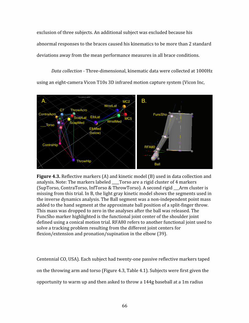
66
exclusion of three subjects. An additional subject was excluded because his
abnormal responses to the braces caused his kinematics to be more than 2 standard
deviations away from the mean performance measures in all brace conditions.
Data collection -‐ Three-‐dimensional, kinematic data were collected at 1000Hz
using an eight-‐camera Vicon T10s 3D infrared motion capture system (Vicon Inc,
Figure 4.3. Reflective markers (A) and kinetic model (B) used in data collection and analysis. Note: The markers labeled ____Torso are a rigid cluster of 4 markers (SupTorso, ContraTorso, InfTorso & ThrowTorso). A second rigid ___Arm cluster is missing from this trial. In B, the light gray kinetic model shows the segments used in the inverse dynamics analysis. The Ball segment was a non-‐independent point mass added to the hand segment at the approximate ball position of a split-‐finger throw. This mass was dropped to zero in the analyses after the ball was released. The FuncSho marker highlighted is the functional joint center of the shoulder joint defined using a conical motion trial. RFA80 refers to another functional joint used to solve a tracking problem resulting from the different joint centers for flexion/extension and pronation/supination in the elbow (39).
Centennial CO, USA). Each subject had twenty-‐one passive reflective markers taped
on the throwing arm and torso (Figure 4.3, Table 4.1). Subjects were first given the
opportunity to warm up and then asked to throw a 144g baseball at a 1m radius
A. B.
ThrowHip
ContraHip
MC2
MC5WristMed
WristLat
ElbLat
ElbMed
(below)
C7 ThrowAcro
ContraAcro
ScapMed
ScapLat
___Torso
FuncSho
Ball
RFA80

67
Table 4.1. Segment parameters used in the kinetic model. Segment length and diameter is obtained using the defining marker sets. Segmental movements are recorded using the tracking markers around the defined axes.
target positioned 10m from the subject. Each subject threw approximately 10-‐20
pitches in each of five randomly ordered experimental conditions (see below). Ball
Segment Definitions
Segment Segment
Geometry
Defining Markers Tracking Markers
Axes
Thorax/Pelvis Cylinder Proximal: ThrowHip, ContraHip
Distal: ThrowAcro, ContraAcro
midHip (calc)
ThrowAcro
ContraAcro
X: axial flex/extension
Y: lateral flex/extension
Z: axial rotation
Arm Cone Proximal: FuncSho (calc), ThrowAcro
Distal: ElbMed, ElbLat
FuncSho (calc)
ElbMed
EldLat
X: flex/extension
Y: ab/adduction
Z: int/external rotation
ForearmUpper Cone Proximal: ElbMed, ElbLat
Distal: RFA80 (calc)
ElbMed
ElbLat
RFA80 (calc)
X: flex/extension
ForearmLower Cone Proximal: RFA80 (calc)
Distal: WriMed, WriLat
RFA80 (calc)
WriMed
WriLat
Z: pro/supination
Hand Sphere Proximal: WriMed, WriLat
Distal: MC5, MC2
midWrist (calc)
MC5
MC2
X: flex/extension
Y: ulnar/radial deviation
Ball Sphere Single marker: Ball Prox (calc)
midWrist (calc)
MC5
MC2
No independent motion

68
speed was measured using a Sports Radar Model 3600 radar gun, and ball accuracy
was calculated using Image J software (v 1.41) from digital video collected with a 30
Hz Canon Vixia HV30 digital video camera (Canon Inc, Tokyo, Japan) recording the
ball impacting the target. Ball release was timed using a synched FlexiForce A201
force sensor (Tekscan Inc, Boston MA, USA) collecting at 1000Hz taped to the
palmar side of the distal phalanx of the third digit.
Experimental treatments -‐ Five different experimental treatments were used:
normal unrestricted, torso restricted, clavicle restricted, shoulder restricted, and
wrist restricted. All restricted conditions were accompanied by sham trials in which
the brace was applied but not tightened to test the effect of the brace itself
independent of the restriction (except for the torso restriction condition for which
this was not possible). All brace conditions used commercially available therapeutic
braces. Subjects were given ad libitum practice throws to acquaint themselves with
both the sham and restricted portions of each condition. The torso restriction
condition employed a DonJoy Dual TLSO back brace (DonJoy Inc., Vista, CA) to limit
intervertebral motion from T2/T3 to the sacrum. This back brace uses wide, elastic
Velcro straps to fasten a rigid plastic plate on the dorsal side of the body with a
similar, smaller plate on the ventral side. A steel bar with a wide, padded end
extends cranially from the ventral plate and is fixed against the clavicles to prevent
rotational motion in the torso. The clavicle restriction condition used a DonJoy
Clavicle Posture Support brace to hold the shoulder in a superiorly rotated,
“shrugged” posture. This brace contains two self-‐adhering straps that run over the
shoulders, under the armpits, and join in the back. During brace tightening, the

69
subject was requested to shrug his shoulders and the clavicle brace was tightened to
prevent a relaxation. For the shoulder restriction condition, a DonJoy Shoulder
Stabilizer brace was used to limit external rotational ROM at the shoulder. This
shoulder brace is comprised of a tight, elastic Velcro vest with a single, elastic half-‐
sleeve on the throwing arm. To restrict external rotation of the humerus, the arm
was internally rotation and a Velcro strap was affixed from the dorsal side of the
vest to the ventral side of the sleeve. The wrist restriction condition employed an
Allsport Dynamics IMC Wrist brace (Allsport Dynamics Inc., Nacogdoches, TX) with
the 0° extension stop either employed (restricted) or absent (sham). This brace
affixed a rigid plastic sleeve and a small plastic plate to the dorsal side of the distal
forearm and hand respectively. When the extension stop was screwed into place, the
plastic sleeve contacted the hand plate and prevented the wrist from
hyperextending.
Analysis -‐ Raw marker data were identified using Vicon Nexus software (v
1.7.1) and exported to C-‐Motion Visual3D software (v4) for additional analysis. A
Butterworth 2nd order low-‐pass filter with a cutoff frequency of 25Hz was chosen
and applied after a residual analysis (41) of the data was conducted (39). Marker
gaps of up to 100 frames were interpolated using Visual3D. Joint Euler angles were
calculated and inverse dynamics analyses were performed using mass distribution
ratios from Dempster (42) in Visual3D. Joint angular velocities, moments, and
power were calculated using each joint’s instantaneous axis of rotation. The Cardan
sequence of rotations at each joint was XYZ. Joint work was calculated in MATLAB (v
7.11.0) using the trapz function. To help simplify analysis, each throwing motion

70
was divided into five standard throwing phases: windup/stride, arm-‐cocking, arm
acceleration, arm deceleration, and follow-‐through (3). Because positive projectile
linear velocity is generated during the arm-‐cocking and acceleration phases (Figure
4.4), we present data from just these two phases. Individual subject means from a
number of performance measures were compared across experimental conditions
using repeated measures ANOVA or MANOVA when variances were not equal
Figure 4.4. The arm-‐cocking and acceleration phases of the throw. Arm-‐cocking begins with the contralateral foot touching the ground at the end of a large stride (STR) in the target direction. During the cocking phase, the humerus externally rotates until it reaches maximum external rotation (MER). Following MER, the very brief acceleration phase occurs in which the humerus rapidly internally rotates, the elbow extends and the wrist is flexed. Acceleration ends with the release (REL) of the projectile.
Arm-cocking phase Acceleration phase
End of Stride Maximum External Rotation Release
Shoulder external rotation Shoulder internal rotation
Elbow !exion Elbow extensionShoulder extension Shoulder !exion
Wrist extension
Torso rotation

71
(Mauchley’s sphericity chi-‐square p < 0.05). Differences were taken as significant at
the alpha < 0.05 level. Post-‐hoc matched pairs t-‐tests were used to determine which
condition (normal, sham, and/or restricted) accounted for any significance. Given
that the torso restriction condition does not have sham, all reported statistics for
this condition are matched pairs t-‐tests.
4.6 Results
Normal unrestricted condition: During normal unrestricted throwing,
maximum ball speed was 27.7±3.8 m/s (mean of all subject means±st. dev. of
subject means) with the ball striking the target on average 0.3±0.2 m from the
bullseye. The angular velocity of torso rotation peaked (848±160 °/s) during the
latter half of the cocking phase (Figure 4.5), before producing an opposing braking
torque during the brief acceleration phase. This opposing torque resulted in a short
period of power absorption during acceleration, generating net negative work
during this phase (-‐74±44 J). During the cocking phase at the shoulder, the humerus
was externally rotated while simultaneously a large, opposing internal rotation
torque was generated. This resulted in a sustained period of power absorption and
negative work (-‐201±70 J) at the shoulder. As the acceleration phase began, with the
initiation of shoulder internal rotation, the shoulder rotation torque and angular
velocity became in-‐phase resulting in very high angular velocities (4290± 1127 °/s)
and peak power (11838±4170 W). At the elbow, the cocking phase began with the
elbow continuing to flex, while a moderate flexion torque was produced. However,

72
Figure 4.5. Joint kinematic and kinetic data from the four critical joint axes during normal throwing. Mean data across all subjects are shown with bolded lines, while 95% confidence intervals are reported with dashed lines. Differences in phase timing between individuals were standardized by interpolation each phase 1000 fold and subsequently down sampling each throw to a standard length. The length ratio of the two phases was kept proportional to the normal, unrestricted phase duration ratio. The arm-‐cocking phase (between STR and MER) is shown in the white field and labeled C in the work plots, while the acceleration phase (between MER and REL) is shown in a gray field and labeled A in the work plots.
during the last quarter of the cocking phase the elbow began to extend rapidly (-‐
2434±552 °/s), peaking midway through the acceleration phase. During this rapid
extension, the opposing flexion moment at the elbow intensified, resulting in large
Angular Velocity (deg/sec) Torque (Nm) Power (W) Work (J)
Wris
tEl
bow
Sh
ould
er
Tors
o
0
0
200
400
600
800
STR MER REL
0
200
400
600
C A
-800
-400
0
400
800
STR MER REL
-4000
0
4000
8000
STR MER REL
-200
0
200
C A
2000
4000
STR MER REL
0
100
200
STR MER REL
0
4000
8000
12000
STR MER REL-4000
-300
-200
-100
0
C A-3000
-2000
-1000
0
STR MER REL 0
100
200
300
400
STR MER REL
-12000
-8000
-4000
0
STR MER REL
0
1000
2000
STR MER REL-2
0
2
4
6
STR MER REL-100
0
100
200
STR MER REL
0
1
2
C A
internal rotation (+)
external rotation (-)
extension (-) flexion (+)
flexion (+)
extension (-)
towards target (+)
away from target (-)
50%
50%
50%
50%
50%
50%
50% 50%
50%
50%
50%50%

73
Torso Restriction
Normal Torso Restriction
Performance Max. ball speed (m/s) 27.7(± 3.8) 26.3(± 4)*
Accuracy (m from bullseye) 0.3(± 0.2) 0.4(± 0.3)
Timing Duration of Cocking (ms) 387(± 182) 303(± 122)*
Duration of Acceleration (ms) 40(± 12) 37(± 12)
Torso rotation Peak Ang. Vel. (deg/s) 848(± 160) 835(± 158)
(+ throw arm torwards target, Peak Torque (Nm) 828(± 437) 773(± 424)
-‐ away from target) Peak Power (W) 6207(± 2190) 5968(± 2166)
Work -‐ Cocking (J) 609(± 171) 536(± 149)*
Work -‐ Acceleration (J) -‐74(± 44) -‐79(± 40)
Shoulder rotation Peak Ang. Vel. (deg/s) 4290(± 1127) 3970(± 1226)*
(+ internal, -‐ external) Peak Torque (Nm) 206(± 42) 178(± 42)
Peak Power (W) 11838(± 4170) 9872(± 4005)*
Work -‐ Cocking (J) -‐201(± 70) -‐184(± 73)*
Work -‐ Acceleration (J) 132(± 52) 100(± 56)*
Elbow flexion/extension Peak Ang. Vel. (deg/s) -‐2434(± 552) -‐2284(± 582)*
(+ flexion, -‐ extension) Peak Torque (Nm) 392(± 116) 357(± 121)
Peak Power (W) 2028(± 3983) 2473(± 4498)
Work -‐ Cocking (J) 2(± 27) -‐1(± 22)
Work -‐ Acceleration (J) -‐246(± 63) -‐218(± 53)*
Wrist flexion/extension Peak Ang. Vel. (deg/s) 1593(± 336) 1645(± 280)
(+ flexion, -‐ extension) Peak Torque (Nm) 7(± 3) 6(± 3)
Peak Power (W) 138(± 90) 155(± 109)
Work -‐ Cocking (J) 0.2(± 1) 0.3(± 1)
Work -‐ Acceleration (J) 1(± 2) 1(± 2)
Table 4.2. Kinematic and kinetic performance measures for the four critical joint axes during normal and torso restriction conditions. Given that no sham was possible for this condition, restricted values that statistically differ (p <0.05) from normal values are indicated with *. Bold values indicate hypothesized reductions from normal values, while italic values indicate unexpected changes.
amounts of power being absorbed and negative work generated (-‐246±63 J). Finally,
at the wrist, a sustained extension occurred until the very end of the cocking phase,

74
when the wrist began to rapidly flex, continuing through the acceleration phase. A
small flexion moment was produced (peak -‐ 7±3 Nm) at the end of cocking, which
then dropped to near zero and recovered during the acceleration phase. This
oscillating power resulted in a small, net positive amount of work (1±2 J) generated
during acceleration. Importantly, while there was considerable variation between
subjects in the timing and duration of the throwing phases, as well as significant
differences in overall performance, there was very little variation in the pattern of
throwing kinematics across subjects.
Torso restriction condition: The torso restriction brace completely restricted
intervertebral rotational motion, allowing only the hip rotators to rotate the trunk.
Given the design of the torso brace, no sham condition was possible. Contrary to
expectations, there were no significant reductions in torso rotation peak power,
angular velocity, or torque (Table 4.2). However, a significant drop in torso rotation
work during cocking (-‐11±18%) was recorded. As expected, significant reductions
were seen in shoulder rotation peak angular velocity (-‐7±13%), power (-‐14±20%),
and work (cocking: -‐7±25%; acceleration: -‐19±30%). Reductions also occurred in
elbow extension peak angular velocity (-‐6±7%) and elbow work during acceleration
(-‐7±14%). No significant reductions were measured in any performance variables at
the wrist. As hypothesized, when the torso restriction brace was applied, maximum
ball speed dropped -‐5±6%.
Clavicle restriction condition: When applied, the clavicle brace superiorly
rotated the scapula 7±4° and also limited scapular protraction. The clavicle sham

75
Clavicle Restriction
Normal Clavicle Sham Clavicle
Restriction Performance Max. ball speed (m/s) 27.7(± 3.8) 27(± 4.2)* 26.3(± 4.1)*† Accuracy (m from bullseye) 0.3(± 0.2) 0.4(± 0.2) 0.4(± 0.2) Timing Duration of Cocking (ms) 387(± 182) 300(± 72)* 306(± 93)* Duration of Acceleration (ms) 40(± 12) 32(± 9)* 34(± 11) Torso rotation Peak Ang. Vel. (deg/s) 848(± 160) 862(± 144) 870(± 155) (+ throw arm torwards target, -‐ away from target)
Peak Torque (Nm) 828(± 437) 580(± 167) 588(± 240)
Peak Power (W) 6207(± 2190) 6869(± 2617) 6492(± 2159) Work -‐ Cocking (J) 609(± 171) 638(± 177) 607(± 174) Work -‐ Acceleration (J) -‐74(± 44) -‐109(± 53)* -‐107(± 63)* Shoulder rotation Peak Ang. Vel. (deg/s) 4290(± 1127) 3852(± 1175)* 3778(± 1126)* (+ internal, -‐ external) Peak Torque (Nm) 206(± 42) 180(± 38) 169(± 37)*† Peak Power (W) 11838(± 4170) 8431(± 4455)* 8958(± 4552)* Work -‐ Cocking (J) -‐201(± 70) -‐188(± 60) -‐173(± 63)*† Work -‐ Acceleration (J) 132(± 52) 54(± 35)* 58(± 32)* Elbow flexion/extension Peak Ang. Vel. (deg/s) -‐2434(± 552) -‐2326(± 517) -‐2220(± 517)*† (+ flexion, -‐ extension) Peak Torque (Nm) 392(± 116) 357(± 80) 336(± 84)† Peak Power (W) 2028(± 3983) 631(± 1050) 630(± 1064) Work -‐ Cocking (J) 2(± 27) 4(± 21) 5(± 22) Work -‐ Acceleration (J) -‐246(± 63) -‐225(± 58)* -‐206(± 59)*† Wrist flexion/extension Peak Ang. Vel. (deg/s) 1593(± 336) 1505(± 255) 1510(± 276) (+ flexion, -‐ extension) Peak Torque (Nm) 7(± 3) 6(± 2) 6(± 2) Peak Power (W) 138(± 90) 113(± 80) 107(± 73) Work -‐ Cocking (J) 0.2(± 1) 0.4(± 1) 0.3(± 1) Work -‐ Acceleration (J) 1(± 2) 1(± 1) 1(± 1)
Table 4.3. Kinematic and kinetic performance measures for the four critical joint axes during normal and clavicle brace restricted conditions. Restricted and sham values that statistically differ (p <0.05) from normal values are indicated with *, while restricted values that statistically differ from the sham are shown with †. Bold values indicate hypothesized reductions from normal values, while italic values indicate unexpected changes.
showed no significant effects on joint motion. However, data from the sham trials
(Table 4.3) indicate some perturbation of the normal throwing motion as there were

76
reductions in: mean maximum ball speed (-‐3±5%), phase duration (cocking -‐
12±27%, acceleration -‐16±27%), torso rotation work – acceleration (-‐42±42%),
elbow extension work – acceleration (-‐10±17%), shoulder rotation work –
acceleration (-‐56±25%), and shoulder rotation angular velocity (-‐11±12%) and
power (-‐28±32%). When the restriction was added, there was a further drop in
maximum ball speed of -‐3±6%. Reductions in shoulder rotation peak torque (-‐
13±31%) and work during acceleration (-‐9±10%) were recorded relative to normal
values. Elbow extension angular velocity dropped -‐4±8% relative to normal, while
elbow work during acceleration dropped -‐8±13% relative to the reduced sham
values. No significant changes were observed in the wrist.
Shoulder restriction condition: When tightened, the shoulder brace reduced
shoulder external rotation by 24±9°. However, when acting as a sham, the shoulder
brace also restricted external rotation by 11±7°, much like a tight jacket. During the
sham trials, there were also slight reductions in mean maximum ball speed (-‐3±5%),
shoulder rotation work during the cocking phase (-‐9±13%), and a slight lengthening
of the duration of the cocking phase (3±5%) (Table 4.4). However, for each of these
significant changes in the sham condition, the shoulder restriction condition
statistically differed from both the sham and normal conditions. The restricted trials
showed a reduction in maximum ball speed from the sham trials of -‐8±6%. The
duration of the cocking phase of the restricted trials shortened (-‐9±14%), while the

77
Shoulder Restriction
Normal Shoulder Sham Shoulder
Restriction Performance Max. ball speed (m/s) 27.7(± 3.8) 27(± 4)* 24.9(± 4)*† Accuracy (m from bullseye) 0.3(± 0.2) 0.4(± 0.3) 0.4(± 0.3) Timing Duration of Cocking (ms) 387(± 182) 392(± 167) 354(± 196)*† Duration of Acceleration (ms) 40(± 12) 38(± 14)* 55(± 27)*† Torso rotation Peak Ang. Vel. (deg/s) 848(± 160) 859(± 161) 833(± 156) (+ throw arm torwards target, -‐ away from target)
Peak Torque (Nm) 828(± 437) 827(± 404) 890(± 453)
Peak Power (W) 6207(± 2190) 6111(± 2036) 5559(± 2000) Work -‐ Cocking (J) 609(± 171) 587(± 152) 513(± 154)*† Work -‐ Acceleration (J) -‐74(± 44) -‐78(± 42) -‐42(± 57)*† Shoulder rotation Peak Ang. Vel. (deg/s) 4290(± 1127) 4261(± 1106) 4038(± 1141) (+ internal, -‐ external) Peak Torque (Nm) 206(± 42) 206(± 45) 198(± 45) Peak Power (W) 11838(± 4170) 12145(± 4628) 13566(± 6141) Work -‐ Cocking (J) -‐201(± 70) -‐181(± 63)* -‐113(± 57)*† Work -‐ Acceleration (J) 132(± 52) 124(± 63) 138(± 58) Elbow flexion/extension Peak Ang. Vel. (deg/s) -‐2434(± 552) -‐2341(± 505) 1923(± 471)*† (+ flexion, -‐ extension) Peak Torque (Nm) 392(± 116) 355(± 88) 326(± 100) Peak Power (W) 2028(± 3983) 1229(± 2050) 1180(± 2383) Work -‐ Cocking (J) 2(± 27) 3(± 20) 4(± 24) Work -‐ Acceleration (J) -‐246(± 63) -‐236(± 58) -‐190(± 46)*† Wrist flexion/extension Peak Ang. Vel. (deg/s) 1593(± 336) 1549(± 490) 1515(± 332) (+ flexion, -‐ extension) Peak Torque (Nm) 7(± 3) 6(± 2) 6(± 2) Peak Power (W) 138(± 90) 128(± 74) 127(± 70) Work -‐ Cocking (J) 0.2(± 1) 0.4(± 1) -‐0.1(± 1)*† Work -‐ Acceleration (J) 1(± 2) 1(± 2) 2(± 1)*†
Table 4.4. Kinematic and kinetic performance measures for the four critical joint axes during normal and shoulder brace restricted conditions. Restricted and sham values that statistically differ (p <0.05) from normal values are indicated with *, while restricted values that statistically differ from the sham are shown with †. Bold values indicate hypothesized reductions from normal values, while italic values indicate unexpected changes.
acceleration phase lengthened (38±56%). Reductions in torso rotation work
were recorded for both phases (cocking -‐14±20%, acceleration -‐41±57%). Although

78
shoulder rotation work during the cocking phase dropped significantly (-‐45±17%)
from normal values, no further significant reductions occurred in shoulder rotation
peak angular velocity, torque, power, or work during acceleration. As predicted
there were significant reductions in elbow extension angular velocity (-‐21±10%)
and elbow work during acceleration (-‐20±21%). Wrist flexion peak angular velocity
remained unchanged, but a significant reduction in wrist work during cocking was
followed by a significant increase in wrist work during acceleration.
Wrist restriction condition: When restricted by a brace, wrist extension ROM
was reduced 62±7°, but there was no significant reduction in the sham condition.
However, the wrist restriction sham trials showed numerous significant reductions
in the measured performance variables (Table 4.5). Although the restricted
condition showed significant reductions in ball speed and phase duration from
normal values, these reductions were not significantly different from the sham
condition. Similarly, while torso rotation performance metrics differed from normal
values, only torso rotation work during acceleration differed between the sham and
restricted trials (-‐6±17%). However, restricted shoulder rotation peak angular
velocity dropped -‐6±8% from sham values, while shoulder rotation peak power and
work values all dropped relative to normal and increased relative to sham trials.
Elbow extension peak angular velocity did not differ between sham and restricted
conditions, although restricted elbow work during acceleration dropped -‐19±14%
from sham levels. The wrist flexion peak angular velocity dropped -‐34±37% relative

79
to the sham trials, while wrist flexion/extension peak power and work during
acceleration increased relative to the sham.
Wrist Restriction
Normal Shoulder Sham Shoulder
Restriction Performance Max. ball speed (m/s) 27.7(± 3.8) 26.1(± 3.9)* 25.8(± 4)* Accuracy (m from bullseye) 0.3(± 0.2) 0.4(± 0.2) 0.4(± 0.2) Timing Duration of Cocking (ms) 387(± 182) 309(± 72)* 311(± 95)* Duration of Acceleration (ms) 40(± 12) 34(± 9)* 33(± 10)* Torso rotation Peak Ang. Vel. (deg/s) 848(± 160) 838(± 175) 831(± 188) (+ throw arm torwards target, -‐ away from target)
Peak Torque (Nm) 828(± 437) 431(± 96)* 409(± 101)*
Peak Power (W) 6207(± 2190) 5488(± 1670) 5676(± 2149) Work -‐ Cocking (J) 609(± 171) 541(± 136)* 550(± 155)* Work -‐ Acceleration (J) -‐74(± 44) -‐69(± 42) -‐62(± 39)*† Shoulder rotation Peak Ang. Vel. (deg/s) 4290(± 1127) 3799(± 1237)* 3579(± 1217)*† (+ internal, -‐ external) Peak Torque (Nm) 206(± 42) 191(± 53) 185(± 53) Peak Power (W) 11838(± 4170) 3534(± 1415)* 4852(± 2614)*† Work -‐ Cocking (J) -‐201(± 70) -‐181(± 66)* -‐192(± 68)† Work -‐ Acceleration (J) 132(± 52) 33(± 27)* 49(± 31)*† Elbow flexion/extension Peak Ang. Vel. (deg/s) -‐2434(± 552) -‐2308(± 587)* -‐2261(± 588)* (+ flexion, -‐ extension) Peak Torque (Nm) 392(± 116) 351(± 116) 276(± 81)*† Peak Power (W) 2028(± 3983) 537(± 466) 354(± 178) Work -‐ Cocking (J) 2(± 27) 15(± 19)* -‐0.5(± 24)† Work -‐ Acceleration (J) -‐246(± 63) -‐215(± 63)* -‐174(± 61)*† Wrist flexion/extension Peak Ang. Vel. (deg/s) 1593(± 336) 1406(± 331)* 881(± 243)*† (+ flexion, -‐ extension) Peak Torque (Nm) 7(± 3) 3(± 2)* 6(± 2)† Peak Power (W) 138(± 90) 24(± 28)* 47(± 29)*† Work -‐ Cocking (J) 0.2(± 1) 0.5(± 1) 0.5(± 0.4) Work -‐ Acceleration (J) 1(± 2) -‐3(± 2)* 1(± 1)†
Table 4.5. Kinematic and kinetic performance measures for the four critical joint axes during normal and wrist brace restricted conditions. Restricted and sham values that statistically differ (p <0.05) from normal values are indicated with *, while restricted values that statistically differ from the sham are shown with †. Bold values indicate hypothesized reductions from normal values, while italic values indicate unexpected changes.

80
4.7 Discussion
Overall, the results of the brace restriction trials are largely consistent with
our model of power generation during throwing. We found support for both the use
of an elastic energy storage mechanism at the shoulder as well as the importance of
a kinetic chain in transferring power between body segments. These data also show
some unanticipated responses to sham conditions, especially in the wrist, which
bear further explanation and investigation.
We hypothesized (H1) that by restricting rotational movement between the
vertebrae by using a brace, measures of throwing performance would experience
small, but significant reductions. However, despite the fact that the restriction of
intervertebral motion was essentially complete, we saw a significant reduction in
torso rotation work during only the cocking phase. All other torso performance
measures remained unchanged. This result suggests that during normal throwing,
most (~90%) of the work required to achieve high projectile velocity is generated at
the hips. The reduction seen in torso rotation work during the cocking phase may be
driven by a significant reduction in the duration of this phase. However, the fact that
other brace conditions result in similar changes in phase duration without affecting
the amount of torso rotation work performed suggest this is not the case. These data
strongly support the hypothesis that the hip rotators are almost wholly responsible
for powering the rapid rotation of the torso (34-‐36). Further work is necessary to
determine which hip rotator muscles are primarily responsible for generating this
power.

81
Although the torso brace restriction resulted in mostly minor, non-‐significant
reductions in performance at the torso, the effects of this brace do seem to be
amplified at the shoulder. We had hypothesized (H2) that if torso rotation was in
fact powering the elastic storage mechanism at the shoulder (39), even minor
reductions in torso rotation performance should result in less elastic energy stored
at the shoulder and, consequently, large reductions in shoulder rotation
performance. The data from the torso restriction trials support this hypothesis.
Negative shoulder rotation work during the cocking phase, which can be used as a
proxy for elastic energy storage, is reduced by more than 10% during the torso
brace trials. Further reductions in shoulder rotation angular velocity, power, and
work during the acceleration phase accompany this reduction in work during the
cocking phase. While it is again possible that a reduction in the duration of the
cocking phase is partly responsible for changes in the shoulder performance
measures, data from other experimental conditions in which the cocking phase
shortened without affecting these same measures make this unlikely. Furthermore,
no change is seen in the duration of the acceleration phase in which peak shoulder
rotation angular velocity and power are achieved. These results provide support for
torso rotation contributing to elastic energy storage in the shoulder during the arm-‐
cocking phase.
The other potential source of power we proposed (H3) could contribute to
elastic energy storage at the shoulder is Pectoralis major. By using the clavicle brace
to hold the shoulder complex in a superiorly rotated position, we sought to change
this muscle’s major line of action, effectively excluding contributions from the

82
inferior fibers of the muscle to horizontal flexion at the shoulder. While no
significant effects of the sham were measured on static shoulder ROM, significant
changes in performance measures during the sham trials suggest that the unloaded
brace did cause some restriction potentially by limiting scapular protraction around
the moment of release. However, despite the unexpected restriction caused by the
sham and the performance responses that resulted during the sham trials, the
restricted trials did show further significant decreases in shoulder rotation work
during the critical cocking phase and also in peak shoulder torque. Shoulder
rotation angular velocity, power, and work during acceleration all changed relative
to the normal condition, but did not differ from the sham trials. While the reductions
in shoulder rotation torque and work during cocking confirm the effect of the
restriction and support the role of P. major in loading the elastic elements in the
shoulder, the lack of significant shifts in the other performance measures (e.g.
shoulder rotation angular velocity, power, and work during acceleration) leaves this
hypothesis only partially supported. It is possible that the minor superior rotation
the brace induced was insufficient to yield a large enough effect on these
performance measures to overcome the high degree of variance they show.
However, it is worth noting that the clavicle brace restriction did lead to
performance reductions in peak elbow extension angular velocity and work during
the acceleration phase.
It has long been thought that, despite very rapid angular velocities at the
distal elbow and wrist joints, the work and power necessary to achieve rapid
angular movements in these joints during throwing are derived from power

83
generated at more proximal joints (7-‐9, 20, 21). As hypothesized (H4), work during
both the cocking and acceleration phases of a normal throw is very low at the wrist
and low or negative at the elbow. This finding suggests that both motions are largely
passive. Furthermore, when examining all brace conditions (torso, clavicle,
shoulder) in which proximal joint actions (torso rotation and/or shoulder rotation)
were affected, significant performance reductions were seen at the elbow, but not
the wrist. These data suggest that the elbow is likely powered primarily by kinetic
transfer from upstream joints (7-‐9, 20). This kinetic chain may also apply to the
wrist, although the lack of response to the restrictions at the wrist suggests some
compensatory action is possible. It is worth noting that given the brevity of the
acceleration phase, having multiple passive components in the shoulder, elbow, and
wrist could serve to simplify the difficult neural control problem that rapid
throwing poses. Such reduced complexity would allow the throwing motion to be
more easily, effectively, and consistently produced.
Another hypothesis (H5) was that wrist hyperextension allows throwers to
delay projectile release, enabling additional acceleration of the proximal joints,
while still maintaining an accurate release trajectory. Although the wrist brace
restriction did show a reduction in the duration of the acceleration phase relative to
the normal condition, it did not differ from the reduction seen during the sham trials.
In fact, unlike responses to the other brace’s sham conditions, the performance
effects of the wrist sham were pervasive and severe. Significant performance
reductions were seen in all segments analyzed during the wrist sham condition.
Given that no measurable effect of wrist ROM was noted with the sham, it is likely

84
that the effects seen during the sham trials were due to the addition of the brace’s
mass to the distal forelimb. Although the mass of the brace was ~165 g, this
additional mass more than doubles the previously added ball’s mass at the hand.
These sham result suggest that the elastic storage mechanism at the shoulder could
be quite sensitive to changes in projectile or distal forearm mass. Without postural
adjustments, such a change in mass could significantly increase the forelimb’s
moment of inertia during the critical cocking phase and disrupt a system finely
tuned for passive action.
The wrist brace restriction did show significant performance effects relative
to the wrist sham where expected (shoulder rotation angular velocity, elbow
extension work – acceleration, and wrist flexion angular velocity). However, these
reductions appear to be independent of any change in phase duration. Furthermore,
a number of performance variables that were predicted to drop because of the
restriction (shoulder rotation work -‐ acceleration, wrist peak power and work -‐
acceleration) actually increased over the sham values, potentially because of
compensation for the restriction. While these data do not lead to the outright
rejection of our wrist hyperextension hypothesis, further data on adding mass at the
wrist without using a brace are needed to address the effects of both factors on
throwing performance.
While the use of braces to induce variation in joint ROM can be an effective
way to address how skeletal morphology can contribute to complex motions such as
throwing, such an approach is not without limitations. Although all brace

85
restrictions (except for the wrist) showed significant reductions in ball speed
relative to both their sham and normal trials, these reductions were relatively minor
(0.5-‐9%). Such minor reductions highlight the ability of the subjects -‐ all
accomplished throwers -‐ to compensate for minor to moderate disruptions of their
normal throwing motion. Brace restrictions can also lead to unintended responses,
such as limiting scapular protraction (clavicle brace) or causing compensatory
flexion action at the wrist (shoulder brace). Our experimental design targeted key
motions responsible for projectile speed, but the effect of these braces were not
restricted to only the motions reported here. Furthermore, even when tasked to
throw as hard as possible in all brace restricted conditions, the tendency of some
subjects to fight through the restrictions in order to maintain performance can lead
to variable effort between trials. Despite these drawbacks of our experimental
approach, both the kinematic data and the fact that accuracy remained unchanged
across all conditions does provide some confirmation that the normal throwing
motion was mostly preserved.
In conclusion, the results of this study confirm previous findings that power
generation during rapid overhand throwing is strongly affected by two phenomena:
the transfer of power/work from proximal segments to distal joints via a kinetic
chain (6, 7, 17, 20, 43) and elastic energy storage at the shoulder (23, 39). The hips
rotators were shown to account for most of the torso rotation power and work
produced during throwing. Through a kinetic transfer of power, torso rotation
enabled the passive loading of the elastic elements in the shoulder during the
cocking phase. Much of the energy absorbed at the shoulder during cocking was

86
then recovered in the acceleration phase, resulting in faster shoulder internal
rotation and elbow extension just prior to release (39). The Pectoralis major was
also shown to contribute to the amount of energy absorption (negative work) at the
shoulder during the cocking phase and again showed significant effects on elbow
extension angular velocity. Kinetic power transfers like that used to load the elastic
storage mechanism are also likely driving the rapid elbow extension and wrist
flexion motions that occur at the end of the throw (7-‐9, 20). Both the wrist and
elbow show little to no positive work performed during either the cocking or
acceleration phases suggesting that both motions are powered by more proximal
joint actions. The effects of wrist hyperextension on the throwing motion remain
unclear. While there are clearly some performance benefits to hyperextension, the
unexpected effects of adding mass at the wrist makes interpreting these results
difficult.
A number of important questions remain unanswered and need further
study. While the storage and use of elastic energy at the shoulder is strongly
supported, where this energy is stored in unknown. There are numerous short,
elastic ligaments in the shoulder capsule and broad, flat muscles tendons crossing
the shoulder joint that, when stretched, could store elastic energy. There are also
elastic components within the muscle tissue of the many shoulder muscles that
could also contribute. Further investigation of these elastic elements using in vivo
soft tissue imaging during the throwing motion would be invaluable in addressing
which elements are storing elastic energy. It is also unknown how variations in
throwing kinematics would affect elastic energy storage. Increasing the variance in

87
subjects’ throwing performance or including alternative throwing styles, such as
windmill-‐type cricket bowls, could provide important insights into differences in
elastic energy storage and throwing performance.
Finally, how power is generated during throwing may be used to better
understand common throwing injuries. Three of the most common injuries in
throwing athletes occur at the shoulder (shoulder labrum tears, shoulder
instability) and elbow (medial epicondylitis) (6, 10, 44-‐50). Damage to the superior
portion of the shoulder labrum at the attachment site of the biceps (a SLAP
lesion/tear) likely results from a high degree of passive external rotation of the
humerus during the arm-‐cocking phase (51, 52). During arm-‐cocking, the biceps are
activated in order to maintain the flexed elbow that is vital for elastic energy storage
(11). However, the combination of bicep flexion and passive external rotation of the
humerus cause high concentrations of stress at the biceps’ tendinous insertion on
the labrum. Such stretching and sometimes tearing of the biceps tendon suggests
that this tendon may in fact provide some elastic storage potential irrespective of
the role of the biceps in positioning the forearm. Similarly, shoulder instability and
the problems it can cause (anterior ligament laxity, increased likelihood of
dislocation (48)) may be driven by use of the capsular ligaments to store elastic
energy at the shoulder to power rapid throwing. During cocking, the arm is
abducted in line with the shoulders, horizontally extending, and externally rotated.
This arm position pulls taut the anterior band of the inferior glenohumeral ligament,
the middle glenohumeral ligament, and the coracohumeral ligament (53). When the
arm is then passively externally rotated as its mass lags behind the accelerating

88
torso, these ligaments are stretched. Such stretching could enable the storage of
elastic energy in these ligaments, although with overuse it could lead to deformation
of these ligaments and, consequently, shoulder instability. Finally, the elbow flexion
positioning vital to increase the forelimb’s moment of inertia and enable elastic
energy storage also results in very high valgus torques at the elbow during both the
arm-‐cocking and early acceleration phases (10, 54-‐56). This high, out-‐of-‐the-‐joint-‐
plane torque stretches the ulnar collateral ligament, which stabilizes the elbow, and
can lead to damage and painful inflammation (6, 10, 50). All of these injuries directly
result from forelimb positioning necessary to maximize elastic energy storage.
Other frequent shoulder injuries, such as rotator cuff tears, are less directly related
to power generation, but likely result from forces generated to decelerate the
rapidly moving forelimb after the projectile is released (10, 49, 57).
Although the elastic energy used to power the throwing motion is vital to
throwing performance, the results from this study suggest steps that might be taken
to reduce the risk of injury to throwers. A number of studies have shown that
limiting the frequency of high-‐speed throwing does have some protective effect
against these overuse injuries (6, 47, 50, 55, 58, 59). Furthermore, it has been shown
that the amount of passive external rotation at the shoulder during the arm-‐cocking
phase (when elastic energy is stored) is correlated with injury (37, 38). This result
suggests that using mildly restrictive garments (such as a compression shirt) would
have some protective effect against over-‐rotation injuries. However, it should be
cautioned that because the same factors that enhance throwing performance also

89
increase the risk of injury, completely preventing these injuries is probably
impossible.
4.8 Acknowledgements
I would like to thank Dan Lieberman for his help in designing this study and
tirelessly commenting on this manuscript; the National Science Foundation, the
American School for Prehistoric Research, and the Harvard Department of Human
Evolutionary Biology for generously funding this research; our subjects for
generously giving their time; the Wyss Institute Motion Capture lab for the use of
their equipment and facilities; Mike Rainbow, Madhu Venkadesan, Leia Stirling, Matt
Vincent, George Karas, Randi Griffin, Gulus Emre, Wes Gordon, Adanma Ekeledo, and
Amanda Lobell for their invaluable help.

90
4.9 Literature Cited
1. Larson SG (1993) Functional Morphology of the Shoulder in Primates. Postcranial Adaptation in Non-‐human Primates, ed Gebo DL (Northern Illinois University Press, Chicago, IL), pp 45-‐69.
2. Larson SG (2007) Evolutionary Transformation of the Hominin Shoulder.
Evolutionary Anthropology 16:172-‐187. 3. Dillman CJ, Fleisig GS, & Andrews JR (1993) Biomechanics of pitching with
emphasis upon shoulder kinematics. J Orthop Sports Phys Ther 18(2):402-‐408.
4. Pappas AM, Zawacki RM, & Sullivan TJ (1985) Biomechanics of baseball
pitching. A preliminary report. Am J Sports Med 13(4):216-‐222. 5. Hong DA, Cheung TK, & Roberts EM (2001) A three-‐dimensional, six-‐segment
chain analysis of forceful overarm throwing. J Electromyogr Kinesiol 11(2):95-‐112.
6. Fleisig GS, Barrentine SW, Escamilla RF, & Andrews JR (1996) Biomechanics
of overhand throwing with implications for injuries. Sports Med 21(6):421-‐437.
7. Hirashima M, Yamane K, Nakamura Y, & Ohtsuki T (2008) Kinetic chain of
overarm throwing in terms of joint rotations revealed by induced acceleration analysis. J Biomech 41(13):2874-‐2883.
8. Hirashima M, Kudo K, Watarai K, & Ohtsuki T (2007) Control of 3D limb
dynamics in unconstrained overarm throws of different speeds performed by skilled baseball players. J Neurophysiol 97(1):680-‐691.
9. Putnam CA (1993) Sequential motions of body segments in striking and
throwing skills: descriptions and explanations. J Biomech 26 Suppl 1:125-‐135. 10. Fleisig GS, Andrews JR, Dillman CJ, & Escamilla RF (1995) Kinetics of baseball
pitching with implications about injury mechanisms. Am J Sports Med 23(2):233-‐239.
11. Hirashima M, Kadota H, Sakurai S, Kudo K, & Ohtsuki T (2002) Sequential
muscle activity and its functional role in the upper extremity and trunk during overarm throwing. J Sports Sci 20(4):301-‐310.
12. Roberts EM (1971) Cinematography in biomedical investigation. Selected
Topics in Biomedical Invesitgation. Proceedings of the C.I.C. Symposium on Biomechanics, ed Cooper JM (The Athletic Institute, Chicago), pp 71-‐81.

91
13. DiGiovine NM, Jobe FW, Pink M, & Perry J (1992) An electromyography analysis of the upper extremity in pitching. J Shoulder Elbow Surg 1:15-‐25.
14. Gowan ID, Jobe FW, Tibone JE, Perry J, & Moynes DR (1987) A comparative
electromyographic analysis of the shoulder during pitching. Professional versus amateur pitchers. Am J Sports Med 15(6):586-‐590.
15. Gans C (1992) Electromyography. Biomechanics -‐ Structures and Systems: A
Practical Approach, ed Biewener AA (Oxford University Press, Oxford), pp 123-‐147.
16. Bell DG (1993) The influence of air temperature on the EMG/force
relationship of the quadriceps. Eur J Appl Physiol Occup Physiol 67(3):256-‐260.
17. Atwater AE (1979) Biomechanics of overarm throwing movements and of
throwing injuries. Exerc Sport Sci Rev 7:43-‐85. 18. Hore J, O'Brien M, & Watts S (2005) Control of joint rotations in overarm
throws of different speeds made by dominant and nondominant arms. J Neurophysiol 94(6):3975-‐3986.
19. Kibler WB & Sciascia A (2004) Kinetic chain contributions to elbow function
and dysfunction in sports. Clin Sports Med 23(4):545-‐552, viii. 20. Feltner ME (1989) 3-‐Dimensional Interactions in a 2-‐Segment Kinetic
Chain .2. Application to the Throwing Arm in Baseball Pitching. Int J Sport Biomech 5(4):420-‐450.
21. Hirashima M, Ohgane K, Kudo K, Hase K, & Ohtsuki T (2003) Counteractive
relationship between the interaction torque and muscle torque at the wrist is predestined in ball-‐throwing. J Neurophysiol 90(3):1449-‐1463.
22. Alexander RM (1991) Optimum timing of muscle activation for simple
models of throwing. J Theor Biol 150(3):349-‐372. 23. Wilk KE, et al. (1993) Stretch-‐shortening drills for the upper extremities:
theory and clinical application. J Orthop Sports Phys Ther 17(5):225-‐239. 24. Newman S, et al. (1984) Respiratory muscle length measured by
sonomicrometry. J Appl Physiol 56(3):753-‐764. 25. Biewener AA, Corning WR, & Tobalske BW (1998) In vivo pectoralis muscle
force-‐length behavior during level flight in pigeons (Columba livia). J Exp Biol 201 (Pt 24):3293-‐3307.

92
26. Finni T, Komi PV, & Lepola V (2000) In vivo human triceps surae and quadriceps femoris muscle function in a squat jump and counter movement jump. Eur J Appl Physiol 83(4 -‐5):416-‐426.
27. Komi PV, Salonen M, Jarvinen M, & Kokko O (1987) In vivo registration of
Achilles tendon forces in man. I. Methodological development. Int J Sports Med 8 Suppl 1:3-‐8.
28. Lewis JL, Lew WD, & Schmidt J (1982) A note on the application and
evaluation of the buckle transducer for the knee ligament force measurement. J Biomech Eng 104(2):125-‐128.
29. Pourcelot P, Defontaine M, Ravary B, Lematre M, & Crevier-‐Denoix N (2005)
A non-‐invasive method of tendon force measurement. J Biomech 38(10):2124-‐2129.
30. Goodall J (1986) The Chimpanzees of Gombe: Patterns of Behavior (Harvard
University Press, Cambridge). 31. Goodall J (1964) Tool-‐Using and Aimed Throwing in a Community of Free-‐
Living Chimpanzees. Nature 201:1264-‐1266. 32. Kohler W (1925) The Mentality of Apes (Harcourt and Brace, New York). 33. Sugiyama Y & Koman J (1979) Tool-‐using and making behavior in wild
chimpanzees at Bossou, Guinea. Primates 20:513-‐524. 34. Wight J, Richards J, & Hall S (2004) Influence of pelvis rotation styles on
baseball pitching mechanics. Sports Biomech 3(1):67-‐83. 35. Stodden DF, Fleisig GS, McLean SP, Lyman SL, & Andrews JR (2001)
Relationship of pelvis and upper torso kinematics to pitched baseball velocity. J Appl Biomech 17(1):164-‐172.
36. Matsuo T, Escamilla RF, Fleisig GS, Barrentine SW, & Andrews JR (2001)
Comparison of kinematic and temporal patterns between different pitch velocity groups. J Appl Biomech 17(1):1-‐13.
37. Miyashita K, et al. (2008) Relationship between maximum shoulder external
rotation angle and throwing and physical variables. J Sports Sci Med 7:47-‐53. 38. Miyashita K, et al. (2008) The role of shoulder maximum external rotation
during throwing for elbow injury prevention in baseball players. J Sports Sci Med 7:223-‐228.

93
39. Roach NT (2012) Chapter 3 -‐ Elastic energy storage in the shoulder and the evolution of high-‐speed throwing in Homo; in The Biomechanics and Evolution of High-‐Speed Throwing PhD dissertation (Harvard University, Cambridge, MA).
40. Lee YH & Chun SI (1991) Congenital absence of pectoralis major: a case
report and isokinetic analysis of shoulder motion. Yonsei medical journal 32(1):87-‐90.
41. Winter DA (1991) The biomechanics of motor control of human gait: normal,
elderly and pathological (University of Waterloo Press, Waterloo, Ontario). 42. Dempster WT (1955) Space requirements for the seated operator (Wright Air
Development Center, Wright-‐Patterson Air Force Base, OH). 43. Higgins JR (1977) Human Movement: an integrated approach (The C.V. Mosby
Company, Saint Louis). 44. Altchek DW & Dines DM (1995) Shoulder Injuries in the Throwing Athlete. J
Am Acad Orthop Surg 3(3):159-‐165. 45. Anz AW, et al. (2010) Correlation of torque and elbow injury in professional
baseball pitchers. Am J Sports Med 38(7):1368-‐1374. 46. Badia A & Stennett C (2006) Sports-‐related injuries of the elbow. J Hand Ther
19(2):206-‐226. 47. Fleisig GS, et al. (2011) Risk of serious injury for young baseball pitchers: a
10-‐year prospective study. Am J Sports Med 39(2):253-‐257. 48. Meister K (2000) Injuries to the shoulder in the throwing athlete. Part one:
Biomechanics/pathophysiology/classification of injury. Am J Sports Med 28(2):265-‐275.
49. Jobe CM, Pink MM, Jobe FW, & Shaffer B (1996) Anterior shoulder instability,
impingement, and rotator cuff tear: theories and concepts. Operative Techniques in Upper Extremity Sports Injuries, ed Jobe FW (Mosby, St. Louis, MO).
50. Rizio L & Uribe JW (2001) Overuse injuries of the upper extremity in baseball.
Clin Sports Med 20(3):453-‐468. 51. Burkhart SS, Morgan CD, & Kibler WB (2003) The disabled throwing
shoulder: spectrum of pathology Part I: pathoanatomy and biomechanics. Arthroscopy 19(4):404-‐420.

94
52. Burkhart SS, Morgan CD, & Kibler WB (2000) Shoulder injuries in overhead athletes. The "dead arm" revisited. Clin Sports Med 19(1):125-‐158.
53. Clemente C (1985) Gray's Anatomy of the Human Body (Lea & Febiger,
Philadelphia) 30th American Ed. 54. Loftice J, Fleisig GS, Zheng N, & Andrews JR (2004) Biomechanics of the
elbow in sports. Clin Sports Med 23(4):519-‐530, vii-‐viii. 55. Sabick MB, Torry MR, Lawton RL, & Hawkins RJ (2004) Valgus torque in
youth baseball pitchers: A biomechanical study. J Shoulder Elbow Surg 13(3):349-‐355.
56. Werner SL, Fleisig GS, Dillman CJ, & Andrews JR (1993) Biomechanics of the
elbow during baseball pitching. J Orthop Sports Phys Ther 17(6):274-‐278. 57. Andrews JR & Angelo RL (1988) Shoulder arthroscopy for the throwing
athlete. Tech Orthop 3(1):75-‐82. 58. Gainor BJ, Piotrowski G, Puhl J, Allen WC, & Hagen R (1980) The throw:
biomechanics and acute injury. Am J Sports Med 8(2):114-‐118. 59. Feltner M & Dapena J (1986) Dynamics of the Shoulder and Elbow Joints of
the Throwing Arm during a Baseball Pitch. Int J Sport Biomech 2(4):235-‐259.

95
Chapter 5 -‐ Throwing and the Evolution of the Hominin Forelimb
5.1 Chapter Summary
High-‐speed, accurate throwing is a uniquely human behavior that is universal
across all cultures. The ability to produce high-‐speed throws was likely important in
the past, but when high-‐speed throwing ability evolved is unknown. Archaeological
data addressing this question remain incomplete as the earliest projectiles, untipped
spears and stones, are rarely preserved or identified. An alternative approach to
testing when and how throwing evolved is to integrate comparative anatomy of
apes and humans, biomechanical models of throwing, and experiments that test
predicted relationships between anatomical variation among humans and throwing
performance. When applied to the hominin fossil record, these insights on the
functional morphology of throwing suggest that high-‐speed throwing first appears
in Homo erectus. This enhanced throwing ability results from the accumulation of
exapted morphological changes in the hominin torso and forelimb as well as a novel,
adaptive shift in the position of the shoulder. High-‐speed throwing by Homo erectus
was likely a vital component of a flexible early hunting/scavenging strategy that
radically altered hominin diet, morphology, and life history.

96
5.2 Introduction
In his 1871 book, The Descent of Man and Selection in Relation to Sex, Darwin
noted that, “To throw a stone with as true an aim as a Fuegian in defending himself,
or in killing birds, requires the most consummate perfection in the correlated action
of the muscles of the hand, arm, and shoulder, and further, a fine sense of touch” (1).
Darwin’s observation, made over 100 years ago, highlights both humans’ unique
ability to throw objects with speed and accuracy and the mechanical complexity of
the throwing motion. Since Darwin, throwing has played a major role in our thinking
about the course of human evolution. Humans’ unique ability to throw probably had
significant effects on the evolution of hunting, which represents a major transition
in the hominin diet (2-‐5). The ability to throw projectiles such as sticks, stones, or
crude spears with accuracy and force almost surely helped hominins to kill or
disable prey (6, 7) or drive off other predators from their own kills (8, 9). However,
other adaptive arguments have been proposed for the evolution of throwing. Fifer
(10) postulates that the use of high-‐performance throws very early in hominin
evolution was an important defensive strategy for driving off dangerous terrestrial
predators, allowing early hominins to traverse more open habitats safely. He further
suggests that morphological modifications in the upper body to improve throwing
performance eventually made quadrupedalism less effective and drove the adoption
of habitual bipedalism. Carrier (11) has also proposed that advantages in generated
force from a bipedal versus quadrupedal posture during intraspecific aggression
bouts, involving striking and throwing behavior, may have favored early bipedal
posture. Other researchers have suggested that although high-‐speed throwing may

97
have evolved in the context of hunting/scavenging, the more important adaptive
context was social competition (9, 12, 13). The ability to throw well could be a low
risk way of punishing non-‐cooperators (9, 12) or serve to equalize body size
differences by emphasizing skill over strength (13, 14), fostering social cooperation
and improving group bonds. Bingham (9, 15, 16) further suggests that human’s
unique throwing ability and the role this ability plays in the evolution of cooperation
had significant effects well into recent human history. The complexity and rapidity
of the throwing motion are also thought to be very neurologically demanding and
some authors have suggested this complexity created strong selection pressures for
a larger brain and contributed to cognitive advances such as language ability long
assumed to be linked to increased brain size (13, 17-‐19).
Despite the likely adaptive importance of high-‐speed, accurate throwing in
our evolutionary past, we actually know very little about the evolution of this
behavior. We not only do not know when high performance throwing arose, but we
also do now know what morphological features evolved to improve throwing
performance, and whether these features were adaptations for throwing or for
other derived behaviors.. The inability to recognize and date the origins of this
important behavior limits our capacity to evaluate hypotheses regarding why
throwing evolved and what selective pressures drove its emergence. The most
direct evidence for the antiquity of throwing comes from the presence of projectiles
in the archaeological record. However, lithic projectile points appear only recently
in hominin evolution (20, 21) and preservational and identification biases make the
recovery of early projectiles such as untipped wooden spears, stones, and throwing

98
sticks sporadic and uninformative. In the absence of useful data from the
archaeological record, more indirect evidence of throwing behavior must be gleaned
from functional morphological analyses of hominin fossils. However, analyses of the
skeletal signatures of throwing behavior are necessarily correlational and often
their effects on throwing performance have been based on untested models (10, 22).
Furthermore, many of these functional studies focus primarily on differences
between more recent taxa (especially H. neanderthalensis versus H. sapiens) and are
unable to address the antiquity of throwing behavior in hominin evolution.
The approach I adopt here is to evaluate what is currently known about
throwing behavior and mechanics in a comparative context. Comparing differences
in throwing behavior and anatomy in humans and non-‐human primates provides a
starting point for examining how shifts in morphology during hominin evolution
affect throwing performance. Biomechanical data on how humans generate high-‐
speed throws are used to further refine these hypotheses by linking morphological
shifts to actual measures of throwing performance. Because of interest in the fossil
record, I specifically focus on morphological shifts evident in the skeleton that most
significantly affect throwing performance and evaluate their occurrence in the
hominin fossil record. The timing of these shifts provides insight into why high-‐
speed throwing evolved and what role this behavior may have played in the
evolution of our genus.

99
5.3 Who throws?
A good place to begin considering the evolution of human’s unique ability to
throw projectiles at high-‐speeds is to examine the comparative evidence for
throwing among non-‐human animals and within livings humans. Although we tend
to think of throwing as a distinctively human behavior, there are many accounts of
animals using projectiles for defense (23, 24), prey capture (25, 26), play (27), and
even to maintain hygiene (28). However, aimed throwing of solid projectiles, in
which an object such as a stick or stone is directed along an elevated trajectory
towards another object or individual, is far less common. With the single exception
of lone African elephant using her trunk to hurl large branches at a nearby
rhinoceros (29), only primates have been observed to produce aimed throws (30-‐
40). In addition, throwing behavior among non-‐human primates is actually quite
rare. Even the most prolific non-‐human throwers, the common chimpanzee (Pan
troglodytes), throw very infrequently.
Despite the infrequency of chimpanzee throwing, this behavior among
chimpanzees is important to consider because chimps still throw more frequently
than other non-‐human primates and because they are humans’ closest relatives (41-‐
44). Furthermore, the last common ancestor (LCA) of chimpanzees and humans
must have been chimpanzee-‐like in some respects (45). Chimpanzees of both sexes
and all ages have been observed to throw objects both overhand (bipedally) and
underhand (tripedally) (33, 34, 36, 37, 46). Bipedal throws are typically performed
with an overhand, windmill-‐type motion (Figure 5.1), in which the throwing arm is

100
Figure 5.1. Juvenile male chimpanzee throwing a rock overhand, bipedally (from (33)). Note the high arm abduction angle, extended elbow, and stride with the throwing side leg.
abducted 135° or more and the elbow is close to fully extended. During these
overhand throws, a chimpanzee will take a step in the direction of the target with
the throwing side leg, which is followed by flexion at the hips and shoulder prior to
release of the projectile. Tripedal throws are typically accomplished using a
sidearm-‐type motion that sweeps the projectile under the quadrupedally positioned
torso by internally rotating and adducting the humerus and flexing the elbow.

101
Researchers have qualitatively noted that although chimpanzee throws are
generally fairly accurate in terms of direction, they tend to lack power and often fall
short of their intended targets (33, 34, 37). Preliminary quantitative data on the
throwing performance of two juvenile male chimpanzees, 3 and 9 years old, that had
been trained to throw baseballs overhand bipedally support these assessments. The
throws of both individuals averaged only 6±1 m/s over 17/18 throws, and were
generally within a meter of the target. Given the impressive strength of chimpanzee
upper bodies (47-‐49), this lack of throwing power raises the hypothesis that their
poor performance is more than just a lack of familiarity or training, but rather
results from morphological limitations that limit power production during throwing
(50).
The context and frequency of chimpanzee throwing behavior are known to
vary by sex and age. Although there are no known sex differences in throwing
performance, adult male chimps throw three times more frequently than females,
and juvenile males throw at least eight times more frequently than their adult
counterparts (33). Furthermore, adult male chimps direct most of their throws at
conspecifics during aggressive displays, while female chimpanzees direct most of
their throws towards interspecifics (such as baboons) in competition for resources
(33). Juveniles show no such targeting preferences.
Compared to other animals, including apes, humans are remarkably good
throwers capable of throwing with both great accuracy and high velocity. Humans
throw using a variety of kinematic styles including underhand tosses, sidearm

102
throws, windwill or cricket style throws, dart-‐throwing or wrist-‐flick motions, and
overhand throws. The fastest throws humans generate are produced using the
overhand style. Elite male professional baseball players routinely throw 44m/s or
faster during competition. Although such speeds represent the pinnacle of human
throwing performance, even the average maximum throwing speed in 12-‐year old
little league athletes (23±2 m/s)(51) far exceeds what chimpanzees can do.
The human overhand throwing motion also differs kinematically from the
chimpanzee overhand throw (52, 53). In humans, the throwing motion begins with a
windup phase, in which the throwing side torso and pelvis are rotated away from
the target. The windup is followed by a large stride in the goal direction with the
contralateral leg (Figure 5.2). As the striding leg contacts the ground, the pelvis and
torso begin to rapidly rotate towards the target in quick succession. At the same
time, the arm is “cocked,” defined as when the shoulder is abducted approximately
90°, fully horizontally extended, and externally rotated and the elbow flexed
approximately 90°. As the humerus reaches its maximum external rotation, the arm-‐
cocking phase ends and is following by a brief acceleration phase, lasting
approximately 30-‐35ms for a 25m/s throw (54). During this acceleration phase, the
humerus internally rotates very rapidly, reaching angular velocities of up to 9000°/s
(53). This rotation is the fastest motion the human body produces. At the same time,
the shoulder horizontally flexes, the elbow rapidly extends, and the wrist flexes as
the projectile is released by decreasing digital flexor force in the hand.

103
Figure 5.2. The arm cocking and acceleration phases of the human throwing motion. Arm-‐cocking begins with the contralateral foot touching the ground at the end of a large stride in the target direction. During the cocking phase that follows, the humerus externally rotates until it reaches maximum external rotation. Cocking is followed by a very brief acceleration phase in which the humerus rapidly internally rotates, the elbow extends and the wrist is flexed. Acceleration ends with the release of the projectile.
Although all humans are capable of throwing well compared to other species,
there is much variation in human throwing ability, especially between the sexes.
Men typically throw with higher average velocity than do comparably aged women
(55-‐58). Additionally, heterosexual men have also been shown to throw with higher
average speed and accuracy than homosexual men (59). That sex differences in
throwing performance appear well before hormonal changes during puberty (as
early as 3 years of age (60-‐62)) and seem to be weaker in non-‐Western children
Arm-cocking phase Acceleration phase
End of Stride Maximum External Rotation Release
Shoulder external rotation Shoulder internal rotation
Elbow !exion Elbow extensionShoulder extension Shoulder !exion
Wrist extension
Torso rotation

104
(63) strongly suggest that environmental and social factors are critical in the
development of these differences. Although most throws performed by both men
and women today are in the context of sports and recreation, these sex differences
likely mirror the historical context of differences in throwing behaviors. There are
numerous ethnographic reports of men and boys throwing in the context of hunting
(64-‐71), warfare (72-‐79), ritualized aggression (80-‐82), enforcement of social
norms (81, 83-‐85), driving off predators and nuisance animals (86-‐88), and sports
(76, 89, 90). Ethnographic reports of women throwing in any context are rare or
absent suggesting a long-‐standing sex bias in this behavior.
Such differences in gender roles, and the sexual division of labor with which
they are associated, have been at the foundation of early theoretical studies
addressing the origins of human hunting (2, 91). Given the important role throwing
may have played in early hunting behavior (2, 6), it is not unreasonable to infer that
the gender differences in throwing behavior and performance are ancient and may
represent a strategy for mitigating failure risks by hunters (92). However, it is
important to note that although men are typically responsible for more hunting
activity than women, women do also hunt in some societies (93). Furthermore,
gender differences in throwing behavior may be a relatively recent behavioral
innovation designed to increase group hunting/foraging efficiency (94).
5.4 How do we throw?
How humans power high-‐velocity throws has been the subject of
considerable research (50, 54, 95-‐105). Electromyography (EMG) studies

105
consistently reveal that during throwing a rapid sequential activation of muscles
occurs, starting in the distal segments (legs and hips) and proceeding to the
proximal segments (forearm and hand) (98). The torques which these muscular
contractions generate produce angular motions around many joint axes throughout
the body. Analyses of the angular velocity contributions of all upper body joint
motions to projectile speed show that the largest contributors at the moment of
release are internal rotation of the humerus, elbow extension, torso rotation, and
wrist flexion (Figure 5.3) (99). However, there are reasons to believe that these
rapid motions are not all powered solely by contractions of the muscles crossing
each joint. For example, despite humeral internal rotation during the acceleration
phase of the throw being the fastest motion the human body produces, EMG studies
of contractile intensity in the shoulder internal rotator muscles show very high
variability across the internal rotators (106, 107), and modeling of these muscles
power generation capabilities indicates that they cannot provide all of the power
actually generated (Chapter 3). Furthermore, one report of an individual with a
paralyzed Triceps muscle shows that this athlete could still achieve more than 80%
of his pre-‐injury throwing velocity. We can therefore conclude that muscles of the
shoulder alone cannot account for all of the joint power necessary to achieve these
high angular velocities (108).
There are two complementary factors that may help amplify muscular power
contributions, allowing humans to produce very large amounts of joint power
during throwing. The first is the transfer of power between body segments or a
“kinetic chain”. An illustrative example of this principle is the use of nunchucks,

106
Figure 5.3. The four critical upper body motions for generating high speed throws are: A. torso rotation; B. shoulder rotation; C. elbow flexion/extension; D. wrist flexion/extension.
where a muscular force is applied to one end of the nunchucks, generating
centrifugal forces that are transferred to the opposite side via a chain connection,
resulting in rapid movement of the opposite end. Biomechanical analyses
investigating how power is generated at each joint and the contributions of adjacent
body segments to this power generation show that both elbow extension and wrist
flexion during the acceleration phase are primarily driven by passive forces
generated in more proximal segments (50, 54, 100, 101, 104). These studies
towards target (+)
away from target (-)
flexion (+)
extension (-)
external (-) internal (+)
extension (-)
flexion (+)
A.
C. D.
B.

107
highlight the importance of the transfer of kinetic power between adjacent
segments, or the kinetic chain, which has long been thought to be an important
component of throwing performance (105, 109).
Chapters 2 and 3 suggest another source of power enhancement at the
shoulder: the storage and release of elastic energy. Elastic energy is stored when
two opposing forces are separated by a stretchy, elastic material that lengthens and
absorbs some of the energy of both forces. For example, elastic energy is stored
when one hand pulls the pouch of a slingshot back, while the other hand holds the
handle, resisting the pull of the rubber bands. When one of the two opposing forces
drops (ie. the slingshot pouch is released), the stretched elastic material will recoil,
rapidly releasing it stored energy. This thesis proposes that a such a mechanism is
employed at the shoulder during the human overhand throwing motion (50, 54). By
positioning the arm and forearm during the arm-‐cocking phase, the forelimb’s
moment of inertia is increased (Figure 5.4). Thus, when the torso rotation motion
accelerates and the largest shoulder horizontal flexor (P. major) is activated, this
large rotation force is resisted by the inertial forces from the forelimb. These inertial
forces cause the forelimb to externally rotate passively, stretching the elastic
elements (ligaments, tendons, elastic components of muscle) that cross the shoulder,
storing elastic energy. Only when the Biceps are briefly deactivated at the beginning
of the acceleration phase (98), does the elbow begin to extend, reducing the
forelimb’s moment of inertia and allowing the elastic elements to recoil and release
their stored energy. Chapter 2 shows that during arm-‐cocking a large amount of
negative work (or energy absorption) occurs at the shoulder (50, 54). We infer that

108
Figure 5.4. Free-‐body diagram of the throwing arm during cocking. During the cocking phase, the arm is abducted (θ1) nearly 90° at the shoulder, positioning the mass of the arm, forearm, hand, and projectile away from the rotational axis of the torso. By flexing the elbow (θ2) slightly less than 90°, the moment of inertia (Mx) is maximized around the long axis of the humerus. As torso rotation accelerates and the Pectoralis major contracts (increasing τin) the forelimb’s large moment of inertia cause it to lag behind the accelerating torso, passively externally rotating the humerus, and stretching the elastic elements crossing the shoulder.
this energy is recovered elastically because the internal rotation power production
at the shoulder during the following acceleration phase far exceeds the capacity of
the shoulder internal rotator muscles to produce such a rapid motion. Elastic energy
storage (EES) has been shown in humans previously (110-‐112). However, these
instances of elastic energy usage are typically associated with locomotion and often
serve to increase energetic efficiency. Although reported instances of elastic energy
usage for power amplification in humans are rare (111, 113, 114), such usage is well
documented in other taxa (115-‐117).
τ in
θ2
1θL 1
2L
I 1
I2
m
M x

109
If correct, the EES model for throwing helps explain how humans can
produce so much power at the shoulder, but exactly where this elastic energy is
stored is not currently known. The clinical literature suggests that energy storage
most likely occurs in the muscles, tendons, and ligaments that cross the anterior
surface of the shoulder. Repetitive injury data from throwing athletes show an
increased prevalence of shoulder instability and stretching related tears and laxity
in the glenohumeral ligaments and at the origin of the biceps tendon on the superior
glenoid labrum (103, 118, 119). Such stretching related injuries to elastic tissues
suggest (but do not prove) that they could store elastic energy. Furthermore,
remodeling of the glenohumeral ligaments in those suffering from shoulder
instability show higher amounts of elastin present in these ligaments and an
increase in the number of large diameter collagen fibrils (120), effectively making
these ligaments more tendon-‐like. Although these elastic structures are not typical
of those used by many animals to store elastic energy to increase locomotor
efficiency (121, 122), their short length and large aggregate cross sectional area
make the shoulder ligaments and muscle tendons ideal for returning elastic energy
very rapidly. It should be noted, however, that studies of the inferior glenohumeral
ligament show lower stress at failure (~5-‐9 MPa) than other elastic structures such
as the Achilles tendon (123, 124). These low failure stresses may help to explain
why, with repetitive stretching, these structures are so frequently injured in
throwing athletes. Although more research is needed to determine exactly which
structures store and release elastic energy in the shoulder, it is nonetheless clear

110
that elastic energy storage and release is an important source of power
amplification in human throwing.
In short, the use of a kinetic chain to transfer power between segments
combined with active muscle contraction and elastic energy storage and recoil at the
shoulder to amplify that power enables humans to consistently produce an
extremely rapid, whip-‐like throwing motion. This overhand throwing motion can
both affect and be affected by underlying soft tissue and skeletal anatomy. For
example, morphological variations that effect range of motion (ROM) at critical
joints could reduce throwing performance. Similarly, high muscular forces
generated during the throwing motion can lead to identifiable damage or
remodeling of muscle attachment sites on bone. Therefore, variations in these
morphological features evident in both humans and in the hominin fossil record can
be used to test hypotheses regarding how throwing performance changed over time
and when high-‐speed throwing first arose.
5.5 Who threw first?
Two types of evidence exist that can shed light on when hominins first began
to throw with high-‐speed and accuracy: First, the presence of projectiles in the
archaeological record, and second, morphological signatures of throwing capacity or
usage. Both lines of evidence are addressed here.
Archaeological evidence -‐ The archaeological evidence for the presence or use
of projectiles is the most direct way to address the antiquity of high-‐speed throwing
behavior. However, the earliest appearance of projectile points in the archaeological

111
record is a subject of ongoing debate (20, 21). Levallois points first appear in east
Africa at the beginning of the Middle Stone Age at approximately 275 ka (21, 125).
These points were likely hafted (21, 126) and some may have been used as
projectiles (21). Other researchers have suggested that early Levallois points were
not used for throwing but instead for thrusting and cutting and that the earliest
lithic tipped projectiles may have been significantly more recent (~90-‐45 ka) (20,
127, 128). Aside from hafted spear/dart points, there have been claims that older,
Early Stone Age lithic tools such as handaxes (129), spheroids (130), and manuports
(131) may have also been used as projectiles, although such hypotheses are difficult
to assess and have not been supported by experiments (132, 133). It is also possible
and in fact likely that the earliest projectiles that were be thrown with considerable
force were minimally modified stones, sticks, or untipped wooden spears which
could be difficult to identify or fail to preserve in the archaeological record. The only
archaeological evidence of such projectiles are three, 400 ka asymmetrically
weighted spears from Schöningen, Germany which have been interpreted as
throwing spears (134). However, the size of these spears and the range over which
could have been thrown have been used to call into question their use as thrown
projectiles (20, 135).
Other corollary evidence of throwing behavior from the archaeological
record comes from signs of projectile damage in faunal remains. Although evidence
for damage from projectiles is common in recent assemblages (136-‐138), very few
bones with hunting lesions exists for the Middle Stone Age/Middle Paleolithic or
earlier. A single equid scapula from the site of Boxgrove in the United Kingdom,

112
dated to ~500 ka, shows a rounded lesion that has been interpreted as an projectile
impact wound from an untipped spear (139). The only other known hunting lesion
of any antiquity is an imbedded, partial Levallois point in an equid cervical
vertebrae from the site of Um el Tlel in Syria (dated to >50 ka), which has also been
cautiously suggested as a possible projectile lesion (140). Although these limited
and controversial specimens are intriguing, they do not provide a clear record for
thrown projectiles.
In short, while throwing projectiles probably predates 45ka, the
archaeological record does not provide enough information to evaluate how often
high-‐speed throws were produced, nor the context in which such throws occurred.
In this light, examination of functional morphological signatures of throwing
behavior in hominin fossils remains the only avenue for addressing the antiquity of
high-‐speed throwing.
Fossil evidence -‐ Although interpreting the archaeological record for throwing
is straightforward, evaluating morphological evidence of this behavior is less clear
and bears further explanation. Two approaches can be used to identify signatures of
throwing capacity or behavior in skeletal material. The first approach relies on
identifying derived morphological features in humans and hominins that enable
high-‐speed, accurate throwing. Such features should have important and testable
performance effects on the throwing motion. Further, determining whether a given
feature is an adaptation or exaptation for high-‐speed throwing requires knowing
when the feature arose, its role in other mechanical actions, and whether the feature

113
leads to trade-‐offs in the performance of other tasks, such as climbing or stone tool
manufacture/usage, exist. The second approach involves the identification of
osseous markers of throwing usage in the form of skeletal modifications or damage
from forces generated during the throwing action. For such epigenetic markers to
be useful, they must be both consistently present in modern human throwers and
unambiguously related to throwing behavior.
Here, I review both types of morphological data for the upper body and
evaluate how useful these data are for addressing the evolution of high-‐speed
throwing. For clarity, I discuss throwing mechanics and functional hypotheses
regarding each major segment of the upper body separately. I then review the fossil
evidence for each segment for morphological indications of high-‐speed throwing.
The torso
Mechanics -‐ The torso is highly mobile during the human overhand throwing
motion (52, 53). During the windup, the torso rotates the throwing arm away from
the target. This rotation is followed by lateral flexion towards the throwing side as
the contralateral leg strides forward. During the following arm-‐cocking phase, the
torso begins to rotate the throwing side arm rapidly towards the target while also
laterally extending to its neutral, upright posture. The rapid rotation of the torso
during this phase peaks in the last quarter of the cocking phase and can exceed
1000°/s (54, 100, 102, 141). The angular velocity of torso rotation drops during the
late cocking phase and through the brief acceleration phase. Even so, an analysis of
the angular velocity contributions of each upper body joint motion to projectile

114
velocity shows that this reduced torso rotation velocity is still the 3rd largest
contributor to projectile speed at release (99).
Chapter 4 hypothesizes that this rapid, rotational acceleration of the torso is
critical for loading an elastic energy storage mechanism at the shoulder. We suggest
that by positioning the mass of the forelimb away from the rotational axis of the
torso, the inertial mass of the forelimb is increased, causing the throwing side
forelimb to lag behind the accelerating torso. The lagging forelimb thus stretches the
ligaments, muscles, and tendons crossing the shoulder and stores some of the
energy generated by the torso in these elastic elements. Only when the torso begins
to decelerate at the end of the cocking phase and the elbow begins to extend does
the inertial mass of the forelimb drop, and elastic energy is recovered. When we
tested the effect of torso rotation on the proposed mechanism by restricting ROM
using a therapeutic back brace, we found that complete restriction of intervertebral
rotation caused a greater than 5% drop in negative work at the shoulder during the
cocking phase (a proxy for elastic energy storage) (54). This resulted in an
equivalent reduction in projectile speed, which we interpreted as support for the
role of torso rotation in powering elastic energy storage at the shoulder.
It is worth noting that the mechanics of torso rotation and pelvic rotation
during throwing are not entirely independent. Previous studies have noted that the
initiation of pelvic rotation at the hips immediately precedes the initiation of torso
rotation (52, 53, 95). However, variation in pelvic rotation onset does not affect
maximum torso rotation angular velocity achieved nor projectile speed (141).

115
Chapter 4 also showed that even when intervertebral torso rotation ROM was
completely removed using a back brace, that torso rotation angular velocity
remained unchanged (54). This finding suggests that the rapid torso rotation motion
is primarily powered by the hip rotator muscles and not the intrinsic spinal rotators.
Given the important role that these lower body, hip rotator muscles play in
generating angular velocity in the upper body, it is worth discussing their
morphology as well.
Functional anatomy – Although most evolutionary studies of throwing focus
on morphological shifts in the shoulder and forelimb, Fifer (10) has suggested an
important role for the torso. Fifer proposes that humans’ tall, mobile waists are
inefficient during bipedal locomotion and are therefore best explained by the role of
torso rotation in the throwing motion. Although there are numerous studies that
dispute Fifer’s assertion that torso rotation was detrimental to locomotor efficiency
(142-‐144), assessing the importance of the human waist during throwing bears
further consideration. The expansion of the human waist results from a shift in the
positional identity of the last thoracic/first lumbar vertebrae (Pan T13 becomes
Humans L1, increasing lumbar number from 4 (Pan) to 5 (Homo))(145), an increase
in lumbar centrum size(142, 146), and shift in the orientation of pelvic ilia (from a
coronal orientation that entrapped the lower lumbar vertebrae (Pan) to a para-‐
sagittal orientation (Homo))(144, 147). However, this expanded lumbar region
probably had little effect on torso rotation ROM as very little axial rotation occurs in
this region(148). It is more likely that a narrowing of the pelvis and inferior rib cage

116
(142) and the reduction in size of the thoracolumbar fascia are responsible for the
increased rotational ROM.
Torso restriction data confirm that increased torso rotational mobility does
significantly increase throwing performance (54). By limiting torso rotation ROM,
Chapter 4 showed significant reductions in projectile speed and in kinetic
performances measures in the forelimb. Such data support the hypothesis that
morphological shifts that increase torso rotation ROM would benefit throwing
performance. There is good evidence for the expansion of the lumbar region of the
spine in the hominin fossil record dating back to the australopiths (A. africanus -‐ Stw
H8/H41, Sts 14, Stw 431; A. afarensis – AL 288-‐1; A./P. robustus – Skw 14002) (143,
146), however, evidence for the potentially more important narrowing of the waist
does not appear until the emergence of Homo (142, 149). Although many adaptive
explanations for these modifications to the lower back do not include throwing (142,
144, 150), these morphological changes would clearly affect throwing performance.
The Gluteus maximus, especially its cranial portions are expanded in the
modern humans, probably as an adaptation for trunk stabilization during running
(142, 151, 152). Fifer also suggested that humans’ expanded Gluteus maximus would
have been very useful in generating power at the hips during the throwing motion
(10). The cranial portion of the G. maximus (which externally rotates and extends
the hip) is significantly expanded in humans relative to chimpanzees (151, 153).
Electromyography on the human G. maximus during throwing shows that this
muscle is highly active and that the activation intensity in the throwing side Gluteus

117
is positively correlated with pelvic rotation velocity (154, 155). Chapter 4 has
further suggested that the pelvic rotator muscles at the hips (such as the G.
maximus) are primarily responsible for powering the rapid rotation of the torso
(54). Such data provide strong, albeit indirect support for modifications to the G.
maximus having significant effects on throwing performance. Fossil evidence of
expanded gluteal muscles is thought to be definitively present in H. erectus pelvic
fossils (KNM WT 15000, KNM ER 3228, BSN49/P27), which have very human-‐like
pelves (142, 151, 156-‐158). However, it has been suggested that expanded gluteals
were also present in Australopithecus (AL 288-‐1, Sts 14, MH1, MH2) (144, 159, 160),
although this remains the subject of ongoing debate (151, 161).
The shoulder
Mechanics – Shoulder mechanics during the throwing motion are complex,
involving both small movements of the scapula and larger actions at the
glenohumeral joint. Patterned motion at the shoulder begins at the end of the stride
phase, when the humerus is abducted approximately 90° (52, 53) and the scapula
has already been rotated slightly upward (cranially) (162). As the arm-‐cocking
phase begins, the humerus is horizontally extended fully and externally rotated to
its passive ROM maxima. During the arm-‐cocking phase the scapula slightly retracts,
externally rotates approximately 20°, cranially rotates ~30°, and posteriorly tilts
~10°, with each motion reaching its maxima with the maximum external rotation of
the humerus (162). In the brief acceleration phase that follows, the humerus is
horizontally flexed and rapidly internally rotated, reaching angular velocities of up

118
to 9000°/s (53). During acceleration, the scapula internally rotates ~10° and tilts
anteriorly ~5° (162). Larger scapular movements (major protraction, caudal
rotation, anterior tilt) occur during the deceleration and follow-‐through phases after
projectile release.
Analysis of angular velocity contributions to projectile speed show that
internal rotation of the humerus is the single largest contributor, accounting for 25-‐
45% of projectile linear velocity at release (99). The power generated around this
rotational joint axis during acceleration is very high and exceeds even generous
estimates of muscle power production capacity (50). Chapter 3 and 4 hypothesized
that this power discrepancy is evidence of power amplification at the shoulder via
elastically stored energy (50). I propose that evolutionary shifts that enable
increased torso rotational mobility and the “relaxation”, or caudal rotation, of the
shoulder complex allow humans to take advantage of large torques generated
around the spinal axis of the body. By positioning the mass of the forelimb away
from this rotational axis, passive motion at the shoulder, caused by the forelimb’s
inertial lag, can stretch the elastic ligaments, tendons, and muscles crossing the
shoulder and store elastic energy. The implications of this hypothesis are that some
of the derived features of the shoulder are likely adaptations for EES. Most
importantly, the relaxation of the shoulder complex would result in two critical
changes. First, such a reorganization of the shoulder would reorient the major line
of action of the Pectoralis major muscle, enabling action by this muscle to contribute
to elastic energy storage (54). Secondly, this shoulder relaxation would prevent a

119
significant performance tradeoff between internal rotation of the humerus and
extension of the elbow, the two largest contributors to projectile velocity.
To experimentally test this hypothesis, chapter 4 used braces to alter the
Pectoralis major’s line of action in throwers. These experiments showed significant
performance reductions in projectile speed and shoulder rotational power (54).
Consequently, it seems likely that the P. major powers the rapid internal rotation of
the humerus both actively through it own muscular action and passively by helping
to load the elastic elements crossing the shoulder. Interestingly, while the P. major’s
large horizontal flexion torque at the shoulder helps to load the elastic storage
mechanism, horizontal flexion of the shoulder does not contribute to the upper
limb’s angular velocity at release (99). Further experimental data limiting the
external rotational ROM at the shoulder also showed reductions in ball speed and
shoulder rotational power (50, 54). The data suggest anatomical modifications at
the shoulder that would alter rotation ROM, for example changes in humeral torsion,
would likely have significant performance effects during throwing.
Scapula – Chapter 4 proposes that a laterally oriented glenoid fossa,
indicative of a relaxed, caudally rotated scapula would enable effective use of an
elastic storage mechanism at the shoulder. Brace restriction data confirm that
scapular orientation affects throwing performance (54). The most informative
scapular measure, vertebral-‐glenoid angle, shows that chimpanzees have
significantly more cranially oriented glenoid fossa (P. troglodytes – 32.6°) than
humans (H. sapiens – 3.4°)(163). However, measuring this angle in fossil scapulae is

120
often impossible as the thin scapular body typically does not preserve. The bar-‐
glenoid and axillo-‐glenoid angles have been used as better-‐preserved proxy
measures for glenoid orientation in fossil taxa and show a moderately cranially
oriented glenoid in the Australopithecines (A. afarensis -‐ AL 288-‐1l, KSD-‐VP-‐1/1g,
DIK-‐1-‐1; A. africanus – Sts 7; A. sediba – MH 2) (143, 164-‐168) and more caudally
directed, human-‐like glenoid angle in Homo (H. erectus -‐ KNM WT 15000; H.
floresiensis – LB 6/4; H. heidelbergensis – AT-‐320, AT-‐801; H. neanderthalensis – see
(169) ) (164, 169-‐171). Interestingly, a fragmentary H. erectus scapula from Dmanisi
(D4166) appears to show a more cranially oriented glenoid, although no angular
measures have been published yet (172). These data, with the potential exception of
Dmanisi, suggest a more human-‐like scapula is first consistently present in H.
erectus indicating a shift in shoulder girdle morphology from a “shrugged” cranially
oriented posture to a more “relaxed” laterally oriented shoulder. As the shoulder
orientation brace restriction data indicate, such a shift likely had significant effects
on throwing performance. It is worth noting, however, that Larson (164) proposes
an alternate reconstruction of shoulder position in H. erectus based on clavicular
length (discussed below).
One potentially important signature of throwing may be the shape of the
glenoid fossa, which Churchill and Rhodes (22) have suggested may be an
adaptation for withstanding high forces generated at the shoulder during throwing.
They note that both anterior/posterior as well as compressive forces at the
glenohumeral joint are quite high (102, 103, 119) during throwing and that an
expanded glenoid may help to distribute these forces and prevent injury. Using
121
index measurements of glenoid breadth/height, they further suggest that significant
differences between Neandertals (lower indices) and modern humans (higher
indices) may be evidence that Neandertals did not habitually throw. An expansion of
this hypothesis to look at earlier fossil hominins finds low glenoid indices in all non-‐
human taxa (22, 171, 173), suggesting that if Churchill and Rhodes are correct about
the relationship between glenoid index and throwing, that habitual throwing is
relatively recent. However, it is worth noting that although the shallow glenoid will
experience large compressive forces during the throwing motion, its flat shape
precludes any significant application of force in the anterior/posterior plane. These
forces are instead borne by the soft tissues surrounding the joint. Further, although
expanded glenoid fossae would be useful in distributing large compressional forces,
Neandertals actually have absolutely larger glenoid surfaces than do modern
humans (171, 173). What drives the increased glenoid index in humans is thus a
reduction in glenoid height compared to Neandertals and not an expansion in
breadth.
Another potential osteological indicator of habitual throwing suggested by
Knusel (174) is the pronounced dorsally oriented sulcus on the axillary border of
the Neandertal scapula (169, 175, 176). This sulcus, or groove in the bone, is present
at the attachment site of individuals with enlarged Teres minor muscles (177, 178).
However, given T. minor is a lateral rotator of the shoulder, its role in producing
high-‐speed throws is unclear. It is possible that such an expanded T. minor is
important in decelerating the humerus after projectile release. Such sulci have been
noted to occur in high frequency in H. heidelbergensis (171), H. neanderthalensis

122
(175, 177), and moderate frequency in Upper Paleolithic H. sapiens (177). However,
the absence/low prevalence of pronounced sulci in modern humans (177) suggest
this trait is more a marker of upper body robusticity than throwing behavior.
Clavicle – Morphological variation in clavicle length has been proposed to
have significant effects on shoulder position and throwing ability. Larson (164) has
proposed that despite a “relaxation” or more caudal orientation of the scapula in H.
erectus, that the presence of a short clavicle has resulted in an a novel, intermediate
morphology in which the glenoid is very anteriorly oriented. According to her model,
this anterior glenoid orientation would result in a detrimental shift to the rotational
ROM at the shoulder and thus reduced throwing ability. This hypothesis is based on
a low claviculohumeral ratio (clavicle length/humeral length *100) calculated for
the reconstructed KNM WT 15000 skeleton (40.9) (164, 179). However, although
the claviculohumeral value for KNM WT 15000 is low for humans, it does fall within
the measured range of variation for modern East Africans (164), in whom no deficit
in throwing ability has been reported. Furthermore, estimates of claviculohumeral
ratios from the contemporaneous, reconstructed Dmanisi fossils fall squarely within
the human range (Adult male – 46.5L, 46R; juvenile – 43.7) (172). Although the
accuracy of the reconstructed Dmanisi values is unknown (no length estimation
methods are reported), these values call into question this intermediate morphology.
Further, a number of complete/nearly complete Homo clavicles that do not have
associated postcrania for size standardization have long absolute lengths that fall
within the human range (H. habilis -‐ OH 48; H. antecessor -‐ ATD6-‐50; H.
neanderthalensis -‐ see (180), also long relative lengths) (180-‐182). The H.

123
floresiensis (LB1) clavicles are an exception, with very low claviculohumeral ratios
(164, 170). However, whether the claviculohumeral ratio of this species provides
support for an anteriorly positioned shoulder (164) is complicated by unusual limb
scaling associated with dwarfing that may result in a relatively longer humerus (183,
184).
A related issue to consider is whether humeral length is a useful scalar for
clavicle length when trying to reconstruct the position of the scapula. Jungers has
shown that humeral length scales isometrically with body mass among
primates(185), but it is not clear that a body mass correction is necessarily
appropriate for reconstructing shoulder position. It is thought that size and shape
changes in both the clavicle and the superior portion of the rib cage affect scapular
position (50, 164, 186). However, size and shape changes in the upper torso are
likely only partially explained by changes in body mass. Accordingly, mass
independent changes in the superior torso will affect shoulder position, but not the
claviclohumeral ratio. For example, chimpanzees have low claviculohumeral ratios
(164) and yet their scapulae are dorsally positioned due to their small superior rib
cage volume. For this reason, it remains to be seen whether claviculohumeral ratio
alone can be used to reconstruct shoulder position. In the fossil record, there is
evidence for an expansion of the superior rib cage in H. erectus (142, 149) which has
been suggested to predate Homo (163). In addition to affecting shoulder position,
this lateral broadening of the superior rib cage in Homo will result in increased
linear velocity of the shoulder and thus projectile speed.

124
Humerus -‐ Many arguments about the evolution of throwing have focused on
the humerus, especially humeral torsion, defined as the highly variable angle
between the orientation of the humeral head and the distal condyle in the elbow
(Figure 5.5). Humeral torsion has been shown to be 10-‐20° lower in the throwing
arm of pain-‐free athletes than both their own non-‐preferred arms and the arms of
non-‐throwing controls (187-‐190). Studies have shown that this reduced throwing
arm torsion is significantly correlated with an increased external rotational ROM at
the shoulder (188-‐192). Chapter 2 also found that individuals with lower throwing
arm torsion achieved higher maximum throwing speeds (50). They suggest that this
performance enhancement occurs because reduced torsion both increases the
amount of elastic energy storage at the shoulder and allows for more internal
rotation acceleration prior to projectile release.
Applying these performance data to the fossil record is complex because
multiple factors affect humeral torsion. Chimpanzees have human-‐like levels of
humeral torsion (164, 193), likely resulting from the need to position the hands
near the midline of the body during quadrupedal locomotion. However, both
australopiths (A. afarensis – AL 288-‐1r; A. africanus – Sts 7; A. sp. indet. – Omo 119-‐
73-‐2718; A./P. boisei – KNM ER 739; A. sediba – MH1, MH2)(164, 168, 193) and early
Homo (H. erectus – KNM WT 15000, D4507)(164, 172, 194) show moderately low to
very low torsion respectively, consistent with the moderately low torsion seen in
throwing athletes dominant arms (50, 187). Low torsion in earlier hominins is
thought to represent a relaxation of functional demands on the humerus at this
point during human evolution (164). However, since such low torsion provides a

125
Figure 5.5. Humeral torsion (blue) is measured as the angle between the orientation of the humeral head and the distal condyle of the humerus. In the clinical literature, the same angle is referred to as humeral retroversion (yellow) and is measured in the opposite direction.
performance benefit during throwing, it could also be an exaptation for this
behavior. Moderately higher torsion and asymmetry in torsion appear in later Homo
(H. heidelbergensis – AT -‐ Hum II; H. neanderthalensis – see (180); H. sapiens – see
(195)) (164, 171, 180, 195), with the notable exception of H. floresiensis (164, 194).
One possibility is that this increase in torsion may be related to changes in torso
shape and scapular position (195, 196). This model suggests that as the torso
broadens, higher torsion angles are required to maintain anterior ROM useful for

126
manipulative tasks. However, such shape changes cannot explain the pattern of
lower torsion values found in modern human throwing athletes. Furthermore,
although humeral torsion is highly plastic, early ontogenetic differences in torsion
between human populations suggests a heritable component (197, 198).
Several studies have noted that side asymmetries in the upper body (199-‐
202) and in humeral torsion specifically (198, 203, 204) may be related to throwing
ability. However, the results from studies attempting to link asymmetry levels and
throwing activity have been equivocal (198, 204). Furthermore, it is worth noting
that asymmetries in torsion develop when moderately low, juvenile levels of
humeral torsion are maintained in the dominant arm although non-‐dominant arm
torsion increases over ontogeny (197). Whether this lower dominant arm torsion is
the result of selective pressures and/or activity patterns, it is not the presence of
asymmetries in torsion but the maintenance of lower torsion into adulthood that
provides a performance advantage during throwing (50, 187). Accordingly, the
absence of humeral torsion asymmetry in individuals or species with low humeral
torsion is unlikely to provide insight into throwing performance or behavior.
The humerus is also a likely site for the presence of osseous markers of
throwing behavior. Knusel (174) has suggested that the presence of enthesopathies
(osseous lesions) at muscle/ligament attachment sites that experience high stress
during the throwing motion may provide useful indicators of throwing behavior.
Unilateral enthesopathies on the medial epicondyle of the distal humerus have
proven to be useful markers of throwing behavior (205-‐210). However, although

127
these entheses are useful for evaluating recent material (210), older fossils are often
too poorly preserved to determine their presence.
Churchill and Rhodes (211) find that relative deltoid tuberosity size in
Neandertals and early Upper Paleolithic humans is smaller than later Paleolithic and
modern human populations. They use this data to again infer that these Neandertals
and early humans did not throw (198, 211). However, although the deltoid is
important in positioning the arm during the early cocking phase, electromyography
data on the deltoid during this phase and the following acceleration phase show
only moderate to low activity (106, 107, 212-‐214). Furthermore, the abduction
motion, for which the deltoids are primarily responsible, provides no angular
velocity contribution to projectile speed at release (99), nor does it induce
accelerations in any other joint (100). Accordingly, the performance effects this
reduction in deltoid size would cause during throwing are likely minimal.
The cross sectional geometry of the humeral shaft also has been argued to
provide insight into habitual throwing behavior. Churchill and colleagues (195, 200)
note that the cross sectional shape of the humeral shaft in early Upper Paleolithic H.
sapiens were less well suited to resisting torsional loads than later Upper Paleolithic
H. sapiens. This observation is inferred to suggest that early Upper Paleolithic
humans likely did not throw (195, 198, 200). They further note that the early Upper
Paleolithic humeri were similar in cross section to modern humans. However, given
that modern human throw very well, this performance inference is not supported.
Furthermore, the size standardization used in this analysis (polar moment

128
standardized to body size) obscures what is likely a higher actual torsional
resistance in humeri of the robust, large bodied Paleolithic humans (215-‐218)
compared to modern throwers.
Trinkaus and colleagues point out that there is considerable bilateral
asymmetry in humeral cross sectional strength measures in Neandertals and Upper
Paelolithic humans (199, 219). In modern humans, similar levels of humeral
strength asymmetry are typically only found in overarm athletes (199, 202, 220).
Churchill and colleagues (200) have hypothesized that such strength asymmetry
(with the right arm typically more robust) results from habitual use of thrusting
spears. However, a recent electromyography study by Shaw and colleagues (221)
show that right handed subjects produced higher activation in the muscles of their
left arm during the spear thrusting motion. They argue that given that right-‐
handedness predominates and has probably done so for a long time, spear thrusting
is likely not the source of Neandertal and Upper Paleolithic humeral strength
asymmetry. Although the presence of such bilateral strength asymmetry is
potentially useful is recognizing throwing behavior, determining what activities
caused the asymmetry is difficult. Furthermore, the preservation of both of an
individuals humeri is exceedingly rare before the advent of burial and therefore of
limited utility before the European Middle Paleolithic.
The elbow
Mechanics – Another highly active region during the throwing motion is the
elbow. Early in the arm-‐cocking phase, the elbow is typically flexed just less than 90°,

129
where it stays until the last quarter of the cocking phase (52, 53). At this time the
Biceps are briefly deactivated (98) and the forearm begins to extend rapidly at
angular velocities up 6000°/s (53). This flexion acceleration continues through the
first half of the following acceleration phase and slows as release approaches (53, 54,
102). Elbow extension is the second largest angular velocity contributor to
projectile speed at release (99). However, kinetic analyses of the elbow show that
this rapid extension is largely powered by centrifugal forces generated in the trunk
and shoulder and not by action in the Triceps (50, 54, 100, 101, 104).
Electromyography data showing highly variable activation of the triceps during this
rapid acceleration, as well as performance data from an individual with a paralyzed
Triceps, support these findings (106, 108).
During both the cocking and early acceleration phases the forearm is slightly
pronated (52, 53). At the end of the acceleration phase (and into the following
deceleration phase), the forearm hyperpronates, providing spin to the projectile.
The angular velocity of hyperpronation during acceleration is low and the peak
torque produced is very small (222-‐224). Pronation provides no angular velocity
contribution to projectile velocity at release (99) and has been suggested to be a
passive byproduct of more proximal joint actions (52). However, a recent study of
pronation and throwing performance has noted a weak relationship between
pronation acceleration and vertical projectile movement (225).
Functional anatomy – Studies have noted that the length and mass of the
forearm will affect projectile speed (198, 226). For a given elbow extension angular

130
velocity a longer forearm will increase the linear velocity of the hand. Similarly, a
reduction in forearm mass will reduce the moment of inertia around the elbow, thus
requiring less power to achieve higher angular velocities. However, some
consideration of scaling effects is required when comparing across taxa as overall
body size will affect both the absolute length of the forearm as well as the inertial
mass of the forearm and the ability to generate torque to overcome that inertia.
Brachial index (radius length/humerus length *100) measures in chimpanzees (92)
show they have relatively longer forearms than humans do (78) (147). Chimpanzee
forearm mass also represents a significantly larger percentage of overall body mass
compared to humans (142, 227). Brachial indices in Australopithecus fossils show
relatively elongated forearms, in some cases exceeding chimpanzee values (A. garhi
– BOU-‐VP-‐12/1; A. afarensis – AL 288-‐1, AL 438-‐1; A. sediba – MH2) (168, 228-‐230).
Estimates of A./H. habilis show a similarly high brachial index, although caution is
advised given the fragmentary nature of this fossil (OH 62) (231). All other
members of the genus Homo exhibit modern human-‐like brachial indices (H. erectus
– KNM WT 15000; H. neanderthalensis – see (232); H. floresiensis – LB1) (230, 232-‐
235).
In considering the effects of these differences on throwing performance, it is
worth noting that although humans, and most fossil specimens attributed to Homo,
have relatively shorter forearms compared to chimpanzees and australopiths, this
relative reduction in forearm length is accompanied by a significant decrease in
forearm mass. For example, while the body size corrected chimpanzee forearm is
~18% longer than the human forearm (aiding in the production of projectile linear

131
velocity), the relative mass of the forearm is 50% higher in the chimpanzees (142,
147, 227). Thus, the laws of scaling dictate that any beneficial increase in forearm
length will be offset by a much larger detrimental increase in the forearm’s inertial
mass. Furthermore, chapter 4 suggests that even minor increases in relative
forearm mass may have large effects on elastic energy storage, resulting in
significantly lower throwing speeds. Churchill and Rhodes have commented that the
robust Neandertal forearm may have limited forearm acceleration during throwing
(22). Given similar robusticity is observed in Neandertal humeri (199, 236) and that
Neandertals have a low, human-‐like brachial index (232), this seems unlikely.
Although an absolutely larger forearm necessitates more power to accelerate the
larger mass, the large Neandertal body was likely capable of providing such power.
Churchill and Rhodes (22) propose that hypertrophy of the ulnar supinator
crest may also provide a useful osseous indicator of throwing behavior. Although
the supinators are not involved in generating propulsive power during the throw,
they propose that this muscle may be highly active during the deceleration of the
arm. However, electromyography data from the single published study which
quantifies supinator activity shows very low activity in this muscle through all
phases of the throw (237). Churchill and Rhodes analysis of ulnar supinator crest
size and asymmetry in Neanderthals, Upper Paleolithic, and modern humans
typically found relatively larger supinator crests on the left ulna and no statistical
difference between sides in any group (22). They do find significant differences in
supinator index between Neandertals and a number of H. sapiens populations as
well as within H. sapiens. The authors interpreted these results as support for a

132
Middle Paleolithic origin of habitual throwing. However, there are several reasons to
question this interpretation. Firstly, the most significant difference in supinator
index reported is between Neandertals and early Upper Paleolithic H. sapiens, both
of whom the authors suggest do not habitually throw. Secondly, no significant
differences are found in either asymmetry nor between males and females, both of
which would be expected if this morphology was an accurate osseous indicator of
habitual throwing. The lack of related performance data for the supinator muscle
further calls into question the functional importance of this character in the
throwing motion. Finally, it is worth noting that the variable presence of a
prominent supinator crest in males across a number of early hominin taxa (A.
afarensis – AL 438-‐1; A./P. boisei – OH 36; H. erectus – KNM WT 15000; H. sp. indet. –
KNM BK 66) (238-‐240) suggests that this trait is a signature of skeletal robusticity
and not necessarily throwing behavior.
Churchill and Rhodes (211) also point out that Neandertals have significantly
longer olecranon processes relative to ulnar length than humans. They note that
although these longer processes would increase the mechanical efficiency of the
Triceps, this increased efficiency could result in costly reductions in elbow extension
velocity. They suggest that these elongated olecranon processes would have
negatively impacted Neandertal throwing ability. However, given that rapid elbow
extension during throwing is driven largely by passive forces (50, 54, 100, 101, 104),
a small increase in ulnar olecranon process length is not likely to affect throwing
performance.
133
The wrist & hand
Mechanics – The final region of the upper body that is involved in throwing is
the wrist and hand. Action at the wrist begins during the cocking phase, when the
wrist slowly hyperextends from its neutral position (53, 54, 241). As the
acceleration phase begins, the wrist is rapidly flexed reaching angular velocities in
excess of 1000°/sec (54, 241), making this motion the fourth largest contributor to
projectile velocity at release (99). During the acceleration phase, this rapid wrist
flexion action is accompanied by a small, low velocity (>30°/s) ulnar deviation
motion (241). Similar to the elbow extension motion, kinetic analyses of wrist
flexion show that this rapid motion is largely powered by passive forces generated
in more proximal joints and not by action in the wrist flexors (50, 54, 100, 101, 104).
It is assumed that at release, the pollical adductors and digital flexors relax
(potentially coupled with antagonistic contractions), loosening the grip, and
allowing the projectile to come free (242). The neural control of this release timing
and its role in achieving accurate throws is a subject of considerable study and
debate (18, 57, 243-‐250). It should be noted that precision and accuracy in rapid
mechanical tasks is strongly correlated with practice and familiarity (251-‐254).
Although these complex neurological mechanics are beyond the scope of this study,
it is useful to discuss how proposed functional morphological signatures of the
precision grip are thought to be vital to achieving accurate projectile release.
Functional anatomy – Wrist – Marzke has suggested that humans’ derived
ability to hyperextend the wrist would have been critical to throwing ability (255).

134
Chapter 4 has further hypothesized that during throwing the ability to hyperextend
the wrist allows for the point of release to be delayed while still maintaining an
accurate release trajectory. They further propose that such a delay would enable
additional time for angular motions at the shoulder and elbow to accelerate.
However, a test of this performance hypothesis using a wrist brace restricting
hyperextension showed equivocal results. Although limiting wrist hyperextension
had significant performance effects at the shoulder and elbow, these effects do not
appear to be related to timing differences. Furthermore, the added mass of the brace
at the wrist led to unexpected complications during these trials. Accordingly, further
experimental data are needed to assess the validity of this hypothesis and test the
performance effects of morphological shifts at the wrist.
Cadaveric studies of human and chimpanzee wrist mobility have shown
significant differences between these two species in wrist extension ROM (Pan
~30°; H. sapiens ~70°) (256, 257). A number of researchers have hypothesized that
wrist hyperextension in chimpanzees may be limited by a dorsal ridge on the distal
radius (257-‐262). Richmond and colleagues (261, 262) note that a pronounced
dorsal ridge on the distal radius has been found in a number of early australopiths
(A. anamensis – KNM ER 20419; A. afarensis – AL 288-‐1), but is absent in later
australopiths (A. africanus -‐ Stw46; A./P. robustus – SKX 3602) and Homo. However,
a recent geometric morphometrics study by Tallman suggests that this dorsal ridge
is present to varying degrees in all australopiths (263). Adding to the uncertainty,
the presence and morphology of a dorsal ridge has not been assessed for recent
Australopithus (168) and Ardipithecus (264) radial finds and no distal radii exist for

135
early Homo. While it may still be reasonable to assert a more extendable wrist was
present in the later australopiths and Homo, more fossils are needed to better
address this hypothesis.
Functional anatomy – Hand -‐ The ability of the hand to produce a precision
grip has been hypothesized to be vital to the accurate release of projectiles (242,
265, 266). The precision grip requires both digit lengths that allow thumb
opposition and strong pollical flexors (267-‐270). Chimpanzees’ elongated fingers,
metacarpals, and carpals coupled with a short, weakly muscled pollex make the
production of a precision grip impossible (242, 267-‐270). Humans, on the other
hand, have elongated, muscular thumbs and shorter digits. Furthermore, humans’
expanded fleshy, palmar finger pads may provide additional sensory feedback vital
to the fine motor control required for projectile release (242).
Fossil evidence for the emergence of precision gripping comes from relative
digit lengths, the size of the apical tufts of the distal phalanges, the presence of a
fossa for the attachment of the Flexor pollicis muscle. The earliest hominin fossil
evidence, a well-‐preserved Ardipithecus ramidus (ARA-‐VP-‐6/500) hand, shows very
ape-‐like digit lengths (264). A. afarensis (AL 333-‐x) phalangeal and metacarpal
fossils show an intermediate morphology with a relatively elongated thumb and
slightly reduced non-‐pollical digit lengths when compared to chimpanzees, but not
quite human-‐like proportions (265, 271). Marzke (265) argues that A. afarensis was
capable of producing a three-‐jaw chuck precision grip that could have enabled
throwing ability. A slight expansion of the apical tufts is also reported for A.

136
afarensis (272). An A. africanus distal phalanx of the thumb (Stw 294) is reported to
show an expanded apical tuft and a prominent Flexor pollicis fossa (273). Ricklan
(273) interprets these data, and additional measures of a number of metacarpals, as
consistent with throwing ability. Well developed apical tufts and prominent Flexor
pollicis fossae are also reported for A./P. robustus (Skx 5016, Skx 8963) (274). A
nearly complete A. sediba hand shows an elongated thumb and shortened fingers
consistent with the production of a precision grip (275). In Homo, all available
fossils are consistent with human hand proportions, strength, and expanded
fingertips and indicate the ability to produce precision grips (170, 172, 175, 242,
274, 276-‐279).
Summary of evidence
The archaeological and morphological evidence for the evolution of high-‐
speed throwing is complex, and often incomplete. However, much can be gleaned
from integrating the information currently available. Archaeological evidence of the
presence and use of projectiles remains the best way to positively confirm the
capacity and usage of high-‐speed throwing. However, although there is wide
agreement on recent evidence of projectiles associated with H. sapiens in Europe
and the Levant, there is still no consensus on whether projectiles were made by
earlier H. sapiens let alone in other hominin taxa. The lack of unequivocal material
evidence of projectiles has been used to suggest that high-‐speed throwing behavior
dates back to only the later Middle Paleolithic/Middle Stone Age (20, 22, 198).
However, absence of evidence is not always evidence of absence, and the poor
137
preservation of organic material and the inability to identify minimally modified
tools likely obscures the presence of the earliest thrown implements, such as
untipped spears, rocks, and throwing sticks. Accordingly, although archaeological
evidence provides excellent data on the minimum earliest appearance of throwing
behavior it yields very poor information on the maximum or true first appearance of
high-‐speed throwing.
The best evidence for the evolution of high-‐speed throwing thus falls to the
functional interpretation of hominin fossils. Taken as a whole, the morphological
evidence for high-‐speed throwing does not accord with the archaeological evidence
and suggests that the capacity to produce high-‐speed throws is considerably older.
First, morphological shifts in the torso that enable increased rotational motion have
been shown to significantly affect throwing performance. Modern human-‐like torso
rotation mobility appears to date back at least to Homo erectus and possibly earlier.
Second, a lateral shoulder positioning has also been shown to have significant
performance effects on throwing velocity. Fossil scapular glenoid orientation data
show a more “relaxed” caudally rotated morphology is present in Homo. However, it
has been suggested that H. erectus may have had short clavicles resulting in an
anteriorly oriented shoulder thought to be detrimental to throwing behavior. Other
H. erectus fossil material has been used to call this reconstruction into question.
Further fossils and analyses of H. erectus clavicles, humeri, and superior rib cage
size and shape are needed to address this hypothesis more fully. Third, low humeral
torsion is found in both the australopiths and early Homo and has been linked to
throwing performance. In fact, higher torsion only appears in later Homo, although

138
lower torsion is maintained in the dominant arm, resulting in side asymmetries.
Side asymmetries in humeral bone strength also appear in later Homo, although the
cause of such strength asymmetries is non-‐specific. Fourth, reductions in forearm
length also appear with H. erectus, and when coupled with even larger reductions in
forearm mass are inferred to result in improved throwing performance. Shifts in
wrist morphology enabling increased wrist extension are thought to occur in the
later australopiths, although the timing of this shift as well as its effect on throwing
performance remain subjects of debate. Finally, changes in digit proportions and
evidence of expanded fleshy fingertips also suggest that the ability to produce a
precision grip, thought to be vital for accurate projectile release, dates back well into
the australopiths.
With the exception of inconclusive evidence regarding relative clavicle length,
all of these morphological data suggest the capacity to produce high-‐speed throws
was present dating back to Homo erectus. Additionally, morphological shifts in the H.
erectus lower body (increased Gluteus maximus, longer legs (142, 147)) are also
consistent with increase throwing ability. Furthermore, data on upper body
asymmetries and entheses that have been used to assert a much later origin for
high-‐speed throwing are not well correlated with throwing performance measures
and show no consistent morphological shifts.
5.6 Why do we throw?
Skeletal evidence (264, 280, 281) and parsimony reconstructions (45)
suggest that the earliest hominins were quite chimpanzee-‐like in many ways. Given

139
that neither chimpanzees nor early hominins show morphological adaptations to
throwing, it is unlikely that the human/chimpanzee LCA threw well. However, even
low velocity chimpanzee throws are sufficient to scatter their compatriots during
aggressive displays. Throws by early hominins likely were used in similar contexts
and to similar effect.
The most likely evolutionary pressures driving the emergence of high-‐speed
throwing behavior relate to its role in hunting. The timing of the emergence of high-‐
speed throwing behavior in Homo erectus suggests that the evolutionary origins of
this behavior may be related to early hunting and possibly also scavenging. The
paleoanthropological record shows hominins had access to meat dating back at least
2 million years (282-‐284) and potentially significantly earlier (285, 286). Analyses
of lithic cutmarks suggest that by 1.5-‐2 million years ago hominins enjoyed early
access to medium to large animal carcasses (282, 287-‐289). Such data suggest that
hunting or power scavenging (in which predators are driven from the own fresh
kills) dates back to H. erectus. These lithic and faunal cutmark data accord well with
significant increases in brain and body size in H. erectus consistent with an increase
in dietary quality (3, 142, 147, 290). How H. erectus was hunting or driving
predators from their kills has been a subject of debate (2, 8, 142, 291). Evidence for
the emergence of endurance running capabilities in H. erectus suggests that
persistence hunting may have played an important role in early meat procurement
(142). Strong morphological evidence for high-‐speed throwing capabilities at this
time indicates an important role for throwing as well. Although these dual
signatures of throwing and endurance running suggest a complex behavioral

140
strategy for early hunting, throwing may provide a number of adaptive advantages.
First, while early thrown projectiles, such as untipped spears, were unlikely to
result in the instantaneous death of prey, they would have resulted in substantial
injury. These injured animals would have been easier to track and incapable of
moving over long distances therefore greatly reducing the energetic cost of their
capture. Furthermore, injuring a prey animal would greatly increase the chance of
success during a persistence hunt. Secondly, the ability to kill or injure animals from
a distance significantly reduces the risk to a hunter. Evidence of persistence hunting
data among !Kung foragers show that persistence hunts frequently ended with a
hand thrown spear killing an exhausted, but still unpredictable animal (292, 293).
Although ethnographic data on hand thrown spears suggests short effective ranges
for these projectiles (294), even these short distances would have greatly reduced
danger to the hunter. Finally, throwing would also provide a significant safety
advantage in driving scavengers away from hominin kills as well as scavenging kills
from other predators.
Throwing ability, in addition to being an advantage to the earliest hunters,
would also have provided significant advantages to later hunters as well. Throwing
was likely an important component of the flexible hunting strategies required of
hominins as they migrated out of Africa as early as 1.85 million years ago (295).
High-‐speed throwing would have been increasingly important as hominins
dispersed to more varied and seasonal environments in which persistence hunting
was not possible for much of the year. Increased reliance on throwing ability later in
human evolution is further supported by the accumulation of technological

141
improvements in throwing armament in the form of lithic spear points and
atlatls/spear throwers.
It is worth noting that although early hunting provides a strong adaptive
context for the emergence of throwing behavior, throwing may also provide
additional (exapted) benefits in other contexts including social enforcement and
reducing the costs of aggression. Furthermore, although the functionally relevant
morphologies discussed here all provide significant performance enhancements
during throwing, it is always difficult to identify the specific selective pressures
driving morphological shifts. The fact that some of the traits identified here as vital
to throwing performance predate the emergence of H. erectus suggests that these
novel morphologies were originally selected for other functions and later were
useful for high-‐speed throwing. In this way, improvement in hominin throwing
ability was likely somewhat gradual. For example, expansion of the waist first began
in the australopiths, likely related to locomotor demands and was exapted for high-‐
speed throwing. Similarly, evolutionary changes in the wrist and hand may have
originally been driven by increased tool use and the manufacture of stone tools, only
to be co-‐opted for improved throwing performance. However, the lower (and wider)
shoulders in H. erectus are a different story. As chapters 3 and 4 argued, this change
in shoulder position likely had the largest effect on throwing performance by
enabling large amounts of elastic energy to be stored and released (50). I have
hypothesized that low, wide shoulders likely reduced climbing efficiency, probably
had little effect on stone tool use or manufacture, and are only adaptive for high-‐
speed throwing. Altogether, the accumulated presence of functional important

142
morphologies in H. erectus provides a novel, adaptive suite that when combined
would enable high-‐speed throwing.
5.7 What questions remain?
Despite strong evidence for high-‐speed throwing ability dating back to Homo
erectus, a number of important questions remain. Morphologically, the length of the
H. erectus clavicle and its effect on shoulder positioning and throwing performance
are still unclear. Although additional fossil material will always be the best way to
address this question, further analyses of existing material are still needed. For
example, little is known about how superior rib cage shape and size affect shoulder
position or whether a low claviculohumeral ratio accurately reflects a shortened
clavicle and anteriorly oriented shoulder. In terms of the throwing performance,
more data are needed on the effects of wrist extension in order to accurately assess
whether morphological shifts at this joint are relevant to throwing performance.
Archaeologically, little is known about the penetration and wounding potential of
the untipped wooden spears that likely played an important role in early throwing
behavior.
More biomechanical studies using similar analytical methods are needed to
address the competing functional requirements of the upper body. For example,
while much is known about the mechanics of primate climbing, very little is known
about how power is generated during human climbing and what the actual
performance costs of changes in the hominin shoulder are. Even less is known about
the mechanics of human stick digging. Digging up energy rich roots and tubers using

143
a minimally modified stick has been proposed to be an important dietary and
behavior shift early in human evolution (296). Mechanically, the digging motion
probably requires significant force and can occupy several hours of work a day for
hunter-‐gatherer women (297). However, how this force is generated likely differs
from the requirements of high-‐speed throwing. I would hypothesize that significant
force is generated by the adductor and extensor muscles of the shoulder, neither of
which contributes significantly to throwing. Biomechanical data are needed to test
this hypothesis. Finally, and potentially most importantly, how power is generated
during stone tool manufacture and usage is still poorly understood. The intensified
use of tools and the novel incorporation of modified lithic tools especially, were
likely hallmark events in hominin evolution. While knapping bears some similarities
to the throwing motion, including a whip-‐like production of power involving
internal rotation of the humerus, these motions are much less rapid in knapping and
likely require significantly less power. Inverse dynamics and induced acceleration
data on knapping will be invaluable in comparing the two motions and determining
the mechanical similarities and differences between these tasks.
Finally, Larson notes the evolutionary importance of the use and
manufacture of tools in describing a problem Homo erectus may have faced as a
result of having very low humeral torsion (164). She hypothesizes that when
combined with a laterally oriented shoulder, low humeral torsion would result in a
restricted range of motion for manipulative tasks and infers that the ability to use
and manufacture tools could suffer as a result. She further proposes this problem is
overcome in H. erectus via an anteriorly directed shoulder, resulting from a

144
relatively shorter clavicle. While I have questioned the reconstruction of the H.
erectus clavicle as relatively short (Chapters 2 and 5), this does raise interesting
questions regarding how H. erectus both threw well and knapped stone tools. To
address this question, further data are needed on the costs to knapping
performance imposed by very low torsion, as well as on whether simple postural
shifts at the shoulder (shoulder flexion, scapular protraction) are capable of
recovering any performance loss due to low torsion. Addressing these remaining
questions will allow us to better refine our understanding of the evolutionary
history of high-‐speed throwing and of the unique human behaviors that throwing
ability enables.
5.8 Acknowledgements
Many thanks are due to Dan Lieberman for his help in developing and
refining the ideas presented in this paper. I would also like to thank Amanda Lobell
for her helpful comments on the manuscript and many members of the Harvard
HEB department for interesting discussions of throwing over my time there.

145
5.9 Literature Cited
1. Darwin C (1874) The descent of man, and selection in relation to sex (A.L. Burt, New York,) Reprinted from 2d English Ed pp vii, 797 p.
2. DeVore I & Lee RB eds (1968) Man the Hunter (Aldine de Gruyter, New York). 3. Aiello LC & Wheeler P (1995) The expensive-‐tissue hypothesis: the brain and
the digestive system in human and primate evolution. Current Anthropology 36(2):199-‐221.
4. Stanford CB & Bunn HT (1999) Meat Eating and Hominid Evolution. Curr
Anthropol 40(5):726-‐728. 5. Milton K (1999) A Hypothesis to explain the Role of Meat-‐Eating in Human
Evolution. Evolutionary Anthropology 8(1):11-‐21. 6. Darwin C (1871) Descent of Man (John Murray, London). 7. Isaac B (1987) Throwing and human evolution. The African Archaeological
Review 5:3-‐17. 8. O'Connell JF, Hawkes K, & Blurton Jones NG (1988) Hadza Scavenging:
Implication for Plio/Pleistocene hominid subsistance. Curr Anthropol 29(2):356-‐363.
9. Bingham PM (1999) Human uniqueness: A general theory. Q Rev Biol
74(2):133-‐169. 10. Fifer F (1987) The adoption of bipedalism by the hominids: A new
hypothesis. Human Evolution 2(2):135-‐147. 11. Carrier DR (2011) The advantage of standing up to fight and the evolution of
habitual bipedalism in hominins. PLoS One 6(5):e19630. 12. Dunsworth H, Challis JH, & Walker A (2003) Throwing and bipedalism: a new
look at an old idea. Upright Walking, eds Franzen JL, Dohler M, & Moya-‐Sola S (Senckenberg Institute, Frankfurt), pp 105-‐110.
13. Darlington PJ (1975) Group Selection, Altruism, Reinforcement, and
Throwing in Human Evolution. Proceedings of the National Academy of Sciences of the United States of America 72(9):3748-‐3752.
14. Chapais B (2011) The evolutionary history of pair-‐bonding and parental
collaboration. The Oxford Handbook of Evolutionary Family Psychology, eds Salmon CA & Shackelford TK (Oxford University Press, Oxford, UK), pp 33-‐50.

146
15. Bingham PM (2000) Human evolution and human history: A complete theory. Evol Anthropol 9(6):248-‐257.
16. Bingham PM & Souza J (2009) Death from a distance and the birth of a
humane universe (BookSurge, Charleston, SC). 17. Calvin WH (1982) Did Throwing Stones Shape Hominid Brain Evolution.
Ethol Sociobiol 3(3):115-‐124. 18. Calvin WH (1983) A stone's throw and its launch window: timing precision
and its implications for language and hominid brains. J Theor Biol 104(1):121-‐135.
19. Calvin WH (1983) The Throwing Madonna: Essays on the Brain (Bantam, New
York). 20. Shea JJ (2006) The origins of lithic projectile point technology: evidence from
Africa, the Levant, and Europe. Journal of Archaeological Science 33:823-‐846. 21. McBrearty S & Brooks AS (2000) The revolution that wasn't: a new
interpretation of the origin of modern human behavior. J Hum Evol 39(5):453-‐563.
22. Churchill SE & Rhodes JA (2009) The evolution of the human capacity for
"killing at a distance": The human fossil evidence for the evolution of projectile weaponry. The evolution of hominin diets: Integrating approaches to the study of Paleolithic subsistence, eds Hublin JJ & Richards MP (Springer, New York).
23. Coss RG & Biardi JE (1997) Individual variation in the antisnake behavior of
California Ground Squirrels (Spermophilus beecheyi). J Mammology 78(2):294-‐310.
24. Cooke JAL, Roth VD, & Miller FH (1972) The urticating hairs of the
Theraphosid spiders. Am Museum Novitates 2498:1-‐43. 25. Rossel S, Corlija J, & Schuster S (2002) Predicting three-‐dimensional target
motion: how archer fish determine where to catch their dislodged prey. J Exp Biol 205(Pt 21):3321-‐3326.
26. de Groot JH & van Leeuwen JL (2004) Evidence for an elastic projection
mechanism in the chameleon tongue. Proc Biol Sci 271(1540):761-‐770. 27. Heimlich S & Boran J (2001) Killer Whales (Voyageur Press, Stillwater, MN).

147
28. Meyer-‐Rochow VB & Gal J (2003) Pressures produces when penguins pooh -‐ calculations of avian defaecation. Polar Biol 27:56-‐58.
29. Wickler W & Seibt U (1997) Aimed object-‐throwing by a wild African
elephant in an interspecific encounter. Ethology 103(5):365-‐368. 30. Cleveland A, Rocca AR, Wendt EL, & Westergaard GC (2003) Throwing
behavior and mass distribution of stone selection in tufted capuchin monkeys (Cebus apella). Am J Primatol 61(4):159-‐172.
31. Westergaard GC & Suomi SJ (1997) Transfer of tools and food between
groups of tufted capuchins (Cebus apella). Am J Primatol 43(1):33-‐41. 32. Leca JB, Nahallage CA, Gunst N, & Huffman MA (2008) Stone-‐throwing by
Japanese macaques: form and functional aspects of a group-‐specific behavioral tradition. J Hum Evol.
33. Goodall J (1986) The Chimpanzees of Gombe: Patterns of Behavior (Harvard
University Press, Cambridge). 34. Goodall J (1964) Tool-‐Using and Aimed Throwing in a Community of Free-‐
Living Chimpanzees. Nature 201:1264-‐1266. 35. Hopkins WD, Russell JL, & Schaeffer JA (2012) The neural and cognitive
correlates of aimed throwing in chimpanzees: a magnetic resonance image and behavioural study on a unique form of social tool use. Philos Trans R Soc Lond B Biol Sci 367(1585):37-‐47.
36. Kohler W (1925) The Mentality of Apes (Harcourt and Brace, New York). 37. Sugiyama Y & Koman J (1979) Tool-‐using and making behavior in wild
chimpanzees at Bossou, Guinea. Primates 20:513-‐524. 38. Galdikas BMF (1982) Orang-‐utan tool-‐use at Tanjung Puting Reserve, Central
Indonesian Borneo (Kalimantan Tengah). J Hum Evol 10:19-‐33. 39. Jordan C (1982) Object manipulation and tool-‐use in captive pygmy
chimpanzees (Pan paniscus). J Hum Evol 11:35-‐39. 40. Nakamichi M (1999) Spontaneous use of sticks as tools by captive gorillas
(Gorilla gorilla gorilla). Primates 40(3):487-‐498. 41. Sibley CG & Ahlquist JE (1987) DNA hybridization evidence of hominoid
phylogeny: results from an expanded data set. J Mol Evol 26(1-‐2):99-‐121.

148
42. Caccone A & Powell JR (1989) DNA divergence among hominoids. Evolution 43(5):925-‐942.
43. Ruvolo M (1997) Molecular phylogeny of the hominoids: inferences from
multiple independent DNA sequence data sets. Mol Biol Evol 14(3):248-‐265. 44. Anonymous (2005) Initial sequence of the chimpanzee genome and
comparison with the human genome. Nature 437(7055):69-‐87. 45. Wrangham R & Pilbeam D (2001) African Apes as Time Machines. All Great
Apes Great and Small, Volume I : African Apes, African Apes, ed Galdikas BMF, Briggs, N.E., Sheeran, L.K., Shapiro, G.L. and Goodall, J. (Kluwer Academics/Plenum Publishers, New York), Vol I, pp ch. 1, pg. 5-‐17.
46. Hopkins WD, Russell JL, Cantalupo C, Freeman H, & Schapiro SJ (2005)
Factors influencing the prevalence and handedness for throwing in captive chimpanzees (Pan troglodytes). J Comp Psychol 119(4):363-‐370.
47. Bauman JE (1923) The strength of the chimpanzee and orang. The Sci
Monthly 16:432-‐439. 48. Finch G (1943) The bodily strength of chimpanzees. J Mammal 24:224-‐228. 49. Walker A (2009) The strength of great apes and the speed of humans. Curr
Anthropol 50(2):229-‐234. 50. Roach NT (2012) Chapter 3 -‐ Elastic energy storage in the shoulder and the
evolution of high-‐speed throwing in Homo; in The Biomechanics and Evolution of High-‐Speed Throwing PhD dissertation (Harvard University, Cambridge, MA).
51. Axe MJ, Snyder-‐Mackler L, Konin JG, & Strube MJ (1996) Development of a
distance-‐based interval throwing program for Little League-‐aged athletes. Am J Sports Med 24(5):594-‐602.
52. Dillman CJ, Fleisig GS, & Andrews JR (1993) Biomechanics of pitching with
emphasis upon shoulder kinematics. J Orthop Sports Phys Ther 18(2):402-‐408.
53. Pappas AM, Zawacki RM, & Sullivan TJ (1985) Biomechanics of baseball
pitching. A preliminary report. Am J Sports Med 13(4):216-‐222. 54. Roach NT (2012) Chapter 4 -‐ The biomechanics of power generation during
rapid, overhand throwing in humans; in The Biomechanics and Evolution of High-‐Speed Throwing PhD dissertation (Harvard University, Cambridge, MA).

149
55. Westergaard GC, Liv C, Haynie MK, & Suomi SJ (2000) A comparative study of aimed throwing by monkeys and humans. Neuropsychologia 38(11):1511-‐1517.
56. Watson NV & Kimura D (1991) Nontrivial sex differences in throwing and
intercepting: relation to psychometrically-‐defined spatial function. Person. Individ. Diff. 12(5):375-‐385.
57. Jardine R & Martin NG (1983) Spatial Ability and Throwing Accuracy. Behav
Genet 13(4):331-‐340. 58. van den Tillaar R & Ettema G (2004) Effect of body size and gender in
overarm throwing performance. Eur J Appl Physiol 91(4):413-‐418. 59. Hall JA & Kimura D (1995) Sexual orientation and performance on sexually
dimorphic motor tasks. Archives of sexual behavior 24(4):395-‐407. 60. Thomas JR & French KE (1985) Gender differences across age in motor
performance a meta-‐analysis. Psychological bulletin 98(2):260-‐282. 61. Maccoby EE & Jacklin CN (1974) The psychology of sex differences (Stanford
University Press, Palo Alto, CA). 62. Lorson KM & Goodway JD (2008) Gender differences in throwing form of
children ages 6-‐8 years during a throwing game. Res Q Exerc Sport 79(2):174-‐182.
63. Thomas JR, Alderson JA, Thomas KT, Campbell AC, & Elliott BC (2010)
Developmental gender differences for overhand throwing in Aboriginal Australian children. Res Q Exerc Sport 81(4):432-‐441.
64. Barton RF (1922) Ifugao economics (Unviersity of California Press, Berkeley). 65. Conzemius E (1932) Ethnographical survey of the Mishkito and Sumu
Indians of Honduras and Nicaragua. (Washington, DC). 66. Dieterlen G (1951) Essai sur la religion bambara. (Paris ). 67. Hart CWM, Pilling AR, & Goodale JC (1987) The Tiwi of North Australia (Holt
Rinehart and Winston, New York). 68. Merker M (1910) Die Masai: Ethnographische Monographie eines
ostrafrikanischen Semitenvolkes (Dietrich Reimer (Ernst Vohsen), Berlin). 69. Nimuendaju C (1952) The Tukuna (University of California Press, Berkeley).

150
70. Puccioni N (1936) Anthropologia e etnografia delle genti della Somalia (Nicola Zanichelli, Bologna).
71. Titiev M (1971) Old Oraibi: a study of the Hopi Indians of the Third Mesa
(Kraus Reprint Company, New York). 72. Parmentier RJ (1987) The sacred remains: myth, history, and polity in Belau
(University of Chicago Press, Chicago). 73. Pollard S (1921) In unknown China: a record of the observartions, adventures
and experiences of a pioneer missionary during a prolonged sojourn amongst the wild and unknown Nosu tribe of western China (J. B. Lippincott, Philadelphia).
74. Clark WF (1975) Population, agriculture and urbanization in the Kingdom of
Tonga (Michigan State University, Ann Arbor, MI). 75. Edgerton RB (1988) Like lions they fought: the Zulu war and the last Black
empire in South Africa (Collier MacMillan, New York). 76. Foster GM (1944) A summary of Yuki culture (University of California Press,
Berkeley). 77. Gulliver PH (1951) A preliminary survey of the Turkana: a report complied
for the government of Kenya. (Rondebosch, Cape Town). 78. Homer (1961) The Iliad (University of Chicago Press, Chicago, IL). 79. Lang C ed (1869) Epitoma Rei Militaris (Teubner, Leipzig). 80. Chagnon NA (1968) Yanomamo: the fierce people (Holt, Rinehart and Winston,
New York). 81. Murphy RF (1959) Intergroup hostility and social cohesion. American
Anthropologist 59:1018-‐1035. 82. Uberoi JP (1971) Politics of the kula ring: an analysis of the findings of
Bronislaw Malinowski (Manchester University Press, Manchester, UK). 83. Hambly WD (1934) The Ovimbundu of Angola. in Museum Ethnological
Expedition to West Africa 1929-‐30, ed Laufer B (Field Museum of Natural History, Chicago, IL).
84. Griaule M (1938) Masques Dogon (Institut d'Ethnologie, Paris).

151
85. Glatzer B (1977) Nomaden von Gharjistan: Aspekte der wirtschaftlichen, sozialen und politischen Organisation nomadischer Durrani-‐Paschtunen in Nordwestafghanistan (Franz Steiner Verlag, Wiesbaden).
86. Routledge WS (1910) With a prehistoric people: the Akikuyu of British East
Africa, being some account of the method of life and mode of thought found existent amongst a nation on its first contact with European civilisation (Ebward Arnold, London).
87. Hill WW (1938) The agricultural and hunting methods of the Navaho Indians
(Yale University Press, New Haven). 88. Grinnell GB (1962) Blackfoot lodge tales: the story of a prairie people
(University of Nebraska Press, Lincoln, NE). 89. Handy ES, Handy EG, & Pukui MK (1972) Native planters in old Hawaii: their
life, lore, and environment (Bishop Museum Press, Honolulu). 90. Sutlive VH (1973) From longhouse to pasar: urbanization in Sarawak, East
Malaysia (University Microfilms, Ann Arbor, MI). 91. Tooby J & DeVore I (1987) The reconstruction of hominid behavioral
evolution through strategic modeling. The evolution of human behavior: Primate models, ed Kinzey WG (SUNY Press, Albany, NY).
92. Wrangham R, Jones JH, Laden G, Pilbeam D, & Conklin-‐Brittain N (1999) The
Raw and the Stolen : Cooking and the Ecology of Human Origins. Current Anthropology 40(5):567-‐594.
93. Dahlberg F ed (1983) Woman the Gatherer (Yale University Press, New
Haven, CT). 94. Kuhn SL & Stiner MC (2006) What's a Mother to Do? A Hypothesis about the
Division of Labor among Neandertals and Modern Humans. Current Anthropology 47(6):953-‐980.
95. Hong DA, Cheung TK, & Roberts EM (2001) A three-‐dimensional, six-‐segment
chain analysis of forceful overarm throwing. J Electromyogr Kinesiol 11(2):95-‐112.
96. Debicki DB, Gribble PL, Watts S, & Hore J (2011) Wrist muscle activation,
interaction torque and mechanical properties in unskilled throws of different speeds. Exp Brain Res 208(1):115-‐125.
152
97. Hore J, Debicki DB, Gribble PL, & Watts S (2011) Deliberate utilization of interaction torques brakes elbow extension in a fast throwing motion. Exp Brain Res 211(1):63-‐72.
98. Hirashima M, Kadota H, Sakurai S, Kudo K, & Ohtsuki T (2002) Sequential
muscle activity and its functional role in the upper extremity and trunk during overarm throwing. J Sports Sci 20(4):301-‐310.
99. Hirashima M, Kudo K, Watarai K, & Ohtsuki T (2007) Control of 3D limb
dynamics in unconstrained overarm throws of different speeds performed by skilled baseball players. J Neurophysiol 97(1):680-‐691.
100. Hirashima M, Yamane K, Nakamura Y, & Ohtsuki T (2008) Kinetic chain of
overarm throwing in terms of joint rotations revealed by induced acceleration analysis. J Biomech 41(13):2874-‐2883.
101. Putnam CA (1993) Sequential motions of body segments in striking and
throwing skills: descriptions and explanations. J Biomech 26 Suppl 1:125-‐135. 102. Fleisig GS, Andrews JR, Dillman CJ, & Escamilla RF (1995) Kinetics of baseball
pitching with implications about injury mechanisms. Am J Sports Med 23(2):233-‐239.
103. Fleisig GS, Barrentine SW, Escamilla RF, & Andrews JR (1996) Biomechanics
of overhand throwing with implications for injuries. Sports Med 21(6):421-‐437.
104. Feltner ME (1989) 3-‐Dimensional Interactions in a 2-‐Segment Kinetic
Chain .2. Application to the Throwing Arm in Baseball Pitching. Int J Sport Biomech 5(4):420-‐450.
105. Atwater AE (1979) Biomechanics of overarm throwing movements and of
throwing injuries. Exerc Sport Sci Rev 7:43-‐85. 106. DiGiovine NM, Jobe FW, Pink M, & Perry J (1992) An electromyography
analysis of the upper extremity in pitching. J Shoulder Elbow Surg 1:15-‐25. 107. Gowan ID, Jobe FW, Tibone JE, Perry J, & Moynes DR (1987) A comparative
electromyographic analysis of the shoulder during pitching. Professional versus amateur pitchers. Am J Sports Med 15(6):586-‐590.
108. Roberts EM (1971) Cinematography in biomedical investigation. Selected
Topics in Biomedical Invesitgation. Proceedings of the C.I.C. Symposium on Biomechanics, ed Cooper JM (The Athletic Institute, Chicago), pp 71-‐81.

153
109. Higgins JR (1977) Human Movement: an integrated approach (The C.V. Mosby Company, Saint Louis).
110. Ishikawa M, Komi PV, Grey MJ, Lepola V, & Bruggemann GP (2005) Muscle-‐
tendon interaction and elastic energy usage in human walking. J Appl Physiol 99(2):603-‐608.
111. Anderson FC & Pandy MG (1993) Storage and utilization of elastic strain
energy during jumping. J Biomech 26(12):1413-‐1427. 112. Biewener AA, Farley CT, Roberts TJ, & Temaner M (2004) Muscle mechanical
advantage of human walking and running: implications for energy cost. Journal of Applied Physiology 97:2266-‐2274.
113. Komi PV & Bosco C (1978) Utilization of stored elastic energy in leg extensor
muscles by men and women. Medicine and science in sports 10(4):261-‐265. 114. Kubo K, Kawakami Y, & Fukunaga T (1999) Influence of elastic properties of
tendon structures on jump performance in humans. J Appl Physiol 87(6):2090-‐2096.
115. Patek SN, Nowroozi BN, Baio JE, Caldwell RL, & Summers AP (2007) Linkage
mechanics and power amplification of the mantis shrimp's strike. J Exp Biol 210(Pt 20):3677-‐3688.
116. Peplowski MM & Marsh RL (1997) Work and power output in the hindlimb
muscles of Cuban tree frogs Osteopilus septentrionalis during jumping. J Exp Biol 200(Pt 22):2861-‐2870.
117. Roberts TJ & Azizi E (2011) Flexible mechanisms: the diverse roles of
biological springs in vertebrate movement. J Exp Biol 214(Pt 3):353-‐361. 118. Altchek DW & Dines DM (1995) Shoulder Injuries in the Throwing Athlete. J
Am Acad Orthop Surg 3(3):159-‐165. 119. Meister K (2000) Injuries to the shoulder in the throwing athlete. Part one:
Biomechanics/pathophysiology/classification of injury. Am J Sports Med 28(2):265-‐275.
120. Rodeo SA, Suzuki K, Yamauchi M, Bhargava M, & Warren RF (1998) Analysis
of collagen and elastic fibers in shoulder capsule in patients with shoulder instability. Am J Sports Med 26(5):634-‐643.
121. Biewener AA, Konieczynski DD, & Baudinette RV (1998) In vivo muscle force-‐
length behavior during steady-‐speed hopping in tammar wallabies. J Exp Biol 201(Pt 11):1681-‐1694.

154
122. Astley HC & Roberts TJ (2012) Evidence for a vertebrate catapult: elastic energy storage in the plantaris tendon during frog jumping. Biol Lett 8(3):386-‐389.
123. Bigliani LU, et al. (1992) Tensile properties of the inferior glenohumeral
ligament. J Orthop Res 10(2):187-‐197. 124. McMahon PJ, Dettling J, Sandusky MD, Tibone JE, & Lee TQ (1999) The
anterior band of the inferior glenohumeral ligament. Assessment of its permanent deformation and the anatomy of its glenoid attachment. J Bone Joint Surg Br 81(3):406-‐413.
125. Brooks AS, Yellen JE, Nevell L, & Hartman G (2005) Projectile technologies of
the African MSA: implications for modern human origins. Transitions before the transition: Evolution and stability in the Middle Paleolithic and Middle Stone Age, eds Hovers E & Kuhn S (Kluwer, Nerw York), pp 233-‐255.
126. Wendt WE (1975) 'Art mobilier' aus der Apollo 11 -‐ Grotte in Sudwest-‐
Afrika: die altesten datierten Kunstwerke Afrikas. Acta Praehist Archaeol 5:1-‐42.
127. Volman RP (1984) Early prehistory of Southern Africa. Southern African
Prehistory and Paleoenvironments, ed Klein RG (Balkema, Rotterdam), pp 169-‐220.
128. Debenath A, Raynal JP, Roche J, Texier JP, & Ferembach D (1986)
Stratigraphie, habitat, typologie et devinir de l'Aterien Marocain: donnees recentes. L'Anthropologie 90:233-‐246.
129. O'Brien EM (1981) The projectile capabilities of an Acheulian handaxe fro
Olorgesailie. Curr Anthropol 22(1):76-‐79. 130. Clark JD (1955) The stone ball: its associations and use by prehistoric man in
Africa. Congres Panafricain de Prehistorie: Actes de la IIe Session (Alger 1952), ed Balout L (Arts et Metiers Graphiques, Paris).
131. Cannell A (2002) Throwing behaviour and the mass distribution of geological
hand samples, hand grenades and Olduvian manuports. Journal of Archaeological Science 29(4):335-‐339.
132. Schick KD & Toth N (1994) Early Stone Age technology in Africa: a review
and case study into the nature and function of spheroids and subspheroids. Integrative Paths to the Past: Paleoanthropological Advances in Honor of F. Clark Howell, eds Robert SC & Ciochon RL (Prentice Hall, Englewood Cliffs, NJ), pp 429-‐449.

155
133. Whittaker JC & McCall G (2001) Handaxe-‐hurling Hominids: an unlikely story. Curr Anthropol 42:566-‐572.
134. Thieme H (1997) Lower Palaeolithic hunting spears from Germany. Nature
385(6619):807-‐810. 135. Schmitt D, Churchill SE, & Hylander WL (2003) Experimental evidence
concerning spear use in Neandertals and early modern humans. Journal of Archaeological Science 30(1):103-‐114.
136. Rust A (1937) Das altsteinzeitliche Rentierjagerlager Meiendorf (Karl
Wachholtz Verlag, Neumunster). 137. Bratlund B (1991) A study of hunting lesions containing flint fragments on
reindeer bones at Stellmoor, Schleswig-‐Holstein, Germany. The Late Glacial in north-‐west Europe: human adaptation and environmental change at the end of the Pleistocene, eds Barton N, Roberts AJ, & Roe DA (Alden Press, Oxford), pp 193-‐207.
138. Letourneux C & Petillon JM (2008) Hunting lesions caused by osseous
projectile points: experimental results and archaeological implications. Journal of Archaeological Science 35(10):2849-‐2862.
139. Roberts MB & Parfitt SA (1999) Boxgrove: A Middle Pleistocene Hominid Site
at Eartham Quarry, Boxgrove, West Sussex (English Heritage, London). 140. Boeda E, et al. (1999) A Levallois point embedded in the vertebra of a wild
ass (Equus africanus): Hafting, projectiles and Mousterian hunting weapons. Antiquity 73(280):394-‐402.
141. Wight J, Richards J, & Hall S (2004) Influence of pelvis rotation styles on
baseball pitching mechanics. Sports Biomech 3(1):67-‐83. 142. Bramble DM & Lieberman DE (2004) Endurance Running and the Evolution
of Homo. Nature 432:345-‐352. 143. Robinson JT (1972) Early hominid posture and locomotion (University of
Chicago Press, Chicago). 144. Lovejoy CO (1988) Evolution of human walking. Sci Am 259(5):118-‐125. 145. Pilbeam D (2004) The anthropoid postcranial axial skeleton: comments on
development, variation, and evolution. J Exp Zoolog B Mol Dev Evol 302(3):241-‐267.

156
146. Sanders WJ (1998) Comparative morphometric study of the australopithecine vertebral series Stw-‐H8/H41. Journal of Human Evolution 34:249-‐302.
147. Aiello LC & Dean C (1990) An Introduction to Human Evolutionary Anatomy
(Academic Press, New York) p 596. 148. Gregersen GG & Lucas DB (1967) An in vivo study of the axial rotation of the
human thoracolumbar spine. J Bone Joint Surg Am 49(2):247-‐262. 149. Jellema LM, Latimer B, & Walker A (1993) The Rib Cage. The Nariokotome
Homo erectus Skeleton, eds Walker A & Leakey REF (Harvard University Press, Cambridge), pp 294-‐325.
150. Whitcome KK, Shapiro LJ, & Lieberman DE (2007) Fetal load and the
evolution of lumbar lordosis in bipedal hominins. Nature 450(7172):1075-‐1078.
151. Lieberman DE, Raichlen D, Pontzer H, Bramble D, & Cutright-‐Smith E (2006)
The Human Gluteus Maximus and its Role in Running. J Exp Biol 209:2143-‐2155.
152. Marzke MW, Longhill JM, & Rasmussen SA (1988) Gluteus maximus muscle
function and the origin of hominid bipedality. Am J Phys Anthropol 77:519-‐528.
153. Stern JT, Jr. (1972) Anatomical and functional specializations of the human
gluteus maximus. Am J Phys Anthropol 36(3):315-‐339. 154. Oliver GD & Keeley DW (2010) Pelvis and torso kinematics and their
relationship to shoulder kinematics in high-‐school baseball pitchers. J Strength Cond Res 24(12):3241-‐3246.
155. Oliver GD & Keeley DW (2010) Gluteal muscle group activation and its
relationship with pelvic and trunk kinematics in high school baseball pitchers. J Strength Cond Res 24(11):3015-‐3022.
156. Rose MD (1984) A hominine hip bone, KNM-‐ER 3228, from East Lake
Turkana, Kenya. Am J Phys Anthropol 63(4):371-‐378. 157. Ruff CB (1995) Biomechanics of the hip and birth in early Homo. Am J Phys
Anthropol 98(4):527-‐574. 158. Simpson SW, et al. (2008) A female Homo erectus pelvis from Gona, Ethiopia.
Science 322(5904):1089-‐1092.

157
159. Haeusler M (2002) New insights into the locomotion of Australopithecus africanus based on the pelvis. Evol Anthropol 11:53-‐57.
160. Kibii JM, et al. (2011) A partial pelvis of Australopithecus sediba. Science
333(6048):1407-‐1411. 161. Toussaint M, Macho GA, Tobias PV, Partridge TC, & Hughes AR (2003) The
third partial skeleton of a late Pliocene hominin (Stw 431) from Sterkfontein, South Africa. S Afr J Sci 99:215-‐233.
162. Meyer KE, et al. (2008) Three-‐dimensional scapular kinematics during the
throwing motion. J Appl Biomech 24(1):24-‐34. 163. Haile-‐Selassie Y, et al. (2010) An early Australopithecus afarensis
postcranium from Woranso-‐Mille, Ethiopia. Proc Natl Acad Sci U S A 107(27):12121-‐12126.
164. Larson SG (2007) Evolutionary Transformation of the Hominin Shoulder.
Evolutionary Anthropology 16:172-‐187. 165. Campbell BG (1966) Human Evolution (Heinemann, London). 166. Oxnard CE (1968) A note on the fragmentary Sterkfontein scapula. American
Journal of Physical Anthropology 28(213-‐217). 167. Vrba E (1979) A new study of the scapula of Australopithecus africanus from
Sterkfontein. American Journal of Physical Anthropology 51:117-‐130. 168. Berger LR, et al. (2010) Australopithecus sediba: a new species of Homo-‐like
australopith from South Africa. Science 328(5975):195-‐204. 169. Trinkaus E (1983) Neandertal postcrania and the adaptive shift to modern
humans. in The Mousterian Legacy: Human biocultural change in the Upper Pleistocene, ed Trinkaus E (Oxford, UK).
170. Larson SG, et al. (2008) Descriptions of the upper limb skeleton of Homo
floresiensis. J Hum Evol. 171. Carretero JM, Arsuaga JL, & Lorenzo C (1997) Clavicles, scapulae and humeri
from the Sima de los Huesos site (Sierra de Atapuerca, Spain). J Hum Evol 33(2-‐3):357-‐408.
172. Lordkipanidze D, et al. (2007) Postcranial evidence from early Homo from
Dmanisi, Georgia. Nature 449(7160):305-‐310.

158
173. Churchill SE & Trinkaus E (1990) Neandertal scapular glenoid morphology. Am J Phys Anthropol 83(2):147-‐160.
174. Knusel C (1992) The throwing hypothesis and hominid origins. Human
Evolution 7(1):1-‐7. 175. Trinkaus E (1983) The Shanidar Neandertals (Academic Press, New York). 176. Trinkaus E (1977) A functional interpretation of the axillary border of the
Neandertal scapula. J Hum Evol 6(3):231-‐234. 177. Trinkaus E (2008) Kiik-‐Koba 2 and Neandertal axillary border ontogeny.
Anthropol Sci 116(3):231-‐236. 178. Schwalbe G (1914) Kritische Besprechung von Boule’s Werk: “L’homme
fossile de La Chapelle-‐aux-‐Saints” mit eigenen Untersuchungen. Zeitschrift für Morphologie und Anthropologie 16:527-‐610.
179. Larson SG (2009) Evolution of the hominin shoulder: Early Homo. The first
humans: Origins of the genus Homo, Vertebrate Paleobiology and Paleoanthropology Series, eds Grine FE & Leakey RE (Springer, New York).
180. Vandermeesch B & Trinkaus E (1995) The postcranial remains of the
Regourdou 1 Neandertal: the shoulder and arm remains. Journal of Human Evolution 28:439-‐476.
181. Napier JR (1965) Comment on "New discoveries in Tanganyika: their bearing
on hominid evolution by PV Tobias. Curr Anthropol 5:402-‐403. 182. Carretero JM, Lorenzo C, & Arsuaga JL (1999) Axial and appendicular
skeleton of Homo antecessor. J Hum Evol 37(3-‐4):459-‐499. 183. Jungers WL, et al. (2008) Descriptions of the lower limb skeleton of Homo
floresiensis. J Hum Evol. 184. Jungers WL, et al. (2009) The foot of Homo floresiensis. Nature
459(7243):81-‐84. 185. Jungers WL (1994) Ape and hominid limb length. Nature 369:194. 186. Kagaya M, Ogihara N, & Nakatsukasa M (2010) Is the clavicle of apes long? An
investigation of clavicular length in relation to body mass and upper thoracic width. Int J Primatol 31:209-‐217.
187. Pieper HG (1998) Humeral torsion in the throwing arm of handball players.
Am J Sports Med 26(2):247-‐253.

159
188. Crockett HC, et al. (2002) Osseous adaptation and range of motion at the glenohumeral joint in professional baseball pitchers. Am J Sports Med 30(1):20-‐26.
189. Reagan KM, et al. (2002) Humeral retroversion and its relationship to
glenohumeral rotation in the shoulder of college baseball players. Am J Sports Med 30(3):354-‐360.
190. Osbahr DC, Cannon DL, & Speer KP (2002) Retroversion of the humerus in
the throwing shoulder of college baseball pitchers. Am J Sports Med 30(3):347-‐353.
191. Chant CB, Litchfield R, Griffin S, & Thain LM (2007) Humeral head
retroversion in competitive baseball players and its relationship to glenohumeral rotation range of motion. J Orthop Sports Phys Ther 37(9):514-‐520.
192. Roach NT, Lieberman DE, Gill TJ, IV, Palmer WE, & Gill TJ, III (2012) The
effect of humeral torsion on rotational range of motion in the shoulder and throwing performance. J Anat 220:293-‐301.
193. Larson SG (1996) Estimating humeral torsion on incomplete fossil
anthropoid humeri. Journal of Human Evolution 31:239-‐257. 194. Larson SG, et al. (2007) Homo floresiensis and the evolution of the hominin
shoulder. J Hum Evol. 195. Churchill SE (1994) Human upper body evolution in the Eurasian Late
Pleistocene. PhD (University of New Mexico, Albequerque). 196. Churchill SE (1996) Particulate versus integrated evolution of the upper
body of Late Pleistocene hominids: a test of two models. American Journal of Physical Anthropology 100(559-‐583).
197. Cowgill LW (2007) Humeral torsion revisited: a functional and ontogenetic
model for populational variation. Am J Phys Anthropol 134(4):472-‐480. 198. Rhodes JA & Churchill SE (2009) Throwing in the Middle and Upper
Paleolithic: inferences from an analysis of humeral retroversion. J Hum Evol 56(1):1-‐10.
199. Trinkaus E, Churchill SE, & Ruff CB (1994) Postcranial robusticity in Homo.
II: Humeral bilateral asymmetry and bone plasticity. Am J Phys Anthropol 93(1):1-‐34.

160
200. Churchill SE, Weaver AH, & Niewoehner WA (1996) Late Pleistocene human technological and subsistence behavior: Functional interpretations of upper limb morphology. Reduction Processes ("Chaines Operatoires") in the European Mousterian, eds Bietti A & Grimaldi S (Quaternaria Nova), pp 18-‐51.
201. Churchill SE & Formicola V (1997) A case of marked bilateral asymmetry in
the upper limbs of an Upper Paleolithic male from Barma Grande (Liguria), Italy. Int J Osteoarchaeol 7:18-‐38.
202. Shaw CN & Stock JT (2009) Habitual throwing and swimming correspond
with upper limb diaphyseal strength and shape in modern human athletes. Am J Phys Anthropol 140(1):160-‐172.
203. Rhodes JA & Knusel CJ (2005) Activity-‐related skeletal change in medieval
humeri: cross-‐sectional and architectural alterations. Am J Phys Anthropol 128(3):536-‐546.
204. Rhodes JA (2006) Adaptations to humeral torsion in medieval Britain. Am J
Phys Anthropol 130(2):160-‐166. 205. Commandre F (1977) Pathologie Abarticulaire (Cetrane, Maurecourt). 206. Dutour O (1986) Enthesopathies (lesions of muscular insertions) as
indicators of the activities of neolithic Saharan populations. Am J Phys Anthropol 71(2):221-‐224.
207. Dutour O (1992) Activities physiques et squelette humain: le difficile passge
de l'actuel au fossile. Bull Mem Soc Anthropol Paris 4:233-‐241. 208. Dutour O (2000) Chasse et activites physiques dans la Prehistorie: les
marqueurs osseux d'activites chez l'homme fossile. Anthropologie et Prehistorie 111:156-‐165.
209. Ouellette H, Bredella M, Labis J, Palmer WE, & Torriani M (2008) MR imaging
of the elbow in baseball pitchers. Skeletal radiology 37(2):115-‐121. 210. Villotte S, Churchill SE, Dutour OJ, & Henry-‐Gambier D (2010) Subsistence
activities and the sexual division of labor in the European Upper Paleolithic and Mesolithic: evidence from upper limb enthesopathies. J Hum Evol 59(1):35-‐43.
211. Churchill SE & Rhodes JA (2006) How strong were the Neandertals? Leverage
and muscularity at the shoulder and elbow in Mousterian foragers. Periodicum Biologorum 108(4):457-‐470.

161
212. Jobe FW, Tibone JE, Perry J, & Moynes D (1983) An EMG analysis of the shoulder in throwing and pitching. A preliminary report. Am J Sports Med 11(1):3-‐5.
213. Kelly BT, Backus SI, Warren RF, & Williams RJ (2002) Electromyographic
analysis and phase definition of the overhead football throw. Am J Sports Med 30(6):837-‐844.
214. Glousman R, et al. (1988) Dynamic electromyographic analysis of the
throwing shoulder with glenohumeral instability. J Bone Joint Surg Am 70(2):220-‐226.
215. Shackelford LL (2007) Regional variation in the postcranial robusticity of
late Upper Paleolithic humans. Am J Phys Anthropol 133(1):655-‐668. 216. Duarte C, et al. (1999) The early Upper Paleolithic human skeleton from the
Abrigo do Lagar Velho (Portugal) and modern human emergence in Iberia. Proc Natl Acad Sci U S A 96(13):7604-‐7609.
217. Holliday TW (2002) Body size and postcranial robusticity of European Upper
Paleolithic hominins. J Hum Evol 43(4):513-‐528. 218. Ruff CB, Trinkaus E, & Holliday TW (1997) Body Mass and Encephalization in
Pleistocene Homo. Nature 387:173-‐176. 219. Trinkaus E (2010) The Tianyuan 1 upper limb remains. The early modern
human from Tianyuan Cave, China, eds Shang H & Trinkaus E (Texas A&M University Anthropology Series, College Station, TX), pp 96-‐131.
220. Warden SJ, Bogenschutz ED, Smith HD, & Gutierrez AR (2009) Throwing
induces substantial torsional adaptation within the midshaft humerus of male baseball players. Bone 45(5):931-‐941.
221. Shaw CN, Hofmann CL, Petraglia MD, Stock JT, & Gottschall JS (2012)
Neandertal humeri may reflect adaptation to scraping tasks, but not spear thrusting. PLoS One 7(7):e40349.
222. Feltner M & Dapena J (1986) Dynamics of the Shoulder and Elbow Joints of
the Throwing Arm during a Baseball Pitch. Int J Sport Biomech 2(4):235-‐259. 223. Sakurai S, Ikegami Y, Okamoto A, Yabe K, & Toyoshima S (1993) A 3-‐
Dimensional Cinematographic Analysis of Upper Limb Movement during Fastball and Curveball Baseball Pitches. Journal of Applied Biomechanics 9(1):47-‐65.

162
224. Fleisig GS, et al. (1996) Kinematic and kinetic comparison between baseball pitching and football passing. Journal of Applied Biomechanics 12(2):207-‐224.
225. Keeley DW, Wicke J, Alford K, & Oliver GD (2010) Biomechanical Analysis of
Forearm Pronation and Its Relationship to Ball Movement for the Two-‐Seam and Four-‐Seam Fastball Pitches. Journal of Strength and Conditioning Research 24(9):2366-‐2371.
226. Tullos HS & King JW (1973) Throwing mechanism in sports. The Orthopedic
clinics of North America 4(3):709-‐720. 227. Zihlman AL & Brunker L (1979) Hominid bipedalism: then and now. Yb
Physical Anthropol 22:132-‐162. 228. Asfaw B, et al. (1999) Australopithecus garhi: a new species of early hominid
from Ethiopia. Science 284(5414):629-‐635. 229. Drapeau MS (2001) Functional analysis of the associated partial forelimb
skeleton from Hadar, Ethiopia (AL 438-‐1). Ph. D (University of Missouri -‐ Columbia, Columbia, MO).
230. Richmond BG, Aiello LC, & Wood BA (2002) Early hominin limb proportions.
J Hum Evol 43(4):529-‐548. 231. Johanson DC, et al. (1987) New partial skeleton of Homo habilis from Olduvai
Gorge, Tanzania. Nature 327(21):205-‐209. 232. Trinkaus E (1981) Neanderthal limb proportions and cold adaptation.
Aspects of human evolution, ed Stringer CB (Taylor and Francis, London), pp 187-‐224.
233. Walker A & Leakey RE eds (1993) The Nariokotome Homo Erectus skeleton
(Harvard University Press, Cambridge, MA). 234. Holliday TW (1997) Body proportions in Late Pleistocene Europe and
modern human origins. J Hum Evol 32(5):423-‐448. 235. Brown P, et al. (2004) A new small-‐bodied hominin from the Late Pleistocene
of Flores, Indonesia. Nature 431(7012):1055-‐1061. 236. Trinkaus E, Churchill SE, Ruff CB, & Vandermeesch B (1999) Long Bone Shaft
Robusticity and Body Proportions of the Saint-‐Cesaire 1 Chatelperronian Neanderthal. J Archaeol Sci 26:753-‐773.
237. Sisto DJ, Jobe FW, Moynes DR, & Antonelli DJ (1987) An electromyographic
analysis of the elbow in pitching. Am J Sports Med 15(3):260-‐263.

163
238. Drapeau MS, Ward CV, Kimbel WH, Johanson DC, & Rak Y (2005) Associated cranial and forelimb remains attributed to Australopithecus afarensis from Hadar, Ethiopia. J Hum Evol 48(6):593-‐642.
239. Aiello LC, Wood B, Key C, & Lewis M (1999) Morphological and taxonomic
affinities of the Olduvai ulna (OH 36). Am J Phys Anthropol 109(1):89-‐110. 240. Anton SC (2003) Natural history of Homo erectus. Am J Phys Anthropol Suppl
37:126-‐170. 241. Pappas AM, Morgan WJ, Schulz LA, & Diana R (1995) Wrist kinematics during
pitching. A preliminary report. Am J Sports Med 23(3):312-‐315. 242. Young RW (2003) Evolution of the human hand: the role of throwing and
clubbing. Journal of Anatomy 202:165-‐174. 243. Chowdhary AG & Challis JH (1999) Timing accuracy in human throwing. J
Theor Biol 201(4):219-‐229. 244. Hore J (1996) Motor control, excitement, and overarm throwing. Can J Physiol
Pharmacol 74(4):385-‐389. 245. Hore J, Ritchie R, & Watts S (1999) Finger opening in an overarm throw is not
triggered by proprioceptive feedback from elbow extension or wrist flexion. Exp Brain Res 125(3):302-‐312.
246. Hore J, Watts S, & Tweed D (1996) Errors in the control of joint rotations
associated with inaccuracies in overarm throws. J Neurophysiol 75(3):1013-‐1025.
247. Hore J, Watts S, Tweed D, & Miller B (1996) Overarm throws with the
nondominant arm: kinematics of accuracy. J Neurophysiol 76(6):3693-‐3704. 248. Jardine R & Martin NG (1983) Throwing Accuracy and the Genetics of Spatial
Ability. Behav Genet 13(5):538-‐538. 249. Cordo P, Carlton L, Bevan L, Carlton M, & Kerr GK (1994) Proprioceptive
coordination of movement sequences: role of velocity and position information. J Neurophysiol 71(5):1848-‐1861.
250. Cordo PJ (1990) Kinesthetic control of a multijoint movement sequence. J
Neurophysiol 63(1):161-‐172. 251. Karni A (1996) The acquisition of perceptual and motor skills: a memory
system in the adult human cortex. Brain research. Cognitive brain research 5(1-‐2):39-‐48.

164
252. Karni A, et al. (1995) Functional MRI evidence for adult motor cortex plasticity during motor skill learning. Nature 377(6545):155-‐158.
253. Karni A & Sagi D (1993) The time course of learning a visual skill. Nature
365(6443):250-‐252. 254. Sanes JN & Donoghue JP (2000) Plasticity and primary motor cortex. Annual
review of neuroscience 23:393-‐415. 255. Marzke MW (1971) Origin of the human hand. American Journal of Physical
Anthropology 34:61-‐84. 256. Schreibner H (1936) Die extrembewegungen der schimpsenhand.
Gegnbauers Morph. Jahr. 77:22-‐60. 257. Tuttle RH (1967) Knuckle-‐walking and the Evolution of Hominoid Hands.
American Journal of Physical Anthropology 26:171-‐206. 258. Corruccini RS (1978) Comparative Osteometrics of the hominoid wrist joint,
with special reference to knuckle-‐walking. Journal of Human Evolution 7:307-‐321.
259. Jenkins FA, Jr. & Fleagle JG (1975) Knuckle-‐walking and the functional
antomy of the wrists in living apes. Primate functional morphology and evolution, ed Tuttle RH (Mouton, The Hague), pp 213-‐231.
260. Napier JR (1960) Studies of the hands of living primates. Proceedings of the
Zoological Society of London 134:647-‐657. 261. Richmond BG & Strait DS (2000) Evidence that Humans Evolved from a
Knuckle-‐Walking Ancestor. Nature 404:382-‐385. 262. Richmond BG, Begun DR, & Strait DS (2001) Origin of Human Bipedalism:
The Knuckle-‐Walking Hypothesis Revisited. Yearbook of Physical Anthropology 44:70-‐105.
263. Tallman M (2012) Morphology of the distal radius in extant hominoids and
fossil hominins: implications for the evolution of bipedalism. Anat Rec (Hoboken) 295(3):454-‐464.
264. White TD, et al. (2009) Ardipithecus ramidus and the paleobiology of early
hominids. Science 326(5949):75-‐86. 265. Marzke MW (1983) Joint Functions and Grips of the Australopithecus-‐
Afarensis Hand, with Special Reference to the Region of the Capitate. Journal of Human Evolution 12(2):197-‐211.

165
266. Kirschmann E (1999) Das Zeitalter der Werfer (Eduard Kirschmann Grunlinde 4, 30459, Hannover, Germany).
267. Napier JR (1956) The Prehensile Movements of the Human Hand. Journal of
Bone and Joint Surgery-‐British Volume 38(4):902-‐913. 268. Napier JR (1980) Hands (George Allen & Unwin, London). 269. Marzke MW (1992) Evolutionary development of the human thumb. Hand
Clin 8(1):1-‐8. 270. Marzke MW (1997) Precision grips, hand morphology, and tools. Am J Phys
Anthropol 102(1):91-‐110. 271. Stern JT, Jr. & Susman RL (1983) The locomotor anatomy of Australopithecus
afarensis. American Journal of Physical Anthropology 60:279-‐317. 272. Bush ME, Lovejoy CO, Johanson DC, & Coppens Y (1982) Hominid Carpal,
Metacarpal, and Phalangeal Bones Recovered from the Hadar Formation -‐ 1974-‐1977 Collections. American Journal of Physical Anthropology 57(4):651-‐677.
273. Ricklan DE (1988) Functional anatomy of the hand of Australopithecus
africanus. J Hum Evol 16:643-‐664. 274. Susman RL (1989) New hominid fossils from the Swartkrans formation
(1979-‐1986 excavations): postcranial specimens. Am J Phys Anthropol 79(4):451-‐474.
275. Kivell TL, Kibii JM, Churchill SE, Schmid P, & Berger LR (2011)
Australopithecus sediba hand demonstrates mosaic evolution of locomotor and manipulative abilities. Science 333(6048):1411-‐1417.
276. Napier J (1962) Fossil Hand Bones from Olduvai Gorge. Nature
196(4853):409-‐&. 277. Susman RL (1979) Comparative and functional morphology of hominoid
fingers. Am J Phys Anthropol 50(2):215-‐236. 278. Lorenzo C, Arsuaga JL, & Carretero JM (1998) Hand and foot remains from
the Gran Dolina Early Pleistocene site (Sierra de Atapuerca, Spain). J Hum Evol 37:501-‐522.
279. Arsuaga JL, et al. (1997) Sima de los Huesos (Sierra de Atapuerca, Spain). The
site. J Hum Evol 33(2-‐3):109-‐127.

166
280. Guy F, et al. (2005) Morphological affinities of the Sahelanthropus tchadensis (Late Miocene hominid from Chad) cranium. Proc Natl Acad Sci U S A 102(52):18836-‐18841.
281. Haile-‐Selassie Y, Suwa G, & White TD (2004) Late Miocene teeth from Middle
Awash, Ethiopia, and early hominid dental evolution. Science 303(5663):1503-‐1505.
282. Braun DR, et al. (2010) Early hominin diet included diverse terrestrial and
aquatic animals 1.95 Ma in East Turkana, Kenya. Proc Natl Acad Sci U S A 107(22):10002-‐10007.
283. de Heinzelin J, et al. (1999) Environment and behavior of 2.5-‐million-‐year-‐
old Bouri hominids. Science 284(5414):625-‐629. 284. Dominguez-‐ Rodrigo M & Barba R (2005) A study of cut marks on small-‐sized
carcasses and its application to the study of cut-‐marked bones from small mammals at the FLK Zinj site. J Taphon 3:121-‐134.
285. McPherron SP, et al. (2010) Evidence for stone-‐tool-‐assisted consumption of
animal tissues before 3.39 million years ago at Dikika, Ethiopia. Nature 466(7308):857-‐860.
286. Semaw S, et al. (2003) 2.6-‐million-‐year-‐old stone tools and associated bones
from OGS-‐6 and OGS-‐7, Gona, Afar, Ethiopia. Journal of Human Evolution 45(2):169-‐177.
287. Pobiner BL, Rogers MJ, Monahan CM, & Harris JWK (2008) New evidence for
hominin carcass processing strategies at 1.5 Ma, Koobi Fora, Kenya. Journal of Human Evolution 55(1):103-‐130.
288. Dominguez-‐Rodrigo M & Barba R (2006) New estimates of tooth mark and
percussion mark frequencies at the FLK Zinj site: The carnivore-‐hominid-‐carnivore hypothesis falsified. Journal of Human Evolution 50(2):170-‐194.
289. Bunn HT (1981) Archaeological Evidence for Meat-‐Eating by Plio-‐Pleistocene
Hominids from Koobi Fora and Olduvai Gorge. Nature 291(5816):574-‐577. 290. Aiello LC & Key C (2002) Energetic Consequences of Being a Homo erectus
Female. American Journal of Human Biology 14:551-‐565. 291. Pickering TR & Bunn HT (2007) The endurance running hypothesis and
hunting and scavenging in savanna-‐woodlands. J Hum Evol. 292. Attenborough D (2002) Food for thought. in The Life of Mammals, ed
Attenborough D (BBC).

167
293. Farbman N (1947) Bushman throwing his spear at a winded gemsbok. in Life Magazine (Life digital archive (images.google.com/hosted/life), New York).
294. Churchill S (1993) Weapon technology, Prey Size Selection, and Hunting
Methods in Modern Hunter Gatherers: Implications for hunting in the Palaeolithic and Mesolithic. Hunting and Animal Exploitation in the Later Palaeolithic and Mesolithic of Eurasia, eds Peterkin GL, Bricker HM, & Mellars P (Archaeological Papers of the American Anthropological Association Washington, DC), Vol 4.
295. Ferring R, et al. (2011) Earliest human occupations at Dmanisi (Georgian
Caucasus) dated to 1.85-‐1.78 Ma. Proceedings of the National Academy of Sciences of the United States of America 108(26):10432-‐10436.
296. Laden G & Wrangham R (2005) The Rise of the Hominids as an Adaptive Shift
in Fallback Foods: Plant Underground Storgae Organs (USOs) and Australopith Origins. Journal of Human Evolution 49:482-‐498.
297. Marlowe FW (2010) The Hadza: Hunter-‐Gatherers of Tanzania (University of
California Press, Berkeley, CA).

168
Appendix 1 – Supplemental data for Chapter 2
Supplemental Table 2.1. Measurement reliability data for humeral torsion and range of motion data.
Supplemental table 2.1. Measurement reliability using
Intra-class correlation coefficients (ICC)
Average Measures
ICC Single Measures
ICC Significance
Goniometric ROM
External ROM maxima 0.918 0.848 0.000
Internal ROM maxima 0.947 0.899 0.000
Kinematic ROM
External ROM maxima 0.932 0.872 0.000
Internal ROM maxima 0.965 0.933 0.000
CT scan measures
Superior scans 0.973 0.922 0.000
Inferior scans 0.999 0.996 0.000
All intra-class correlations were calculated using a two-way random model for absolute agreement

169
Appendix 2 – Supplemental Material for Chapter 3
Pre-‐enrollment performance task & data exclusion criteria – All subjects were
required to pass an initial performance task prior to enrollment in the study. Each
potential subject was given 5 tries to hit a 1m x 1m target with a baseball from 10
meters away at a minimum speed of 22.35 m/sec (50 mph). Twenty-‐five subjects
successfully completed the task and were enrolled in the study (one subject
subsequently removed himself prior to data collection). Of the 24 total subjects, 3
failed to achieve any throw of at least 22.35 m/sec during the full data collection and
were excluded from the study. One further subject was removed from normal vs.
restricted analysis due to an abnormal response to the shoulder restricted
condition, rendering him an outlier in all analyzed data. The final sample size for all
conditions is 20 subjects.
Kinematics – Twenty-‐one 25mm passive reflective markers were used to
define the torso, arm, forearm and hand (Sup. Figure 3.1). The markers were placed
on bony landmarks using both double-‐sided toupee tape and cloth medical tape. The
torso was defined with 5 markers (C7, C7 prominens; ThrowAcro, throwing side
acromion; ContraAcro, contralateral side acromion; ThrowHip, throwing side
greater trochanter of the femur; and ContraHip, contralateral side greater
trochanter of the femur). A rigid cluster containing 4 markers was also placed on the
back between the scapula on trials were no brace restricted its application
(TorsoSup, superior; TorsoInf, inferior; TorsoThrow, throwing side; and
TorsoContra, contralateral side). Two markers were placed on the scapular spine
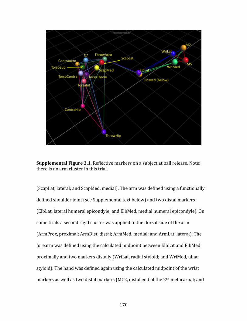
170
Supplemental Figure 3.1. Reflective markers on a subject at ball release. Note: there is no arm cluster in this trial.
(ScapLat, lateral; and ScapMed, medial). The arm was defined using a functionally
defined shoulder joint (see Supplemental text below) and two distal markers
(ElbLat, lateral humeral epicondyle; and ElbMed, medial humeral epicondyle). On
some trials a second rigid cluster was applied to the dorsal side of the arm
(ArmProx, proximal; ArmDist, distal; ArmMed, medial; and ArmLat, lateral). The
forearm was defined using the calculated midpoint between ElbLat and ElbMed
proximally and two markers distally (WriLat, radial styloid; and WriMed, ulnar
styloid). The hand was defined again using the calculated midpoint of the wrist
markers as well as two distal markers (MC2, distal end of the 2nd metacarpal; and

171
MC5, distal end of the 5th metacarpal). All markers were identified using Vicon
Nexus v. 1.7.1 software.
Experimental conditions – All subjects performed both “slow” and “fast”
throws in each of 5 conditions. During slow throws the subject was instructed to
make a comfortable throw with no consideration of speed. During fast throws the
subject was tasked to throw as fast as he felt comfortable throwing. The conditions
were normal unrestricted kinematics and four brace restricted conditions
(clavicular elevation, torso immobilization, shoulder rotation, and wrist extension).
Condition order was randomized across subjects. The brace restrictions used
commercially available therapeutic braces to limit range of motion at a specific joint.
All brace conditions (except for torso immobilization) also included an intermediate
sub-‐condition in which the brace was worn but no restriction was applied. During
loaded brace conditions, subjects were instructed to attempt their normal throwing
motion. In this paper, only data from the normal unrestricted and shoulder rotation
restricts are used.
Kinetics – The kinetic model was created using C-‐Motion Visual3D software.
Segment definitions and rotational axes are described in Sup. Table 3.1. All segment
masses, centers of mass, and radii of gyration are taken directly from Dempster (1).
A number of the markers used to define and track segments are virtual calculated
markers. The markers midHip and midWrist represent the calculated midpoint
between the hip and wrist markers respectively. The FuncSho marker is calculated
as a functional joint from a movement trial. In this movement trial, the subject keeps
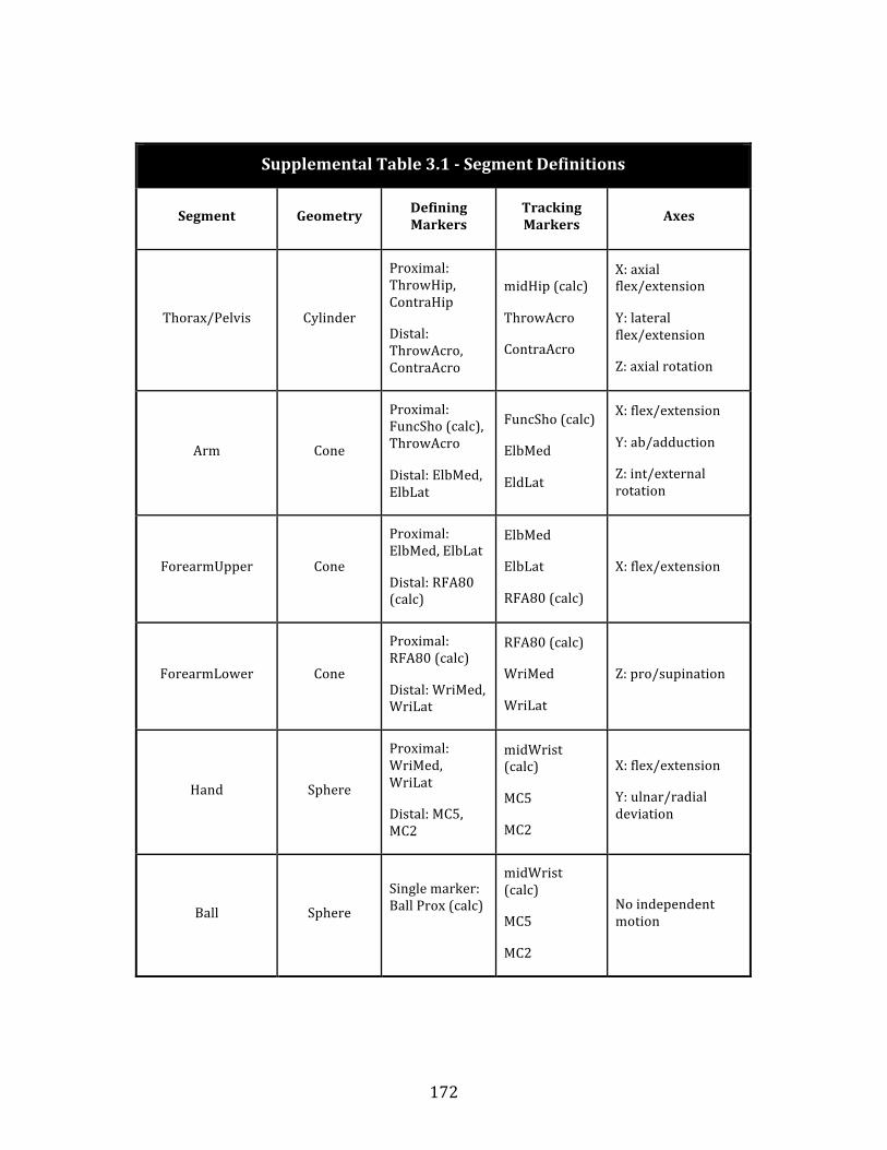
172
Supplemental Table 3.1 -‐ Segment Definitions
Segment Geometry Defining Markers
Tracking Markers Axes
Thorax/Pelvis Cylinder
Proximal: ThrowHip, ContraHip
Distal: ThrowAcro, ContraAcro
midHip (calc)
ThrowAcro
ContraAcro
X: axial flex/extension
Y: lateral flex/extension
Z: axial rotation
Arm Cone
Proximal: FuncSho (calc), ThrowAcro
Distal: ElbMed, ElbLat
FuncSho (calc)
ElbMed
EldLat
X: flex/extension
Y: ab/adduction
Z: int/external rotation
ForearmUpper Cone
Proximal: ElbMed, ElbLat
Distal: RFA80 (calc)
ElbMed
ElbLat
RFA80 (calc)
X: flex/extension
ForearmLower Cone
Proximal: RFA80 (calc)
Distal: WriMed, WriLat
RFA80 (calc)
WriMed
WriLat
Z: pro/supination
Hand Sphere
Proximal: WriMed, WriLat
Distal: MC5, MC2
midWrist (calc)
MC5
MC2
X: flex/extension
Y: ulnar/radial deviation
Ball Sphere
Single marker: Ball Prox (calc)
midWrist (calc)
MC5
MC2
No independent motion
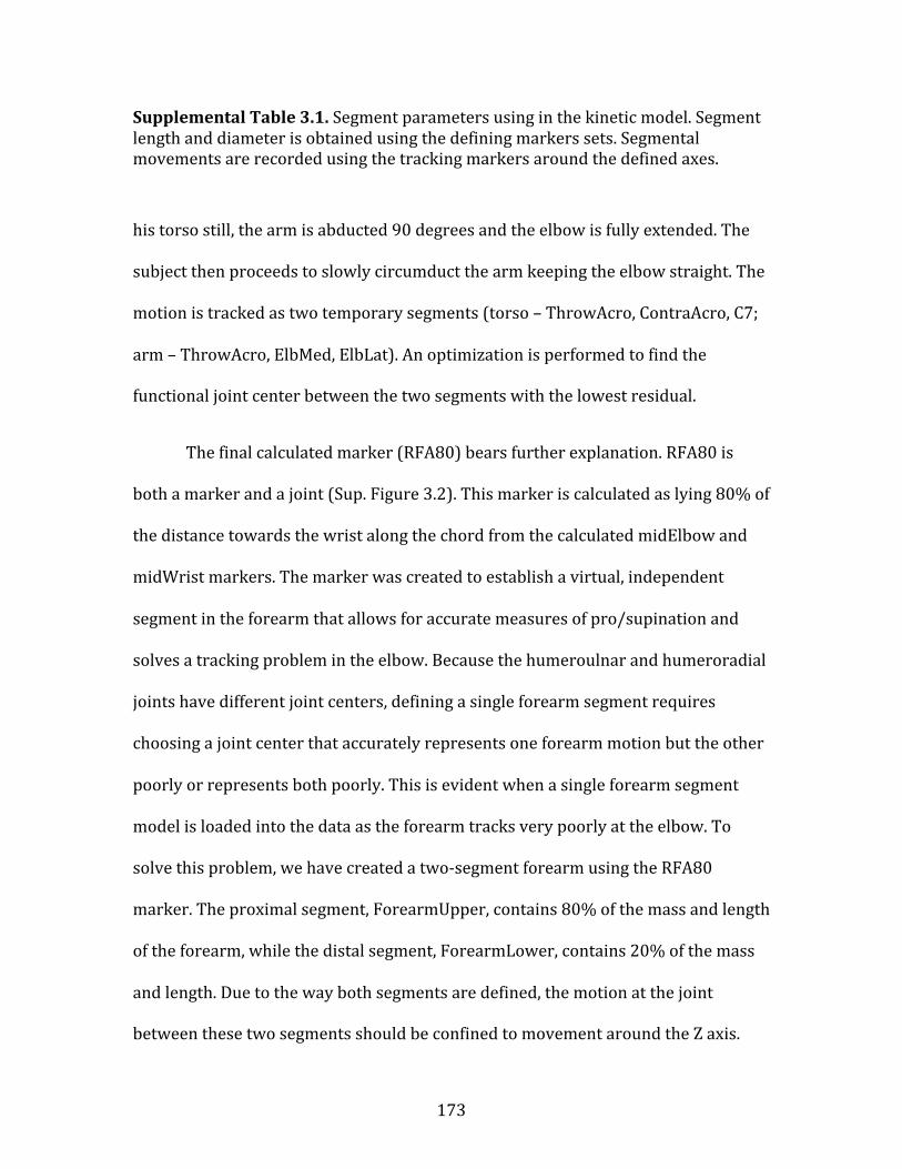
173
Supplemental Table 3.1. Segment parameters using in the kinetic model. Segment length and diameter is obtained using the defining markers sets. Segmental movements are recorded using the tracking markers around the defined axes.
his torso still, the arm is abducted 90 degrees and the elbow is fully extended. The
subject then proceeds to slowly circumduct the arm keeping the elbow straight. The
motion is tracked as two temporary segments (torso – ThrowAcro, ContraAcro, C7;
arm – ThrowAcro, ElbMed, ElbLat). An optimization is performed to find the
functional joint center between the two segments with the lowest residual.
The final calculated marker (RFA80) bears further explanation. RFA80 is
both a marker and a joint (Sup. Figure 3.2). This marker is calculated as lying 80% of
the distance towards the wrist along the chord from the calculated midElbow and
midWrist markers. The marker was created to establish a virtual, independent
segment in the forearm that allows for accurate measures of pro/supination and
solves a tracking problem in the elbow. Because the humeroulnar and humeroradial
joints have different joint centers, defining a single forearm segment requires
choosing a joint center that accurately represents one forearm motion but the other
poorly or represents both poorly. This is evident when a single forearm segment
model is loaded into the data as the forearm tracks very poorly at the elbow. To
solve this problem, we have created a two-‐segment forearm using the RFA80
marker. The proximal segment, ForearmUpper, contains 80% of the mass and length
of the forearm, while the distal segment, ForearmLower, contains 20% of the mass
and length. Due to the way both segments are defined, the motion at the joint
between these two segments should be confined to movement around the Z axis.
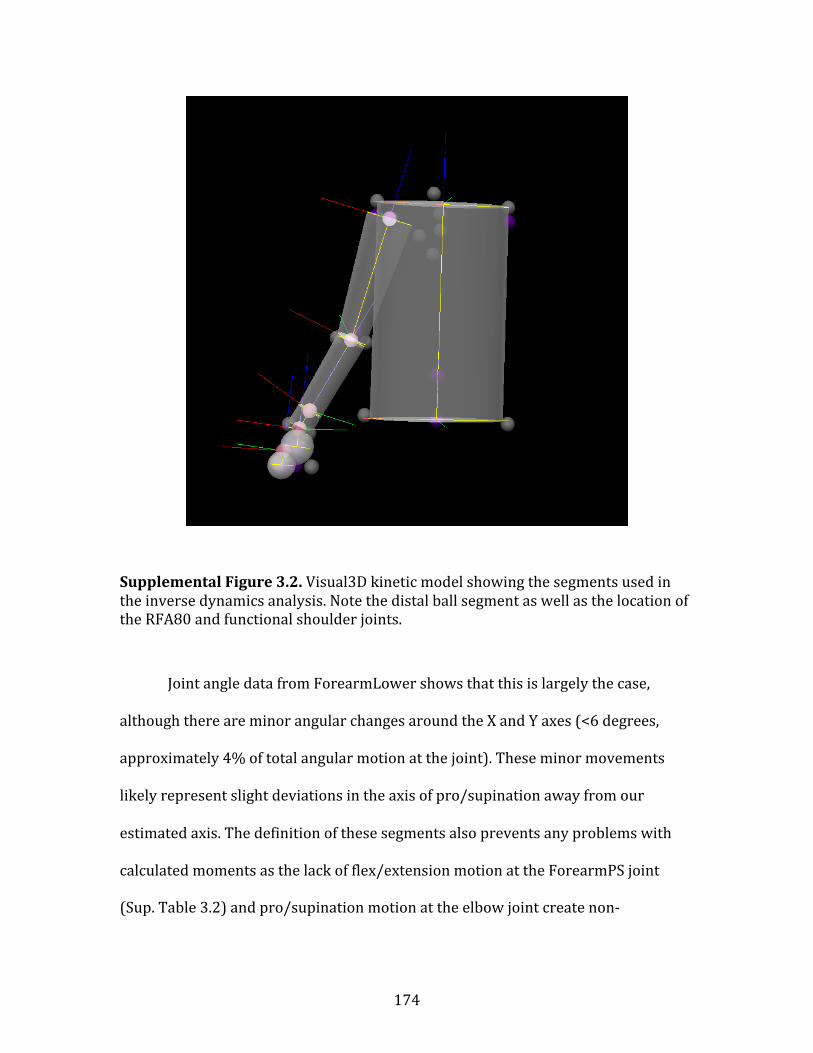
174
Supplemental Figure 3.2. Visual3D kinetic model showing the segments used in the inverse dynamics analysis. Note the distal ball segment as well as the location of the RFA80 and functional shoulder joints.
Joint angle data from ForearmLower shows that this is largely the case,
although there are minor angular changes around the X and Y axes (<6 degrees,
approximately 4% of total angular motion at the joint). These minor movements
likely represent slight deviations in the axis of pro/supination away from our
estimated axis. The definition of these segments also prevents any problems with
calculated moments as the lack of flex/extension motion at the ForearmPS joint
(Sup. Table 3.2) and pro/supination motion at the elbow joint create non-‐

175
Supplemental Table 3.2 -‐ Joint Definitions
Joint Segment Reference Segment
Resolution Coordinate System
Cardan Sequence for Joint Angles
Torso Thorax/Pelvis Lab Lab X,Y,Z
Shoulder Arm Thorax/Pelvis Arm X,Y,Z
Elbow ForearmUpper Arm Arm X,Y,Z
ForearmPS ForearmLower ForearmUpper ForearmUpper X,Y,Z
Wrist Hand ForearmLower ForearmLower X,Y,Z
Supplemental Table 3.2. Joint parameters used in the kinetic model. The cardan sequence is the series of rotations used to define both the joint Euler angles and the joint velocity, which is calculated using the instantaneous axis of rotation and not the first derivative of the Euler angles.
independent masses for the entire forearm. The use of a two-‐segment forearm also
resolves the model-‐tracking problem in the elbow.
The most distal segment in our kinetic model is the ball. The ball segment is
defined as a calculated marker, Ball, which is placed 130% of the distance along the
chord from the WriLat to MC2 marker and 20% of the distance along the chord
between the MC2 and MC5 markers. This places the ball between and slightly distal
to the 2nd and 3rd metacarpal heads (a common placement for a standard split finger

176
throw). The ball was given the appropriate mass (144g) and radius (3.68cm) of a
standard baseball. The ball segment was tracked using the same markers as the
hand segment and therefore is non-‐independent. In all kinetic data from after the
moment of release, the mass of the Ball segment is dropped to zero.
All defined joints in the kinetic model are described in Sup. Table 3.2. It
should be noted that all joint angles and velocities are defined using a X,Y,Z Cardan
sequence of rotations and all joint measures are resolved relative to the proximal
segment with the exception of the torso/pelvis, which is necessarily resolved
relative to the lab global coordinate system, and the shoulder, which is resolved
relative to its own axis to bring angular velocities and related measures in line with
previously published data (2, 3).
Filtering – Residual analysis, as described in Winter (4), was conducted on
throwing trials from 5 randomly chosen subjects using a custom MATLAB script.
The residual was calculated for 2nd, 4th and 6th order Butterworth lowpass filters
with the cutoff frequency iterated between 1Hz and the Nyquist frequency of the
data (500Hz). The residual was calculated for the ThrowHip, ThrowAcro, ElbLat,
WristLat and MC2 markers using the formula;
Equation S1: (Eq. S1)
where R is the residual in meters, is the cutoff frequency, is the raw data of the
ith sample, and is the filtered data of the ith sample. The full residual analysis was
€
R( fc ) =1N
(Xii=1
N
∑ − ˆ X i)2
€
fc
€
Xi
€
ˆ X i

177
Supplemental Figure 3.3. Sample residual analysis data from the full trial ElbLat marker data using a 2nd order Butterworth filter (Subject 006, trial 8).
Supplemental Figure 3.4. Sample residual analysis data from the critical period ElbLat marker data using a 2nd order Butterworth filter (Subject 006, trial 8).
178
conducted twice; once with the full throwing trial (Sup. Figure 3.3) and once using
only the critical period from the initiation of internal rotation of the humerus to ball
release (Sup. Figure 3.4). Cutoff frequencies were manually chosen following Winter
(4).
A 2nd order filter was chosen as the best fit for the data as it consistently
returned the lowest actual residuals across all markers. Full trial and critical period
cutoff frequency means were statistically different (p < 0.05) using a matched pairs
t-‐test (Sup. Table 3.3). The mean was calculated from the X, Y, and Z residuals from
each marker and then again from all markers (all marker XYZ mean: 17.3 Hz – full
data set; 36.7 Hz – critical period). For data processing, we split the difference
between the full data period and the critical period frequencies by using a 25 Hz
cutoff frequency. As a final check on the sensitivity of our analyses, we added
random noise (2mm width, zero mean) to all markers from a single subject’s raw,
unfiltered data. After filtering, no significant differences were detected in any kinetic
measures.
Throwing phase standardization – Differences in timing between throws are
controlled for in the kinematic and kinetic data by assigning portions of the motion
to 5 standardized phases (5). The windup and stride phases are collapsed into a
single phase as we are unable to differentiate these two phases with our marker
setup. The two phases reported here are defined as: Arm Cocking – from the
moment of leading foot contact with the ground at the end of stride (STR) to the
maximum external rotation (MER) of the shoulder, & Acceleration – from MER to

179
Supplemental Table 3.3 -‐ Cutoff frequency data (Hz)
-‐ 2nd order Butterworth filter
Marker Data set X Y Z Mean XYZ
ThrowHip full 14.8(4.8) 12.2(3) 10.2(3.2) 12.4(3.7)
critical 41(11.5) 35(7.8) 37.4(6.8) 37.8(8.7)
ThrowAcro full 21(12.9) 12.8(4.4) 18(11.3) 17.3(9.5)
critical 67.8(46.5) 58.4(45.7) 26.6(9.7) 50.4(34)
ElbLat full 18.8(8.9) 15.2(3) 14.8(4.8) 16.3(5.6)
critical 35.6(8.4) 34.2(8.5) 36.8(7.3) 35.5(8.1)
WristLat full 30.2(15.7) 15.8(5) 13.2(1.6) 19.7(7.4)
critical 41.8(11.9) 25(4.7) 25.2(4.3) 30.7(7)
MC2 full 34.4(10.9) 16(4.4) 12.4(1.8) 17.1(5.7)
critical 25.2(9.4) 24.8(8.4) 24.2(7.5) 25.3(8.4)
Supplemental Table 3.3. Mean cutoff frequencies (and standard deviations) from 5 subjects using a 2nd order Butterworth filter.
the moment of release (REL). STR was defined using the minimum point of mid-‐
pelvis linear velocity. MER was defined as the shoulder rotation angular velocity
zero point following STR. Data from the entire internal rotation motion (including

180
the full acceleration and deceleration phases) were further calculated from MER to
DECEL, defined as the shoulder rotation angular velocity zero point following REL.
Given that these phases often differ in duration between throws, further
standardization is required in order to calculate mean values and variances for all
parameters. Accordingly, all joint angles, angular velocities, torques and power
values were processed in MATLAB by interpolating each phase 1000 fold and
subsequently down sampling each throw to a set length. The acceleration phase was
down sampled to 100 data points and the arm-‐cocking phase to 894 points (in
keeping with the mean arm cocking/acceleration duration ratio). It should be noted
that the different throwing conditions did vary in this ratio (see Supplemental Data
below), but that in order to achieve comparable data between conditions the mean
8.94:1 ratio was used for all conditions.
Effects of projectile type – For practical reasons, we chose to collect data on
individuals throwing standard baseballs. However, given that hominin throwers
were likely throwing objects such as rocks and untipped spears, it is worth noting
how throwing kinematics differ between projectile types. While the throwing
literature largely focuses on baseball throwing, there are a number of studies
addressing the throwing kinematics of footballs (6-‐9), handballs (10-‐12), and
javelins (13-‐15). These projectiles differ from baseballs in both mass and shape.
Results from these studies show that a standard sequence and timing of motions is
conserved across all throw types (6, 16). Importantly, these standard kinematic
patterns include consistent external rotation of the arm into the passive ROM during

181
the arm-‐cocking phase, when we propose that elastic energy is stored (6, 17, 18).
Modeling research by Alexander (19) suggests that projectile mass may be
important in dictating arm position differences during the arm-‐cocking phase. These
positioning differences, where heavier projectiles are positioned closer to the
shoulder joint by reducing arm abduction angle and increasing elbow flexion, may
help to reduce the arm’s moment of inertia during cocking to prevent damage to the
ligaments and tendons crossing the shoulder. Data on arm positioning during the
cocking phase suggest that this may be the case for a number of heavier projectiles
(handball ~ 450g; shot put -‐ 7260g) (18, 20). Such a constraint may have
compromised hominin throwing performance when throwing heavy projectiles or
affected the choice of how heavy a projectile was thrown. Scarce ethnographic data
on hand-‐thrown spears suggests that human hunter-‐gatherers choose lighter spears
(averaging 184g), potentially to avoid injury or compromised throwing mechanics
(21). Projectile shape likely also has effects on throwing kinematics, especially in the
positioning of the hand.
Elongated projectiles (such as footballs, javelins, or spears) require that the
palm of the hand is positioned alongside the elongated axis the projectile, as
opposed to spherical projectiles (such as baseballs, handballs, etc…) where the palm
is typically positioned behind the projectile (22, 23). When throwing elongated
projectiles, this is accomplished through a more supinated position of the forearm.
While this more supinated forearm position is required of both football
quarterbacks and javelin throwers, the kinematics of hominin spear throwing were
likely more similar to those of football passing than javelin throwing. This is because

182
spear throwing for hunting as well as football passing require both power and
accuracy in order to be effective, while javelin throwing requires only power, as the
marker of performance is simply the distance the javelin has travelled from the
thrower. For this reason, the differences between baseball pitching and javelin
throwing (including the run up and unstable release) will not be discussed here.
Detailed comparisons of the football passing and baseball throwing motions show
that while these motions are largely similar, football quarterbacks tend to achieve
slower torso rotational velocities and faster shoulder flexion velocities (6).
Quarterbacks also externally rotate their arm ~9° less than baseball pitchers during
the cocking phase (6). It has been suggested that these differences in football
passing kinematics may be accommodations to the increased mass of the football (~
415g), in order to reduce the risk of injury to the shoulder and elbow (6). Similar
kinematic accommodations may have been employed by spear throwing hominins
for the same purpose. Finally, it is worth noting that throwing athletes from all
sports show reduced humeral torsion in their dominant arm versus non-‐dominant
arm (24-‐28).
Timing differences between experimental conditions – Experimental
conditions involving the brace showed significantly different timing in both the
duration and relative duration of the arm-‐cocking (repeated measures ANOVA, p =
0.0255, Mauchly’s sphericity = 0.2404) and acceleration phases (MANOVA, p =
0.0024, Mauchly’s sphericity < 0.0001) (Sup. Figure 3.5). Post-‐hoc pairwise testing
shows these differences are due to reduction of the arm-‐cocking phase and
elongation of the acceleration phase in the restricted condition. The reason for the

183
Supplemental Figure 3.5. Duration of the arm cocking phase (blue) and acceleration phase (red) for the normal and shoulder brace conditions. Restricted throw timing differed significantly from both normal and intermediate throws.
change in cocking duration is not clear, but the elongated acceleration seems to
represent a slowdown associated with reduced acceleration of the arm due to the
lack of elastic energy storage in this condition. Such reduced acceleration could
delay the moment of release and elongate the acceleration phase.
Muscle data – Muscle volumes used for muscle modeling are taken from the
Visible Human Project data set (29). These volumes are calculated from a single
male subject and are the largest available in the literature (which contains other
males in the 95th percentile in height and the 90th percentile in weight) (30). Muscle
volumes for forelimb muscle (Sup. Table 3.4) were calculated from MR images by
0 100 200 300 400 500 600

184
Garner and Pandy (31). The values used for the anterior deltoid were calculated by
dividing the entire deltoid volume by three. These volumes were then converted to
masses by multiplying by an empirically derived measure of mammalian muscle
density from the literature (32). The summed muscle mass of all internal rotators of
the humerus was then used to normalize our power data by potentially contributing
muscle mass. Our use of these very large muscle volumes in our analysis, as well as
the assumption that all fibers are contributing at 100% of their capacity and solely
to humeral internal rotation, intentionally overestimate the potential muscular
contribution to joint power at the shoulder. This significant overestimation is
designed to reduce the effects of error within the inverse dynamics torque
estimates.
Modeled vs. actual power output -‐ One way to test the hypothesis that elastic
energy provides most of the rotational power for throwing is to compare estimates
of the maximum power production capacity of all the muscles potentially
responsible for internally rotating the humerus with the actual calculated power.
The maximum power production of all internal rotators of the humerus is calculated
using the average actual power during the acceleration phase, thus minimizing the
effects of instantaneous changes in angular acceleration. This average actual power
value is then adjusted by a mass estimate for all potentially contributing muscles
(see Supplemental Methods above). This mass specific power estimate (in W/kg-‐1) is
then compared to published maximum mass specific power values from the
literature, which range from ~250-‐500 W/kg-‐1 (33-‐35). This conservative model

185
Supplemental Table 3.4 –
Shoulder Internal Rotator Muscle Volumes (cm3)
Muscle (Garner & Pandy 2003)
Pectoralis major 676.40
Latissimus dorsi 549.69
Anterior deltoid 792.87/3 = (264.29)
Teres major 231.40
Subscapularis 318.52
Total internal rotator volume (cm3) 2040.30
Muscle Density (Mendez & Keys, 1960) 1.06 g/cm3
Total internal rotator muscle mass (kg) 2.16
Supplemental Table 3.4. Calculated total muscle volume and mass for internal rotators of the shoulder. The largest volumes for each are taken from the literature.
shows that the average actual power at the shoulder exceeds the maximum isotonic
power production capacity for muscle by at least 3-‐7 fold (Sup. Table 3.5). However,
it is well known that when muscle is actively stretched during or just prior to
concentric contraction there is a notable increase in force production (36-‐39).
Plyometric models propose that the pre-‐stretching of the shoulder internal rotator
muscles during arm-‐cocking augment force enhancement during the subsequent
acceleration phase of the throw (40). Accordingly, we also compared our average
actual power values to force enhancement adjusted (1.75x -‐ (38)) values of

186
Supplemental Table 3.5 – Modeled Power Comparison
Maximum isotonic power -‐ skeletal muscle (W kg -‐1) 250-‐500
Adjustment for force enhancement due to stretching 1.75x
Maximum modeled muscle power -‐ skeletal muscle (W kg -‐1) 438-‐800
________________________________
Predicted values
Shoulder rotation work during acceleration (J) 132 ± 52
Time of acceleration (sec) 0.034 ± 0.013
Average joint power (W) 3,847 ± 1967
Estimated total muscle mass (kg) 2.16
Adjusted average kinetic power -‐ acceleration (W kg -‐1) 1,781 ± 911
Average kinetic power > Maximum muscle power
Supplemental Table 3.5. Average kinetic power (shoulder rotation) adjusted by modeled total internal rotator muscle mass compared to known maximum power values for skeletal muscle.
maximum muscle force, ranging from ~438-‐875 W/kg-‐1. Even with this significant
force enhancement, our measured average rotational power in the shoulder exceeds
the maximum muscle values by 2-‐4 times. Thus, in order to achieve this joint power
using muscular power alone, the individuals in this analysis would require internal
rotator muscles at least 2-‐4 times larger than those used here. Given that the muscle
masses used are the largest published (31), this is highly unlikely and suggests that

187
elastic energy stored in the shoulder is used to enhance the internal rotation power
output.
Location of elastic energy storage – Exactly where elastic energy is being
stored at the shoulder is currently unknown. While further in vivo soft tissue
imaging of the ligaments, tendons, and muscles crossing this joint are needed to
resolve this question, clinical data on repetitive injuries in the shoulder do provide
some insight. Throwing athletes show an increased prevalence of shoulder
instability and stretching related tears and laxity in the glenohumeral ligaments and
at the origin of the biceps tendon on the superior glenoid labrum (41-‐43). Over-‐
stretching injuries to these elastic structures suggest they may be involved in
storing elastic energy. For example, by actively positioning the arm during the early
cocking phase a number of shoulder ligaments are pulled taut (anterior band of the
inferior glenohumeral ligament, the middle glenohumeral ligament, and the
coracohumeral ligament) (44). It is quite likely that during following late cocking
phase, when the humerus is further passively rotated, these already taut ligaments
would stretch considerably. Furthermore, microscopic analyses of the glenohumeral
ligaments in those suffering from shoulder instability show higher amounts of
elastin present in these ligaments and an increase in the number of large diameter
collagen fibrils (45), effectively making these ligaments more tendon-‐like. Although
these elastic structures are not typical of those used by many animals to store elastic
energy to increase locomotor efficiency (46, 47), their short length and large
aggregate cross sectional area make the shoulder ligaments and muscle tendons
ideal for returning elastic energy very rapidly.

188
Supplemental Figure 3.6. Maximum ball speed across treatment conditions showed significant differences between all conditions as expected.
Ball speed & accuracy – Mean maximum ball speed was compared across
experimental conditions (Sup. Figure 3.6). Significant differences were found
between condition means (MANOVA – p < 0.0001), but not between condition
variances (Levene’s -‐ p = 0.94). Post-‐hoc pairwise comparisons showed significant
differences between all three conditions. As expected, the intermediate condition in
which the brace was worn but not tightened showed an intermediate reduction in
ball speed, while the full restricted showed a greater reduction. Mean accuracy was
also compared across conditions. No significant differences were found between
conditions for mean accuracy (repeated measures ANOVA -‐ p = 0.1485) or variance

189
Supplemental Figure 3.7. Mean joint power (green dashed) and work (green) for the sham condition. The mean values for the normal condition (blue dashed, grey) and restricted condition (red dashed, red) are shown for comparison. A 95% confidence interval (± 2 st. error) bounds the mean sham values in the power curve and all work values. Significant reductions (p < 0.05) from both the normal and intermediate conditions are indicated by **, while the †† indicates significant differences from both the normal and restricted conditions.
(Levene’s – p = 0.4574). Within all conditions, no condition order effects were found
in either speed or accuracy.
Intermediate/sham condition – Data were collected for an intermediate
condition in which the shoulder brace was applied, but the restriction mechanism
Time STR MER REL
-12000
-10000
-8000
-6000
-4000
-2000
0
2000
STR MER REL
-2000
0
2000
4000
6000
8000
10000
12000
14000
16000
18000
Pow
er (W
) Po
wer
(W)
**
**
-300
-200
-100
0
100
200
Wor
k (J
)
C A
-300
-200
-100
0
50
Wor
k (J
)
C A
††

190
was not loaded. This intermediate condition allows for the analysis of the effect of
the brace itself somewhat independently of the restriction. In this condition, the
shoulder brace acted like a tight-‐fitting jacket and reduced external rotational range
of motion at the shoulder approximately -‐11° (compared to the restricted condition
which limited external rotational ROM -‐28.3°). This intermediate level of restriction
resulted in a small but significant reduction in maximum projectile velocity from the
normal trials. However, the intermediate condition showed significantly higher
projectile velocity than the restricted condition. Kinetic data show this intermediate
condition is largely indistinguishable from the normal condition (Sup. Figure 3.7),
with the exception of the shoulder rotation work done during the arm cocking phase
which drops -‐9±13% (p < 0.05) from the normal condition.
Chimpanzee throwing performance – Although data exist on the frequency of
chimpanzee throwing behavior (48-‐51), all published descriptions of throwing
performance have been qualitative. We collected preliminary throwing velocity data
(Table S6) from 3 chimpanzees with previous overhand throwing training at a
sanctuary in Auburn, CA. While not directly comparable to human throwing data
due to limited practice and training, these data further illustrate the lack of
projectile velocity, corroborating previous published accounts.
Although chimpanzees are facultative rather than skilled throwers,
chimpanzee throwing behavior and anatomy can provide insight into the

191
Supplemental Table 3.6 – Chimpanzee throwing velocity (mph)
Juvenile Male
(3 years)
Juvenile Male (9 years)
Adult female (~30 years)
Mean velocity
12.4 12.5 19*
N
17 18 12*
Maximum velocity
14.3 15.5 22.4*
Standard Deviation
1.4 1.7 2.4*
Supplemental Table 3.6. Throwing velocity data from 3 chimpanzees. Note: while the adult female had previously been trained to throw overhand, during data collection she chose to throw underhand (while standing bipedally). Her data has been asterisked accordingly.
performance effects of morphological differences between these two species. I use a
model that compares human and chimpanzee throwing performance because I am
interested in assessing how differences in skeletal morphology affect throwing
ability. Furthermore, because many aspects of early hominin upper body anatomy
more closely resembles that of chimpanzees (52-‐54), they are a useful comparator
for humans and are helpful in assessing throwing performance in the fossil record.
Regardless of whether the human-‐chimpanzee last common ancestor was
behaviorally chimpanzee-‐like (53-‐55) or not (56, 57), comparisons of human
throwing mechanics and performance to that of chimpanzees can provide valuable

192
insights for interpreting how throwing performance and skeletal anatomy are
related.
Clavicle length in Homo erectus -‐ Previous research reconstructing the
hominin shoulder has suggested that early H. erectus fossils have relatively short
clavicles when standardized by humeral length (58). Given that the clavicle is the
only bony attachment of the shoulder girdle to the torso, clavicle length is inferred
to affect scapular positioning and thus range of motion at the shoulder (58). Using
the claviculohumeral ratio (clavicle length/humeral length) from the KNM WT
15000 and LB1 fossils, Larson (58) reconstructs early H. erectus as having novel,
intermediate shoulder morphology with low, very anteriorly positioned scapula and
suggests that as a result early H. erectus may have had compromising throwing
ability.
However, while the KNM WT 15000 claviculohumeral ratio (40.9) is low for
modern humans, it does fall within the measured range of normal variation seen in
East African populations today (58), in whom no deficit in throwing performance
has been reported. Additionally, reconstructed fossil clavicles from the early H.
erectus site of Dmanisi show claviculohumeral ratios squarely within the modern
human range (Adult male – 46.5L, 46R; juvenile – 43.7) (59). Given that both the
Dmanisi and Nariokotome clavicle fossils are incomplete, assessing the accuracy of
the length reconstruction methods used is important to resolve this discrepancy.
However, these methods are not reported in the Dmanisi analysis. Further research
is required to address this interesting discrepancy.

193
Finally, while standardizing clavicle length using the humerus has been
shown to be a reasonable proxy for body mass interspecifically (58, 60), the high
variability in this relationship intraspecifically suggests that this standardization
method may not be very useful within a species. Furthermore, the claviculohumeral
ratio may not be very useful for reconstructing shoulder position. To illustrate the
problem with this metric, we look to chimpanzees, who have low claviculohumeral
ratios and yet have dorsally positioned, cranially oriented scapula. This dorsal
shoulder position is driven not by the length of the clavicle relative to body mass,
but by the length of the clavicle relative to the breadth of the superior portion of the
ribcage, which is evolving independently of body mass (61). Recent work
investigating clavicular length in apes suggests that when chimpanzee clavicles are
normalized to thorax width they fall closer to the rest of the apes in having a
relatively elongated clavicle relative to monkeys (62). Such a thorax width
standardization better accounts for the shoulder positioning seen in chimpanzees
and humans. In sum, better data is needed to determine how clavicle length and
shape, as well as thorax width and shape, relate to shoulder positioning in hominins.
However, at the moment equivocal data about H. erectus clavicle length do not
preclude high-‐speed throwing.
Glenoid orientation as a throwing adaptation – The “relaxation” or caudal
rotation of the scapula in H. erectus likely had significant effects on many behaviors.
We have proposed that this modification to shoulder position may be an adaptation
for throwing. The logic behind this hypothesis is simply that shifts in shoulder
position would result in large increases in throwing performance, while having

194
relatively minor or even negative effects on the performance of other important
behaviors. For instance, laterally oriented shoulders likely positioned the
glenohumeral joint further inferiorly on the thorax, resulting in a wider shoulder
breadth that would reduce the mechanical advantage of the shoulder muscles
during climbing. In addition, a more relaxed shoulder would result in a lateral
reorientation of the P. major muscle, removing any useful flexion contribution of this
muscle during stick digging. The effects that shoulder position had on knapping are
more difficult to address. Given that power requirements of stone tool manufacture
are probably much lower than during high-‐speed throwing and that the muscle
most significantly affected by a change in shoulder positioning, P. major, is likely
minimally active during knapping, little direct effect of this shoulder position shift is
expected. However, Larson (58) notes that a laterally oriented glenoid in
combination with very low torsion in H. erectus (below most modern human values,
including most throwing athletes) could result in reduced range of motion for
manipulative tasks such as knapping or tool use. This problem, in combination with
potentially short clavicles relative to humerus length, have led Larson to propose
that the shoulder in H. erectus was more anteriorly positioned. Given that any deficit
in manipulative range of motion could be overcome by movements at the highly
mobile shoulder (shoulder flexion, scapular protraction), we expect the negative
effects of shoulder positioning on stone tool knapping and use were minimal. The
only other context in which a low laterally oriented shoulder has been suggested to
improve performance is during running (61). Bramble and Lieberman suggest that
shifts in shoulder position represent a decoupling of the head and shoulders and

195
help to increase gaze stability during running. Although this decoupling would
clearly insulate the head from jarring motions that would perturb the vestibule-‐
ocular reflex, such impacts are partly damped elsewhere in the body (63, 64)
reducing the performance effects of decoupled shoulders. In sum, much additional
research is needed to further quantify the performance effects of a laterally oriented
shoulder on many diverse activities, but there is reason to hypothesize that shifts in
shoulder position have the important performance effects during throwing and may
represent an adaptation for this behavior.
Static optimization of arm positioning – We have proposed that throwers
store elastic energy at the shoulder by maximizing the arm’s moment of inertia,
causing the arm to lag behind the accelerating torso, stretching the shoulder
ligaments and tendons. We have further proposed that the arm’s moment of inertia
is maximized by abducting the humerus in line with the torso rotation and P. major
torques lines of action and by flexing the elbow. To test these assumptions, a static
optimization of the shoulder abduction and elbow flexion angles, maximizing the
arm’s moment of inertia, was performed (Sup. Figure 3.8). This optimization was
conducted using both chimpanzee and human morphologies (1, 65-‐68) and varied
projectile mass. A cosine cost function was applied at the shoulder to penalize
deviations in humeral abduction away from alignment with the two sources of
torque, which the arm’s inertial mass is proposed to resist. Results show that, as
predicted, humans maximize the arm’s moment of inertia by positioning the arm at
a lower shoulder abduction angle and a higher elbow flexion angle than
chimpanzees do (for low mass projectiles: human shoulder abduction ~80°, elbow
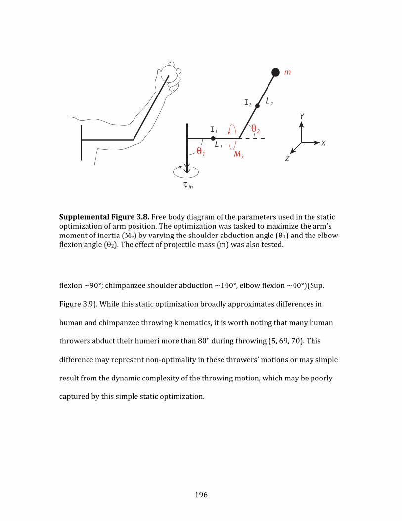
196
Supplemental Figure 3.8. Free body diagram of the parameters used in the static optimization of arm position. The optimization was tasked to maximize the arm’s moment of inertia (Mx) by varying the shoulder abduction angle (θ1) and the elbow flexion angle (θ2). The effect of projectile mass (m) was also tested.
flexion ~90°; chimpanzee shoulder abduction ~140°, elbow flexion ~40°)(Sup.
Figure 3.9). While this static optimization broadly approximates differences in
human and chimpanzee throwing kinematics, it is worth noting that many human
throwers abduct their humeri more than 80° during throwing (5, 69, 70). This
difference may represent non-‐optimality in these throwers’ motions or may simple
result from the dynamic complexity of the throwing motion, which may be poorly
captured by this simple static optimization.
I
I

197
Supplemental Figure 3.9. The results of a static optimization maximizing the arm’s moment of inertia (Mx) by varying the shoulder abduction angle (θ1) and the elbow flexion angle (θ2). Results for low mass projectiles correspond to known differences in human and chimpanzee arm position during the arm-‐cocking phase, when elastic energy storage is proposed to occur.
1. Dempster WT (1955) Space requirements for the seated operator (Wright Air
Development Center, Wright-‐Patterson Air Force Base, OH). 2. Hirashima M, Kudo K, Watarai K, & Ohtsuki T (2007) Control of 3D limb
dynamics in unconstrained overarm throws of different speeds performed by skilled baseball players. J Neurophysiol 97(1):680-‐691.
3. Hong DA, Cheung TK, & Roberts EM (2001) A three-‐dimensional, six-‐segment
chain analysis of forceful overarm throwing. J Electromyogr Kinesiol 11(2):95-‐112.
4. Winter DA (1990) Biomechanics and motor control of human movement
(Wiley-‐Interscience, New York) 2nd Ed. 5. Dillman CJ, Fleisig GS, & Andrews JR (1993) Biomechanics of pitching with
emphasis upon shoulder kinematics. J Orthop Sports Phys Ther 18(2):402-‐408.
6. Fleisig GS, et al. (1996) Kinematic and kinetic comparison between baseball
pitching and football passing. Journal of Applied Biomechanics 12(2):207-‐224. 7. Escamilla RF & Andrews JR (2009) Shoulder muscle recruitment patterns
and related biomechanics during upper extremity sports. Sports Med 39(7):569-‐590.
50
70
90
110
130
150
170
190
0 1 2 3 4 5 6 7 8 9 1050
70
90
110
130
150
170
190
0 1 2 3 4 5 6 7 8 9 10
(θ1)
(θ1)
(θ1+θ2) (θ1+θ2)
Projectile mass (m) in kg
Arm
Ang
le in
deg
rees
Human Chimpanzee

198
8. Kelly BT, Barnes RP, Powell JW, & Warren RF (2004) Shoulder injuries to quarterbacks in the national football league. Am J Sports Med 32(2):328-‐331.
9. Kelly BT, Backus SI, Warren RF, & Williams RJ (2002) Electromyographic
analysis and phase definition of the overhead football throw. Am J Sports Med 30(6):837-‐844.
10. Wagner H, Buchecker M, von Duvillard SP, & Muller E (2010) Kinematic
comparison of team handball throwing with two different arm positions. International journal of sports physiology and performance 5(4):469-‐483.
11. van den Tillaar R & Ettema G (2009) Is there a proximal-‐to-‐distal sequence in
overarm throwing in team handball? J Sports Sci 27(9):949-‐955. 12. van den Tillaar R & Ettema G (2007) A three-‐dimensional analysis of
overarm throwing in experienced handball players. J Appl Biomech 23(1):12-‐19.
13. Liu H, Leigh S, & Yu B (2010) Sequences of upper and lower extremity
motions in javelin throwing. J Sports Sci 28(13):1459-‐1467. 14. Best RJ, Bartlett RM, & Morriss CJ (1993) A three-‐dimensional analysis of
javelin throwing technique. J Sports Sci 11(4):315-‐328. 15. Morriss C & Bartlett R (1996) Biomechanical factors critical for performance
in the men's javelin throw. Sports Med 21(6):438-‐446. 16. Atwater AE (1979) Biomechanics of overarm throwing movements and of
throwing injuries. Exerc Sport Sci Rev 7:43-‐85. 17. Chagneau F, Delamarche P, & Levasseur M (1992) Stroboscopic
computerized determination of humeral rotation in overarm throwing. Br J Sports Med 26(1):59-‐62.
18. Fradet L, Kulpa R, Multon F, & Delamarche P (2002) Kinematic analysis of
handball throwing. International Symposium on Biomechanics in Sports, ed Gianikellis KE (ISBS), pp 119-‐122.
19. Alexander RM (1991) Optimum timing of muscle activation for simple
models of throwing. J Theor Biol 150(3):349-‐372. 20. Coh M, Stuhec S, & Supej M (2008) Comparative biomechanical analysis of
the rotational shot put technique. Collegium antropologicum 32(1):249-‐256. 21. Hughes SS (1998) Getting to the Point: Evolutionary Change in Prehistoric
Weaponry. Journal of Archaeological Method and Theory 5(4):345-‐408.

199
22. Young RW (2003) Evolution of the human hand: the role of throwing and
clubbing. Journal of Anatomy 202:165-‐174. 23. Wolfe SW, Crisco JJ, Orr CM, & Marzke MW (2006) The dart-‐throwing motion
of the wrist: is it unique to humans? J Hand Surg Am 31(9):1429-‐1437. 24. Pieper HG (1998) Humeral torsion in the throwing arm of handball players.
Am J Sports Med 26(2):247-‐253. 25. Reagan KM, et al. (2002) Humeral retroversion and its relationship to
glenohumeral rotation in the shoulder of college baseball players. Am J Sports Med 30(3):354-‐360.
26. Osbahr DC, Cannon DL, & Speer KP (2002) Retroversion of the humerus in
the throwing shoulder of college baseball pitchers. Am J Sports Med 30(3):347-‐353.
27. Herrington L (1998) Glenohumeral joint: internal and external rotation range
of motion in javelin throwers. Br J Sports Med 32(3):226-‐228. 28. Crockett HC, et al. (2002) Osseous adaptation and range of motion at the
glenohumeral joint in professional baseball pitchers. Am J Sports Med 30(1):20-‐26.
29. Spitzer V, Ackerman MJ, Scherzinger AL, & Whitlock D (1996) The visible
human male: a technical report. J Am Med Inform Assoc 3(2):118-‐130. 30. Holzbaur KR, Murray WM, Gold GE, & Delp SL (2007) Upper limb muscle
volumes in adult subjects. J Biomech 40(4):742-‐749. 31. Garner BA & Pandy MG (2003) Estimation of musculotendon properties in
the human upper limb. Ann Biomed Eng 31(2):207-‐220. 32. Mendez J & Keys A (1960) Density and composition of mammalian muscle.
Metabolism 9:184-‐188. 33. McGowan CP, Baudinette RV, Usherwood JR, & Biewener AA (2005) The
mechanics of jumping versus steady hopping in yellow-‐footed rock wallabies. J Exp Biol 208(Pt 14):2741-‐2751.
34. Lutz GJ & Rome LC (1994) Built for jumping: the design of the frog muscular system. Science 263(5145):370-‐372.
35. Peplowski MM & Marsh RL (1997) Work and power output in the hindlimb
muscles of Cuban tree frogs Osteopilus septentrionalis during jumping. J Exp Biol 200(Pt 22):2861-‐2870.

200
36. Katz B (1939) The relation between force and speed in muscular contraction.
J Physiol 96(1):45-‐64. 37. Harry JD, Ward AW, Heglund NC, Morgan DL, & McMahon TA (1990) Cross-‐
bridge cycling theories cannot explain high-‐speed lengthening behavior in frog muscle. Biophys J 57(2):201-‐208.
38. Biewener AA & Blickhan R (1988) Kangaroo rat locomotion: design for
elastic energy storage or acceleration? J Exp Biol 140:243-‐255. 39. McMahon TA (1984) Muscles, Reflexes, and Locomotion (Princeton University
Press, Princeton, NJ). 40. Wilk KE, et al. (1993) Stretch-‐shortening drills for the upper extremities:
theory and clinical application. J Orthop Sports Phys Ther 17(5):225-‐239. 41. Fleisig GS, Barrentine SW, Escamilla RF, & Andrews JR (1996) Biomechanics
of overhand throwing with implications for injuries. Sports Med 21(6):421-‐437.
42. Altchek DW & Dines DM (1995) Shoulder Injuries in the Throwing Athlete. J
Am Acad Orthop Surg 3(3):159-‐165. 43. Meister K (2000) Injuries to the shoulder in the throwing athlete. Part two:
evaluation/treatment. Am J Sports Med 28(4):587-‐601. 44. Clemente C (1985) Gray's Anatomy of the Human Body (Lea & Febiger,
Philadelphia) 30th American Ed. 45. Rodeo SA, Suzuki K, Yamauchi M, Bhargava M, & Warren RF (1998) Analysis
of collagen and elastic fibers in shoulder capsule in patients with shoulder instability. Am J Sports Med 26(5):634-‐643.
46. Biewener AA, Konieczynski DD, & Baudinette RV (1998) In vivo muscle force-‐
length behavior during steady-‐speed hopping in tammar wallabies. J Exp Biol 201(Pt 11):1681-‐1694.
47. Astley HC & Roberts TJ (2012) Evidence for a vertebrate catapult: elastic
energy storage in the plantaris tendon during frog jumping. Biol Lett 8(3):386-‐389.
48. Goodall J (1986) The Chimpanzees of Gombe: Patterns of Behavior (Harvard
University Press, Cambridge).

201
49. Goodall J (1964) Tool-‐Using and Aimed Throwing in a Community of Free-‐Living Chimpanzees. Nature 201:1264-‐1266.
50. Kohler W (1925) The Mentality of Apes (Harcourt and Brace, New York). 51. Sugiyama Y & Koman J (1979) Tool-‐using and making behavior in wild
chimpanzees at Bossou, Guinea. Primates 20:513-‐524. 52. White TD, et al. (2009) Ardipithecus ramidus and the paleobiology of early
hominids. Science 326(5949):75-‐86. 53. Brunet M, et al. (2002) A new hominid from the Upper Miocene of Chad,
Central Africa. Nature 418(6894):145-‐151. 54. Pickford M, Senut B, Gommery D, & Treil J (2002) Bipedalism in Orrorin
tugenensis revealed by its femora. C.R. Paleovol 1:1-‐13. 55. Wrangham R & Pilbeam D (2001) African Apes as Time Machines. All Great
Apes Great and Small, Volume I : African Apes, African Apes, ed Galdikas BMF, Briggs, N.E., Sheeran, L.K., Shapiro, G.L. and Goodall, J. (Kluwer Academics/Plenum Publishers, New York), Vol I, pp ch. 1, pg. 5-‐17.
56. Sayers K & Lovejoy CO (2008) The chimpanzee has no clothes: A critical
examination of Pan troglodytes in models of human evolution. Current Anthropology 49(1):87-‐114.
57. Zihlmann A (1996) Reconstructions reconsidered: chimpanzee models and
human evolution. Great Ape Societies, ed McGrew WC, Marchant, L.F., and Nishida, T. (Cambridge University Press, Cambridge), pp ch. 21, pg. 293-‐304.
58. Larson SG (2007) Evolutionary Transformation of the Hominin Shoulder.
Evolutionary Anthropology 16:172-‐187. 59. Lordkipanidze D, et al. (2007) Postcranial evidence from early Homo from
Dmanisi, Georgia. Nature 449(7160):305-‐310. 60. Jungers WL (1994) Ape and hominid limb length. Nature 369:194. 61. Bramble DM & Lieberman DE (2004) Endurance Running and the Evolution
of Homo. Nature 432:345-‐352. 62. Kagaya M, Ogihara N, & Nakatsukasa M (2010) Is the clavicle of apes long? An
investigation of clavicular length in relation to body mass and upper thoracic width. Int J Primatol 31:209-‐217.

202
63. Lieberman DE, et al. (2010) Foot strike patterns and collision forces in habitually barefoot versus shod runners. Nature 463(7280):531-‐535.
64. Pontzer H, Holloway JH, 3rd, Raichlen DA, & Lieberman DE (2009) Control
and function of arm swing in human walking and running. J Exp Biol 212(Pt 4):523-‐534.
65. Zihlman AL (1984) Body Build and tissue composition in Pan paniscus and
Pan troglodytes, with comparisons to other hominoids. The Pygmy Chimpanzee, ed Susman RL (Plenum Press, New York), pp 179-‐200.
66. Aiello LC & Dean C (1990) An Introduction to Human Evolutionary Anatomy
(Academic Press, New York) p 596. 67. Schultz AH (1930) The skeleton of the trunk and limbs of higher primates.
Human Biology 2:303-‐438. 68. Schultz AH (1937) Proportions, variability and asymmetries of the long
bones of the limbs and the clavicles in man and apes. Hum Biol 9(3):281-‐328. 69. Fleisig GS, Andrews JR, Dillman CJ, & Escamilla RF (1995) Kinetics of baseball
pitching with implications about injury mechanisms. Am J Sports Med 23(2):233-‐239.
70. Pappas AM, Zawacki RM, & Sullivan TJ (1985) Biomechanics of baseball
pitching. A preliminary report. Am J Sports Med 13(4):216-‐222.


