ABSTRACT
Title of Dissertation SYNTHETIC ION CHANNELS FROM
LIPOPHILIC GUANOSINE DERIVATIVES
Ling Ma Doctor of Philosophy 2009
Dissertation directed by Professor Jeffery T Davis
Department of Chemistry and Biochemistry
Synthetic ion channels and pores not only represent models of natural
transmembrane ion channels but also demonstrate their potential applications in the areas
of drug delivery biosensors antimicrobial agents and other molecular devices In this
thesis lipophilic guanosine derivatives that combine both ldquomolecular recognitionrdquo and
ldquomembrane solublerdquo features are utilized for the development of the self-assembled
synthetic ion channels
The potential of lipophilic G-quadruplexes to function as synthetic ion channels
has been investigated by tracing the cation exchange process between free cations and G-
quadruplex bound cations Cation exchange between bulk cations (K+ NH4+) in solution
and the bound cations in G-quadruplexes (G 1)16bull4Na+bull4DNP- was investigated by
electrospray ionization mass spectrometry and by 1H 15N NMR spectroscopy The ESI-
MS and 1H NMR data showed that G-quadruplexes containing ldquomixed cationsrdquo formed
through a sequential ion exchange process The use of NMR-ldquovisiblerdquo 15NH4+ cations in
the NMR titration experiments allowed the determination of two ldquomixed-cationrdquo
intermediates by 15N-filtered 1H NMR and selective NOE spectroscopy A ldquocentral
insertionrdquo pathway was proposed for the cation exchange process from (G 1)16bull 4Na+bull
4DNP- to (G 1)16bull 4NH4+bull 4DNP- In the lipophilic G-quadruplex the ldquocentralrdquo Na+
bound between the 2 symmetry related G8-Na+ octamers is bound less strongly than are
the 2 ldquoouterrdquo Na+ ions sandwiched within the G8-octamers These results demonstrated
the dynamic nature of lipophilic G-quadruplex in solution and directed the design of a
ditopic guanosine-sterol conjugate as an approach toward making synthetic ion channels
Guanosine-sterol conjugate 3-1 was prepared by coupling 2rsquo 3rsquo-bis-TBDMS 5rsquo-
amino guanosine with a bis-lithocholic acid derivative Voltage clamp experiments
demonstrated a series of stable single ion channel conductances when compound 3-1 was
incorporated into a planar phospholipid membrane These channels are large with
nanoSiemens conductance values and they last for seconds of ldquoopenrdquo time This feature
distinguishes them from most synthetic channels which typically conduct in the
picosiemens range with millisecond lifetimes The structural studies using the bis-
lithocholamide linker demonstrated that the guanosine moiety plays an essential role in
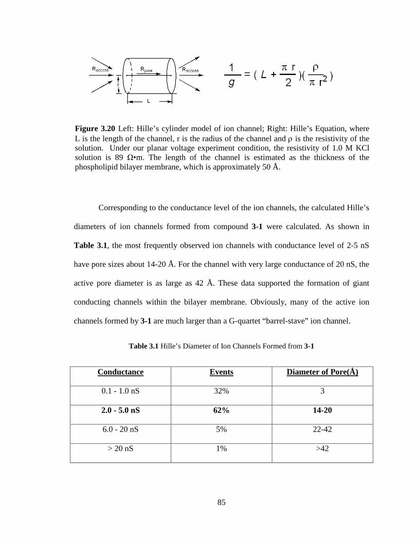
the self-assembly of the transmembrane ion channels The sizes of the most prevalent
single channels calculated by Hillersquos equation are much larger than the diameter of a G-
quartet which suggested that the ion transport proceeded through larger pore(s) that form
upon self-assembly of lipophilic guanosine-lithocholate 3-1 within the phospholipid
membrane The large transmembrane pore(s) could be envisioned as a supramolecular
structure with hydrophobic walls of bis-lithocholate linker and a central pillar of a cation-
filled G-quadruplex
The use of a bis-urea functionality in the bis-lithocholic acid linker generated
guanosine-sterol conjugate 4-1 The ion channel activity of 4-1 was demonstrated by
voltage clamp experiment Large ion channels formed from 4-1 had longer life-times than
those formed from compound 3-1 The extra stabilization of self-assembled ion channels
attributed to the bisurea hydrogen bonding is consistent with the structural hypothesis of
ion channels The stable large transmembrane ion channels self-assembled by lipophilic
guanosine derivatives have potential for delivery of drugs or biomolecules
Synthetic Ion Channels From Lipophilic Guanosine Derivatives
By
Ling Ma
Dissertation submitted to the Faculty of the Graduate School of the
University of Maryland College Park in partial fulfillment
of the requirements for the degree of
Doctor of Philosophy
2009
Advisory Committee
Professor Jeffery T Davis Chair
Professor Dorothy Beckett
Professor Marco Colombini
Professor Lyle Isaacs
Professor Steven E Rokita
copy Copyright by
Ling Ma
2009
ii
Dedication
To my parents my mother-in-law my husband my brother and my daughter for all your
love and support
iii
Acknowledgements
First I would like to express my heartfelt gratitude to my research advisor and
mentor Professor Jeffery T Davis Thank you for giving me the freedom guidance and
encouragement throughout my whole graduate career Special thanks go to Professor
Marco Colombini for providing access to his voltage clamp facilities I am truly grateful
for his generosity and his helpful advice I am also thankful for our discussions on
membranes and on life in general I would also like to thank Prof Lyle Isaacs for his
helpful suggestions in chemistry and career advice I would also like to thank professors
Steven Rokita Dorothy Beckett Michael Doyle and Daniel Falvey for their valuable
discussions and suggestions
I am very grateful to Dr Yiu-Fai Lam and Dr Yinde Wang for their training and
help in NMR spectroscopy Particularly I would like to thank Yiu-Fai for his help with
my research project He taught me so many new techniques and helped me set up
numerous special experiments Yiu-Fai Thank you
I am thankful for the electrospray ionization mass spectrometry training provided
by Noel Whittaker and Dr Yue Li I would also like to acknowledge Dr Fred Khachik
for the use of his HPLC instrument and Dr Peter Zavalij for the crystal structures
I would like to express my gratitude to the Davis research group You have made
my graduate life much more enjoyable I would especially like to thank Dr Mark
Kaucher for helping me start my PhD program teaching me how to use CD
spectrophotometer and providing helpful advice I would also like to thank Jennifer
Seganish Sofya Berezin William Harrell Oluyomi Okunola Doriann Dejesus and
iv
Monique Pichon for their discussion and friendship I would also like to thank Dr Paul
Santacroce for teaching me new techniques and helpful discussions Particularly I would
like to thank Dr Monica Melegari from University of Parma Italy She was always there
to discuss chemistry and made my ldquomembrane-makingrdquo experience fun Thanks also go
to the undergraduates I had the pleasure to work with Emily Ryan and Jonathan Jinhoon
Park Thank you for the excellent synthesis work
I extend my appreciation to my fellow classmates and friends who supported each
other and in shared the graduate school experience Drs Becky Vieira Regan Nally
Soumyadip Ghosh Wei-hao Huang Sofya Berezin JuHee Park In particular I would
like to thank Dr Becky Vieira and Regan Nally for their friendship Dr Becky thank you
for your generous help and patience in correcting my writing
Lastly I would like to send my deepest thanks to my family I have the most
wonderful family in this world I would like to thank my parents and brother for always
being there to love and support me I thank my mother-in law for her help and
encouragement I thank my daughter for being an excellent kid and making me a proud
Mom I would like to especially thank my husband for his understanding patience
support and love I love you all very dearly
v
Table of Contents
LIST OF TABLES x
LIST OF FIGURES xi
LIST OF SCHEMES xvii
Chapter 1 Introduction 1
11 Introduction 1
12 Thesis Organization 1
13 Nucleobases and Lipids as Building Motifs for Supramolecular Structures 2
131 Nucleobases Molecular Recognition Motif 2
132 LipidBilayer Structure Unit of Functional Biological Membrane 5
14 Lipophilic Nucleobases Nucleosides and Oligonucleotides The Nucleolipids 6
141 Natural Nucleolipids 6
142 Molecular Recognitionof Synthetic Nucleolipids 9
143 Membrane Association Properties of Nucleolipids 13
15 Functional Nanostructures from Amphiphilic Nucleobases 14
151 Molecular Recognition in Aqueous Solution Amphiphilic Nucleobase-
Derived Supramolecular Receptors 15
152 Lipophilic Nucleobases Approach Toward Functional Biological Surfaces 17
16 Synthetic Nucleolipid forTransportof Biomolecules 21
161 Lipophilic Nucleobase as Carrier for Nucleotide Monophosphate Transport 21
162 Nucleolipids for DNA Transfection 23
vi
17 Lipophilic Guanosine Building Block of Transmembrane Ion Channels 25
171 Ion Channel Model From DNA G-quadruplex 25
172 Lipophilic Guanosine Derivatives 27
18 Summary 29
Chapter 2 Cation Exchange in Lipophilic G-Quadruplexes Not All Ion Binding Sites
Are Equal 30
21 Introduction 30
211 Cation-Dependent Self-Assembly of DNA G-Quadruplex Structures 31
212 DNA G-quadruplex as an Ion Channel Model 32
213 Lipophilic G-Quadruplexes 35
22 Mass Spectrometry Shows Mixed-Cation G-Quadruplexes Formed by Sequential
Ion Exchange 37
23 Proton NMR Indicates that the Central Na+ Is the First Cation Exchanged for K+ in
Formation of Mixed Na+ K+ G-Quadruplexes 40
24 Use of 15NH4+ Cation to Probe the Cation Exchange Process Within a Lipophilic
G-quadruplex 43
241 NMR Studies with 15NH4+ Confirm Identity of Mixed-Cationic G-
Quadruplexes 43
242 Determination of the First Intermediate in Na+15NH4+ Cation Exchange 46
243 Characterization of the Second Intermediate in the Na+15NH4+ Cation
Exchange Process 48
25 Cation Exchange Pathway in the Lipophilic G-Quadruplex 50
26 The Larger Cs+ Cation But Not the Smaller Li+ Can Displace the Central Cation
vii
in the Na+ G-Quadruplex 53
27 Conclusion 54
Chapter 3 Large and Stable Transmembrane Pores from a Guanosine-Bile Acid
Conjugate 56
31 Introduction 56
311 G-QuartetFolate-Quartet as a Motif for Transmembrane Ion Transporter 58
32 Design of G-quartet Ion Channels Using a Ditopic Guanosine with a Covalent
Linker 61
33 Design of Guanosine-Sterol Conjugate 64
34 Synthesis of Guanosine-Sterol Conjugate 67
35 Synthesis of Bis-lithocholamide for Control Experiment 71
36 Self-Assembly Property Study of Guanosine-Sterol Conjugate 72
37 Guanosine-Sterol Conjugate Forms Transmembrane Ion Channels (Pores) 75
371 Ion Selectivity of the Synthetic Ion Channels 80
372 Proposed Active Structure of the Synthetic Ion Channel 83
38 Conclusion 87
Chapter 4 Synthetic Ion Channel from Ditopic Sterol-Guanosines with Bis-urea Linker
89
41 Introduction 89
42 Bis-urea Macrocycles are Known to Form Self-Assembled Cylinders 91
43 Synthesis of Bis-urea Guanosine-Sterol Conjugate 4-1 95
44 Solution Study of Self-Assembly Bis-Urea Modified Lithocholate vs Bis-
viii
Carbamate Analogue 99
441 1H NMR Spectroscopy Provides Evidence of Intermolecular Hydrogen
Bonding Between Bis-Urea Linker Units 99
442 Intermolecular Hydrogen Bonding Study by FT-IR Spectroscopy 101
45 Large and Stable Ion Channel From Guanosine-Sterol Conjugate With Bis-Urea
Modified Linker 104
46 Structural Study of Guanosine-Sterol Conjugate With the Bis-Urea Linker 108
47 Guanosine-Sterol Conjugates Cause Release of Carboxyfluorescein from
Liposomes 110
48 Conclusion 113
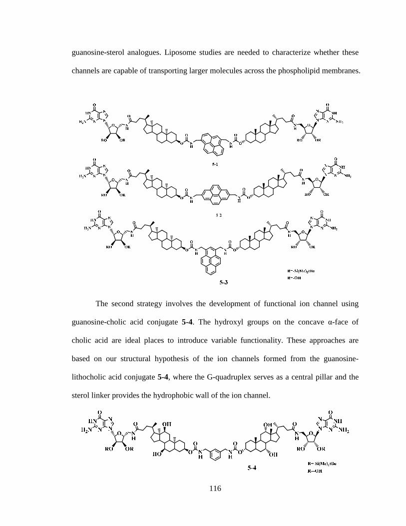
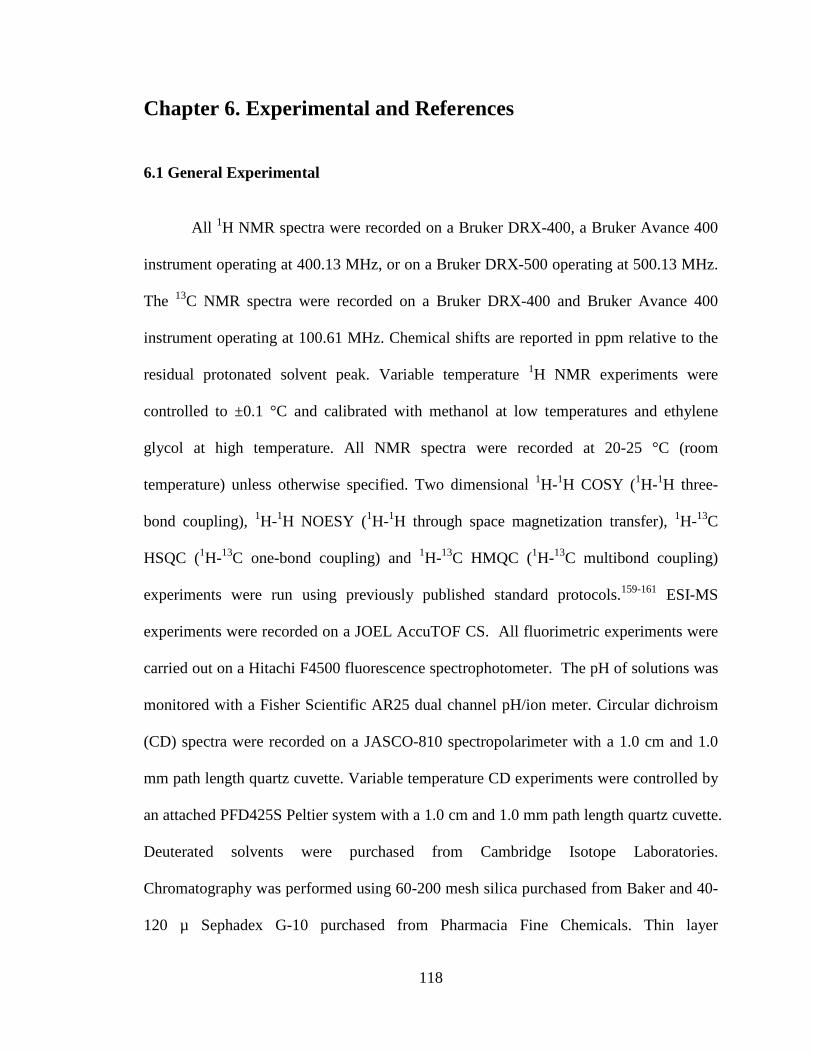
Chapter 5 Final Conclusion and Future Directions 114
Chapter 6 Experimental and References 118
61 General Experimental 118
62 Synthesis 119
63 ESI-MS Experiment of G-quadruplex 128
64 NMR Titration 129
65 15N Filtered 1H NMR Experiment 129
66 NOESY Experiment 130
67 Selective NOE Experiment 130
68 Planar Bilayer Conductance Experiments 131
69 Reversal Potential Measurements 131
ix
610 Liposome Preparation 132
References 133
x
List of Tables
Table 31 Hillersquos diameter of ion channels formed from 3-1 85
Table 41 FT-IR frequencies for bis-urea 4-10 and bis-carbamate lithocholate 3-
13helliphelliphelliphelliphelliphelliphelliphelliphelliphelliphelliphelliphelliphelliphelliphelliphelliphelliphelliphelliphelliphelliphelliphelliphelliphelliphelliphelliphelliphelliphelliphellip103
Table 42 Frequency of Events for Channels formed from 4-1 and 3-1 106
xi
List of Figures
Figure 11 a) Natural Nucleobase b) Watson-Crick base pairing motif c) Hoogsteen
base- pairing motif 4
Figure 12 Self-assembled structures of guanine a) G-ribbon and b) G-quartet 5
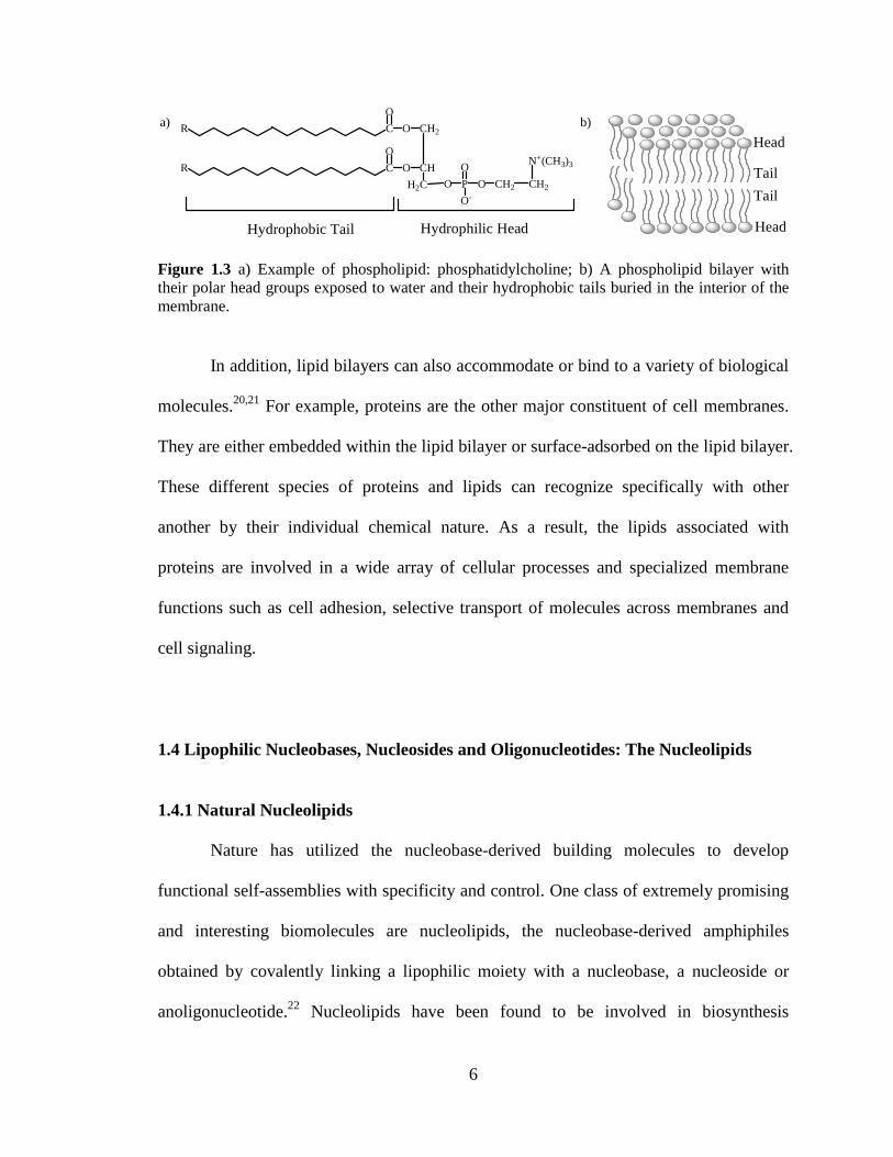
Figure 13 a) Example of phospholipid phosphatidylcholine b) A phospholipid bilayer
with their polar head groups exposed to water and their hydrophobic tails buried in the
interior of the membrane 6
Figure 14 Chemical structure of cytidinediphosphatediacylglycerol 1-6 8
Figure 15 Chemical structure of the nucleolipid antibiotic tunicamycins 9
Figure 16 Some examples of nucleolipids 13
Figure 17 The representation of the incorporation of lipophilic (thyminylocty1) ammo-
nium bromide or alkylthymine into SDS micelles and the hydrogen bond formation
between lipophilic thymine derivatives and lipophilic alkyladenine inside micelles 16
Figure 18 a) Chemical structure of the lipophilic anchor of the oligonucleotides LT
23mer b) The sequence of the LT23mer on the membrane surface can recognize the
complementary oligonucleotides A 20mer to form DNA double helix 19
Figure 19 a) Chemical structures of complementary oligonucleotide amphiphiles ONA1
and ONA2 control DNA sequences A1 and A2 fluorescent 14mer and 9mer DNA probes
b) A liposome anchored ONA can function as either the thermo-controlled reversible
switch or the chemical irreversible switch Below the melting temperature of the duplex
the probe is centered onto the liposome surface (on state) Above melting temperature or
xii
in the presence of a competitive complementary ON) the fluorescent probe is expelled
from the surface (off state) 20
Figure 110 a) Chemical structures of the cationic nucleolipids for 5rsquo-DMP or 5rsquo-AMP
transport b) A cartoon of transport of 5rsquo-DMP or 5rsquo-AMP by a carrier mechanism The
formation of carriersubstrate complex through complementary hydrogen bonding and
electrostatic interaction contribute to the transmembrane transport 23
Figure 111 a) Chemical structures of uridine-based cationic nucleolipid b) Schematic
drawing of lipoplexes formed by CL-DNA and uridine-based nucleolipid where DNA
rods are intercalated between nucleolipid bilayers 24
Figure 112 The crystal structure of DNA G-quadruplex formed form oligonucletidesd
(TG4T) in the presence of Na+ in the solution which provide a nature ion channel model
26
Figure 113 Some examples of lipophilic guanosine 27
Figure 114 Crystal structure of a lipophilic G-quadruplex [G]16middot4K+middot4Picndash 28
Figure 21 ESI-MS from titration of KPh4B into a solution of [G 1]16middot4Na+middot4DNP- in 11 CD2Cl2CD3CN a) [G 1]16middot4Na+middot4DNP- b) after addition of 1 equivalents of KPh4B c) after addition of 2 equivalents of KPh4B d) after addition of 4 equivalents of KPh4B and e) [G 1]16middot4K+middot4DNP- The diagram illustrates the possible mixed-cation G-quadruplexes that could be formed in the cation exchang processhelliphelliphelliphelliphelliphelliphellip40
Figure 22 Region of the 1H NMR spectra (400 MHz) showing the G-quartet NH1 amide protons during titration of [G 1]16middot4Na+middot4DNP- with KPh4B in 11 CD2Cl2CD3CN (a) [G 1]16middot4Na+middot4DNP- The molar ratio of added KPh4B to the [G 1]16middot4Na+middot4DNP- G-quadruplex is (b) 051 (c) 11 (d) 21 (e) 31 (f) 41 and (g) 121 42
Figure 23 A region of the 1H NMR spectrua (400 MHz) for titration of 15NH4Ph4B into a solution of (G 1)16bull 4Na+bull 4DNP- in 11 CD2Cl2CD3CN at 20 oC The signals for the amide N1H protons of the G-quartets are shown a) (G 1)16bull 4Na+bull 4DNP-The mole ratio of 15NH4
+ to (G 1)16bull4Na+bull4DNP- is b) 051 c) 11 d) 21 e) 31 f) 41 g) 121hellip 544
Figure 24 a) Region from the 2-D 15N-1H HSQC-ROESY NMR spectrum of [G 1]16middot4NH4
+middot4DNP- in 11 CD2Cl2CD3CN showing cross-peaks between the 15N NMR resonances and the 15N-filtered 1H resonances b) Region of the 2-D 1H-1H NOESY NMR
xiii
spectra of [G 1]16middot4NH4+middot4DNP- in 11 CD2Cl2CD3CN showing cross-peaks between the
N1H amide resonances and the 1H resonances for the bound NH4+ cations Both spectra
were recorded on a 1 mM sample at 20 degC using a 500 MHz NMR spectrometer 45
Figure 25 15N-filtered 1H NMR (500 MHz) spectra of 15NH4Ph4B titration the solution of [G 1]16middot4Na+middot4DNP- in 11 CD2Cl2CD3CN with mol ratio at (a) 01 (b) 11 (c) 21 (d) 31 (e) 41 (f) 121 and (g) [G 1]16middot4
15NH4+middot4DNP- 47
Figure 26 Portion of a NOESY spectrum of a 11 15NH4 Ph4B titration into [G 1]16middot4Na+middot4DNP- in 11 CD2Cl2CD3CN The 2-D spectrum shows the NOE cross-peak between the 15NH4
+1H resonance with the N1H of the inner G-quartet layer The chemical shifts of δ 1179 and 1140 ppm correspond to the N1H amide protons for the outer and inner G-quartet layers The chemical shift of the 15NH4
+ proton was confirmed from the 15N decoupled 1H NMR spectrum showing a single peak at δ 710 ppm 48
Figure 27 Selective NOE spectra for amide N1H peaks of (G 1)16middot 4Na+ middot4DNP- titrated with 2 equivalents of 15NH4Ph4B in 11CD2Cl2CD3CN Left a) N1H region of 1H NMR spectrum (green dots indicate the N1H amide proton from the first intermediate blue dots indicate the N1H amide proton from the second intermediate) b) 15NH4
+ proton region showing selective NOE correlation to amide proton at δ 1151 δ 1135 δ 1140 and δ 1179 ppm respectively Right structural scheme for the second intermediate [G4 (o) middot Na+ middot G4 (i) middot 15NH4
+ middot G4 (i) middot15NH4+ middot G4 (o)] formed during the Na+ NH4
+ exchange process with the assignments for the different G-quartet N1H amide protonshelliphelliphellip 50
Figure 28 1H NMR spectra of N1H region of different G-quadruplex solutions in CD2Cl2CD3CN at 20 degC (a) [G1]16middot4Na+middot4DNP- (b) 101 mol ratio of LiPh4B and [G1]16middot4Na+middot4DNP- (c) 81 mol ratio of CsPh4B and [G1]16middot4Na+middot4DNP- 54
Figure 31 Ditopic G-sterol 3-1 and control bis-lithocholamide 3-16 Typical traces of conductance vs time after addition of 3-1 or 3-16 are depictedhelliphelliphelliphelliphelliphelliphelliphelliphellip56
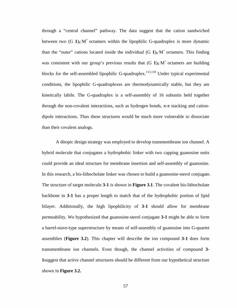
Figure 32 Hypothetical structure of a self-assembled barrel-stave ion channel formed from compound 1 The wedge represents guanosine moiety the line represents the bis-lithocholic acid linker Channel formation would be controlled by the self-assembly of guanosine head groups to give a G-quartet based structure 58
Figure 33 The unimolecular G-quadruplex stabilized by post-covalent modification that can function as a transmembrane Na+ transporter 59
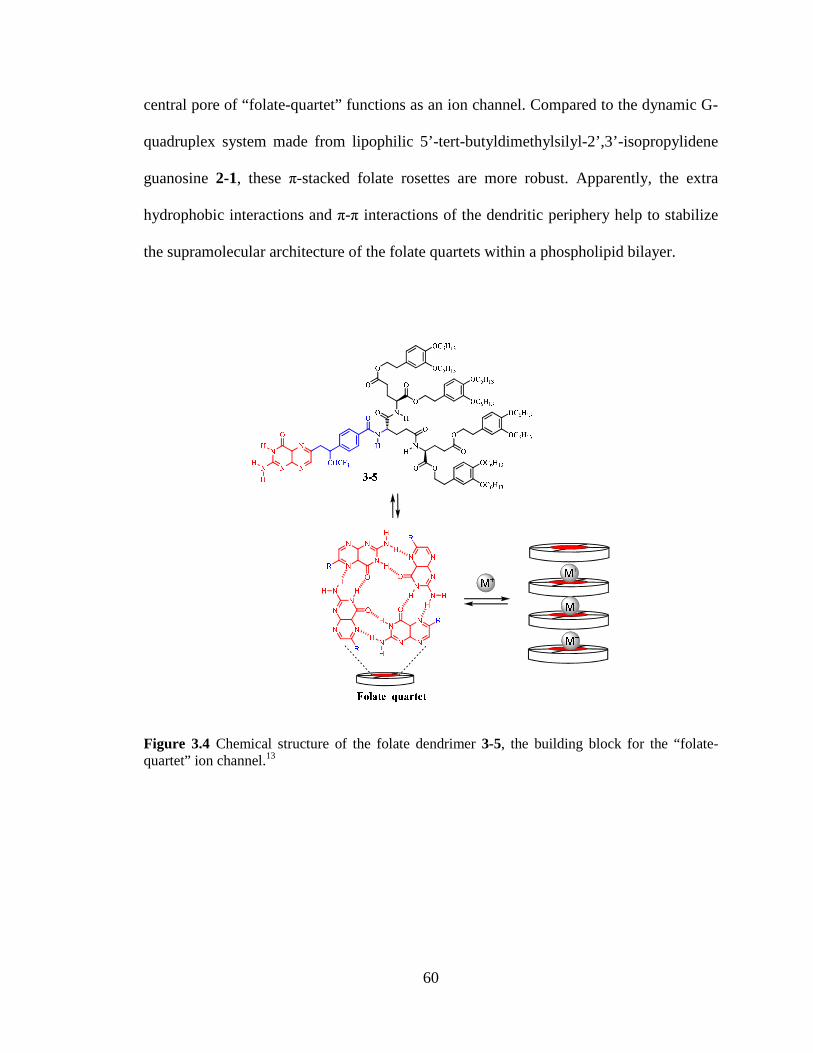
Figure 34 Chemical structure of the folate dendrimer 3-5 the building block for the ldquofolate-quartetrdquo ion channel 60
Figure 35 Schematic illustration of two types of ldquocation channelrdquo formed from a G-quadruplex a) ldquoBarrel Rosetterdquo G-quadruplex formed by 16 lipophilic guanosine
xiv
subunits b) ldquoBarrel Staverdquo G-quadruplex from by 4 ditopic ligands The ball stands for a cation and the stave stand for the ligand 62
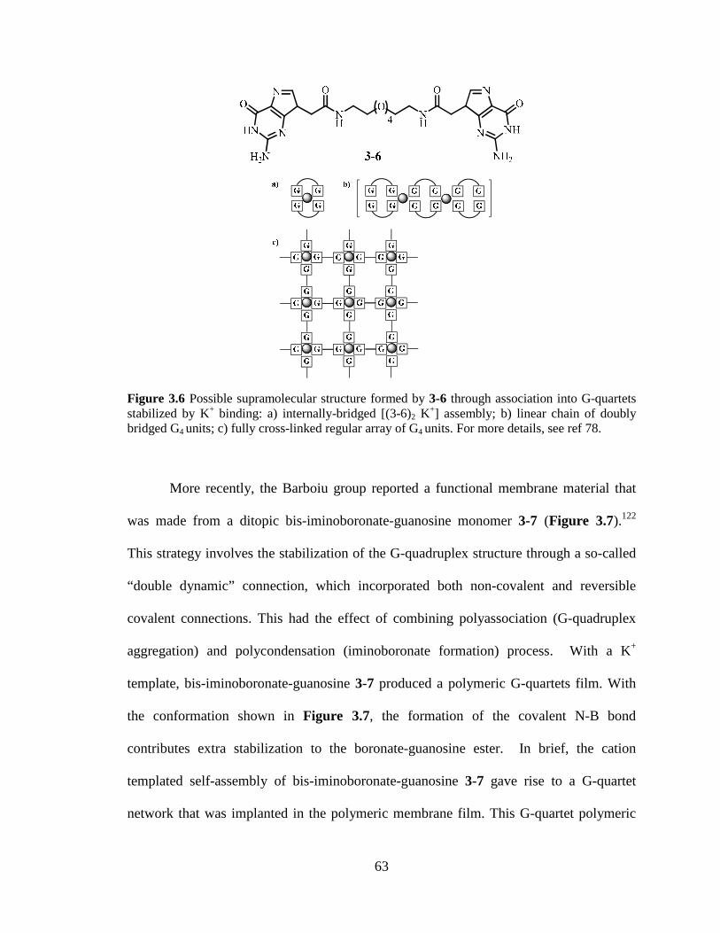
Figure 36 Possible supramolecular structure formed by 3-6 through association into G-quartets stabilized by K+ binding a) internally-bridged [(3-6)2 K+] assembly b) linear chain of doubly bridged G4 units c) fully cross-linked regular array of G4 units 63 Figure 37 Structure of bis-iminoboronate-guanosine 3-7 used to form G4-quartet based membrane 64
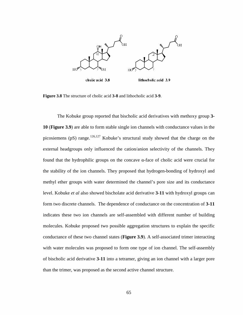
Figure 38 The structure of cholic acid 3-8 and lithocholic acid 3-9 65
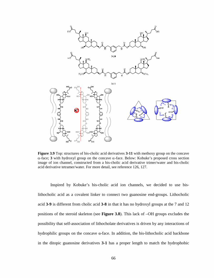
Figure 39 Top structures of bis-cholic acid derivatives 3-11 with methoxy group on the concave α-face 3 with hydroxyl group on the concave α-face Below Kobukersquos proposed cross section image of ion channel constructed from a bis-cholic acid derivative trimerwater and bis-cholic acid derivative tetramerwater 66
Figure 310 a)1HNMR spectrum of guanosine-bislithocholate conjugate 3-1 inDMSO-d6 b) Selective NOE experiment was carried out by irradiating new amide proton peak at δ 808 ppm 69
Figure 311 1H- 1HNOESY spectrum of guanosine-bislithocholic acid conjugate 3-1 in DMSO-d6 The expansion of the F1 (vertical) axis correlations near δ 808 ppm showing NOE of amide N-H proton is correlated with Harsquo Hardquo H5rsquo H5rdquo H4rsquo H3rsquo 70 Figure 312 ESI-Mass spectrum of guanosine-sterol conjugate 3-1 71
Figure 313 a)1HNMR spectrum of guanosine-bislithocholic acid conjugate 3-1 in DMSO-d6 and CDCl3 b) 1HNMR spectrum of bis-lithocholamide control compound 3-16 in CDCl3 73
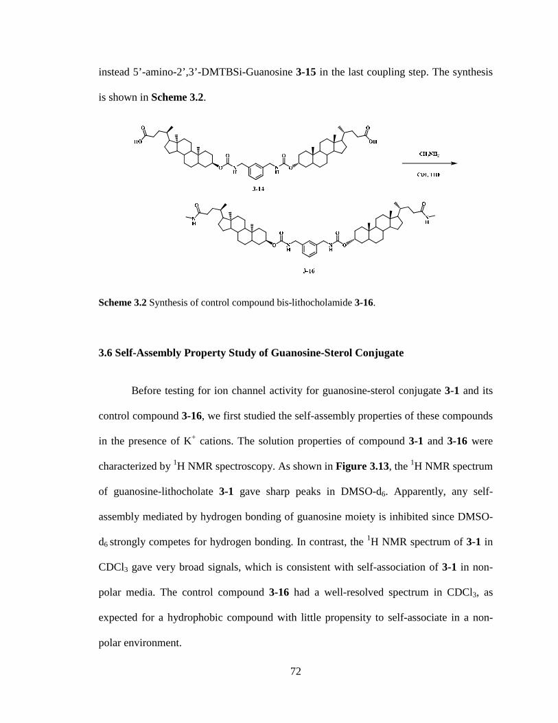
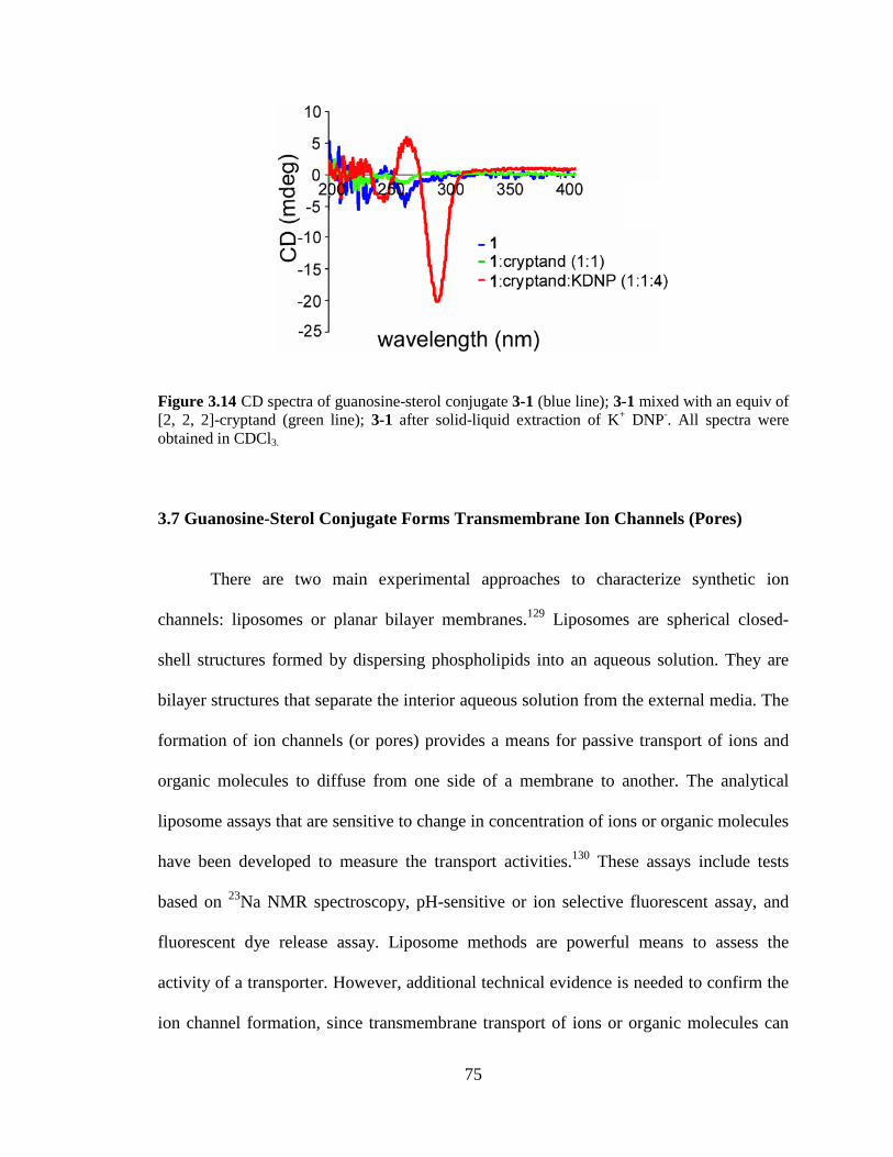
Figure 314 CD spectra of guanosine-sterol conjugate 3-1 (blue line) 3-1 mixed with an equiv of [2 2 2]-cryptand (green line) 3-1 after solid-liquid extraction of K+ DNP- All spectra were obtained in CDCl3 75
Figure 315 The setup of voltage clamp experiment The bilayer membrane was prepared with soybean phospholipid supplemented with 05 asolectin 05 DPPC and 01 cholesterol The buffer solution is 1M KCl 2mM MgCl2 and 5mM Pipes All the measurements were done at 25 degC 76
Figure 316 Representative traces from voltage-clamp experiments The distinct conduc-tance values were recorded in the presence of 3-1 at -10 mV in 1 M KCl The number of open events was counted from a total of six experiments Three of the experiment results were from method A another three were from method B 78
xv
Figure 317 Distribution of conductance change The frequency was calculated based on the total number of increment (channel open) events or decrement (channel close) events 80
Figure 318 The data analysis showed that ion selectivity (PK
+ PCl-) decreases with a
corresponding increase in conductance of ion channels These results suggested guanosine-sterol conjugate 1 forms a variety of ion channels with different sizes Panel A Panel B showed the ion selectivity data obtained by applying guanosine-sterol conjugate 3-1 using the sample loading method A and method B respectively 82
Figure 319 Barrel-stave ion channel formed from guanosine-sterol conjugate 3-1 The central G-quartet pore is responsible for the lt01 nS conductance during voltage clamp experiment 84
Figure 320 Left Hillersquos cylinder model of ion channel Right Hillersquos Equation where L is the length of the channel r is the radius of the channel and ρ is the resistivity of the solution Under our planar voltage experiment condition the resistivity of 10 M KCl solution is 89 Ωbullm The length of the channel is estimated as the thickness of the phospholipid bilayer membrane which is approximately 50 Aring 85
Figure 321 Molecular modeling of one possible self-assembly of two molecules of guanosine-lithocholate 3-1 ( Modeling was made by Dr Monica Melegari) 86
Figure 322 Possible G4-quartet stacks formed by bis-G-lithocholate 1 within bilayer membrane 87
Figure 41 a) Structure represents the modification of sterol linker b) Scheme depicting the one possible self-assembled structure from bis-urea 4-1 The bis-urea modified sterol linker might introduce additional hydrogen bonding through urea stacking to stabilize this synthetic ion channelhelliphelliphelliphelliphelliphelliphelliphelliphelliphelliphelliphelliphelliphelliphelliphelliphelliphelliphellip90
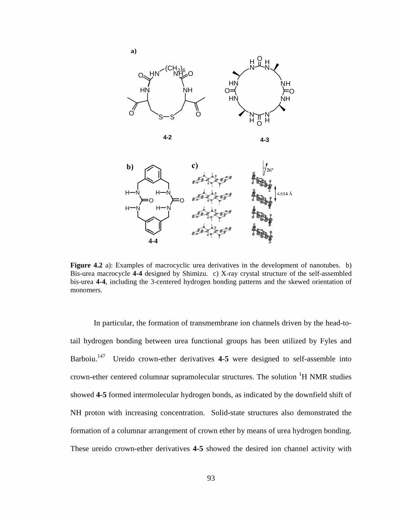
Figure 42 a)Examples of macrocyclic urea derivatives in the development of nanotubes b) Bis-urea macrocycle 4-4 designed by Shimizu c) X-ray crystal structure of the self-assembled bis-urea 4-4 including the 3-centered hydrogen bonding patterns and the skewed orientation of monomers 93
Figure 43 Dynamic self-organization of heteroditopic receptors 4-5 94
Figure 44 a) Crystal structures of lipophilic G-quadruplex templated by the sodium cation b) One of the proposed ion channels from self-assembled bis-urea modified guanosine-sterol conjugate The urea-urea hydrogen bond between the linkers was envisioned to improve the stability of the pore 95
Figure 45 1H NMR spectrum of 3α-amino lithocholate 4-9 in CDCl3 96
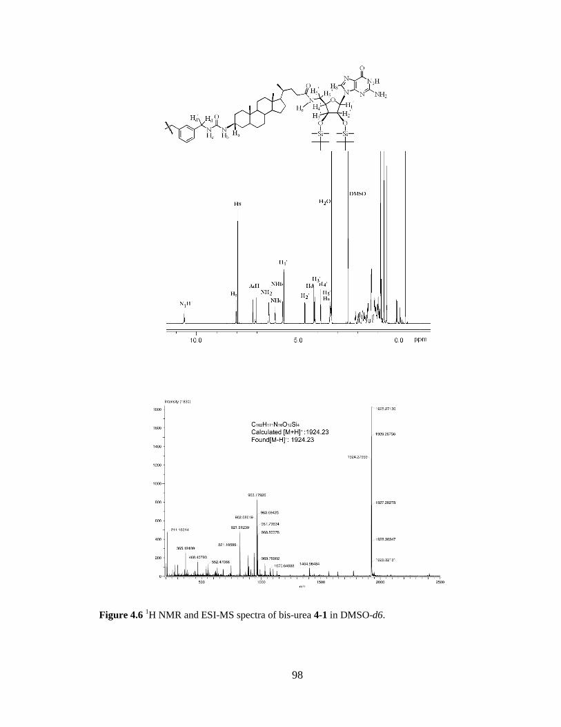
Figure 46 1H NMR and ESI-MS Spectra of Bis-Urea 4-1 in DMSO-d6 98
xvi
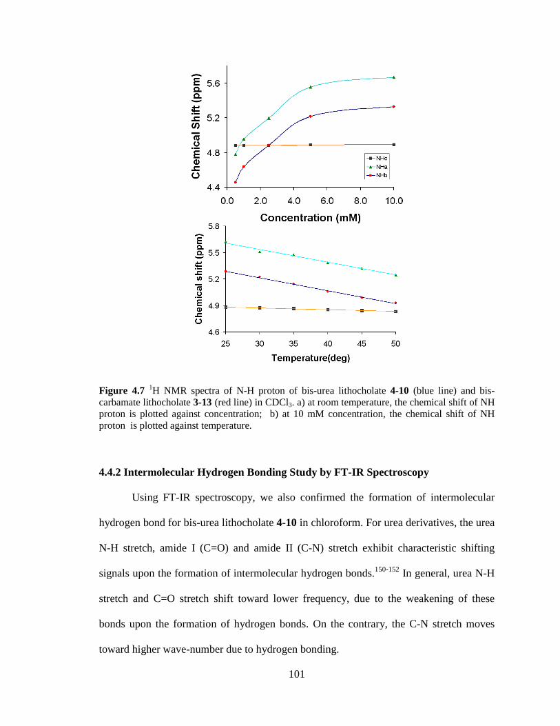
Figure 47 1H NMR spectra of N-H proton of bis-urea lithocholate 4-10 (blue line) and bis-carbamate lithocholate 3-13 (red line) in CDCl3 a) at room temperature the chemical shift of NH proton is plotted against concentration b) at 10 mM concentration the chemical shift of NH proton is plotted against temperature 101
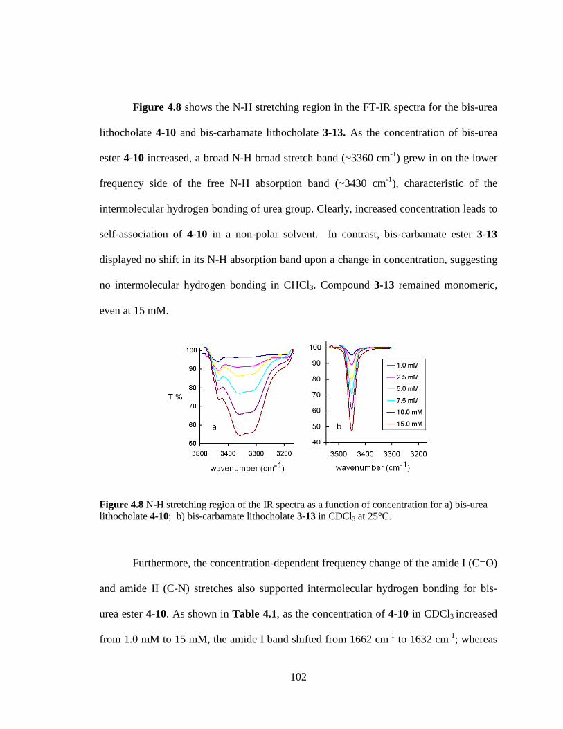
Figure 48 N-H stretching region of the IR spectra as a function of concentration for a) bis-urea lithocholate 4-10 b) bis-carbamate lithocholate 3-13 in CDCl3 at 25degC 102 Figure 49 Representative conductance records from voltage clamp experiment Ion channels formed by 4-1 in a planar bilayer at an applied voltage of -10 mV in 1M KCl (trans) KCl (cis) solution (pH 70) 104
Figure 410 Distribution of conductance change for the ion channels from guanosine-sterol conjugate 4-1 and 3-1 The frequency was calculated based on the total number of increment (channel open) events or decrement (channel close) events a) data from compound 4-1 b) data from compound 3-1 106
Figure 411 Histogram showing the various open lifetimes for 1-5 nS channels formed from 3-1 and 4-1 Distribution of conductance change the frequency was calculated based on the total number of increment (channel open) events 108
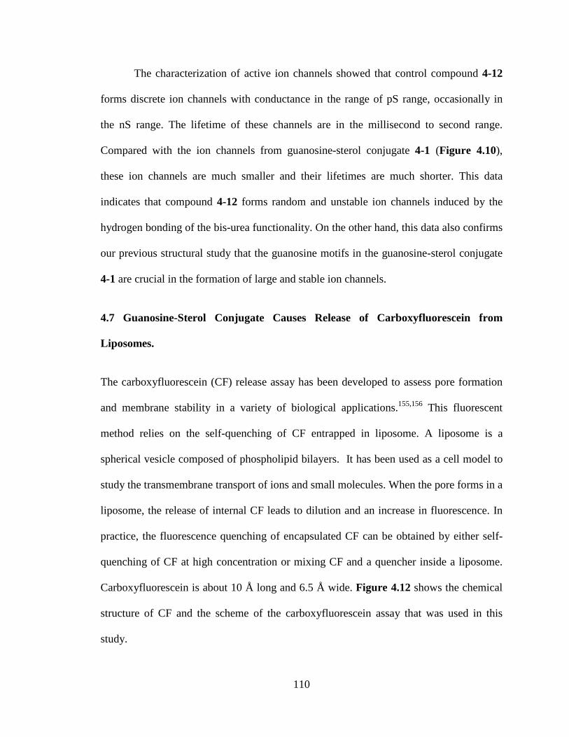
Figure 412 a) Chemical structure of carboxyfluorescein b) Scheme of a fluorescent assay of carboxyfluorescein release from liposome 111
Figure 413 Carboxyfluorescein release assay confirmed the pore formation mediated by guanosine-sterol conjugate 3-1 and 4-1 112
xvii
List of Schemes
Scheme 21 Schematic structure of (A) G-quartet and (B) G8-octamer 32
Scheme 22 Highndashresolution crystal structure of a DNA G-quadruplex formed by oligonucleotide [d(TG4T)]4 b) Natural potassium ion channel 34
Scheme 23 Schematic representation of 15NH4
+ movement through the Oxy-15 quadruplex a) bulk NH4
+ cation only exchanges with the outer 15NH4+ b) exchange of
two bound 15NH4+ cationsare random Black yellow and red ball represent the bulk
ammonium ion and the boundammonium ions located in outer and inner binding site of the G-quadruplex 35
Scheme 24 G-quadruplex formed through self-assembly of G 1 in the presence of K+DNP- 36
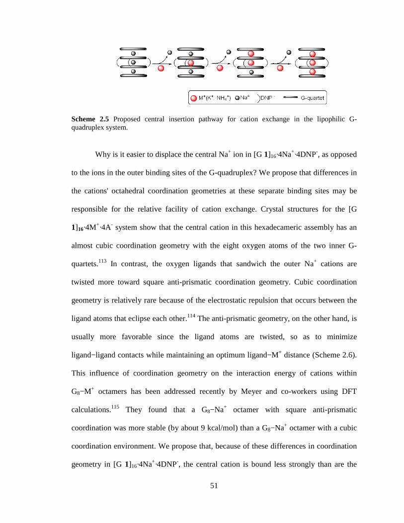
Scheme 25 Proposed central insertion pathway for cation exchange in the lipophilic G-quadruplex system 51
Scheme 26 These depictions are taken from the crystal structure for [G1]16middot4Na+middot4pic- The illustrations show the octahedral coordination geometry for one of the outer Na+ cations (on the left) and for the central Na+ (right) The central Na+ has a almost cubic coordination geometry whereas the oxygen ligands are twisted toward the energetically more favorable square anti-prism geometry for the outer Na+ 52
Scheme 31 Synthesis of bis-guanosine-lithocholic acid conjugate 3-1helliphelliphelliphelliphelliphellip68
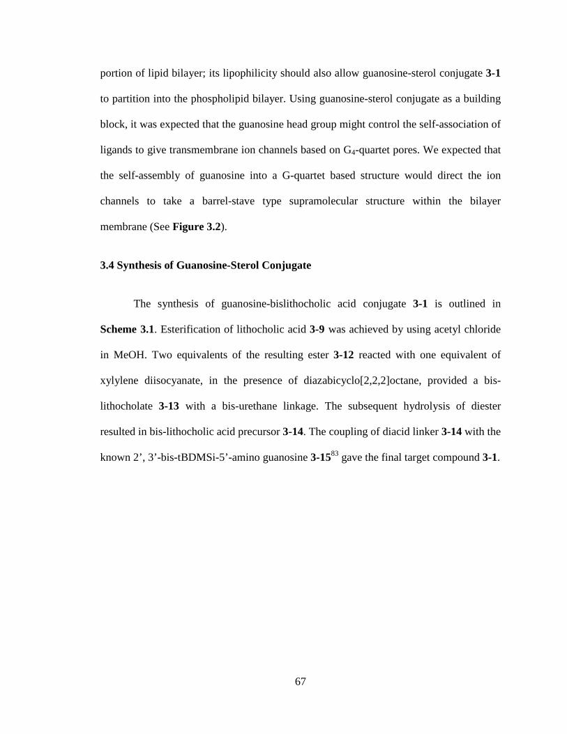
Scheme 32 Synthesis of control compound bis-lithocholamide 3-16 72
Scheme 41 Three center hydrogen bonding net-work formed by urea derivativehelliphellip 92
Scheme 42 Synthesis of 3α-amino lithocholate 4-9 96
Scheme 43 Synthesis of bis-urea modified guanosine-sterol conjugate 4-1 97 Scheme 44 Synthesis of bis-urea lithocholamide 4-12 for the control experiment 109
1
Chapter 1 Introduction
11 Introduction
Molecular self-assembly plays a fundamental role in Nature and is a widely used
principle to build supramolecular structures from individual molecular components1-3
For example self-assembly has been a useful tool to mimic biological functions and
fabricate nanomaterials45 In recent years many research activities have focused on the
development of novel functional materials using well-defined building blocks This thesis
ldquoSynthetic Ion Channels from Lipophilic Guanosine Derivativesrdquo describes the design
and development of amphiphilic building blocks for the self-assembly of functional
structures
12 Thesis Organization
This thesis is organized into six chapters The overall goal of this research was to
develop trans-membrane synthetic ion channels using lipophilic guanosine as building
blocks Chapter 1 briefly discusses the molecular recognition properties of the naturally
occurring nucleobases and then goes on to review the potential use of lipophilic
nucleobases nucleosides and oligonucleotides as functional nanostructures and
biomaterials The cation exchange studies in Chapter 2 give insight into the stability of
self-assembled lipophilic G-quadruplex in solution The kinetic lability of lipophilic G-
quadruplex inspired the design of a ditopic lipophilic guanosine to build trans-membrane
ion channels Chapter 3 describes large and stable ion channels formed from a ditopic
2
guanosine-lithocholic acid conjugate The ion channel activity and structure-properties
characterization suggested the key role of the guanosine subunit in the formation of ion
channels Chapter 4 explores the enhanced stability and activity of ion channels through
the modification of guanosine-sterol building blocks Chapter 5 describes the conclusion
and directions of future research Finally Chapter 6 contains the experimental protocols
for the research described in Chapters 2-4
13 Nucleobases and Lipids as Building Motifs for Supramolecular Structures
131 Nucleobases Molecular Recognition Motif
In Nature the self-assembly of single strand DNA into double helix and other
higher order structures is mediated by many noncovalent interactions such as hydrogen
bonding π-π stacking and van der Waals forces Among these intermolecular
interactions complementary hydrogen bonding via nucleobase-pairing is of primary
importance in the molecular recognition of DNA and RNA sequences Thus the
molecular recognition capability of nucleobases via specific hydrogen-bonding pattern is
considered to play an essential role in the storage translation and replication of genetic
information6-8
The naturally occurring nucleobases are nitrogen-containing aromatic
heterocycles including the double-ringed purines (adenine 1-1 guanine 1-2) and single-
ringed pyrimidines (cytosine 1-3 thymine 1-4 and uracil 1-5) They are the key
constituents of DNA and RNA that are involved in molecular recognition Cytosine 1-3
3
guanine 1-2 adenine 1-1 and thymine 1-4 are found in DNA while uracil 1-5 replaces
thymine 1-4 in RNA Nucleobases are able to recognize each other through a
complementary hydrogen bonding interaction known as Watson-Crick pairing9 Watson-
Crick pairing is one of the most recognized observations in a variety of DNA- and RNA-
structures In this specific hydrogen-bonding motif guanine 1-2 pairs with cytosine 1-3
through a three-point hydrogen bonding while adenine 1-1 interacts with thymine 1-4 (or
uracil in RNA) in a two-point hydrogen bonding fashion (Figure 11)
Furthermore various alternative hydrogen-bonding motifs are also possible71011
These non-Watson-Crick pairings include mismatch-pairing nucleobase self-
dimerization and Hoogsteen base pairing that associates the C6 and N7 binding sites of
purine nucleobases Hoogsteen base pairing through the self-association of guanosine 1-2
is another prevalent hydrogen bonding motif found in a variety of G-rich DNA and RNA
suprastructures12
4
Figure 11 a) Natural Nucleobase b) Watson-Crick base pairing motif c) Hoogsteen base- pairing motif
Of the natural nucleobases guanosine 1-2 possesses a unique self-association
property since it incorporates both Watson-Crick and Hoogsteen edges for the self-
complementary hydrogen bonding The guanine-guanine interactions involve the two
Hoogsteen pairings between hydrogen bonding acceptor O6 and N7 at Hoogsteen edges
and hydrogen bonding donor N1H amide and N2H amine at Watson-Crick edges Under
certain conditions guanine self-assembles into several suprastructures that differ from the
classic nucleobase dimers13-17 These higher-ordered structures include the G-ribbon
structure or cyclic structure known as the G-quartet (Figure 12b) Figure 12a shows
the hydrogen-bonded ribbon G-structure in the absence of cations Whereas the G-
quartet is a preferential self-assembled structure in the presence of cations since cations
5
stabilize this cyclic tetramer through the electrostatic interaction between cation and the
carbonyl oxygens (Figure 13b)
N
NN
N
O
NH
H
H
R
N
NN
N
O
NH
H
H
R
N
NN
N
O
NH
H
H
R
N
NN
N
O
NH
H
H
Ra)
N
NN
N
O
N
H
H
H
R
N
NN
N
O
N
H
H
H
R
NN
NN
O
N HH
H
R
NN
N N
O
NHH
H
Rb)
Figure 12 Self-assembled structures of guanine a) G-ribbon and b) G-quartet
132 Lipid Bilayer Structural Unit of Functional Biological Membrane
The lipid bilayer is the basic structural unit of all biological membranes1819 It
acts as the hydrophobic barrier that segregates cellular compartments and isolates the
cells from the extracellular environment As shown in Figure 13 phospholipids the
fundamental building blocks of all cell membranes consist of a polar phosphate-
containing head group and two nonpolar hydrocarbon tails These amphiphilic molecules
self-assemble in a tail-to-tail fashion to form a lipid bilayer with a hydrophobic interior
and a hydrophilic surface Lipid bilayers are versatile structures that can be formed by a
number of phospholipids and other lipid molecules such as cholesterol and glycolipids
6
CH2OP
O
O-
OH2C
CHO
CH2O
C
C
R
O
O
R
CH2
N+(CH3)3
Hydrophobic Tail Hydrophilic Head
Tail
Head
Head
Tail
b)a)
Figure 13 a) Example of phospholipid phosphatidylcholine b) A phospholipid bilayer with their polar head groups exposed to water and their hydrophobic tails buried in the interior of the membrane
In addition lipid bilayers can also accommodate or bind to a variety of biological
molecules2021 For example proteins are the other major constituent of cell membranes
They are either embedded within the lipid bilayer or surface-adsorbed on the lipid bilayer
These different species of proteins and lipids can recognize specifically with other
another by their individual chemical nature As a result the lipids associated with
proteins are involved in a wide array of cellular processes and specialized membrane
functions such as cell adhesion selective transport of molecules across membranes and
cell signaling
14 Lipophilic Nucleobases Nucleosides and Oligonucleotides The Nucleolipids
141 Natural Nucleolipids
Nature has utilized the nucleobase-derived building molecules to develop
functional self-assemblies with specificity and control One class of extremely promising
and interesting biomolecules are nucleolipids the nucleobase-derived amphiphiles
obtained by covalently linking a lipophilic moiety with a nucleobase a nucleoside or
anoligonucleotide22 Nucleolipids have been found to be involved in biosynthesis
7
processes14 and a variety of biological activities such as antimicrobial antivirus anti
tumor activities in eukaryotic and prokaryotic cells2324
Cytidinediphosphatediacylglycerol (CDP-diacylglycerol) 1-6 is one of the most
ubiquitous nucleolipids present in cells (Figure 14)25 It plays an essential role in the
biosynthesis of the membrane phospholipids In mammalian cells CDP-diacylglycerol1-
6is formed from cytidinetriphosphate and phosphatidic acid and serves as an activated
energy-rich intermediate Catalyzed by different specific membrane-bound enzymes
CDP-diacylglycerol 1-6 is able to transfer a phosphatidic acid to a hydroxyl group of a
carbohydrate or glycolipid to generate phospholipids The specific enzymes are located in
mitochondria for the biosynthesis of phosphatidylglycerol the precursor of cardiolipin or
in microsomes for the biosynthesis of phosphatidylinositol2627 This indicates that
nucleoside-based lipid can be incorporated into a variety of phospholipids in the cell
membrane In prokaryotes CDP-diacylglycerol 1-6 functions as the precursor for the
biosynthesis of phosphatidylserine as well28
Moreover CDP-diacylglycerol 1-6 is involved in the enzymatic activities of
phosphatidylserine synthases occurring in gram-negative bacteria such as E coli
Salmonella typhimurium Serratiamarcescenceetc The membrane association of these
phosphatidylserine synthases is induced by the lipid substrate CDP-diacylglycerol 1-
61819 The CDP-diacylglycerol-dependent activity of phosphatidylserine suggested that
phosphatidylserine synthases are peripheral membrane proteins that specifically bind to
the nucleolipid CDP-diacylglycerol 1-6 via an intermolecular interaction such as
8
nucleobase-pairing hydrophobic interactions andor van der Waals interactions
Nucleolipid CDP-diacylglycerol 1-6 can reside in the membrane due to its high degree of
amphiphilicity
Figure 14 Chemical structure of cytidinediphosphatediacylglycerol 1-6
Tunicamycins 1-7 was the first discovered nucleolipid antibiotic that was
produced from the fermentation broths of Streptomyceslysosuperificus (Figure 15)
Tunicamycins1-7 is composed of the heterocyclic nucleobase uracil and a branched fatty
acid side chain and it has demonstrated the ability to inhibit a variety of biosynthesis of
oligosaccharide thus preventing the glycosylation of proteins in both prokaryotic and
eukaryotic cells21-24 The antibiotic can function as an irreversible inhibitor for the
GlcNAc-1-phosphate transferaseGlcNAc-1-phosphate transferase is the eukaryotic
enzyme that catalyzes the transfer of N-acetylglucosamine1-phosphate from UDP-N-
acetylglucosamine to dolichylphosphate to generate N-
acetylglucosaminylpyrophosphoryldolichol Mechanistic studies on the actions of
tunicamycin 1-7 have revealed that these nucleolipids bind preferentially to the GlcNAc-
1-phosphate transferase and also interact with the membrane phospholipid2930 The
binding of the tunicamycins 1-7 totransferase occurs when tunicamycins 1-7 is
incorporated into biological membranes Tunicamycins 1-7 also acts as reversible
inhibitors for the phospho-N-acetylmuramyl-pentapeptidetranslocase (translocase I) in
9
the peptidoglycan biosynthesis as well Translocase I is the specific enzyme that
catalyzes the relocation of phospho-MurNAc-L-Ala-g-D-Glum-DAP-D-Ala-D-Ala from
UMP to a membrane-bound carrier undecaprenyl phosphate The studies of actions of
tunicamycins have showed that the inhibition by tunicamycins 1-7 is competitive with
regard to the substrate UDP-MurNAc-pentapeptide since both molecules share the same
uridine subunit This result demonstrated the recognition characteristic of nucleobase in
the observed bioactivity The capability of tunicamycins 1-7 to locate within the
membrane bilayer can be attributed to the hydrophobicity of the tunicamycins
Figure 15 Chemical structure of the nucleolipid antibiotic tunicamycins
142 Molecular Recognition of Synthetic Nucleolipids
The design of synthetic amphiphiles that are derived from biologically functional
nucleobases nucleosides and oligonucleotides has become a major strategy to build
interesting self-assembled structures with complex and desirable properties Like the
typical amphiphiles nucleolipid amphiphiles will self-assemble in aqueous solution to
10
form distinct structures such as micelles vesicles and other self-aggregates31-33 However
nucleolipids have unique physicochemical properties due to their informative nucleobase
constituents The specific base-pairing of nucleobases through hydrogen bonding and π-π
stacking is cooperative with the hydrophobic effect from the lipid segment contributing
to the self-assembly
In the 1980s the molecular recognition between two complementary nucleolipids
1-8 and 1-9 was first reported by Ringsdorf and coworkers34 Using surface pressure
measurements UV and DSC techniques they found that adenine- and thymine-
nucleolipids base-paired at the air-water interface Afterward numerous techniques
including Langmuir-Blodgett NMR FTIR RA ATR fluorescent probes etc have
provided direct evidence of the complementary hydrogen bonding between nucleolipids
at the air-water interface35-38
The base pairing of nucleolipid was also observed at the mesoscopic interfaces of
the self-assembly of nucleolipids in either micellar or vesiclar structures39 For instance it
was reported by Berti and Baglioni that short-chain phospholiponucleosides diC8P-
adenosine 1-10 and diC8P-uridine 1-11 self-assembled into mixed micelles with CMC
values around 10-3 M40 The data from NMR UVminusVis and CD spectroscopies revealed
the stacking and hydrogen-bonding of the complementary adenosine and uridine occurred
at the micellar surface The continued study of phospholiponucleosides with longer alkyl
chain (DOP-adenosine 1-12 DOP-uridine 1-13 and DOP-cytosine 1-14) demonstrated
that these nucleolipidsare able to self-assemble into liposomes41 The liposomes formed
11
from a mixture of DOP-adenosine 1-12 and DOP-uridine 1-13 showed the UV CD and
NMR properties for base-pairing while the liposome formed from a mixture of DOP-
adenosine 1-12 and DOP-cytosine 1-14 failed to give any spectroscopic change These
data supporting that the molecular recognition properties of nucleolipids are controlled by
specific Watson-Crick base pairing
Specific base pairing that resembles biofunctional supramolecular structures has
been found for certain nucleolipids Itojima and coworkers reported that lipophilic
dimyristoyl-5rsquo-phosphatidylnucleosides 1-15 can self-assemble into helical strands that
are similar to DNA duplex in aqueous solution42 Dimyristoyl-5rsquo-phosphatidyladenosine
1-15 can form various self-assemblies including multi-helical strands or tubular
structure depending on the pH and alkalinity of the solution The formation of a hybrid
helix from the equimolar mixture of dimyristoyl-5rsquo-phosphatidyladenosine 1-15
anddimyristoyl-5rsquo-phosphatidyluridine 1-16 suggested that the specific hydrogen bonding
between nucleobases directs the self-assembly of nucleolipids The research of Gottarelli
and Spada established that in the presence of certain cations liphophilic guanosine 1-17
can self-associate into columnar structures that contain stacked G-quartet in organic
solution144344 The self-assembly is driven by Watson-Crick and Hoogsteen base pairing
of guanine π-π stacking and cation-dipole interactions Similar to the liquid crystals
formed from guanosine oligonucletides in aqueous solution the G-quartet is the
fundamental building block for these supramolecular structures Such a G-quartet was
also found in the DNA secondary structure that regulates the telomerase activity in
chromosomes
12
The molecular recognition characteristics of nucleolipids have attracted
substantial research including attention to the design and synthesis of novel amphiphilies
derived from nucleobases for both the fundamental aspects and application purposes For
these reasons a wide range of nucleolipids have been synthesized by covalently linking
the hydrophobic segment to nucleobases nucleosides and oligonucleotides The
lipophilic moieties such as alkyl chain cholesterol glycerol etc are attached either
directly to the nucleobase or to the sugar moiety of nucleoside or oligonucleotides
Nucleolipid structures can be classified as either neutral cationic anionic or zwitterionic
nucleolipids depending on their charge properties Figure 16 lists the nucleolipids
reported in the literature mentioned in section 14
13
Figure 16 Some examples of nucleolipids
143 Membrane Association Properties of Nucleolipids
The lipophilic moiety of certain nucleolipids provides another important feature
for these compounds namely membrane association A variety of nucleolipids have been
found to be lipophilic components that can serve as an anchor to attach lipophilic
nucleobase nucleosides and oligonucleotides into the lipid membrane45-48 For the
membrane incorporation purpose the lipid anchor should be sufficiently hydrophobic to
14
overcome the self-association propensity of the amphiphiles and also allow it to be stable
in the membrane The Huster and Liebscher groups have designed lipophilic nucleosides
that conjugate adenine and uracil nucleobases with 1-octadecynyl or sterol hydrophobic
moiety1-18 1-1949 Using solid-state 2H and 31P 1H MAS NMR spectroscopy they
identified the membrane location of the functional groups of these nucleolipids The lipid
moieties are incorporated into the phospholipid membrane while the nucleobase
functional groups are exposed on the surface of the membrane The effects of the inserted
hydrophobic moiety on the membrane show that the steroidal ring of 1-19 causes the
decrease of lipid order parameters and packing density while the alkyl chain of 1-18 has
no pronounced influence on the structure of lipid membrane Compound 1-18 was
proposed as an ideal nucleolipid for building the functional membrane surface This
result suggested that nucleolipids can be used to build functionalized membrane surfaces
In addition lipophilic moieties are implicated to associate the membrane permeation for
the antiviral and antitumor activities of nucleolipid antibiotics50-52
15 Functional Nanostructures from Amphiphilic Nucleobases
Various researches on the lipophilic nucelobases nucleosides and
oligonucleotides have demonstrated their very interesting and unique properties5354 The
combination of molecular recognition and hydrophobic characteristics of nucleolipids
may allow them to target specific lipid membrane or deliver biologically active molecules
which provides a wide opening for the development of bio-devices drug delivery
biosensor and therapeutic strategy This section will describe a few but promising
15
nucleolipid systems that can base-pair within membranes or function as membrane
transporters
151 Molecular Recognition in Aqueous Solution Amphiphilic Nucleobase-Derived
Supramolecular Receptors
The molecular recognition of lipophilic nucleobases through base-paring in
aqueous environment was first reported by Nowick and co-workers55-57 In their
pioneering work lipophilic (thyminylocty1)ammonium bromide 1-20 and sodium
dodecyl sulfate (SDS) self-assembled into micelles and served as supramolecular
receptors55 The resulting receptor consisting of thymine functionality was able to
recognize N6-actyle-9-alkyladenine 1-21 in aqueous solution through an A-T base-pair
(Figure17) The incorporation of (thyminylocty1) ammonium bromide 1-20 into SDS
micelles was assisted by the electrostatic interaction between the positive-charged
ammonium group of 1-20 and the negatively charged sulfate group of SDS and the
hydrophobic interaction between alkyl chain of 1-20 and SDS The complete
incorporation of 10mM of 1-20 into micelles was achieved at an SDS concentration of
20 mMNMR titration studies showed that thymine derivative 1-20 can bind to
alkyladenine derivative 1-21 by means of intermolecular hydrogen bonding in the
presence of SDS This specific binding was determined to be 11 stoichiometry by a Jobrsquos
plot which is consistent with a 11 adenine-thymine base-pairing pattern It was also
found that when SDS is absent in the aqueous solution only aromatic stacking exists
between 1-20 and 1-21 This result suggests that the presence of SDS is crucial for the
hydrogen bond formation and that the adenine-thymine base-pairing occurs within the
hydrophobic core of SDS micelles The author also investigated the effect of lipophilicity
16
of adenine derivatives 1-21 on the hydrogen binding between 1-20and 1-21 By varying
the length of alkyl chain on adenine derivatives 1-21 the binding constants were
measured and compared The adenine derivatives 1-21 bearing longer alkyl-chains
exhibited a larger binding affinity with thymine derivatives 1-20 indicating that the
hydrophobicity of adenine derivatives 1-21 is required for micellar incorporation and
base pairing
Figure 17 The representation of the incorporation of lipophilic (thyminylocty1) ammonium bromide or alkylthymine into SDS micelles and the hydrogen bond formation between lipophilic thymine derivatives and lipophilic alkyladenine inside micelles
The continued effort by the Nowick group provided another successful example
of molecular recognition between neutral lipophilic nucleobases in SDS micelles57 Here
the neutral 1-alkylthymine derivative 1-22 incorporated into SDS micelles functioned as
supramolecular receptors Likewise the receptors can bind N6-acyl-9-alkyladenine 1-21
through base-pairing in aqueous solution Nowick found that 1-alkylthymine derivative
1-22 must be sufficiently hydrophobic to be incorporated within SDS micelles The
incorporation of 1-22 into micelles plays a critical role in the adenine-thymine base-
pairing within micelles The studies on molecular recognition between neutral lipophilic
17
nucleobases in aqueous SDS micelles confirmed the model of supramolecular receptor
proposed in their previous study The hydrophobic core of SDS micelles provides a
suitable environment for hydrogen bonding between the complementary lipophilic
nucleobases The hydrophobic interaction between lipophilic nucleobases and SDS and
the base-pairing between two complementary nucleobases moieties provides a driving
force for molecular recognition in water By means of micelles Nowickrsquos strategy should
have a wide application in the recognition or transport of nucleobase related biomolecules
152 Lipophilic Nucleobases Approach Toward Functional Biological Surfaces
In biological systems phospholipid membrane surfaces represent one of the
active microscopic interfaces for molecular recognition As we mentioned before
membranes are versatile structures that are composed of lipids and a variety of biological
molecules For example proteins embedded within the lipid bilayer or surface adsorbed
on the lipid bilayer are involved in most biological activities such as signal transduction
enzymatic reaction etc To mimic and understand the natural recognition and binding
process on the membrane surface a number of researchers have focused on the
development of functional membranes using nucleolipids
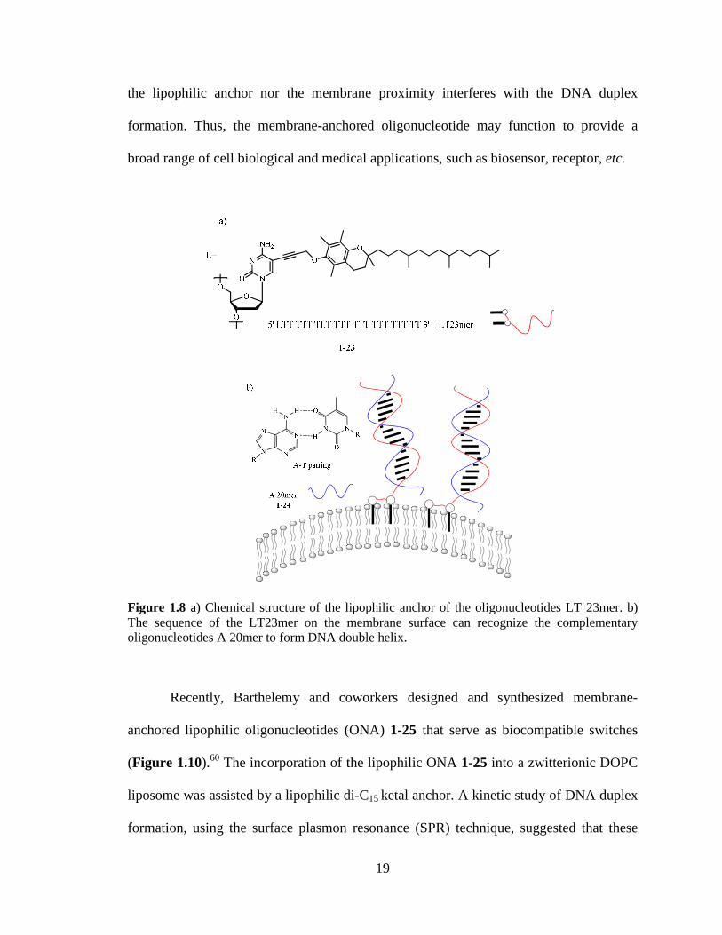
Liebecher Huster and Herrmann reported an oligonucleotide (LT 23mer) 1-23
that anchors into the lipid membrane and forms a DNA duplex58 LT-23mer is designed
as an amphiphile that consists of 21 thymidine (T) units and two lipophilic nucleotides L
in at positions 1 and 8 The α-tocopherol subunit located in the 1-position of cytosine
serves as a lipophilic anchor (Figure 18) A doubly lipophilic anchor separated by 6
nucleotides was designed to prevent the oligonucleotide from self-associating in aqueous
18
environment which improved the membrane insertion The membrane incorporation of
lipophilic LT 23mer 1-23 was studied by incubating LT 23mer and fluorescently labeled
3rsquoRh-or 5rsquoRh-A20mer 1-24 with giant unilamellar vesicles (GUVs) using fluorescence
microscopy Rh fluorescence was detected over the entire membrane indicating the
complementary binding of LT 23mer 1-23 to 3rsquoRh- or 5rsquoRh-A20mer 1-24 occurs on the
GUV surface By means of 1H 31P 1H NMR EPR DSC and fluorescence techniques
Huster et al established that LT 23mer 1-23 are enriched in the liquid-disordered
membrane domains59 LT 23mer 1-23 inserts stably into lipid membranes and imposes
insignificant modification to the bilayer membrane structure The ribosecytosine moiety
of LT 23mer is located in the lipid-water interface while the non-lipidated nucleotide end
is exposed to the aqueous environment This result suggests that the nucleolipids could be
used to build a functional surface on the membrane surface
After LT 23mer 1-23 anchored into the lipid membrane NMR and FRET
techniques were used to characterize the functionality of the membrane surface The
GUVs containing LT 23mer and fluorescent phospholipid N-NBD-PE were incubated
with 3rsquoRh-A20mer 1-24 The resulting strong FRET was attributed to the efficient
complementary binding between membrane-bound LT 23mers1-23and lipophilic 3rsquoRh-
A20mer1-24 The formation of Watson-Crick A-T base pairs which was confirmed by
1H NMR chemical shifts suggested that the oligonucleotides exposed to the aqueous
media are capable of recognizing the complementary oligonucleotide to form the DNA
double helix This membrane-bound DNA double strand presents similar structural
feature and melting properties as the free double-stranded DNA indicating that neither
19
the lipophilic anchor nor the membrane proximity interferes with the DNA duplex
formation Thus the membrane-anchored oligonucleotide may function to provide a
broad range of cell biological and medical applications such as biosensor receptor etc
Figure 18 a) Chemical structure of the lipophilic anchor of the oligonucleotides LT 23mer b) The sequence of the LT23mer on the membrane surface can recognize the complementary oligonucleotides A 20mer to form DNA double helix
Recently Barthelemy and coworkers designed and synthesized membrane-
anchored lipophilic oligonucleotides (ONA) 1-25 that serve as biocompatible switches
(Figure 110)60 The incorporation of the lipophilic ONA 1-25 into a zwitterionic DOPC
liposome was assisted by a lipophilic di-C15 ketal anchor A kinetic study of DNA duplex
formation using the surface plasmon resonance (SPR) technique suggested that these
20
lipophilic membranendashbound ONA recognize their complementary sequences (ON) 1-26
or fluorescent-labeled complementary sequences (fONA) 1-27 via Watson-Crick base
pairings The DOPC lipid membrane has no effect on this DNA duplex formation Thus
the oligonucleotide-tagged liposome can function as a supramolecular platform for
detection and delivery of bioactive molecules
Figure 19 a) Chemical structures of complementary oligonucleotide amphiphiles ONA1 and ONA2 control DNA sequences A1 and A2 fluorescent 14mer and 9mer DNA probes b) A liposome anchored ONA can function as either the thermo-controlled reversible switch or the chemical irreversible switch Below the melting temperature of the duplex the probe is centered onto the liposome surface (on state) Above melting temperature or in the presence of a competitive complementary ON) the fluorescent probe is expelled from the surface (off state)
Barthelemy demonstrated that liposome-anchored ONA 1-25 can be fluorescently
switched ldquoonrdquo or ldquooffrdquo depending on the stability of DNA duplex formed from ONA and
ONA1 5- U GGC TCA CAA CAG GC-3
A1 5- GGC TCA CAA CAG GC-3
ONA2 5- U GCC TGT TGT GAG CC-3
A2 5- GCC TGT TGT GAG CC-3
9mer-probe 5- Fluoroscein GGC TCA CAA-3
14mer-probe 5- Fluoroscein-GGC TCA CAA CAG GC-3
1-25
1-27
1-26
21
its fluorescent labeled complementary sequences (fONA) 1-27 The
formationdissociation of ONA-fONA could be tuned by certain physical (temperature)
or chemical (presence of competitive complementary ON sequences) stimuli They found
ONA-tagged liposomes serve as a reversible thermal switch At temperatures above Tm
(melting temperature of ONA-fONA duplex) the ONA-fONA duplex melts and the
switch is turned ldquooffrdquo while at temperatures below Tm the ONA-fONA duplex anneals
at the surface of liposome and the switch is ldquoonrdquo On the other hand ONA-tagged
liposome was demonstrated to be irreversibly switched ldquooffrdquo when the competitive ON
sequences 1-26 to either ONA or fONA 1-27 are present in the system However this
switch was not sensitive to the scrambled ON complementary to ONA 1-25 indicating
that the switching process is driven by specific Watson-Crick base paring These findings
suggested that the oligonucletide-tagged liposome may provide a potential delivery
system for drug or other biological molecules The release of drug or other biological
molecules could be triggered by a temperature change or the presence of a competitive
RNA or DNA
16 Synthetic Nucleolipid for Transport of Biomolecules
161 Lipophilic Nucleobase as Carrier for Nucleotide MonophosphateTransport
Hong and colleagues described that lipophilic phosphonium-nucleobase
conjugates 1-28 and 1-29 can function as carriers for the trans-membrane transport of 5rsquo-
AMP or 5rsquo-GMP6162 The combination of phosphonium and lipophilic nucleobase
thymine or cytosine rendered phosphonium-nucleobase 1-28 or 1-29 an ideal structure to
22
recognize 5rsquo-AMP or 5rsquo-GMP in aqueous solution The extraction and transport activity
showed that phosphonium-nucleobase conjugates 1-28 and 1-29 transport their
complementary nucleotide monophosphate much more efficiently than their nucleobase-
free analogues The transport rates were increased by a factor of ~100 for 5rsquo-AMP and
5rsquo-GMP respectively The control experiment using non-complementary nucleobase-
phosphonium conjugates resulted in the decreased transport of 5rsquo-AMP or 5rsquo-GMP
These data suggested that the base pairing between nucleolipid 1-28 or 1-29 and their
complementary nucleotide monophosphate can be attributed to the increased transport
The concentration-dependent extraction study provided a proposed carrier mechanism for
5rsquo-AMP or 5rsquo-GMP transport The electrostatic interaction between phosphonium and
phosphate and the hydrogen bonding and π-π stacking between two complementary
nucleobases allowed for the formation of nucleolipidnucleotide monophosphate complex
in aqueous solution thus leading to the transmembrane transport (Figure 110) Based on
the same design strategy Hong prepared lipophilic thiouronium-thymine conjugate 1-30
for 5rsquo-AMP transport Likewise thiouronium-thymine demonstrated the enhanced
nucleotide monophosphate transport ability The structure-related transport activity
studies confirmed that the complementary Watson-Crick base pairing between lipophilic
thiouronium-thymine (carrier) and 5rsquo-AMP (substrate) contributed to the increased
transport
23
Figure 110 a) Chemical structures of the cationic nucleolipids for 5rsquo-GMP or 5rsquo-AMP transport b) A cartoon of transport of 5rsquo-GMP or 5rsquo-AMP by a carrier mechanism The formation of carriersubstrate complex through complementary hydrogen bonding and electrostatic interaction contribute to the transmembrane transport
162 Nucleolipids for DNA Transfection
The Barthelemy group has pioneered the development of synthetic vectors using
cationic lipophilic uridine64 The nucleolipids O-Et-DOUPC 1-31 are composed of
positive-charged ammonium group a nucleobase (eg uridine) and a lipophilic oleyl
alkyl group attached at the 2rsquo 3rsquo position of ribose by an ester linkage (Figure 111) O-
Et-DOUPC 1-31 is able to self-aggregate into unilamellar vesicles Its positive-charge
and nucleobase components enable the formation of nucleolipoplexes (vectorDNA
24
assembly) Data from TEM SAXS experiments established that O-Et-DOUPCcalf-
thymus DNA (CT-DNA) lipoplexes formed as a multilamellar structure where DNA is
intercalated in the lipid bilayer The binding of nucleolipid to DNA was confirmed by an
ethidium bromide (EB 38-diamino-5-ethyl-6-phenylphenanthridinium bromide)
fluorescence assay The fluorescence change of ED was observed when O-Et-DOUPC 1-
31 displaced CT-DNA bound EB in the solution This result indicated competitive
binding of O-Et-DOUPC1-31 to CT-DNA
Figure 111 a) Chemical structures of uridine-based cationic nucleolipid b) Schematic drawing of lipoplexes formed by CL-DNA and uridine-based nucleolipid where DNA rods are intercalated between nucleolipid bilayers
The reporter β-gal gene assay revealed that O-Et-DOUPC 1-31 at high
nucleolipidDNA (w w) ratios (181ndash361) can efficiently transport plasmid DNA to
Chinese hamster ovarian (CHO) cells While transfection assays of mammalian cell lines
((HeLa and MCF-7) indicated that DOTAU can transfect expression vector (pEGFP)
encoding GFP efficiently The in vitro cytotoxicity measurement results showed that O-
Et-DOUPC 1-31 does not inhibit the proliferation of CHO cell line suggesting the non-
toxic nature of this nucleolipid
nucleolipid bilayer
DNA rod
b)
25
Shortly thereafter Grinstaff and Barthelemy reported an efficient delivery of a
gene using a similar uridine-based nucleolipid N-[5rsquo-(2rsquo3rsquo-dioleoyl)uridine]-NrsquoNrsquoNrsquo-
trimethylammoniumtosylate (DOTAU) 1-32 Transfection assays of mammalian cell
lines (HeLa and MCF-7) suggested that DOTAU is an efficient transfection reagent for
an expression vector (pEGFP) encoding GFP Structural characterization by TEM SAXS
IR and ethidium bromide fluorescence assays showed typical multilamellar lipoplexes
formation form DOTAU vesicles and calf-thymus DNA The compact structure of
lipoplexes of DOTAUpoly A suggests that a uracil moiety is involved in the stabilization
of complex through specific U-A base pairing The proliferation assays on mammalian
cell lines showed that the toxicity of DOTAU 1-32 is insignificant compared with the
commercially-available Lipofectamine The cationic nucleolipids provide a future trend
for the development of synthetic transfection agent
17 Lipophilic Guanosine Building Block of Transmembrane Ion Channels
171 Ion Channel Model from DNA G-quadruplex
As mentioned previously guanine is able to self-assemble into G-quartets through
Watson-Crick and Hoogsteen hydrogen bonding In nature guanine-rich sequences of
DNA and RNA can fold into G-quadruplexes These supramolecular structures are
involved in the inhibition of telomerase activity in cancer cells playing an important role
in the biology of cancer and ageing65-67 Numerous NMR biophysical and
crystallography studies have established that the G-quartet serves as an essential building
unit of the DNA G-quadruplex6869 In addition certain cations (eg Na+ K+) are
26
important for the stabilization of this supramolecular structure70-72 Self-assembly of the
G-quadruplex is driven by the specific guanine base pairing π-π stacking and cationndash
dipole interactions Figure 112 shows the crystal structure of the G-quadruplex formed
by the hexanucleotide [d(TGGGGT)]4 in the presence of Na+ cations73 In this four-
stranded DNA G-quadruplex the G-quartets are found stacked on top of each other with
a space of 313 Aring and 30deg twist angle It is noteworthy that monovalent Na+ ions are
either located in the central cavity of G-quartet or sandwiched between the stacked G-
quartet
[d(TG4T)]4
The alignment of cations along the central tunnels of DNA G-quadruplex is
reminiscent of the structure of transmembrane ion channels That cations can flow
through the central channel of DNA G-quadruplex was demonstrated by Feigonrsquos elegant
work7475 In this study oligonucleotide [G4T4G4] (Oxy-15) was templated by 15NH4+
ions to form a dimeric G-quadruplex Utilizing 15N and 1H NMR spectroscopy three ion
binding sites within the Oxy-15 G-quadruplex were identified The dynamic analysis
revealed that these 15NH4+ ions flow along the central axis of the DNA G-quadruplex
Figure 112 The crystal structure of DNA G-quadruplex formed form oligonucletidesd (TG4T) in the presence of Na+ in the solution which provide a nature ion channel model
27
172 Lipophilic Guanosine Derivatives
A number of research groups have been active in the development of lipophilic
guanosine to build functional supramolecular structures The lipophilic guanosine
derivatives can self-assemble into a variety of supramolecular structures such as
organogelators liquid crystals nanowires membrane films etc4376-78 In these G-quartet-
based assemblies hydrogen bonding between guanines takes either ribbon-type or
stacking nanotube-type structures depending on experimental condition For example
lipophilic guanosine 1-33 is an organogelator of alkanes that has a guanine sheet-like
structure The organogel can undergo gel-to-liquid crystal phase transition by heating
due to the selective breakage of hydrogen bonds network in the structure79 For lipophilic
guanosine 1-34 in the presence of alkaline cations NMR and SANS results revealed
lipophilic guanosine 1-34 self-assembles into a columnar structure due to G-quartet
stacking while in the absence of cations NMR and crystal X-ray diffraction data
confirmed it only forms ribbon-likestructures80
Figure 113 Some examples of lipophilic guanosine
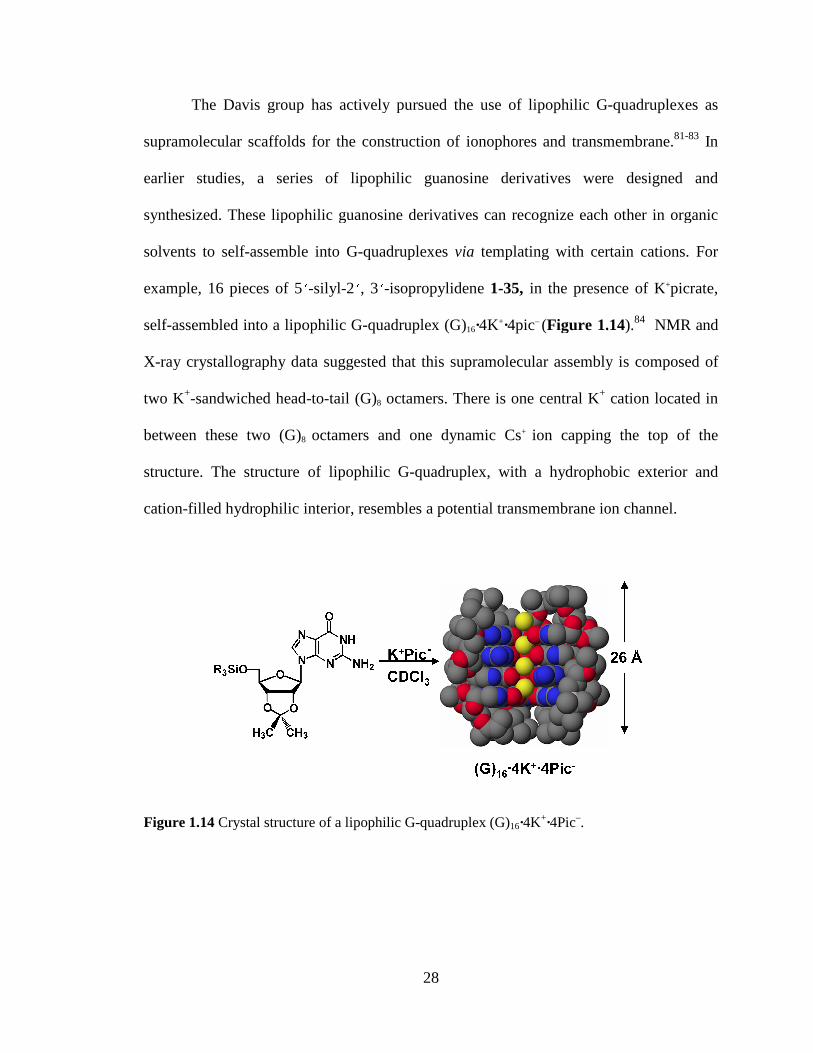
The Davis group has actively pursued the use of
supramolecular scaffolds for the construction of
earlier studies a serie
synthesized These lipophilic guanosine
solvents to self-assemble into G
example 16 pieces of 5
self-assembled into a lipophilic
X-ray crystallography data
two K+-sandwiched head
between these two (G)8
structure The structure of li
cation-filled hydrophilic interior
Figure 114 Crystal structur
28
The Davis group has actively pursued the use of lipophilic G-
s for the construction of ionophores and transmembrane
es of lipophilic guanosine derivatives were
hese lipophilic guanosine derivatives can recognize each other in organic
assemble into G-quadruplexes via templating with certain cations
-silyl-2 3 -isopropylidene 1-35 in the presence of K
lipophilic G-quadruplex (G)16middot4K+middot4picndash (Figure 11
ray crystallography data suggested that this supramolecular assembly
head-to-tail (G)8 octamers There is one central K+
8 octamers and one dynamic Cs+ ion capping the top of the
The structure of lipophilic G-quadruplex with a hydrophobic
filled hydrophilic interior resembles a potential transmembrane ion channel
Crystal structure of a lipophilic G-quadruplex (G)16middot4K+middot4Picndash
-quadruplexes as
transmembrane81-83 In
were designed and
can recognize each other in organic
certain cations For
in the presence of K+picrate
Figure 114)84 NMR and
assembly is composed of
cation located in
pping the top of the
hydrophobic exterior and
transmembrane ion channel
29
18 Summary
This chapter has described the self-assembly of lipophobic nucleobases
nucleosides and oligonucleotides using their molecular recognition properties In
particular nucleolipids have demonstrated the capability to insert into lipid membranes or
function as carriers for membrane transport of biomolecules The lipophilic G-
quadruplexes bearing the resemblance to the natural DNA G-quadruplex provide the
potential scaffolds to function as synthetic ion channels My research project described in
chapter 2 will focus on whether this lipophilic G-quadruplex can function as a synthetic
ion channel85 Subsequently the design and synthesis of lipophilic guanosine derivatives
that can form self-assembled ion channels will be described in the following chapters
30
Chapter 2 Cation Exchange in Lipophilic G-
Quadruplexes Not All Ion Binding Sites Are Equal
The majority of this chapter has been published in reference 85
bull Ma L Iezzi M A Kaucher M S Lam Y-F Davis J T J Am Chem Soc 2006 128 15269-15277
21 Introduction
The overall goal of the research described in this chapter was to investigate the
potential use of lipophilic G-quadruplexes to function as ion channels Initial studies
involved the study of cation exchange between G-quadruplex-bound cations and cations
in solution
In our group extensive efforts have been made to study the structure and dynamic
properties of lipophilic G-quadruplexes with the aim to develop transmembrane synthetic
ion channels1482838687 The diffusion NMR analysis demonstrated that the
hexadecameric [G 1]16middot4K+middot4pic- (where G 1 is 5lsquo-tert-butyldimethylsilyl-2lsquo3lsquo-
isopropylidene guanosine) observed in the solid state also predominates in organic
solution8288 The dynamic study of G-quadruplex showed that the identity of both the
bound anions and the bound cations significantly attenuate the kinetic stability of the G-
quadruplex and modulate the rate of ligand exchange between G-quadruplex and
monomer in solution In this chapter we have turned our attention to studying the cation
exchange process within the lipophilic G-quadruplex In short we would like to
determine if the cations exchange through the ends of the G-quadruplex stack much like
31
an ion channel or if cation exchange occurs through the sides of the G-quadruplex
especially since there is no covalent backbone connecting the different G-quartet layers
The preliminary result of my research has been the identification of the
intermediates in the cation exchange process between G-quadruplex bound Na+ cation
and free K+ (NH4+) cations in solution A ldquocentral insertionrdquo pathway was proposed for
the cation exchange process This finding suggested that the kinetic labiality of the self-
assembled hexadecameric G-quadruplex might be too great to function as a synthetic ion
channel The stabilization of this supramolecule by introducing hydrophobic covalent
linkers between the individuals G-quartet layers will be pursued in the future Self-
assembly of the modified lipophilic G-quadruplex will provide a ldquoDNA-approachrdquo for
building supramolecular ion channels
211 Cation-Dependent Self-Assembly of DNA G-Quadruplex Structures
The G-quartet structure was first discovered by Gellert and co-workers as the
fundamental unit in the formation of hydrogels by 5rsquo-GMP89 This cyclic structure
consists of two hydrogen-bonding arrays from four guanine subunits where the N1H
amideO6 and N2H amideN7 are complementary donoracceptors Pinnavaia and co-
workers later showed that Na+ and K+ stabilize diastereomeric G8-M+ octamers by
coordinating to the eight carbonyl oxygens of stacked G-quartets (Scheme 21)90 This
example demonstrated that different cations may selectively bind to a 5rsquo-GMP self-
assembled structure Since those studies many nucleosides and oligonucleotides have
been shown to form the secondary G-quadruplex structures779192 In the last decade the
implicated significance of the G-quadruplex in controlling biological functions has driven
32
considerable research on the stabilization of DNA and RNA G-quadruplexes1293 By
means of melting temperature measurement90 dehydration energy calculations94 X-ray
diffraction and NMR studies9596 insight into the cation selectivity of the G-quadruplex
has been established The combination of the correct size for a cation binding pocket and
the desolvation energy of cations determine the selectivity trend of monovalent cations in
DNA quadruplex structures In general monovalent cations stabilize a DNA quadruplex
in the following order K+gtNH4+gt Rb+ Na+gt Cs+ Li+ This difference in cation binding
affinity can result in the conformational changes within the DNA G-quadruplex
particularly when different competitive cations are present in solution9798
Scheme 21 Schematic structure of (A) G-quartet and (B) G8-octamer
212 DNA G-quadruplex as an Ion Channel Model
The localization of cations bound inside the DNA G-quadruplexes has been
achieved directly by crystallography and solid-state NMR The direct evidence of cation
binding site inside a DNA G-quadruplex was reported by Rich and co-workers in their
crystallographic study of an Oxytricha nova telemetric DNA d(G4T4G4)(Oxy-15)95 The
G-quadruplex bound K+ ions were detected within the central cavity of two G-quartets
Afterward various crystallographic studies demonstrated that different alkali and alkaline
33
cations such as Na+ K+ Ba2+ Sr2+ Pb2+ are sandwiched between two G-quartets99100
One of the excellent examples is Luisirsquos highndashresolution crystal structure of a DNA G-
quadruplex formed by oligonucleotide [d(TG4T)]4 (Scheme 22)73 In this parallel-
stranded quadruplex a string of Na+ ions located along the central cavities either within
a G-quartet or sandwiched by G-quartets Solid-state NMR technique also provided
complementary evidence of alkali metal cations binding to nucleic acids71101 Griffin and
Rovnyakrsquos pioneering work demonstrated the direct observation of 23Na+ ions bound to
the DNA G-quadruplex using 23Na solid-state NMR102 A series of solid-state 23Na and
39K NMR methods have been developed by the Wu group that allowed the identification
of Na+ and K+ ions bound to a G-quadruplex and the determination of cation
coordination geometry in a telemetric Oxy-15 DNA qudruplex101103 Both
crystallography and solid-state NMR studies demonstrated that an array of alkali and
alkaline cations can locate within the central channel formed by the stacking G-quartets
The DNA G-quadruplex resembles the crystal structure of natural potassium ion channel
formed from Streptomyces lividans (Scheme 22)104
34
[d(TG4T)]4 K+ ion channel
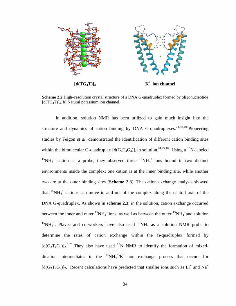
In addition solution NMR has been utilized to gain much insight into the
structure and dynamics of cation binding by DNA G-quadruplexes7498105Pioneering
studies by Feigon et al demonstrated the identification of different cation binding sites
within the bimolecular G-quadruplex [d(G4T4G4)]2 in solution7475106 Using a 15N-labeled
15NH4+ cation as a probe they observed three 15NH4
+ ions bound in two distinct
environments inside the complex one cation is at the inner binding site while another
two are at the outer binding sites (Scheme 23) The cation exchange analysis showed
that 15NH4+ cations can move in and out of the complex along the central axis of the
DNA G-quadruplex As shown in scheme 23 in the solution cation exchange occurred
between the inner and outer 15NH4+ ions as well as between the outer 15NH4
+and solution
15NH4+ Plavec and co-workers have also used 15NH4 as a solution NMR probe to
determine the rates of cation exchange within the G-quadruplex formed by
[d(G3T4G3)]2107 They also have used 15N NMR to identify the formation of mixed-
dication intermediates in the 15NH4+K+ ion exchange process that occurs for
[d(G3T4G3)]2 Recent calculations have predicted that smaller ions such as Li+ and Na+
Scheme 22 Highndashresolution crystal structure of a DNA G-quadruplex formed by oligonucleotide [d(TG4T)]4 b) Natural potassium ion channel
35
should move through the DNA G-quadruplex channels with relatively low activation
barriers when compared to the larger NH4+ and K+ cations
Scheme 23 Schematic representation of 15NH4+ movement through the Oxy-15 quadruplex a)
bulk NH4+ cation only exchanges with the outer 15NH4
+ b) exchange of two bound 15NH4+ cations
is random Black yellow and red ball represent the bulk ammonium ion and the bound ammonium ions located in outer and inner binding site of the G-quadruplex
213 Lipophilic G-quadruplexes
The Davis group and others have been exploring the properties of lipophilic G-
quadruplexes with an eye toward using them as self-assembled ionophores for selective
sequestration of radioactive 137Cs+ and 226Ra2+ and as ion channels for transporting
cations across lipid membranes108 To design such functional assemblies it is imperative
to understand their structural and dynamic properties In the presence of cations 5lsquo-tert-
butyldimethylsilyl-2lsquo3lsquo-isopropylidene guanosine (G 1) 2-1 self-assembles into a
lipophilic G-quadruplex [G 1]16middot3K+middotCs+middot4pic- (Scheme 24) A crystal structure shows
the complex to be composed of four stacked G-quartets83 This pseudo-D4 symmetric G-
quadruplex can be described as a pair of head-to-tail C4 symmetric [G 1]8 octamers that
are coaxially stacked on one another in a head-to-head arrangement99 The two G8
octamers use eight carbonyl oxygens to coordinate K+ in a square anti-prismatic
geometry A third K+ with a nearly cubic coordination geometry holds the two C4
36
symmetric [G 1]8 octamers together by binding to the two inner G-quartets A solvated
Cs+ caps the G-quadruplex by sitting in a cavity above one of the outer G4-quartets This
capping ion is dynamic in solution since the NMR spectra of the G-quadruplex shows
only two sets of signals If the capping ion was tightly bound one would expect four sets
of NMR signals for the four different G-quartet layers in [G 1]16middot3K+middotCs+middot4pic- The
bound cations in these G-quadruplexes have been observed with 23Na and 39K solid-state
NMR For 23Na all four channel ions (including the capping ion) were resolved Crystal
structures and solution NMR also showed that four phenolate anions hydrogen bond to
the two inner G-quartets
Unlike DNA G-quadruplexes this lipophilic G-quadruplex has no covalent
backbone between stacked G-quartet layers We reasoned that the study of the cation
Scheme 24 G-quadruplex formed through self-assembly of G 1 in the presence of K+DNP-
37
exchange process between cations in solution and cations bound to the G-quadruplex
bound would allow us to evaluate the stability of the G-quadruplex and its potential to
function as a transmembrane ion channel
22 Mass Spectrometry Shows Mixed-Cation G-Quadruplexes Formed by
Sequential Ion Exchange
In general G-quartets are stabilized by monovalent cations in the order K+gt NH4+gt
Na+gtgtCs+gt Li+525 Because of this difference in relative binding strength the addition of
a higher affinity cation to a G-quadruplex containing a weaker binding cation results in
replacement of that initially bound ion Such exchange has been amply demonstrated by
solution 1H NMR in water for Na+K+ exchange in the hairpin G-quadruplex
d(G3T4G3)294 In that study exchange between distinct cationic forms of the d(G3T4G3)2
G-quadruplexes was fast on the NMR chemical shift time scale giving rise to a single
time-averaged set of signals In our present case the different cationic forms of the
lipophilic G-quadruplex are in slow exchange on the chemical shift time scale and what
we hypothesized to be mixed-cation intermediates could be readily distinguished from
the homomeric all-Na+ and all-K+ G-quadruplexes by solution 1H NMR (see the
following sections) In the DNA study Feigon and colleagues demonstrated that the free
energy of cation dehydration helped determine the relative binding affinity of different
ions for DNA G-quartets in water In an organic solvent however free cations are not
hydrated so there may be other factors that determine the cations binding affinity for the
G-quartet in an organic phase Nonetheless the mass spectrometric and solution NMR
38
data described next indicate that G-quartets also have a higher affinity for K+ over Na+
in organic solvents such as CD2Cl2
To unequivocally identify mixed-cation G-quadruplexes that form during the
Na+K+ ion exchange process we first used electrospray ionization mass spectrometry
(ESI-MS) a powerful method for characterizing noncovalent assemblies109 ESI-MS has
been used to monitor the formation of G-quadruplexes by a variety of G-nucleosides and
G-rich-oligonucleotides110111 To the best of our knowledge however the Na+K+ cation
exchange process within a G-quadruplex has not been previously characterized by mass
spectrometry Thus crystalline samples of [G1]16middot4 Na+middot4DNP- and [G1]16middot4 K+middot4DNP-
were prepared according to standard procedures83 and their purity was confirmed by both
NMR and mass spectrometry ESI-MS analysis in the positive-ion mode showed distinct
peaks for the intact Na+ and K+ G-quadruplexes at mz 74353 and 74845 respectively
(Figure 21ae) These singly charged ions correspond to the molecular weight for the
hexadecamer [[G 1]16middot3Mmiddot2DNP]+ a species formed in the gas phase by losing one cation
(presumably the weakly bound capping ion) and two DNP- anions from the starting G-
quadruplexes As depicted in Figure 21bminusd we then monitored the cation exchange
process by adding increasing amounts of KPh4B to a solution of [G 1]16middot4Na+middot4DNP-in
11 CD2Cl2CD3CN The major peaks in Figure 21b 21c had mz ratios intermediate
between the two homomeric species [[G 1]16middot3Namiddot2DNP]+ and [[G 1]16middot3Kmiddot2DNP]+
Significantly the dominant molecular ion in each titration sample showed a 16 amu
increase in mass corresponding to the sequential displacement of a bound Na+ by a
single K+ during the titration For example Figure 21b corresponding to a sample
39
containing 1 equivalent of KPh4B for each equivalent of [G 1]16middot4Na+middot4DNP- showed a
significant decrease in the [[G 1]16middot3Namiddot2DNP]+ species (mz 74353) with the
concomitant formation of a new major species with mz 74520 This new peak
corresponds to a mixed-cation G-quadruplex of formula [[G 1]16middotKmiddot2Namiddot2DNP]+ As
depicted in Figure 21 there are two possible isomers for [[G 1]16middotKmiddot2Namiddot2DNP]+
depending on whether one of the outer cations or the central cation is first replaced by K+
The addition of a second equivalent of KPh4B led to the formation of [[G
1]16middot2KmiddotNamiddot2DNP]+ (mz 74682) as the dominant species in that region of the ESI mass
spectrum (Figure 21c) Again as depicted in Figure 21 there are two possible isomers
for this second intermediate Essentially complete conversion to the K+ G-quadruplex
(mz 74845) occurred after the addition of just 4 equivalents of KPh4B to the solution of
the Na+ G-quadruplex (Figure 21d) The ESI-MS results depicted in Figure 21 are
significant for the following reasons as this data (1) confirms the pronounced K+Na+
binding selectivity shown by this lipophilic G-quadruplex (2) demonstrate the formation
of discrete mixed-cationic assemblies such as [[G 1]16middotKmiddot2Namiddot2DNP]+ and [[G
1]16middot2KmiddotNamiddot2DNP]+ under conditions where sub-stochiometric K+ is present and (3)
illustrate the stepwise displacement of Na+ cations by the stronger binding K+ ion This is
the first time to our knowledge that mass spectrometric data has revealed the sequential
ion exchange process in a G-quadruplex Such ESI-MS titration experiments should also
be informative for understanding the details of structure and ion binding in the rich array
of DNA and RNA G-quadruplexes that have been discovered to date
40
Figure 21 ESI-MS from titration of KPh4B into a solution of [G1]16middot4Na+middot4DNP- in 11 CD2Cl2CD3CN a) [G 1]16middot4Na+middot4DNP- b) after addition of 1 equivalents of KPh4B c) after addition of 2 equivalents of KPh4B d) after addition of 4 equivalents of KPh4B and e) [G 1]16middot4K+middot4DNP- The diagram illustrates the possible mixed-cation G-quadruplexes that could be formed in the cation exchange process
23 Proton NMR Indicates that the Central Na+ Is the First Cation Exchanged for
K+ in Formation of Mixed Na+ K+ G-Quadruplexes
ESI-MS analysis of the titration of [G 1]16middot4Na+middot4DNP- with KPh4B provided
unequivocal evidence for the formation of the mixed Na+ K+ G-quadruplexes [[G
1]16middotKmiddot2Namiddot2DNP]+ and [[G 1]16middot2KmiddotNamiddot2DNP]+ However the isomeric structures of
these mixed G-quadruplexes and the location of bound cations could not be ascertained
from the mass spectrometry data alone For example as shown in Figure 21 the first
intermediate formed during Na+K+ ion exchange could involve the replacement of either
the outer or the centrally bound Na+ In theory these isomers should be distinguished by
41
1H NMR based on their differences in symmetry To identify the structures of these
discrete intermediates and to define specific cation exchange pathways we turned to 1H
NMR spectroscopy knowing that the chemical shifts of the G-quartets amide N1H
protons are sensitive to the identity of the bound cation8294 Figure 22a shows two
distinct N1H amide peaks for the all-Na+ G-quadruplex [G 1]16middot4Na+middot4DNP- in 11
CD2Cl2CD3CN with the signal at δ 1146 ppm corresponding to the N1H amide for the
two inner G4-quartets and the signal at δ 1171 ppm due to the outer G4-quartets In
contrast the G-quartet N1H amide peaks for the K+ G-quadruplex [G 1]16middot4K+middot4DNP-
occur at δ 1135 (inner) and δ 1158 ppm (outer) respectively (Figure 22g)
Figure 22b shows that two new amide N1H signals (δ 1142 and δ 1175 ppm)
appear after the addition of 05 equivalents of KPh4B to a solution of [G 1]16middot4Na+middot4DNP-
These new N1H signals are in slow exchange on the chemical shift time scale with signals
for [G 1]16middot4Na+middot4DNP- a feature that helped us recognize this new species as a mixed
Na+ K+ G-quadruplex Further addition of KPh4B (1 equivalent) resulted in these new
N1H resonances becoming predominant while the signals for [G 1]16middot4Na+middot4DNP- greatly
diminished (Figure 22c) The new G-quadruplex in Figure 22c has only two N1H
signals (one set for the outer G-quartets and one set for the inner G-quartets) an
indication that cation exchange occurred with the central Na+ in [G 1]16middot4Na+middot4DNP- to
give an intermediate with pseudo-D4 symmetry namely [[G 1]4(o)middotNa+middot[G 1]4(i)middotK+middot[G
1]4(i)middotNa+middot[G 1]4(o)] If one of the outer Na+ cations had been displaced by the first
equivalent of K+ we would expect a lower symmetry hexadecamer [[G 1]4(o)middotK+middot[G
1]4(i)middotNa+middot[G 1]4(i)middotNa+middot[G 1]4(o)] with four separate signals for its nonequivalent G-quartet
layers
42
Only after the addition of 2 equivalents of KPh4B did the 1H NMR spectra show
that the major G-quadruplex in solution had four new N1H amide peaks of similar
intensity consistent with the formation of a mixed hexadecamer of lower symmetry
[[G1]4(o)middotK+middot[G1]4(i)middotK
+middot[G1]4(i)middotNa+middot[G1]4(o)] where one of the two outer Na+ cations must
have been exchanged for a higher affinity K+ (see the four labeled peaks in Figure
22de) Conversion into the all-K+ G-quadruplex was essentially complete after the
addition of just 4 equivalents of KPh4B (Figure 22f) a result that was consistent with
the ESI-MS titration experiment (Figure 21)
Figure 22 Region of the 1H NMR spectra (400 MHz) showing the G-quartet NH1 amide protons during titration of [G 1]16middot4Na+middot4DNP- with KPh4B in 11 CD2Cl2CD3CN (a) [G 1]16middot4Na+middot4DNP- The mol ratio of added KPh4B to the [G 1]16middot4Na+middot4DNP- G-quadruplex is (b) 051 (c) 11 (d) 21 (e) 31 (f) 41 and (g) 121
Both ESI-MS and 1H NMR titration data indicated that replacement of bound Na+
by K+ is a thermodynamically favorable process particularly since the addition of only 4
equivalents of KPh4B led to essentially complete transformation of the all-Na+ G-
quadruplex to the all-K+ G-quadruplex In addition mixed-cationic G-quadruplexes were
43
detected in conversion of the all-Na+ G-quadruplex to the all-K+ G-quadruplex Since Na+
and K+ are invisible using standard solution NMR spectroscopy we were not able to
directly detect the different Na+ and K+ cations bound to various G-quadruplex species
during the exchange process To overcome this limitation we used the NMR-active
15NH4+ as a surrogate for K+ to identify the mixed-cation G-quadruplexes formed upon
displacement of Na+ by a higher affinity cation
24 Use of 15NH4+ Cation to Probe the Cation Exchange Process Within a Lipophilic
G-quadruplex
241 NMR Studies with 15NH4+ Confirm Identity of Mixed-Cationic G-
Quadruplexes
The NH4+ cation has a similar coordination chemistry as K+ and 15NH4
+ has been
used as an NMR probe to localize cations in DNA G-quadruplexes7475 Wong and Wu
have also used solid-state 23Na NMR competition experiments to show that K+ and NH4+
have similar free energies of binding to G-quadruplexes101103 NMR titrations indicated
that the order of the site exchange between free NH4+ and Na+ bound to [G
1]16middot4Na+middot4DNP- was similar to that observed for the K+Na+ exchange in Figure 22 The
1H NMR spectra of various amounts of 15NH4Ph4B titrated into a solution of [G
1]16middot4Na+middot4DNP-in 11 CD2Cl2CD3CN is shown in Figure 23 A new set of N1H amide
protons appeared upon addition of one equivalent of 15NH4+ to the solution of [G
1]16bull4Na+bull4DNP- (Figure 23c) This indicates that the first intermediate is a highly
symmetric structure Several new N1H amide proton peaks appeared as additional
44
equivalents of 15NH4Ph4B were titrated into the solution (Figure 23d-g)This result
indicated that sequential cation exchange continued and a second asymmetric G-
quadruplex intermediate was formed Twelve equivalents of 15NH4+ cations had to be
used to complete the conversion from the all-Na+ G-quadruplex into the all-15NH4+
Gquadruplex (Figure 23g) providing evidence that 15NH4+ cation has a lower preference
than does K+ for the G-quadruplex Thus mixed-cationic species that were in slow
exchange with the all-Na+ and all-NH4+ forms of the G-quadruplexes were identified by
their separate sets of 1H NMR signals
Figure 23 A region of the 1H NMR spectra (400 MHz) for titration of 15NH4Ph4B into a solution of (G 1)16middot4Na+middot4DNP- in 11 CD2Cl2CD3CN at 20 oC The signals for the amide N1H protons of the G-quartets are shown a) (G 1)16middot 4Na+middot 4DNP- The mole ratio of 15NH4
+ to (G 1)16bull4Na+bull4DNP- is b) 051 c) 11 d) 21 e) 31 f) 41 g) 121
Both 2-D 15N-1H HSQC-ROESY112 and 1H-1H NOESY data (Figure 24) for the
all-NH4+ species [G 1]16middot4 NH4
+middot4DNP- showed two types of bound 15NH4+ with one
cation occupying the central location between the inner G-quartets and two 15NH4+
cations bound to the symmetry-related outer sites The 15N-1H HSQC spectrum in Figure
24a shows two cross-peaks between the larger 15N signal at δ 302 ppm and the larger
45
15N-decoupled 1H signal at δ 719 ppm This correlation corresponds to the two 15NH4+
cations in the outer cation binding sites The other cross-peak between the 15N signal at δ
274 ppm and the 1H signal at δ 730 ppm corresponds to 15NH4+ in the central binding
site These assignments were confirmed by NOESY (Figure 24b) NOE cross-peaks are
observed between the two different 15NH4+ signals and the N1H amide protons for the
inner and outer G-quartets The 15NH4+ cation bound in the central binding site shows
cross-peaks only with the N1H amide of the inner G-quartet (δ 1141 ppm) In contrast
the stronger 15NH4+ signals show cross-peaks with N1H amide protons for both the inner
and the outer G-quartets as would be expected for an ammonium cation bound between
both types of layers No 15NH4+ binding to the capping site was observed in these solution
studies
Figure 24 a) Region from the 2-D 15N-1H HSQC-ROESY NMR spectrum of [G1]16middot4NH4
+middot4DNP- in 11 CD2Cl2CD3CN showing cross-peaks between the 15N NMR resonances and the 15N-filtered 1H resonances b) Region of the 2-D 1H-1H NOESY NMR spectra of [G1]16middot4NH4
+middot4DNP- in 11 CD2Cl2CD3CN showing cross-peaks between the N1H amide resonances and the 1H resonances for the bound NH4
+ cations Both spectra were recorded on a 1 mM sample at 20 degC using a 500 MHz NMR spectrometer
46
The Na+K+ exchange data in Figure 22 suggest that the central cation in (G
1)16middot4Na+middot4DNP- which exchanges first is bound less tightly than the outer cations
coordinated within G8-octamers This hypothesis was supported by 15N longitudinal
relaxation measurements for the bound NH4+ cations in [G 1]16middot4NH4
+middot4DNP- Thus 15N
T1 spinminuslattice relaxation times were found to be 125 s for the 15N signal of the outer
ions at δ 302 ppm and just 23 s for the central ion at δ 274 ppm Although there are
many factors that influence T1 relaxation the much shorter value for the centrally bound
15NH4+ may be because it is undergoing chemical exchange with solvated cations more
readily than are the more tightly bound 15NH4+ cations in the two outer binding sites
242 Determination of the First Intermediate in Na+15NH4+ Cation Exchange
Using 15NH4+ as a visible replacement ion for K+ we performed a series of 15N-
filtered 1H NMR experiments to unambiguously assign the two different cation binding
sites within the G-quadruplex84 The 15N-filtered and 15N-decoupled 1H NMR spectra in
Figure 25 show the 15NH4+Na+ exchange that occurs upon titration of 15NH4Ph4B into a
solution of [G1]16middot4Na+middot4DNP- The composition of mixed-cationic G-quadruplexes
varies with added 15NH4+ After the addition of 1 equivalent of 15NH4
+ to [G
1]16middot4Na+middot4DNP- (Figure 25b) one resonance (δ 710 ppm) was observed in the 15N-
filtered 1H NMR spectra consistent with 15NH4+ being bound to a single site within the
hexadecameric G-quadruplex As the amount of 15NH4+ increased additional 15N-
decoupled signals for NH4+ resonances were observed indicating further displacement of
specific Na+ ions by 15NH4+ The titration sample containing 12 equivalents of
15NH4Ph4B showed two 15NH4+ proton peaks (in a 12 ratio) at δ 731 ppm and δ 717
47
ppm respectively (Figure 25f) This pattern is consistent with the structure of the all-
NH4+ G-quadruplex (G 1)16middot4
15NH4+middot4DNP- (Figure 25g) The other two equal intensity
15NH4+ signals that appear and then disappear during the course of the titration (marked
by blue circles in Figure 25cminusf) are due to a G-quadruplex that contains two bound
15NH4+ cations and one bound Na+ cation namely [[G 1]4(o)middot
15NH4+middot[G 1]4(i)middot
15NH4+middot[G
1]4(i)middotNa+middot[G 1]4(o)]
Figure 25 15N-filtered 1H NMR (500 MHz) spectra of 15NH4Ph4B titration the solution of [G 1]16middot4Na+middot4DNP- in 11 CD2Cl2CD3CN with mol ratio at (a) 01 (b) 11 (c) 21 (d) 31 (e) 41 (f) 121 and (g) [G 1]16middot4
15NH4+middot4DNP-
We reasoned that the species formed in Figure 25b after the addition of 1
equivalent of 15NH4Ph4B to [G 1]16middot4Na+middot4DNP- must be a D4 symmetric mixed G-
quadruplex with the 15NH4+ cation bound in the central cavity between two G8-Na+-
octamers This assignment for the species [[G 1]4(o)middotNa+middot[G 1]4(i)middot15NH4
+middot[G 1]4(i)middotNa+middot[G
1]4(o)] was confirmed by a 2-D 1H 1H NOESY experiment Figure 26 shows a cross-peak
between the 15NH4+ protons centered at δ 710 ppm and the N1H protons of the inner G-
48
quartet layers at δ 1140 ppm In marked contrast there were no 1H-1H NOEs between
this bound 15NH4+ and the N1H amide of the outer G-quartet layers at δ 1179 ppm
Figure 26 Portion of a NOESY spectrum of a 11 15NH4 Ph4B titration into [G 1]16middot4Na+middot4DNP- in 11 CD2Cl2CD3CN The 2-D spectrum shows the NOE cross-peak between the 15NH4
+1H resonance with the N1H of the inner G-quartet layer The chemical shifts of δ 1179 and 1140 ppm correspond to the N1H amide protons for the outer and inner G-quartet layers The chemical shift of the 15NH4
+ proton was confirmed from the 15N decoupled 1H NMR spectrum showing a single peak at δ 710 ppm
The NMR data in Figures 25 and 26 conclusively demonstrate that the initial
NH4+Na+ cation exchange occurs between solvated 15NH4
+ and the centrally bound D4
symmetric Na+ cation to give [[G 1]4(o)middotNa+middot[G 1]4(i)middot15NH4
+middot[G 1]4(i)middotNa+middot[G 1]4(o)] as the
first discrete intermediate in the cation exchange process
243 Characterization of the Second Intermediate in the Na+15NH4+ Cation
Exchange Process
A series of 1D selective NOE analyses was carried out on the solution of (G
1)16bull4Na+bull4DNP- that contained 2 equivalents of 15NH4Ph4B in 11 CD2Cl2CD3CN
Figure 27 shows the selective NOE data that allowed to identify a mixed 21 NH4+Na+
49
species [[G 1]4(o)middot15NH4
+middot[G 1]4(i)middot15NH4
+middot[G 1]4(i)middotNa+middot[G 1]4(o)] as the second
intermediate in the exchange process In the N1H proton region the two stronger peaks at
δ1179 and δ1140 ppm were irradiated first No NOE was observed between 15NH4+ and
the N1H proton of the outer layer at δ1179 ppm (see bottom trace in Figure 27b) Only
the N1H amide proton of the inner layer at δ1140 ppm had a selective NOE with 15NH4+
protons showing a pair of peaks (due to 15N-1H coupling) in the 15NH4+ region (labeled
with green) These data agree with the previous assignment that the first intermediate in
the exchange process is asymmetric G-quadruplex [G 1]4(o) middotNa+middot [G 1]4(o) middot15NH4+middot [G
1]4(o) middotNa+middot [G 1]4(o) It was noticed that the N1H signal at δ1140 ppm also correlated
with another 15NH4+ proton in a different intermediate (blue lsquoirsquo label in Figure 27b) This
finding suggested the overlap of N1H amide protons of the second intermediate with that
of the first intermediate at δ1140 ppm The overlap of N1H peaks probably results from
the similar chemical environment of the ldquoinnerrdquo G-quartets in these two intermediates
We next irradiated the G-quartet N1H signal at δ1135 ppm a new peak in the titration
solution Two new 15NH4+resonances displayed selective NOEs to this amide peak at
δ1135 ppm One 15NH4+ was the same as previously identified to be part of the second
intermediate (blue lsquoirsquo label in Figure 27b) The other 15NH4+ signal corresponded to one
that also had an NOE with the N1H proton at δ1151 ppm Combining all of this
information from the selective NOE experiments allowed us to identify the second
intermediate as [G 1]4(o)middot15NH4
+middot[G 1]4(i)middot15NH4
+middot[G 1]4(i)middotNa+middot[G 1]4(o)] Because of its
asymmetric structure the four G-quartet layers of this mixed cation G-quadruplex have
quite different chemical environments with four N1H resonances at δ1179 1140
1135and 1151 ppm respectively
50
Figure 27 Selective NOE spectra for amide N1H peaks of [G 1]16middot 4Na+middot4DNP- titrated with 2 equivalents of 15NH4Ph4B in 11CD2Cl2CD3CN Left a) N1H region of 1H NMR spectrum (green dots indicate the N1H amide proton from the first intermediate blue dots indicate the N1H amide proton from the second intermediate) b) 15NH4
+ proton region showing selective NOE correlation to amide proton at δ1151 δ1135 δ1140 and δ 1179 ppm respectively Right structural scheme for the second intermediate [G 1]4 (o) middot Na+middot [G 1]4 (i) middot
15NH4+middot [G 1]4 (i) middot
15NH4+middot
[G 1]4 (o)] formed during the Na+ NH4+ exchange process with the assignments for the different
G-quartet N1H amide protons
25 Cation Exchange Pathway in the Lipophilic G-Quadruplex
Firm identification of these mixed-cationic G-quadruplexes using the combined
ESI-MS and 1H 15N NMR data allowed us to define a pathway for exchange of
monovalent cations in this lipophilic G-quadruplex (Scheme 25) Thus higher affinity
cations such as K+ and NH4+ preferentially displace the central Na+ cation in the G-
quadruplex [G 1]16middot4Na+middot4DNP- to give the pseudo-D4 symmetric mixed-cation G-
quadruplex [[G 1]4(o)middotNa+middot[G 1]4(i)middot15NH4
+middot[G 1]4(i)middotNa+middot[G 1]4(o)] as the first intermediate
Consequently sequential exchange of the two tighter-bound outer cations finished the ion
exchange process Control experiments where components were mixed in the appropriate
ratios confirmed that the process depicted in Scheme 25 is under complete
thermodynamic control (data not shown)
51
Scheme 25 Proposed central insertion pathway for cation exchange in the lipophilic G-quadruplex system
Why is it easier to displace the central Na+ ion in [G 1]16middot4Na+middot4DNP- as opposed
to the ions in the outer binding sites of the G-quadruplex We propose that differences in
the cations octahedral coordination geometries at these separate binding sites may be
responsible for the relative facility of cation exchange Crystal structures for the [G
1]16middot4M+middot4A- system show that the central cation in this hexadecameric assembly has an
almost cubic coordination geometry with the eight oxygen atoms of the two inner G-
quartets113 In contrast the oxygen ligands that sandwich the outer Na+ cations are
twisted more toward square anti-prismatic coordination geometry Cubic coordination
geometry is relatively rare because of the electrostatic repulsion that occurs between the
ligand atoms that eclipse each other114 The anti-prismatic geometry on the other hand is
usually more favorable since the ligand atoms are twisted so as to minimize
ligandminusligand contacts while maintaining an optimum ligandminusM+ distance (Scheme 26)
This influence of coordination geometry on the interaction energy of cations within
G8minusM+ octamers has been addressed recently by Meyer and co-workers using DFT
calculations115 They found that a G8minusNa+ octamer with square anti-prismatic
coordination was more stable (by about 9 kcalmol) than a G8minusNa+ octamer with a cubic
coordination environment We propose that because of these differences in coordination
geometry in [G 1]16middot4Na+middot4DNP- the central cation is bound less strongly than are the
52
outer cations and therefore exchanges more readily with the higher affinity K+or NH4+
Indeed X-ray data show that the separation between the two inner G-quartets is greater
than the average separation between the inner and the outer G-quartet layers There is
more space in the central ion binding site than in the outer sites
Scheme 26 These depictions are taken from the crystal structure for [G 1]16middot4Na+middot4pic- (ref 16) The illustrations show the octahedral coordination geometry for one of the outer Na+ cations (on the left) and for the central Na+ (right) The central Na+ has a almost cubic coordination geometry whereas the oxygen ligands are twisted toward the energetically more favorable square anti-prism geometry for the outer Na+
This central cation may serve to help dimerize discrete G8minusM+ octamer units into
more highly ordered superstructures Indeed we have previously shown for another
lipophilic G-quadruplex that the K+ ion concentration can influence the degree of self-
association21 Thus at low K+ concentrations 5lsquo-(35-bis(methoxy)benzoyl)-2lsquo3lsquo-
isopropylidene (G 2) forms an octamer Upon the addition of extra K+ these [G 2]8middotK+
octamers dimerize to form a stable [G 2]16-hexadecamer in solution A similar
phenomenon has been seen for the tetrahymena telomere sequence d(T2G4) and for the
human telomere sequence d(TTAGGG) which are both monomeric G-quadruplexes at
lower K+ ion concentrations but form head-to-head G-quadruplex dimers at higher K+ ion
concentrations
53
26 The Larger Cs+ Cation But Not the Smaller Li+ Can Displace the Central
Cation in the Na+ G-Quadruplex
The final experiments in this study were based on the hypothesis that the near
cubic coordination geometry for the central cation binding site should accommodate a
larger cation much more readily than a smaller cation We reasoned that a larger cation
should maximize the separation between the eclipsing oxygen atoms within the central
ion binding site As shown in Figure 28 this hypothesis appears reasonable as we could
substitute this position with Cs+ (r = 167 A) a large cation that typically is not thought to
stabilize G8-octamers Thus the addition of 8 equivalents of CsPh4B to a solution of the
Na+ G-quadruplex led to the telltale formation of two new G-quartet N1H amide signals
consistent with displacement of the central Na+ cation and formation of a pseudo-D4
symmetric G-quadruplex [[G 1]4(o)middotNa+middot[G 1]4(i)middotCs+middot[G 1]4(i)middotNa+middot[G 1]4(o)] In marked
contrast the addition of 10 equivalents of LiPh4B to the same Na+ G-quadruplex gave no
noticeable formation of any mixed-cationic species Thus unlike Cs+ (r = 167 Aring) the
smaller Li+ (r = 059 Aring) does not displace Na+ (r = 097 Aring) from the central ion binding
site in this G-quadruplex These experiments show that new mixed-cationic G-
quadruplexes can be rationally prepared based on the combined structural information
from X-ray crystallography ESI mass spectrometry and solution NMR spectroscopy
These results also suggest that large ions such as Cs+ may well stabilize higher ordered
DNA G-quadruplexes that are capable of forming head-to-head dimers with a cubic
coordination geometry116
54
Figure 28 1H NMR spectra of N1H region of different G-quadruplex solutions in CD2Cl2CD3CN at 20 degC (a) [G 1]16middot4Na+middot4DNP- (b) 101 mol ratio of LiPh4B and [G 1]16middot4Na+middot4DNP- (c) 81 mol ratio of CsPh4B and [G 1]16middot4Na+middot4DNP-
27 Conclusion
In summary we have studied the cation exchange between competitive cations in
solution and Na+ ions bound to the lipophilic G-quadruplex [G 1]16middot4Na+middot4DNP- Cations
with a stronger binding affinity for G-quadruplexes such as K+ drive the cation
exchange process Both ESI-MS and NMR measurements of the K+ titration into [G
1]16middot4Na+middot4DNP- revealed that cation exchange is a sequential process as discrete mixed-
cation intermediates were detected Using the 15NH4+ cation as a probe the identity of
these mixed-cation G-quadruplex isomers was determined by 15N-filtered 1H NMR
NOESY and selective NOE experiments A central insertion pathway in which free
cations first replace the central cation in [G 1]16middot4Na+middot4DNP- is operative in this
lipophilic G-quadruplex A structural rationale based on the different solid-state
octahedral coordination geometries in [G 1]16middot4Na+middot4DNP- was proposed to explain the
differences in site exchange between the central and the outer binding sites for these
lipophilic G-quadruplexes
55
The strategy that we have outlined in this chapter may also be useful for
identification of specific ion exchange pathways in G-quadruplexes formed by DNA and
RNA oligonucleotides particularly for those systems that have been shown to form
dimeric G-quadruplexes either in the solid-state or in solution Finally these mechanistic
studies on cation exchange which clearly couple aspects of the crystal structure to
solution state properties should help us better understand the factors that control
assembly and disassembly of lipophilic G-quadruplexes Such knowledge will guide our
future efforts to build selective ionophores and synthetic ion channels For example
unlike in DNA G-quadruplexes the cations in these lipophilic G-quadruplexes apparently
do not move through the ends of the central channel In fact the cation in the middle of
the assembly is the easiest to displace Thus it is likely that covalent side chains will be
needed to link together lipophilic G-units so as to generate an appropriate building block
for a synthetic ion channel Such covalent linkages should stabilize these G-quadruplex
structures
56
Chapter 3 Large and Stable Transmembrane Pores from a
Guanosine-Bile Acid Conjugate
The majority of this chapter has been published in reference 117
bull Ma L Melegari M Colombini M Davis J T ldquoLarge and Stable Transmembrane Pores from Guanosine-Bile Acid Conjugatesrdquo J Am Chem Soc 2008 130 2938-2939
31 Introduction
The research in this chapter reports that the guanosine-lithocholate 3-1 is able to
self-assemble into discrete synthetic ion channels in phospholipid membranes (Figure
31)117 The pores are large (nS conductance) and stable with ldquoopenrdquo times of seconds
distinguishing them from most synthetic channels which typically conduct in the pS
range with millisecond lifetimes
Figure 31 Ditopic G-sterol 3-1 and control bis-lithocholamide 3-16 Typical traces of conductance vs time after addition of 3-1or 3-16 are depicted See Reference 115 for detail
The preliminary results reported in chapter 2 indicate that cation exchange
between cations bound to a lipophilic G-quadruplex and free cations in solution proceed
3-1
3-16
57
through a ldquocentral channelrdquo pathway The data suggest that the cation sandwiched
between two (G 1)8M+ octamers within the lipophilic G-quadruplex is more dynamic
than the ldquoouterrdquo cations located inside the individual (G 1)8M+ octamers This finding
was consistent with our grouprsquos previous results that (G 1)8M+ octamers are building
blocks for the self-assembled lipophilic G-quadruplex113118 Under typical experimental
conditions the lipophilic G-quadruplexes are thermodynamically stable but they are
kinetically labile The G-quadruplex is a self-assembly of 16 subunits held together
through the non-covalent interactions such as hydrogen bonds π-π stacking and cation-
dipole interactions Thus these structures would be much more vulnerable to dissociate
than their covalent analogs
A ditopic design strategy was employed to develop transmembrane ion channel A
hybrid molecule that conjugates a hydrophobic linker with two capping guanosine units
could provide an ideal structure for membrane insertion and self-assembly of guanosine
In this research a bis-lithocholate linker was chosen to build a guanosine-sterol conjugate
The structure of target molecule 3-1 is shown in Figure 31 The covalent bis-lithocholate
backbone in 3-1 has a proper length to match that of the hydrophobic portion of lipid
bilayer Additionally the high lipophilicity of 3-1 should allow for membrane
permeability We hypothesized that guanosine-sterol conjugate 3-1 might be able to form
a barrel-stave-type superstructure by means of self-assembly of guanosine into G-quartet
assemblies (Figure 32) This chapter will describe the ion compound 3-1 does form
transmembrane ion channels Even though the channel activities of compound 3-
1suggest that active channel structures should be different from our hypothetical structure
shown in Figure 32
58
311 G-QuartetFolate-Quartet as a Motif for Transmembrane Ion Transporter
As a hydrogen-bonded macrocyclic array with ionophore properties the G-quartet
represents a promising scaffold on which to build synthetic ion channels A variety of
lipophilic guanosine derivatives have been designed as self-assembled ionophores They
all combine a central hydrophilic cation binding pore with hydrophobic periphery
Several previous efforts include Matilersquos rigid-rods attached guanosine on their
periphery119and Davisrsquo guanosine-calix[4]arene conjugate87 These synthetic strategies
have been used to stabilize the formation of G-quartets In all these cases the resulting
self-assembled G-quartet exhibited the tubular architecture that resembles an ion channel
However there was no evidence in any of these systems that they function as
transmembrane ion channels
The first synthetic ion transporter containing G-quartet units was developed by
the Davis group using a synthetic strategy based on the ldquocovalent capturerdquo of a self-
assembled G-quadruplex (Figure 33)120 In this case lipophilic guanosine 3-2 was
Figure 32 Hypothetical structure of a self-assembled barrel-stave ion channel formed from compound 1 The wedge represents guanosine moiety the line represents the bis-lithocholic acid linker Channel formation would be controlled by the self-assembly of guanosine head groups to give a G-quartet based structure
35-50 Aring
59
designed with terminal alkene attachments This new lipophilic guanosine readily forms a
G-quadruplex 3-3 in the presence of a K+ template Then postmodification of the G-
quadruplex 3-3 via olefin metathesis yields a unimolecular G-quadruplex 3-4 This
covalently modified supramolecule 3-4 acts as a transmembrane Na+ transporter in
phospholipid liposomes This unimolecular approach suggests that the self-assembled G-
quadruplex 3-3 was stabilized by post-covalent modification
Figure 33 The unimolecular G-quadruplex stabilized by post-covalent modification that can function as a transmembrane Na+ transporter120
Recently synthetic transmembrane ion channels built from folate dendrimers 3-5
were reported by the Matile and Kato groups (Figure 34)121 Folic acid and guanine base
have similar hydrogen bonding motif Folic acid self-assembles into a folic acid quartet
that is similar to the G-quartet Serving as the central core of the folate dendrimer the
folate analog is surrounded by a hydrogen-bonded amide network phenyl ring and alkyl
dendritic periphery The report from Matile and Kato revealed that the folate dendrimer
could self-assemble into π-stacked folate rosettes in hydrophobic media with or without a
cation template The ion channel activity of these dendritic folate rosettes shows that the
3-2
3-3 3-4
60
central pore of ldquofolate-quartetrdquo functions as an ion channel Compared to the dynamic G-
quadruplex system made from lipophilic 5rsquo-tert-butyldimethylsilyl-2rsquo3rsquo-isopropylidene
guanosine 2-1 these π-stacked folate rosettes are more robust Apparently the extra
hydrophobic interactions and π-π interactions of the dendritic periphery help to stabilize
the supramolecular architecture of the folate quartets within a phospholipid bilayer
Figure 34 Chemical structure of the folate dendrimer 3-5 the building block for the ldquofolate-quartetrdquo ion channel13
61
32 Design of G-quartet Ion Channels Using a Ditopic Guanosine with a Covalent
Linker
Based on the crystal structures of the lipophilic G-quadruplex83 the structural
requirements for the skeleton of the ditopic guanosine monomer can be conceived The
design strategy for the backbone will focus on the following factors 1) proper length that
matches the width of the hydrophobic portion of a lipid membrane 2) high lipophilicity
to help with membrane permeability Such a ditopic guanosine analog may provide a
better system to study the structure related properties of the lipophilic G-quadruplex and
its function as transmembrane ion channel In addition the backbone length and
functionality of the covalent linker can be adjusted by organic synthesis Therefore the
self-assembly of an optimized pre-organized ditopic guanosine monomer may result in
synthetic ion channels
In this work a ditopic lipophilic guanosine 3-1 with a hydrophobic covalent
linker was the initial design for the ion channel precursor We reasoned that the kinetic
instability of a completely non-covalent assembly would be overcome by the covalent
backbone that connects two guanosine monomers In the presence of monovalent cations
we hypothesized that the self-association of the guanosine moieties into a G-quartet
would transform four building blocks into a columnar supramolecular structure In this
case a ldquobarrel-staverdquo type of ion channel would be formed instead of the ldquobarrel-rosetterdquo
channel represented by the hexadecameric G-quadruplex (Figure 35) Furthermore this
self-assembly strategy to build synthetic ion channels should have several advantages
over the covalent synthesis Self-assembled ion channels can be prepared from molecules
62
with high synthetic efficiency the size and functionality of those ion channels can be
manipulated by adjusting the building subunits
Figure 35 Schematic illustration of two types of ldquocation channelrdquo formed from a G-quadruplex a) ldquoBarrel Rosetterdquo G-quadruplex formed by 16 lipophilic guanosine subunits b) ldquoBarrel Staverdquo G-quadruplex from by 4 ditopic ligands The ball stands for a cation and the stave stand for the ligand
Various ditopic guanosine derivatives have been designed and used to develop
functional supramolecular materials78122 As shown in Figure 36 Lehn used the ditopic
αω-bis-guanine monomer 3-6 to study the reversible sol-gel interconversion of self-
assembled polymer hydrogels via a K+ template and a K+ release pathway78 Their results
show that supramolecular polymer forms gel through the self-complementary hydrogen
bonding of guanine units They proposed that supramolecular macrocycles with a G-
quartet core were stabilized by K+ complexation
63
Figure 36 Possible supramolecular structure formed by 3-6 through association into G-quartets stabilized by K+ binding a) internally-bridged [(3-6)2 K+] assembly b) linear chain of doubly bridged G4 units c) fully cross-linked regular array of G4 units For more details see ref 78
More recently the Barboiu group reported a functional membrane material that
was made from a ditopic bis-iminoboronate-guanosine monomer 3-7 (Figure 37)122
This strategy involves the stabilization of the G-quadruplex structure through a so-called
ldquodouble dynamicrdquo connection which incorporated both non-covalent and reversible
covalent connections This had the effect of combining polyassociation (G-quadruplex
aggregation) and polycondensation (iminoboronate formation) process With a K+
template bis-iminoboronate-guanosine 3-7 produced a polymeric G-quartets film With
the conformation shown in Figure 37 the formation of the covalent N-B bond
contributes extra stabilization to the boronate-guanosine ester In brief the cation
templated self-assembly of bis-iminoboronate-guanosine 3-7 gave rise to a G-quartet
network that was implanted in the polymeric membrane film This G-quartet polymeric
64
membrane demonstrates both electronproton transfer and cation transport properties In
spite of the impressive progress mentioned above there are still considerable challenges
to meet to develop stable self-assembled structures that can function as discrete ion
channels
Figure 37 Structure of bis-iminoboronate-guanosine 3-7 used to form G4-quartet based membrane 14
33 Design of Guanosine-Sterol Conjugate
Bile acids are cholesterol-derived amphiphilic molecules that facilitate dietary fat
processing through formation of mixed micelles123All bile acids are composed of a rigid
steroid skeleton and an alkyl side-chain with a terminal carboxylic acid group The bile
acids are structurally distinguished from each other by their side-chain structure the
stereochemistry of the steroid ring and the number and position of hydroxyl groups on the
steroid ring Figure 38 shows the structure of cholic acid 3-8 one of the most prevalent
bile acid in humans and lithocholic acid 3-9 the compound we chose as the hydrophobic
backbone for the ditopic guanosine 3-1 Due to the rigidity of their concave skeletons and
the ability of variable functionalization bile acids and their analogues have been
promising building blocks used extensively in molecular recognition host-guest
chemistry and biomimetic chemistry 124125
65
Figure 38 The structure of cholic acid 3-8 and lithocholic acid 3-9
The Kobuke group reported that bischolic acid derivatives with methoxy group 3-
10 (Figure 39) are able to form stable single ion channels with conductance values in the
picosiemens (pS) range126127 Kobukersquos structural study showed that the charge on the
external headgroups only influenced the cationanion selectivity of the channels They
found that the hydrophilic groups on the concave α-face of cholic acid were crucial for
the stability of the ion channels They proposed that hydrogen-bonding of hydroxyl and
methyl ether groups with water determined the channelrsquos pore size and its conductance
level Kobuke et al also showed bischolate acid derivative 3-11 with hydroxyl groups can
form two discrete channels The dependence of conductance on the concentration of 3-11
indicates these two ion channels are self-assembled with different number of building
molecules Kobuke proposed two possible aggregation structures to explain the specific
conductance of these two channel states (Figure 39) A self-associated trimer interacting
with water molecules was proposed to form one type of ion channel The self-assembly
of bischolic acid derivative 3-11 into a tetramer giving an ion channel with a larger pore
than the trimer was proposed as the second active channel structure
66
Figure 39 Top structures of bis-cholic acid derivatives 3-11 with methoxy group on the concave α-face 3 with hydroxyl group on the concave α-face Below Kobukersquos proposed cross section image of ion channel constructed from a bis-cholic acid derivative trimerwater and bis-cholic acid derivative tetramerwater For more detail see reference 126 127
Inspired by Kobukersquos bis-cholic acid ion channels we decided to use bis-
lithocholic acid as a covalent linker to connect two guanosine end-groups Lithocholic
acid 3-9 is different from cholic acid 3-8 in that it has no hydroxyl groups at the 7 and 12
positions of the steroid skeleton (see Figure 38) This lack of ndashOH groups excludes the
possibility that self-association of lithocholate derivatives is driven by any interactions of
hydrophilic groups on the concave α-face In addition the bis-lithocholic acid backbone
in the ditopic guanosine derivatives 3-1 has a proper length to match the hydrophobic
67
portion of lipid bilayer its lipophilicity should also allow guanosine-sterol conjugate 3-1
to partition into the phospholipid bilayer Using guanosine-sterol conjugate as a building
block it was expected that the guanosine head group might control the self-association of
ligands to give transmembrane ion channels based on G4-quartet pores We expected that
the self-assembly of guanosine into a G-quartet based structure would direct the ion
channels to take a barrel-stave type supramolecular structure within the bilayer
membrane (See Figure 32)
34 Synthesis of Guanosine-Sterol Conjugate
The synthesis of guanosine-bislithocholic acid conjugate 3-1 is outlined in
Scheme 31 Esterification of lithocholic acid 3-9 was achieved by using acetyl chloride
in MeOH Two equivalents of the resulting ester 3-12 reacted with one equivalent of
xylylene diisocyanate in the presence of diazabicyclo[222]octane provided a bis-
lithocholate 3-13 with a bis-urethane linkage The subsequent hydrolysis of diester
resulted in bis-lithocholic acid precursor 3-14 The coupling of diacid linker 3-14 with the
known 2rsquo 3rsquo-bis-tBDMSi-5rsquo-amino guanosine 3-1583 gave the final target compound 3-1
68
Scheme 31 Synthesis of bis-guanosine-lithocholic acid conjugate 3-1
Characterization of the desired final product was carried out by 1H NMR
spectroscopy and by ESI-mass spectrometry The 1HNMR spectrum of compound 3-1 in
DMSO-d6 gave a well-resolved set of signals This spectrum showed a new amide N-H
proton at δ 808 ppm (Figure 310a) Since there are two amino groups in 2rsquo 3rsquo-bis-
tBDMSi-5rsquo-amino guanosine 3-15 at the C2 and C5rsquopositions it was necessary to clarify
the regioselectivity of the coupling reaction between 5rsquo-amino guanosine 3-15 and bis-
lithocholic acid 3-14 A selective NOE experiment was carried out in order to assign the
new amide N-H signal in conjugate 3-1 As shown in Figure 310b irradiating the N-H
proton peak at δ 808 ppm showed characteristic NOE signal to the α-proton of carbonyl
on the lithocholate side chain as well as NOEs to the protons on the C5rsquo C4rsquo C3rsquo carbon
69
of the ribose moiety These NOEs are characteristic features that indicated that amide
formation had occurred as desired by coupling bis-lithocholic acid 3-14 with the 5rsquo-
amino group of guanosine 3-15
Figure 310 a)1HNMR spectrum of guanosine-bislithocholate conjugate 3-1 inDMSO-d6 b) Selective NOE experiment was carried out by irradiating new amide proton peak at δ 808 ppm
a) 3-1 in DMSO
b)
70
Further structural evidence for the identity of 3-1 was obtained from a series of
COSY and NOESY experiments Figure 311 exhibits the 2D NOESY of guanosine-
sterol conjugate 3-1 Consistent with the selective NOE result in Figure 310b the
expansion of the F1(vertical) axis correlations near δ 808 ppm show NOE correlation of
the 5rsquo-amide proton with carbonyl α-proton (CONH---Harsquo Hardquo) and guanosine ribose C
5rsquo C 4rsquo C 3rsquo protons The new amide bond forms from coupling of bis-lithocholic acid
derivative 3-15 with the 5rsquo-amino group of 2rsquo3rsquo-bis-TBDMS5rsquo-amino guanosine 3-15
Figure 311 1H- 1HNOESY spectrum of guanosine-bislithocholic acid conjugate 3-1 in MSO-d6 The expansion of the F1 (vertical) axis correlations near δ 808 ppm showing NOE of amide N-H proton is correlated with Harsquo Hardquo H5rsquo H5rdquo H4rsquo H3rsquo
71
The ESI-MS characterization of 3-1 showed the (M-H)+ peak at mz 19262
which is consistent with the theoretical calculation (MW= 19262) (Figure 312) These
NMR and MS characterization confirmed the formation of ditopic guanosine-sterol
conjugate 3-1
35 Synthesis of Bis-lithocholamide for Control Experiment
To understand the importance of the guanosine end-groups in the self-assembly of
ion channels made from guanosine-sterol conjugate 3-16 we decided to prepare bis-
lithocholamide 3-16 with -NHCH3 end groups as a control compound Comparing the ion
channel activity of both compound 3-1 and 3-16 would allow us to distinguish the
contribution of the guanosine subunits to any active ion channels The synthesis of 3-16
in Scheme 32 was based on the similar synthetic strategy as described in the bis-
lithocholate formation (Scheme 31) the only difference is introducing methylamine
Figure 312 ESI-Mass spectrum of guanosine-sterol conjugate 3-1
72
instead 5rsquo-amino-2rsquo3rsquo-DMTBSi-Guanosine 3-15 in the last coupling step The synthesis
is shown in Scheme 32
Scheme 32 Synthesis of control compound bis-lithocholamide 3-16
36 Self-Assembly Property Study of Guanosine-Sterol Conjugate
Before testing for ion channel activity for guanosine-sterol conjugate 3-1 and its
control compound 3-16 we first studied the self-assembly properties of these compounds
in the presence of K+ cations The solution properties of compound 3-1 and 3-16 were
characterized by 1H NMR spectroscopy As shown in Figure 313 the 1H NMR spectrum
of guanosine-lithocholate 3-1 gave sharp peaks in DMSO-d6 Apparently any self-
assembly mediated by hydrogen bonding of guanosine moiety is inhibited since DMSO-
d6 strongly competes for hydrogen bonding In contrast the 1H NMR spectrum of 3-1 in
CDCl3 gave very broad signals which is consistent with self-association of 3-1 in non-
polar media The control compound 3-16 had a well-resolved spectrum in CDCl3 as
expected for a hydrophobic compound with little propensity to self-associate in a non-
polar environment
The self-assembly property of guanosine
K+ cations was further characterized by circular dichroism (CD) spectroscopy CD
spectroscopy has been widely used to characterize the structures of
quadruplexes and lipophilic G
cations the stacking of G
formation of self-assembled structures
a) 3-1 in DMSO
b) 3-16 in CDCl3
Figure 313 a) 1HNMR spectrum of guanosineCDCl3 b) 1HNMR spectrum of bis
73
assembly property of guanosine-sterol conjugate 3-1 in the presence of
cations was further characterized by circular dichroism (CD) spectroscopy CD
spectroscopy has been widely used to characterize the structures of
lipophilic G-quadruplexes128 In the presence of certain
of G-quartets give rise to characteristic CD bands represe
assembled structures
3-1 in CDCl3
HNMR spectrum of guanosine-bislithocholic acid conjugate 3-1 HNMR spectrum of bis-lithocholamide control compound 3-16 in CDCl
in the presence of
cations was further characterized by circular dichroism (CD) spectroscopy CD
spectroscopy has been widely used to characterize the structures of both DNA G-
In the presence of certain templating
give rise to characteristic CD bands representing
1 in DMSO-d6 and in CDCl3
74
In this study CD spectroscopy showed compound 3-1 can form stacked G-
quartets in a non-polar environment Figure 314 shows a series of CD spectra for
different samples of 3-1 in CHCl3 First the CD spectrum of 3-1 (blue line) was taken
after its isolation from a silica gel column This sample showed some CD activity in the
200-280 nm region suggesting that there might be stacked G-quartets that are templated
by the trace cations in solution Then one equivalent of [222]-cryptand was added to
remove any adventitious cations that were bound to 3-1 The resulting CD spectrum
(green line) showed less CD activity after addition of the ionophore Finally we stirred
the solution of 3-1 and [222]-cryptand in the presence of excess K+DNP- salt After
extraction of K+ DNP- the CD spectrum of the guanosine-lithocholate 3-1 (red line)
showed a CD signature that was diagnostic for stacked G-quartets with a positive band at
λ= 266 nm and a negative peak at λ= 240 nm128In addition the complex formed by
ditopic conjugate3-1 and K+ showed a strong negative band at λ = 296 nm a signal that
has been observed for DNA G-quadruplexes43As expected the control compound bis-
lithocholamide 3-16 showed no CD activity under identical conditions Overall the CD
data indicate that K+ can template the formation of stacked G-quartets by compound 3-1
in a non-polar environment
75
Figure 314 CD spectra of guanosine-sterol conjugate 3-1 (blue line) 3-1 mixed with an equiv of [2 2 2]-cryptand (green line) 3-1 after solid-liquid extraction of K+ DNP- All spectra were obtained in CDCl3
37 Guanosine-Sterol Conjugate Forms Transmembrane Ion Channels (Pores)
There are two main experimental approaches to characterize synthetic ion
channels liposomes or planar bilayer membranes129 Liposomes are spherical closed-
shell structures formed by dispersing phospholipids into an aqueous solution They are
bilayer structures that separate the interior aqueous solution from the external media The
formation of ion channels (or pores) provides a means for passive transport of ions and
organic molecules to diffuse from one side of a membrane to another The analytical
liposome assays that are sensitive to change in concentration of ions or organic molecules
have been developed to measure the transport activities130 These assays include tests
based on 23Na NMR spectroscopy pH-sensitive or ion selective fluorescent assay and
fluorescent dye release assay Liposome methods are powerful means to assess the
activity of a transporter However additional technical evidence is needed to confirm the
ion channel formation since transmembrane transport of ions or organic molecules can
76
occur via either a carrier or channel mechanism Alternatively planar bilayer membranes
measurement is an electrophysiological method for the study of ion channels In this
technique the current flowing though the phospholipid bilayer is monitored under
voltage-clamp conditions Figure 315 shows a diagram of a voltage clamp experiment
Planar bilayer membrane is formed in a microscopic orifice on a central partition between
two chambers Two electrodes are immersed in the buffer solutions loaded inside the
chambers As a sealed bilayer membrane has good resistance when there is no ion
channel present in the membrane few ions can move across the bilayer and the observed
current is zero When channels are formed within a planar bilayer membrane in response
to the applied potential ions from the electrolyte solution can move across the membrane
to produce an observed electric current This measurement allows direct observation of
ion transport through a channel embedded in a phospholipid membrane
Figure 315 The setup of voltage clamp experiment The bilayer membrane was prepared with soybean phospholipid supplemented with 05 asolectin 05 DPPC and 01 cholesterol The buffer solution is 1M KCl 2mM MgCl2 and 5mM Pipes All the measurements were done at 25 degC
77
In this study the ion channel activity of guanosine-sterol conjugate 3-1 was
assessed using the planar bilayer experiment Acting as a cell membrane model planar
phospholipid membranes were prepared by the monolayer method across a 100-microm
diameter hole in a Saran partition131 The monolayers were made from a solution of 05
wv asolectin 05 wv dipalmitoylphosphatidylcholine (DPPC) and 01 wv
cholesterolin hexane Because of the poor water solubility of compound 3-1 we used two
different methods to incorporate 3-1 into the bilayer membrane Method A involved
adding 25 microL of a solution of 3-1 in DMSO (314 microM final concentration) to the cis side
of the chamber after the planar bilayer membrane was formed Alternatively Method B
involved pre-mixing compound 3-1 (26 microM final concentration) with the lipid mixture
(asolectin DPPC and cholesterol) to form a bilayer membrane
In both methods the conductance changes were observed during the voltage
clamp experiment indicating compound 3-1 was successfully incorporated into the
membrane to form single ion channels Figure 316 shows representative conductance
records for ion channels formed by guanosine-sterol 3-1 at an applied voltage of 10 mV
under 1M KCl(trans)KCl(cis) solution Some of these ion channels formed by 3-1 had
remarkably large conductances in the nanoSiemens (nS) range and long lifetime of
seconds (s) as compared with many other synthetic ion channels reported in the literature
with typical conductance values in picosiemens range (pS) and ms life-time129 Similar to
the natural ion channels these synthetic ion channels formed from 3-1 showed
conductance values of different magnitudes that appeared and disappeared over a 2-3
hour period This pattern of ldquoopenrdquo and ldquoclosedrdquo conductance state is consistent with the
78
dynamic formation and disintegration of self-assembled ion channels from guanosine-
sterol conjugate 3-1
Figure 316 Representative traces from voltage-clamp experiments The distinct conductance values were recorded in the presence of 3-1 at -10 mV in 1 M KCl The number of open events was counted from a total of six experiments Three of the experiment results were from method A another three were from method B
The conductance data shown in Figure 316 were obtained from 6 separate
experiments including 3 experiments that employed sample introduction using either
method A or method B We observed that the magnitude and lifetimes of ion conductance
produced by 3-1 varied during each single experiment Channels with conductance values
of 01-10 nS typically had the shortest open lifetimes (10-80 ms) suggesting these
smaller channels are much more labile than those larger channels Larger channels with
conductance levels of 1-5 nS had much longer open lifetimes typically lasting longer
than 10 s Statistical analysis shows that formation and disassembly of pores with 1-5 nS
conductance were also the most frequent events observed during any experiment
Occasionally long periods of multi-step larger conductance (gt20 nS) were observed in
the three experiments that used the Method A protocol for introduction of compound 3-1
79
We also noticed that the voltage clamp experiments using Method B where the
sample was loaded into the membrane displayed discrete single ion channels during most
of the recording time While under the same test conditions ion channels with smaller
conductance were observed for the experiments using Method A This phenomenon
might arise from the different amount of compound 3-1 in the solution where the
concentration of 3-1 in Method A (314 microM) was about 12 times greater than that in
Method B (26 microM) The macroscopic ion channels might result from either one
independent giant channel or several small ion channels that open cooperatively at the
same time This dilemma will be addressed in the next section 351
The table in Figure 316 shows the statistical summary of the 6 voltage-clamp
experiments Apparently sample introduction using Method A resulted in larger ion
channel conductance than when using Method B In addition the ion channels with
conductance level of 1-5 nS occurred most frequently regardless of how much 3-1 was
loaded into the bilayer membrane This result indicates that the formation of the
particular type of ion channel with conductance range of 1-5 nS from guanosine-sterol
conjugate 3-1 is a thermodynamically favorable process
Analysis of data from these 6 separate experiments showed similar numbers of
increments and decrements at discrete conductance values consistent with the opening
and closing of channels of the same size (Figure 317) In addition the conductance data
clearly revealed that the most frequent conductance value was of 1to 5 nS magnitude
regardless of how compound 3-1 was incorporated into the phospholipid bilayer These
results indicate the formation of certain active pores mediated by compound 3-1 within
lipid bilayer Significantly addition of the control compound 3-16 to planar bilayer
80
membranes never resulted in any measurable conductance This outcome rules out the
possibility that the channels are able to self-assemble solely due to the hydrophobic effect
of the bis-sterol linker Apparently the guanosine end groups in compound 3-1 are
absolutely essential for pore formation and transmembrane ion transport
Figure 317 Distribution of conductance change The frequency was calculated based on the total number of increment (channel open) events or decrement (channel close) events
371 Ion Selectivity of the Synthetic Ion Channels
In nature ion channels possess the remarkable ability to discriminate among
different ions and transport specific ions across cell membranes For instance potassium
sodium calcium and chloride channels selectively conduct their namesake ions through
the membrane132 Ion selectivity reflects the fact that there is a free energy difference for
ions that compete to flow through the channel Different combination of phase cavity
size rigidity and chemical structure controls the different mechanisms for ion selectivity
In this research the determination of ion selectivity would help us to gain insight into the
hydrophilic properties of the ion channels formed from guanosine-sterol conjugate 3-1 It
81
would also allow us to estimate the effect of channel size on the ion selectivity since an
increase in channel size usually leads to a decrease in ion selectivity The final results
would help us to understand whether the macroscopic ion channels observed in the
conductance measurements (Figure 314) are originated from single ion channels or from
multiple smaller structures that gate simultaneously
Typically electrophysiological measurements are used to quantitatively
characterize the ion selectivity of channels Using the reversal potential (zero net current)
under bionic conditions the permeability ratio PK+ PCl
-can be calculated by using the
Goldman-Hodgkin-Katz (GHK) equation 133
where [K]o [K] i and [Cl]o [Cl] i refer to the bulk activities of K+ ion species and Cl- ion
species at concentrations of 1 M and 01 M respectively R is the gas constant T is the
absolute temperature and F is Faradayrsquos constant
Here the ion selectivity between opposite charged K+Cl- ions was first
characterized by voltage clamp experiments under an asymmetric condition with 01 M
KCl on the cis side of the membrane and 1 M KCl on the trans side The reversal
potential Erev following a series of conductance changes was recorded at zero net current
The permeability ratios of the ion channels calculated from the GHK equation revealed
that compound 3-1 forms cation-selective channels For example the large ion channels
with conductance values of 2-5 nS displayed a cation-selective permeability with PK+
PCl- ratio of 64 plusmn 03 The statistical analysis showed that the ion selectivity (PK
+ PCl-)
decreased from 13 to 3 with a corresponding increase in conductance of the ion channel
82
from 1 nS to 80 nS (Figure 316) Here the reversal potential corresponding to the single
ion channel with a conductance of 01 nS was neglected due to its very short lifetime
during the recording This charge selectivity as a function of conductance level suggested
that multiple pore structures are present in the membrane Additionally we also observed
ion channels with microscopic conductance occurred for the experiments using sample
loading Method A
Figure 318 The data analysis showed that ion selectivity (PK+ PCl
-) decreases with a corresponding increase in conductance of ion channels These results suggested guanosine-sterol conjugate 1 forms a variety of ion channels with different sizes Panel A Panel B showed the ion selectivity data obtained by applying guanosine-sterol conjugate 1 using the sample loading method A and method B respectively
(A)
0
2
4
6
8
10
12
14
16
0 20 40 60 80 100
Conductance (nS)
Ch
arg
e S
elec
tivi
ty(K
Cl)
(B)
0
2
4
6
8
10
12
0 2 4 6 8 10
Conductane (nS)
Ch
arg
e S
elec
tivi
ty (
KC
l)
83
These microscopic ion channels gave quite different PK+ PCl
- permeability ratio
as shown in Figure 318 (panel A) For example at conductance level of 20 nS the
permeability ratios varied from 13 to 2 This result suggested the large conductance could
be originated from a large discrete single ion channel with lower ion selectivity or from
cooperatively gated multiple small ion channels that show higher ion selectivity The
cation preference between equivalently charged K+ and Na+ was determined using 01 M
KCl on the cis side of the membrane and 01 M NaCl on the trans side The reversal
potentials measurement showed that the ion channels formed from guanosine-sterol
conjugate 1 have no cation selectivity for either K+ or Na+
372 Proposed Active Structure of the Synthetic Ion Channel
Both the conductance measurements and ion selectivity results suggest multiple
types of ion channels formed by guanosine-sterol conjugate 3-1 in the membrane In
addition the control experiment using bis-lithocholamide 3-16 indicates that the
guanosine units in compound 3-1 play an essential role in the formation of ion channels
in bilayer membrane However the structure of active ion channels formed by guanosine-
sterol conjugate 3-1 is still unclear
As mentioned in section 311 the dendritic folate rosettes ion
channels121exhibited a maximal single-channel conductance of 21 pS corresponding to
the conductance that would be expected for ldquofolate-quartetrdquo pore with a Hillersquos diameter
of 37 Aring133 In a similar fashion small single ion channels conductance in the pS range (lt
01 nS at 1 M KCl) were observed during the planar bilayer conductance measurements
using guanosine-sterol conjugate 3-1 The pore diameter of 37 Aring was estimated by the
corrected Hille equation which is consistent with the G
self-assembly of 3-1 would be conceived as a barrel
guanosine end groups from four molecules
quartet The bis-lithocholic
(Figure 319) The conductance
through the G4-quartetrsquos central pore
NH
O
O
NH
O
O
OO
HN
N
NO
H2N N
Si Si
The more frequently observed large conductan
bilayer experiment suggested the formation of ion channel
than that of the G-quartet
pore size of these ion channels formed f
assumed to be a uniform cylinder bathed in a solution The conductance
can be calculated from the radius and le
equation(Figure 320)133
Figure 319 Barrel-stave ion channel formed from guanosinecentral G-quartet pore is responsible for the lt01 nS conductance during voltage clamp experiment
84
corrected Hille equation which is consistent with the G4-quartetrsquos central cavity The
would be conceived as a barrel-stave type ion channel where
groups from four molecules are templated by K+ to self-
lithocholic acid linker forms hydrophobic wall to span the membrane
) The conductance for the lt 01 nS events could derive from ion
quartetrsquos central pore
NH
O
NH
O
O
O
O O
NH
N
NO
NH2N
SiSi
3-1
The more frequently observed large conductances in the nS range from the planar
bilayer experiment suggested the formation of ion channels with pore size
quartetrsquos central cavity Here Hillersquos equation was used
pore size of these ion channels formed from 3-136 In Hillersquos model the ion channel was
a uniform cylinder bathed in a solution The conductance
can be calculated from the radius and length of the channel as shown
133
stave ion channel formed from guanosine-sterol conjugate quartet pore is responsible for the lt01 nS conductance during voltage clamp
quartetrsquos central cavity The
stave type ion channel where
-associate into G-
ic wall to span the membrane
could derive from ions flowing
range from the planar
with pore sizes much larger
was used to estimate the
In Hillersquos model the ion channel was
a uniform cylinder bathed in a solution The conductance g of a channel
ngth of the channel as shown in Hillersquos
sterol conjugate 3-1 The quartet pore is responsible for the lt01 nS conductance during voltage clamp
85
Corresponding to the conductance level of the ion channels the calculated Hillersquos
diameters of ion channels formed from compound 3-1 were calculated As shown in
Table 31 the most frequently observed ion channels with conductance level of 2-5 nS
have pore sizes about 14-20 Aring For the channel with very large conductance of 20 nS the
active pore diameter is as large as 42 Aring These data supported the formation of giant
conducting channels within the bilayer membrane Obviously many of the active ion
channels formed by 3-1 are much larger than a G-quartet ldquobarrel-staverdquo ion channel
Table 31 Hillersquos Diameter of Ion Channels Formed from 3-1
Conductance Events Diameter of Pore(Aring)
01 - 10 nS 32 3
20 - 50 nS 62 14-20
60 - 20 nS 5 22-42
gt 20 nS 1 gt42
Figure 320 Left Hillersquos cylinder model of ion channel Right Hillersquos Equation where L is the length of the channel r is the radius of the channel and ρ is the resistivity of the solution Under our planar voltage experiment condition the resistivity of 10 M KCl solution is 89 Ωbullm The length of the channel is estimated as the thickness of the phospholipid bilayer membrane which is approximately 50 Aring
86
Since the guanosine is the key functional group for the self-assembly of 3-1 into
ion channels it is rationalized that the formation of columnar G-quadruplex structures
may be the driving force for self-association of a given pore With this hypothesis in
mind we proposed a bicyclical ion channel structure from a dimer of guanosine-sterol
conjugate 3-1 The molecular modeling shown in Figure 321 demonstrated that four
guanosine end-groups could self-assemble into a G-quartet which has a central
ionophore pore of about 3-35 Aring The bis-lithocholic acid linkers are flexible enough to
bend into two peripheral large pores with diameters of 15 Aring Such a pore size is in the
range of the calculated diameters for channels with large conductance levels of 10-50 nS
This modeling study supported our proposal for the active ion channel formed from
compound 3-1
Figure 321 Molecular modelling of one possible self-assembly of two molecules of guanosine-lithocholate 3-1(Modelling was made by Dr Monica Melegari)
So far all the experimental data suggest that the ion channels formed from
guanosine-sterol conjugate 3-1 with large conductance could be considered to be the
87
consequence of the formation of giant supramolecular macrocycles Molecular modeling
also supported the proposal that in the active channel structure the lithocholate linker
interconnects the G-quartet cores Similar to the polymeric G-quartet sheet proposed by
Lehn and Barboiu et al (see Figure 36)78122self-assembly of stacked G4-quartets from
ditopic compound 3-1 would be result in a bis-sterol linker network within the membrane
The structures of the ion channels could be envisioned to be due to the stacking of such
bicyclical dimers where the G-quadruplex functions as the central pillar and the
hydrophobic lithocholic acid linker acts as the giant pore for ion conduction Based on
this hypothesis several other possible models of a ldquobarrelndashstaverdquo ion channel are
proposed (Figure 322) There are probably other structures that might be considered as
well
Figure 322 Possible G4-quartet stacks formed by bis-G-lithocholate 3-1 within bilayer membrane
38 Conclusion
In conclusion ditopic guanosine-sterol conjugate 3-1 is able to incorporate into a
planar phospholipid membrane to self-assemble into single ion channels Planar bilayer
88
experiments revealed the formation of discrete stable and large ion channels The
conductance of distinct magnitudes was ldquoopenrdquo and ldquocloserdquo during recording
demonstrating their dynamic formation and disintegration properties The control
experiment and CD study showed that stacked G-quartets play a key role in the self-
assembly of this ion channel The Hille diameter of the most prevalent single channels
(conductance 2-5 nS) is in the nanometer range a diameter that is much larger than the
diameter of a G-quartet This result indicated that ion transport proceeded through larger
pore(s) that form upon self-assembly of guanosine-lithocholate 3-1 These
transmembrane pore(s) of nanometer diameter size could be envisioned as a ldquobarrel-
rosetterdquo supramolecular structure with hydrophobic walls provided by the bis-lithocholate
linker and a central pillar of a cation-filled G-quadruplex
The ability of guanosine-sterol conjugate 3-1 to form transmembrane pores with
nanometer diameters provides the potential for a multitude of applications such as a
chemical sensor and drug delivery Future efforts are needed to develop more stable and
functional systems that are capable of recognizing and transporting molecules from one
side of membrane to another
89
Chapter 4 Synthetic Ion Channel from Ditopic Sterol-
Guanosine with Bis-urea Linker
The majority of this chapter has been published in reference 134
bull Ma L Harrell W H Davis J T ldquoStabilizing Guanosine-Sterol Channels With a Carbamate to Urea Modification In the Linkerrdquo Org Lett 2009 11 1539-1542
41 Introduction
The goal of the research in this chapter was to develop more stable
transmembrane ion channels using the modified guanosine-sterol conjugate compound 4-
1 containing a bisurea linkage (Figure 41)134 The previous research described in
Chapter 3 demonstrated that the guanosine-bile acid conjugate 3-1 with a bis-carbamate
linker was able to self-assemble into large ion channels with nano-Siemens (nS)
conductance within the phospholipid membrane The study of structure related ion
channel activity revealed that the guanosine units in compound 3-1 played an essential
role in the formation of the transmembrane ion channel Thus the bis-carbamate
lithocholic derivatives lacking the guanosine end groups did not form ion channels in
the bilayer membrane
To rationalize the large conductances from guanosine-sterol 3-1 we proposed the
supramolecular structures shown in Figure 322 These possible structures of which
there may be many consist of a hydrophobic wall of bis-carbamate lithocholate linkers
and a central pillar of self-assembled G-quadruplexes117 To test this hypothesis we
90
modified the backbone of the linker region in order to introduce additional noncovalent
interactions We hypothesized that this strategy could further promote the self-association
and improve the stability of supramolecular structures controlled by the guanosine motif
The cooperative stabilization of a self-assembly of G-quadruplex has been reported by
Hamilton and coworkers135 By linking a porphyrin macrocycle at the 5rsquo-termini of the
oligonucleotide d(TGnT4) (where n=3-6) a dramatically enhanced stabilization of a
parallel G-quadruplex structure was achieved Hamilton found that allosteric porphyrin-
porphyrin self-association was essential for the formation and subsequent stabilization of
the DNA G-quadruplex
a) b)
Figure 41 a) Structure represents the modification of sterol linker b) Scheme depicting the one possible self-assembled structure from bis-urea 4-1 The bis-urea modified sterol linker might introduce additional hydrogen bonding through urea stacking to stabilize this synthetic ion channel
In this work we reasoned that the self-assembly of guanosine-sterol conjugate 4-1
could be stabilized by replacing the bis-carbamate linker unit in 3-1 with a bis-urea group
Figure 41b shows one possible self-assembled structure formed by the modified
guanosine-sterol 3-1 with the bis-urea linker The three-centered intermolecular bis-urea
91
hydrogen bonding on the hydrophobic wall might help cooperatively stabilize the self-
assembly of the guanosine-sterol conjugate This structural modification on the channel
precursor (from bis-carbamate 3-1 to bis-urea 4-1) was expected to result in
supramolecular structures with enhanced stability and improved ion channel activity
42 Bis-urea Macrocycles are Known to Form Self-Assembled Cylinders
There has been great interest in the development of functional materials and
biological devices by means of self-association5136 Hydrogen bonding has proven to be
one effective non-covalent interaction in the self-assembly system as a result of its
highly tailored recognition elements in the manner of hydrogen-bond donor-acceptor
arrays In particular NNrsquo-disubstituted ureas as building blocks for the hydrogen-bond-
mediated self-assembly has attracted much attention137138 The urea group is able to self-
associate into supramolecular structures in a diversity of environment such as solutions
gels fibers and crystals139-141 The X-ray crystallography of disubstituted ureas revealed
that ureas form hydrogen-bonded network through a three-center intermolecular
hydrogen-bonding motif (Scheme 41)142 Two N-H protons in one urea molecule
function as hydrogen-bond donors and carbonyl oxygen atom in the adjacent molecule
function as a hydrogen-bond acceptor This head-to-tail multiple hydrogen-bonding array
tends to form a spacing of about between 45 Aring -47 Aring The specific space depends on the
rigidity size and shape of the substitution in the urea groups
92
ON
N
H
H
R
R
ON
N
H
H
R
R
ON
N
H
H
R
R
ON
N
H
H
R
R
Scheme 41 Three center hydrogen bonding net-work formed by urea derivative
The directional property of urea hydrogen bonding has been utilized by many
research groups to develop self-assembled organic nanotubes139143144 The design
strategy involved in using the macrocyclic urea derivatives include Karlersquos peptide
nanotubes formed by cystine-based cyclic bis-urea 4-2140and Guichardrsquos chiral
nanotubes assembled by a cyclo-tetraurea substituted with alanine residues 4-3144 The
structural studies of these different nanotubes exhibited that the contiguous urea-urea
hydrogen bonding directed the extended stacking of macrocyles to form tube-like
structures Another inspiring example of this strategy is Shimizursquos columnar nanotube
using the self-assembly of rigid bis-urea macrocycles 4-4145146 In their approach two
meta-xylene spacers are incorporated in a bis-urea macrocycle to introduce the
intramolecular hydrogen bond and inhibit the collapse of the central cavity The X-ray
crystallographic analysis revealed a columnar structure (Figure 42) The macrocycles
stacked on top of each other through intermolecular urea-urea hydrogen bonding and π-π
interaction of phenyl rings The hydrogen-bonded urea groups were aligned parallel with
a general space of 4614 Aring The macrocyclic ring tilted 26deg off the urea center to optimize
the aryl π-π stacking within the distance of 3568 Aring Shimizursquos topology of bis-urea
assembly in the macrocyclic structure provided a detail structural evidence of bis-urea
compound stacking in a columnar fashion
HN
O
O
a)
N
NO
H
H
4
b)
Figure 42 a) Examples of macrocyclic urea derivatives in the development of nanotubes b) Bis-urea macrocycle 4-4 designed by Shimizu c) Xbis-urea 4-4 including the 3monomers
In particular the formation of transmembrane ion channels driven by the head
tail hydrogen bonding between urea functional groups has been utilized by Fyles and
Barboiu147 Ureido crown
crown-ether centered columnar supramolecular structures The solution
showed 4-5 formed intermolecular hydrogen bonds as indi
NH proton with increasing concentration Solid
formation of a columnar arrangement of crown ether by means of urea hydrogen bonding
These ureido crown-ether derivatives
93
NH
NH
NH
NH
HN
HN
HN
HN
O
O
O
ON NH
NH(CH2)6HN
S S O
O
4-2 4-3
ON
NO
H
H
4-4
a) Examples of macrocyclic urea derivatives in the development of nanotubes b) designed by Shimizu c) X-ray crystal structure of the self
including the 3-centered hydrogen bonding patterns and the skewed orientation of
In particular the formation of transmembrane ion channels driven by the head
tail hydrogen bonding between urea functional groups has been utilized by Fyles and
Ureido crown-ether derivatives 4-5 were designed to self
ether centered columnar supramolecular structures The solution
formed intermolecular hydrogen bonds as indicated by the downfield shift of
NH proton with increasing concentration Solid-state structures also demonstrated the
formation of a columnar arrangement of crown ether by means of urea hydrogen bonding
ether derivatives 4-5 showed the desired ion channel activity with
a) Examples of macrocyclic urea derivatives in the development of nanotubes b) ray crystal structure of the self-assembled
centered hydrogen bonding patterns and the skewed orientation of
In particular the formation of transmembrane ion channels driven by the head-to-
tail hydrogen bonding between urea functional groups has been utilized by Fyles and
were designed to self-assemble into
ether centered columnar supramolecular structures The solution 1H NMR studies
cated by the downfield shift of
state structures also demonstrated the
formation of a columnar arrangement of crown ether by means of urea hydrogen bonding
desired ion channel activity with
94
conductance in the pS (pico-Siemens) range Fyles proposed that the urea hydrogen
bonding directed the stacking of the ureido crowns to form an ion channel (Figure 43)
The crown ether arrays function as the central pore for cation transport Fylersquos ureido-
crown ether channel gave a nice example that urea-urea hydrogen bonding could be used
to direct the self-assembly of ion channels
Figure 43 Dynamic self-organization of heteroditopic receptors 4-5
In this study we rationalized that the self-assembly of bis-urea 4-1 would be
enhanced by the positive cooperative hydrogen-bonding of the urea groups in the sterol
linker (Figure 41) As mentioned in Chapter 3 the bis-carbamate 3-1 ion channel can be
envisioned as a central pillar of self-assembled G-quadruplex The bis-lithocholate linker
may well provide the walls for the transmembrane pore From the reported crystal
structure of lipophilic G-quadruplex (Figure 44a)83 we know that the space between
two stacking individual G-quartet layers is 33 Aring In the case of self-association of 2-1
the stacking central G-quartet with 33 Aring apart would be in proper distance range to allow
the bis-urea group in the sterol linker to form a hydrogen-bonding array146 (Figure 44b)
With the bis-urea modified link
associate into the structures that are even more stable than those formed by
a) b)
Figure 44 a) Crystal structures of lipophilic GOne of the proposed ion channels from selfconjugate The urea-urea hydrogen bond between the listability of the pore
43 Synthesis of Bis-urea Guanosine
To achieve the synthesis of bis
has to be substituted by an amino group We decided that
retention was necessary to rule out the possible conformational changes due to the
configurational conversion at the C3
lithocholate 4-9 was synthesized using the published double in
As shown in Scheme 42
of configuration was achieved by Mitsunobu reaction in the
acid The subsequent SN2 reaction of 3
derivative 4-8 The reduction of the azide group under Staudinger reaction conditions
gave the desired 3α-amino lithocholate
95
urea modified linker guanosinendashsterol conjugate 4-1 should be able to self
associate into the structures that are even more stable than those formed by
a) b)
a) Crystal structures of lipophilic G-quadruplex templated by the sodium cation b) One of the proposed ion channels from self-assembled bis-urea modified guanosine
urea hydrogen bond between the linkers was envisioned to improve the
urea Guanosine-Sterol Conjugate 4-1
To achieve the synthesis of bis-urea 4-1 the 3α-hydroxy group of lithocholic acid
has to be substituted by an amino group We decided that the displacement with stereo
retention was necessary to rule out the possible conformational changes due to the
configurational conversion at the C3-carbon on the sterol backbone The 3
was synthesized using the published double inversion transformation
Scheme 42 mesylation of 3α-hydroxy lithocholate 4-6 with
of configuration was achieved by Mitsunobu reaction in the presence of methanesulfonic
2 reaction of 3-mesylate 4-7 with NaN3 gave the equatorial azide
The reduction of the azide group under Staudinger reaction conditions
amino lithocholate 4-9 The stereochemistry of the 3
should be able to self-
associate into the structures that are even more stable than those formed by 3-1
quadruplex templated by the sodium cation b) urea modified guanosine-sterol
nkers was envisioned to improve the
hydroxy group of lithocholic acid
the displacement with stereo-
retention was necessary to rule out the possible conformational changes due to the
carbon on the sterol backbone The 3α-amino
version transformation148
with strict inversion
presence of methanesulfonic
gave the equatorial azide
The reduction of the azide group under Staudinger reaction conditions
eochemistry of the 3α-amino
96
lithocholate 4-9 was confirmed by 1H NMR spectroscopy The axial 3α-proton exhibited
a triplet of triplet splitting pattern with two anti and two gauche coupling constants of
112 Hz and 40 Hz respectively (Figure 45)
Scheme 42 Synthesis of 3α-amino lithocholate 4-9
3α-amino lithocholate
H
Figure 45 1H NMR spectrum of 3α-amino lithocholate 4-9 in CDCl3
97
The bis-urea lithocholate ester 4-10 was prepared by reacting 3α-amino
lithocholate 4-9 with m-xylene diisocyanate in THF Hydrolysis of the resulting diester 4-
10 gave the corresponding diacid 4-11 Coupling of 9 with the known 2rsquo 3rsquo-bis-
tBDMSi-5rsquo-amino guanosine83 gave the target bis-urea guanosine-sterol 4-1 (Scheme
43) The desired final product 4-1 was confirmed by 1HNMR and ESI-MS
characterization (Figure 36) The ESI-MS characterization of 4-1 shows the (M-H)+
peak at mz 19262 which is consistent with the theoretical calculation (MW= 19262)
(Figure 36)
Scheme 43 Synthesis of bis-urea modified guanosine-sterol conjugate 4-1
Figure 46 1H NMR and ESI
98
H NMR and ESI-MS spectra of bis-urea 4-1 in DMSO-d6
99
44 Solution Study of Self-Assembly Bis-Urea Modified Lithocholate vs Bis-
Carbamate Analogue
441 1H NMR Spectroscopy Provides Evidence of Intermolecular Hydrogen
Bonding Between Bis-Urea Linker Units
Various NMR techniques have been successfully applied to the study of hydrogen
bonding in either solution or in the solid-state137149 For example 1H NMR spectroscopy
can provide the information of intermolecular hydrogen bond by monitoring the chemical
shift of protons involved in the hydrogen bond The formation of a hydrogen bond in the
structure of XmdashH---Y results in the decrease of electron density around the H atoms
Therefore the H nucleus becomes less shielded from the applied magnetic field and the
chemical shift of the hydrogen-bonded proton moves downfield On the contrary the
increase in temperature or dilution are expected to disfavor the hydrogen-bond network
and thus the chemical shift of the proton involved in a hydrogen bond interaction moves
upfield toward the spectral position of the non-hydrogen bonded species
The impact of the carbamate to urea modification on the self-association of these
modified sterols was first evaluated by solution 1H NMR spectroscopy We chose to
compare bis-lithocholate ester 3-13 and 4-10 rather than guanosine-sterol conjugate 3-1
and 4-1 so as to eliminate the complications in the spectra that arise from the amide
protons of guanosine 1H NMR experiments were conducted on the CDCl3 solution of
diester 3-13 and 4-10 and the chemical shift of NH protons was monitored as a function
of concentration and temperature As shown in Figure 47a at room temperature an
increase in concentration of bis-urea lithocholate 4-10 in CDCl3 resulted in downfield
shift of both urea NH protons (∆δ ~ 08 ppm from 05 to 10 mM) This data suggested the
100
electron density of the proton decreases as a result of the formation of intermolecular
hydrogen bonding Both urea protons showed very similar downfield chemical shifts
which would be consistent with urearsquos characteristic three-center hydrogen bonding
pattern In contrast the chemical shift of the carbamate NH in compound 3-13 was
insensitive to the change in concentration This result suggested that bis-urea 4-10 has the
greater propensity to self-assemble than does the analogous carbamate 3-13
Using a 10 mM solution of compound 3-13 and 4-10 in CDCl3 the temperature
dependence of the NH chemical shifts for these two compounds was also investigated
The NH chemical shifts in 4-10 were also sensitive to temperature another signature of
intermolecular hydrogen bonding As shown in Figure 46b increasing the temperature
of the solution of 4-10 caused a significant upfield shift for both urea N-H protons
consistent with increased dissociation of hydrogen bonds with increasing temperature On
the contrary the carbamate NH proton from bis-carbamate analogue 3-13 displayed little
dependence on temperature corresponding to a monomeric structure in CDCl3
101
Figure 47 1H NMR spectra of N-H proton of bis-urea lithocholate 4-10 (blue line) and bis-carbamate lithocholate 3-13 (red line) in CDCl3 a) at room temperature the chemical shift of NH proton is plotted against concentration b) at 10 mM concentration the chemical shift of NH proton is plotted against temperature
442 Intermolecular Hydrogen Bonding Study by FT-IR Spectroscopy
Using FT-IR spectroscopy we also confirmed the formation of intermolecular
hydrogen bond for bis-urea lithocholate 4-10 in chloroform For urea derivatives the urea
N-H stretch amide I (C=O) and amide II (C-N) stretch exhibit characteristic shifting
signals upon the formation of intermolecular hydrogen bonds150-152 In general urea N-H
stretch and C=O stretch shift toward lower frequency due to the weakening of these
bonds upon the formation of hydrogen bonds On the contrary the C-N stretch moves
toward higher wave-number due to hydrogen bonding
102
Figure 48 shows the N-H stretching region in the FT-IR spectra for the bis-urea
lithocholate 4-10 and bis-carbamate lithocholate 3-13 As the concentration of bis-urea
ester 4-10 increased a broad N-H broad stretch band (~3360 cm-1) grew in on the lower
frequency side of the free N-H absorption band (~3430 cm-1) characteristic of the
intermolecular hydrogen bonding of urea group Clearly increased concentration leads to
self-association of 4-10 in a non-polar solvent In contrast bis-carbamate ester 3-13
displayed no shift in its N-H absorption band upon a change in concentration suggesting
no intermolecular hydrogen bonding in CHCl3 Compound 3-13 remained monomeric
even at 15 mM
Figure 48 N-H stretching region of the IR spectra as a function of concentration for a) bis-urea lithocholate 4-10 b) bis-carbamate lithocholate 3-13 in CDCl3 at 25degC
Furthermore the concentration-dependent frequency change of the amide I (C=O)
and amide II (C-N) stretches also supported intermolecular hydrogen bonding for bis-
urea ester 4-10 As shown in Table 41 as the concentration of 4-10 in CDCl3 increased
from 10 mM to 15 mM the amide I band shifted from 1662 cm-1 to 1632 cm-1 whereas
103
the amide band II shifted from 1527 cm-1 to 1684 cm-1 These concentration-dependent
band shifts are consistent with the characteristic bis-urea hydrogen-bonding association
pattern24 Increasing the concentration of 4-10 shifted the equilibrium toward self-
assembly via the intermolecular H-bonding between the urea moieties As for bis-
carbamate ester 3-13 little shift of amide I and amide II absorption band was observed as
the concentration changed suggesting no intermolecular hydrogen bonding for
compound 3-13
Table 41 FT-IR frequencies for bis-urea 4-10 and bis-carbamate lithocholate 3-13
Compound Conc [mM] ν max [cm-]
νf N-H νb N-H ν Amide I ν Amide II
4-10 10 mM 3437 3407 1662 1527
25 mM 3436 3381 1660 1529
50 mM 3435 3371 1659 1529
75 mM 3435 3365 1632 1564
100 mM 3433 3363 1632 1564
150 mM 3431 3361 1632 1564
3-13 10 mM 3452 NA 1716 1508
25 mM 3452 NA 1716 1508
50 mM 3452 NA 1716 1508
75 mM 3452 NA 1716 1512
100 mM 3452 NA 1716 1510
150 mM 3452 NA 1716 1510
104
45 Large and Stable Ion Channel From Guanosine-Sterol Conjugate With Bis-Urea
Modified Linker
Guanosine-sterol conjugate 4-1 containing the bis-urea modified linker was
designed so that it might further stabilize self-assembled ion channels within the
phospholipid bilayer The ion channel formation from 4-1 in a bilayer membrane was
characterized by voltage clamp techniques previously described in Chapter 3 Thus 50
nM of compound 4-1 (final concentration in isopropanol) was loaded into the cis side of
the planar membrane under 10 M KCl symmetric condition A voltage-ramp potential of
-10mV was applied Active ion channels with multiple levels of conductance were
observed Figure 49 shows representative conductance changes generated after addition
of 4-1 These multiple open states clearly demonstrate the formation of discrete ion
channels with large conductance in the nS range and long lifetimes in the second to
minutes range The transits from ldquoopenrdquo to ldquoclosedrdquo state indicated the dynamic nature of
self-assembled ion channels
Figure 49 Representative conductance records from voltage clamp experiment Ion channels formed by 4-1 in a planar bilayer at an applied voltage of -10 mV in 1M KCl (trans) KCl (cis) solution (pH 70)
105
During the voltage clamp experiment we first noticed that the bis-urea 4-1 is
membrane active at a much lower concentration than is bis-carbamate 3-1 Bis-urea 4-1
was active at a concentration of 50 nM but compound 3-1 had to be added to the solution
bathing the planar membrane at a much higher concentration (314 mM) in order to
observe channel formation within a reasonable time Addition of 4-1 at such mM
concentrations typically caused breakage of the planar bilayer membrane during the
voltage clamp experiment This dramatic difference in effective concentrations may be
attributed to the fact that bis-urea 4-1 is better able to self-associate into some active
structure than is bis-carbamate 3-1 Despite such difference both guanosine-sterol
conjugate 4-1 and 3-1 gave multiple channels with similar type of conductance states In
particular the most frequent conductance increments and decrements were observed on
the order of 1-5 nS for both guanosine-sterol 4-1 and 3-1 (Table 42) This result
indicated 4-1 and 3-1 forms very similar pore structures within the phospholipid bilayer
regardless of the differences in their sterol backbone This result supports our previous
conclusion that the guanosine moiety plays an essential role in the self-assembly of the
ion channels
To compare the ion channel activity of guanosine-sterol conjugates 4-1 and 3-1
we characterized the ldquoopen staterdquo of the active ion channels formed from these two
different analogues Histograms of the frequencies of conductance recorded are shown in
Figure 410 While compound 3-1 and 4-1 both formed multiple ion channels with very
similar conductance levels the frequency and life-time of these channel openings are
quite different Bis-urea 4-1 formed on average larger channels than did bis-carbamate
3-1 The statistical data is summarized in Table 1 ldquoGiantrdquo channels with conductance
106
levels greater than 5 nS were formed more frequently by bis-urea 4-1 (43 of all events)
than by bis-carbamate 3-1 (12 of all events)
Figure 410 Distribution of conductance change for the ion channels from guanosine-sterol conjugate 4-1 and 3-1 The frequency was calculated based on the total number of increment (channel open) events or decrement (channel close) events a) data from compound 4-1 b) data from compound 3-1
Table 42 Frequency of Events for Channels formed from 4-1 and 3-1
Bis-Carbamate 3-1 Bis-Urea 4-1 Conductance Events Events Events Events 01 - 10 nS 100 20 16 22 11 - 50 nS 331 68 26 35 51 - 20 nS 43 9 17 23
gt 20 nS 13 3 13 20
Note The number and frequency of ion channel open events for 4-1 were taken from the 3
experiments that used Method 1 reported in Chapter 3 For compound 3-1 the data were taken
from at total of 4 experiments These experiments were conducted by adding either compound 3-
1 (314 microM) or 4-1 (50 nM)to the cis side of the chamber after the planar bilayer membrane was
formed (10 M KCl and -10 mV applied potential)
107
The lifetimes of the ldquoopenrdquo state have been used to assess the stability of self-
assembled ion channels153154 Overall more stable ion channels give longer lifetimes for
active ion transport To compare the relative stability of channels formed by guanosine-
sterol 4-1 and 3-1 we analyzed the ldquoopenrdquo lifetimes for channels formed these
compounds We chose conductance values between 1 and 5 nS since these conductances
were the most frequent events observed for both guanosine-sterols (see Table 42) As
shown in the histogram in Figure 411 about 70 of the 1-5 nS channels formed from
bis-carbamate 3-1 had open lifetimes that were less than 10 seconds In contrast over 80
of the 1-5 nS channels formed by bis-urea 4-1 had open lifetimes that were greater than
10 seconds Remarkably about 50 of these channels formed by 4-1 were open for 30
seconds or longer The average lifetime of 1-5 nS channels formed from bis-urea 4-1 was
66 secondsevent as compared to 16 secondsevent for the 1-5 nS channels formed from
bis-carbamate 1 Our data indicates that the bis-urea units in the linker region of 4-1 help
to stabilize transmembrane ion channels formed by this guanosine-lithocholate derivative
relative to those channels formed by compound 3-1
108
Figure 411 Histogram showing the various open lifetimes for 1-5 nS channels formed from 3-1 and 4-1 Distribution of conductance change the frequency was calculated based on the total number of increment (channel open) events
46 Structural Study of Guanosine-Sterol Conjugate With the Bis-Urea Linker
We wanted to gain more insight about the structure-function relationship between
ion channels and the molecular precursor guanosine-sterol conjugate 4-1 Thus bis-urea
lithocholamide 4-12 lacking the guanosine end group was designed as a control
compound to evaluate the functionality of the guanosine end-group or the sterol linker in
the formation of ion channels The synthesis of 4-12 was accomplished by coupling bis-
lithocholic diacid 4-11 with CH3NH2 in the presence of the coupling reagent CDI
(Scheme 44)
109
Scheme 44 Synthesis of bis-urea lithocholamide 4-12 for the control experiment
Using the experimental method described in section 46 for compound 4-1 the
channel activity of 4-12 in a bilayer membrane was characterized by voltage clamp
experiment After 250 nM of compound 4-12 (final concentration in isopropanol) was
loaded into the cis side of the preformed membrane under a 10 M KCl symmetric
condition it was observed that compound 4-12 was able to form ion channels within
phospholipid bilayer Compared with its channel inactive bis-carbamate analogue 3-16
examined in chapter 3 the bis-urea lithocholate 4-12 demonstrated quite different ion
channel properties This result indicates that the bis-urea moiety in the sterol linker 4-12
plays an essential role in the self-assembly of ion channel Intermolecular hydrogen
bonding between urea groups likely allow for the self-association that occurs within the
bilayer membrane This result is consistent with the previous NMR and IR study that the
urea modified sterol backbone can form intermolecular hydrogen bonding in a non-polar
environment while the carbamate backbone could not form intermolecular hydrogen
bonds
110
The characterization of active ion channels showed that control compound 4-12
forms discrete ion channels with conductance in the range of pS range occasionally in
the nS range The lifetime of these channels are in the millisecond to second range
Compared with the ion channels from guanosine-sterol conjugate 4-1 (Figure 410)
these ion channels are much smaller and their lifetimes are much shorter This data
indicates that compound 4-12 forms random and unstable ion channels induced by the
hydrogen bonding of the bis-urea functionality On the other hand this data also confirms
our previous structural study that the guanosine motifs in the guanosine-sterol conjugate
4-1 are crucial in the formation of large and stable ion channels
47 Guanosine-Sterol Conjugate Causes Release of Carboxyfluorescein from
Liposomes
The carboxyfluorescein (CF) release assay has been developed to assess pore formation
and membrane stability in a variety of biological applications155156 This fluorescent
method relies on the self-quenching of CF entrapped in liposome A liposome is a
spherical vesicle composed of phospholipid bilayers It has been used as a cell model to
study the transmembrane transport of ions and small molecules When the pore forms in a
liposome the release of internal CF leads to dilution and an increase in fluorescence In
practice the fluorescence quenching of encapsulated CF can be obtained by either self-
quenching of CF at high concentration or mixing CF and a quencher inside a liposome
Carboxyfluorescein is about 10 Aring long and 65 Aring wide Figure 412 shows the chemical
structure of CF and the scheme of the carboxyfluorescein assay that was used in this
study
111
a) b)
Carboxyfluorescein
The study of CF release as mediated by guanosine-sterol conjugate 3-1 and 4-1
was monitored by the change in CF fluorescence Here liposomes (100 nm) were
prepared from EYPC (egg yolk phosphatidylcholine) phospholipids Both CF and the p-
xylene-bis-pyridinium bromide quencher were pre-loaded inside the liposome The final
concentration of liposome was maintained at 330 microM in a HEPES buffer solution (10
mM HEPES 50 mM NaCl 1 mM EDTA pH = 7) Upon addition of a 100 microM DMSO
solution of either 3-1 or 4-1 into the liposome solution a significant fluorescence increase
occurred indicating CF efflux across the phospholipid bilayer Figure 413 demonstrates
the CF release data as a function of time It is clear that both guanosine-sterol conjugates
3-1 and 4-1 promote CF transport from liposome into the extravesicular solution The
data provides direct evidence for the formation of large pore mediated by these
compounds within liposome
O
COOH
HO O
HOOC
Figure 412 a) Chemical structure of carboxyfluorescein b) Scheme of a fluorescent assay of carboxyfluorescein release from liposome
112
Figure 413 Carboxyfluorescein release assay confirmed the pore formation mediated by guanosine-sterol conjugate 3-1 and 4-1
Compound 4-1 with a urea linker was slightly more active than 3-1 with a
biscarbamate linker There was no transport of CF for the control bis-urea lithocholate 4-
1 that lacked guanosine end groups consistent with our previous structural study that the
guanosine moiety play a key role in the self-assembly of the ion channels C2-ceramide
was chosen as a positive control in these experiments Ceramides are sphingosine-based
lipid molecules that are believed to be key regulators in apoptosis by inducing
cytochrome c release from mitochondria157 Siskind and Colombini reported that C(2)-
ceramide forms large channels in phospholipid membrane131158 The formation of
ceramide channels is thought to be the origin for the increasing the permeability of the
mitochondrial outer membrane to small proteins The CF release study shown in Figure
415 supported the formation of pores by guanosine-sterol conjugates 3-1 and 4-1 within
phospholipid bilayers
113
48 Conclusion
Guanosine-sterol conjugate 4-1 containing a bis-urea group in its linker region
was designed so that it might further stabilize self-assembled ion channels within the
phospholipid bilayer Our hypothesis is that strengthening the intermolecular interactions
between the bis-lithocholate linker regions might work cooperatively with the guanosine-
based self-association to provide highly stable conducting structures Bis-urea 4-1 forms
large ion channels whose lifetimes are longer than those channels formed by bis-
carbamate 3-1 and is consistent with our structural hypothesis Our future work will
focus on investigating whether this new guanosine-lithocholic acid derivative 4-1 which
can form channels that must have nm diameters can be used to transport larger
compounds in and out of phospholipid vesicles
114
Chapter 5 Final Conclusion and Future Directions
The study of cation exchange between solvated cations (K+ NH4+) and bound
cations in the G-quadruplex [G 1]16bull 4Na+bull 4DNP- revealed that cations of these
lipophilic G-quadruplex do not move through the ends of the central channel (Chapter 2)
In reality the cation exchange occurs first in the middle of the assembly This result
suggested that the lipophilic G-quadruplex is too kinetically labile to function as an intact
structure for ion transport To overcome this problem covalent linkers are needed to
connect lipophilic G units so as to generate an appropriate building block for a synthetic
ion channel Such covalent linkages should stabilize the G-quadruplex structures
A ditopic design strategy involving a bis-sterol linker capped with two guanosine
end-groups was described in Chapter 3 This guanosine-lithocholate conjugate 3-1 is
able to self-assemble into discrete synthetic ion channels in phospholipid membranes
The pores are large (nS conductance) and stable with ldquoopenrdquo times of seconds
distinguishing them from most synthetic channels which typically conduct in the pS
range with millisecond lifetimes The structure-property studies suggested that the
guanosine subunits play an important role in the self-assembly of an active ion channel
The importance of the hydrophobic effect of the bis-sterol linker on the channel
formation was ruled out
The replacement of the carbamate group with the urea group in the sterol linker of
3-1 was designed to achieve a more stable and efficient system (Chapter 4) As a result
guanosine-sterol 4-1 with bis-urea modifications also resulted in formation of large and
115
stable ion channels The channels were longer-lived than those formed by the
corresponding bis-carbamate analogue In addition both guanosine-sterol 3-1 and 4-1 can
transport organic molecule (carboxyfluorescein) across phospholipid bilayer membrane
The research discussed in this thesis has demonstrated our approach toward
synthetic ion channels based on ditopic lipophilic guanosine building molecules The
insight into the structure properties and functionality of this lipophilic nucleobase may
lead to more potential applications in biomembranes or as functional biomaterials So far
there are only limited numbers of examples of membrane active lipophilic guanosine
derivatives that have been reported Since nanometer-size pores formed by both ditopic
guanosine-sterol 3-1 and 4-1 were proposed by the planar bilayer membrane study
stabilization and functionalization of these large pores may create new systems for
biosensor or drug delivery
The first strategy involves incorporating additional noncovalent interactions into
the linker region of ditopic guanosine-sterol conjugate similar to that of compound 4-1
A possible target molecule includes pyrene-modified guanosine-sterol conjugate 5-1 5-2
5-3 with varying substitution positions on the pyrene ring This strategy is expected to
generate more stable ion channels since strong π-π interaction between pyrene subunit in
the sterol linker might work cooperatively with the self-association of guanosine group
In addition compounds 5-1 5-2 5-3 can be further modified by replacing bis-carbamate
with bis-urea moiety in the sterol linker Planar bilayer measurements are a must to
evaluate the activity and stability of the self-assembled ion channel formed by these
116
guanosine-sterol analogues Liposome studies are needed to characterize whether these
channels are capable of transporting larger molecules across the phospholipid membranes
The second strategy involves the development of functional ion channel using
guanosine-cholic acid conjugate 5-4 The hydroxyl groups on the concave α-face of
cholic acid are ideal places to introduce variable functionality These approaches are
based on our structural hypothesis of the ion channels formed from the guanosine-
lithocholic acid conjugate 5-4 where the G-quadruplex serves as a central pillar and the
sterol linker provides the hydrophobic wall of the ion channel
117
The incorporation of certain functionality into cholic acid linker of compound 5-4
may provide molecular recognition property for the self-assembled pores This research
may direct our approach to synthetic channels or pores that can be used to recognize and
eventually selectively transport larger biomolecules in and out of liposomes andor cells
118
Chapter 6 Experimental and References
61 General Experimental
All 1H NMR spectra were recorded on a Bruker DRX-400 a Bruker Avance 400
instrument operating at 40013 MHz or on a Bruker DRX-500 operating at 50013 MHz
The 13C NMR spectra were recorded on a Bruker DRX-400 and Bruker Avance 400
instrument operating at 10061 MHz Chemical shifts are reported in ppm relative to the
residual protonated solvent peak Variable temperature 1H NMR experiments were
controlled to plusmn01 degC and calibrated with methanol at low temperatures and ethylene
glycol at high temperature All NMR spectra were recorded at 20-25 degC (room
temperature) unless otherwise specified Two dimensional 1H-1H COSY (1H-1H three-
bond coupling) 1H-1H NOESY (1H-1H through space magnetization transfer) 1H-13C
HSQC (1H-13C one-bond coupling) and 1H-13C HMQC (1H-13C multibond coupling)
experiments were run using previously published standard protocols159-161 ESI-MS
experiments were recorded on a JOEL AccuTOF CS All fluorimetric experiments were
carried out on a Hitachi F4500 fluorescence spectrophotometer The pH of solutions was
monitored with a Fisher Scientific AR25 dual channel pHion meter Circular dichroism
(CD) spectra were recorded on a JASCO-810 spectropolarimeter with a 10 cm and 10
mm path length quartz cuvette Variable temperature CD experiments were controlled by
an attached PFD425S Peltier system with a 10 cm and 10 mm path length quartz cuvette
Deuterated solvents were purchased from Cambridge Isotope Laboratories
Chromatography was performed using 60-200 mesh silica purchased from Baker and 40-
120 micro Sephadex G-10 purchased from Pharmacia Fine Chemicals Thin layer
119
chromatography was performed on Kieselgel 60 F254 and UniplateTM Silica Gel GF
silica-coated glass plates and visualized by UV or by staining with I2 or an aqueous
solution of ceric ammonium molybdate (CAM) All chemicals and solvents were
purchased from Sigma Fluka or Acros Phospholipids used in the voltage clamp and
liposome experiments were purchased from Avanti Polar Lipids Guanosine (G 1) (G
1)164K+4DNP- G-quadruplex and the potassium sodium phenolates and ammonium
(15N) tetraphenyl-borate were prepared following the published methods All solvents
were dried and distilled following standard procedures
62 Synthesis
5rsquo-t-butyl-dimethylsilyl-2rsquo3rsquo-O-isopropylidene-Guanosine (G 1) To a suspension of
guanosine (100 g 350 mmol) in acetone (500 mL) was added p-TsOH-H2O (135 g
710 mmol) and 22-dimethoxypropane (53 mL 430 mmol) The resulting solution was
stirred at room temperature until it turned clear Triethylamine (10 mL 710 mmol) was
added to neutralize the solution A precipitate formed and the solvent was removed in
vacuo To a suspension of the residue and imidazole (194 g 290 mmol) in methylene
chloride (150 mL) was added t-butyl dimethylsilyl chloride (432 g 290 mmol) The
reaction mixture was stirred overnight and TLC analysis indicated the reaction was
complete The reaction mixture was washed with 01 N HCl saturated NaHCO3 and
saturated brine and concentrated in vacuo Recrystallization of the crude product from
isopropanol gave a white solid G 1 (385 g 59 )1H NMR (400 MHz 11
CD2Cl2CD3CN) δ 959 (s 1HNH1) 766 ( s 1H H8) 593 (d 1H H1rsquo J = 20 Hz)
120
550 (s 2H N2) 515 (dd 1H H2rsquo J = 22 60 Hz) 494 (dd 1H H3rsquo J =27 60 Hz)
423 (m 1H H4rsquo J=27 Hz 100 Hz) 376 (m 2H H5rsquo H5rdquo J= 60 100 Hz 60 Hz)
155 (s 3H CH3) 134 (s 3H CH3) 085 (s 9H t-Bu) 001 (s 3H Si (CH3)) 000 (s
3H Si (CH3))
Hexadecamer of (G 1)16bull4Na+bull4DNP+ A suspension of G 1(100 mg 228 micromol) in 6 mL
of CH2Cl2 was stirred with a NaDNP solution in water (4 mL 228 mM) at room
temperature for 5 h The organic layer was separated and dried under vacuum The
residue was dissolved in 4 mL of CH2Cl2 and 4 mL of CH3CN The resulting clear
solution was allowed to evaporate slowly at room temperature Yellow crystals of
G16bull4Na+bull4DNP- formed after 3 days 1H NMR (400 MHz 11 CD2Cl2CD3CN) δ 1171
(s 1H NH1a) 1146 (s 1H NH1b) 995 (s 1H N2 HAa) 809 (d 2H DNP anion m-H J
= 81 Hz) 758 (s 1H H8a) 689 (s 1H H8b) 633 (s 1H H1a) 620 (t 1H anion p-H
J = 81 Hz) 601 (d 1H H2a J = 60 Hz) 568 (s 1H N2 HBa) 556 (t 1H H2b J = 40
Hz) 478 (d 1H H1b J = 37 Hz) 438 (d 1H H3a J = 60 Hz) 428-410 (m 4H H4
H5) 372 (d 1H H3b J = 64 Hz) 368 (d 1H H5b J = 93 Hz) 319-316 (m 2H
H5) 157 (s 3H CH3a) 154 (s 3H CH3b) 147 (s 3H CH3b) 137 (s 3H CH3a) 085
(s 9H Si-tBua) 047 (s 9H Si-tBub) 015 (s 6H Si-CH3a) 014 (s 6H Si-CH3b) -041
(s 3H Si-CH3b) -046 (s 3H Si-CH3a) The designators a) and b) reflect the upper and
lower G-quartets respectively
Hexadecamer of the G16bull4NH4+bull4DNP- To a solution of 33N NH4OH (138 microl 457
micromol) in H2O (4 mL) was added 26-dinitrophenol (84 mg 457 micromol) The mixture
solution was stirred at room temperature for 40min A suspension of G 1 (100 mg 228
micromol) in 6 mL of CH
temperature for 4h The organic layer was separated and dried under vacuum The
organic layer was separated and dried under vacuum The residue was dissolved in 4 mL
of CH2Cl2 and 4 mL of CH
slowly at room temperature Orange rod crystals of G
days 1H NMR (400 MHz 11 CD
CD2Cl2CD3CN) δ 1150 (s 1H NH1
(d 2H DNP anion m-H J = 81
H1a) 621 (t 1H anion p
HBa) 474 (t 1H H2b J = 40 Hz) 434 (d 1H H1
H5) 368 (d 1H H3a J = 60 Hz) 313 (d 1H H3
93 Hz) 319-316 (m 2H H5) 157 (s 3H CH
CH3b) 137 (s 3H CH3a
CH3a) 014 (s 6H Si-
designators a) and b) reflect the upper and lower G
Methyl lithocholate (3
procedure of Thchtrop and Covey
give a white solid in 96 yield mp 122
3 H -CO2CH3) 360 (m 1 H
NMR (100 MHz CDCl3)
354 353 346 311 310 305 282 272 264 242 234 208 183 120 ESI
[M+H] + calcd for C25H43O
121
mol) in 6 mL of CH2Cl2 was stirred with NH4DNP aqueous solution at room
mperature for 4h The organic layer was separated and dried under vacuum The
organic layer was separated and dried under vacuum The residue was dissolved in 4 mL
and 4 mL of CH3CN The resulting clear solution was allowed to evaporate
room temperature Orange rod crystals of G16bull4NH4+bull4DNP
H NMR (400 MHz 11 CD2Cl2CD3CN) 1H NMR (400 MHz 11
1150 (s 1H NH1a) 1136 (s 1H NH1b) 925 (s 1H N2 H
H J = 81 Hz) 762 (s 1H H8a) 707 (s 1H H8
p-H J = 81 Hz) 572 (d 1H H2a J = 60 Hz) 558 (s 1H N2
J = 40 Hz) 434 (d 1H H1b J = 37 Hz) 421-409 (m 4H H4
J = 60 Hz) 313 (d 1H H3b J = 64 Hz) 301 (d 1H H5
316 (m 2H H5) 157 (s 3H CH3a) 154 (s 3H CH
3a) 085 (s 9H Si-tBua) 047 (s 9H Si-tBub) 015 (s 6H Si
-CH3b) -041 (s 3H Si-CH3b) -046 (s 3H Si
designators a) and b) reflect the upper and lower G-quartets respectively
Methyl lithocholate (3-12)Methyl lithocholate 3-12 was prepared following the
procedure of Thchtrop and Covey 1 and purified by crystallization from
give a white solid in 96 yield mp 122-123 degC 1H NMR (400 MHz CDCl
) 360 (m 1 H -CH-OH) 235-089 (m 34 H) 062 (s 3 H
) δ 1748 719 565559 515 427421 404 402 364 358
354 353 346 311 310 305 282 272 264 242 234 208 183 120 ESI
O3 3913 found 3911
DNP aqueous solution at room
mperature for 4h The organic layer was separated and dried under vacuum The
organic layer was separated and dried under vacuum The residue was dissolved in 4 mL
CN The resulting clear solution was allowed to evaporate
bull4DNP- formed after 2
H NMR (400 MHz 11
) 925 (s 1H N2 HAa) 811
) 707 (s 1H H8b) 628 (s 1H
J = 60 Hz) 558 (s 1H N2
409 (m 4H H4
J = 64 Hz) 301 (d 1H H5b J =
) 154 (s 3H CH3b) 147 (s 3H
) 015 (s 6H Si-
046 (s 3H Si-CH3a) The
quartets respectively
was prepared following the
and purified by crystallization from acetonitrile to
H NMR (400 MHz CDCl3) 364 (s
062 (s 3 H -CH3) 13C
1748 719 565559 515 427421 404 402 364 358
354 353 346 311 310 305 282 272 264 242 234 208 183 120 ESI-MS
Bis[(24-methoxycarbonyl)
solution of 3-12 (106 g 270 mmol) in dioxane (23 mL) m
mL 136 mmol) and DABCO (006g 053 mmol) The mixture solution was stirred
under N2 at 70C for two days The organic solvent was evaporated under reduce pressure
and the remaining solid was purified by column chromatography (SiO
EtOAc) to give 3-13 as a white solid (127 g 73)
(m 1 H Ar-H) 718-716 (m 3 H Ar
CH-O-CONH) 433 (d 4 H
089 (m 68 H) 062 (s 6 H 2
1289 1265 750 569 514 448 427 418 404 401 357 353 345 326 310
309 281 270 263 241 232 208 182 120 ESI
C60H96N3O8 9867 found 9866
Bis(24-carboxyl-3-lithocholanyl)
13 (035 g 036 mmol) in MeOH(100 mL) was stirred at 70
Then 18 mL of 1N NaOH solution was added The reaction mixture was allowed to stir
under N2 for 4 h at 70C Then the solution was acidified by 1N HCl to pH 4
solvent was removed under reduced pressure The solid mixture was dissolved in CH
and washed with water The organic solvent was evaporated under reduced pressure to
give 6as white solid (017 g 50)
NH-CH2Ar) 723 (m 1 H Ar
412 (d 4 H J = 32 Hz 2
NMR (100 MHz DMSO
122
methoxycarbonyl)-3-lithocholanyl]-NrsquoN-xylylene Dicarbamate (3
(106 g 270 mmol) in dioxane (23 mL) m-xylylene diisocyanate (021
mL 136 mmol) and DABCO (006g 053 mmol) The mixture solution was stirred
˚C for two days The organic solvent was evaporated under reduce pressure
and the remaining solid was purified by column chromatography (SiO
as a white solid (127 g 73) 1H NMR (400 MHz CDCl
716 (m 3 H Ar-H) 487 (m 2 H 2-NH-CH2Ar) 463 (m 2 H 2
CONH) 433 (d 4 H J = 60 Hz2-NH-CH2Ar) 364 (s 6 H 2
089 (m 68 H) 062 (s 6 H 2-CH3) 13C NMR (100 MHz CDCl3) δ 1747 1562 1391
1289 1265 750 569 514 448 427 418 404 401 357 353 345 326 310
309 281 270 263 241 232 208 182 120 ESI-MS [M+NH
9867 found 9866
lithocholanyl)-NrsquoN-xylylene Dicarbamate (3-14)
(035 g 036 mmol) in MeOH(100 mL) was stirred at 70 C until complete dissolution
Then 18 mL of 1N NaOH solution was added The reaction mixture was allowed to stir
˚C Then the solution was acidified by 1N HCl to pH 4
solvent was removed under reduced pressure The solid mixture was dissolved in CH
and washed with water The organic solvent was evaporated under reduced pressure to
white solid (017 g 50) 1H NMR (100 MHz DMSO- d6)
Ar) 723 (m 1 H Ar-H) 710 (m 3 H Ar-H) 446 (m 2 H 2
= 32 Hz 2-NH-CH2Ar) 220-089 (m 68 H) 060 (s 6 H 2
DMSO-d6) 1750 1560 1400 1280 1254 734 560 555 436
xylylene Dicarbamate (3-13)To a
xylylene diisocyanate (021
mL 136 mmol) and DABCO (006g 053 mmol) The mixture solution was stirred
˚C for two days The organic solvent was evaporated under reduce pressure
and the remaining solid was purified by column chromatography (SiO2 61 Benzene
H NMR (400 MHz CDCl3) 728
Ar) 463 (m 2 H 2-
Ar) 364 (s 6 H 2-CO2CH3) 234-
1747 1562 1391
1289 1265 750 569 514 448 427 418 404 401 357 353 345 326 310
MS [M+NH4]+ calcd for
14)A mixture of 3-
˚C until complete dissolution
Then 18 mL of 1N NaOH solution was added The reaction mixture was allowed to stir
˚C Then the solution was acidified by 1N HCl to pH 4-5 The
solvent was removed under reduced pressure The solid mixture was dissolved in CH2Cl2
and washed with water The organic solvent was evaporated under reduced pressure to
750( m 2 H 2-
) 446 (m 2 H 2-CH-O-CONH)
089 (m 68 H) 060 (s 6 H 2-CH3) 13C
6) 1750 1560 1400 1280 1254 734 560 555 436
423 413 403 395 353 348 347 342 324309 308 277 266 260 238 231
204 182 119 ESI-MS [M+NH
Bis[24-(2rsquo3rsquo-bis-TBDMS5rsquo
dicarbamate (3-1) A solution of
in THF(20 mL) was stirred at reflux under N
guanosine (014 g 028 mmol) was added and the reaction mixture was allowed to stir at
reflux for another 24h Solvent was removed under reduced pressure and the crude
product was purified by column chromatography (SiO
as a white solid (012 g 59
807 (t 2 H J = 575 Hz 2
CH2NH-CO2) 723 (m 1 H Ar
2 H J = 80 Hz 2-H1) 466 (dd 2 H
CH-O-CONH) 419 (d 2 H
H J = 65 Hz 2-H4) 341
2-3CH3) 057 (s 6 H 2-
6H 2-SiCH3) 13C NMR
1403 1367 1284 1259 1171 859 852 742 739 736 563 559 439 426 416
412 405 356 352 350 345 327 317 308 280 270 263 260 258 241 234
208 186 181 178 121
19262 found 19263
123
423 413 403 395 353 348 347 342 324309 308 277 266 260 238 231
MS [M+NH4]+ calcd for C58H92N3O8 9588 found 9587
TBDMS5rsquo-amino guanosine)-3-lithocholanyl]
A solution of 3-14(010 g 011 mmol) and CDI (004 g 027 mmol)
was stirred at reflux under N2 for 4 h Then 2rsquo3rsquo-bis-TBDMS5rsquo
mmol) was added and the reaction mixture was allowed to stir at
reflux for another 24h Solvent was removed under reduced pressure and the crude
product was purified by column chromatography (SiO2 1005 CH2Cl2MeOH) to give
as a white solid (012 g 59) 1H NMR (500 MHz DMSO-d6) 1071 (bs 2 H 2
= 575 Hz 2-NH-COCH2) 797 (s 2 H 2-H8) 750 (t 2 H
) 723 (m 1 H Ar-H) 708 (m 3 H Ar-H) 648 (s 4 H 2-
) 466 (dd 2 H J1 = 725 Hz J2 = 425 Hz 2-H2rsquo ) 445 (m 2 H 2
CONH) 419 (d 2 H J = 40 Hz 2-H3) 413 (bd 4 H 2-NH-C
) 341-333 (m 4 H 2-H5 2-H5) 221-088 (m 86 H) 066 (s 18 H
-CH3) 007(d 12 H -SiCH3) -006 (s 6 H 2-
C NMR (100 MHz DMSO- d6) 1737 1572 1566 1539 1519
1403 1367 1284 1259 1171 859 852 742 739 736 563 559 439 426 416
412 405 356 352 350 345 327 317 308 280 270 263 260 258 241 234
208 186 181 178 121 -44 -45 -54 ESI-MS [M+H]+ calcd for C
423 413 403 395 353 348 347 342 324309 308 277 266 260 238 231
9588 found 9587
lithocholanyl] -NrsquoN-xylylene
(004 g 027 mmol)
TBDMS5rsquo-amino
mmol) was added and the reaction mixture was allowed to stir at
reflux for another 24h Solvent was removed under reduced pressure and the crude
MeOH) to give 1
1071 (bs 2 H 2-NH)
) 750 (t 2 H J = 525 Hz 2-
-C2-NH2) 569 (d
) 445 (m 2 H 2-
CH2Ar) 384 (t 2
088 (m 86 H) 066 (s 18 H
-SiCH3) -031 (s
6) 1737 1572 1566 1539 1519
1403 1367 1284 1259 1171 859 852 742 739 736 563 559 439 426 416
412 405 356 352 350 345 327 317 308 280 270 263 260 258 241 234
calcd for C102H169N14O14Si4
Bis[(24-N-methylamide)
mixture of 3-14 (015 g 027 mmol) in CH
mL 020 mmol) EDC (002g 013 mmol) and DMAP (001g 010 mmol) were added
The reaction mixture was stirred under N
concentrated and the remaining solid was purified by column chromatography (SiO
201 CH2Cl2MeOH) to give
CDCl3) 728 (m 1 H Ar
(m 2H 2-NH-CH2Ar) 463 (m 2 H 2
CH2Ar) 279 (d 6 H J
ESI-MS [M+H]+ calcd for C
Methyl 3β-mesyl lithocholate (4
triphenylphosphine (1007 g 3840 mmol) in dry THF (40 mL) was added
methanesulfonic acid (166 mL 2560 mmol) The reaction was heated to 40 degC and
DIAD (756 mL 3840 mm
became viscous white suspension The reaction was stirred vigorously under N
for 24 hours The suspension was filtered and washed by THF the filtrate was dried by
remove the solvent under
chromatography (SiO2 91 hexaneEtOAc) to give mesylate
(344 g 57) mp 1035
SO2CH3) 364 (s 3 H -CO
H -CH3) 13C NMR (100 MHz CDCl
124
methylamide)-3-lithocholanyl]-NrsquoN- xylylene Dicarbamate (3
(015 g 027 mmol) in CH2Cl2 (60 mL) 20M CH3NH
mL 020 mmol) EDC (002g 013 mmol) and DMAP (001g 010 mmol) were added
The reaction mixture was stirred under N2 at 40degC for 48 h The crude mixture was
concentrated and the remaining solid was purified by column chromatography (SiO
MeOH) to give 3-16 as a white solid (20 mg 40) 1H NMR (400 MHz
728 (m 1 H Ar-H) 718-716 (m 3 H Ar-H) 538 (bs 2H 2
Ar) 463 (m 2 H 2-CH-O-CONH) 433 (d 4 H J
=32 Hz 2-NHCH3) 234-089 (m 68 H) 062 (s 6H 2
calcd for C60H95N4O6 9677 found 9677
mesyl lithocholate (4-7) To a solution of 4-6 (500 g 1280 mmol) and
triphenylphosphine (1007 g 3840 mmol) in dry THF (40 mL) was added
methanesulfonic acid (166 mL 2560 mmol) The reaction was heated to 40 degC and
DIAD (756 mL 3840 mmol) was added dropwise over 15 min The mixture solution
became viscous white suspension The reaction was stirred vigorously under N
for 24 hours The suspension was filtered and washed by THF the filtrate was dried by
remove the solvent under reduced pressure The crude product was purified by flash
91 hexaneEtOAc) to give mesylate 4-7 as a white crystalline
(344 g 57) mp 1035-105 degC 1H NMR (400 MHz CDCl3) δ 502 (m 1H
CO2CH3) 297 (s 3 H -SO3CH3) 234-089 (m 34 H) 063 (s 3
C NMR (100 MHz CDCl3) δ 1747 806 565 560 515 427 401 400
xylylene Dicarbamate (3-16) To a
NH2 in THF (010
mL 020 mmol) EDC (002g 013 mmol) and DMAP (001g 010 mmol) were added
at 40degC for 48 h The crude mixture was
concentrated and the remaining solid was purified by column chromatography (SiO2
H NMR (400 MHz
538 (bs 2H 2-NHCH3) 487
= 56 Hz 2-NH-
089 (m 68 H) 062 (s 6H 2-CH3)
(500 g 1280 mmol) and
triphenylphosphine (1007 g 3840 mmol) in dry THF (40 mL) was added
methanesulfonic acid (166 mL 2560 mmol) The reaction was heated to 40 degC and
ol) was added dropwise over 15 min The mixture solution
became viscous white suspension The reaction was stirred vigorously under N2 at 40 degC
for 24 hours The suspension was filtered and washed by THF the filtrate was dried by
reduced pressure The crude product was purified by flash
as a white crystalline
502 (m 1H -CH-O-
089 (m 34 H) 063 (s 3
1747 806 565 560 515 427 401 400
125
386 368 355 353 347 317 310 309 300 281 262 261 260 241 236 210
182 120 ESI-MS [M-H]+ calcd for C26H45O5S 4693 found 4693
Methyl 3α-azido lithocholate (4-8) 4-7 (311 g 665 mmol) and sodium azide (259 g
3991 mmol) were dissolved in DMPU The reaction was stirred at 60 degC for 48 hours
Water (60 mL) was added to the solution and a white precipitate formed The white solid
was filtered and washed with water to yield 4-8 (255 g 93) mp 92-94 degC 1H NMR
(400 MHz CDCl3) δ 364 (s 3 H -CO2CH3) 327 (m 1H -CH-N3) 234-089 (m 34 H)
063 (s 3 H -CH3) 13C NMR (100 MHz CDCl3) δ 1747 612 563 559 515 427
423 404 400 357 355 353 346 324 310 309 281 270 267 263 241 234
208 182 120 ESI-MS [M+H]+ calcd for C25H42 N3O2 4163 found 4164
Methyl 3α-amino lithocholate (4-9) To a solution of 4-8 (151 g 363 mmol) in THF
(56 mL) was added triphenylphosphine (134 g 509 mmol) The mixture was stirred at
RT for 1 hour water (30 mL) was then added into the solution The reaction was stirred
at RT overnight The solvent was removed by the deduced pressure the solid residue was
crystallized with CH3CN to give 4-9 as a white crystalline (082 g 58) mp 98-99 degC
1H NMR (400 MHz CDCl3) δ 364 (s 3 H -CO2CH3) 268 (m 1 H -CH-NH2) 234-
089 (m 34 H) 063 (s 3 H -CH3) 13C NMR (100 MHz CDCl3) δ 1748 565 559
515 427 425 404 401 375 361 358 354 346 314 310 309 282 273 264
242 236 208 182 120 (missing 1 C) ESI-MS [M+H]+ calcd for C25H45 NO2 3903
found 3903
126
Bis[(24-methoxycarbonyl)-3-lithocholanyl]-NrsquoN-xylylene Diurea (4-10) A m-
xylylene diisocyannate (013mL 084 mmol) THF (2 mL) solution was added dropwise
to a solution of 4-9 (082 g 210 mmol) in THF (6 mL) A white precipitate formed after
5 min The reaction was stirred at RT overnight The solid was filtered and washed with
THF to yield 4-10 (053 g 65) mp 184-186 degC 1H NMR (400 MHz CDCl3) δ 711
(m 1 H Ar-H) 705-700 (m 3 H Ar-H) 563 (m 2 H 2-NH-CH2Ar) 531 (m 2 H 2-
CONH-CH) 408 (m 4H 2 NH-CH2Ar) 364 (s 6 H 2-CO2CH3) 332 (m 2H 2NH-
CH--CH) 234-089 (m 68 H) 062 (s 6 H 2-CH3) 13C NMR (100 MHz CDCl3) δ
1747 1585 1399 1285 1262 1260 564 560 515 504 438 426 425 404
401 361 357 354 346 343 311 310 285 282 271 264 241 236 208 182
120 ESI-MS [M+H]+ calcd for C60H95N4O6 9677 found 9677
Bis[(24-carboxyl)-3-lithocholanyl]-NrsquoN-xylylene Diurea (4-11) To a solution of 4-10
(082 g 210 mmol) in THF (100 mL) was added 1N LiOH solution (25 mL) The
mixture was reflux overnight Then the solution was acidified with 1N HCl (28 mL) and
white solid crushed out of solution Filtered the precipitate and washed it with water
provide desired compound 4-11 (068g 97) mp 236 degC (dec) 1H NMR (400 MHz
DMSO) δ 120 (S 2H COOH) 722 (m 1 H Ar-H) 705-700 (m 3 H Ar-H) 612 (t 2
H J = 4Hz 2-NH-CH2Ar) 577 (d 2 H J = 8 Hz 2-CONH-CH) 415 (m 4H 2 NH-
CH2Ar) 332 (m 2H 2NH-CH--CH) 234-089 (m 68 H) 062 (s 6 H 2-CH3) 13C
NMR (100 MHz DMSO) δ 1757 1582 1418 1289 1265 1262 569 565 501
127
437 431 429 367 362 357 351 348 316 315 290 286 276 270 247 242
213 190 127(missing 2 C) ESI-MS [M+H]+ calcd for C58H91N4O6 9397 found 9397
Bis[24-(2rsquo3rsquo-bis-TBDMS5rsquo-amino guanosine)-3-lithocholanyl]-NrsquoN-xylylene diurea
(4-1) CDI (011g 066 mmol) was added into the a solution of 4-11(025 g 026 mmol)
in THF(20 mL) The mixture was stirred at reflux under N2 for 4 h Then 2rsquo3rsquo-bis-
TBDMS5rsquo-amino guanosine (035 g 068 mmol) was added and the reaction mixture
was allowed to stir at reflux for another 24h Solvent was removed under reduced
pressure and the crude product was purified by column chromatography (SiO2 1005
CH2Cl2MeOH) to give 4-1 as a pale-white solid (009 g 34) mp 250 degC (dec)1H
NMR (600 MHz DMSO-d6) δ 1062 (bs 2 H 2-NH) 806 (m 2 H 2-NH-COCH2) 799
(s 2 H 2-H8) 722 (m 1 H Ar-H) 708 (m 3 H Ar-H) 643 (s 4 H 2-C2-NH2) 612 (t
2 H J = 72 Hz Ar-CH2-NH-CO) 576 (d 2 H J = 78 Hz 3α-CH-NH-CO) 569 (d 2 H
J = 78 Hz 2-H1) 466 (dd 2 H J1 = 96 Hz J2 = 30 Hz 2-H2rsquo ) 418 (d 4 H J = 42
Hz 2-NH-CH2Ar) 414 (m 2 H 2-H3) 384 (t 2 H J = 66 Hz 2-H4) 342-333 (m 4
H 2-H5 2-H5) 213-089 (m 86 H) 071 (s 18 H 2-3CH3) 059 (s 6 H 2-CH3)
008(d 12 H J =24 Hz -SiCH3) -009 (s 6 H 2-SiCH3) -034 (s 6H 2-SiCH3) 13C
NMR (100 MHz DMSO- d6) 1728 1573 1567 1536 1515 1409 1360 1280
1256 12531168 853 848 742 734 560 556 493 428 422 420 409 358
353 349 342 339 322 314 281 277 268 261 257 254 238 233 208 183
178 175 118 -470 -477 -483 -568 ESI-MS [M+H]+ calcd C102H171N16O12Si4
192423 found 192429
128
Bis[(24-amino)-3-lithocholanyl]-NrsquoN-xylylene diurea (4-12)
Carbonyl di-imidazole (004 g 027 mmol) was added to a solution of 4-11(010 g 011
mmol) in THF (10 mL) The reaction mixture was stirred at reflux under N2 for 4 h Then
2M CH3NH2 in CH3CN solution (014 mL 028 mmol) was added and the reaction
mixture was allowed to stir at reflux for another 24 h White crude product was
precipitated out when the solvent was reduced under vacuum Filtered the crude product
and the washed it with 11 CH3CN and THF giving 4-12 as a white solid (005 g 49)
mp 226 degC (dec)1H NMR (400 MHz DMSO-d6) δ 1197 (bs 2H -NHCH3) 767 (m 1
H Ar-H) 766-705 (m 3 H Ar-H) 611 (t 2 H J = 56 Hz 2-CONH-CH2Ar) 577 (d 2
H J = 76 Hz 2-CONH-CH) 414 (m 4 H 2 NH-CH2Ar) 331 (s 6 H 2NH-CH3) 221-
088 (m 68 H) 060 (s 6 H 2-CH3) 13C NMR (100 MHz DMSO-d6) δ 1729 1573
1410 1280 1253 561 557 556 493 429 423 421 359 354 350 348 342
340 281 277 268 261 254 239 234 204 183 181 119 ESI-MS [M+H]+
calcd for C60H97N6O4 96475 found 96475
63 ESI-MS Experiment of G-quadruplex
Electrospray ionization mass experiments were performed on AccuTOF
spectrometry using the positive ionization mode The G-quadruplexsalt mixture
solutions (~ 71 microM) in 11 CD2Cl2 CD3CN were injected into the mass spectrometer at a
flow rate of 4 microLmin The ion intensities increased with the ion spray potential and the
orifice 1 voltage which were set to 4-5 kV and 200-300 V respectively Spectra were
required over an mz range of 7000 ndash 8100
129
64 NMR Titration
A stock solution of the G16bull4Na+bull4DNP- (2 mL) was prepared at a concentration
of 357 mM in CD2Cl2 and 2 mL sample of cation solution (KPh4B and 15NH4Ph4B ) in
a range of 178-357 mM was prepared in CD3CN Equal amount of G16bull4Na+bull4DNP-
(200 microl) was loaded into six 5 mm NMR tubes An increasing amount of cation solution
(from 10 microl to 24 microl) was added into the aliquots of G16bull4Na+bull4DNP- in NMR tubes
giving mixtures the following mole ratios 10 105 11 12 13 14 The extra solvents
were added into the NMR tube to make a titration sample in 11 CD2Cl2CD3CN A
constant volume of 500 microL was used in the NMR tube for each titration so that the
concentration of G16bull4Na+bull4DNP- remained constant The NMR spectra were recorded
after each titration
1H NMR titration the chemical shifts of the amide protons of the G-quadruplex were
monitored as the concentration of free cation (K+ and 15NH4+) increased in the titration
15 N filtered 1H NMR titration the chemical shift of the 15N-labeled ammonium protons
were monitored as the concentration of 15NH4+ cation increased in the titration
65 15N Filtered 1H NMR Experiment
15N filtered 1H NMR experiments of 15NH4 Ph4B titration a solution of
G16bull4Na+bull4DNP- in 11 CD2Cl2 CD3CN were recorded at 298 K with the HSQC pulse
sequence112The experiments were conducted in the phase sensitive mode using the
EchoAntiecho-TPPT gradient selection method The relaxation delay was 80 s The
130
spectra were taken with a sweep of 160 ppm A total of 48 scans were applied for data
collection
66 NOESY Experiment
The NOESY experiments of equimolar 15NH4 Ph4B titration a solution of
G16bull4Na+bull4DNP- in 11 CD2Cl2 CD3CN were recorded at 298 K with the NOESYGPTP
pulse sequence160The experiments were conducted in the phase sensitive mode using the
TPPI method The relaxation delay was 25 s and the mixing time was 500 ms The
spectral width was 8012 Hz in each dimension A total of 48 scans were collected for
each time increment A total of 512 serial files were collected resulting in a data matrix
of 512 times 2048
67 Selective NOE Experiment
The selective NOE experiments of two equivalent 15NH4 Ph4B titration of a
solution of G16bull4Na+bull4DNP- in 11 CD2Cl2 CD3CN were recorded at 298 K with the 1 D
NOE pulse sequence161The experiments were conducted using a selective inversion
pulse on the peak of interest The relaxation delay was 50 s and mixing time was 500 ms
The spectra were taken with a sweep of 160 ppm A total of 1600 scans were applied for
the data collection
131
68 Planar Bilayer Conductance Experiments
Planar phospholipid membranes were prepared by the monolayer method across a
100-microm-diameter hole in a Saran partition Monolayers were produced using a solution of
05 wv asolectin 05 wv DiPhyPC 01 wv cholesterolin hexane This technique
produces solvent-free phospholipidmembranes whose lipid composition (focusing on the
polar head-groups) is similar to that found in the mitochondrial outer membrane It differs
from the natural membrane in lacking proteins The aqueous solution contained 10 M
KCl 1 mM MgCl2 and 5mM PIPES (pH 70) The voltage was clamped (trans side was
ground) and the current recorded
69 Reversal Potential Measurements
Planar membranes were formed as above using a 05 (wv) asolectin 05 (wv)
DiPhyPC 01 (wv) cholesterol in hexane solution The aqueous solution on the trans
side contained 100 mM KCl 1 mM MgCl2 and 5 mM PIPES (pH 70) and on the cis
side 10 M KCl 1 mM MgCl2 and 5 mM PIPES (pH 70) The voltage was clamped at
10 mV and the current recorded Guanosine-sterol 3-1 was added either by stirring into
the water phase on the cis side of sealed membrane (after-loading method) or by
premixing with phosphate lipid (premix method) As the compound 3-1 started to form
channel the voltage required to achieve a zero net current across the membrane was
measured
132
610 Liposome Preparation
Liposomes were prepared by previously published methods279 High pressure
extrusion was performed on the AvantiTM mini-extruder with a 100 nm polycarbonate
membrane EYPC was purchased from Avanti Polar Lipids Egg yolk L-α-
phosphatidylcholine (EYPC ethanol solution 70 microL 92 micromol) was dissolved in CHCl3 (3
mL) the solution was slowly evaporated under reduced pressure overnight The resulting
thin lipid film was hydrated in 39 mM NaCl 60 mM DPX 15 mM carboxyfluorescein
and subjected to four freeze-thaw cycles The large multilamellar liposome suspension
(10 mL) was submitted to a high-pressure extrusion at room temperature (21 extrusions)
through a 100 nm polycarbonate membrane A sequential gel filtration column that
equilibrated and eluted with HEPES buffer solution (10 mM HEPES 50 mM NaCl 1
mM EDTA pH = 7) afforded a suspension of unicellular vesicles (LUVs)
133
References
(1) Lehn J M Supramolecular chemistry - scope and perspectives Molecules-supermolecules-molecular devices Nobel lecture 8 December 1987 Chemica Scripta 1988 28 237-262 (2) Lehn J-M Toward self-organization and complex matter Science (Washington DC U S) 2002 295 2400-2403 (3) Wilson A J Supramolecular chemistryAnnu Rep Prog Chem Sect B Org Chem 2007 103 174-192 (4) Tamerler C Sarikaya M Molecular biomimetics building materials natures way one molecule at a time Nanofabr Biomed Appl 2005 119-134 (5) Schalley C A Luetzen A Albrecht M Approaching supramolecular functionality ChemEur J 2004 10 1072-1080 (6) Stryer L Biochemistry 3rd ed W H Freeman and Co New York 1988 (7) Bloomfield V A C D M Tinoco Jr I Nucleic acids properties structures and funtions 2000 (8) Saenger W Principles of Nucleic Acid Structure Springer Verlag New York 1984 (9) Watson J D Crick F H C Molecular structure of nucleic acids - a structure for deoxyribose nucleic acid Nature 1953 171 737-738 (10) Sessler J L Lawrence C M Jayawickramarajah J Molecular recognition via base-pairing Chem Soc Rev 2007 36 314-325 (11) Hoogsteen K The structure of crystals containing a hydrogen-bonded complex of 1-methylthymine and 9-methyladenine Acta Crystallographica 1959 12 822-823 (12) Incles C M Schultes C M Kempski H Koehler H Kelland L R Neidle S A G-quadruplex telomere targeting agent produces p16-associated senescence and chromosomal fusions in human prostate cancer cells MolCancer Ther 2004 3 1201-1206 (13) Marsh T C Henderson E G-wires - self-assembly of a telomeric oligonucleotide d(GGGGTTGGGG) into large superstructures Biochemistry 1994 33 10718-10724
134
(14) Davis J T G-quartets 40 years later From 5 -GMP to molecular biology and supramolecular chemistry Angew Chem Inte Ed 2004 43 668-698 (15) Masiero S Gottarelli G Pieraccini S G-quartets as a self-assembled scaffold for circular porphyrin arrays Chem Commun 2000 1995-1996 (16) Gottarelli G Masiero S Mezzina E Pieraccini S Spada G P Mariani P A new lyotropic liquid crystalline phase formed in hydrocarbon solvents by a deoxyguanosine derivative through extensive hydrogen bonding Liq Cryst 1999 26 965-971 (17) Gottarelli G Spada G P The stepwise supramolecular organisation of guanosine derivatives Chem Rec 2004 4 39-49 (18) Garidel P Johann C Blume A Thermodynamics of lipid organization and domain formation in phospholipid bilayers J Liposome Res 2000 10 131-158 (19) Kumar V V Lipid Molecular Shapes And Membrane Architecture Indian J Biochem Biophys 1993 30 135-138 (20) Lee A G Lipid-protein interactions in biological membranes a structural perspective Biochim Biophys Acta-Biomembr 2003 1612 1-40 (21) Pomorski T Menon A K Lipid flippases and their biological functions Cell Mol Life Sci 2006 63 2908-2921 (22) Rosemeyer H Nucleolipids Natural occurrence synthesis molecular recognition and supramolecular assemblies as potential precursors of life and bioorganic materials Chem Biodiversity 2005 2 977-1062 (23) Kimura K Kagami S Ikeda Y Takahashi H Yoshihama M Kusakabe H Osada H Isono K New types of liposidomycins that inhibit bacterial peptidoglycan synthesis and are produced by Streptomyces - I Producing organism and medium components J Antibiot 1998 51 640-646 (24) Fu X Schmitz F J Tanner R S Kelly-Borges M Agelasines H and I 9-methyladenine-containing diterpenoids from an Agelas sponge J Nat Prod 1998 61 548-550 (25) Stryer L Biochemie F Vieweg amp Sohn Braunschweig 1979 (26) Carstensen S Pliska-Matyshak G Bhuvarahamurthy N Robbins K M Murthy P P N Biosynthesis and localization of phosphatidyl-scyllo-inositol in barley aleurone cells Lipids 1999 34 67-73
135
(27) Houtkooper R H Vaz F M Cardiolipin the heart of mitochondrial metabolism Cell Mol Life Sci 2008 65 2493-2506 (28) Larson T J Dowhan W Ribosomal-Associated Phosphatidylserine Synthetase From Escherichia-Coli - Purification By Substrate-Specific Elution From Phosphocellulose Using Cytidine 5-Diphospho-12-Diacyl-Sn-Glycerol Biochemistry 1976 15 5212-5218 (29) Heifetz A Keenan R W Elbein A D Mechanism Of Action Of Tunicamycin On The Udp-Glcnac-Dolichyl-Phosphate Glcnac-1-Phosphate Transferase Biochemistry 1979 18 2186-2192 (30) Brandish P E Kimura K Inukai M Southgate R Lonsdale J T Bugg T D H Modes of action of tunicamycin liposidomycin B and mureidomycin A Inhibition of phospho-N-acetylmuramyl-pentapeptide translocase from Escherichia coli Antimicrob Agents Chemother 1996 40 1640-1644 (31) Bonaccio S Capitani D Segre A L Walde P Luisi P L Liposomes from phosphatidyl nucleosides An NMR investigation Langmuir 1997 13 1952-1956 (32) Bonaccio S Walde P Luisi P L Liposomes Containing Purine And Pyrimidine-Bases - Stable Unilamellar Liposomes From Phosphatidyl Nucleosides J Phys Chem 1994 98 6661-6663 (33) Yun Y J Park S M Kim B H Novel thymidine-based organogelators and their gelation behaviours Chem Commun 2003 254-255 (34) Kitano H Ringsdorf H Surface behaviors of nucleic-acid base-containing lipids in monolayer and bilayer systems Bull Chem Soc Jpn 1985 58 2826-2828 (35) Ahlers M Ringsdorf H Rosemeyer H Seela F Orientation recognition and photoreaction of nucleolipids in model membranes Colloid Polym Sci 1990 268 132-142 (36) Paleos C M Tsiourvas D Molecular recognition of organized assemblies via hydrogen bonding in aqueous media Adv Matter 1997 9 695-710 (37) Huang J G Liang Y Q Fourier transform infrared spectroscopic investigation on Langmuir-Blodgett films of octadecanoyl ester of 1-(2-carboxyethyl) thymine Molecular orientation molecular recognition to complementary base of nucleic acid and order-disorder transition Spectrosc Lett 1997 30 1441-1466 (38) Huang J G Liang Y Q Molecular recognition of nucleolipid amphiphile octadecanoyl ester of 1-(2-carboxyethyl) adenine to the complementary nucleobases Part II a Fourier transform infrared spectroscopic study of Langmuir-Blodgett films Thin Solid Films 1998 325 210-217
136
(39) Baglioni P Berti D Self assembly in micelles combining stacking and H-bonding Curr Opin Colloid Interface Sci 2003 8 55-61 (40) Berti D Baglioni P Bonaccio S Barsacchi-Bo G Luisi P L Base complementarity and nucleoside recognition in phosphatidylnucleoside vesicles J Phys Chem B 1998 102 303-308 (41) Berti D Barbaro P Bucci I Baglioni P Molecular recognition through H-bonding in micelles formed by dioctylphosphatidyl nucleosides J Phys Chem B 1999 103 4916-4922 (42) Itojima Y Ogawa Y Tsuno K Handa N Yanagawa H Spontaneous formation of selical structures from phospholipid-nucleoside conjugates Biochemistry 1992 31 4757-4765 (43) Gottarelli G Masiero S Spada G P Self-assembly in organic solvents of a deoxyguanosine derivative induced by alkali metal picrates Chem Commun 1995 2555-2557 (44) Gottarelli G Masiero S Mezzina E Spada G P Mariani P Recanatini M The self-assembly of a lipophilic deoxyguanosine derivative and the formation of a liquid-crystalline phase in hydrocarbon solvents Helv Chim Acta 1998 81 2078-2092 (45) Gosse C Boutorine A Aujard I Chami M Kononov A Cogne-Laage E Allemand J F Li J Jullien L Micelles of lipid-oligonucleotide conjugates Implications for membrane anchoring and base pairing J Phys Chem B 2004 108 6485-6497 (46) Pincet F Lebeau L Cribier S Short-range specific forces are able to induce hemifusion Eur Biophy J Biophy Lett 2001 30 91-97 (47) Cruciani O Mannina L Sobolev A P Segre A Luisi P Multilamellar Liposomes formed by phosphatidyl nucleosides An NMR-HR-MAS characterization Langmuir 2004 20 1144-1151 (48) Yoshina-Ishii C Miller G P Kraft M L Kool E T Boxer S G General method for modification of liposomes for encoded assembly on supported bilayers J Am Chem Soc 2005 127 1356-1357 (49) Scheidt H A Flasche W Cismas C Rost M Herrmann A Liebscher J Huster D Design and application of lipophilic nucleosides as building blocks to obtain highly functional biological surfaces J Phys Chem B 2004 108 16279-16287
137
(50) Mackellar C Graham D Will D W Burgess S Brown T Synthesis and physical-properties of anti-Hiv antisense oligonucleotides bearing terminal lipophilic groups Nucleic Acids Res 1992 20 3411-3417 (51) Chillemi R Aleo D Granata G Sciuto S Synthesis of very short chain lysophosphatidyloligodeoxynucleotides Bioconjugate Chem 2006 17 1022-1029 (52) Lorenz C Hadwiger P John M Vornlocher H P Unverzagt C Steroid and lipid conjugates of siRNAs to enhance cellular uptake and gene silencing in liver cells Bioorg Med Chem Lett 2004 14 4975-4977 (53) Barthelemy P Nucleoside-based lipids at work From supramolecular assemblies to biological applications C R Chim 2009 12 171-179 (54) Gissot A Camplo M Grinstaff M W Barthelemy P Nucleoside nucleotide and oligonucleotide based amphiphiles a successful marriage of nucleic acids with lipids Org Biomol Chem 2008 6 1324-1333 (55) Nowick J S Chen J S Molecular recognition in aqueous micellar solution - adenine thymine base-pairing in SDS micelles J Am Chem Soc 1992 114 1107-1108 (56) Nowick J S Cao T Noronha G Molecular recognition between uncharged molecules in aqueous micelles J Am Chem Soc 1994 116 3285-3289 (57) Nowick J S Chen J S Noronha G Molecular recognition in micelles - the roles of hydrogen-bonding and hydrophobicity in adenine thymine base-pairing in SDS micelles J Am Chem Soc 1993 115 7636-7644 (58) Kurz A Bunge A Windeck A K Rost M Flasche W Arbuzova A Strohbach D Mueller S Liebscher J Huster D Herrmann A Lipid-anchored oligonucleotides for stable double-helix formation in distinct membrane domains Angew Chem Int Ed 2006 45 4440-4444 (59) Bunge A Kurz A Windeck A K Korte T Flasche W Liebscher J Herrmann A Huster D Lipophilic oligonucleotides spontaneously insert into lipid membranes bind complementary DNA strands and sequester into lipid-disordered domains Langmuir 2007 23 4455-4464 (60) Gissot A Di Primo C Bestel I Giannone G Chapuis H Barthelemy P Sensitive liposomes encoded with oligonucleotide amphiphiles a biocompatible switch Chem Commun 2008 5550-5552 (61) Jung Y G Yeo W S Lee S B Hong J I Improved transport of nucleotide monophosphates by lipophilic phosphonium-nucleobase conjugates Chem Commun 1997 1061-1062
138
(62) Yeo W S Hong J I Thiouroninm-thymine conjugate as a new carrier for selective transport of 5 -AMP Tetrahedron Lett 1998 39 3769-3772 (63) Chabaud P Camplo M Payet D Serin G Moreau L Barthelemy P Grinstaff M W Cationic nucleoside lipids for gene delivery Bioconjugate Chem 2006 17 466-472 (64) Moreau L Barthelemy P Li Y G Luo D Prata C A H Grinstaff M W Nucleoside phosphocholine amphiphile for in vitro DNA transfection Mol BioSyst 2005 1 260-264 (65) Sen D Gilbert W Novel DNA superstructures formed by telomere-like oligomers Biochemistry 1992 31 65-70 (66) Kerwin S M G-quadruplex DNA as a target for drug design Curr Pharm Des 2000 6 441-471 (67) Han H Y Hurley L H G-quadruplex DNA a potential target for anti-cancer drug design Trends Pharmacol Sci 2000 21 136-142 (68) Kato Y Ohyama T Mita H Yamamoto Y Dynamics and thermodynamics of dimerization of parallel G-quadruplexed DNA formed from d(TTAG(n)) (n=3-5) J Am Chem Soc 2005 127 9980-9981 (69) Simonsson T G-quadruplex DNA structures - Variations on a theme Biol Chem 2001 382 621-628 (70) Chowdhury S Bansal M G-quadruplex structure can be stable with only some coordination sites being occupied by cations A six-nanosecond molecular dynamics study J Phys Chem B 2001 105 7572-7578 (71) Basu S Szewczak A A Cocco M Strobel S A Direct detection of monovalent metal ion binding to a DNA G-quartet by Tl-205 NMR J Am Chem Soc 2000 122 3240-3241 (72) Wong A Ida R Wu G Direct NMR detection of the invisible alkali metal cations tightly bound to G-quadruplex structures Biochem Biophys Res Commun 2005 337 363-366 (73) Phillips K Dauter Z Murchie A I H Lilley D M J Luisi B The crystal structure of a parallel-stranded guanine tetraplex at 095 angstrom resolution J Mol Biol 1997 273 171-182 (74) Hud N V Schultze P Feigon J Ammonium ion as an NMR probe for monovalent cation coordination sites of DNA quadruplexes J Am Chem Soc 1998 120 6403-6404
139
(75) Hud N V Schultze P Sklenar V Feigon J Binding sites and dynamics of ammonium ions in a telomere repeat DNA quadruplex J Mol Biol 1999 285 233-243 (76) Marlow A L Mezzina E Spada G P Masiero S Davis J T Gottarelli G Cation-templated self-assembly of a lipophilic deoxyguanosine Solution structure of a K+-dG(8) octamer J Org Chem 1999 64 5116-5123 (77) Spada G P Gottarelli G The disclosure of the stepwise supramolecular organization of guanosine derivatives Serendipity or programmed design Synlett 2004 596-602 (78) Ghoussoub A Lehn J M Dynamic sol-gel interconversion by reversible cation binding and release in G-quartet-based supramolecular polymers Chem Commun 2005 5763-5765 (79) Araki K Takasawa R Yoshikawa I Design fabrication and properties of macroscale supramolecular fibers consisted of fully hydrogen-bonded pseudo-polymer chains Chem Commun 2001 1826-1827 (80) Pieraccini S Gottarelli G Mariani P Masiero S Saturni L Spada G P Columnar lyomesophases formed in hydrocarbon solvents by chiral lipophilic guanosine-alkali metal complexes Chirality 2001 13 7-12 (81) Cai M M Shi X D Sidorov V Fabris D Lam Y F Davis J T Cation-directed self-assembly of lipophilic nucleosides the cations central role in the structure and dynamics of a hydrogen-bonded assembly Tetrahedron 2002 58 661-671 (82) Shi X D Mullaugh K M Fettinger J C Jiang Y Hofstadler S A Davis J T Lipophilic G-quadruplexes are self-assembled ion pair receptors and the bound anion modulates the kinetic stability of these complexes J Am Chem Soc 2003 125 10830-10841 (83) Forman S L Fettinger J C Pieraccini S Gottareli G Davis J T Toward artificial ion channels A lipophilic G-quadruplex J Am Chem Soc 2000 122 4060-4067 (84) Wei A Raymond M K Roberts J D N-15 nuclear magnetic resonance spectroscopy Changes in nuclear overhauser effects and T-1 with viscosity J Am Chem Soc 1997 119 2915-2920 (85) Ma L Iezzi M Kaucher M S Lam Y F Davis J T Cation exchange in lipophilic G-quadruplexes Not all ion binding sites are equal J Am Chem Soc 2006 128 15269-15277
140
(86) Shi X Fettinger J C Davis J T Ion-pair recognition by nucleoside self-assembly guanosine hexadecamers bind cations and anions Angew Chem Int Ed 2001 40 2827-2831 (87) Kotch F W Sidorov V Lam Y F Kayser K J Li H Kaucher M S Davis J T Water-mediated association provides an ion pair receptor J Am Chem Soc 2003 125 15140-15150 (88) Kaucher M S Lam Y F Pieraccini S Gottarelli G Davis J T Using diffusion NMR to characterize guanosine self-association Insights into structure and mechanism Chem-Eur J 2005 11 164-173 (89) Gellert M Lipsett M N Davies D R Helix formation by guanylic acid Proc Natl Acad Sci U S A 1962 48 2013-2018 (90) Pinnavaia T J Miles H T Becker E D Self-assembled 5-guanosine monophosphate - nuclear magnetic-resonance evidence for a regular ordered structure and slow chemical exchange J Am Chem Soc 1975 97 7198-7200 (91) Guschlbauer W Chantot J F Thiele D 4-Stranded nucleic-acid structures 25 years later - from guanosine gels to telomer DNA J Biomol Struct Dyn 1990 8 491-511 (92) Pinnavaia T J Marshall C L Mettler C M Fisk C I Miles H T Becker E D Alkali-metal ion specificity in solution ordering of a nucleotide 5-guanosine monophosphate J Am Chem Soc 1978 100 3625-3627 (93) Mergny J L Helene C G-quadruplex DNA A target for drug design Nature Medicine 1998 4 1366-1367 (94) Hud N V Smith F W Anet F A L Feigon J The selectivity for K+ versus Na+ in DNA quadruplexes is dominated by relative free energies of hydration A thermodynamic analysis by H-1 NMR Biochemistry 1996 35 15383-15390 (95) Kang C Zhang X H Ratliff R Moyzis R Rich A Crystal-structure of 4-stranded oxytricha telomeric DNA Nature 1992 356 126-131 (96) Wong A Wu G Selective binding of monovalent cations to the stacking G-quartet structure formed by guanosine 5 -monophosphate A solid-state NMR study J Am Chem Soc 2003 125 13895-13905 (97) Crnugelj M Hud N V Plavec J The solution structure of d(G(4)T(4)G(3))2 a bimolecular G-quadruplex with a novel fold J Mol Bio 2002 320 911-924 (98) Sket P Crnugelj M Plavec J Identification of mixed di-cation forms of G-quadruplex in solution Nucleic Acids Research 2005 33 3691-3697
141
(99) Laughlan G Murchie A I H Norman D G Moore M H Moody P C E Lilley D M J Luisi B The High-resolution crystal-structure of a parallel-stranded guanine tetraplex Science 1994 265 520-524 (100) Haider S Parkinson G N Neidle S Crystal structure of the potassium form of an Oxytricha nova G-quadruplex J Mol Biol 2002 320 189-200 (101) Wu G Wong A Gan Z H Davis J T Direct detection of potassium cations bound to G-quadruplex structures by solid-state K-39 NMR at 196 T J Am Chem Soc 2003 125 7182-7183 (102) Rovnyak D Baldus M Wu G Hud N V Feigon J Griffin R G Localization of Na-23(+) in a DNA quadruplex by high-field solid-state NMR J Am Chem Soc 2000 122 11423-11429 (103) Wong A Fettinger J C Forman S L Davis J T Wu G The sodium ions inside a lipophilic G-quadruplex channel as probed by solid-state Na-23 NMR J Am Chem Soc 2002 124 742-743 (104) Jiang Y X Lee A Chen J Y Ruta V Cadene M Chait B T MacKinnon R X-ray structure of a voltage-dependent K+ channel Nature 2003 423 33-41 (105) Deng H Braunlin W H Kinetics of sodium ion binding to DNA quadruplexes J Mol Biol 1996 255 476-483 (106) Hud N V Sklenar V Feigon J Localization of ammonium lone in the minor groove of DNA duplexes in solution and the origin of DNA A-tract bending J Mol Biol 1999 286 651-660 (107) Sket P Crnugelj M Kozminski W Plavec J (NH4+)-N-15 ion movement inside d(G(4)T(4)G(4))(2) G-quadruplex is accelerated in the presence of smaller Na+ ions Org Biomol Chem 2004 2 1970-1973 (108) Cai M Marlow A L Fettinger J C Fabris D Haverlock T J Moyer B A Davis J T Binding cesium ions with nucleosides Templated self-assembly of isoguanosine pentamers Angew Chem Int Ed 2000 39 1283-1285 (109) Schalley C A Supramolecular chemistry goes gas phase the mass spectrometric examination of noncovalent interactions in host-guest chemistry and molecular recognition Int J Mass Spectrom 2000 194 11-39 (110) Fukushima K Iwahashi H 1 1 Complex of guanine quartet with alkali metal cations detected by electrospray ionization mass spectrometry Chem Commun 2000 895-896
142
(111) Manet I Francini L Masiero S Pieraccini S Spada G P Gottarelli G An ESI-MS and NMR study of the self-assembly of guanosine derivatives Helv Chim Acta 2001 84 2096-2107 (112) Bodenhausen G Ruben D J Natural abundance N-15 NMR by enhanced heteronuclear spectroscopy Chemical Physics Letters 1980 69 185-189 (113) Shi X Fettinger J C Davis J T Homochiral G-Quadruplexes with Ba2+ but Not with K+ The Cation Programs Enantiomeric Self-Recognition J Am Chem Soc 2001 123 6738-6739 (114) Cotton F A W G Advanced Inorganic Chemistry Wiley and Sons New York 1980 (115) Meyer M Hocquet A Suhnel J Interaction of sodium and potassium ions with sandwiched cytosine- guanine- thymine- and uracil-base tetrads J Comput Chem 2005 26 352-364 (116) Wang Y Patel D J Guanine residues in d(T2AG3) And d(T2G4) Form Parallel-stranded potassium cation stabilized G-quadruplexes with antiglycosidic torsion angles in solution Biochemistry 1992 31 8112-8119 (117) Ma L Melegari M Colombini M Davis J T Large and stable transmembrane pores from guanosine-bile acid conjugates J Am Chem Soc 2008 130 2938-2939 (118) Davis J T Kaucher M S Kotch F W Iezzi M A Clover B C Mullaugh K M Kinetic control in noncovalent synthesis Regioselective ligand exchange into a hydrogen bonded assembly Org Lett 2004 6 4265-4268 (119) Chen L Sakai N Moshiri S T Matile S Toward supramolecular ion channels formed by oligonucleotide analogs Hydrophobic guanine dimers Tetrahedron Lett 1998 39 3627-3630 (120) Kaucher M S Harrell W A Davis J T A unimolecular G-quadruplex that functions as a synthetic transmembrane Na+ transporter J Am Chem Soc 2006 128 8-39 (121) Sakai N Kamikawa Y Nishii M Matsuoka T Kato T Matile S Dendritic folate rosettes as ion channels in lipid bilayers J Am Chem Soc 2006 128 2218-2219 (122) Arnal-Herault C Pasc A Michau M Cot D Petit E Barboiu M Functional G-quartet macroscopic membrane films Angew Chem Int Ed 2007 46 8409-8413
143
(123) Hofmann A F The continuing importance of bile acids in liver and intestinal disease ArchInter Med 1999 159 2647-2658 (124) Paquet V Zumbuehl A Carreira E M Biologically active amphotericin B-calix[4] arene conjugates Bioconjugate Chem 2006 17 1460-1463 (125) Alrefai W A Gill R K Bile acid transporters Structure function regulation and pathophysiological implications Pharm Res 2007 24 1803-1823 (126) Kobuke Y Nagatani T Transmembrane ion channels constructed of cholic acid derivatives J Org Chem 2001 66 5094-5101 (127) Yoshii M Yamamura M Satake A Kobuke Y Supramolecular ion channels from a transmembrane bischolic acid derivative showing two discrete conductances Org Biomol Chem 2004 2 2619-2623 (128) Gottarelli G Masiero S Spada G P The use of CD spectroscopy for the study of the self-assembly of guanine derivatives Enantiomer 1998 3 429-438 (129) Fyles T M Synthetic ion channels in bilayer membranes Chem Soc Rev 2007 36 335-347 (130) Matile S Som A Sorde N Recent synthetic ion channels and pores Tetrahedron 2004 60 6405-6435 (131) Siskind L J Colombini M The lipids C-2- and C-16-ceramide form large stable channels - Implications for apoptosis J Biol Chem 2000 275 38640-38644 (132) Gokel G W Murillo O Synthetic organic chemical models for transmembrane channels Acc Chem Res 1996 29 425-432 (133) Hille B Ionic Channels of Excitable Membrane Sinauer Associates Sunderland MA 2001 (134) Ma L Harrell W A Jr Davis J T Stabilizing guanosine-sterol ion channels with a carbamate to urea modification in the linker Org Lett 2009 11 1539-1542 (135) Jayawickramarajah J Tagore D M Tsou L K Hamilton A D Allosteric control of self-assembly Modulating the formation of guanine quadruplexes through orthogonal aromatic interactions Angew Chem Int Ed 2007 46 7583-7586 (136) Etter M C Encoding and decoding hydrogen-bond patterns of organic-compounds Acc Chem Res 1990 23 120-126
144
(137) Schoonbeek F S van Esch J H Hulst R Kellogg R M Feringa B L Geminal bis-ureas as gelators for organic solvents Gelation properties and structural studies in solution and in the gel state Chem-Eur J 2000 6 2633-2643 (138) Dewal M B Lufaso M W Hughes A D Samuel S A Pellechia P Shimizu L S Absorption properties of a porous organic crystalline apohost formed by a self-assembled bis-urea macrocycle Chem Mater 2006 18 4855-4864 (139) Ranganathan D Lakshmi C Karle I L Hydrogen-bonded self-assembled peptide nanotubes from cystine-based macrocyclic bisureas J Am Chem Soc 1999 121 6103-6107 (140) Ranganathan D Kurur S Madhusudanan K P Karle I L Self-assembling urea-based peptidomimetics A simple one-step synthesis and crystal structure of core beta-alanyl ureylene retro-bispeptides (MeO-A(aa)-[NH-CO-NH]-CH2-CH2-CO-NH-A(aa)-OMe A(aa)=amino acid A) Tetrahedron Lett 1997 38 4659-4662 (141) de Loos M Friggeri A van Esch J Kellogg R M Feringa B L Cyclohexane bis-urea compounds for the gelation of water and aqueous solutions Org Biomol Chem 2005 3 1631-1639 (142) Custelcean R Crystal engineering with urea and thiourea hydrogen-bonding groups Chem Commun 2008 295-307 (143) Ranganathan D Haridas V Gilardi R Karle I L Self-assembling aromatic-bridged serine-based cyclodepsipeptides (serinophanes) A demonstration of tubular structures formed through aromatic pi-pi interactions J Am Chem Soc 1998 120 10793-10800 (144) Ghadiri M R Granja J R Buehler L K Artificial Transmembrane Ion Channels From Self-Assembling Peptide Nanotubes Nature 1994 369 301-304 (145) Shimizu L S Hughes A D Smith M D Davis M J Zhang B P zur Loye H C Shimizu K D Self-assembled nanotubes that reversibly bind acetic acid guests J Am Chem Soc 2003 125 14972-14973 (146) Shimizu L S Smith M D Hughes A D Shimizu K D Self-assembly of a bis-urea macrocycle into a columnar nanotube Chem Commun 2001 1592-1593 (147) Cazacu A Tong C van der Lee A Fyles T M Barboiu M Columnar self-assembled ureido crown ethers An example of ion-channel organization in lipid bilayers J Am Chem Soc 2006 128 9541-9548 (148) Davis A P Dresen S Lawless L J Mitsunobu reactions with methanesulfonic acid The replacement of equatorial hydroxyl groups by azide with net retention of configuration Tetrahedron Lett 1997 38 4305-4308
145
(149) Carr A J Melendez R Geib S J Hamilton A D The design of organic gelators Solution and solid state properties of a family of bis-ureas Tetrahedron Lett 1998 39 7447-7450 (150) Dobrowolski J C Jamroz M H Mazurek A P Infrared study on the double hydrogen-bond between the urea molecule and halogenated aliphatic hydrocarbon solvents Vib Spectrosc 1994 8 53-60 (151) Mido Y Infrared-spectra and configurations of dialkylureas Spectrochimi Acta Part A 1972 A 28 1503-1518 (152) Mido Y Gohda T Steric effects of alkyl-groups on N-H stretching vibrations and rotational-isomerism of alkylureas Bull Chem Soc Jpn 1975 48 2704-2707 (153) Li J H Y Cragoe E J Lindemann B Structure-activity relationship of amiloride analogs as blockers of epithelial Na channels2 Side-chain modifications J Membr Biol 1987 95 171-185 (154) Jung G Redemann T Kroll K Meder S Hirsch A Boheim G Template-free self-assembling fullerene and lipopeptide conjugates of alamethicin form voltage-dependent ion channels of remarkable stability and activity J Pept Sci 2003 9 784-798 (155) Ferdani R Li R Q Pajewski R Pajewska J Winter R K Gokel G W Transport of chloride and carboxyfluorescein through phospholipid vesicle membranes by heptapeptide amphiphiles Org Biomol Chem 2007 5 2423-2432 (156) Pajewski R Ferdani R Pajewska J Djedovic N Schlesinger P H Gokel G W Evidence for dimer formation by an amphiphilic heptapeptide that mediates chloride and carboxyfluorescein release from liposomes Org Biomol Chem 2005 3 619-625 (157) Colombini M Siskind L J Stiban J Anishkin A Permeabilization of the outer membrane by ceramide channels role in apoptosis Biophys J 2007 1A-1A (158) Siskind L J Davoody A Lewin N Marshall S Colombini M Enlargement and contracture of C-2-ceramide channels Biophys J 2003 85 1560-1575 (159) Nowak I Robins M J Protection of the amino group of adenosine and guanosine derivatives by elaboration into a 25-dimethylpyrrole moiety Organic Letters 2003 5 3345-3348
146
(160) Wagner R Berger S Heteronuclear edited gradient selected 1D and 2D NOE spectra Determination of the NOE effect between chemically equivalent protons Magnetic Resonance In Chemistry 1997 35 199-202 (161) Stott K Stonehouse J Keeler J Hwang T L Shaka A J Excitation sculpting in high-resolution nuclear-magnetic-resonances spectroscopy - application to selective NOE experiments J Am Chem Soc 1995 117 4199-4200
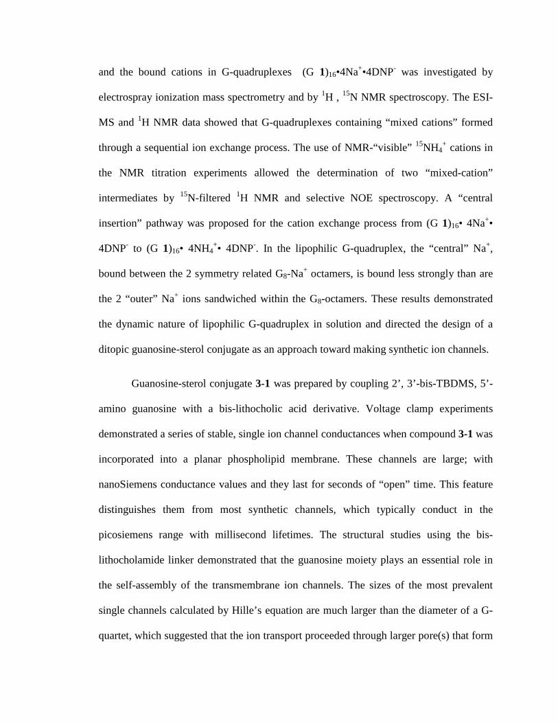
and the bound cations in G-quadruplexes (G 1)16bull4Na+bull4DNP- was investigated by
electrospray ionization mass spectrometry and by 1H 15N NMR spectroscopy The ESI-
MS and 1H NMR data showed that G-quadruplexes containing ldquomixed cationsrdquo formed
through a sequential ion exchange process The use of NMR-ldquovisiblerdquo 15NH4+ cations in
the NMR titration experiments allowed the determination of two ldquomixed-cationrdquo
intermediates by 15N-filtered 1H NMR and selective NOE spectroscopy A ldquocentral
insertionrdquo pathway was proposed for the cation exchange process from (G 1)16bull 4Na+bull
4DNP- to (G 1)16bull 4NH4+bull 4DNP- In the lipophilic G-quadruplex the ldquocentralrdquo Na+
bound between the 2 symmetry related G8-Na+ octamers is bound less strongly than are
the 2 ldquoouterrdquo Na+ ions sandwiched within the G8-octamers These results demonstrated
the dynamic nature of lipophilic G-quadruplex in solution and directed the design of a
ditopic guanosine-sterol conjugate as an approach toward making synthetic ion channels
Guanosine-sterol conjugate 3-1 was prepared by coupling 2rsquo 3rsquo-bis-TBDMS 5rsquo-
amino guanosine with a bis-lithocholic acid derivative Voltage clamp experiments
demonstrated a series of stable single ion channel conductances when compound 3-1 was
incorporated into a planar phospholipid membrane These channels are large with
nanoSiemens conductance values and they last for seconds of ldquoopenrdquo time This feature
distinguishes them from most synthetic channels which typically conduct in the
picosiemens range with millisecond lifetimes The structural studies using the bis-
lithocholamide linker demonstrated that the guanosine moiety plays an essential role in
the self-assembly of the transmembrane ion channels The sizes of the most prevalent
single channels calculated by Hillersquos equation are much larger than the diameter of a G-
quartet which suggested that the ion transport proceeded through larger pore(s) that form
upon self-assembly of lipophilic guanosine-lithocholate 3-1 within the phospholipid
membrane The large transmembrane pore(s) could be envisioned as a supramolecular
structure with hydrophobic walls of bis-lithocholate linker and a central pillar of a cation-
filled G-quadruplex
The use of a bis-urea functionality in the bis-lithocholic acid linker generated
guanosine-sterol conjugate 4-1 The ion channel activity of 4-1 was demonstrated by
voltage clamp experiment Large ion channels formed from 4-1 had longer life-times than
those formed from compound 3-1 The extra stabilization of self-assembled ion channels
attributed to the bisurea hydrogen bonding is consistent with the structural hypothesis of
ion channels The stable large transmembrane ion channels self-assembled by lipophilic
guanosine derivatives have potential for delivery of drugs or biomolecules
Synthetic Ion Channels From Lipophilic Guanosine Derivatives
By
Ling Ma
Dissertation submitted to the Faculty of the Graduate School of the
University of Maryland College Park in partial fulfillment
of the requirements for the degree of
Doctor of Philosophy
2009
Advisory Committee
Professor Jeffery T Davis Chair
Professor Dorothy Beckett
Professor Marco Colombini
Professor Lyle Isaacs
Professor Steven E Rokita
copy Copyright by
Ling Ma
2009
ii
Dedication
To my parents my mother-in-law my husband my brother and my daughter for all your
love and support
iii
Acknowledgements
First I would like to express my heartfelt gratitude to my research advisor and
mentor Professor Jeffery T Davis Thank you for giving me the freedom guidance and
encouragement throughout my whole graduate career Special thanks go to Professor
Marco Colombini for providing access to his voltage clamp facilities I am truly grateful
for his generosity and his helpful advice I am also thankful for our discussions on
membranes and on life in general I would also like to thank Prof Lyle Isaacs for his
helpful suggestions in chemistry and career advice I would also like to thank professors
Steven Rokita Dorothy Beckett Michael Doyle and Daniel Falvey for their valuable
discussions and suggestions
I am very grateful to Dr Yiu-Fai Lam and Dr Yinde Wang for their training and
help in NMR spectroscopy Particularly I would like to thank Yiu-Fai for his help with
my research project He taught me so many new techniques and helped me set up
numerous special experiments Yiu-Fai Thank you
I am thankful for the electrospray ionization mass spectrometry training provided
by Noel Whittaker and Dr Yue Li I would also like to acknowledge Dr Fred Khachik
for the use of his HPLC instrument and Dr Peter Zavalij for the crystal structures
I would like to express my gratitude to the Davis research group You have made
my graduate life much more enjoyable I would especially like to thank Dr Mark
Kaucher for helping me start my PhD program teaching me how to use CD
spectrophotometer and providing helpful advice I would also like to thank Jennifer
Seganish Sofya Berezin William Harrell Oluyomi Okunola Doriann Dejesus and
iv
Monique Pichon for their discussion and friendship I would also like to thank Dr Paul
Santacroce for teaching me new techniques and helpful discussions Particularly I would
like to thank Dr Monica Melegari from University of Parma Italy She was always there
to discuss chemistry and made my ldquomembrane-makingrdquo experience fun Thanks also go
to the undergraduates I had the pleasure to work with Emily Ryan and Jonathan Jinhoon
Park Thank you for the excellent synthesis work
I extend my appreciation to my fellow classmates and friends who supported each
other and in shared the graduate school experience Drs Becky Vieira Regan Nally
Soumyadip Ghosh Wei-hao Huang Sofya Berezin JuHee Park In particular I would
like to thank Dr Becky Vieira and Regan Nally for their friendship Dr Becky thank you
for your generous help and patience in correcting my writing
Lastly I would like to send my deepest thanks to my family I have the most
wonderful family in this world I would like to thank my parents and brother for always
being there to love and support me I thank my mother-in law for her help and
encouragement I thank my daughter for being an excellent kid and making me a proud
Mom I would like to especially thank my husband for his understanding patience
support and love I love you all very dearly
v
Table of Contents
LIST OF TABLES x
LIST OF FIGURES xi
LIST OF SCHEMES xvii
Chapter 1 Introduction 1
11 Introduction 1
12 Thesis Organization 1
13 Nucleobases and Lipids as Building Motifs for Supramolecular Structures 2
131 Nucleobases Molecular Recognition Motif 2
132 LipidBilayer Structure Unit of Functional Biological Membrane 5
14 Lipophilic Nucleobases Nucleosides and Oligonucleotides The Nucleolipids 6
141 Natural Nucleolipids 6
142 Molecular Recognitionof Synthetic Nucleolipids 9
143 Membrane Association Properties of Nucleolipids 13
15 Functional Nanostructures from Amphiphilic Nucleobases 14
151 Molecular Recognition in Aqueous Solution Amphiphilic Nucleobase-
Derived Supramolecular Receptors 15
152 Lipophilic Nucleobases Approach Toward Functional Biological Surfaces 17
16 Synthetic Nucleolipid forTransportof Biomolecules 21
161 Lipophilic Nucleobase as Carrier for Nucleotide Monophosphate Transport 21
162 Nucleolipids for DNA Transfection 23
vi
17 Lipophilic Guanosine Building Block of Transmembrane Ion Channels 25
171 Ion Channel Model From DNA G-quadruplex 25
172 Lipophilic Guanosine Derivatives 27
18 Summary 29
Chapter 2 Cation Exchange in Lipophilic G-Quadruplexes Not All Ion Binding Sites
Are Equal 30
21 Introduction 30
211 Cation-Dependent Self-Assembly of DNA G-Quadruplex Structures 31
212 DNA G-quadruplex as an Ion Channel Model 32
213 Lipophilic G-Quadruplexes 35
22 Mass Spectrometry Shows Mixed-Cation G-Quadruplexes Formed by Sequential
Ion Exchange 37
23 Proton NMR Indicates that the Central Na+ Is the First Cation Exchanged for K+ in
Formation of Mixed Na+ K+ G-Quadruplexes 40
24 Use of 15NH4+ Cation to Probe the Cation Exchange Process Within a Lipophilic
G-quadruplex 43
241 NMR Studies with 15NH4+ Confirm Identity of Mixed-Cationic G-
Quadruplexes 43
242 Determination of the First Intermediate in Na+15NH4+ Cation Exchange 46
243 Characterization of the Second Intermediate in the Na+15NH4+ Cation
Exchange Process 48
25 Cation Exchange Pathway in the Lipophilic G-Quadruplex 50
26 The Larger Cs+ Cation But Not the Smaller Li+ Can Displace the Central Cation
vii
in the Na+ G-Quadruplex 53
27 Conclusion 54
Chapter 3 Large and Stable Transmembrane Pores from a Guanosine-Bile Acid
Conjugate 56
31 Introduction 56
311 G-QuartetFolate-Quartet as a Motif for Transmembrane Ion Transporter 58
32 Design of G-quartet Ion Channels Using a Ditopic Guanosine with a Covalent
Linker 61
33 Design of Guanosine-Sterol Conjugate 64
34 Synthesis of Guanosine-Sterol Conjugate 67
35 Synthesis of Bis-lithocholamide for Control Experiment 71
36 Self-Assembly Property Study of Guanosine-Sterol Conjugate 72
37 Guanosine-Sterol Conjugate Forms Transmembrane Ion Channels (Pores) 75
371 Ion Selectivity of the Synthetic Ion Channels 80
372 Proposed Active Structure of the Synthetic Ion Channel 83
38 Conclusion 87
Chapter 4 Synthetic Ion Channel from Ditopic Sterol-Guanosines with Bis-urea Linker
89
41 Introduction 89
42 Bis-urea Macrocycles are Known to Form Self-Assembled Cylinders 91
43 Synthesis of Bis-urea Guanosine-Sterol Conjugate 4-1 95
44 Solution Study of Self-Assembly Bis-Urea Modified Lithocholate vs Bis-
viii
Carbamate Analogue 99
441 1H NMR Spectroscopy Provides Evidence of Intermolecular Hydrogen
Bonding Between Bis-Urea Linker Units 99
442 Intermolecular Hydrogen Bonding Study by FT-IR Spectroscopy 101
45 Large and Stable Ion Channel From Guanosine-Sterol Conjugate With Bis-Urea
Modified Linker 104
46 Structural Study of Guanosine-Sterol Conjugate With the Bis-Urea Linker 108
47 Guanosine-Sterol Conjugates Cause Release of Carboxyfluorescein from
Liposomes 110
48 Conclusion 113
Chapter 5 Final Conclusion and Future Directions 114
Chapter 6 Experimental and References 118
61 General Experimental 118
62 Synthesis 119
63 ESI-MS Experiment of G-quadruplex 128
64 NMR Titration 129
65 15N Filtered 1H NMR Experiment 129
66 NOESY Experiment 130
67 Selective NOE Experiment 130
68 Planar Bilayer Conductance Experiments 131
69 Reversal Potential Measurements 131
ix
610 Liposome Preparation 132
References 133
x
List of Tables
Table 31 Hillersquos diameter of ion channels formed from 3-1 85
Table 41 FT-IR frequencies for bis-urea 4-10 and bis-carbamate lithocholate 3-
13helliphelliphelliphelliphelliphelliphelliphelliphelliphelliphelliphelliphelliphelliphelliphelliphelliphelliphelliphelliphelliphelliphelliphelliphelliphelliphelliphelliphelliphelliphelliphellip103
Table 42 Frequency of Events for Channels formed from 4-1 and 3-1 106
xi
List of Figures
Figure 11 a) Natural Nucleobase b) Watson-Crick base pairing motif c) Hoogsteen
base- pairing motif 4
Figure 12 Self-assembled structures of guanine a) G-ribbon and b) G-quartet 5
Figure 13 a) Example of phospholipid phosphatidylcholine b) A phospholipid bilayer
with their polar head groups exposed to water and their hydrophobic tails buried in the
interior of the membrane 6
Figure 14 Chemical structure of cytidinediphosphatediacylglycerol 1-6 8
Figure 15 Chemical structure of the nucleolipid antibiotic tunicamycins 9
Figure 16 Some examples of nucleolipids 13
Figure 17 The representation of the incorporation of lipophilic (thyminylocty1) ammo-
nium bromide or alkylthymine into SDS micelles and the hydrogen bond formation
between lipophilic thymine derivatives and lipophilic alkyladenine inside micelles 16
Figure 18 a) Chemical structure of the lipophilic anchor of the oligonucleotides LT
23mer b) The sequence of the LT23mer on the membrane surface can recognize the
complementary oligonucleotides A 20mer to form DNA double helix 19
Figure 19 a) Chemical structures of complementary oligonucleotide amphiphiles ONA1
and ONA2 control DNA sequences A1 and A2 fluorescent 14mer and 9mer DNA probes
b) A liposome anchored ONA can function as either the thermo-controlled reversible
switch or the chemical irreversible switch Below the melting temperature of the duplex
the probe is centered onto the liposome surface (on state) Above melting temperature or
xii
in the presence of a competitive complementary ON) the fluorescent probe is expelled
from the surface (off state) 20
Figure 110 a) Chemical structures of the cationic nucleolipids for 5rsquo-DMP or 5rsquo-AMP
transport b) A cartoon of transport of 5rsquo-DMP or 5rsquo-AMP by a carrier mechanism The
formation of carriersubstrate complex through complementary hydrogen bonding and
electrostatic interaction contribute to the transmembrane transport 23
Figure 111 a) Chemical structures of uridine-based cationic nucleolipid b) Schematic
drawing of lipoplexes formed by CL-DNA and uridine-based nucleolipid where DNA
rods are intercalated between nucleolipid bilayers 24
Figure 112 The crystal structure of DNA G-quadruplex formed form oligonucletidesd
(TG4T) in the presence of Na+ in the solution which provide a nature ion channel model
26
Figure 113 Some examples of lipophilic guanosine 27
Figure 114 Crystal structure of a lipophilic G-quadruplex [G]16middot4K+middot4Picndash 28
Figure 21 ESI-MS from titration of KPh4B into a solution of [G 1]16middot4Na+middot4DNP- in 11 CD2Cl2CD3CN a) [G 1]16middot4Na+middot4DNP- b) after addition of 1 equivalents of KPh4B c) after addition of 2 equivalents of KPh4B d) after addition of 4 equivalents of KPh4B and e) [G 1]16middot4K+middot4DNP- The diagram illustrates the possible mixed-cation G-quadruplexes that could be formed in the cation exchang processhelliphelliphelliphelliphelliphelliphellip40
Figure 22 Region of the 1H NMR spectra (400 MHz) showing the G-quartet NH1 amide protons during titration of [G 1]16middot4Na+middot4DNP- with KPh4B in 11 CD2Cl2CD3CN (a) [G 1]16middot4Na+middot4DNP- The molar ratio of added KPh4B to the [G 1]16middot4Na+middot4DNP- G-quadruplex is (b) 051 (c) 11 (d) 21 (e) 31 (f) 41 and (g) 121 42
Figure 23 A region of the 1H NMR spectrua (400 MHz) for titration of 15NH4Ph4B into a solution of (G 1)16bull 4Na+bull 4DNP- in 11 CD2Cl2CD3CN at 20 oC The signals for the amide N1H protons of the G-quartets are shown a) (G 1)16bull 4Na+bull 4DNP-The mole ratio of 15NH4
+ to (G 1)16bull4Na+bull4DNP- is b) 051 c) 11 d) 21 e) 31 f) 41 g) 121hellip 544
Figure 24 a) Region from the 2-D 15N-1H HSQC-ROESY NMR spectrum of [G 1]16middot4NH4
+middot4DNP- in 11 CD2Cl2CD3CN showing cross-peaks between the 15N NMR resonances and the 15N-filtered 1H resonances b) Region of the 2-D 1H-1H NOESY NMR
xiii
spectra of [G 1]16middot4NH4+middot4DNP- in 11 CD2Cl2CD3CN showing cross-peaks between the
N1H amide resonances and the 1H resonances for the bound NH4+ cations Both spectra
were recorded on a 1 mM sample at 20 degC using a 500 MHz NMR spectrometer 45
Figure 25 15N-filtered 1H NMR (500 MHz) spectra of 15NH4Ph4B titration the solution of [G 1]16middot4Na+middot4DNP- in 11 CD2Cl2CD3CN with mol ratio at (a) 01 (b) 11 (c) 21 (d) 31 (e) 41 (f) 121 and (g) [G 1]16middot4
15NH4+middot4DNP- 47
Figure 26 Portion of a NOESY spectrum of a 11 15NH4 Ph4B titration into [G 1]16middot4Na+middot4DNP- in 11 CD2Cl2CD3CN The 2-D spectrum shows the NOE cross-peak between the 15NH4
+1H resonance with the N1H of the inner G-quartet layer The chemical shifts of δ 1179 and 1140 ppm correspond to the N1H amide protons for the outer and inner G-quartet layers The chemical shift of the 15NH4
+ proton was confirmed from the 15N decoupled 1H NMR spectrum showing a single peak at δ 710 ppm 48
Figure 27 Selective NOE spectra for amide N1H peaks of (G 1)16middot 4Na+ middot4DNP- titrated with 2 equivalents of 15NH4Ph4B in 11CD2Cl2CD3CN Left a) N1H region of 1H NMR spectrum (green dots indicate the N1H amide proton from the first intermediate blue dots indicate the N1H amide proton from the second intermediate) b) 15NH4
+ proton region showing selective NOE correlation to amide proton at δ 1151 δ 1135 δ 1140 and δ 1179 ppm respectively Right structural scheme for the second intermediate [G4 (o) middot Na+ middot G4 (i) middot 15NH4
+ middot G4 (i) middot15NH4+ middot G4 (o)] formed during the Na+ NH4
+ exchange process with the assignments for the different G-quartet N1H amide protonshelliphelliphellip 50
Figure 28 1H NMR spectra of N1H region of different G-quadruplex solutions in CD2Cl2CD3CN at 20 degC (a) [G1]16middot4Na+middot4DNP- (b) 101 mol ratio of LiPh4B and [G1]16middot4Na+middot4DNP- (c) 81 mol ratio of CsPh4B and [G1]16middot4Na+middot4DNP- 54
Figure 31 Ditopic G-sterol 3-1 and control bis-lithocholamide 3-16 Typical traces of conductance vs time after addition of 3-1 or 3-16 are depictedhelliphelliphelliphelliphelliphelliphelliphelliphellip56
Figure 32 Hypothetical structure of a self-assembled barrel-stave ion channel formed from compound 1 The wedge represents guanosine moiety the line represents the bis-lithocholic acid linker Channel formation would be controlled by the self-assembly of guanosine head groups to give a G-quartet based structure 58
Figure 33 The unimolecular G-quadruplex stabilized by post-covalent modification that can function as a transmembrane Na+ transporter 59
Figure 34 Chemical structure of the folate dendrimer 3-5 the building block for the ldquofolate-quartetrdquo ion channel 60
Figure 35 Schematic illustration of two types of ldquocation channelrdquo formed from a G-quadruplex a) ldquoBarrel Rosetterdquo G-quadruplex formed by 16 lipophilic guanosine
xiv
subunits b) ldquoBarrel Staverdquo G-quadruplex from by 4 ditopic ligands The ball stands for a cation and the stave stand for the ligand 62
Figure 36 Possible supramolecular structure formed by 3-6 through association into G-quartets stabilized by K+ binding a) internally-bridged [(3-6)2 K+] assembly b) linear chain of doubly bridged G4 units c) fully cross-linked regular array of G4 units 63 Figure 37 Structure of bis-iminoboronate-guanosine 3-7 used to form G4-quartet based membrane 64
Figure 38 The structure of cholic acid 3-8 and lithocholic acid 3-9 65
Figure 39 Top structures of bis-cholic acid derivatives 3-11 with methoxy group on the concave α-face 3 with hydroxyl group on the concave α-face Below Kobukersquos proposed cross section image of ion channel constructed from a bis-cholic acid derivative trimerwater and bis-cholic acid derivative tetramerwater 66
Figure 310 a)1HNMR spectrum of guanosine-bislithocholate conjugate 3-1 inDMSO-d6 b) Selective NOE experiment was carried out by irradiating new amide proton peak at δ 808 ppm 69
Figure 311 1H- 1HNOESY spectrum of guanosine-bislithocholic acid conjugate 3-1 in DMSO-d6 The expansion of the F1 (vertical) axis correlations near δ 808 ppm showing NOE of amide N-H proton is correlated with Harsquo Hardquo H5rsquo H5rdquo H4rsquo H3rsquo 70 Figure 312 ESI-Mass spectrum of guanosine-sterol conjugate 3-1 71
Figure 313 a)1HNMR spectrum of guanosine-bislithocholic acid conjugate 3-1 in DMSO-d6 and CDCl3 b) 1HNMR spectrum of bis-lithocholamide control compound 3-16 in CDCl3 73
Figure 314 CD spectra of guanosine-sterol conjugate 3-1 (blue line) 3-1 mixed with an equiv of [2 2 2]-cryptand (green line) 3-1 after solid-liquid extraction of K+ DNP- All spectra were obtained in CDCl3 75
Figure 315 The setup of voltage clamp experiment The bilayer membrane was prepared with soybean phospholipid supplemented with 05 asolectin 05 DPPC and 01 cholesterol The buffer solution is 1M KCl 2mM MgCl2 and 5mM Pipes All the measurements were done at 25 degC 76
Figure 316 Representative traces from voltage-clamp experiments The distinct conduc-tance values were recorded in the presence of 3-1 at -10 mV in 1 M KCl The number of open events was counted from a total of six experiments Three of the experiment results were from method A another three were from method B 78
xv
Figure 317 Distribution of conductance change The frequency was calculated based on the total number of increment (channel open) events or decrement (channel close) events 80
Figure 318 The data analysis showed that ion selectivity (PK
+ PCl-) decreases with a
corresponding increase in conductance of ion channels These results suggested guanosine-sterol conjugate 1 forms a variety of ion channels with different sizes Panel A Panel B showed the ion selectivity data obtained by applying guanosine-sterol conjugate 3-1 using the sample loading method A and method B respectively 82
Figure 319 Barrel-stave ion channel formed from guanosine-sterol conjugate 3-1 The central G-quartet pore is responsible for the lt01 nS conductance during voltage clamp experiment 84
Figure 320 Left Hillersquos cylinder model of ion channel Right Hillersquos Equation where L is the length of the channel r is the radius of the channel and ρ is the resistivity of the solution Under our planar voltage experiment condition the resistivity of 10 M KCl solution is 89 Ωbullm The length of the channel is estimated as the thickness of the phospholipid bilayer membrane which is approximately 50 Aring 85
Figure 321 Molecular modeling of one possible self-assembly of two molecules of guanosine-lithocholate 3-1 ( Modeling was made by Dr Monica Melegari) 86
Figure 322 Possible G4-quartet stacks formed by bis-G-lithocholate 1 within bilayer membrane 87
Figure 41 a) Structure represents the modification of sterol linker b) Scheme depicting the one possible self-assembled structure from bis-urea 4-1 The bis-urea modified sterol linker might introduce additional hydrogen bonding through urea stacking to stabilize this synthetic ion channelhelliphelliphelliphelliphelliphelliphelliphelliphelliphelliphelliphelliphelliphelliphelliphelliphelliphelliphellip90
Figure 42 a)Examples of macrocyclic urea derivatives in the development of nanotubes b) Bis-urea macrocycle 4-4 designed by Shimizu c) X-ray crystal structure of the self-assembled bis-urea 4-4 including the 3-centered hydrogen bonding patterns and the skewed orientation of monomers 93
Figure 43 Dynamic self-organization of heteroditopic receptors 4-5 94
Figure 44 a) Crystal structures of lipophilic G-quadruplex templated by the sodium cation b) One of the proposed ion channels from self-assembled bis-urea modified guanosine-sterol conjugate The urea-urea hydrogen bond between the linkers was envisioned to improve the stability of the pore 95
Figure 45 1H NMR spectrum of 3α-amino lithocholate 4-9 in CDCl3 96
Figure 46 1H NMR and ESI-MS Spectra of Bis-Urea 4-1 in DMSO-d6 98
xvi
Figure 47 1H NMR spectra of N-H proton of bis-urea lithocholate 4-10 (blue line) and bis-carbamate lithocholate 3-13 (red line) in CDCl3 a) at room temperature the chemical shift of NH proton is plotted against concentration b) at 10 mM concentration the chemical shift of NH proton is plotted against temperature 101
Figure 48 N-H stretching region of the IR spectra as a function of concentration for a) bis-urea lithocholate 4-10 b) bis-carbamate lithocholate 3-13 in CDCl3 at 25degC 102 Figure 49 Representative conductance records from voltage clamp experiment Ion channels formed by 4-1 in a planar bilayer at an applied voltage of -10 mV in 1M KCl (trans) KCl (cis) solution (pH 70) 104
Figure 410 Distribution of conductance change for the ion channels from guanosine-sterol conjugate 4-1 and 3-1 The frequency was calculated based on the total number of increment (channel open) events or decrement (channel close) events a) data from compound 4-1 b) data from compound 3-1 106
Figure 411 Histogram showing the various open lifetimes for 1-5 nS channels formed from 3-1 and 4-1 Distribution of conductance change the frequency was calculated based on the total number of increment (channel open) events 108
Figure 412 a) Chemical structure of carboxyfluorescein b) Scheme of a fluorescent assay of carboxyfluorescein release from liposome 111
Figure 413 Carboxyfluorescein release assay confirmed the pore formation mediated by guanosine-sterol conjugate 3-1 and 4-1 112
xvii
List of Schemes
Scheme 21 Schematic structure of (A) G-quartet and (B) G8-octamer 32
Scheme 22 Highndashresolution crystal structure of a DNA G-quadruplex formed by oligonucleotide [d(TG4T)]4 b) Natural potassium ion channel 34
Scheme 23 Schematic representation of 15NH4
+ movement through the Oxy-15 quadruplex a) bulk NH4
+ cation only exchanges with the outer 15NH4+ b) exchange of
two bound 15NH4+ cationsare random Black yellow and red ball represent the bulk
ammonium ion and the boundammonium ions located in outer and inner binding site of the G-quadruplex 35
Scheme 24 G-quadruplex formed through self-assembly of G 1 in the presence of K+DNP- 36
Scheme 25 Proposed central insertion pathway for cation exchange in the lipophilic G-quadruplex system 51
Scheme 26 These depictions are taken from the crystal structure for [G1]16middot4Na+middot4pic- The illustrations show the octahedral coordination geometry for one of the outer Na+ cations (on the left) and for the central Na+ (right) The central Na+ has a almost cubic coordination geometry whereas the oxygen ligands are twisted toward the energetically more favorable square anti-prism geometry for the outer Na+ 52
Scheme 31 Synthesis of bis-guanosine-lithocholic acid conjugate 3-1helliphelliphelliphelliphelliphellip68
Scheme 32 Synthesis of control compound bis-lithocholamide 3-16 72
Scheme 41 Three center hydrogen bonding net-work formed by urea derivativehelliphellip 92
Scheme 42 Synthesis of 3α-amino lithocholate 4-9 96
Scheme 43 Synthesis of bis-urea modified guanosine-sterol conjugate 4-1 97 Scheme 44 Synthesis of bis-urea lithocholamide 4-12 for the control experiment 109
1
Chapter 1 Introduction
11 Introduction
Molecular self-assembly plays a fundamental role in Nature and is a widely used
principle to build supramolecular structures from individual molecular components1-3
For example self-assembly has been a useful tool to mimic biological functions and
fabricate nanomaterials45 In recent years many research activities have focused on the
development of novel functional materials using well-defined building blocks This thesis
ldquoSynthetic Ion Channels from Lipophilic Guanosine Derivativesrdquo describes the design
and development of amphiphilic building blocks for the self-assembly of functional
structures
12 Thesis Organization
This thesis is organized into six chapters The overall goal of this research was to
develop trans-membrane synthetic ion channels using lipophilic guanosine as building
blocks Chapter 1 briefly discusses the molecular recognition properties of the naturally
occurring nucleobases and then goes on to review the potential use of lipophilic
nucleobases nucleosides and oligonucleotides as functional nanostructures and
biomaterials The cation exchange studies in Chapter 2 give insight into the stability of
self-assembled lipophilic G-quadruplex in solution The kinetic lability of lipophilic G-
quadruplex inspired the design of a ditopic lipophilic guanosine to build trans-membrane
ion channels Chapter 3 describes large and stable ion channels formed from a ditopic
2
guanosine-lithocholic acid conjugate The ion channel activity and structure-properties
characterization suggested the key role of the guanosine subunit in the formation of ion
channels Chapter 4 explores the enhanced stability and activity of ion channels through
the modification of guanosine-sterol building blocks Chapter 5 describes the conclusion
and directions of future research Finally Chapter 6 contains the experimental protocols
for the research described in Chapters 2-4
13 Nucleobases and Lipids as Building Motifs for Supramolecular Structures
131 Nucleobases Molecular Recognition Motif
In Nature the self-assembly of single strand DNA into double helix and other
higher order structures is mediated by many noncovalent interactions such as hydrogen
bonding π-π stacking and van der Waals forces Among these intermolecular
interactions complementary hydrogen bonding via nucleobase-pairing is of primary
importance in the molecular recognition of DNA and RNA sequences Thus the
molecular recognition capability of nucleobases via specific hydrogen-bonding pattern is
considered to play an essential role in the storage translation and replication of genetic
information6-8
The naturally occurring nucleobases are nitrogen-containing aromatic
heterocycles including the double-ringed purines (adenine 1-1 guanine 1-2) and single-
ringed pyrimidines (cytosine 1-3 thymine 1-4 and uracil 1-5) They are the key
constituents of DNA and RNA that are involved in molecular recognition Cytosine 1-3
3
guanine 1-2 adenine 1-1 and thymine 1-4 are found in DNA while uracil 1-5 replaces
thymine 1-4 in RNA Nucleobases are able to recognize each other through a
complementary hydrogen bonding interaction known as Watson-Crick pairing9 Watson-
Crick pairing is one of the most recognized observations in a variety of DNA- and RNA-
structures In this specific hydrogen-bonding motif guanine 1-2 pairs with cytosine 1-3
through a three-point hydrogen bonding while adenine 1-1 interacts with thymine 1-4 (or
uracil in RNA) in a two-point hydrogen bonding fashion (Figure 11)
Furthermore various alternative hydrogen-bonding motifs are also possible71011
These non-Watson-Crick pairings include mismatch-pairing nucleobase self-
dimerization and Hoogsteen base pairing that associates the C6 and N7 binding sites of
purine nucleobases Hoogsteen base pairing through the self-association of guanosine 1-2
is another prevalent hydrogen bonding motif found in a variety of G-rich DNA and RNA
suprastructures12
4
Figure 11 a) Natural Nucleobase b) Watson-Crick base pairing motif c) Hoogsteen base- pairing motif
Of the natural nucleobases guanosine 1-2 possesses a unique self-association
property since it incorporates both Watson-Crick and Hoogsteen edges for the self-
complementary hydrogen bonding The guanine-guanine interactions involve the two
Hoogsteen pairings between hydrogen bonding acceptor O6 and N7 at Hoogsteen edges
and hydrogen bonding donor N1H amide and N2H amine at Watson-Crick edges Under
certain conditions guanine self-assembles into several suprastructures that differ from the
classic nucleobase dimers13-17 These higher-ordered structures include the G-ribbon
structure or cyclic structure known as the G-quartet (Figure 12b) Figure 12a shows
the hydrogen-bonded ribbon G-structure in the absence of cations Whereas the G-
quartet is a preferential self-assembled structure in the presence of cations since cations
5
stabilize this cyclic tetramer through the electrostatic interaction between cation and the
carbonyl oxygens (Figure 13b)
N
NN
N
O
NH
H
H
R
N
NN
N
O
NH
H
H
R
N
NN
N
O
NH
H
H
R
N
NN
N
O
NH
H
H
Ra)
N
NN
N
O
N
H
H
H
R
N
NN
N
O
N
H
H
H
R
NN
NN
O
N HH
H
R
NN
N N
O
NHH
H
Rb)
Figure 12 Self-assembled structures of guanine a) G-ribbon and b) G-quartet
132 Lipid Bilayer Structural Unit of Functional Biological Membrane
The lipid bilayer is the basic structural unit of all biological membranes1819 It
acts as the hydrophobic barrier that segregates cellular compartments and isolates the
cells from the extracellular environment As shown in Figure 13 phospholipids the
fundamental building blocks of all cell membranes consist of a polar phosphate-
containing head group and two nonpolar hydrocarbon tails These amphiphilic molecules
self-assemble in a tail-to-tail fashion to form a lipid bilayer with a hydrophobic interior
and a hydrophilic surface Lipid bilayers are versatile structures that can be formed by a
number of phospholipids and other lipid molecules such as cholesterol and glycolipids
6
CH2OP
O
O-
OH2C
CHO
CH2O
C
C
R
O
O
R
CH2
N+(CH3)3
Hydrophobic Tail Hydrophilic Head
Tail
Head
Head
Tail
b)a)
Figure 13 a) Example of phospholipid phosphatidylcholine b) A phospholipid bilayer with their polar head groups exposed to water and their hydrophobic tails buried in the interior of the membrane
In addition lipid bilayers can also accommodate or bind to a variety of biological
molecules2021 For example proteins are the other major constituent of cell membranes
They are either embedded within the lipid bilayer or surface-adsorbed on the lipid bilayer
These different species of proteins and lipids can recognize specifically with other
another by their individual chemical nature As a result the lipids associated with
proteins are involved in a wide array of cellular processes and specialized membrane
functions such as cell adhesion selective transport of molecules across membranes and
cell signaling
14 Lipophilic Nucleobases Nucleosides and Oligonucleotides The Nucleolipids
141 Natural Nucleolipids
Nature has utilized the nucleobase-derived building molecules to develop
functional self-assemblies with specificity and control One class of extremely promising
and interesting biomolecules are nucleolipids the nucleobase-derived amphiphiles
obtained by covalently linking a lipophilic moiety with a nucleobase a nucleoside or
anoligonucleotide22 Nucleolipids have been found to be involved in biosynthesis
7
processes14 and a variety of biological activities such as antimicrobial antivirus anti
tumor activities in eukaryotic and prokaryotic cells2324
Cytidinediphosphatediacylglycerol (CDP-diacylglycerol) 1-6 is one of the most
ubiquitous nucleolipids present in cells (Figure 14)25 It plays an essential role in the
biosynthesis of the membrane phospholipids In mammalian cells CDP-diacylglycerol1-
6is formed from cytidinetriphosphate and phosphatidic acid and serves as an activated
energy-rich intermediate Catalyzed by different specific membrane-bound enzymes
CDP-diacylglycerol 1-6 is able to transfer a phosphatidic acid to a hydroxyl group of a
carbohydrate or glycolipid to generate phospholipids The specific enzymes are located in
mitochondria for the biosynthesis of phosphatidylglycerol the precursor of cardiolipin or
in microsomes for the biosynthesis of phosphatidylinositol2627 This indicates that
nucleoside-based lipid can be incorporated into a variety of phospholipids in the cell
membrane In prokaryotes CDP-diacylglycerol 1-6 functions as the precursor for the
biosynthesis of phosphatidylserine as well28
Moreover CDP-diacylglycerol 1-6 is involved in the enzymatic activities of
phosphatidylserine synthases occurring in gram-negative bacteria such as E coli
Salmonella typhimurium Serratiamarcescenceetc The membrane association of these
phosphatidylserine synthases is induced by the lipid substrate CDP-diacylglycerol 1-
61819 The CDP-diacylglycerol-dependent activity of phosphatidylserine suggested that
phosphatidylserine synthases are peripheral membrane proteins that specifically bind to
the nucleolipid CDP-diacylglycerol 1-6 via an intermolecular interaction such as
8
nucleobase-pairing hydrophobic interactions andor van der Waals interactions
Nucleolipid CDP-diacylglycerol 1-6 can reside in the membrane due to its high degree of
amphiphilicity
Figure 14 Chemical structure of cytidinediphosphatediacylglycerol 1-6
Tunicamycins 1-7 was the first discovered nucleolipid antibiotic that was
produced from the fermentation broths of Streptomyceslysosuperificus (Figure 15)
Tunicamycins1-7 is composed of the heterocyclic nucleobase uracil and a branched fatty
acid side chain and it has demonstrated the ability to inhibit a variety of biosynthesis of
oligosaccharide thus preventing the glycosylation of proteins in both prokaryotic and
eukaryotic cells21-24 The antibiotic can function as an irreversible inhibitor for the
GlcNAc-1-phosphate transferaseGlcNAc-1-phosphate transferase is the eukaryotic
enzyme that catalyzes the transfer of N-acetylglucosamine1-phosphate from UDP-N-
acetylglucosamine to dolichylphosphate to generate N-
acetylglucosaminylpyrophosphoryldolichol Mechanistic studies on the actions of
tunicamycin 1-7 have revealed that these nucleolipids bind preferentially to the GlcNAc-
1-phosphate transferase and also interact with the membrane phospholipid2930 The
binding of the tunicamycins 1-7 totransferase occurs when tunicamycins 1-7 is
incorporated into biological membranes Tunicamycins 1-7 also acts as reversible
inhibitors for the phospho-N-acetylmuramyl-pentapeptidetranslocase (translocase I) in
9
the peptidoglycan biosynthesis as well Translocase I is the specific enzyme that
catalyzes the relocation of phospho-MurNAc-L-Ala-g-D-Glum-DAP-D-Ala-D-Ala from
UMP to a membrane-bound carrier undecaprenyl phosphate The studies of actions of
tunicamycins have showed that the inhibition by tunicamycins 1-7 is competitive with
regard to the substrate UDP-MurNAc-pentapeptide since both molecules share the same
uridine subunit This result demonstrated the recognition characteristic of nucleobase in
the observed bioactivity The capability of tunicamycins 1-7 to locate within the
membrane bilayer can be attributed to the hydrophobicity of the tunicamycins
Figure 15 Chemical structure of the nucleolipid antibiotic tunicamycins
142 Molecular Recognition of Synthetic Nucleolipids
The design of synthetic amphiphiles that are derived from biologically functional
nucleobases nucleosides and oligonucleotides has become a major strategy to build
interesting self-assembled structures with complex and desirable properties Like the
typical amphiphiles nucleolipid amphiphiles will self-assemble in aqueous solution to
10
form distinct structures such as micelles vesicles and other self-aggregates31-33 However
nucleolipids have unique physicochemical properties due to their informative nucleobase
constituents The specific base-pairing of nucleobases through hydrogen bonding and π-π
stacking is cooperative with the hydrophobic effect from the lipid segment contributing
to the self-assembly
In the 1980s the molecular recognition between two complementary nucleolipids
1-8 and 1-9 was first reported by Ringsdorf and coworkers34 Using surface pressure
measurements UV and DSC techniques they found that adenine- and thymine-
nucleolipids base-paired at the air-water interface Afterward numerous techniques
including Langmuir-Blodgett NMR FTIR RA ATR fluorescent probes etc have
provided direct evidence of the complementary hydrogen bonding between nucleolipids
at the air-water interface35-38
The base pairing of nucleolipid was also observed at the mesoscopic interfaces of
the self-assembly of nucleolipids in either micellar or vesiclar structures39 For instance it
was reported by Berti and Baglioni that short-chain phospholiponucleosides diC8P-
adenosine 1-10 and diC8P-uridine 1-11 self-assembled into mixed micelles with CMC
values around 10-3 M40 The data from NMR UVminusVis and CD spectroscopies revealed
the stacking and hydrogen-bonding of the complementary adenosine and uridine occurred
at the micellar surface The continued study of phospholiponucleosides with longer alkyl
chain (DOP-adenosine 1-12 DOP-uridine 1-13 and DOP-cytosine 1-14) demonstrated
that these nucleolipidsare able to self-assemble into liposomes41 The liposomes formed
11
from a mixture of DOP-adenosine 1-12 and DOP-uridine 1-13 showed the UV CD and
NMR properties for base-pairing while the liposome formed from a mixture of DOP-
adenosine 1-12 and DOP-cytosine 1-14 failed to give any spectroscopic change These
data supporting that the molecular recognition properties of nucleolipids are controlled by
specific Watson-Crick base pairing
Specific base pairing that resembles biofunctional supramolecular structures has
been found for certain nucleolipids Itojima and coworkers reported that lipophilic
dimyristoyl-5rsquo-phosphatidylnucleosides 1-15 can self-assemble into helical strands that
are similar to DNA duplex in aqueous solution42 Dimyristoyl-5rsquo-phosphatidyladenosine
1-15 can form various self-assemblies including multi-helical strands or tubular
structure depending on the pH and alkalinity of the solution The formation of a hybrid
helix from the equimolar mixture of dimyristoyl-5rsquo-phosphatidyladenosine 1-15
anddimyristoyl-5rsquo-phosphatidyluridine 1-16 suggested that the specific hydrogen bonding
between nucleobases directs the self-assembly of nucleolipids The research of Gottarelli
and Spada established that in the presence of certain cations liphophilic guanosine 1-17
can self-associate into columnar structures that contain stacked G-quartet in organic
solution144344 The self-assembly is driven by Watson-Crick and Hoogsteen base pairing
of guanine π-π stacking and cation-dipole interactions Similar to the liquid crystals
formed from guanosine oligonucletides in aqueous solution the G-quartet is the
fundamental building block for these supramolecular structures Such a G-quartet was
also found in the DNA secondary structure that regulates the telomerase activity in
chromosomes
12
The molecular recognition characteristics of nucleolipids have attracted
substantial research including attention to the design and synthesis of novel amphiphilies
derived from nucleobases for both the fundamental aspects and application purposes For
these reasons a wide range of nucleolipids have been synthesized by covalently linking
the hydrophobic segment to nucleobases nucleosides and oligonucleotides The
lipophilic moieties such as alkyl chain cholesterol glycerol etc are attached either
directly to the nucleobase or to the sugar moiety of nucleoside or oligonucleotides
Nucleolipid structures can be classified as either neutral cationic anionic or zwitterionic
nucleolipids depending on their charge properties Figure 16 lists the nucleolipids
reported in the literature mentioned in section 14
13
Figure 16 Some examples of nucleolipids
143 Membrane Association Properties of Nucleolipids
The lipophilic moiety of certain nucleolipids provides another important feature
for these compounds namely membrane association A variety of nucleolipids have been
found to be lipophilic components that can serve as an anchor to attach lipophilic
nucleobase nucleosides and oligonucleotides into the lipid membrane45-48 For the
membrane incorporation purpose the lipid anchor should be sufficiently hydrophobic to
14
overcome the self-association propensity of the amphiphiles and also allow it to be stable
in the membrane The Huster and Liebscher groups have designed lipophilic nucleosides
that conjugate adenine and uracil nucleobases with 1-octadecynyl or sterol hydrophobic
moiety1-18 1-1949 Using solid-state 2H and 31P 1H MAS NMR spectroscopy they
identified the membrane location of the functional groups of these nucleolipids The lipid
moieties are incorporated into the phospholipid membrane while the nucleobase
functional groups are exposed on the surface of the membrane The effects of the inserted
hydrophobic moiety on the membrane show that the steroidal ring of 1-19 causes the
decrease of lipid order parameters and packing density while the alkyl chain of 1-18 has
no pronounced influence on the structure of lipid membrane Compound 1-18 was
proposed as an ideal nucleolipid for building the functional membrane surface This
result suggested that nucleolipids can be used to build functionalized membrane surfaces
In addition lipophilic moieties are implicated to associate the membrane permeation for
the antiviral and antitumor activities of nucleolipid antibiotics50-52
15 Functional Nanostructures from Amphiphilic Nucleobases
Various researches on the lipophilic nucelobases nucleosides and
oligonucleotides have demonstrated their very interesting and unique properties5354 The
combination of molecular recognition and hydrophobic characteristics of nucleolipids
may allow them to target specific lipid membrane or deliver biologically active molecules
which provides a wide opening for the development of bio-devices drug delivery
biosensor and therapeutic strategy This section will describe a few but promising
15
nucleolipid systems that can base-pair within membranes or function as membrane
transporters
151 Molecular Recognition in Aqueous Solution Amphiphilic Nucleobase-Derived
Supramolecular Receptors
The molecular recognition of lipophilic nucleobases through base-paring in
aqueous environment was first reported by Nowick and co-workers55-57 In their
pioneering work lipophilic (thyminylocty1)ammonium bromide 1-20 and sodium
dodecyl sulfate (SDS) self-assembled into micelles and served as supramolecular
receptors55 The resulting receptor consisting of thymine functionality was able to
recognize N6-actyle-9-alkyladenine 1-21 in aqueous solution through an A-T base-pair
(Figure17) The incorporation of (thyminylocty1) ammonium bromide 1-20 into SDS
micelles was assisted by the electrostatic interaction between the positive-charged
ammonium group of 1-20 and the negatively charged sulfate group of SDS and the
hydrophobic interaction between alkyl chain of 1-20 and SDS The complete
incorporation of 10mM of 1-20 into micelles was achieved at an SDS concentration of
20 mMNMR titration studies showed that thymine derivative 1-20 can bind to
alkyladenine derivative 1-21 by means of intermolecular hydrogen bonding in the
presence of SDS This specific binding was determined to be 11 stoichiometry by a Jobrsquos
plot which is consistent with a 11 adenine-thymine base-pairing pattern It was also
found that when SDS is absent in the aqueous solution only aromatic stacking exists
between 1-20 and 1-21 This result suggests that the presence of SDS is crucial for the
hydrogen bond formation and that the adenine-thymine base-pairing occurs within the
hydrophobic core of SDS micelles The author also investigated the effect of lipophilicity
16
of adenine derivatives 1-21 on the hydrogen binding between 1-20and 1-21 By varying
the length of alkyl chain on adenine derivatives 1-21 the binding constants were
measured and compared The adenine derivatives 1-21 bearing longer alkyl-chains
exhibited a larger binding affinity with thymine derivatives 1-20 indicating that the
hydrophobicity of adenine derivatives 1-21 is required for micellar incorporation and
base pairing
Figure 17 The representation of the incorporation of lipophilic (thyminylocty1) ammonium bromide or alkylthymine into SDS micelles and the hydrogen bond formation between lipophilic thymine derivatives and lipophilic alkyladenine inside micelles
The continued effort by the Nowick group provided another successful example
of molecular recognition between neutral lipophilic nucleobases in SDS micelles57 Here
the neutral 1-alkylthymine derivative 1-22 incorporated into SDS micelles functioned as
supramolecular receptors Likewise the receptors can bind N6-acyl-9-alkyladenine 1-21
through base-pairing in aqueous solution Nowick found that 1-alkylthymine derivative
1-22 must be sufficiently hydrophobic to be incorporated within SDS micelles The
incorporation of 1-22 into micelles plays a critical role in the adenine-thymine base-
pairing within micelles The studies on molecular recognition between neutral lipophilic
17
nucleobases in aqueous SDS micelles confirmed the model of supramolecular receptor
proposed in their previous study The hydrophobic core of SDS micelles provides a
suitable environment for hydrogen bonding between the complementary lipophilic
nucleobases The hydrophobic interaction between lipophilic nucleobases and SDS and
the base-pairing between two complementary nucleobases moieties provides a driving
force for molecular recognition in water By means of micelles Nowickrsquos strategy should
have a wide application in the recognition or transport of nucleobase related biomolecules
152 Lipophilic Nucleobases Approach Toward Functional Biological Surfaces
In biological systems phospholipid membrane surfaces represent one of the
active microscopic interfaces for molecular recognition As we mentioned before
membranes are versatile structures that are composed of lipids and a variety of biological
molecules For example proteins embedded within the lipid bilayer or surface adsorbed
on the lipid bilayer are involved in most biological activities such as signal transduction
enzymatic reaction etc To mimic and understand the natural recognition and binding
process on the membrane surface a number of researchers have focused on the
development of functional membranes using nucleolipids
Liebecher Huster and Herrmann reported an oligonucleotide (LT 23mer) 1-23
that anchors into the lipid membrane and forms a DNA duplex58 LT-23mer is designed
as an amphiphile that consists of 21 thymidine (T) units and two lipophilic nucleotides L
in at positions 1 and 8 The α-tocopherol subunit located in the 1-position of cytosine
serves as a lipophilic anchor (Figure 18) A doubly lipophilic anchor separated by 6
nucleotides was designed to prevent the oligonucleotide from self-associating in aqueous
18
environment which improved the membrane insertion The membrane incorporation of
lipophilic LT 23mer 1-23 was studied by incubating LT 23mer and fluorescently labeled
3rsquoRh-or 5rsquoRh-A20mer 1-24 with giant unilamellar vesicles (GUVs) using fluorescence
microscopy Rh fluorescence was detected over the entire membrane indicating the
complementary binding of LT 23mer 1-23 to 3rsquoRh- or 5rsquoRh-A20mer 1-24 occurs on the
GUV surface By means of 1H 31P 1H NMR EPR DSC and fluorescence techniques
Huster et al established that LT 23mer 1-23 are enriched in the liquid-disordered
membrane domains59 LT 23mer 1-23 inserts stably into lipid membranes and imposes
insignificant modification to the bilayer membrane structure The ribosecytosine moiety
of LT 23mer is located in the lipid-water interface while the non-lipidated nucleotide end
is exposed to the aqueous environment This result suggests that the nucleolipids could be
used to build a functional surface on the membrane surface
After LT 23mer 1-23 anchored into the lipid membrane NMR and FRET
techniques were used to characterize the functionality of the membrane surface The
GUVs containing LT 23mer and fluorescent phospholipid N-NBD-PE were incubated
with 3rsquoRh-A20mer 1-24 The resulting strong FRET was attributed to the efficient
complementary binding between membrane-bound LT 23mers1-23and lipophilic 3rsquoRh-
A20mer1-24 The formation of Watson-Crick A-T base pairs which was confirmed by
1H NMR chemical shifts suggested that the oligonucleotides exposed to the aqueous
media are capable of recognizing the complementary oligonucleotide to form the DNA
double helix This membrane-bound DNA double strand presents similar structural
feature and melting properties as the free double-stranded DNA indicating that neither
19
the lipophilic anchor nor the membrane proximity interferes with the DNA duplex
formation Thus the membrane-anchored oligonucleotide may function to provide a
broad range of cell biological and medical applications such as biosensor receptor etc
Figure 18 a) Chemical structure of the lipophilic anchor of the oligonucleotides LT 23mer b) The sequence of the LT23mer on the membrane surface can recognize the complementary oligonucleotides A 20mer to form DNA double helix
Recently Barthelemy and coworkers designed and synthesized membrane-
anchored lipophilic oligonucleotides (ONA) 1-25 that serve as biocompatible switches
(Figure 110)60 The incorporation of the lipophilic ONA 1-25 into a zwitterionic DOPC
liposome was assisted by a lipophilic di-C15 ketal anchor A kinetic study of DNA duplex
formation using the surface plasmon resonance (SPR) technique suggested that these
20
lipophilic membranendashbound ONA recognize their complementary sequences (ON) 1-26
or fluorescent-labeled complementary sequences (fONA) 1-27 via Watson-Crick base
pairings The DOPC lipid membrane has no effect on this DNA duplex formation Thus
the oligonucleotide-tagged liposome can function as a supramolecular platform for
detection and delivery of bioactive molecules
Figure 19 a) Chemical structures of complementary oligonucleotide amphiphiles ONA1 and ONA2 control DNA sequences A1 and A2 fluorescent 14mer and 9mer DNA probes b) A liposome anchored ONA can function as either the thermo-controlled reversible switch or the chemical irreversible switch Below the melting temperature of the duplex the probe is centered onto the liposome surface (on state) Above melting temperature or in the presence of a competitive complementary ON) the fluorescent probe is expelled from the surface (off state)
Barthelemy demonstrated that liposome-anchored ONA 1-25 can be fluorescently
switched ldquoonrdquo or ldquooffrdquo depending on the stability of DNA duplex formed from ONA and
ONA1 5- U GGC TCA CAA CAG GC-3
A1 5- GGC TCA CAA CAG GC-3
ONA2 5- U GCC TGT TGT GAG CC-3
A2 5- GCC TGT TGT GAG CC-3
9mer-probe 5- Fluoroscein GGC TCA CAA-3
14mer-probe 5- Fluoroscein-GGC TCA CAA CAG GC-3
1-25
1-27
1-26
21
its fluorescent labeled complementary sequences (fONA) 1-27 The
formationdissociation of ONA-fONA could be tuned by certain physical (temperature)
or chemical (presence of competitive complementary ON sequences) stimuli They found
ONA-tagged liposomes serve as a reversible thermal switch At temperatures above Tm
(melting temperature of ONA-fONA duplex) the ONA-fONA duplex melts and the
switch is turned ldquooffrdquo while at temperatures below Tm the ONA-fONA duplex anneals
at the surface of liposome and the switch is ldquoonrdquo On the other hand ONA-tagged
liposome was demonstrated to be irreversibly switched ldquooffrdquo when the competitive ON
sequences 1-26 to either ONA or fONA 1-27 are present in the system However this
switch was not sensitive to the scrambled ON complementary to ONA 1-25 indicating
that the switching process is driven by specific Watson-Crick base paring These findings
suggested that the oligonucletide-tagged liposome may provide a potential delivery
system for drug or other biological molecules The release of drug or other biological
molecules could be triggered by a temperature change or the presence of a competitive
RNA or DNA
16 Synthetic Nucleolipid for Transport of Biomolecules
161 Lipophilic Nucleobase as Carrier for Nucleotide MonophosphateTransport
Hong and colleagues described that lipophilic phosphonium-nucleobase
conjugates 1-28 and 1-29 can function as carriers for the trans-membrane transport of 5rsquo-
AMP or 5rsquo-GMP6162 The combination of phosphonium and lipophilic nucleobase
thymine or cytosine rendered phosphonium-nucleobase 1-28 or 1-29 an ideal structure to
22
recognize 5rsquo-AMP or 5rsquo-GMP in aqueous solution The extraction and transport activity
showed that phosphonium-nucleobase conjugates 1-28 and 1-29 transport their
complementary nucleotide monophosphate much more efficiently than their nucleobase-
free analogues The transport rates were increased by a factor of ~100 for 5rsquo-AMP and
5rsquo-GMP respectively The control experiment using non-complementary nucleobase-
phosphonium conjugates resulted in the decreased transport of 5rsquo-AMP or 5rsquo-GMP
These data suggested that the base pairing between nucleolipid 1-28 or 1-29 and their
complementary nucleotide monophosphate can be attributed to the increased transport
The concentration-dependent extraction study provided a proposed carrier mechanism for
5rsquo-AMP or 5rsquo-GMP transport The electrostatic interaction between phosphonium and
phosphate and the hydrogen bonding and π-π stacking between two complementary
nucleobases allowed for the formation of nucleolipidnucleotide monophosphate complex
in aqueous solution thus leading to the transmembrane transport (Figure 110) Based on
the same design strategy Hong prepared lipophilic thiouronium-thymine conjugate 1-30
for 5rsquo-AMP transport Likewise thiouronium-thymine demonstrated the enhanced
nucleotide monophosphate transport ability The structure-related transport activity
studies confirmed that the complementary Watson-Crick base pairing between lipophilic
thiouronium-thymine (carrier) and 5rsquo-AMP (substrate) contributed to the increased
transport
23
Figure 110 a) Chemical structures of the cationic nucleolipids for 5rsquo-GMP or 5rsquo-AMP transport b) A cartoon of transport of 5rsquo-GMP or 5rsquo-AMP by a carrier mechanism The formation of carriersubstrate complex through complementary hydrogen bonding and electrostatic interaction contribute to the transmembrane transport
162 Nucleolipids for DNA Transfection
The Barthelemy group has pioneered the development of synthetic vectors using
cationic lipophilic uridine64 The nucleolipids O-Et-DOUPC 1-31 are composed of
positive-charged ammonium group a nucleobase (eg uridine) and a lipophilic oleyl
alkyl group attached at the 2rsquo 3rsquo position of ribose by an ester linkage (Figure 111) O-
Et-DOUPC 1-31 is able to self-aggregate into unilamellar vesicles Its positive-charge
and nucleobase components enable the formation of nucleolipoplexes (vectorDNA
24
assembly) Data from TEM SAXS experiments established that O-Et-DOUPCcalf-
thymus DNA (CT-DNA) lipoplexes formed as a multilamellar structure where DNA is
intercalated in the lipid bilayer The binding of nucleolipid to DNA was confirmed by an
ethidium bromide (EB 38-diamino-5-ethyl-6-phenylphenanthridinium bromide)
fluorescence assay The fluorescence change of ED was observed when O-Et-DOUPC 1-
31 displaced CT-DNA bound EB in the solution This result indicated competitive
binding of O-Et-DOUPC1-31 to CT-DNA
Figure 111 a) Chemical structures of uridine-based cationic nucleolipid b) Schematic drawing of lipoplexes formed by CL-DNA and uridine-based nucleolipid where DNA rods are intercalated between nucleolipid bilayers
The reporter β-gal gene assay revealed that O-Et-DOUPC 1-31 at high
nucleolipidDNA (w w) ratios (181ndash361) can efficiently transport plasmid DNA to
Chinese hamster ovarian (CHO) cells While transfection assays of mammalian cell lines
((HeLa and MCF-7) indicated that DOTAU can transfect expression vector (pEGFP)
encoding GFP efficiently The in vitro cytotoxicity measurement results showed that O-
Et-DOUPC 1-31 does not inhibit the proliferation of CHO cell line suggesting the non-
toxic nature of this nucleolipid
nucleolipid bilayer
DNA rod
b)
25
Shortly thereafter Grinstaff and Barthelemy reported an efficient delivery of a
gene using a similar uridine-based nucleolipid N-[5rsquo-(2rsquo3rsquo-dioleoyl)uridine]-NrsquoNrsquoNrsquo-
trimethylammoniumtosylate (DOTAU) 1-32 Transfection assays of mammalian cell
lines (HeLa and MCF-7) suggested that DOTAU is an efficient transfection reagent for
an expression vector (pEGFP) encoding GFP Structural characterization by TEM SAXS
IR and ethidium bromide fluorescence assays showed typical multilamellar lipoplexes
formation form DOTAU vesicles and calf-thymus DNA The compact structure of
lipoplexes of DOTAUpoly A suggests that a uracil moiety is involved in the stabilization
of complex through specific U-A base pairing The proliferation assays on mammalian
cell lines showed that the toxicity of DOTAU 1-32 is insignificant compared with the
commercially-available Lipofectamine The cationic nucleolipids provide a future trend
for the development of synthetic transfection agent
17 Lipophilic Guanosine Building Block of Transmembrane Ion Channels
171 Ion Channel Model from DNA G-quadruplex
As mentioned previously guanine is able to self-assemble into G-quartets through
Watson-Crick and Hoogsteen hydrogen bonding In nature guanine-rich sequences of
DNA and RNA can fold into G-quadruplexes These supramolecular structures are
involved in the inhibition of telomerase activity in cancer cells playing an important role
in the biology of cancer and ageing65-67 Numerous NMR biophysical and
crystallography studies have established that the G-quartet serves as an essential building
unit of the DNA G-quadruplex6869 In addition certain cations (eg Na+ K+) are
26
important for the stabilization of this supramolecular structure70-72 Self-assembly of the
G-quadruplex is driven by the specific guanine base pairing π-π stacking and cationndash
dipole interactions Figure 112 shows the crystal structure of the G-quadruplex formed
by the hexanucleotide [d(TGGGGT)]4 in the presence of Na+ cations73 In this four-
stranded DNA G-quadruplex the G-quartets are found stacked on top of each other with
a space of 313 Aring and 30deg twist angle It is noteworthy that monovalent Na+ ions are
either located in the central cavity of G-quartet or sandwiched between the stacked G-
quartet
[d(TG4T)]4
The alignment of cations along the central tunnels of DNA G-quadruplex is
reminiscent of the structure of transmembrane ion channels That cations can flow
through the central channel of DNA G-quadruplex was demonstrated by Feigonrsquos elegant
work7475 In this study oligonucleotide [G4T4G4] (Oxy-15) was templated by 15NH4+
ions to form a dimeric G-quadruplex Utilizing 15N and 1H NMR spectroscopy three ion
binding sites within the Oxy-15 G-quadruplex were identified The dynamic analysis
revealed that these 15NH4+ ions flow along the central axis of the DNA G-quadruplex
Figure 112 The crystal structure of DNA G-quadruplex formed form oligonucletidesd (TG4T) in the presence of Na+ in the solution which provide a nature ion channel model
27
172 Lipophilic Guanosine Derivatives
A number of research groups have been active in the development of lipophilic
guanosine to build functional supramolecular structures The lipophilic guanosine
derivatives can self-assemble into a variety of supramolecular structures such as
organogelators liquid crystals nanowires membrane films etc4376-78 In these G-quartet-
based assemblies hydrogen bonding between guanines takes either ribbon-type or
stacking nanotube-type structures depending on experimental condition For example
lipophilic guanosine 1-33 is an organogelator of alkanes that has a guanine sheet-like
structure The organogel can undergo gel-to-liquid crystal phase transition by heating
due to the selective breakage of hydrogen bonds network in the structure79 For lipophilic
guanosine 1-34 in the presence of alkaline cations NMR and SANS results revealed
lipophilic guanosine 1-34 self-assembles into a columnar structure due to G-quartet
stacking while in the absence of cations NMR and crystal X-ray diffraction data
confirmed it only forms ribbon-likestructures80
Figure 113 Some examples of lipophilic guanosine
The Davis group has actively pursued the use of
supramolecular scaffolds for the construction of
earlier studies a serie
synthesized These lipophilic guanosine
solvents to self-assemble into G
example 16 pieces of 5
self-assembled into a lipophilic
X-ray crystallography data
two K+-sandwiched head
between these two (G)8
structure The structure of li
cation-filled hydrophilic interior
Figure 114 Crystal structur
28
The Davis group has actively pursued the use of lipophilic G-
s for the construction of ionophores and transmembrane
es of lipophilic guanosine derivatives were
hese lipophilic guanosine derivatives can recognize each other in organic
assemble into G-quadruplexes via templating with certain cations
-silyl-2 3 -isopropylidene 1-35 in the presence of K
lipophilic G-quadruplex (G)16middot4K+middot4picndash (Figure 11
ray crystallography data suggested that this supramolecular assembly
head-to-tail (G)8 octamers There is one central K+
8 octamers and one dynamic Cs+ ion capping the top of the
The structure of lipophilic G-quadruplex with a hydrophobic
filled hydrophilic interior resembles a potential transmembrane ion channel
Crystal structure of a lipophilic G-quadruplex (G)16middot4K+middot4Picndash
-quadruplexes as
transmembrane81-83 In
were designed and
can recognize each other in organic
certain cations For
in the presence of K+picrate
Figure 114)84 NMR and
assembly is composed of
cation located in
pping the top of the
hydrophobic exterior and
transmembrane ion channel
29
18 Summary
This chapter has described the self-assembly of lipophobic nucleobases
nucleosides and oligonucleotides using their molecular recognition properties In
particular nucleolipids have demonstrated the capability to insert into lipid membranes or
function as carriers for membrane transport of biomolecules The lipophilic G-
quadruplexes bearing the resemblance to the natural DNA G-quadruplex provide the
potential scaffolds to function as synthetic ion channels My research project described in
chapter 2 will focus on whether this lipophilic G-quadruplex can function as a synthetic
ion channel85 Subsequently the design and synthesis of lipophilic guanosine derivatives
that can form self-assembled ion channels will be described in the following chapters
30
Chapter 2 Cation Exchange in Lipophilic G-
Quadruplexes Not All Ion Binding Sites Are Equal
The majority of this chapter has been published in reference 85
bull Ma L Iezzi M A Kaucher M S Lam Y-F Davis J T J Am Chem Soc 2006 128 15269-15277
21 Introduction
The overall goal of the research described in this chapter was to investigate the
potential use of lipophilic G-quadruplexes to function as ion channels Initial studies
involved the study of cation exchange between G-quadruplex-bound cations and cations
in solution
In our group extensive efforts have been made to study the structure and dynamic
properties of lipophilic G-quadruplexes with the aim to develop transmembrane synthetic
ion channels1482838687 The diffusion NMR analysis demonstrated that the
hexadecameric [G 1]16middot4K+middot4pic- (where G 1 is 5lsquo-tert-butyldimethylsilyl-2lsquo3lsquo-
isopropylidene guanosine) observed in the solid state also predominates in organic
solution8288 The dynamic study of G-quadruplex showed that the identity of both the
bound anions and the bound cations significantly attenuate the kinetic stability of the G-
quadruplex and modulate the rate of ligand exchange between G-quadruplex and
monomer in solution In this chapter we have turned our attention to studying the cation
exchange process within the lipophilic G-quadruplex In short we would like to
determine if the cations exchange through the ends of the G-quadruplex stack much like
31
an ion channel or if cation exchange occurs through the sides of the G-quadruplex
especially since there is no covalent backbone connecting the different G-quartet layers
The preliminary result of my research has been the identification of the
intermediates in the cation exchange process between G-quadruplex bound Na+ cation
and free K+ (NH4+) cations in solution A ldquocentral insertionrdquo pathway was proposed for
the cation exchange process This finding suggested that the kinetic labiality of the self-
assembled hexadecameric G-quadruplex might be too great to function as a synthetic ion
channel The stabilization of this supramolecule by introducing hydrophobic covalent
linkers between the individuals G-quartet layers will be pursued in the future Self-
assembly of the modified lipophilic G-quadruplex will provide a ldquoDNA-approachrdquo for
building supramolecular ion channels
211 Cation-Dependent Self-Assembly of DNA G-Quadruplex Structures
The G-quartet structure was first discovered by Gellert and co-workers as the
fundamental unit in the formation of hydrogels by 5rsquo-GMP89 This cyclic structure
consists of two hydrogen-bonding arrays from four guanine subunits where the N1H
amideO6 and N2H amideN7 are complementary donoracceptors Pinnavaia and co-
workers later showed that Na+ and K+ stabilize diastereomeric G8-M+ octamers by
coordinating to the eight carbonyl oxygens of stacked G-quartets (Scheme 21)90 This
example demonstrated that different cations may selectively bind to a 5rsquo-GMP self-
assembled structure Since those studies many nucleosides and oligonucleotides have
been shown to form the secondary G-quadruplex structures779192 In the last decade the
implicated significance of the G-quadruplex in controlling biological functions has driven
32
considerable research on the stabilization of DNA and RNA G-quadruplexes1293 By
means of melting temperature measurement90 dehydration energy calculations94 X-ray
diffraction and NMR studies9596 insight into the cation selectivity of the G-quadruplex
has been established The combination of the correct size for a cation binding pocket and
the desolvation energy of cations determine the selectivity trend of monovalent cations in
DNA quadruplex structures In general monovalent cations stabilize a DNA quadruplex
in the following order K+gtNH4+gt Rb+ Na+gt Cs+ Li+ This difference in cation binding
affinity can result in the conformational changes within the DNA G-quadruplex
particularly when different competitive cations are present in solution9798
Scheme 21 Schematic structure of (A) G-quartet and (B) G8-octamer
212 DNA G-quadruplex as an Ion Channel Model
The localization of cations bound inside the DNA G-quadruplexes has been
achieved directly by crystallography and solid-state NMR The direct evidence of cation
binding site inside a DNA G-quadruplex was reported by Rich and co-workers in their
crystallographic study of an Oxytricha nova telemetric DNA d(G4T4G4)(Oxy-15)95 The
G-quadruplex bound K+ ions were detected within the central cavity of two G-quartets
Afterward various crystallographic studies demonstrated that different alkali and alkaline
33
cations such as Na+ K+ Ba2+ Sr2+ Pb2+ are sandwiched between two G-quartets99100
One of the excellent examples is Luisirsquos highndashresolution crystal structure of a DNA G-
quadruplex formed by oligonucleotide [d(TG4T)]4 (Scheme 22)73 In this parallel-
stranded quadruplex a string of Na+ ions located along the central cavities either within
a G-quartet or sandwiched by G-quartets Solid-state NMR technique also provided
complementary evidence of alkali metal cations binding to nucleic acids71101 Griffin and
Rovnyakrsquos pioneering work demonstrated the direct observation of 23Na+ ions bound to
the DNA G-quadruplex using 23Na solid-state NMR102 A series of solid-state 23Na and
39K NMR methods have been developed by the Wu group that allowed the identification
of Na+ and K+ ions bound to a G-quadruplex and the determination of cation
coordination geometry in a telemetric Oxy-15 DNA qudruplex101103 Both
crystallography and solid-state NMR studies demonstrated that an array of alkali and
alkaline cations can locate within the central channel formed by the stacking G-quartets
The DNA G-quadruplex resembles the crystal structure of natural potassium ion channel
formed from Streptomyces lividans (Scheme 22)104
34
[d(TG4T)]4 K+ ion channel
In addition solution NMR has been utilized to gain much insight into the
structure and dynamics of cation binding by DNA G-quadruplexes7498105Pioneering
studies by Feigon et al demonstrated the identification of different cation binding sites
within the bimolecular G-quadruplex [d(G4T4G4)]2 in solution7475106 Using a 15N-labeled
15NH4+ cation as a probe they observed three 15NH4
+ ions bound in two distinct
environments inside the complex one cation is at the inner binding site while another
two are at the outer binding sites (Scheme 23) The cation exchange analysis showed
that 15NH4+ cations can move in and out of the complex along the central axis of the
DNA G-quadruplex As shown in scheme 23 in the solution cation exchange occurred
between the inner and outer 15NH4+ ions as well as between the outer 15NH4
+and solution
15NH4+ Plavec and co-workers have also used 15NH4 as a solution NMR probe to
determine the rates of cation exchange within the G-quadruplex formed by
[d(G3T4G3)]2107 They also have used 15N NMR to identify the formation of mixed-
dication intermediates in the 15NH4+K+ ion exchange process that occurs for
[d(G3T4G3)]2 Recent calculations have predicted that smaller ions such as Li+ and Na+
Scheme 22 Highndashresolution crystal structure of a DNA G-quadruplex formed by oligonucleotide [d(TG4T)]4 b) Natural potassium ion channel
35
should move through the DNA G-quadruplex channels with relatively low activation
barriers when compared to the larger NH4+ and K+ cations
Scheme 23 Schematic representation of 15NH4+ movement through the Oxy-15 quadruplex a)
bulk NH4+ cation only exchanges with the outer 15NH4
+ b) exchange of two bound 15NH4+ cations
is random Black yellow and red ball represent the bulk ammonium ion and the bound ammonium ions located in outer and inner binding site of the G-quadruplex
213 Lipophilic G-quadruplexes
The Davis group and others have been exploring the properties of lipophilic G-
quadruplexes with an eye toward using them as self-assembled ionophores for selective
sequestration of radioactive 137Cs+ and 226Ra2+ and as ion channels for transporting
cations across lipid membranes108 To design such functional assemblies it is imperative
to understand their structural and dynamic properties In the presence of cations 5lsquo-tert-
butyldimethylsilyl-2lsquo3lsquo-isopropylidene guanosine (G 1) 2-1 self-assembles into a
lipophilic G-quadruplex [G 1]16middot3K+middotCs+middot4pic- (Scheme 24) A crystal structure shows
the complex to be composed of four stacked G-quartets83 This pseudo-D4 symmetric G-
quadruplex can be described as a pair of head-to-tail C4 symmetric [G 1]8 octamers that
are coaxially stacked on one another in a head-to-head arrangement99 The two G8
octamers use eight carbonyl oxygens to coordinate K+ in a square anti-prismatic
geometry A third K+ with a nearly cubic coordination geometry holds the two C4
36
symmetric [G 1]8 octamers together by binding to the two inner G-quartets A solvated
Cs+ caps the G-quadruplex by sitting in a cavity above one of the outer G4-quartets This
capping ion is dynamic in solution since the NMR spectra of the G-quadruplex shows
only two sets of signals If the capping ion was tightly bound one would expect four sets
of NMR signals for the four different G-quartet layers in [G 1]16middot3K+middotCs+middot4pic- The
bound cations in these G-quadruplexes have been observed with 23Na and 39K solid-state
NMR For 23Na all four channel ions (including the capping ion) were resolved Crystal
structures and solution NMR also showed that four phenolate anions hydrogen bond to
the two inner G-quartets
Unlike DNA G-quadruplexes this lipophilic G-quadruplex has no covalent
backbone between stacked G-quartet layers We reasoned that the study of the cation
Scheme 24 G-quadruplex formed through self-assembly of G 1 in the presence of K+DNP-
37
exchange process between cations in solution and cations bound to the G-quadruplex
bound would allow us to evaluate the stability of the G-quadruplex and its potential to
function as a transmembrane ion channel
22 Mass Spectrometry Shows Mixed-Cation G-Quadruplexes Formed by
Sequential Ion Exchange
In general G-quartets are stabilized by monovalent cations in the order K+gt NH4+gt
Na+gtgtCs+gt Li+525 Because of this difference in relative binding strength the addition of
a higher affinity cation to a G-quadruplex containing a weaker binding cation results in
replacement of that initially bound ion Such exchange has been amply demonstrated by
solution 1H NMR in water for Na+K+ exchange in the hairpin G-quadruplex
d(G3T4G3)294 In that study exchange between distinct cationic forms of the d(G3T4G3)2
G-quadruplexes was fast on the NMR chemical shift time scale giving rise to a single
time-averaged set of signals In our present case the different cationic forms of the
lipophilic G-quadruplex are in slow exchange on the chemical shift time scale and what
we hypothesized to be mixed-cation intermediates could be readily distinguished from
the homomeric all-Na+ and all-K+ G-quadruplexes by solution 1H NMR (see the
following sections) In the DNA study Feigon and colleagues demonstrated that the free
energy of cation dehydration helped determine the relative binding affinity of different
ions for DNA G-quartets in water In an organic solvent however free cations are not
hydrated so there may be other factors that determine the cations binding affinity for the
G-quartet in an organic phase Nonetheless the mass spectrometric and solution NMR
38
data described next indicate that G-quartets also have a higher affinity for K+ over Na+
in organic solvents such as CD2Cl2
To unequivocally identify mixed-cation G-quadruplexes that form during the
Na+K+ ion exchange process we first used electrospray ionization mass spectrometry
(ESI-MS) a powerful method for characterizing noncovalent assemblies109 ESI-MS has
been used to monitor the formation of G-quadruplexes by a variety of G-nucleosides and
G-rich-oligonucleotides110111 To the best of our knowledge however the Na+K+ cation
exchange process within a G-quadruplex has not been previously characterized by mass
spectrometry Thus crystalline samples of [G1]16middot4 Na+middot4DNP- and [G1]16middot4 K+middot4DNP-
were prepared according to standard procedures83 and their purity was confirmed by both
NMR and mass spectrometry ESI-MS analysis in the positive-ion mode showed distinct
peaks for the intact Na+ and K+ G-quadruplexes at mz 74353 and 74845 respectively
(Figure 21ae) These singly charged ions correspond to the molecular weight for the
hexadecamer [[G 1]16middot3Mmiddot2DNP]+ a species formed in the gas phase by losing one cation
(presumably the weakly bound capping ion) and two DNP- anions from the starting G-
quadruplexes As depicted in Figure 21bminusd we then monitored the cation exchange
process by adding increasing amounts of KPh4B to a solution of [G 1]16middot4Na+middot4DNP-in
11 CD2Cl2CD3CN The major peaks in Figure 21b 21c had mz ratios intermediate
between the two homomeric species [[G 1]16middot3Namiddot2DNP]+ and [[G 1]16middot3Kmiddot2DNP]+
Significantly the dominant molecular ion in each titration sample showed a 16 amu
increase in mass corresponding to the sequential displacement of a bound Na+ by a
single K+ during the titration For example Figure 21b corresponding to a sample
39
containing 1 equivalent of KPh4B for each equivalent of [G 1]16middot4Na+middot4DNP- showed a
significant decrease in the [[G 1]16middot3Namiddot2DNP]+ species (mz 74353) with the
concomitant formation of a new major species with mz 74520 This new peak
corresponds to a mixed-cation G-quadruplex of formula [[G 1]16middotKmiddot2Namiddot2DNP]+ As
depicted in Figure 21 there are two possible isomers for [[G 1]16middotKmiddot2Namiddot2DNP]+
depending on whether one of the outer cations or the central cation is first replaced by K+
The addition of a second equivalent of KPh4B led to the formation of [[G
1]16middot2KmiddotNamiddot2DNP]+ (mz 74682) as the dominant species in that region of the ESI mass
spectrum (Figure 21c) Again as depicted in Figure 21 there are two possible isomers
for this second intermediate Essentially complete conversion to the K+ G-quadruplex
(mz 74845) occurred after the addition of just 4 equivalents of KPh4B to the solution of
the Na+ G-quadruplex (Figure 21d) The ESI-MS results depicted in Figure 21 are
significant for the following reasons as this data (1) confirms the pronounced K+Na+
binding selectivity shown by this lipophilic G-quadruplex (2) demonstrate the formation
of discrete mixed-cationic assemblies such as [[G 1]16middotKmiddot2Namiddot2DNP]+ and [[G
1]16middot2KmiddotNamiddot2DNP]+ under conditions where sub-stochiometric K+ is present and (3)
illustrate the stepwise displacement of Na+ cations by the stronger binding K+ ion This is
the first time to our knowledge that mass spectrometric data has revealed the sequential
ion exchange process in a G-quadruplex Such ESI-MS titration experiments should also
be informative for understanding the details of structure and ion binding in the rich array
of DNA and RNA G-quadruplexes that have been discovered to date
40
Figure 21 ESI-MS from titration of KPh4B into a solution of [G1]16middot4Na+middot4DNP- in 11 CD2Cl2CD3CN a) [G 1]16middot4Na+middot4DNP- b) after addition of 1 equivalents of KPh4B c) after addition of 2 equivalents of KPh4B d) after addition of 4 equivalents of KPh4B and e) [G 1]16middot4K+middot4DNP- The diagram illustrates the possible mixed-cation G-quadruplexes that could be formed in the cation exchange process
23 Proton NMR Indicates that the Central Na+ Is the First Cation Exchanged for
K+ in Formation of Mixed Na+ K+ G-Quadruplexes
ESI-MS analysis of the titration of [G 1]16middot4Na+middot4DNP- with KPh4B provided
unequivocal evidence for the formation of the mixed Na+ K+ G-quadruplexes [[G
1]16middotKmiddot2Namiddot2DNP]+ and [[G 1]16middot2KmiddotNamiddot2DNP]+ However the isomeric structures of
these mixed G-quadruplexes and the location of bound cations could not be ascertained
from the mass spectrometry data alone For example as shown in Figure 21 the first
intermediate formed during Na+K+ ion exchange could involve the replacement of either
the outer or the centrally bound Na+ In theory these isomers should be distinguished by
41
1H NMR based on their differences in symmetry To identify the structures of these
discrete intermediates and to define specific cation exchange pathways we turned to 1H
NMR spectroscopy knowing that the chemical shifts of the G-quartets amide N1H
protons are sensitive to the identity of the bound cation8294 Figure 22a shows two
distinct N1H amide peaks for the all-Na+ G-quadruplex [G 1]16middot4Na+middot4DNP- in 11
CD2Cl2CD3CN with the signal at δ 1146 ppm corresponding to the N1H amide for the
two inner G4-quartets and the signal at δ 1171 ppm due to the outer G4-quartets In
contrast the G-quartet N1H amide peaks for the K+ G-quadruplex [G 1]16middot4K+middot4DNP-
occur at δ 1135 (inner) and δ 1158 ppm (outer) respectively (Figure 22g)
Figure 22b shows that two new amide N1H signals (δ 1142 and δ 1175 ppm)
appear after the addition of 05 equivalents of KPh4B to a solution of [G 1]16middot4Na+middot4DNP-
These new N1H signals are in slow exchange on the chemical shift time scale with signals
for [G 1]16middot4Na+middot4DNP- a feature that helped us recognize this new species as a mixed
Na+ K+ G-quadruplex Further addition of KPh4B (1 equivalent) resulted in these new
N1H resonances becoming predominant while the signals for [G 1]16middot4Na+middot4DNP- greatly
diminished (Figure 22c) The new G-quadruplex in Figure 22c has only two N1H
signals (one set for the outer G-quartets and one set for the inner G-quartets) an
indication that cation exchange occurred with the central Na+ in [G 1]16middot4Na+middot4DNP- to
give an intermediate with pseudo-D4 symmetry namely [[G 1]4(o)middotNa+middot[G 1]4(i)middotK+middot[G
1]4(i)middotNa+middot[G 1]4(o)] If one of the outer Na+ cations had been displaced by the first
equivalent of K+ we would expect a lower symmetry hexadecamer [[G 1]4(o)middotK+middot[G
1]4(i)middotNa+middot[G 1]4(i)middotNa+middot[G 1]4(o)] with four separate signals for its nonequivalent G-quartet
layers
42
Only after the addition of 2 equivalents of KPh4B did the 1H NMR spectra show
that the major G-quadruplex in solution had four new N1H amide peaks of similar
intensity consistent with the formation of a mixed hexadecamer of lower symmetry
[[G1]4(o)middotK+middot[G1]4(i)middotK
+middot[G1]4(i)middotNa+middot[G1]4(o)] where one of the two outer Na+ cations must
have been exchanged for a higher affinity K+ (see the four labeled peaks in Figure
22de) Conversion into the all-K+ G-quadruplex was essentially complete after the
addition of just 4 equivalents of KPh4B (Figure 22f) a result that was consistent with
the ESI-MS titration experiment (Figure 21)
Figure 22 Region of the 1H NMR spectra (400 MHz) showing the G-quartet NH1 amide protons during titration of [G 1]16middot4Na+middot4DNP- with KPh4B in 11 CD2Cl2CD3CN (a) [G 1]16middot4Na+middot4DNP- The mol ratio of added KPh4B to the [G 1]16middot4Na+middot4DNP- G-quadruplex is (b) 051 (c) 11 (d) 21 (e) 31 (f) 41 and (g) 121
Both ESI-MS and 1H NMR titration data indicated that replacement of bound Na+
by K+ is a thermodynamically favorable process particularly since the addition of only 4
equivalents of KPh4B led to essentially complete transformation of the all-Na+ G-
quadruplex to the all-K+ G-quadruplex In addition mixed-cationic G-quadruplexes were
43
detected in conversion of the all-Na+ G-quadruplex to the all-K+ G-quadruplex Since Na+
and K+ are invisible using standard solution NMR spectroscopy we were not able to
directly detect the different Na+ and K+ cations bound to various G-quadruplex species
during the exchange process To overcome this limitation we used the NMR-active
15NH4+ as a surrogate for K+ to identify the mixed-cation G-quadruplexes formed upon
displacement of Na+ by a higher affinity cation
24 Use of 15NH4+ Cation to Probe the Cation Exchange Process Within a Lipophilic
G-quadruplex
241 NMR Studies with 15NH4+ Confirm Identity of Mixed-Cationic G-
Quadruplexes
The NH4+ cation has a similar coordination chemistry as K+ and 15NH4
+ has been
used as an NMR probe to localize cations in DNA G-quadruplexes7475 Wong and Wu
have also used solid-state 23Na NMR competition experiments to show that K+ and NH4+
have similar free energies of binding to G-quadruplexes101103 NMR titrations indicated
that the order of the site exchange between free NH4+ and Na+ bound to [G
1]16middot4Na+middot4DNP- was similar to that observed for the K+Na+ exchange in Figure 22 The
1H NMR spectra of various amounts of 15NH4Ph4B titrated into a solution of [G
1]16middot4Na+middot4DNP-in 11 CD2Cl2CD3CN is shown in Figure 23 A new set of N1H amide
protons appeared upon addition of one equivalent of 15NH4+ to the solution of [G
1]16bull4Na+bull4DNP- (Figure 23c) This indicates that the first intermediate is a highly
symmetric structure Several new N1H amide proton peaks appeared as additional
44
equivalents of 15NH4Ph4B were titrated into the solution (Figure 23d-g)This result
indicated that sequential cation exchange continued and a second asymmetric G-
quadruplex intermediate was formed Twelve equivalents of 15NH4+ cations had to be
used to complete the conversion from the all-Na+ G-quadruplex into the all-15NH4+
Gquadruplex (Figure 23g) providing evidence that 15NH4+ cation has a lower preference
than does K+ for the G-quadruplex Thus mixed-cationic species that were in slow
exchange with the all-Na+ and all-NH4+ forms of the G-quadruplexes were identified by
their separate sets of 1H NMR signals
Figure 23 A region of the 1H NMR spectra (400 MHz) for titration of 15NH4Ph4B into a solution of (G 1)16middot4Na+middot4DNP- in 11 CD2Cl2CD3CN at 20 oC The signals for the amide N1H protons of the G-quartets are shown a) (G 1)16middot 4Na+middot 4DNP- The mole ratio of 15NH4
+ to (G 1)16bull4Na+bull4DNP- is b) 051 c) 11 d) 21 e) 31 f) 41 g) 121
Both 2-D 15N-1H HSQC-ROESY112 and 1H-1H NOESY data (Figure 24) for the
all-NH4+ species [G 1]16middot4 NH4
+middot4DNP- showed two types of bound 15NH4+ with one
cation occupying the central location between the inner G-quartets and two 15NH4+
cations bound to the symmetry-related outer sites The 15N-1H HSQC spectrum in Figure
24a shows two cross-peaks between the larger 15N signal at δ 302 ppm and the larger
45
15N-decoupled 1H signal at δ 719 ppm This correlation corresponds to the two 15NH4+
cations in the outer cation binding sites The other cross-peak between the 15N signal at δ
274 ppm and the 1H signal at δ 730 ppm corresponds to 15NH4+ in the central binding
site These assignments were confirmed by NOESY (Figure 24b) NOE cross-peaks are
observed between the two different 15NH4+ signals and the N1H amide protons for the
inner and outer G-quartets The 15NH4+ cation bound in the central binding site shows
cross-peaks only with the N1H amide of the inner G-quartet (δ 1141 ppm) In contrast
the stronger 15NH4+ signals show cross-peaks with N1H amide protons for both the inner
and the outer G-quartets as would be expected for an ammonium cation bound between
both types of layers No 15NH4+ binding to the capping site was observed in these solution
studies
Figure 24 a) Region from the 2-D 15N-1H HSQC-ROESY NMR spectrum of [G1]16middot4NH4
+middot4DNP- in 11 CD2Cl2CD3CN showing cross-peaks between the 15N NMR resonances and the 15N-filtered 1H resonances b) Region of the 2-D 1H-1H NOESY NMR spectra of [G1]16middot4NH4
+middot4DNP- in 11 CD2Cl2CD3CN showing cross-peaks between the N1H amide resonances and the 1H resonances for the bound NH4
+ cations Both spectra were recorded on a 1 mM sample at 20 degC using a 500 MHz NMR spectrometer
46
The Na+K+ exchange data in Figure 22 suggest that the central cation in (G
1)16middot4Na+middot4DNP- which exchanges first is bound less tightly than the outer cations
coordinated within G8-octamers This hypothesis was supported by 15N longitudinal
relaxation measurements for the bound NH4+ cations in [G 1]16middot4NH4
+middot4DNP- Thus 15N
T1 spinminuslattice relaxation times were found to be 125 s for the 15N signal of the outer
ions at δ 302 ppm and just 23 s for the central ion at δ 274 ppm Although there are
many factors that influence T1 relaxation the much shorter value for the centrally bound
15NH4+ may be because it is undergoing chemical exchange with solvated cations more
readily than are the more tightly bound 15NH4+ cations in the two outer binding sites
242 Determination of the First Intermediate in Na+15NH4+ Cation Exchange
Using 15NH4+ as a visible replacement ion for K+ we performed a series of 15N-
filtered 1H NMR experiments to unambiguously assign the two different cation binding
sites within the G-quadruplex84 The 15N-filtered and 15N-decoupled 1H NMR spectra in
Figure 25 show the 15NH4+Na+ exchange that occurs upon titration of 15NH4Ph4B into a
solution of [G1]16middot4Na+middot4DNP- The composition of mixed-cationic G-quadruplexes
varies with added 15NH4+ After the addition of 1 equivalent of 15NH4
+ to [G
1]16middot4Na+middot4DNP- (Figure 25b) one resonance (δ 710 ppm) was observed in the 15N-
filtered 1H NMR spectra consistent with 15NH4+ being bound to a single site within the
hexadecameric G-quadruplex As the amount of 15NH4+ increased additional 15N-
decoupled signals for NH4+ resonances were observed indicating further displacement of
specific Na+ ions by 15NH4+ The titration sample containing 12 equivalents of
15NH4Ph4B showed two 15NH4+ proton peaks (in a 12 ratio) at δ 731 ppm and δ 717
47
ppm respectively (Figure 25f) This pattern is consistent with the structure of the all-
NH4+ G-quadruplex (G 1)16middot4
15NH4+middot4DNP- (Figure 25g) The other two equal intensity
15NH4+ signals that appear and then disappear during the course of the titration (marked
by blue circles in Figure 25cminusf) are due to a G-quadruplex that contains two bound
15NH4+ cations and one bound Na+ cation namely [[G 1]4(o)middot
15NH4+middot[G 1]4(i)middot
15NH4+middot[G
1]4(i)middotNa+middot[G 1]4(o)]
Figure 25 15N-filtered 1H NMR (500 MHz) spectra of 15NH4Ph4B titration the solution of [G 1]16middot4Na+middot4DNP- in 11 CD2Cl2CD3CN with mol ratio at (a) 01 (b) 11 (c) 21 (d) 31 (e) 41 (f) 121 and (g) [G 1]16middot4
15NH4+middot4DNP-
We reasoned that the species formed in Figure 25b after the addition of 1
equivalent of 15NH4Ph4B to [G 1]16middot4Na+middot4DNP- must be a D4 symmetric mixed G-
quadruplex with the 15NH4+ cation bound in the central cavity between two G8-Na+-
octamers This assignment for the species [[G 1]4(o)middotNa+middot[G 1]4(i)middot15NH4
+middot[G 1]4(i)middotNa+middot[G
1]4(o)] was confirmed by a 2-D 1H 1H NOESY experiment Figure 26 shows a cross-peak
between the 15NH4+ protons centered at δ 710 ppm and the N1H protons of the inner G-
48
quartet layers at δ 1140 ppm In marked contrast there were no 1H-1H NOEs between
this bound 15NH4+ and the N1H amide of the outer G-quartet layers at δ 1179 ppm
Figure 26 Portion of a NOESY spectrum of a 11 15NH4 Ph4B titration into [G 1]16middot4Na+middot4DNP- in 11 CD2Cl2CD3CN The 2-D spectrum shows the NOE cross-peak between the 15NH4
+1H resonance with the N1H of the inner G-quartet layer The chemical shifts of δ 1179 and 1140 ppm correspond to the N1H amide protons for the outer and inner G-quartet layers The chemical shift of the 15NH4
+ proton was confirmed from the 15N decoupled 1H NMR spectrum showing a single peak at δ 710 ppm
The NMR data in Figures 25 and 26 conclusively demonstrate that the initial
NH4+Na+ cation exchange occurs between solvated 15NH4
+ and the centrally bound D4
symmetric Na+ cation to give [[G 1]4(o)middotNa+middot[G 1]4(i)middot15NH4
+middot[G 1]4(i)middotNa+middot[G 1]4(o)] as the
first discrete intermediate in the cation exchange process
243 Characterization of the Second Intermediate in the Na+15NH4+ Cation
Exchange Process
A series of 1D selective NOE analyses was carried out on the solution of (G
1)16bull4Na+bull4DNP- that contained 2 equivalents of 15NH4Ph4B in 11 CD2Cl2CD3CN
Figure 27 shows the selective NOE data that allowed to identify a mixed 21 NH4+Na+
49
species [[G 1]4(o)middot15NH4
+middot[G 1]4(i)middot15NH4
+middot[G 1]4(i)middotNa+middot[G 1]4(o)] as the second
intermediate in the exchange process In the N1H proton region the two stronger peaks at
δ1179 and δ1140 ppm were irradiated first No NOE was observed between 15NH4+ and
the N1H proton of the outer layer at δ1179 ppm (see bottom trace in Figure 27b) Only
the N1H amide proton of the inner layer at δ1140 ppm had a selective NOE with 15NH4+
protons showing a pair of peaks (due to 15N-1H coupling) in the 15NH4+ region (labeled
with green) These data agree with the previous assignment that the first intermediate in
the exchange process is asymmetric G-quadruplex [G 1]4(o) middotNa+middot [G 1]4(o) middot15NH4+middot [G
1]4(o) middotNa+middot [G 1]4(o) It was noticed that the N1H signal at δ1140 ppm also correlated
with another 15NH4+ proton in a different intermediate (blue lsquoirsquo label in Figure 27b) This
finding suggested the overlap of N1H amide protons of the second intermediate with that
of the first intermediate at δ1140 ppm The overlap of N1H peaks probably results from
the similar chemical environment of the ldquoinnerrdquo G-quartets in these two intermediates
We next irradiated the G-quartet N1H signal at δ1135 ppm a new peak in the titration
solution Two new 15NH4+resonances displayed selective NOEs to this amide peak at
δ1135 ppm One 15NH4+ was the same as previously identified to be part of the second
intermediate (blue lsquoirsquo label in Figure 27b) The other 15NH4+ signal corresponded to one
that also had an NOE with the N1H proton at δ1151 ppm Combining all of this
information from the selective NOE experiments allowed us to identify the second
intermediate as [G 1]4(o)middot15NH4
+middot[G 1]4(i)middot15NH4
+middot[G 1]4(i)middotNa+middot[G 1]4(o)] Because of its
asymmetric structure the four G-quartet layers of this mixed cation G-quadruplex have
quite different chemical environments with four N1H resonances at δ1179 1140
1135and 1151 ppm respectively
50
Figure 27 Selective NOE spectra for amide N1H peaks of [G 1]16middot 4Na+middot4DNP- titrated with 2 equivalents of 15NH4Ph4B in 11CD2Cl2CD3CN Left a) N1H region of 1H NMR spectrum (green dots indicate the N1H amide proton from the first intermediate blue dots indicate the N1H amide proton from the second intermediate) b) 15NH4
+ proton region showing selective NOE correlation to amide proton at δ1151 δ1135 δ1140 and δ 1179 ppm respectively Right structural scheme for the second intermediate [G 1]4 (o) middot Na+middot [G 1]4 (i) middot
15NH4+middot [G 1]4 (i) middot
15NH4+middot
[G 1]4 (o)] formed during the Na+ NH4+ exchange process with the assignments for the different
G-quartet N1H amide protons
25 Cation Exchange Pathway in the Lipophilic G-Quadruplex
Firm identification of these mixed-cationic G-quadruplexes using the combined
ESI-MS and 1H 15N NMR data allowed us to define a pathway for exchange of
monovalent cations in this lipophilic G-quadruplex (Scheme 25) Thus higher affinity
cations such as K+ and NH4+ preferentially displace the central Na+ cation in the G-
quadruplex [G 1]16middot4Na+middot4DNP- to give the pseudo-D4 symmetric mixed-cation G-
quadruplex [[G 1]4(o)middotNa+middot[G 1]4(i)middot15NH4
+middot[G 1]4(i)middotNa+middot[G 1]4(o)] as the first intermediate
Consequently sequential exchange of the two tighter-bound outer cations finished the ion
exchange process Control experiments where components were mixed in the appropriate
ratios confirmed that the process depicted in Scheme 25 is under complete
thermodynamic control (data not shown)
51
Scheme 25 Proposed central insertion pathway for cation exchange in the lipophilic G-quadruplex system
Why is it easier to displace the central Na+ ion in [G 1]16middot4Na+middot4DNP- as opposed
to the ions in the outer binding sites of the G-quadruplex We propose that differences in
the cations octahedral coordination geometries at these separate binding sites may be
responsible for the relative facility of cation exchange Crystal structures for the [G
1]16middot4M+middot4A- system show that the central cation in this hexadecameric assembly has an
almost cubic coordination geometry with the eight oxygen atoms of the two inner G-
quartets113 In contrast the oxygen ligands that sandwich the outer Na+ cations are
twisted more toward square anti-prismatic coordination geometry Cubic coordination
geometry is relatively rare because of the electrostatic repulsion that occurs between the
ligand atoms that eclipse each other114 The anti-prismatic geometry on the other hand is
usually more favorable since the ligand atoms are twisted so as to minimize
ligandminusligand contacts while maintaining an optimum ligandminusM+ distance (Scheme 26)
This influence of coordination geometry on the interaction energy of cations within
G8minusM+ octamers has been addressed recently by Meyer and co-workers using DFT
calculations115 They found that a G8minusNa+ octamer with square anti-prismatic
coordination was more stable (by about 9 kcalmol) than a G8minusNa+ octamer with a cubic
coordination environment We propose that because of these differences in coordination
geometry in [G 1]16middot4Na+middot4DNP- the central cation is bound less strongly than are the
52
outer cations and therefore exchanges more readily with the higher affinity K+or NH4+
Indeed X-ray data show that the separation between the two inner G-quartets is greater
than the average separation between the inner and the outer G-quartet layers There is
more space in the central ion binding site than in the outer sites
Scheme 26 These depictions are taken from the crystal structure for [G 1]16middot4Na+middot4pic- (ref 16) The illustrations show the octahedral coordination geometry for one of the outer Na+ cations (on the left) and for the central Na+ (right) The central Na+ has a almost cubic coordination geometry whereas the oxygen ligands are twisted toward the energetically more favorable square anti-prism geometry for the outer Na+
This central cation may serve to help dimerize discrete G8minusM+ octamer units into
more highly ordered superstructures Indeed we have previously shown for another
lipophilic G-quadruplex that the K+ ion concentration can influence the degree of self-
association21 Thus at low K+ concentrations 5lsquo-(35-bis(methoxy)benzoyl)-2lsquo3lsquo-
isopropylidene (G 2) forms an octamer Upon the addition of extra K+ these [G 2]8middotK+
octamers dimerize to form a stable [G 2]16-hexadecamer in solution A similar
phenomenon has been seen for the tetrahymena telomere sequence d(T2G4) and for the
human telomere sequence d(TTAGGG) which are both monomeric G-quadruplexes at
lower K+ ion concentrations but form head-to-head G-quadruplex dimers at higher K+ ion
concentrations
53
26 The Larger Cs+ Cation But Not the Smaller Li+ Can Displace the Central
Cation in the Na+ G-Quadruplex
The final experiments in this study were based on the hypothesis that the near
cubic coordination geometry for the central cation binding site should accommodate a
larger cation much more readily than a smaller cation We reasoned that a larger cation
should maximize the separation between the eclipsing oxygen atoms within the central
ion binding site As shown in Figure 28 this hypothesis appears reasonable as we could
substitute this position with Cs+ (r = 167 A) a large cation that typically is not thought to
stabilize G8-octamers Thus the addition of 8 equivalents of CsPh4B to a solution of the
Na+ G-quadruplex led to the telltale formation of two new G-quartet N1H amide signals
consistent with displacement of the central Na+ cation and formation of a pseudo-D4
symmetric G-quadruplex [[G 1]4(o)middotNa+middot[G 1]4(i)middotCs+middot[G 1]4(i)middotNa+middot[G 1]4(o)] In marked
contrast the addition of 10 equivalents of LiPh4B to the same Na+ G-quadruplex gave no
noticeable formation of any mixed-cationic species Thus unlike Cs+ (r = 167 Aring) the
smaller Li+ (r = 059 Aring) does not displace Na+ (r = 097 Aring) from the central ion binding
site in this G-quadruplex These experiments show that new mixed-cationic G-
quadruplexes can be rationally prepared based on the combined structural information
from X-ray crystallography ESI mass spectrometry and solution NMR spectroscopy
These results also suggest that large ions such as Cs+ may well stabilize higher ordered
DNA G-quadruplexes that are capable of forming head-to-head dimers with a cubic
coordination geometry116
54
Figure 28 1H NMR spectra of N1H region of different G-quadruplex solutions in CD2Cl2CD3CN at 20 degC (a) [G 1]16middot4Na+middot4DNP- (b) 101 mol ratio of LiPh4B and [G 1]16middot4Na+middot4DNP- (c) 81 mol ratio of CsPh4B and [G 1]16middot4Na+middot4DNP-
27 Conclusion
In summary we have studied the cation exchange between competitive cations in
solution and Na+ ions bound to the lipophilic G-quadruplex [G 1]16middot4Na+middot4DNP- Cations
with a stronger binding affinity for G-quadruplexes such as K+ drive the cation
exchange process Both ESI-MS and NMR measurements of the K+ titration into [G
1]16middot4Na+middot4DNP- revealed that cation exchange is a sequential process as discrete mixed-
cation intermediates were detected Using the 15NH4+ cation as a probe the identity of
these mixed-cation G-quadruplex isomers was determined by 15N-filtered 1H NMR
NOESY and selective NOE experiments A central insertion pathway in which free
cations first replace the central cation in [G 1]16middot4Na+middot4DNP- is operative in this
lipophilic G-quadruplex A structural rationale based on the different solid-state
octahedral coordination geometries in [G 1]16middot4Na+middot4DNP- was proposed to explain the
differences in site exchange between the central and the outer binding sites for these
lipophilic G-quadruplexes
55
The strategy that we have outlined in this chapter may also be useful for
identification of specific ion exchange pathways in G-quadruplexes formed by DNA and
RNA oligonucleotides particularly for those systems that have been shown to form
dimeric G-quadruplexes either in the solid-state or in solution Finally these mechanistic
studies on cation exchange which clearly couple aspects of the crystal structure to
solution state properties should help us better understand the factors that control
assembly and disassembly of lipophilic G-quadruplexes Such knowledge will guide our
future efforts to build selective ionophores and synthetic ion channels For example
unlike in DNA G-quadruplexes the cations in these lipophilic G-quadruplexes apparently
do not move through the ends of the central channel In fact the cation in the middle of
the assembly is the easiest to displace Thus it is likely that covalent side chains will be
needed to link together lipophilic G-units so as to generate an appropriate building block
for a synthetic ion channel Such covalent linkages should stabilize these G-quadruplex
structures
56
Chapter 3 Large and Stable Transmembrane Pores from a
Guanosine-Bile Acid Conjugate
The majority of this chapter has been published in reference 117
bull Ma L Melegari M Colombini M Davis J T ldquoLarge and Stable Transmembrane Pores from Guanosine-Bile Acid Conjugatesrdquo J Am Chem Soc 2008 130 2938-2939
31 Introduction
The research in this chapter reports that the guanosine-lithocholate 3-1 is able to
self-assemble into discrete synthetic ion channels in phospholipid membranes (Figure
31)117 The pores are large (nS conductance) and stable with ldquoopenrdquo times of seconds
distinguishing them from most synthetic channels which typically conduct in the pS
range with millisecond lifetimes
Figure 31 Ditopic G-sterol 3-1 and control bis-lithocholamide 3-16 Typical traces of conductance vs time after addition of 3-1or 3-16 are depicted See Reference 115 for detail
The preliminary results reported in chapter 2 indicate that cation exchange
between cations bound to a lipophilic G-quadruplex and free cations in solution proceed
3-1
3-16
57
through a ldquocentral channelrdquo pathway The data suggest that the cation sandwiched
between two (G 1)8M+ octamers within the lipophilic G-quadruplex is more dynamic
than the ldquoouterrdquo cations located inside the individual (G 1)8M+ octamers This finding
was consistent with our grouprsquos previous results that (G 1)8M+ octamers are building
blocks for the self-assembled lipophilic G-quadruplex113118 Under typical experimental
conditions the lipophilic G-quadruplexes are thermodynamically stable but they are
kinetically labile The G-quadruplex is a self-assembly of 16 subunits held together
through the non-covalent interactions such as hydrogen bonds π-π stacking and cation-
dipole interactions Thus these structures would be much more vulnerable to dissociate
than their covalent analogs
A ditopic design strategy was employed to develop transmembrane ion channel A
hybrid molecule that conjugates a hydrophobic linker with two capping guanosine units
could provide an ideal structure for membrane insertion and self-assembly of guanosine
In this research a bis-lithocholate linker was chosen to build a guanosine-sterol conjugate
The structure of target molecule 3-1 is shown in Figure 31 The covalent bis-lithocholate
backbone in 3-1 has a proper length to match that of the hydrophobic portion of lipid
bilayer Additionally the high lipophilicity of 3-1 should allow for membrane
permeability We hypothesized that guanosine-sterol conjugate 3-1 might be able to form
a barrel-stave-type superstructure by means of self-assembly of guanosine into G-quartet
assemblies (Figure 32) This chapter will describe the ion compound 3-1 does form
transmembrane ion channels Even though the channel activities of compound 3-
1suggest that active channel structures should be different from our hypothetical structure
shown in Figure 32
58
311 G-QuartetFolate-Quartet as a Motif for Transmembrane Ion Transporter
As a hydrogen-bonded macrocyclic array with ionophore properties the G-quartet
represents a promising scaffold on which to build synthetic ion channels A variety of
lipophilic guanosine derivatives have been designed as self-assembled ionophores They
all combine a central hydrophilic cation binding pore with hydrophobic periphery
Several previous efforts include Matilersquos rigid-rods attached guanosine on their
periphery119and Davisrsquo guanosine-calix[4]arene conjugate87 These synthetic strategies
have been used to stabilize the formation of G-quartets In all these cases the resulting
self-assembled G-quartet exhibited the tubular architecture that resembles an ion channel
However there was no evidence in any of these systems that they function as
transmembrane ion channels
The first synthetic ion transporter containing G-quartet units was developed by
the Davis group using a synthetic strategy based on the ldquocovalent capturerdquo of a self-
assembled G-quadruplex (Figure 33)120 In this case lipophilic guanosine 3-2 was
Figure 32 Hypothetical structure of a self-assembled barrel-stave ion channel formed from compound 1 The wedge represents guanosine moiety the line represents the bis-lithocholic acid linker Channel formation would be controlled by the self-assembly of guanosine head groups to give a G-quartet based structure
35-50 Aring
59
designed with terminal alkene attachments This new lipophilic guanosine readily forms a
G-quadruplex 3-3 in the presence of a K+ template Then postmodification of the G-
quadruplex 3-3 via olefin metathesis yields a unimolecular G-quadruplex 3-4 This
covalently modified supramolecule 3-4 acts as a transmembrane Na+ transporter in
phospholipid liposomes This unimolecular approach suggests that the self-assembled G-
quadruplex 3-3 was stabilized by post-covalent modification
Figure 33 The unimolecular G-quadruplex stabilized by post-covalent modification that can function as a transmembrane Na+ transporter120
Recently synthetic transmembrane ion channels built from folate dendrimers 3-5
were reported by the Matile and Kato groups (Figure 34)121 Folic acid and guanine base
have similar hydrogen bonding motif Folic acid self-assembles into a folic acid quartet
that is similar to the G-quartet Serving as the central core of the folate dendrimer the
folate analog is surrounded by a hydrogen-bonded amide network phenyl ring and alkyl
dendritic periphery The report from Matile and Kato revealed that the folate dendrimer
could self-assemble into π-stacked folate rosettes in hydrophobic media with or without a
cation template The ion channel activity of these dendritic folate rosettes shows that the
3-2
3-3 3-4
60
central pore of ldquofolate-quartetrdquo functions as an ion channel Compared to the dynamic G-
quadruplex system made from lipophilic 5rsquo-tert-butyldimethylsilyl-2rsquo3rsquo-isopropylidene
guanosine 2-1 these π-stacked folate rosettes are more robust Apparently the extra
hydrophobic interactions and π-π interactions of the dendritic periphery help to stabilize
the supramolecular architecture of the folate quartets within a phospholipid bilayer
Figure 34 Chemical structure of the folate dendrimer 3-5 the building block for the ldquofolate-quartetrdquo ion channel13
61
32 Design of G-quartet Ion Channels Using a Ditopic Guanosine with a Covalent
Linker
Based on the crystal structures of the lipophilic G-quadruplex83 the structural
requirements for the skeleton of the ditopic guanosine monomer can be conceived The
design strategy for the backbone will focus on the following factors 1) proper length that
matches the width of the hydrophobic portion of a lipid membrane 2) high lipophilicity
to help with membrane permeability Such a ditopic guanosine analog may provide a
better system to study the structure related properties of the lipophilic G-quadruplex and
its function as transmembrane ion channel In addition the backbone length and
functionality of the covalent linker can be adjusted by organic synthesis Therefore the
self-assembly of an optimized pre-organized ditopic guanosine monomer may result in
synthetic ion channels
In this work a ditopic lipophilic guanosine 3-1 with a hydrophobic covalent
linker was the initial design for the ion channel precursor We reasoned that the kinetic
instability of a completely non-covalent assembly would be overcome by the covalent
backbone that connects two guanosine monomers In the presence of monovalent cations
we hypothesized that the self-association of the guanosine moieties into a G-quartet
would transform four building blocks into a columnar supramolecular structure In this
case a ldquobarrel-staverdquo type of ion channel would be formed instead of the ldquobarrel-rosetterdquo
channel represented by the hexadecameric G-quadruplex (Figure 35) Furthermore this
self-assembly strategy to build synthetic ion channels should have several advantages
over the covalent synthesis Self-assembled ion channels can be prepared from molecules
62
with high synthetic efficiency the size and functionality of those ion channels can be
manipulated by adjusting the building subunits
Figure 35 Schematic illustration of two types of ldquocation channelrdquo formed from a G-quadruplex a) ldquoBarrel Rosetterdquo G-quadruplex formed by 16 lipophilic guanosine subunits b) ldquoBarrel Staverdquo G-quadruplex from by 4 ditopic ligands The ball stands for a cation and the stave stand for the ligand
Various ditopic guanosine derivatives have been designed and used to develop
functional supramolecular materials78122 As shown in Figure 36 Lehn used the ditopic
αω-bis-guanine monomer 3-6 to study the reversible sol-gel interconversion of self-
assembled polymer hydrogels via a K+ template and a K+ release pathway78 Their results
show that supramolecular polymer forms gel through the self-complementary hydrogen
bonding of guanine units They proposed that supramolecular macrocycles with a G-
quartet core were stabilized by K+ complexation
63
Figure 36 Possible supramolecular structure formed by 3-6 through association into G-quartets stabilized by K+ binding a) internally-bridged [(3-6)2 K+] assembly b) linear chain of doubly bridged G4 units c) fully cross-linked regular array of G4 units For more details see ref 78
More recently the Barboiu group reported a functional membrane material that
was made from a ditopic bis-iminoboronate-guanosine monomer 3-7 (Figure 37)122
This strategy involves the stabilization of the G-quadruplex structure through a so-called
ldquodouble dynamicrdquo connection which incorporated both non-covalent and reversible
covalent connections This had the effect of combining polyassociation (G-quadruplex
aggregation) and polycondensation (iminoboronate formation) process With a K+
template bis-iminoboronate-guanosine 3-7 produced a polymeric G-quartets film With
the conformation shown in Figure 37 the formation of the covalent N-B bond
contributes extra stabilization to the boronate-guanosine ester In brief the cation
templated self-assembly of bis-iminoboronate-guanosine 3-7 gave rise to a G-quartet
network that was implanted in the polymeric membrane film This G-quartet polymeric
64
membrane demonstrates both electronproton transfer and cation transport properties In
spite of the impressive progress mentioned above there are still considerable challenges
to meet to develop stable self-assembled structures that can function as discrete ion
channels
Figure 37 Structure of bis-iminoboronate-guanosine 3-7 used to form G4-quartet based membrane 14
33 Design of Guanosine-Sterol Conjugate
Bile acids are cholesterol-derived amphiphilic molecules that facilitate dietary fat
processing through formation of mixed micelles123All bile acids are composed of a rigid
steroid skeleton and an alkyl side-chain with a terminal carboxylic acid group The bile
acids are structurally distinguished from each other by their side-chain structure the
stereochemistry of the steroid ring and the number and position of hydroxyl groups on the
steroid ring Figure 38 shows the structure of cholic acid 3-8 one of the most prevalent
bile acid in humans and lithocholic acid 3-9 the compound we chose as the hydrophobic
backbone for the ditopic guanosine 3-1 Due to the rigidity of their concave skeletons and
the ability of variable functionalization bile acids and their analogues have been
promising building blocks used extensively in molecular recognition host-guest
chemistry and biomimetic chemistry 124125
65
Figure 38 The structure of cholic acid 3-8 and lithocholic acid 3-9
The Kobuke group reported that bischolic acid derivatives with methoxy group 3-
10 (Figure 39) are able to form stable single ion channels with conductance values in the
picosiemens (pS) range126127 Kobukersquos structural study showed that the charge on the
external headgroups only influenced the cationanion selectivity of the channels They
found that the hydrophilic groups on the concave α-face of cholic acid were crucial for
the stability of the ion channels They proposed that hydrogen-bonding of hydroxyl and
methyl ether groups with water determined the channelrsquos pore size and its conductance
level Kobuke et al also showed bischolate acid derivative 3-11 with hydroxyl groups can
form two discrete channels The dependence of conductance on the concentration of 3-11
indicates these two ion channels are self-assembled with different number of building
molecules Kobuke proposed two possible aggregation structures to explain the specific
conductance of these two channel states (Figure 39) A self-associated trimer interacting
with water molecules was proposed to form one type of ion channel The self-assembly
of bischolic acid derivative 3-11 into a tetramer giving an ion channel with a larger pore
than the trimer was proposed as the second active channel structure
66
Figure 39 Top structures of bis-cholic acid derivatives 3-11 with methoxy group on the concave α-face 3 with hydroxyl group on the concave α-face Below Kobukersquos proposed cross section image of ion channel constructed from a bis-cholic acid derivative trimerwater and bis-cholic acid derivative tetramerwater For more detail see reference 126 127
Inspired by Kobukersquos bis-cholic acid ion channels we decided to use bis-
lithocholic acid as a covalent linker to connect two guanosine end-groups Lithocholic
acid 3-9 is different from cholic acid 3-8 in that it has no hydroxyl groups at the 7 and 12
positions of the steroid skeleton (see Figure 38) This lack of ndashOH groups excludes the
possibility that self-association of lithocholate derivatives is driven by any interactions of
hydrophilic groups on the concave α-face In addition the bis-lithocholic acid backbone
in the ditopic guanosine derivatives 3-1 has a proper length to match the hydrophobic
67
portion of lipid bilayer its lipophilicity should also allow guanosine-sterol conjugate 3-1
to partition into the phospholipid bilayer Using guanosine-sterol conjugate as a building
block it was expected that the guanosine head group might control the self-association of
ligands to give transmembrane ion channels based on G4-quartet pores We expected that
the self-assembly of guanosine into a G-quartet based structure would direct the ion
channels to take a barrel-stave type supramolecular structure within the bilayer
membrane (See Figure 32)
34 Synthesis of Guanosine-Sterol Conjugate
The synthesis of guanosine-bislithocholic acid conjugate 3-1 is outlined in
Scheme 31 Esterification of lithocholic acid 3-9 was achieved by using acetyl chloride
in MeOH Two equivalents of the resulting ester 3-12 reacted with one equivalent of
xylylene diisocyanate in the presence of diazabicyclo[222]octane provided a bis-
lithocholate 3-13 with a bis-urethane linkage The subsequent hydrolysis of diester
resulted in bis-lithocholic acid precursor 3-14 The coupling of diacid linker 3-14 with the
known 2rsquo 3rsquo-bis-tBDMSi-5rsquo-amino guanosine 3-1583 gave the final target compound 3-1
68
Scheme 31 Synthesis of bis-guanosine-lithocholic acid conjugate 3-1
Characterization of the desired final product was carried out by 1H NMR
spectroscopy and by ESI-mass spectrometry The 1HNMR spectrum of compound 3-1 in
DMSO-d6 gave a well-resolved set of signals This spectrum showed a new amide N-H
proton at δ 808 ppm (Figure 310a) Since there are two amino groups in 2rsquo 3rsquo-bis-
tBDMSi-5rsquo-amino guanosine 3-15 at the C2 and C5rsquopositions it was necessary to clarify
the regioselectivity of the coupling reaction between 5rsquo-amino guanosine 3-15 and bis-
lithocholic acid 3-14 A selective NOE experiment was carried out in order to assign the
new amide N-H signal in conjugate 3-1 As shown in Figure 310b irradiating the N-H
proton peak at δ 808 ppm showed characteristic NOE signal to the α-proton of carbonyl
on the lithocholate side chain as well as NOEs to the protons on the C5rsquo C4rsquo C3rsquo carbon
69
of the ribose moiety These NOEs are characteristic features that indicated that amide
formation had occurred as desired by coupling bis-lithocholic acid 3-14 with the 5rsquo-
amino group of guanosine 3-15
Figure 310 a)1HNMR spectrum of guanosine-bislithocholate conjugate 3-1 inDMSO-d6 b) Selective NOE experiment was carried out by irradiating new amide proton peak at δ 808 ppm
a) 3-1 in DMSO
b)
70
Further structural evidence for the identity of 3-1 was obtained from a series of
COSY and NOESY experiments Figure 311 exhibits the 2D NOESY of guanosine-
sterol conjugate 3-1 Consistent with the selective NOE result in Figure 310b the
expansion of the F1(vertical) axis correlations near δ 808 ppm show NOE correlation of
the 5rsquo-amide proton with carbonyl α-proton (CONH---Harsquo Hardquo) and guanosine ribose C
5rsquo C 4rsquo C 3rsquo protons The new amide bond forms from coupling of bis-lithocholic acid
derivative 3-15 with the 5rsquo-amino group of 2rsquo3rsquo-bis-TBDMS5rsquo-amino guanosine 3-15
Figure 311 1H- 1HNOESY spectrum of guanosine-bislithocholic acid conjugate 3-1 in MSO-d6 The expansion of the F1 (vertical) axis correlations near δ 808 ppm showing NOE of amide N-H proton is correlated with Harsquo Hardquo H5rsquo H5rdquo H4rsquo H3rsquo
71
The ESI-MS characterization of 3-1 showed the (M-H)+ peak at mz 19262
which is consistent with the theoretical calculation (MW= 19262) (Figure 312) These
NMR and MS characterization confirmed the formation of ditopic guanosine-sterol
conjugate 3-1
35 Synthesis of Bis-lithocholamide for Control Experiment
To understand the importance of the guanosine end-groups in the self-assembly of
ion channels made from guanosine-sterol conjugate 3-16 we decided to prepare bis-
lithocholamide 3-16 with -NHCH3 end groups as a control compound Comparing the ion
channel activity of both compound 3-1 and 3-16 would allow us to distinguish the
contribution of the guanosine subunits to any active ion channels The synthesis of 3-16
in Scheme 32 was based on the similar synthetic strategy as described in the bis-
lithocholate formation (Scheme 31) the only difference is introducing methylamine
Figure 312 ESI-Mass spectrum of guanosine-sterol conjugate 3-1
72
instead 5rsquo-amino-2rsquo3rsquo-DMTBSi-Guanosine 3-15 in the last coupling step The synthesis
is shown in Scheme 32
Scheme 32 Synthesis of control compound bis-lithocholamide 3-16
36 Self-Assembly Property Study of Guanosine-Sterol Conjugate
Before testing for ion channel activity for guanosine-sterol conjugate 3-1 and its
control compound 3-16 we first studied the self-assembly properties of these compounds
in the presence of K+ cations The solution properties of compound 3-1 and 3-16 were
characterized by 1H NMR spectroscopy As shown in Figure 313 the 1H NMR spectrum
of guanosine-lithocholate 3-1 gave sharp peaks in DMSO-d6 Apparently any self-
assembly mediated by hydrogen bonding of guanosine moiety is inhibited since DMSO-
d6 strongly competes for hydrogen bonding In contrast the 1H NMR spectrum of 3-1 in
CDCl3 gave very broad signals which is consistent with self-association of 3-1 in non-
polar media The control compound 3-16 had a well-resolved spectrum in CDCl3 as
expected for a hydrophobic compound with little propensity to self-associate in a non-
polar environment
The self-assembly property of guanosine
K+ cations was further characterized by circular dichroism (CD) spectroscopy CD
spectroscopy has been widely used to characterize the structures of
quadruplexes and lipophilic G
cations the stacking of G
formation of self-assembled structures
a) 3-1 in DMSO
b) 3-16 in CDCl3
Figure 313 a) 1HNMR spectrum of guanosineCDCl3 b) 1HNMR spectrum of bis
73
assembly property of guanosine-sterol conjugate 3-1 in the presence of
cations was further characterized by circular dichroism (CD) spectroscopy CD
spectroscopy has been widely used to characterize the structures of
lipophilic G-quadruplexes128 In the presence of certain
of G-quartets give rise to characteristic CD bands represe
assembled structures
3-1 in CDCl3
HNMR spectrum of guanosine-bislithocholic acid conjugate 3-1 HNMR spectrum of bis-lithocholamide control compound 3-16 in CDCl
in the presence of
cations was further characterized by circular dichroism (CD) spectroscopy CD
spectroscopy has been widely used to characterize the structures of both DNA G-
In the presence of certain templating
give rise to characteristic CD bands representing
1 in DMSO-d6 and in CDCl3
74
In this study CD spectroscopy showed compound 3-1 can form stacked G-
quartets in a non-polar environment Figure 314 shows a series of CD spectra for
different samples of 3-1 in CHCl3 First the CD spectrum of 3-1 (blue line) was taken
after its isolation from a silica gel column This sample showed some CD activity in the
200-280 nm region suggesting that there might be stacked G-quartets that are templated
by the trace cations in solution Then one equivalent of [222]-cryptand was added to
remove any adventitious cations that were bound to 3-1 The resulting CD spectrum
(green line) showed less CD activity after addition of the ionophore Finally we stirred
the solution of 3-1 and [222]-cryptand in the presence of excess K+DNP- salt After
extraction of K+ DNP- the CD spectrum of the guanosine-lithocholate 3-1 (red line)
showed a CD signature that was diagnostic for stacked G-quartets with a positive band at
λ= 266 nm and a negative peak at λ= 240 nm128In addition the complex formed by
ditopic conjugate3-1 and K+ showed a strong negative band at λ = 296 nm a signal that
has been observed for DNA G-quadruplexes43As expected the control compound bis-
lithocholamide 3-16 showed no CD activity under identical conditions Overall the CD
data indicate that K+ can template the formation of stacked G-quartets by compound 3-1
in a non-polar environment
75
Figure 314 CD spectra of guanosine-sterol conjugate 3-1 (blue line) 3-1 mixed with an equiv of [2 2 2]-cryptand (green line) 3-1 after solid-liquid extraction of K+ DNP- All spectra were obtained in CDCl3
37 Guanosine-Sterol Conjugate Forms Transmembrane Ion Channels (Pores)
There are two main experimental approaches to characterize synthetic ion
channels liposomes or planar bilayer membranes129 Liposomes are spherical closed-
shell structures formed by dispersing phospholipids into an aqueous solution They are
bilayer structures that separate the interior aqueous solution from the external media The
formation of ion channels (or pores) provides a means for passive transport of ions and
organic molecules to diffuse from one side of a membrane to another The analytical
liposome assays that are sensitive to change in concentration of ions or organic molecules
have been developed to measure the transport activities130 These assays include tests
based on 23Na NMR spectroscopy pH-sensitive or ion selective fluorescent assay and
fluorescent dye release assay Liposome methods are powerful means to assess the
activity of a transporter However additional technical evidence is needed to confirm the
ion channel formation since transmembrane transport of ions or organic molecules can
76
occur via either a carrier or channel mechanism Alternatively planar bilayer membranes
measurement is an electrophysiological method for the study of ion channels In this
technique the current flowing though the phospholipid bilayer is monitored under
voltage-clamp conditions Figure 315 shows a diagram of a voltage clamp experiment
Planar bilayer membrane is formed in a microscopic orifice on a central partition between
two chambers Two electrodes are immersed in the buffer solutions loaded inside the
chambers As a sealed bilayer membrane has good resistance when there is no ion
channel present in the membrane few ions can move across the bilayer and the observed
current is zero When channels are formed within a planar bilayer membrane in response
to the applied potential ions from the electrolyte solution can move across the membrane
to produce an observed electric current This measurement allows direct observation of
ion transport through a channel embedded in a phospholipid membrane
Figure 315 The setup of voltage clamp experiment The bilayer membrane was prepared with soybean phospholipid supplemented with 05 asolectin 05 DPPC and 01 cholesterol The buffer solution is 1M KCl 2mM MgCl2 and 5mM Pipes All the measurements were done at 25 degC
77
In this study the ion channel activity of guanosine-sterol conjugate 3-1 was
assessed using the planar bilayer experiment Acting as a cell membrane model planar
phospholipid membranes were prepared by the monolayer method across a 100-microm
diameter hole in a Saran partition131 The monolayers were made from a solution of 05
wv asolectin 05 wv dipalmitoylphosphatidylcholine (DPPC) and 01 wv
cholesterolin hexane Because of the poor water solubility of compound 3-1 we used two
different methods to incorporate 3-1 into the bilayer membrane Method A involved
adding 25 microL of a solution of 3-1 in DMSO (314 microM final concentration) to the cis side
of the chamber after the planar bilayer membrane was formed Alternatively Method B
involved pre-mixing compound 3-1 (26 microM final concentration) with the lipid mixture
(asolectin DPPC and cholesterol) to form a bilayer membrane
In both methods the conductance changes were observed during the voltage
clamp experiment indicating compound 3-1 was successfully incorporated into the
membrane to form single ion channels Figure 316 shows representative conductance
records for ion channels formed by guanosine-sterol 3-1 at an applied voltage of 10 mV
under 1M KCl(trans)KCl(cis) solution Some of these ion channels formed by 3-1 had
remarkably large conductances in the nanoSiemens (nS) range and long lifetime of
seconds (s) as compared with many other synthetic ion channels reported in the literature
with typical conductance values in picosiemens range (pS) and ms life-time129 Similar to
the natural ion channels these synthetic ion channels formed from 3-1 showed
conductance values of different magnitudes that appeared and disappeared over a 2-3
hour period This pattern of ldquoopenrdquo and ldquoclosedrdquo conductance state is consistent with the
78
dynamic formation and disintegration of self-assembled ion channels from guanosine-
sterol conjugate 3-1
Figure 316 Representative traces from voltage-clamp experiments The distinct conductance values were recorded in the presence of 3-1 at -10 mV in 1 M KCl The number of open events was counted from a total of six experiments Three of the experiment results were from method A another three were from method B
The conductance data shown in Figure 316 were obtained from 6 separate
experiments including 3 experiments that employed sample introduction using either
method A or method B We observed that the magnitude and lifetimes of ion conductance
produced by 3-1 varied during each single experiment Channels with conductance values
of 01-10 nS typically had the shortest open lifetimes (10-80 ms) suggesting these
smaller channels are much more labile than those larger channels Larger channels with
conductance levels of 1-5 nS had much longer open lifetimes typically lasting longer
than 10 s Statistical analysis shows that formation and disassembly of pores with 1-5 nS
conductance were also the most frequent events observed during any experiment
Occasionally long periods of multi-step larger conductance (gt20 nS) were observed in
the three experiments that used the Method A protocol for introduction of compound 3-1
79
We also noticed that the voltage clamp experiments using Method B where the
sample was loaded into the membrane displayed discrete single ion channels during most
of the recording time While under the same test conditions ion channels with smaller
conductance were observed for the experiments using Method A This phenomenon
might arise from the different amount of compound 3-1 in the solution where the
concentration of 3-1 in Method A (314 microM) was about 12 times greater than that in
Method B (26 microM) The macroscopic ion channels might result from either one
independent giant channel or several small ion channels that open cooperatively at the
same time This dilemma will be addressed in the next section 351
The table in Figure 316 shows the statistical summary of the 6 voltage-clamp
experiments Apparently sample introduction using Method A resulted in larger ion
channel conductance than when using Method B In addition the ion channels with
conductance level of 1-5 nS occurred most frequently regardless of how much 3-1 was
loaded into the bilayer membrane This result indicates that the formation of the
particular type of ion channel with conductance range of 1-5 nS from guanosine-sterol
conjugate 3-1 is a thermodynamically favorable process
Analysis of data from these 6 separate experiments showed similar numbers of
increments and decrements at discrete conductance values consistent with the opening
and closing of channels of the same size (Figure 317) In addition the conductance data
clearly revealed that the most frequent conductance value was of 1to 5 nS magnitude
regardless of how compound 3-1 was incorporated into the phospholipid bilayer These
results indicate the formation of certain active pores mediated by compound 3-1 within
lipid bilayer Significantly addition of the control compound 3-16 to planar bilayer
80
membranes never resulted in any measurable conductance This outcome rules out the
possibility that the channels are able to self-assemble solely due to the hydrophobic effect
of the bis-sterol linker Apparently the guanosine end groups in compound 3-1 are
absolutely essential for pore formation and transmembrane ion transport
Figure 317 Distribution of conductance change The frequency was calculated based on the total number of increment (channel open) events or decrement (channel close) events
371 Ion Selectivity of the Synthetic Ion Channels
In nature ion channels possess the remarkable ability to discriminate among
different ions and transport specific ions across cell membranes For instance potassium
sodium calcium and chloride channels selectively conduct their namesake ions through
the membrane132 Ion selectivity reflects the fact that there is a free energy difference for
ions that compete to flow through the channel Different combination of phase cavity
size rigidity and chemical structure controls the different mechanisms for ion selectivity
In this research the determination of ion selectivity would help us to gain insight into the
hydrophilic properties of the ion channels formed from guanosine-sterol conjugate 3-1 It
81
would also allow us to estimate the effect of channel size on the ion selectivity since an
increase in channel size usually leads to a decrease in ion selectivity The final results
would help us to understand whether the macroscopic ion channels observed in the
conductance measurements (Figure 314) are originated from single ion channels or from
multiple smaller structures that gate simultaneously
Typically electrophysiological measurements are used to quantitatively
characterize the ion selectivity of channels Using the reversal potential (zero net current)
under bionic conditions the permeability ratio PK+ PCl
-can be calculated by using the
Goldman-Hodgkin-Katz (GHK) equation 133
where [K]o [K] i and [Cl]o [Cl] i refer to the bulk activities of K+ ion species and Cl- ion
species at concentrations of 1 M and 01 M respectively R is the gas constant T is the
absolute temperature and F is Faradayrsquos constant
Here the ion selectivity between opposite charged K+Cl- ions was first
characterized by voltage clamp experiments under an asymmetric condition with 01 M
KCl on the cis side of the membrane and 1 M KCl on the trans side The reversal
potential Erev following a series of conductance changes was recorded at zero net current
The permeability ratios of the ion channels calculated from the GHK equation revealed
that compound 3-1 forms cation-selective channels For example the large ion channels
with conductance values of 2-5 nS displayed a cation-selective permeability with PK+
PCl- ratio of 64 plusmn 03 The statistical analysis showed that the ion selectivity (PK
+ PCl-)
decreased from 13 to 3 with a corresponding increase in conductance of the ion channel
82
from 1 nS to 80 nS (Figure 316) Here the reversal potential corresponding to the single
ion channel with a conductance of 01 nS was neglected due to its very short lifetime
during the recording This charge selectivity as a function of conductance level suggested
that multiple pore structures are present in the membrane Additionally we also observed
ion channels with microscopic conductance occurred for the experiments using sample
loading Method A
Figure 318 The data analysis showed that ion selectivity (PK+ PCl
-) decreases with a corresponding increase in conductance of ion channels These results suggested guanosine-sterol conjugate 1 forms a variety of ion channels with different sizes Panel A Panel B showed the ion selectivity data obtained by applying guanosine-sterol conjugate 1 using the sample loading method A and method B respectively
(A)
0
2
4
6
8
10
12
14
16
0 20 40 60 80 100
Conductance (nS)
Ch
arg
e S
elec
tivi
ty(K
Cl)
(B)
0
2
4
6
8
10
12
0 2 4 6 8 10
Conductane (nS)
Ch
arg
e S
elec
tivi
ty (
KC
l)
83
These microscopic ion channels gave quite different PK+ PCl
- permeability ratio
as shown in Figure 318 (panel A) For example at conductance level of 20 nS the
permeability ratios varied from 13 to 2 This result suggested the large conductance could
be originated from a large discrete single ion channel with lower ion selectivity or from
cooperatively gated multiple small ion channels that show higher ion selectivity The
cation preference between equivalently charged K+ and Na+ was determined using 01 M
KCl on the cis side of the membrane and 01 M NaCl on the trans side The reversal
potentials measurement showed that the ion channels formed from guanosine-sterol
conjugate 1 have no cation selectivity for either K+ or Na+
372 Proposed Active Structure of the Synthetic Ion Channel
Both the conductance measurements and ion selectivity results suggest multiple
types of ion channels formed by guanosine-sterol conjugate 3-1 in the membrane In
addition the control experiment using bis-lithocholamide 3-16 indicates that the
guanosine units in compound 3-1 play an essential role in the formation of ion channels
in bilayer membrane However the structure of active ion channels formed by guanosine-
sterol conjugate 3-1 is still unclear
As mentioned in section 311 the dendritic folate rosettes ion
channels121exhibited a maximal single-channel conductance of 21 pS corresponding to
the conductance that would be expected for ldquofolate-quartetrdquo pore with a Hillersquos diameter
of 37 Aring133 In a similar fashion small single ion channels conductance in the pS range (lt
01 nS at 1 M KCl) were observed during the planar bilayer conductance measurements
using guanosine-sterol conjugate 3-1 The pore diameter of 37 Aring was estimated by the
corrected Hille equation which is consistent with the G
self-assembly of 3-1 would be conceived as a barrel
guanosine end groups from four molecules
quartet The bis-lithocholic
(Figure 319) The conductance
through the G4-quartetrsquos central pore
NH
O
O
NH
O
O
OO
HN
N
NO
H2N N
Si Si
The more frequently observed large conductan
bilayer experiment suggested the formation of ion channel
than that of the G-quartet
pore size of these ion channels formed f
assumed to be a uniform cylinder bathed in a solution The conductance
can be calculated from the radius and le
equation(Figure 320)133
Figure 319 Barrel-stave ion channel formed from guanosinecentral G-quartet pore is responsible for the lt01 nS conductance during voltage clamp experiment
84
corrected Hille equation which is consistent with the G4-quartetrsquos central cavity The
would be conceived as a barrel-stave type ion channel where
groups from four molecules are templated by K+ to self-
lithocholic acid linker forms hydrophobic wall to span the membrane
) The conductance for the lt 01 nS events could derive from ion
quartetrsquos central pore
NH
O
NH
O
O
O
O O
NH
N
NO
NH2N
SiSi
3-1
The more frequently observed large conductances in the nS range from the planar
bilayer experiment suggested the formation of ion channels with pore size
quartetrsquos central cavity Here Hillersquos equation was used
pore size of these ion channels formed from 3-136 In Hillersquos model the ion channel was
a uniform cylinder bathed in a solution The conductance
can be calculated from the radius and length of the channel as shown
133
stave ion channel formed from guanosine-sterol conjugate quartet pore is responsible for the lt01 nS conductance during voltage clamp
quartetrsquos central cavity The
stave type ion channel where
-associate into G-
ic wall to span the membrane
could derive from ions flowing
range from the planar
with pore sizes much larger
was used to estimate the
In Hillersquos model the ion channel was
a uniform cylinder bathed in a solution The conductance g of a channel
ngth of the channel as shown in Hillersquos
sterol conjugate 3-1 The quartet pore is responsible for the lt01 nS conductance during voltage clamp
85
Corresponding to the conductance level of the ion channels the calculated Hillersquos
diameters of ion channels formed from compound 3-1 were calculated As shown in
Table 31 the most frequently observed ion channels with conductance level of 2-5 nS
have pore sizes about 14-20 Aring For the channel with very large conductance of 20 nS the
active pore diameter is as large as 42 Aring These data supported the formation of giant
conducting channels within the bilayer membrane Obviously many of the active ion
channels formed by 3-1 are much larger than a G-quartet ldquobarrel-staverdquo ion channel
Table 31 Hillersquos Diameter of Ion Channels Formed from 3-1
Conductance Events Diameter of Pore(Aring)
01 - 10 nS 32 3
20 - 50 nS 62 14-20
60 - 20 nS 5 22-42
gt 20 nS 1 gt42
Figure 320 Left Hillersquos cylinder model of ion channel Right Hillersquos Equation where L is the length of the channel r is the radius of the channel and ρ is the resistivity of the solution Under our planar voltage experiment condition the resistivity of 10 M KCl solution is 89 Ωbullm The length of the channel is estimated as the thickness of the phospholipid bilayer membrane which is approximately 50 Aring
86
Since the guanosine is the key functional group for the self-assembly of 3-1 into
ion channels it is rationalized that the formation of columnar G-quadruplex structures
may be the driving force for self-association of a given pore With this hypothesis in
mind we proposed a bicyclical ion channel structure from a dimer of guanosine-sterol
conjugate 3-1 The molecular modeling shown in Figure 321 demonstrated that four
guanosine end-groups could self-assemble into a G-quartet which has a central
ionophore pore of about 3-35 Aring The bis-lithocholic acid linkers are flexible enough to
bend into two peripheral large pores with diameters of 15 Aring Such a pore size is in the
range of the calculated diameters for channels with large conductance levels of 10-50 nS
This modeling study supported our proposal for the active ion channel formed from
compound 3-1
Figure 321 Molecular modelling of one possible self-assembly of two molecules of guanosine-lithocholate 3-1(Modelling was made by Dr Monica Melegari)
So far all the experimental data suggest that the ion channels formed from
guanosine-sterol conjugate 3-1 with large conductance could be considered to be the
87
consequence of the formation of giant supramolecular macrocycles Molecular modeling
also supported the proposal that in the active channel structure the lithocholate linker
interconnects the G-quartet cores Similar to the polymeric G-quartet sheet proposed by
Lehn and Barboiu et al (see Figure 36)78122self-assembly of stacked G4-quartets from
ditopic compound 3-1 would be result in a bis-sterol linker network within the membrane
The structures of the ion channels could be envisioned to be due to the stacking of such
bicyclical dimers where the G-quadruplex functions as the central pillar and the
hydrophobic lithocholic acid linker acts as the giant pore for ion conduction Based on
this hypothesis several other possible models of a ldquobarrelndashstaverdquo ion channel are
proposed (Figure 322) There are probably other structures that might be considered as
well
Figure 322 Possible G4-quartet stacks formed by bis-G-lithocholate 3-1 within bilayer membrane
38 Conclusion
In conclusion ditopic guanosine-sterol conjugate 3-1 is able to incorporate into a
planar phospholipid membrane to self-assemble into single ion channels Planar bilayer
88
experiments revealed the formation of discrete stable and large ion channels The
conductance of distinct magnitudes was ldquoopenrdquo and ldquocloserdquo during recording
demonstrating their dynamic formation and disintegration properties The control
experiment and CD study showed that stacked G-quartets play a key role in the self-
assembly of this ion channel The Hille diameter of the most prevalent single channels
(conductance 2-5 nS) is in the nanometer range a diameter that is much larger than the
diameter of a G-quartet This result indicated that ion transport proceeded through larger
pore(s) that form upon self-assembly of guanosine-lithocholate 3-1 These
transmembrane pore(s) of nanometer diameter size could be envisioned as a ldquobarrel-
rosetterdquo supramolecular structure with hydrophobic walls provided by the bis-lithocholate
linker and a central pillar of a cation-filled G-quadruplex
The ability of guanosine-sterol conjugate 3-1 to form transmembrane pores with
nanometer diameters provides the potential for a multitude of applications such as a
chemical sensor and drug delivery Future efforts are needed to develop more stable and
functional systems that are capable of recognizing and transporting molecules from one
side of membrane to another
89
Chapter 4 Synthetic Ion Channel from Ditopic Sterol-
Guanosine with Bis-urea Linker
The majority of this chapter has been published in reference 134
bull Ma L Harrell W H Davis J T ldquoStabilizing Guanosine-Sterol Channels With a Carbamate to Urea Modification In the Linkerrdquo Org Lett 2009 11 1539-1542
41 Introduction
The goal of the research in this chapter was to develop more stable
transmembrane ion channels using the modified guanosine-sterol conjugate compound 4-
1 containing a bisurea linkage (Figure 41)134 The previous research described in
Chapter 3 demonstrated that the guanosine-bile acid conjugate 3-1 with a bis-carbamate
linker was able to self-assemble into large ion channels with nano-Siemens (nS)
conductance within the phospholipid membrane The study of structure related ion
channel activity revealed that the guanosine units in compound 3-1 played an essential
role in the formation of the transmembrane ion channel Thus the bis-carbamate
lithocholic derivatives lacking the guanosine end groups did not form ion channels in
the bilayer membrane
To rationalize the large conductances from guanosine-sterol 3-1 we proposed the
supramolecular structures shown in Figure 322 These possible structures of which
there may be many consist of a hydrophobic wall of bis-carbamate lithocholate linkers
and a central pillar of self-assembled G-quadruplexes117 To test this hypothesis we
90
modified the backbone of the linker region in order to introduce additional noncovalent
interactions We hypothesized that this strategy could further promote the self-association
and improve the stability of supramolecular structures controlled by the guanosine motif
The cooperative stabilization of a self-assembly of G-quadruplex has been reported by
Hamilton and coworkers135 By linking a porphyrin macrocycle at the 5rsquo-termini of the
oligonucleotide d(TGnT4) (where n=3-6) a dramatically enhanced stabilization of a
parallel G-quadruplex structure was achieved Hamilton found that allosteric porphyrin-
porphyrin self-association was essential for the formation and subsequent stabilization of
the DNA G-quadruplex
a) b)
Figure 41 a) Structure represents the modification of sterol linker b) Scheme depicting the one possible self-assembled structure from bis-urea 4-1 The bis-urea modified sterol linker might introduce additional hydrogen bonding through urea stacking to stabilize this synthetic ion channel
In this work we reasoned that the self-assembly of guanosine-sterol conjugate 4-1
could be stabilized by replacing the bis-carbamate linker unit in 3-1 with a bis-urea group
Figure 41b shows one possible self-assembled structure formed by the modified
guanosine-sterol 3-1 with the bis-urea linker The three-centered intermolecular bis-urea
91
hydrogen bonding on the hydrophobic wall might help cooperatively stabilize the self-
assembly of the guanosine-sterol conjugate This structural modification on the channel
precursor (from bis-carbamate 3-1 to bis-urea 4-1) was expected to result in
supramolecular structures with enhanced stability and improved ion channel activity
42 Bis-urea Macrocycles are Known to Form Self-Assembled Cylinders
There has been great interest in the development of functional materials and
biological devices by means of self-association5136 Hydrogen bonding has proven to be
one effective non-covalent interaction in the self-assembly system as a result of its
highly tailored recognition elements in the manner of hydrogen-bond donor-acceptor
arrays In particular NNrsquo-disubstituted ureas as building blocks for the hydrogen-bond-
mediated self-assembly has attracted much attention137138 The urea group is able to self-
associate into supramolecular structures in a diversity of environment such as solutions
gels fibers and crystals139-141 The X-ray crystallography of disubstituted ureas revealed
that ureas form hydrogen-bonded network through a three-center intermolecular
hydrogen-bonding motif (Scheme 41)142 Two N-H protons in one urea molecule
function as hydrogen-bond donors and carbonyl oxygen atom in the adjacent molecule
function as a hydrogen-bond acceptor This head-to-tail multiple hydrogen-bonding array
tends to form a spacing of about between 45 Aring -47 Aring The specific space depends on the
rigidity size and shape of the substitution in the urea groups
92
ON
N
H
H
R
R
ON
N
H
H
R
R
ON
N
H
H
R
R
ON
N
H
H
R
R
Scheme 41 Three center hydrogen bonding net-work formed by urea derivative
The directional property of urea hydrogen bonding has been utilized by many
research groups to develop self-assembled organic nanotubes139143144 The design
strategy involved in using the macrocyclic urea derivatives include Karlersquos peptide
nanotubes formed by cystine-based cyclic bis-urea 4-2140and Guichardrsquos chiral
nanotubes assembled by a cyclo-tetraurea substituted with alanine residues 4-3144 The
structural studies of these different nanotubes exhibited that the contiguous urea-urea
hydrogen bonding directed the extended stacking of macrocyles to form tube-like
structures Another inspiring example of this strategy is Shimizursquos columnar nanotube
using the self-assembly of rigid bis-urea macrocycles 4-4145146 In their approach two
meta-xylene spacers are incorporated in a bis-urea macrocycle to introduce the
intramolecular hydrogen bond and inhibit the collapse of the central cavity The X-ray
crystallographic analysis revealed a columnar structure (Figure 42) The macrocycles
stacked on top of each other through intermolecular urea-urea hydrogen bonding and π-π
interaction of phenyl rings The hydrogen-bonded urea groups were aligned parallel with
a general space of 4614 Aring The macrocyclic ring tilted 26deg off the urea center to optimize
the aryl π-π stacking within the distance of 3568 Aring Shimizursquos topology of bis-urea
assembly in the macrocyclic structure provided a detail structural evidence of bis-urea
compound stacking in a columnar fashion
HN
O
O
a)
N
NO
H
H
4
b)
Figure 42 a) Examples of macrocyclic urea derivatives in the development of nanotubes b) Bis-urea macrocycle 4-4 designed by Shimizu c) Xbis-urea 4-4 including the 3monomers
In particular the formation of transmembrane ion channels driven by the head
tail hydrogen bonding between urea functional groups has been utilized by Fyles and
Barboiu147 Ureido crown
crown-ether centered columnar supramolecular structures The solution
showed 4-5 formed intermolecular hydrogen bonds as indi
NH proton with increasing concentration Solid
formation of a columnar arrangement of crown ether by means of urea hydrogen bonding
These ureido crown-ether derivatives
93
NH
NH
NH
NH
HN
HN
HN
HN
O
O
O
ON NH
NH(CH2)6HN
S S O
O
4-2 4-3
ON
NO
H
H
4-4
a) Examples of macrocyclic urea derivatives in the development of nanotubes b) designed by Shimizu c) X-ray crystal structure of the self
including the 3-centered hydrogen bonding patterns and the skewed orientation of
In particular the formation of transmembrane ion channels driven by the head
tail hydrogen bonding between urea functional groups has been utilized by Fyles and
Ureido crown-ether derivatives 4-5 were designed to self
ether centered columnar supramolecular structures The solution
formed intermolecular hydrogen bonds as indicated by the downfield shift of
NH proton with increasing concentration Solid-state structures also demonstrated the
formation of a columnar arrangement of crown ether by means of urea hydrogen bonding
ether derivatives 4-5 showed the desired ion channel activity with
a) Examples of macrocyclic urea derivatives in the development of nanotubes b) ray crystal structure of the self-assembled
centered hydrogen bonding patterns and the skewed orientation of
In particular the formation of transmembrane ion channels driven by the head-to-
tail hydrogen bonding between urea functional groups has been utilized by Fyles and
were designed to self-assemble into
ether centered columnar supramolecular structures The solution 1H NMR studies
cated by the downfield shift of
state structures also demonstrated the
formation of a columnar arrangement of crown ether by means of urea hydrogen bonding
desired ion channel activity with
94
conductance in the pS (pico-Siemens) range Fyles proposed that the urea hydrogen
bonding directed the stacking of the ureido crowns to form an ion channel (Figure 43)
The crown ether arrays function as the central pore for cation transport Fylersquos ureido-
crown ether channel gave a nice example that urea-urea hydrogen bonding could be used
to direct the self-assembly of ion channels
Figure 43 Dynamic self-organization of heteroditopic receptors 4-5
In this study we rationalized that the self-assembly of bis-urea 4-1 would be
enhanced by the positive cooperative hydrogen-bonding of the urea groups in the sterol
linker (Figure 41) As mentioned in Chapter 3 the bis-carbamate 3-1 ion channel can be
envisioned as a central pillar of self-assembled G-quadruplex The bis-lithocholate linker
may well provide the walls for the transmembrane pore From the reported crystal
structure of lipophilic G-quadruplex (Figure 44a)83 we know that the space between
two stacking individual G-quartet layers is 33 Aring In the case of self-association of 2-1
the stacking central G-quartet with 33 Aring apart would be in proper distance range to allow
the bis-urea group in the sterol linker to form a hydrogen-bonding array146 (Figure 44b)
With the bis-urea modified link
associate into the structures that are even more stable than those formed by
a) b)
Figure 44 a) Crystal structures of lipophilic GOne of the proposed ion channels from selfconjugate The urea-urea hydrogen bond between the listability of the pore
43 Synthesis of Bis-urea Guanosine
To achieve the synthesis of bis
has to be substituted by an amino group We decided that
retention was necessary to rule out the possible conformational changes due to the
configurational conversion at the C3
lithocholate 4-9 was synthesized using the published double in
As shown in Scheme 42
of configuration was achieved by Mitsunobu reaction in the
acid The subsequent SN2 reaction of 3
derivative 4-8 The reduction of the azide group under Staudinger reaction conditions
gave the desired 3α-amino lithocholate
95
urea modified linker guanosinendashsterol conjugate 4-1 should be able to self
associate into the structures that are even more stable than those formed by
a) b)
a) Crystal structures of lipophilic G-quadruplex templated by the sodium cation b) One of the proposed ion channels from self-assembled bis-urea modified guanosine
urea hydrogen bond between the linkers was envisioned to improve the
urea Guanosine-Sterol Conjugate 4-1
To achieve the synthesis of bis-urea 4-1 the 3α-hydroxy group of lithocholic acid
has to be substituted by an amino group We decided that the displacement with stereo
retention was necessary to rule out the possible conformational changes due to the
configurational conversion at the C3-carbon on the sterol backbone The 3
was synthesized using the published double inversion transformation
Scheme 42 mesylation of 3α-hydroxy lithocholate 4-6 with
of configuration was achieved by Mitsunobu reaction in the presence of methanesulfonic
2 reaction of 3-mesylate 4-7 with NaN3 gave the equatorial azide
The reduction of the azide group under Staudinger reaction conditions
amino lithocholate 4-9 The stereochemistry of the 3
should be able to self-
associate into the structures that are even more stable than those formed by 3-1
quadruplex templated by the sodium cation b) urea modified guanosine-sterol
nkers was envisioned to improve the
hydroxy group of lithocholic acid
the displacement with stereo-
retention was necessary to rule out the possible conformational changes due to the
carbon on the sterol backbone The 3α-amino
version transformation148
with strict inversion
presence of methanesulfonic
gave the equatorial azide
The reduction of the azide group under Staudinger reaction conditions
eochemistry of the 3α-amino
96
lithocholate 4-9 was confirmed by 1H NMR spectroscopy The axial 3α-proton exhibited
a triplet of triplet splitting pattern with two anti and two gauche coupling constants of
112 Hz and 40 Hz respectively (Figure 45)
Scheme 42 Synthesis of 3α-amino lithocholate 4-9
3α-amino lithocholate
H
Figure 45 1H NMR spectrum of 3α-amino lithocholate 4-9 in CDCl3
97
The bis-urea lithocholate ester 4-10 was prepared by reacting 3α-amino
lithocholate 4-9 with m-xylene diisocyanate in THF Hydrolysis of the resulting diester 4-
10 gave the corresponding diacid 4-11 Coupling of 9 with the known 2rsquo 3rsquo-bis-
tBDMSi-5rsquo-amino guanosine83 gave the target bis-urea guanosine-sterol 4-1 (Scheme
43) The desired final product 4-1 was confirmed by 1HNMR and ESI-MS
characterization (Figure 36) The ESI-MS characterization of 4-1 shows the (M-H)+
peak at mz 19262 which is consistent with the theoretical calculation (MW= 19262)
(Figure 36)
Scheme 43 Synthesis of bis-urea modified guanosine-sterol conjugate 4-1
Figure 46 1H NMR and ESI
98
H NMR and ESI-MS spectra of bis-urea 4-1 in DMSO-d6
99
44 Solution Study of Self-Assembly Bis-Urea Modified Lithocholate vs Bis-
Carbamate Analogue
441 1H NMR Spectroscopy Provides Evidence of Intermolecular Hydrogen
Bonding Between Bis-Urea Linker Units
Various NMR techniques have been successfully applied to the study of hydrogen
bonding in either solution or in the solid-state137149 For example 1H NMR spectroscopy
can provide the information of intermolecular hydrogen bond by monitoring the chemical
shift of protons involved in the hydrogen bond The formation of a hydrogen bond in the
structure of XmdashH---Y results in the decrease of electron density around the H atoms
Therefore the H nucleus becomes less shielded from the applied magnetic field and the
chemical shift of the hydrogen-bonded proton moves downfield On the contrary the
increase in temperature or dilution are expected to disfavor the hydrogen-bond network
and thus the chemical shift of the proton involved in a hydrogen bond interaction moves
upfield toward the spectral position of the non-hydrogen bonded species
The impact of the carbamate to urea modification on the self-association of these
modified sterols was first evaluated by solution 1H NMR spectroscopy We chose to
compare bis-lithocholate ester 3-13 and 4-10 rather than guanosine-sterol conjugate 3-1
and 4-1 so as to eliminate the complications in the spectra that arise from the amide
protons of guanosine 1H NMR experiments were conducted on the CDCl3 solution of
diester 3-13 and 4-10 and the chemical shift of NH protons was monitored as a function
of concentration and temperature As shown in Figure 47a at room temperature an
increase in concentration of bis-urea lithocholate 4-10 in CDCl3 resulted in downfield
shift of both urea NH protons (∆δ ~ 08 ppm from 05 to 10 mM) This data suggested the
100
electron density of the proton decreases as a result of the formation of intermolecular
hydrogen bonding Both urea protons showed very similar downfield chemical shifts
which would be consistent with urearsquos characteristic three-center hydrogen bonding
pattern In contrast the chemical shift of the carbamate NH in compound 3-13 was
insensitive to the change in concentration This result suggested that bis-urea 4-10 has the
greater propensity to self-assemble than does the analogous carbamate 3-13
Using a 10 mM solution of compound 3-13 and 4-10 in CDCl3 the temperature
dependence of the NH chemical shifts for these two compounds was also investigated
The NH chemical shifts in 4-10 were also sensitive to temperature another signature of
intermolecular hydrogen bonding As shown in Figure 46b increasing the temperature
of the solution of 4-10 caused a significant upfield shift for both urea N-H protons
consistent with increased dissociation of hydrogen bonds with increasing temperature On
the contrary the carbamate NH proton from bis-carbamate analogue 3-13 displayed little
dependence on temperature corresponding to a monomeric structure in CDCl3
101
Figure 47 1H NMR spectra of N-H proton of bis-urea lithocholate 4-10 (blue line) and bis-carbamate lithocholate 3-13 (red line) in CDCl3 a) at room temperature the chemical shift of NH proton is plotted against concentration b) at 10 mM concentration the chemical shift of NH proton is plotted against temperature
442 Intermolecular Hydrogen Bonding Study by FT-IR Spectroscopy
Using FT-IR spectroscopy we also confirmed the formation of intermolecular
hydrogen bond for bis-urea lithocholate 4-10 in chloroform For urea derivatives the urea
N-H stretch amide I (C=O) and amide II (C-N) stretch exhibit characteristic shifting
signals upon the formation of intermolecular hydrogen bonds150-152 In general urea N-H
stretch and C=O stretch shift toward lower frequency due to the weakening of these
bonds upon the formation of hydrogen bonds On the contrary the C-N stretch moves
toward higher wave-number due to hydrogen bonding
102
Figure 48 shows the N-H stretching region in the FT-IR spectra for the bis-urea
lithocholate 4-10 and bis-carbamate lithocholate 3-13 As the concentration of bis-urea
ester 4-10 increased a broad N-H broad stretch band (~3360 cm-1) grew in on the lower
frequency side of the free N-H absorption band (~3430 cm-1) characteristic of the
intermolecular hydrogen bonding of urea group Clearly increased concentration leads to
self-association of 4-10 in a non-polar solvent In contrast bis-carbamate ester 3-13
displayed no shift in its N-H absorption band upon a change in concentration suggesting
no intermolecular hydrogen bonding in CHCl3 Compound 3-13 remained monomeric
even at 15 mM
Figure 48 N-H stretching region of the IR spectra as a function of concentration for a) bis-urea lithocholate 4-10 b) bis-carbamate lithocholate 3-13 in CDCl3 at 25degC
Furthermore the concentration-dependent frequency change of the amide I (C=O)
and amide II (C-N) stretches also supported intermolecular hydrogen bonding for bis-
urea ester 4-10 As shown in Table 41 as the concentration of 4-10 in CDCl3 increased
from 10 mM to 15 mM the amide I band shifted from 1662 cm-1 to 1632 cm-1 whereas
103
the amide band II shifted from 1527 cm-1 to 1684 cm-1 These concentration-dependent
band shifts are consistent with the characteristic bis-urea hydrogen-bonding association
pattern24 Increasing the concentration of 4-10 shifted the equilibrium toward self-
assembly via the intermolecular H-bonding between the urea moieties As for bis-
carbamate ester 3-13 little shift of amide I and amide II absorption band was observed as
the concentration changed suggesting no intermolecular hydrogen bonding for
compound 3-13
Table 41 FT-IR frequencies for bis-urea 4-10 and bis-carbamate lithocholate 3-13
Compound Conc [mM] ν max [cm-]
νf N-H νb N-H ν Amide I ν Amide II
4-10 10 mM 3437 3407 1662 1527
25 mM 3436 3381 1660 1529
50 mM 3435 3371 1659 1529
75 mM 3435 3365 1632 1564
100 mM 3433 3363 1632 1564
150 mM 3431 3361 1632 1564
3-13 10 mM 3452 NA 1716 1508
25 mM 3452 NA 1716 1508
50 mM 3452 NA 1716 1508
75 mM 3452 NA 1716 1512
100 mM 3452 NA 1716 1510
150 mM 3452 NA 1716 1510
104
45 Large and Stable Ion Channel From Guanosine-Sterol Conjugate With Bis-Urea
Modified Linker
Guanosine-sterol conjugate 4-1 containing the bis-urea modified linker was
designed so that it might further stabilize self-assembled ion channels within the
phospholipid bilayer The ion channel formation from 4-1 in a bilayer membrane was
characterized by voltage clamp techniques previously described in Chapter 3 Thus 50
nM of compound 4-1 (final concentration in isopropanol) was loaded into the cis side of
the planar membrane under 10 M KCl symmetric condition A voltage-ramp potential of
-10mV was applied Active ion channels with multiple levels of conductance were
observed Figure 49 shows representative conductance changes generated after addition
of 4-1 These multiple open states clearly demonstrate the formation of discrete ion
channels with large conductance in the nS range and long lifetimes in the second to
minutes range The transits from ldquoopenrdquo to ldquoclosedrdquo state indicated the dynamic nature of
self-assembled ion channels
Figure 49 Representative conductance records from voltage clamp experiment Ion channels formed by 4-1 in a planar bilayer at an applied voltage of -10 mV in 1M KCl (trans) KCl (cis) solution (pH 70)
105
During the voltage clamp experiment we first noticed that the bis-urea 4-1 is
membrane active at a much lower concentration than is bis-carbamate 3-1 Bis-urea 4-1
was active at a concentration of 50 nM but compound 3-1 had to be added to the solution
bathing the planar membrane at a much higher concentration (314 mM) in order to
observe channel formation within a reasonable time Addition of 4-1 at such mM
concentrations typically caused breakage of the planar bilayer membrane during the
voltage clamp experiment This dramatic difference in effective concentrations may be
attributed to the fact that bis-urea 4-1 is better able to self-associate into some active
structure than is bis-carbamate 3-1 Despite such difference both guanosine-sterol
conjugate 4-1 and 3-1 gave multiple channels with similar type of conductance states In
particular the most frequent conductance increments and decrements were observed on
the order of 1-5 nS for both guanosine-sterol 4-1 and 3-1 (Table 42) This result
indicated 4-1 and 3-1 forms very similar pore structures within the phospholipid bilayer
regardless of the differences in their sterol backbone This result supports our previous
conclusion that the guanosine moiety plays an essential role in the self-assembly of the
ion channels
To compare the ion channel activity of guanosine-sterol conjugates 4-1 and 3-1
we characterized the ldquoopen staterdquo of the active ion channels formed from these two
different analogues Histograms of the frequencies of conductance recorded are shown in
Figure 410 While compound 3-1 and 4-1 both formed multiple ion channels with very
similar conductance levels the frequency and life-time of these channel openings are
quite different Bis-urea 4-1 formed on average larger channels than did bis-carbamate
3-1 The statistical data is summarized in Table 1 ldquoGiantrdquo channels with conductance
106
levels greater than 5 nS were formed more frequently by bis-urea 4-1 (43 of all events)
than by bis-carbamate 3-1 (12 of all events)
Figure 410 Distribution of conductance change for the ion channels from guanosine-sterol conjugate 4-1 and 3-1 The frequency was calculated based on the total number of increment (channel open) events or decrement (channel close) events a) data from compound 4-1 b) data from compound 3-1
Table 42 Frequency of Events for Channels formed from 4-1 and 3-1
Bis-Carbamate 3-1 Bis-Urea 4-1 Conductance Events Events Events Events 01 - 10 nS 100 20 16 22 11 - 50 nS 331 68 26 35 51 - 20 nS 43 9 17 23
gt 20 nS 13 3 13 20
Note The number and frequency of ion channel open events for 4-1 were taken from the 3
experiments that used Method 1 reported in Chapter 3 For compound 3-1 the data were taken
from at total of 4 experiments These experiments were conducted by adding either compound 3-
1 (314 microM) or 4-1 (50 nM)to the cis side of the chamber after the planar bilayer membrane was
formed (10 M KCl and -10 mV applied potential)
107
The lifetimes of the ldquoopenrdquo state have been used to assess the stability of self-
assembled ion channels153154 Overall more stable ion channels give longer lifetimes for
active ion transport To compare the relative stability of channels formed by guanosine-
sterol 4-1 and 3-1 we analyzed the ldquoopenrdquo lifetimes for channels formed these
compounds We chose conductance values between 1 and 5 nS since these conductances
were the most frequent events observed for both guanosine-sterols (see Table 42) As
shown in the histogram in Figure 411 about 70 of the 1-5 nS channels formed from
bis-carbamate 3-1 had open lifetimes that were less than 10 seconds In contrast over 80
of the 1-5 nS channels formed by bis-urea 4-1 had open lifetimes that were greater than
10 seconds Remarkably about 50 of these channels formed by 4-1 were open for 30
seconds or longer The average lifetime of 1-5 nS channels formed from bis-urea 4-1 was
66 secondsevent as compared to 16 secondsevent for the 1-5 nS channels formed from
bis-carbamate 1 Our data indicates that the bis-urea units in the linker region of 4-1 help
to stabilize transmembrane ion channels formed by this guanosine-lithocholate derivative
relative to those channels formed by compound 3-1
108
Figure 411 Histogram showing the various open lifetimes for 1-5 nS channels formed from 3-1 and 4-1 Distribution of conductance change the frequency was calculated based on the total number of increment (channel open) events
46 Structural Study of Guanosine-Sterol Conjugate With the Bis-Urea Linker
We wanted to gain more insight about the structure-function relationship between
ion channels and the molecular precursor guanosine-sterol conjugate 4-1 Thus bis-urea
lithocholamide 4-12 lacking the guanosine end group was designed as a control
compound to evaluate the functionality of the guanosine end-group or the sterol linker in
the formation of ion channels The synthesis of 4-12 was accomplished by coupling bis-
lithocholic diacid 4-11 with CH3NH2 in the presence of the coupling reagent CDI
(Scheme 44)
109
Scheme 44 Synthesis of bis-urea lithocholamide 4-12 for the control experiment
Using the experimental method described in section 46 for compound 4-1 the
channel activity of 4-12 in a bilayer membrane was characterized by voltage clamp
experiment After 250 nM of compound 4-12 (final concentration in isopropanol) was
loaded into the cis side of the preformed membrane under a 10 M KCl symmetric
condition it was observed that compound 4-12 was able to form ion channels within
phospholipid bilayer Compared with its channel inactive bis-carbamate analogue 3-16
examined in chapter 3 the bis-urea lithocholate 4-12 demonstrated quite different ion
channel properties This result indicates that the bis-urea moiety in the sterol linker 4-12
plays an essential role in the self-assembly of ion channel Intermolecular hydrogen
bonding between urea groups likely allow for the self-association that occurs within the
bilayer membrane This result is consistent with the previous NMR and IR study that the
urea modified sterol backbone can form intermolecular hydrogen bonding in a non-polar
environment while the carbamate backbone could not form intermolecular hydrogen
bonds
110
The characterization of active ion channels showed that control compound 4-12
forms discrete ion channels with conductance in the range of pS range occasionally in
the nS range The lifetime of these channels are in the millisecond to second range
Compared with the ion channels from guanosine-sterol conjugate 4-1 (Figure 410)
these ion channels are much smaller and their lifetimes are much shorter This data
indicates that compound 4-12 forms random and unstable ion channels induced by the
hydrogen bonding of the bis-urea functionality On the other hand this data also confirms
our previous structural study that the guanosine motifs in the guanosine-sterol conjugate
4-1 are crucial in the formation of large and stable ion channels
47 Guanosine-Sterol Conjugate Causes Release of Carboxyfluorescein from
Liposomes
The carboxyfluorescein (CF) release assay has been developed to assess pore formation
and membrane stability in a variety of biological applications155156 This fluorescent
method relies on the self-quenching of CF entrapped in liposome A liposome is a
spherical vesicle composed of phospholipid bilayers It has been used as a cell model to
study the transmembrane transport of ions and small molecules When the pore forms in a
liposome the release of internal CF leads to dilution and an increase in fluorescence In
practice the fluorescence quenching of encapsulated CF can be obtained by either self-
quenching of CF at high concentration or mixing CF and a quencher inside a liposome
Carboxyfluorescein is about 10 Aring long and 65 Aring wide Figure 412 shows the chemical
structure of CF and the scheme of the carboxyfluorescein assay that was used in this
study
111
a) b)
Carboxyfluorescein
The study of CF release as mediated by guanosine-sterol conjugate 3-1 and 4-1
was monitored by the change in CF fluorescence Here liposomes (100 nm) were
prepared from EYPC (egg yolk phosphatidylcholine) phospholipids Both CF and the p-
xylene-bis-pyridinium bromide quencher were pre-loaded inside the liposome The final
concentration of liposome was maintained at 330 microM in a HEPES buffer solution (10
mM HEPES 50 mM NaCl 1 mM EDTA pH = 7) Upon addition of a 100 microM DMSO
solution of either 3-1 or 4-1 into the liposome solution a significant fluorescence increase
occurred indicating CF efflux across the phospholipid bilayer Figure 413 demonstrates
the CF release data as a function of time It is clear that both guanosine-sterol conjugates
3-1 and 4-1 promote CF transport from liposome into the extravesicular solution The
data provides direct evidence for the formation of large pore mediated by these
compounds within liposome
O
COOH
HO O
HOOC
Figure 412 a) Chemical structure of carboxyfluorescein b) Scheme of a fluorescent assay of carboxyfluorescein release from liposome
112
Figure 413 Carboxyfluorescein release assay confirmed the pore formation mediated by guanosine-sterol conjugate 3-1 and 4-1
Compound 4-1 with a urea linker was slightly more active than 3-1 with a
biscarbamate linker There was no transport of CF for the control bis-urea lithocholate 4-
1 that lacked guanosine end groups consistent with our previous structural study that the
guanosine moiety play a key role in the self-assembly of the ion channels C2-ceramide
was chosen as a positive control in these experiments Ceramides are sphingosine-based
lipid molecules that are believed to be key regulators in apoptosis by inducing
cytochrome c release from mitochondria157 Siskind and Colombini reported that C(2)-
ceramide forms large channels in phospholipid membrane131158 The formation of
ceramide channels is thought to be the origin for the increasing the permeability of the
mitochondrial outer membrane to small proteins The CF release study shown in Figure
415 supported the formation of pores by guanosine-sterol conjugates 3-1 and 4-1 within
phospholipid bilayers
113
48 Conclusion
Guanosine-sterol conjugate 4-1 containing a bis-urea group in its linker region
was designed so that it might further stabilize self-assembled ion channels within the
phospholipid bilayer Our hypothesis is that strengthening the intermolecular interactions
between the bis-lithocholate linker regions might work cooperatively with the guanosine-
based self-association to provide highly stable conducting structures Bis-urea 4-1 forms
large ion channels whose lifetimes are longer than those channels formed by bis-
carbamate 3-1 and is consistent with our structural hypothesis Our future work will
focus on investigating whether this new guanosine-lithocholic acid derivative 4-1 which
can form channels that must have nm diameters can be used to transport larger
compounds in and out of phospholipid vesicles
114
Chapter 5 Final Conclusion and Future Directions
The study of cation exchange between solvated cations (K+ NH4+) and bound
cations in the G-quadruplex [G 1]16bull 4Na+bull 4DNP- revealed that cations of these
lipophilic G-quadruplex do not move through the ends of the central channel (Chapter 2)
In reality the cation exchange occurs first in the middle of the assembly This result
suggested that the lipophilic G-quadruplex is too kinetically labile to function as an intact
structure for ion transport To overcome this problem covalent linkers are needed to
connect lipophilic G units so as to generate an appropriate building block for a synthetic
ion channel Such covalent linkages should stabilize the G-quadruplex structures
A ditopic design strategy involving a bis-sterol linker capped with two guanosine
end-groups was described in Chapter 3 This guanosine-lithocholate conjugate 3-1 is
able to self-assemble into discrete synthetic ion channels in phospholipid membranes
The pores are large (nS conductance) and stable with ldquoopenrdquo times of seconds
distinguishing them from most synthetic channels which typically conduct in the pS
range with millisecond lifetimes The structure-property studies suggested that the
guanosine subunits play an important role in the self-assembly of an active ion channel
The importance of the hydrophobic effect of the bis-sterol linker on the channel
formation was ruled out
The replacement of the carbamate group with the urea group in the sterol linker of
3-1 was designed to achieve a more stable and efficient system (Chapter 4) As a result
guanosine-sterol 4-1 with bis-urea modifications also resulted in formation of large and
115
stable ion channels The channels were longer-lived than those formed by the
corresponding bis-carbamate analogue In addition both guanosine-sterol 3-1 and 4-1 can
transport organic molecule (carboxyfluorescein) across phospholipid bilayer membrane
The research discussed in this thesis has demonstrated our approach toward
synthetic ion channels based on ditopic lipophilic guanosine building molecules The
insight into the structure properties and functionality of this lipophilic nucleobase may
lead to more potential applications in biomembranes or as functional biomaterials So far
there are only limited numbers of examples of membrane active lipophilic guanosine
derivatives that have been reported Since nanometer-size pores formed by both ditopic
guanosine-sterol 3-1 and 4-1 were proposed by the planar bilayer membrane study
stabilization and functionalization of these large pores may create new systems for
biosensor or drug delivery
The first strategy involves incorporating additional noncovalent interactions into
the linker region of ditopic guanosine-sterol conjugate similar to that of compound 4-1
A possible target molecule includes pyrene-modified guanosine-sterol conjugate 5-1 5-2
5-3 with varying substitution positions on the pyrene ring This strategy is expected to
generate more stable ion channels since strong π-π interaction between pyrene subunit in
the sterol linker might work cooperatively with the self-association of guanosine group
In addition compounds 5-1 5-2 5-3 can be further modified by replacing bis-carbamate
with bis-urea moiety in the sterol linker Planar bilayer measurements are a must to
evaluate the activity and stability of the self-assembled ion channel formed by these
116
guanosine-sterol analogues Liposome studies are needed to characterize whether these
channels are capable of transporting larger molecules across the phospholipid membranes
The second strategy involves the development of functional ion channel using
guanosine-cholic acid conjugate 5-4 The hydroxyl groups on the concave α-face of
cholic acid are ideal places to introduce variable functionality These approaches are
based on our structural hypothesis of the ion channels formed from the guanosine-
lithocholic acid conjugate 5-4 where the G-quadruplex serves as a central pillar and the
sterol linker provides the hydrophobic wall of the ion channel
117
The incorporation of certain functionality into cholic acid linker of compound 5-4
may provide molecular recognition property for the self-assembled pores This research
may direct our approach to synthetic channels or pores that can be used to recognize and
eventually selectively transport larger biomolecules in and out of liposomes andor cells
118
Chapter 6 Experimental and References
61 General Experimental
All 1H NMR spectra were recorded on a Bruker DRX-400 a Bruker Avance 400
instrument operating at 40013 MHz or on a Bruker DRX-500 operating at 50013 MHz
The 13C NMR spectra were recorded on a Bruker DRX-400 and Bruker Avance 400
instrument operating at 10061 MHz Chemical shifts are reported in ppm relative to the
residual protonated solvent peak Variable temperature 1H NMR experiments were
controlled to plusmn01 degC and calibrated with methanol at low temperatures and ethylene
glycol at high temperature All NMR spectra were recorded at 20-25 degC (room
temperature) unless otherwise specified Two dimensional 1H-1H COSY (1H-1H three-
bond coupling) 1H-1H NOESY (1H-1H through space magnetization transfer) 1H-13C
HSQC (1H-13C one-bond coupling) and 1H-13C HMQC (1H-13C multibond coupling)
experiments were run using previously published standard protocols159-161 ESI-MS
experiments were recorded on a JOEL AccuTOF CS All fluorimetric experiments were
carried out on a Hitachi F4500 fluorescence spectrophotometer The pH of solutions was
monitored with a Fisher Scientific AR25 dual channel pHion meter Circular dichroism
(CD) spectra were recorded on a JASCO-810 spectropolarimeter with a 10 cm and 10
mm path length quartz cuvette Variable temperature CD experiments were controlled by
an attached PFD425S Peltier system with a 10 cm and 10 mm path length quartz cuvette
Deuterated solvents were purchased from Cambridge Isotope Laboratories
Chromatography was performed using 60-200 mesh silica purchased from Baker and 40-
120 micro Sephadex G-10 purchased from Pharmacia Fine Chemicals Thin layer
119
chromatography was performed on Kieselgel 60 F254 and UniplateTM Silica Gel GF
silica-coated glass plates and visualized by UV or by staining with I2 or an aqueous
solution of ceric ammonium molybdate (CAM) All chemicals and solvents were
purchased from Sigma Fluka or Acros Phospholipids used in the voltage clamp and
liposome experiments were purchased from Avanti Polar Lipids Guanosine (G 1) (G
1)164K+4DNP- G-quadruplex and the potassium sodium phenolates and ammonium
(15N) tetraphenyl-borate were prepared following the published methods All solvents
were dried and distilled following standard procedures
62 Synthesis
5rsquo-t-butyl-dimethylsilyl-2rsquo3rsquo-O-isopropylidene-Guanosine (G 1) To a suspension of
guanosine (100 g 350 mmol) in acetone (500 mL) was added p-TsOH-H2O (135 g
710 mmol) and 22-dimethoxypropane (53 mL 430 mmol) The resulting solution was
stirred at room temperature until it turned clear Triethylamine (10 mL 710 mmol) was
added to neutralize the solution A precipitate formed and the solvent was removed in
vacuo To a suspension of the residue and imidazole (194 g 290 mmol) in methylene
chloride (150 mL) was added t-butyl dimethylsilyl chloride (432 g 290 mmol) The
reaction mixture was stirred overnight and TLC analysis indicated the reaction was
complete The reaction mixture was washed with 01 N HCl saturated NaHCO3 and
saturated brine and concentrated in vacuo Recrystallization of the crude product from
isopropanol gave a white solid G 1 (385 g 59 )1H NMR (400 MHz 11
CD2Cl2CD3CN) δ 959 (s 1HNH1) 766 ( s 1H H8) 593 (d 1H H1rsquo J = 20 Hz)
120
550 (s 2H N2) 515 (dd 1H H2rsquo J = 22 60 Hz) 494 (dd 1H H3rsquo J =27 60 Hz)
423 (m 1H H4rsquo J=27 Hz 100 Hz) 376 (m 2H H5rsquo H5rdquo J= 60 100 Hz 60 Hz)
155 (s 3H CH3) 134 (s 3H CH3) 085 (s 9H t-Bu) 001 (s 3H Si (CH3)) 000 (s
3H Si (CH3))
Hexadecamer of (G 1)16bull4Na+bull4DNP+ A suspension of G 1(100 mg 228 micromol) in 6 mL
of CH2Cl2 was stirred with a NaDNP solution in water (4 mL 228 mM) at room
temperature for 5 h The organic layer was separated and dried under vacuum The
residue was dissolved in 4 mL of CH2Cl2 and 4 mL of CH3CN The resulting clear
solution was allowed to evaporate slowly at room temperature Yellow crystals of
G16bull4Na+bull4DNP- formed after 3 days 1H NMR (400 MHz 11 CD2Cl2CD3CN) δ 1171
(s 1H NH1a) 1146 (s 1H NH1b) 995 (s 1H N2 HAa) 809 (d 2H DNP anion m-H J
= 81 Hz) 758 (s 1H H8a) 689 (s 1H H8b) 633 (s 1H H1a) 620 (t 1H anion p-H
J = 81 Hz) 601 (d 1H H2a J = 60 Hz) 568 (s 1H N2 HBa) 556 (t 1H H2b J = 40
Hz) 478 (d 1H H1b J = 37 Hz) 438 (d 1H H3a J = 60 Hz) 428-410 (m 4H H4
H5) 372 (d 1H H3b J = 64 Hz) 368 (d 1H H5b J = 93 Hz) 319-316 (m 2H
H5) 157 (s 3H CH3a) 154 (s 3H CH3b) 147 (s 3H CH3b) 137 (s 3H CH3a) 085
(s 9H Si-tBua) 047 (s 9H Si-tBub) 015 (s 6H Si-CH3a) 014 (s 6H Si-CH3b) -041
(s 3H Si-CH3b) -046 (s 3H Si-CH3a) The designators a) and b) reflect the upper and
lower G-quartets respectively
Hexadecamer of the G16bull4NH4+bull4DNP- To a solution of 33N NH4OH (138 microl 457
micromol) in H2O (4 mL) was added 26-dinitrophenol (84 mg 457 micromol) The mixture
solution was stirred at room temperature for 40min A suspension of G 1 (100 mg 228
micromol) in 6 mL of CH
temperature for 4h The organic layer was separated and dried under vacuum The
organic layer was separated and dried under vacuum The residue was dissolved in 4 mL
of CH2Cl2 and 4 mL of CH
slowly at room temperature Orange rod crystals of G
days 1H NMR (400 MHz 11 CD
CD2Cl2CD3CN) δ 1150 (s 1H NH1
(d 2H DNP anion m-H J = 81
H1a) 621 (t 1H anion p
HBa) 474 (t 1H H2b J = 40 Hz) 434 (d 1H H1
H5) 368 (d 1H H3a J = 60 Hz) 313 (d 1H H3
93 Hz) 319-316 (m 2H H5) 157 (s 3H CH
CH3b) 137 (s 3H CH3a
CH3a) 014 (s 6H Si-
designators a) and b) reflect the upper and lower G
Methyl lithocholate (3
procedure of Thchtrop and Covey
give a white solid in 96 yield mp 122
3 H -CO2CH3) 360 (m 1 H
NMR (100 MHz CDCl3)
354 353 346 311 310 305 282 272 264 242 234 208 183 120 ESI
[M+H] + calcd for C25H43O
121
mol) in 6 mL of CH2Cl2 was stirred with NH4DNP aqueous solution at room
mperature for 4h The organic layer was separated and dried under vacuum The
organic layer was separated and dried under vacuum The residue was dissolved in 4 mL
and 4 mL of CH3CN The resulting clear solution was allowed to evaporate
room temperature Orange rod crystals of G16bull4NH4+bull4DNP
H NMR (400 MHz 11 CD2Cl2CD3CN) 1H NMR (400 MHz 11
1150 (s 1H NH1a) 1136 (s 1H NH1b) 925 (s 1H N2 H
H J = 81 Hz) 762 (s 1H H8a) 707 (s 1H H8
p-H J = 81 Hz) 572 (d 1H H2a J = 60 Hz) 558 (s 1H N2
J = 40 Hz) 434 (d 1H H1b J = 37 Hz) 421-409 (m 4H H4
J = 60 Hz) 313 (d 1H H3b J = 64 Hz) 301 (d 1H H5
316 (m 2H H5) 157 (s 3H CH3a) 154 (s 3H CH
3a) 085 (s 9H Si-tBua) 047 (s 9H Si-tBub) 015 (s 6H Si
-CH3b) -041 (s 3H Si-CH3b) -046 (s 3H Si
designators a) and b) reflect the upper and lower G-quartets respectively
Methyl lithocholate (3-12)Methyl lithocholate 3-12 was prepared following the
procedure of Thchtrop and Covey 1 and purified by crystallization from
give a white solid in 96 yield mp 122-123 degC 1H NMR (400 MHz CDCl
) 360 (m 1 H -CH-OH) 235-089 (m 34 H) 062 (s 3 H
) δ 1748 719 565559 515 427421 404 402 364 358
354 353 346 311 310 305 282 272 264 242 234 208 183 120 ESI
O3 3913 found 3911
DNP aqueous solution at room
mperature for 4h The organic layer was separated and dried under vacuum The
organic layer was separated and dried under vacuum The residue was dissolved in 4 mL
CN The resulting clear solution was allowed to evaporate
bull4DNP- formed after 2
H NMR (400 MHz 11
) 925 (s 1H N2 HAa) 811
) 707 (s 1H H8b) 628 (s 1H
J = 60 Hz) 558 (s 1H N2
409 (m 4H H4
J = 64 Hz) 301 (d 1H H5b J =
) 154 (s 3H CH3b) 147 (s 3H
) 015 (s 6H Si-
046 (s 3H Si-CH3a) The
quartets respectively
was prepared following the
and purified by crystallization from acetonitrile to
H NMR (400 MHz CDCl3) 364 (s
062 (s 3 H -CH3) 13C
1748 719 565559 515 427421 404 402 364 358
354 353 346 311 310 305 282 272 264 242 234 208 183 120 ESI-MS
Bis[(24-methoxycarbonyl)
solution of 3-12 (106 g 270 mmol) in dioxane (23 mL) m
mL 136 mmol) and DABCO (006g 053 mmol) The mixture solution was stirred
under N2 at 70C for two days The organic solvent was evaporated under reduce pressure
and the remaining solid was purified by column chromatography (SiO
EtOAc) to give 3-13 as a white solid (127 g 73)
(m 1 H Ar-H) 718-716 (m 3 H Ar
CH-O-CONH) 433 (d 4 H
089 (m 68 H) 062 (s 6 H 2
1289 1265 750 569 514 448 427 418 404 401 357 353 345 326 310
309 281 270 263 241 232 208 182 120 ESI
C60H96N3O8 9867 found 9866
Bis(24-carboxyl-3-lithocholanyl)
13 (035 g 036 mmol) in MeOH(100 mL) was stirred at 70
Then 18 mL of 1N NaOH solution was added The reaction mixture was allowed to stir
under N2 for 4 h at 70C Then the solution was acidified by 1N HCl to pH 4
solvent was removed under reduced pressure The solid mixture was dissolved in CH
and washed with water The organic solvent was evaporated under reduced pressure to
give 6as white solid (017 g 50)
NH-CH2Ar) 723 (m 1 H Ar
412 (d 4 H J = 32 Hz 2
NMR (100 MHz DMSO
122
methoxycarbonyl)-3-lithocholanyl]-NrsquoN-xylylene Dicarbamate (3
(106 g 270 mmol) in dioxane (23 mL) m-xylylene diisocyanate (021
mL 136 mmol) and DABCO (006g 053 mmol) The mixture solution was stirred
˚C for two days The organic solvent was evaporated under reduce pressure
and the remaining solid was purified by column chromatography (SiO
as a white solid (127 g 73) 1H NMR (400 MHz CDCl
716 (m 3 H Ar-H) 487 (m 2 H 2-NH-CH2Ar) 463 (m 2 H 2
CONH) 433 (d 4 H J = 60 Hz2-NH-CH2Ar) 364 (s 6 H 2
089 (m 68 H) 062 (s 6 H 2-CH3) 13C NMR (100 MHz CDCl3) δ 1747 1562 1391
1289 1265 750 569 514 448 427 418 404 401 357 353 345 326 310
309 281 270 263 241 232 208 182 120 ESI-MS [M+NH
9867 found 9866
lithocholanyl)-NrsquoN-xylylene Dicarbamate (3-14)
(035 g 036 mmol) in MeOH(100 mL) was stirred at 70 C until complete dissolution
Then 18 mL of 1N NaOH solution was added The reaction mixture was allowed to stir
˚C Then the solution was acidified by 1N HCl to pH 4
solvent was removed under reduced pressure The solid mixture was dissolved in CH
and washed with water The organic solvent was evaporated under reduced pressure to
white solid (017 g 50) 1H NMR (100 MHz DMSO- d6)
Ar) 723 (m 1 H Ar-H) 710 (m 3 H Ar-H) 446 (m 2 H 2
= 32 Hz 2-NH-CH2Ar) 220-089 (m 68 H) 060 (s 6 H 2
DMSO-d6) 1750 1560 1400 1280 1254 734 560 555 436
xylylene Dicarbamate (3-13)To a
xylylene diisocyanate (021
mL 136 mmol) and DABCO (006g 053 mmol) The mixture solution was stirred
˚C for two days The organic solvent was evaporated under reduce pressure
and the remaining solid was purified by column chromatography (SiO2 61 Benzene
H NMR (400 MHz CDCl3) 728
Ar) 463 (m 2 H 2-
Ar) 364 (s 6 H 2-CO2CH3) 234-
1747 1562 1391
1289 1265 750 569 514 448 427 418 404 401 357 353 345 326 310
MS [M+NH4]+ calcd for
14)A mixture of 3-
˚C until complete dissolution
Then 18 mL of 1N NaOH solution was added The reaction mixture was allowed to stir
˚C Then the solution was acidified by 1N HCl to pH 4-5 The
solvent was removed under reduced pressure The solid mixture was dissolved in CH2Cl2
and washed with water The organic solvent was evaporated under reduced pressure to
750( m 2 H 2-
) 446 (m 2 H 2-CH-O-CONH)
089 (m 68 H) 060 (s 6 H 2-CH3) 13C
6) 1750 1560 1400 1280 1254 734 560 555 436
423 413 403 395 353 348 347 342 324309 308 277 266 260 238 231
204 182 119 ESI-MS [M+NH
Bis[24-(2rsquo3rsquo-bis-TBDMS5rsquo
dicarbamate (3-1) A solution of
in THF(20 mL) was stirred at reflux under N
guanosine (014 g 028 mmol) was added and the reaction mixture was allowed to stir at
reflux for another 24h Solvent was removed under reduced pressure and the crude
product was purified by column chromatography (SiO
as a white solid (012 g 59
807 (t 2 H J = 575 Hz 2
CH2NH-CO2) 723 (m 1 H Ar
2 H J = 80 Hz 2-H1) 466 (dd 2 H
CH-O-CONH) 419 (d 2 H
H J = 65 Hz 2-H4) 341
2-3CH3) 057 (s 6 H 2-
6H 2-SiCH3) 13C NMR
1403 1367 1284 1259 1171 859 852 742 739 736 563 559 439 426 416
412 405 356 352 350 345 327 317 308 280 270 263 260 258 241 234
208 186 181 178 121
19262 found 19263
123
423 413 403 395 353 348 347 342 324309 308 277 266 260 238 231
MS [M+NH4]+ calcd for C58H92N3O8 9588 found 9587
TBDMS5rsquo-amino guanosine)-3-lithocholanyl]
A solution of 3-14(010 g 011 mmol) and CDI (004 g 027 mmol)
was stirred at reflux under N2 for 4 h Then 2rsquo3rsquo-bis-TBDMS5rsquo
mmol) was added and the reaction mixture was allowed to stir at
reflux for another 24h Solvent was removed under reduced pressure and the crude
product was purified by column chromatography (SiO2 1005 CH2Cl2MeOH) to give
as a white solid (012 g 59) 1H NMR (500 MHz DMSO-d6) 1071 (bs 2 H 2
= 575 Hz 2-NH-COCH2) 797 (s 2 H 2-H8) 750 (t 2 H
) 723 (m 1 H Ar-H) 708 (m 3 H Ar-H) 648 (s 4 H 2-
) 466 (dd 2 H J1 = 725 Hz J2 = 425 Hz 2-H2rsquo ) 445 (m 2 H 2
CONH) 419 (d 2 H J = 40 Hz 2-H3) 413 (bd 4 H 2-NH-C
) 341-333 (m 4 H 2-H5 2-H5) 221-088 (m 86 H) 066 (s 18 H
-CH3) 007(d 12 H -SiCH3) -006 (s 6 H 2-
C NMR (100 MHz DMSO- d6) 1737 1572 1566 1539 1519
1403 1367 1284 1259 1171 859 852 742 739 736 563 559 439 426 416
412 405 356 352 350 345 327 317 308 280 270 263 260 258 241 234
208 186 181 178 121 -44 -45 -54 ESI-MS [M+H]+ calcd for C
423 413 403 395 353 348 347 342 324309 308 277 266 260 238 231
9588 found 9587
lithocholanyl] -NrsquoN-xylylene
(004 g 027 mmol)
TBDMS5rsquo-amino
mmol) was added and the reaction mixture was allowed to stir at
reflux for another 24h Solvent was removed under reduced pressure and the crude
MeOH) to give 1
1071 (bs 2 H 2-NH)
) 750 (t 2 H J = 525 Hz 2-
-C2-NH2) 569 (d
) 445 (m 2 H 2-
CH2Ar) 384 (t 2
088 (m 86 H) 066 (s 18 H
-SiCH3) -031 (s
6) 1737 1572 1566 1539 1519
1403 1367 1284 1259 1171 859 852 742 739 736 563 559 439 426 416
412 405 356 352 350 345 327 317 308 280 270 263 260 258 241 234
calcd for C102H169N14O14Si4
Bis[(24-N-methylamide)
mixture of 3-14 (015 g 027 mmol) in CH
mL 020 mmol) EDC (002g 013 mmol) and DMAP (001g 010 mmol) were added
The reaction mixture was stirred under N
concentrated and the remaining solid was purified by column chromatography (SiO
201 CH2Cl2MeOH) to give
CDCl3) 728 (m 1 H Ar
(m 2H 2-NH-CH2Ar) 463 (m 2 H 2
CH2Ar) 279 (d 6 H J
ESI-MS [M+H]+ calcd for C
Methyl 3β-mesyl lithocholate (4
triphenylphosphine (1007 g 3840 mmol) in dry THF (40 mL) was added
methanesulfonic acid (166 mL 2560 mmol) The reaction was heated to 40 degC and
DIAD (756 mL 3840 mm
became viscous white suspension The reaction was stirred vigorously under N
for 24 hours The suspension was filtered and washed by THF the filtrate was dried by
remove the solvent under
chromatography (SiO2 91 hexaneEtOAc) to give mesylate
(344 g 57) mp 1035
SO2CH3) 364 (s 3 H -CO
H -CH3) 13C NMR (100 MHz CDCl
124
methylamide)-3-lithocholanyl]-NrsquoN- xylylene Dicarbamate (3
(015 g 027 mmol) in CH2Cl2 (60 mL) 20M CH3NH
mL 020 mmol) EDC (002g 013 mmol) and DMAP (001g 010 mmol) were added
The reaction mixture was stirred under N2 at 40degC for 48 h The crude mixture was
concentrated and the remaining solid was purified by column chromatography (SiO
MeOH) to give 3-16 as a white solid (20 mg 40) 1H NMR (400 MHz
728 (m 1 H Ar-H) 718-716 (m 3 H Ar-H) 538 (bs 2H 2
Ar) 463 (m 2 H 2-CH-O-CONH) 433 (d 4 H J
=32 Hz 2-NHCH3) 234-089 (m 68 H) 062 (s 6H 2
calcd for C60H95N4O6 9677 found 9677
mesyl lithocholate (4-7) To a solution of 4-6 (500 g 1280 mmol) and
triphenylphosphine (1007 g 3840 mmol) in dry THF (40 mL) was added
methanesulfonic acid (166 mL 2560 mmol) The reaction was heated to 40 degC and
DIAD (756 mL 3840 mmol) was added dropwise over 15 min The mixture solution
became viscous white suspension The reaction was stirred vigorously under N
for 24 hours The suspension was filtered and washed by THF the filtrate was dried by
remove the solvent under reduced pressure The crude product was purified by flash
91 hexaneEtOAc) to give mesylate 4-7 as a white crystalline
(344 g 57) mp 1035-105 degC 1H NMR (400 MHz CDCl3) δ 502 (m 1H
CO2CH3) 297 (s 3 H -SO3CH3) 234-089 (m 34 H) 063 (s 3
C NMR (100 MHz CDCl3) δ 1747 806 565 560 515 427 401 400
xylylene Dicarbamate (3-16) To a
NH2 in THF (010
mL 020 mmol) EDC (002g 013 mmol) and DMAP (001g 010 mmol) were added
at 40degC for 48 h The crude mixture was
concentrated and the remaining solid was purified by column chromatography (SiO2
H NMR (400 MHz
538 (bs 2H 2-NHCH3) 487
= 56 Hz 2-NH-
089 (m 68 H) 062 (s 6H 2-CH3)
(500 g 1280 mmol) and
triphenylphosphine (1007 g 3840 mmol) in dry THF (40 mL) was added
methanesulfonic acid (166 mL 2560 mmol) The reaction was heated to 40 degC and
ol) was added dropwise over 15 min The mixture solution
became viscous white suspension The reaction was stirred vigorously under N2 at 40 degC
for 24 hours The suspension was filtered and washed by THF the filtrate was dried by
reduced pressure The crude product was purified by flash
as a white crystalline
502 (m 1H -CH-O-
089 (m 34 H) 063 (s 3
1747 806 565 560 515 427 401 400
125
386 368 355 353 347 317 310 309 300 281 262 261 260 241 236 210
182 120 ESI-MS [M-H]+ calcd for C26H45O5S 4693 found 4693
Methyl 3α-azido lithocholate (4-8) 4-7 (311 g 665 mmol) and sodium azide (259 g
3991 mmol) were dissolved in DMPU The reaction was stirred at 60 degC for 48 hours
Water (60 mL) was added to the solution and a white precipitate formed The white solid
was filtered and washed with water to yield 4-8 (255 g 93) mp 92-94 degC 1H NMR
(400 MHz CDCl3) δ 364 (s 3 H -CO2CH3) 327 (m 1H -CH-N3) 234-089 (m 34 H)
063 (s 3 H -CH3) 13C NMR (100 MHz CDCl3) δ 1747 612 563 559 515 427
423 404 400 357 355 353 346 324 310 309 281 270 267 263 241 234
208 182 120 ESI-MS [M+H]+ calcd for C25H42 N3O2 4163 found 4164
Methyl 3α-amino lithocholate (4-9) To a solution of 4-8 (151 g 363 mmol) in THF
(56 mL) was added triphenylphosphine (134 g 509 mmol) The mixture was stirred at
RT for 1 hour water (30 mL) was then added into the solution The reaction was stirred
at RT overnight The solvent was removed by the deduced pressure the solid residue was
crystallized with CH3CN to give 4-9 as a white crystalline (082 g 58) mp 98-99 degC
1H NMR (400 MHz CDCl3) δ 364 (s 3 H -CO2CH3) 268 (m 1 H -CH-NH2) 234-
089 (m 34 H) 063 (s 3 H -CH3) 13C NMR (100 MHz CDCl3) δ 1748 565 559
515 427 425 404 401 375 361 358 354 346 314 310 309 282 273 264
242 236 208 182 120 (missing 1 C) ESI-MS [M+H]+ calcd for C25H45 NO2 3903
found 3903
126
Bis[(24-methoxycarbonyl)-3-lithocholanyl]-NrsquoN-xylylene Diurea (4-10) A m-
xylylene diisocyannate (013mL 084 mmol) THF (2 mL) solution was added dropwise
to a solution of 4-9 (082 g 210 mmol) in THF (6 mL) A white precipitate formed after
5 min The reaction was stirred at RT overnight The solid was filtered and washed with
THF to yield 4-10 (053 g 65) mp 184-186 degC 1H NMR (400 MHz CDCl3) δ 711
(m 1 H Ar-H) 705-700 (m 3 H Ar-H) 563 (m 2 H 2-NH-CH2Ar) 531 (m 2 H 2-
CONH-CH) 408 (m 4H 2 NH-CH2Ar) 364 (s 6 H 2-CO2CH3) 332 (m 2H 2NH-
CH--CH) 234-089 (m 68 H) 062 (s 6 H 2-CH3) 13C NMR (100 MHz CDCl3) δ
1747 1585 1399 1285 1262 1260 564 560 515 504 438 426 425 404
401 361 357 354 346 343 311 310 285 282 271 264 241 236 208 182
120 ESI-MS [M+H]+ calcd for C60H95N4O6 9677 found 9677
Bis[(24-carboxyl)-3-lithocholanyl]-NrsquoN-xylylene Diurea (4-11) To a solution of 4-10
(082 g 210 mmol) in THF (100 mL) was added 1N LiOH solution (25 mL) The
mixture was reflux overnight Then the solution was acidified with 1N HCl (28 mL) and
white solid crushed out of solution Filtered the precipitate and washed it with water
provide desired compound 4-11 (068g 97) mp 236 degC (dec) 1H NMR (400 MHz
DMSO) δ 120 (S 2H COOH) 722 (m 1 H Ar-H) 705-700 (m 3 H Ar-H) 612 (t 2
H J = 4Hz 2-NH-CH2Ar) 577 (d 2 H J = 8 Hz 2-CONH-CH) 415 (m 4H 2 NH-
CH2Ar) 332 (m 2H 2NH-CH--CH) 234-089 (m 68 H) 062 (s 6 H 2-CH3) 13C
NMR (100 MHz DMSO) δ 1757 1582 1418 1289 1265 1262 569 565 501
127
437 431 429 367 362 357 351 348 316 315 290 286 276 270 247 242
213 190 127(missing 2 C) ESI-MS [M+H]+ calcd for C58H91N4O6 9397 found 9397
Bis[24-(2rsquo3rsquo-bis-TBDMS5rsquo-amino guanosine)-3-lithocholanyl]-NrsquoN-xylylene diurea
(4-1) CDI (011g 066 mmol) was added into the a solution of 4-11(025 g 026 mmol)
in THF(20 mL) The mixture was stirred at reflux under N2 for 4 h Then 2rsquo3rsquo-bis-
TBDMS5rsquo-amino guanosine (035 g 068 mmol) was added and the reaction mixture
was allowed to stir at reflux for another 24h Solvent was removed under reduced
pressure and the crude product was purified by column chromatography (SiO2 1005
CH2Cl2MeOH) to give 4-1 as a pale-white solid (009 g 34) mp 250 degC (dec)1H
NMR (600 MHz DMSO-d6) δ 1062 (bs 2 H 2-NH) 806 (m 2 H 2-NH-COCH2) 799
(s 2 H 2-H8) 722 (m 1 H Ar-H) 708 (m 3 H Ar-H) 643 (s 4 H 2-C2-NH2) 612 (t
2 H J = 72 Hz Ar-CH2-NH-CO) 576 (d 2 H J = 78 Hz 3α-CH-NH-CO) 569 (d 2 H
J = 78 Hz 2-H1) 466 (dd 2 H J1 = 96 Hz J2 = 30 Hz 2-H2rsquo ) 418 (d 4 H J = 42
Hz 2-NH-CH2Ar) 414 (m 2 H 2-H3) 384 (t 2 H J = 66 Hz 2-H4) 342-333 (m 4
H 2-H5 2-H5) 213-089 (m 86 H) 071 (s 18 H 2-3CH3) 059 (s 6 H 2-CH3)
008(d 12 H J =24 Hz -SiCH3) -009 (s 6 H 2-SiCH3) -034 (s 6H 2-SiCH3) 13C
NMR (100 MHz DMSO- d6) 1728 1573 1567 1536 1515 1409 1360 1280
1256 12531168 853 848 742 734 560 556 493 428 422 420 409 358
353 349 342 339 322 314 281 277 268 261 257 254 238 233 208 183
178 175 118 -470 -477 -483 -568 ESI-MS [M+H]+ calcd C102H171N16O12Si4
192423 found 192429
128
Bis[(24-amino)-3-lithocholanyl]-NrsquoN-xylylene diurea (4-12)
Carbonyl di-imidazole (004 g 027 mmol) was added to a solution of 4-11(010 g 011
mmol) in THF (10 mL) The reaction mixture was stirred at reflux under N2 for 4 h Then
2M CH3NH2 in CH3CN solution (014 mL 028 mmol) was added and the reaction
mixture was allowed to stir at reflux for another 24 h White crude product was
precipitated out when the solvent was reduced under vacuum Filtered the crude product
and the washed it with 11 CH3CN and THF giving 4-12 as a white solid (005 g 49)
mp 226 degC (dec)1H NMR (400 MHz DMSO-d6) δ 1197 (bs 2H -NHCH3) 767 (m 1
H Ar-H) 766-705 (m 3 H Ar-H) 611 (t 2 H J = 56 Hz 2-CONH-CH2Ar) 577 (d 2
H J = 76 Hz 2-CONH-CH) 414 (m 4 H 2 NH-CH2Ar) 331 (s 6 H 2NH-CH3) 221-
088 (m 68 H) 060 (s 6 H 2-CH3) 13C NMR (100 MHz DMSO-d6) δ 1729 1573
1410 1280 1253 561 557 556 493 429 423 421 359 354 350 348 342
340 281 277 268 261 254 239 234 204 183 181 119 ESI-MS [M+H]+
calcd for C60H97N6O4 96475 found 96475
63 ESI-MS Experiment of G-quadruplex
Electrospray ionization mass experiments were performed on AccuTOF
spectrometry using the positive ionization mode The G-quadruplexsalt mixture
solutions (~ 71 microM) in 11 CD2Cl2 CD3CN were injected into the mass spectrometer at a
flow rate of 4 microLmin The ion intensities increased with the ion spray potential and the
orifice 1 voltage which were set to 4-5 kV and 200-300 V respectively Spectra were
required over an mz range of 7000 ndash 8100
129
64 NMR Titration
A stock solution of the G16bull4Na+bull4DNP- (2 mL) was prepared at a concentration
of 357 mM in CD2Cl2 and 2 mL sample of cation solution (KPh4B and 15NH4Ph4B ) in
a range of 178-357 mM was prepared in CD3CN Equal amount of G16bull4Na+bull4DNP-
(200 microl) was loaded into six 5 mm NMR tubes An increasing amount of cation solution
(from 10 microl to 24 microl) was added into the aliquots of G16bull4Na+bull4DNP- in NMR tubes
giving mixtures the following mole ratios 10 105 11 12 13 14 The extra solvents
were added into the NMR tube to make a titration sample in 11 CD2Cl2CD3CN A
constant volume of 500 microL was used in the NMR tube for each titration so that the
concentration of G16bull4Na+bull4DNP- remained constant The NMR spectra were recorded
after each titration
1H NMR titration the chemical shifts of the amide protons of the G-quadruplex were
monitored as the concentration of free cation (K+ and 15NH4+) increased in the titration
15 N filtered 1H NMR titration the chemical shift of the 15N-labeled ammonium protons
were monitored as the concentration of 15NH4+ cation increased in the titration
65 15N Filtered 1H NMR Experiment
15N filtered 1H NMR experiments of 15NH4 Ph4B titration a solution of
G16bull4Na+bull4DNP- in 11 CD2Cl2 CD3CN were recorded at 298 K with the HSQC pulse
sequence112The experiments were conducted in the phase sensitive mode using the
EchoAntiecho-TPPT gradient selection method The relaxation delay was 80 s The
130
spectra were taken with a sweep of 160 ppm A total of 48 scans were applied for data
collection
66 NOESY Experiment
The NOESY experiments of equimolar 15NH4 Ph4B titration a solution of
G16bull4Na+bull4DNP- in 11 CD2Cl2 CD3CN were recorded at 298 K with the NOESYGPTP
pulse sequence160The experiments were conducted in the phase sensitive mode using the
TPPI method The relaxation delay was 25 s and the mixing time was 500 ms The
spectral width was 8012 Hz in each dimension A total of 48 scans were collected for
each time increment A total of 512 serial files were collected resulting in a data matrix
of 512 times 2048
67 Selective NOE Experiment
The selective NOE experiments of two equivalent 15NH4 Ph4B titration of a
solution of G16bull4Na+bull4DNP- in 11 CD2Cl2 CD3CN were recorded at 298 K with the 1 D
NOE pulse sequence161The experiments were conducted using a selective inversion
pulse on the peak of interest The relaxation delay was 50 s and mixing time was 500 ms
The spectra were taken with a sweep of 160 ppm A total of 1600 scans were applied for
the data collection
131
68 Planar Bilayer Conductance Experiments
Planar phospholipid membranes were prepared by the monolayer method across a
100-microm-diameter hole in a Saran partition Monolayers were produced using a solution of
05 wv asolectin 05 wv DiPhyPC 01 wv cholesterolin hexane This technique
produces solvent-free phospholipidmembranes whose lipid composition (focusing on the
polar head-groups) is similar to that found in the mitochondrial outer membrane It differs
from the natural membrane in lacking proteins The aqueous solution contained 10 M
KCl 1 mM MgCl2 and 5mM PIPES (pH 70) The voltage was clamped (trans side was
ground) and the current recorded
69 Reversal Potential Measurements
Planar membranes were formed as above using a 05 (wv) asolectin 05 (wv)
DiPhyPC 01 (wv) cholesterol in hexane solution The aqueous solution on the trans
side contained 100 mM KCl 1 mM MgCl2 and 5 mM PIPES (pH 70) and on the cis
side 10 M KCl 1 mM MgCl2 and 5 mM PIPES (pH 70) The voltage was clamped at
10 mV and the current recorded Guanosine-sterol 3-1 was added either by stirring into
the water phase on the cis side of sealed membrane (after-loading method) or by
premixing with phosphate lipid (premix method) As the compound 3-1 started to form
channel the voltage required to achieve a zero net current across the membrane was
measured
132
610 Liposome Preparation
Liposomes were prepared by previously published methods279 High pressure
extrusion was performed on the AvantiTM mini-extruder with a 100 nm polycarbonate
membrane EYPC was purchased from Avanti Polar Lipids Egg yolk L-α-
phosphatidylcholine (EYPC ethanol solution 70 microL 92 micromol) was dissolved in CHCl3 (3
mL) the solution was slowly evaporated under reduced pressure overnight The resulting
thin lipid film was hydrated in 39 mM NaCl 60 mM DPX 15 mM carboxyfluorescein
and subjected to four freeze-thaw cycles The large multilamellar liposome suspension
(10 mL) was submitted to a high-pressure extrusion at room temperature (21 extrusions)
through a 100 nm polycarbonate membrane A sequential gel filtration column that
equilibrated and eluted with HEPES buffer solution (10 mM HEPES 50 mM NaCl 1
mM EDTA pH = 7) afforded a suspension of unicellular vesicles (LUVs)
133
References
(1) Lehn J M Supramolecular chemistry - scope and perspectives Molecules-supermolecules-molecular devices Nobel lecture 8 December 1987 Chemica Scripta 1988 28 237-262 (2) Lehn J-M Toward self-organization and complex matter Science (Washington DC U S) 2002 295 2400-2403 (3) Wilson A J Supramolecular chemistryAnnu Rep Prog Chem Sect B Org Chem 2007 103 174-192 (4) Tamerler C Sarikaya M Molecular biomimetics building materials natures way one molecule at a time Nanofabr Biomed Appl 2005 119-134 (5) Schalley C A Luetzen A Albrecht M Approaching supramolecular functionality ChemEur J 2004 10 1072-1080 (6) Stryer L Biochemistry 3rd ed W H Freeman and Co New York 1988 (7) Bloomfield V A C D M Tinoco Jr I Nucleic acids properties structures and funtions 2000 (8) Saenger W Principles of Nucleic Acid Structure Springer Verlag New York 1984 (9) Watson J D Crick F H C Molecular structure of nucleic acids - a structure for deoxyribose nucleic acid Nature 1953 171 737-738 (10) Sessler J L Lawrence C M Jayawickramarajah J Molecular recognition via base-pairing Chem Soc Rev 2007 36 314-325 (11) Hoogsteen K The structure of crystals containing a hydrogen-bonded complex of 1-methylthymine and 9-methyladenine Acta Crystallographica 1959 12 822-823 (12) Incles C M Schultes C M Kempski H Koehler H Kelland L R Neidle S A G-quadruplex telomere targeting agent produces p16-associated senescence and chromosomal fusions in human prostate cancer cells MolCancer Ther 2004 3 1201-1206 (13) Marsh T C Henderson E G-wires - self-assembly of a telomeric oligonucleotide d(GGGGTTGGGG) into large superstructures Biochemistry 1994 33 10718-10724
134
(14) Davis J T G-quartets 40 years later From 5 -GMP to molecular biology and supramolecular chemistry Angew Chem Inte Ed 2004 43 668-698 (15) Masiero S Gottarelli G Pieraccini S G-quartets as a self-assembled scaffold for circular porphyrin arrays Chem Commun 2000 1995-1996 (16) Gottarelli G Masiero S Mezzina E Pieraccini S Spada G P Mariani P A new lyotropic liquid crystalline phase formed in hydrocarbon solvents by a deoxyguanosine derivative through extensive hydrogen bonding Liq Cryst 1999 26 965-971 (17) Gottarelli G Spada G P The stepwise supramolecular organisation of guanosine derivatives Chem Rec 2004 4 39-49 (18) Garidel P Johann C Blume A Thermodynamics of lipid organization and domain formation in phospholipid bilayers J Liposome Res 2000 10 131-158 (19) Kumar V V Lipid Molecular Shapes And Membrane Architecture Indian J Biochem Biophys 1993 30 135-138 (20) Lee A G Lipid-protein interactions in biological membranes a structural perspective Biochim Biophys Acta-Biomembr 2003 1612 1-40 (21) Pomorski T Menon A K Lipid flippases and their biological functions Cell Mol Life Sci 2006 63 2908-2921 (22) Rosemeyer H Nucleolipids Natural occurrence synthesis molecular recognition and supramolecular assemblies as potential precursors of life and bioorganic materials Chem Biodiversity 2005 2 977-1062 (23) Kimura K Kagami S Ikeda Y Takahashi H Yoshihama M Kusakabe H Osada H Isono K New types of liposidomycins that inhibit bacterial peptidoglycan synthesis and are produced by Streptomyces - I Producing organism and medium components J Antibiot 1998 51 640-646 (24) Fu X Schmitz F J Tanner R S Kelly-Borges M Agelasines H and I 9-methyladenine-containing diterpenoids from an Agelas sponge J Nat Prod 1998 61 548-550 (25) Stryer L Biochemie F Vieweg amp Sohn Braunschweig 1979 (26) Carstensen S Pliska-Matyshak G Bhuvarahamurthy N Robbins K M Murthy P P N Biosynthesis and localization of phosphatidyl-scyllo-inositol in barley aleurone cells Lipids 1999 34 67-73
135
(27) Houtkooper R H Vaz F M Cardiolipin the heart of mitochondrial metabolism Cell Mol Life Sci 2008 65 2493-2506 (28) Larson T J Dowhan W Ribosomal-Associated Phosphatidylserine Synthetase From Escherichia-Coli - Purification By Substrate-Specific Elution From Phosphocellulose Using Cytidine 5-Diphospho-12-Diacyl-Sn-Glycerol Biochemistry 1976 15 5212-5218 (29) Heifetz A Keenan R W Elbein A D Mechanism Of Action Of Tunicamycin On The Udp-Glcnac-Dolichyl-Phosphate Glcnac-1-Phosphate Transferase Biochemistry 1979 18 2186-2192 (30) Brandish P E Kimura K Inukai M Southgate R Lonsdale J T Bugg T D H Modes of action of tunicamycin liposidomycin B and mureidomycin A Inhibition of phospho-N-acetylmuramyl-pentapeptide translocase from Escherichia coli Antimicrob Agents Chemother 1996 40 1640-1644 (31) Bonaccio S Capitani D Segre A L Walde P Luisi P L Liposomes from phosphatidyl nucleosides An NMR investigation Langmuir 1997 13 1952-1956 (32) Bonaccio S Walde P Luisi P L Liposomes Containing Purine And Pyrimidine-Bases - Stable Unilamellar Liposomes From Phosphatidyl Nucleosides J Phys Chem 1994 98 6661-6663 (33) Yun Y J Park S M Kim B H Novel thymidine-based organogelators and their gelation behaviours Chem Commun 2003 254-255 (34) Kitano H Ringsdorf H Surface behaviors of nucleic-acid base-containing lipids in monolayer and bilayer systems Bull Chem Soc Jpn 1985 58 2826-2828 (35) Ahlers M Ringsdorf H Rosemeyer H Seela F Orientation recognition and photoreaction of nucleolipids in model membranes Colloid Polym Sci 1990 268 132-142 (36) Paleos C M Tsiourvas D Molecular recognition of organized assemblies via hydrogen bonding in aqueous media Adv Matter 1997 9 695-710 (37) Huang J G Liang Y Q Fourier transform infrared spectroscopic investigation on Langmuir-Blodgett films of octadecanoyl ester of 1-(2-carboxyethyl) thymine Molecular orientation molecular recognition to complementary base of nucleic acid and order-disorder transition Spectrosc Lett 1997 30 1441-1466 (38) Huang J G Liang Y Q Molecular recognition of nucleolipid amphiphile octadecanoyl ester of 1-(2-carboxyethyl) adenine to the complementary nucleobases Part II a Fourier transform infrared spectroscopic study of Langmuir-Blodgett films Thin Solid Films 1998 325 210-217
136
(39) Baglioni P Berti D Self assembly in micelles combining stacking and H-bonding Curr Opin Colloid Interface Sci 2003 8 55-61 (40) Berti D Baglioni P Bonaccio S Barsacchi-Bo G Luisi P L Base complementarity and nucleoside recognition in phosphatidylnucleoside vesicles J Phys Chem B 1998 102 303-308 (41) Berti D Barbaro P Bucci I Baglioni P Molecular recognition through H-bonding in micelles formed by dioctylphosphatidyl nucleosides J Phys Chem B 1999 103 4916-4922 (42) Itojima Y Ogawa Y Tsuno K Handa N Yanagawa H Spontaneous formation of selical structures from phospholipid-nucleoside conjugates Biochemistry 1992 31 4757-4765 (43) Gottarelli G Masiero S Spada G P Self-assembly in organic solvents of a deoxyguanosine derivative induced by alkali metal picrates Chem Commun 1995 2555-2557 (44) Gottarelli G Masiero S Mezzina E Spada G P Mariani P Recanatini M The self-assembly of a lipophilic deoxyguanosine derivative and the formation of a liquid-crystalline phase in hydrocarbon solvents Helv Chim Acta 1998 81 2078-2092 (45) Gosse C Boutorine A Aujard I Chami M Kononov A Cogne-Laage E Allemand J F Li J Jullien L Micelles of lipid-oligonucleotide conjugates Implications for membrane anchoring and base pairing J Phys Chem B 2004 108 6485-6497 (46) Pincet F Lebeau L Cribier S Short-range specific forces are able to induce hemifusion Eur Biophy J Biophy Lett 2001 30 91-97 (47) Cruciani O Mannina L Sobolev A P Segre A Luisi P Multilamellar Liposomes formed by phosphatidyl nucleosides An NMR-HR-MAS characterization Langmuir 2004 20 1144-1151 (48) Yoshina-Ishii C Miller G P Kraft M L Kool E T Boxer S G General method for modification of liposomes for encoded assembly on supported bilayers J Am Chem Soc 2005 127 1356-1357 (49) Scheidt H A Flasche W Cismas C Rost M Herrmann A Liebscher J Huster D Design and application of lipophilic nucleosides as building blocks to obtain highly functional biological surfaces J Phys Chem B 2004 108 16279-16287
137
(50) Mackellar C Graham D Will D W Burgess S Brown T Synthesis and physical-properties of anti-Hiv antisense oligonucleotides bearing terminal lipophilic groups Nucleic Acids Res 1992 20 3411-3417 (51) Chillemi R Aleo D Granata G Sciuto S Synthesis of very short chain lysophosphatidyloligodeoxynucleotides Bioconjugate Chem 2006 17 1022-1029 (52) Lorenz C Hadwiger P John M Vornlocher H P Unverzagt C Steroid and lipid conjugates of siRNAs to enhance cellular uptake and gene silencing in liver cells Bioorg Med Chem Lett 2004 14 4975-4977 (53) Barthelemy P Nucleoside-based lipids at work From supramolecular assemblies to biological applications C R Chim 2009 12 171-179 (54) Gissot A Camplo M Grinstaff M W Barthelemy P Nucleoside nucleotide and oligonucleotide based amphiphiles a successful marriage of nucleic acids with lipids Org Biomol Chem 2008 6 1324-1333 (55) Nowick J S Chen J S Molecular recognition in aqueous micellar solution - adenine thymine base-pairing in SDS micelles J Am Chem Soc 1992 114 1107-1108 (56) Nowick J S Cao T Noronha G Molecular recognition between uncharged molecules in aqueous micelles J Am Chem Soc 1994 116 3285-3289 (57) Nowick J S Chen J S Noronha G Molecular recognition in micelles - the roles of hydrogen-bonding and hydrophobicity in adenine thymine base-pairing in SDS micelles J Am Chem Soc 1993 115 7636-7644 (58) Kurz A Bunge A Windeck A K Rost M Flasche W Arbuzova A Strohbach D Mueller S Liebscher J Huster D Herrmann A Lipid-anchored oligonucleotides for stable double-helix formation in distinct membrane domains Angew Chem Int Ed 2006 45 4440-4444 (59) Bunge A Kurz A Windeck A K Korte T Flasche W Liebscher J Herrmann A Huster D Lipophilic oligonucleotides spontaneously insert into lipid membranes bind complementary DNA strands and sequester into lipid-disordered domains Langmuir 2007 23 4455-4464 (60) Gissot A Di Primo C Bestel I Giannone G Chapuis H Barthelemy P Sensitive liposomes encoded with oligonucleotide amphiphiles a biocompatible switch Chem Commun 2008 5550-5552 (61) Jung Y G Yeo W S Lee S B Hong J I Improved transport of nucleotide monophosphates by lipophilic phosphonium-nucleobase conjugates Chem Commun 1997 1061-1062
138
(62) Yeo W S Hong J I Thiouroninm-thymine conjugate as a new carrier for selective transport of 5 -AMP Tetrahedron Lett 1998 39 3769-3772 (63) Chabaud P Camplo M Payet D Serin G Moreau L Barthelemy P Grinstaff M W Cationic nucleoside lipids for gene delivery Bioconjugate Chem 2006 17 466-472 (64) Moreau L Barthelemy P Li Y G Luo D Prata C A H Grinstaff M W Nucleoside phosphocholine amphiphile for in vitro DNA transfection Mol BioSyst 2005 1 260-264 (65) Sen D Gilbert W Novel DNA superstructures formed by telomere-like oligomers Biochemistry 1992 31 65-70 (66) Kerwin S M G-quadruplex DNA as a target for drug design Curr Pharm Des 2000 6 441-471 (67) Han H Y Hurley L H G-quadruplex DNA a potential target for anti-cancer drug design Trends Pharmacol Sci 2000 21 136-142 (68) Kato Y Ohyama T Mita H Yamamoto Y Dynamics and thermodynamics of dimerization of parallel G-quadruplexed DNA formed from d(TTAG(n)) (n=3-5) J Am Chem Soc 2005 127 9980-9981 (69) Simonsson T G-quadruplex DNA structures - Variations on a theme Biol Chem 2001 382 621-628 (70) Chowdhury S Bansal M G-quadruplex structure can be stable with only some coordination sites being occupied by cations A six-nanosecond molecular dynamics study J Phys Chem B 2001 105 7572-7578 (71) Basu S Szewczak A A Cocco M Strobel S A Direct detection of monovalent metal ion binding to a DNA G-quartet by Tl-205 NMR J Am Chem Soc 2000 122 3240-3241 (72) Wong A Ida R Wu G Direct NMR detection of the invisible alkali metal cations tightly bound to G-quadruplex structures Biochem Biophys Res Commun 2005 337 363-366 (73) Phillips K Dauter Z Murchie A I H Lilley D M J Luisi B The crystal structure of a parallel-stranded guanine tetraplex at 095 angstrom resolution J Mol Biol 1997 273 171-182 (74) Hud N V Schultze P Feigon J Ammonium ion as an NMR probe for monovalent cation coordination sites of DNA quadruplexes J Am Chem Soc 1998 120 6403-6404
139
(75) Hud N V Schultze P Sklenar V Feigon J Binding sites and dynamics of ammonium ions in a telomere repeat DNA quadruplex J Mol Biol 1999 285 233-243 (76) Marlow A L Mezzina E Spada G P Masiero S Davis J T Gottarelli G Cation-templated self-assembly of a lipophilic deoxyguanosine Solution structure of a K+-dG(8) octamer J Org Chem 1999 64 5116-5123 (77) Spada G P Gottarelli G The disclosure of the stepwise supramolecular organization of guanosine derivatives Serendipity or programmed design Synlett 2004 596-602 (78) Ghoussoub A Lehn J M Dynamic sol-gel interconversion by reversible cation binding and release in G-quartet-based supramolecular polymers Chem Commun 2005 5763-5765 (79) Araki K Takasawa R Yoshikawa I Design fabrication and properties of macroscale supramolecular fibers consisted of fully hydrogen-bonded pseudo-polymer chains Chem Commun 2001 1826-1827 (80) Pieraccini S Gottarelli G Mariani P Masiero S Saturni L Spada G P Columnar lyomesophases formed in hydrocarbon solvents by chiral lipophilic guanosine-alkali metal complexes Chirality 2001 13 7-12 (81) Cai M M Shi X D Sidorov V Fabris D Lam Y F Davis J T Cation-directed self-assembly of lipophilic nucleosides the cations central role in the structure and dynamics of a hydrogen-bonded assembly Tetrahedron 2002 58 661-671 (82) Shi X D Mullaugh K M Fettinger J C Jiang Y Hofstadler S A Davis J T Lipophilic G-quadruplexes are self-assembled ion pair receptors and the bound anion modulates the kinetic stability of these complexes J Am Chem Soc 2003 125 10830-10841 (83) Forman S L Fettinger J C Pieraccini S Gottareli G Davis J T Toward artificial ion channels A lipophilic G-quadruplex J Am Chem Soc 2000 122 4060-4067 (84) Wei A Raymond M K Roberts J D N-15 nuclear magnetic resonance spectroscopy Changes in nuclear overhauser effects and T-1 with viscosity J Am Chem Soc 1997 119 2915-2920 (85) Ma L Iezzi M Kaucher M S Lam Y F Davis J T Cation exchange in lipophilic G-quadruplexes Not all ion binding sites are equal J Am Chem Soc 2006 128 15269-15277
140
(86) Shi X Fettinger J C Davis J T Ion-pair recognition by nucleoside self-assembly guanosine hexadecamers bind cations and anions Angew Chem Int Ed 2001 40 2827-2831 (87) Kotch F W Sidorov V Lam Y F Kayser K J Li H Kaucher M S Davis J T Water-mediated association provides an ion pair receptor J Am Chem Soc 2003 125 15140-15150 (88) Kaucher M S Lam Y F Pieraccini S Gottarelli G Davis J T Using diffusion NMR to characterize guanosine self-association Insights into structure and mechanism Chem-Eur J 2005 11 164-173 (89) Gellert M Lipsett M N Davies D R Helix formation by guanylic acid Proc Natl Acad Sci U S A 1962 48 2013-2018 (90) Pinnavaia T J Miles H T Becker E D Self-assembled 5-guanosine monophosphate - nuclear magnetic-resonance evidence for a regular ordered structure and slow chemical exchange J Am Chem Soc 1975 97 7198-7200 (91) Guschlbauer W Chantot J F Thiele D 4-Stranded nucleic-acid structures 25 years later - from guanosine gels to telomer DNA J Biomol Struct Dyn 1990 8 491-511 (92) Pinnavaia T J Marshall C L Mettler C M Fisk C I Miles H T Becker E D Alkali-metal ion specificity in solution ordering of a nucleotide 5-guanosine monophosphate J Am Chem Soc 1978 100 3625-3627 (93) Mergny J L Helene C G-quadruplex DNA A target for drug design Nature Medicine 1998 4 1366-1367 (94) Hud N V Smith F W Anet F A L Feigon J The selectivity for K+ versus Na+ in DNA quadruplexes is dominated by relative free energies of hydration A thermodynamic analysis by H-1 NMR Biochemistry 1996 35 15383-15390 (95) Kang C Zhang X H Ratliff R Moyzis R Rich A Crystal-structure of 4-stranded oxytricha telomeric DNA Nature 1992 356 126-131 (96) Wong A Wu G Selective binding of monovalent cations to the stacking G-quartet structure formed by guanosine 5 -monophosphate A solid-state NMR study J Am Chem Soc 2003 125 13895-13905 (97) Crnugelj M Hud N V Plavec J The solution structure of d(G(4)T(4)G(3))2 a bimolecular G-quadruplex with a novel fold J Mol Bio 2002 320 911-924 (98) Sket P Crnugelj M Plavec J Identification of mixed di-cation forms of G-quadruplex in solution Nucleic Acids Research 2005 33 3691-3697
141
(99) Laughlan G Murchie A I H Norman D G Moore M H Moody P C E Lilley D M J Luisi B The High-resolution crystal-structure of a parallel-stranded guanine tetraplex Science 1994 265 520-524 (100) Haider S Parkinson G N Neidle S Crystal structure of the potassium form of an Oxytricha nova G-quadruplex J Mol Biol 2002 320 189-200 (101) Wu G Wong A Gan Z H Davis J T Direct detection of potassium cations bound to G-quadruplex structures by solid-state K-39 NMR at 196 T J Am Chem Soc 2003 125 7182-7183 (102) Rovnyak D Baldus M Wu G Hud N V Feigon J Griffin R G Localization of Na-23(+) in a DNA quadruplex by high-field solid-state NMR J Am Chem Soc 2000 122 11423-11429 (103) Wong A Fettinger J C Forman S L Davis J T Wu G The sodium ions inside a lipophilic G-quadruplex channel as probed by solid-state Na-23 NMR J Am Chem Soc 2002 124 742-743 (104) Jiang Y X Lee A Chen J Y Ruta V Cadene M Chait B T MacKinnon R X-ray structure of a voltage-dependent K+ channel Nature 2003 423 33-41 (105) Deng H Braunlin W H Kinetics of sodium ion binding to DNA quadruplexes J Mol Biol 1996 255 476-483 (106) Hud N V Sklenar V Feigon J Localization of ammonium lone in the minor groove of DNA duplexes in solution and the origin of DNA A-tract bending J Mol Biol 1999 286 651-660 (107) Sket P Crnugelj M Kozminski W Plavec J (NH4+)-N-15 ion movement inside d(G(4)T(4)G(4))(2) G-quadruplex is accelerated in the presence of smaller Na+ ions Org Biomol Chem 2004 2 1970-1973 (108) Cai M Marlow A L Fettinger J C Fabris D Haverlock T J Moyer B A Davis J T Binding cesium ions with nucleosides Templated self-assembly of isoguanosine pentamers Angew Chem Int Ed 2000 39 1283-1285 (109) Schalley C A Supramolecular chemistry goes gas phase the mass spectrometric examination of noncovalent interactions in host-guest chemistry and molecular recognition Int J Mass Spectrom 2000 194 11-39 (110) Fukushima K Iwahashi H 1 1 Complex of guanine quartet with alkali metal cations detected by electrospray ionization mass spectrometry Chem Commun 2000 895-896
142
(111) Manet I Francini L Masiero S Pieraccini S Spada G P Gottarelli G An ESI-MS and NMR study of the self-assembly of guanosine derivatives Helv Chim Acta 2001 84 2096-2107 (112) Bodenhausen G Ruben D J Natural abundance N-15 NMR by enhanced heteronuclear spectroscopy Chemical Physics Letters 1980 69 185-189 (113) Shi X Fettinger J C Davis J T Homochiral G-Quadruplexes with Ba2+ but Not with K+ The Cation Programs Enantiomeric Self-Recognition J Am Chem Soc 2001 123 6738-6739 (114) Cotton F A W G Advanced Inorganic Chemistry Wiley and Sons New York 1980 (115) Meyer M Hocquet A Suhnel J Interaction of sodium and potassium ions with sandwiched cytosine- guanine- thymine- and uracil-base tetrads J Comput Chem 2005 26 352-364 (116) Wang Y Patel D J Guanine residues in d(T2AG3) And d(T2G4) Form Parallel-stranded potassium cation stabilized G-quadruplexes with antiglycosidic torsion angles in solution Biochemistry 1992 31 8112-8119 (117) Ma L Melegari M Colombini M Davis J T Large and stable transmembrane pores from guanosine-bile acid conjugates J Am Chem Soc 2008 130 2938-2939 (118) Davis J T Kaucher M S Kotch F W Iezzi M A Clover B C Mullaugh K M Kinetic control in noncovalent synthesis Regioselective ligand exchange into a hydrogen bonded assembly Org Lett 2004 6 4265-4268 (119) Chen L Sakai N Moshiri S T Matile S Toward supramolecular ion channels formed by oligonucleotide analogs Hydrophobic guanine dimers Tetrahedron Lett 1998 39 3627-3630 (120) Kaucher M S Harrell W A Davis J T A unimolecular G-quadruplex that functions as a synthetic transmembrane Na+ transporter J Am Chem Soc 2006 128 8-39 (121) Sakai N Kamikawa Y Nishii M Matsuoka T Kato T Matile S Dendritic folate rosettes as ion channels in lipid bilayers J Am Chem Soc 2006 128 2218-2219 (122) Arnal-Herault C Pasc A Michau M Cot D Petit E Barboiu M Functional G-quartet macroscopic membrane films Angew Chem Int Ed 2007 46 8409-8413
143
(123) Hofmann A F The continuing importance of bile acids in liver and intestinal disease ArchInter Med 1999 159 2647-2658 (124) Paquet V Zumbuehl A Carreira E M Biologically active amphotericin B-calix[4] arene conjugates Bioconjugate Chem 2006 17 1460-1463 (125) Alrefai W A Gill R K Bile acid transporters Structure function regulation and pathophysiological implications Pharm Res 2007 24 1803-1823 (126) Kobuke Y Nagatani T Transmembrane ion channels constructed of cholic acid derivatives J Org Chem 2001 66 5094-5101 (127) Yoshii M Yamamura M Satake A Kobuke Y Supramolecular ion channels from a transmembrane bischolic acid derivative showing two discrete conductances Org Biomol Chem 2004 2 2619-2623 (128) Gottarelli G Masiero S Spada G P The use of CD spectroscopy for the study of the self-assembly of guanine derivatives Enantiomer 1998 3 429-438 (129) Fyles T M Synthetic ion channels in bilayer membranes Chem Soc Rev 2007 36 335-347 (130) Matile S Som A Sorde N Recent synthetic ion channels and pores Tetrahedron 2004 60 6405-6435 (131) Siskind L J Colombini M The lipids C-2- and C-16-ceramide form large stable channels - Implications for apoptosis J Biol Chem 2000 275 38640-38644 (132) Gokel G W Murillo O Synthetic organic chemical models for transmembrane channels Acc Chem Res 1996 29 425-432 (133) Hille B Ionic Channels of Excitable Membrane Sinauer Associates Sunderland MA 2001 (134) Ma L Harrell W A Jr Davis J T Stabilizing guanosine-sterol ion channels with a carbamate to urea modification in the linker Org Lett 2009 11 1539-1542 (135) Jayawickramarajah J Tagore D M Tsou L K Hamilton A D Allosteric control of self-assembly Modulating the formation of guanine quadruplexes through orthogonal aromatic interactions Angew Chem Int Ed 2007 46 7583-7586 (136) Etter M C Encoding and decoding hydrogen-bond patterns of organic-compounds Acc Chem Res 1990 23 120-126
144
(137) Schoonbeek F S van Esch J H Hulst R Kellogg R M Feringa B L Geminal bis-ureas as gelators for organic solvents Gelation properties and structural studies in solution and in the gel state Chem-Eur J 2000 6 2633-2643 (138) Dewal M B Lufaso M W Hughes A D Samuel S A Pellechia P Shimizu L S Absorption properties of a porous organic crystalline apohost formed by a self-assembled bis-urea macrocycle Chem Mater 2006 18 4855-4864 (139) Ranganathan D Lakshmi C Karle I L Hydrogen-bonded self-assembled peptide nanotubes from cystine-based macrocyclic bisureas J Am Chem Soc 1999 121 6103-6107 (140) Ranganathan D Kurur S Madhusudanan K P Karle I L Self-assembling urea-based peptidomimetics A simple one-step synthesis and crystal structure of core beta-alanyl ureylene retro-bispeptides (MeO-A(aa)-[NH-CO-NH]-CH2-CH2-CO-NH-A(aa)-OMe A(aa)=amino acid A) Tetrahedron Lett 1997 38 4659-4662 (141) de Loos M Friggeri A van Esch J Kellogg R M Feringa B L Cyclohexane bis-urea compounds for the gelation of water and aqueous solutions Org Biomol Chem 2005 3 1631-1639 (142) Custelcean R Crystal engineering with urea and thiourea hydrogen-bonding groups Chem Commun 2008 295-307 (143) Ranganathan D Haridas V Gilardi R Karle I L Self-assembling aromatic-bridged serine-based cyclodepsipeptides (serinophanes) A demonstration of tubular structures formed through aromatic pi-pi interactions J Am Chem Soc 1998 120 10793-10800 (144) Ghadiri M R Granja J R Buehler L K Artificial Transmembrane Ion Channels From Self-Assembling Peptide Nanotubes Nature 1994 369 301-304 (145) Shimizu L S Hughes A D Smith M D Davis M J Zhang B P zur Loye H C Shimizu K D Self-assembled nanotubes that reversibly bind acetic acid guests J Am Chem Soc 2003 125 14972-14973 (146) Shimizu L S Smith M D Hughes A D Shimizu K D Self-assembly of a bis-urea macrocycle into a columnar nanotube Chem Commun 2001 1592-1593 (147) Cazacu A Tong C van der Lee A Fyles T M Barboiu M Columnar self-assembled ureido crown ethers An example of ion-channel organization in lipid bilayers J Am Chem Soc 2006 128 9541-9548 (148) Davis A P Dresen S Lawless L J Mitsunobu reactions with methanesulfonic acid The replacement of equatorial hydroxyl groups by azide with net retention of configuration Tetrahedron Lett 1997 38 4305-4308
145
(149) Carr A J Melendez R Geib S J Hamilton A D The design of organic gelators Solution and solid state properties of a family of bis-ureas Tetrahedron Lett 1998 39 7447-7450 (150) Dobrowolski J C Jamroz M H Mazurek A P Infrared study on the double hydrogen-bond between the urea molecule and halogenated aliphatic hydrocarbon solvents Vib Spectrosc 1994 8 53-60 (151) Mido Y Infrared-spectra and configurations of dialkylureas Spectrochimi Acta Part A 1972 A 28 1503-1518 (152) Mido Y Gohda T Steric effects of alkyl-groups on N-H stretching vibrations and rotational-isomerism of alkylureas Bull Chem Soc Jpn 1975 48 2704-2707 (153) Li J H Y Cragoe E J Lindemann B Structure-activity relationship of amiloride analogs as blockers of epithelial Na channels2 Side-chain modifications J Membr Biol 1987 95 171-185 (154) Jung G Redemann T Kroll K Meder S Hirsch A Boheim G Template-free self-assembling fullerene and lipopeptide conjugates of alamethicin form voltage-dependent ion channels of remarkable stability and activity J Pept Sci 2003 9 784-798 (155) Ferdani R Li R Q Pajewski R Pajewska J Winter R K Gokel G W Transport of chloride and carboxyfluorescein through phospholipid vesicle membranes by heptapeptide amphiphiles Org Biomol Chem 2007 5 2423-2432 (156) Pajewski R Ferdani R Pajewska J Djedovic N Schlesinger P H Gokel G W Evidence for dimer formation by an amphiphilic heptapeptide that mediates chloride and carboxyfluorescein release from liposomes Org Biomol Chem 2005 3 619-625 (157) Colombini M Siskind L J Stiban J Anishkin A Permeabilization of the outer membrane by ceramide channels role in apoptosis Biophys J 2007 1A-1A (158) Siskind L J Davoody A Lewin N Marshall S Colombini M Enlargement and contracture of C-2-ceramide channels Biophys J 2003 85 1560-1575 (159) Nowak I Robins M J Protection of the amino group of adenosine and guanosine derivatives by elaboration into a 25-dimethylpyrrole moiety Organic Letters 2003 5 3345-3348
146
(160) Wagner R Berger S Heteronuclear edited gradient selected 1D and 2D NOE spectra Determination of the NOE effect between chemically equivalent protons Magnetic Resonance In Chemistry 1997 35 199-202 (161) Stott K Stonehouse J Keeler J Hwang T L Shaka A J Excitation sculpting in high-resolution nuclear-magnetic-resonances spectroscopy - application to selective NOE experiments J Am Chem Soc 1995 117 4199-4200